super-enhancers: a new frontier for epigenetic modifiers
TRANSCRIPT
REVIEW Open Access
Super-enhancers: a new frontier forepigenetic modifiers in cancerchemoresistanceGuo-Hua Li1†, Qiang Qu2†, Ting-Ting Qi1, Xin-Qi Teng1, Hai-Hong Zhu1, Jiao-Jiao Wang1, Qiong Lu1* and Jian Qu1*
Abstract
Although new developments of surgery, chemotherapy, radiotherapy, and immunotherapy treatments for cancer haveimproved patient survival, the emergence of chemoresistance in cancer has significant impacts on treatment effects.The development of chemoresistance involves several polygenic, progressive mechanisms at the molecular and cellularlevels, as well as both genetic and epigenetic heterogeneities. Chemotherapeutics induce epigenetic reprogrammingin cancer cells, converting a transient transcriptional state into a stably resistant one. Super-enhancers (SEs) are centralto the maintenance of identity of cancer cells and promote SE-driven-oncogenic transcriptions to which cancer cellsbecome highly addicted. This dependence on SE-driven transcription to maintain chemoresistance offers an Achilles’heel for chemoresistance. Indeed, the inhibition of SE components dampens oncogenic transcription and inhibitstumor growth to ultimately achieve combined sensitization and reverse the effects of drug resistance. No reviews havebeen published on SE-related mechanisms in the cancer chemoresistance. In this review, we investigated the structure,function, and regulation of chemoresistance-related SEs and their contributions to the chemotherapy via regulation ofthe formation of cancer stem cells, cellular plasticity, the microenvironment, genes associated with chemoresistance,noncoding RNAs, and tumor immunity. The discovery of these mechanisms may aid in the development of new drugsto improve the sensitivity and specificity of cancer cells to chemotherapy drugs.
Keywords: Super-enhancer, Chemoresistance, Epigenetic reprogramming, Cancer, Therapy
BackgroundCancer is presently a leading cause of death in 91 coun-tries [1]. According to a report by the InternationalAgency for Research on Cancer, there were 19.3 millionnew cases and nearly 10.0 million deaths from cancer in2020 worldwide, and the incidence will increase in the fu-ture [2, 3]. Beyond traditional chemotherapy approaches,new therapies, including targeted therapies and immuno-therapy, have attracted scientific attention and producedclinical applications [4, 5], however, tumor heterogeneity
and resistance remain major obstacles to cancer treatment[6]. The resistance of tumor cells to chemotherapeutics(chemoresistance) is a critical challenge that oncologicalstudies seek to understand and overcome [7].Chemoresistance describes the reduced toxicity of
chemotherapy drugs to tumor cells, which often leads totreatment failure. The responsiveness of tumor tissue tochemotherapy is determined by three main factors: thetype of drug, the biological characteristics of the cancercells, and the specific tumor microenvironment (TME)[8]. Most studies have focused on the internal factors ofcancer cells, including cancer stem cells (CSCs), multi-drug resistant proteins (MDRPs), autophagy, DNA dam-age repair, and epigenetic regulation [9, 10]. Addressing
© The Author(s). 2021 Open Access This article is licensed under a Creative Commons Attribution 4.0 International License,which permits use, sharing, adaptation, distribution and reproduction in any medium or format, as long as you giveappropriate credit to the original author(s) and the source, provide a link to the Creative Commons licence, and indicate ifchanges were made. The images or other third party material in this article are included in the article's Creative Commonslicence, unless indicated otherwise in a credit line to the material. If material is not included in the article's Creative Commonslicence and your intended use is not permitted by statutory regulation or exceeds the permitted use, you will need to obtainpermission directly from the copyright holder. To view a copy of this licence, visit http://creativecommons.org/licenses/by/4.0/.The Creative Commons Public Domain Dedication waiver (http://creativecommons.org/publicdomain/zero/1.0/) applies to thedata made available in this article, unless otherwise stated in a credit line to the data.
* Correspondence: [email protected]; [email protected]†Guo-Hua Li and Qiang Qu contributed equally to this work.1Department of Pharmacy, the Second Xiangya Hospital, Central SouthUniversity; Institute of Clinical Pharmacy, Central South University, 139 MiddleRenmin Road, Changsha, Hunan 410011, People’s Republic of ChinaFull list of author information is available at the end of the article
Li et al. Journal of Experimental & Clinical Cancer Research (2021) 40:174 https://doi.org/10.1186/s13046-021-01974-y

each decisive factor separately can help solve the prob-lem of chemoresistance.Epigenetic regulation is a way of regulating cell pheno-
type without changing DNA sequence. Recent studiessuggested that chemoresistance is involved in both gen-etic and epigenetic heterogeneities and highlighted therole of epigenetic regulations [11–17]. Chemotherapeu-tics induce epigenetic reprogramming in cancer cells,converting a transient transcriptional state into a stablyresistant one [18, 19].Super-enhancers (SEs), first discovered by Young et al.
in 2013, are a large cluster of multiple enhancers thatcan greatly promote gene expression [20]. Although thetotal number of genetic control elements can reach intothe millions, only a few hundred SEs control the keygenes for cell identity and function [21]. SEs are import-ant elements in epigenetic regulation and play a key rolein the occurrence and progression of diseases, particu-larly cancer, and they have the potential to be developedinto new therapeutic targets and diagnostic markers [22,23]. There have been multiple studies on the mecha-nisms by which SEs affect chemoresistance, providing anew direction to overcoming obstacles in chemotherapy,but there have been few summaries in this field. Thus,we focus here on the emerging role of epigenetics, par-ticularly SEs, on chemoresistance through regulation ofthe formation of CSCs, cellular plasticity, the micro-environment, the genes associated with chemoresistance,and non-coding RNAs (ncRNAs), to contribute newideas to improve the efficacy of chemotherapy.
Chemoresistance overviewIntrinsic and acquired chemoresistanceChemoresistance may appear early in the process oftumorigenesis, whether de novo/primary (intrinsic) re-sistance or acquired/secondary resistance [24]. In intrin-sic resistance, naive tumors do not produce a responseto first-line chemotherapy in the initial treatment,whereas in acquired resistance, tumors are initially sensi-tive to chemotherapy, but it later fails to elicit a response[25]. Intrinsic resistance is selective, and it is related togenetic instability and tissue-specific chemoresistance-related gene expression [26]. Acquired resistance resultsfrom drug induction, meaning that the drug triggerstranscription and signaling pathways related to apoptosisand anti-apoptosis [27, 28]. Some studies have shownthat chemoresistance is the result of random mutationsbut is nevertheless drug specific [29–31]. There are alsosome similarities in gene regulation between intrinsic re-sistance and acquired resistance, including autophagy,mutation of target proteins, and the overexpression ofMDRPs [32, 33]. Both intrinsic and acquired resistancemay exhibit multidrug and cross-resistance to agentsthat are structurally and pharmacologically diverse [34].
Tumor heterogeneity in resistance development hasattracted more interest lately. Tumor heterogeneity is atthe foundation of intrinsic and acquired chemoresis-tance, which can refer to patient heterogeneity, inter-tumor and intratumor cellular heterogeneity, genomicheterogeneity including mutations and gene fusion, andepigenetic heterogeneity with inherent differences be-tween cell populations, as well as the possibility oftherapy-induced epigenetic changes [35]. Due to the het-erogeneity of tumors on the molecular and cellularlevels, many mechanisms can coexist within tumors toinduce chemotherapeutic resistance [35, 36].
Mechanisms of chemoresistanceThe development of chemoresistance involves severalmechanisms at the molecular and cellular levels [11].The complex mechanisms that cross-talk and interactwith each other in chemoresistance are founded in thepharmacokinetics and pharmacodynamics of chemother-apy drugs [11] (Fig. 1). The factors that affect thepharmacokinetics (absorption, distribution, metabolism,and excretion) of chemotherapeutic drugs include drugtransporter-mediated change in drug influx/efflux,exosome-mediated drug export, subcellular drugcompartmentalization and redistribution, and altereddrug metabolism (which involves changes in drug inter-action, inactivation, detoxification, and aberrant drug-metabolizing enzymes) [37, 38]. The mechanisms ofpharmacodynamic chemoresistance include aberrant cellsignaling, dysregulation in target expression and func-tion, high-frequency mutations that target enzymes orchange their catalytic function, genetic instability, oxida-tive stress, mitochondrial metabolic reprogramming,changes to the microenvironment, cellular reprogram-ming and phenotypic plasticity, inefficient apoptosis, andDNA repair [11, 37, 38]. Multiple biomolecular mecha-nisms are involved in the development of chemoresis-tance in cancer cells, including, but not limited to, CSCs,overexpression of MDRPs (e.g., P-glycoprotein; P-gp),dysregulation of apoptosis, TME, DNA damage repair,and epigenetic dysregulation [9].Epigenetics refers to genetic changes in cell pheno-
types that have nothing to do with changes in DNA se-quences; the word is often used to describe theregulation of chromatin during DNA replication, tran-scription, and repair [39]. Related mechanisms includeDNA methylation, histone modification, ncRNAs, andnucleosome positioning [40, 41]. DNA methylation leadsto tighter chromatin, which inhibits gene expression.Conversely, acetylation modification increases chromatinaccessibility and changes the nucleosome positioning topromote gene expression [42].There is growing evidence that chemoresistance is not
only related to genetic changes but is also influenced by
Li et al. Journal of Experimental & Clinical Cancer Research (2021) 40:174 Page 2 of 17

epigenetic regulation. Epigenetics has shed light on theelaborative cellular machinery involved in both tumordevelopment and chemoresistance [11, 43]. The epigen-etic landscape of cancer cells includes both heterogen-eity and plasticity, as well as associated alterations [44].Chemosensitive tumors that respond to primary chemo-therapy can relapse but still respond to second-linechemotherapy, in a pattern that is attributable to hetero-genicity and the relatively stable epigenetic state, whilechemoresistant clones within a chemosensitive tumormay accrue temporal epigenetic changes during chemo-therapy that then would change to a stable chemoresis-tant epigenetic state [43, 44]. Chemotherapeutics induceepigenetic reprogramming in cancer cells, converting atransient transcriptional state to a stably resistant one[19, 45]. Further, genetic changes, such as mutations inthe regions of epigenetic regulating factors, can induceepigenetic aberrations, including changes in DNAmethylation, histone covalent modifications, nucleosomerepositioning, and SE landscape changes [46].ncRNA refers to RNA that does not encode a protein,
including ribosomal RNA, transfer RNA (tRNA), smallnuclear RNA, small nucleolar RNA, microRNA, long non-coding RNA (lncRNA), circular RNA, and ncRNAs withunknown functions [47]. ncRNAs play a vital role in generegulation, either by participating in base complementarypairing, or by acting as scaffolds or molecular chaperonesfor chromatin regulation [48]. Enhancer RNAs (eRNAs)
and SE RNAs (seRNAs), transcribed by enhancers or SEs,in turn regulate the activity of enhancers or SEs through avariety of mechanisms, such as interacting with RNA poly-merase II (RNA pol-II), promoting histone acetylation,and increasing transcription factor (TF) recruitment andchromatin accessibility [49–51].The mechanism of epigenetics in tumor tolerance has
been confirmed by multiple studies. DNA methylationdownregulates the expression of antigen processing andpresentation molecules, such as MHC I and Fas, leadingto immune escape and reducing the sensitivity of thetumor cells to immunotherapy [52]. Histone demethyla-tion can alter the chromatin state so that the cells dy-namically survive drug exposure, that is, a single cell isin a transient and reversible tolerant state [53]. Similarly,epigenetic modifications also occur in CSCs, whereDNA methylation and histone modifications regulate theactivity of key signaling pathways, including wnt/β-ca-tenin, Hedgehog, and Notch, and the expression ofATP-binding cassette transporter proteins [54]. More-over, ncRNAs also play an important role in the che-moresistance of various cancers, such as hepatocellularcarcinoma (HCC) [55], colorectal cancer [56, 57], gastriccancer [58], lung cancer [59], and pancreatic cancer [60].Many reviews have described the role that epigenetics
plays in chemoresistance [12, 43, 44], but there is still in-sufficient detail on the function of SEs in chemoresis-tance. In the following sections, we describe the general
Fig. 1 Pharmacokinetic and pharmacodynamic factors leading to tumor chemoresistance and related mechanisms. Various factors, including manybiomolecular mechanisms, are involved in the induction of chemoresistance through influencing the pharmacokinetics and pharmacodynamicsfactors of chemotherapy drugs. Epigenetic regulation, particularly through SEs, plays an important role in this process
Li et al. Journal of Experimental & Clinical Cancer Research (2021) 40:174 Page 3 of 17

components of SEs, followed by a detailed discussion ofthe potential association between SE aberrations and themechanisms of chemoresistance.
Structures and functions of SEsConcept and structures of SEsThe enhancer is a non-coding cis-regulatory element,bounded by TFs, cofactors, mediators, and RNA Pol-II,that is responsible for transcription regulation in the hu-man genome [61]. SEs are a large cluster of enhancerswith a length of 8–20 kb that are enriched in more TFs,cofactors, mediators, RNA Pol-II, and histone H3 lysine27acetylation (H3K27ac) than typical enhancers [21]. Cyclin-dependent kinase 7 (CDK7) and bromodomain-containingprotein 4 (BRD4) are also important components of SEsand are enriched in SE regions [62]. High signals ofH3K27ac and histone H3 lysine4 methylation (H3K4me1)usually represent active enhancers, and H3K27ac ChIP-seq is widely used to identify SEs [63].SEs strongly upregulate the expression of target genes
by forming a physically interacting SE-promoter loopconsisted of SEs, target genes, TFs, cofactors, mediatorsand RNA Pol-II, which spatially narrows the distance be-tween SE and the promoter through cohesion [64].Interestingly, the target gene is usually located eitherdownstream or upstream of the SE, indicating that theregulation of SE is directionless [65]. Moreover, the dis-tance between the SE and its target gene is uncertain,and SE usually acts through distant chromatin interac-tions [66]. Therefore, it may be that SEs simultaneouslyregulate the expression of multiple genes and may notfollow rules of proximity [67]. ncRNAs transcribed fromthe SE region mediated by RNA Pol-II are called eRNAs[68]. Studies have shown that these eRNAs promote theformation of the SE-promoter loop and contribute to SEactivity [69, 70] (Fig. 2a). Various TFs may occupy theSE region, among which some core TFs could regulatetheir own expression through SE-promoter interaction,thus forming a core transcriptional regulatory circuitry(CRC) [71, 72]. The CRC model can help us to betterunderstand the role of SE in cell type-specific transcrip-tional regulation (Fig. 2b).The three-dimensional (3D) conformation of chroma-
tin influences gene expression and biological processessince DNA is packed in chromatin [73]. Studies haveshown that the 3D organization of chromatin is dynamicin the regulation process of gene expression, and 3C(chromosome conformation capture) and its extendedtechnologies including 4C (circularized chromosomeconformation capture), 5C (chromosome conformationcapture carbon copy) and Hi-C (high-throughputchromosome conformation capture) are often used forconformation research of chromatin [74].
SEs are usually located in the SE domains (SDs), specif-ically within the topologically associating domain (TAD)[75]. TADs, regions enriched in chromatin interactions,are composed of contact domains and multiple subTADscontaining dense genes and inhibitory and activating chro-matin signatures [76]. TADs are chromatin loop architec-tures formed in the process of genome organization, andare basic units of 3D nuclear organization, the propertiesand functions of which are affected by the 3D conform-ation of chromatin [77–79]. Architectural proteins, archi-tectural protein binding sites, tRNAs, short interspersednuclear elements and housekeeping genes form theboundaries of TADs that play a role of insulator and guar-antee the interactions of distant elements [77, 80]. CCCTC-binding factor (CTCF) is an important architecturalprotein that can associate with proteins such as transcrip-tion factor IIIC, condensins and cohesins to build a TADboundary at a specific genomic location, thereby prevent-ing cross-site interactions [81]. Insulated by the strongboundaries with lower chromatin interaction frequencies,SEs can only target genes within the SDs, thus preventingabnormal SE-promoter interactions and transcriptionalactivation [80, 82]. In addition, mediated by low-complexity disordered regions or intrinsically disorderedregions, SEs can form membraneless phase-separatedstructures, which concentrate biologically and physicallysimilar proteins or other molecules, thus enabling efficienttranscription [83]. The transcriptional coactivators BRD4and mediator of RNA Pol-II transcription subunit 1(MED1) were found to form condensates at SEs, therebycompartmentalizing and concentrating the transcriptionapparatus [84]. Moreover, eRNAs serve as a scaffold forSE phase separation [69]. Hnisz et al. established a phaseseparation model to explain the transcriptional control ofSEs, which is helpful for us to understand the formationand function of SEs as well [85] (Fig. 2c).
Roles of SEs in cancerSEs control cell identityThe factors that induce the formation of oncogenic SEsinclude DNA mutations and indels, chromatinrearrangements, changes in the 3D structure of thechromosome, and viral infections [86]. In the unique SE-promoter 3D loop and phase separation structure, SEsusually show greater transcriptional activation abilitythan typical enhancers [21]. SEs are also more sensitiveto perturbations and thus can be targeted by small mo-lecular inhibitors such as JQ1, a BRD4 inhibitor, andTHZ1, a CDK7 inhibitor [87]. Previous studies haveshown that SEs regulate the expression of cell-type-specific genes [88, 89]. Therefore, SEs can be consideredpowerful cis-regulatory elements, defining cell identityand conferring cell fate.
Li et al. Journal of Experimental & Clinical Cancer Research (2021) 40:174 Page 4 of 17

It has been reported that SEs contribute to the main-tenance of stem cell identity, including ESCs [83], hair-follicle stem cells [90], and hematopoietic stem cells[91]. In addition, SEs can regulate uterine development[63], T cell development [92], and myogenic differenti-ation [93]. However, the recurrent gain or loss of SEsusually leads to diseases, including neurodegenerativedisease [94], autoimmune disease [75], and various can-cers [20]. SEs undergo dynamic remodeling in the pro-gression of cell lineage [95]. The formation of SEs forkey TFs associated with the control of cell identities,such as Oct4, Sox2 and Nanog, can reprogram cell fatethrough CRCs in ESCs, even in cancer cells [21, 96].Research by Denes Hnisz et al. showed that SEs havethe potential to become biomarkers of specific cancers,which may provide references for the occurrence, diag-nosis and treatment of cancers [20].
SEs drive tumorigenesis, tumor progression, and prognosisAberrant gene transcription driven by SEs can alwayslead to tumorigenesis, tumor progression, and prog-nosis [97]. Therefore, SEs can be used as effectivebiomarkers for cancer diagnosis, treatment, and prog-nosis. Moreover, the identification of cancer-specificSEs can help us to find new critical oncogenes anduncover novel mechanisms for different cancers. Onthis occasion, many SE-related databases, includingdbSUPER [98], SEA version 3.0 [99], and SEdb [100],have been established to facilitate SE exploration. Dueto the development of genome-wide epigenetic data,tumor epigenetic markers such as SEs have beenattracting more and more attention for their use inpredicting tumor progression, prognosis/disease freesurvival, chemo sensitivity/chemoresistance, andrecurrence.
Fig. 2 Structure and function of SEs. a eRNAs transcribed from SE regions enhance the SE-promoter interaction and contribute to the transcription oftarget genes. b Master TFs form a core transcriptional regulatory circuitry by binding to their SE regions and strongly promote their own expression. cMultiple components including TFs, cofactors, MED1, BRD4, and RNA Pol II form a phase separation structure in the SE region, which promotes cross-link interactions and concentrates the transcription apparatus at SE-associated genes. d Gain or loss of SEs increases tumor proliferation, invasion, andchemoresistance through promotion of the expression of oncogenes or inhibiting the expression of tumor suppressor genes
Li et al. Journal of Experimental & Clinical Cancer Research (2021) 40:174 Page 5 of 17

In HCC, an important regulatory axis related to SEwas found: transcription factor 4 (TCF4) occupies the SEregion and induces extensive interactions between SEand the AJUBA promoter, which strongly promotesAJUBA expression and increases cancer metastasis[101]. In addition to directly regulating the expression ofcoding genes directly, SEs can also regulate the expres-sion of ncRNAs in cancer [102]. seRNAs are transcribedfrom the SE and can recruit TFs, promote the formationof SE-promoter loops, direct chromatin accessibility, andregulate SE acetyltransferase activity [51]. Jiao et al.found that heparinase eRNA enhances chromatin loop-ing between the SE and promoter in several cancer celllines, promoting tumorigenesis in vitro and in vivo [103].Klf6 is a gene responsible for tumorigenesis, and the lossof the Klf6 SE was found to inhibit the proliferation ofliver cancer cells by upregulating miR-1301 [104].lncRNA HCCL5 is an SE-driven gene that confers themalignant phenotypes of liver cancer cells [105].LINC00162, an SE lncRNA, was highly expressed inbladder cancer cells and tissues, which can promote pro-gression of bladder cancer [106]. Our team constructedprognostic models for five -genes associated with SEs forosteosarcoma patients and multiple myeloma patients,which accurately predict the prognosis of these cancerpatients [107, 108].Studies that use the CRC model and are focused on
master TFs and SEs are gradually growing in number.Zhang was recently the first to study SE-associated CRCtranscriptional control in lung adenocarcinoma: masterTFs ELF3, EHF, and TGIF1 were found to co-occupy theSE region and promote each other’s expression throughthe formation of CRC, which induces the malignant pro-gression of lung adenocarcinoma [109]. Similarly, inEwing sarcoma, KLF15, TCF4, and NKX2–2 have beenidentified as the master TFs containing both EWS-FLI1binding motif and SE peaks [110]. Importantly, thesethree CRC TFs co-regulate the PI3K/AKT and MAPKsignaling pathways to promote the aggressiveness ofEwing sarcoma [110].It is worth mentioning that although SEs play an
important role in stem cell identity and contribute tothe development of regenerative medicine, once theyare hijacked by cancer cells, their transcriptional bal-ance is broken and the number of CSCs increases.For example, osteosarcoma-specific SEs promotetumor stemness by directly activating the expressionof leukemia-inhibitory factor (LIF) [111]. In glioblast-oma, the formation of a new SE-promoter loop upre-gulates the expression of genes associated with tumorstemness, such as CDK6 and SOX2 [112, 113]. Inaddition, SEs may also play a role in the possible re-sponse of cancer cells to chemotherapy [114, 115].Many genes may be related to drug resistance,
including characteristic genes of CSCs and sometransporters, are regulated by SEs. Besides, certainfactors can induce the appearance of chemoresistanceby regulating the modification of histone, BRD4 andCDK activity, and the formation of SEs. Details of themechanisms are shown in Fig. 2d.
SE-driven mechanisms of chemoresistanceRecent studies have shown that SEs are related to the che-moresistance of various cancers, including small-cell lungcancer (SCLC), ovarian cancer and adenocarcinoma,breast cancer, glioblastoma, and so forth [45, 116, 117].Moreover, the sensitivity of chemotherapy can be restoredby small molecule epigenetic inhibitors of SEs [45]. In thefollowing sections, we discuss the potential association be-tween SE-driven mechanisms and cancer chemoresis-tance, such as through the regulation of the formation ofCSCs, cellular plasticity, the microenvironment, genes as-sociated with chemoresistance, ncRNAs, and tumor im-munity (Table 1) (Fig. 3).
SEs and CSCs in chemoresistanceSEs can affect the development of chemoresistance byaffecting the formation and markers of CSCs. Accordingto the literature on neuroblastoma, the SE-related genesMEOX2, SIX1, and SIX4, among others, are involved inCSC identity and can lead to increased resistance [21,118]. The retinoic acid receptor-related orphan receptorgamma (RORγ) is a nuclear hormone receptor that hasemerged as a key regulator of stem cells. In pancreaticadenocarcinoma, the expression of RORγ increases withcancer progression, and its blockade via genetic orpharmacologic approaches profoundly depletes the CSCpool and inhibits human and mouse tumor propagationby partly suppressing an SE-associated oncogenic net-work [117]. The high aldehyde dehydrogenase (ALDH)activity due to ALDH1A1 expression contributes tochemotherapy resistance and tumor relapse. Studieshave shown that BETi can inhibit ALDH activity byabolishing BRD4-mediated ALDH1A1 expressionthrough SE elements and its associated eRNAs, therebyinhibiting the growth of cisplatin-treated ovarian cancercells [119]. For their part, SOX2 and SOX9 are stem fac-tors that play an important role in the acquired resist-ance of squamous cell carcinoma (SCC) to cisplatin: thedetailed mechanism of this feature is the switch fromSOX2+ to SOX9+ mediated by SE remodeling [120]. Astudy of genome-wide RNAi screening has shown thatsalinomycin and JQ1 have synergy effects in the inhib-ition of breast CSCs, and JQ1 may be a potential small-molecule drug to overcome the resistance of cancer cellsto chemotherapy [121].
Li et al. Journal of Experimental & Clinical Cancer Research (2021) 40:174 Page 6 of 17

SEs and cellular plasticity in chemoresistanceCellular plasticity refers to changes in cell genetic mole-cules and phenotypes, and it is a mechanism of canceroccurrence and progression [122]. The emergence ofplasticity is related to the stimulation of the microenvir-onment, changes in cell-signaling pathways, and bio-chemical characteristics [123]. The most common typeof cell plasticity is epithelial-mesenchymal transition(EMT) [124], which is involved in the migration, inva-sion, and chemotherapy resistance of cancer cells [122,125]. Other types of plasticity, such as the transition toneuroendocrine phenotype and CSC, also appear in re-sistance to chemotherapy, such as SCLC and ovariancancer [126–128].SEs can regulate the plasticity of cancer cells and may
lead to chemoresistance. The different cell subtypes ofpancreatic ductal adenocarcinoma are inseparable fromthe modulation of SEs. Targeting SEs affects the transi-tion from classic to basal subtypes, thereby controllingthe progression of malignant tumors [129, 130]. ThreeTFs, namely, TBX18, EN1, and TCF4, are involved inregulating the transition in breast cancer cells from the
luminal type to the basal type [131]. Resistance toHedgehog pathway inhibitor (vismodegib) is associatedwith the cell identity switch of the remaining cells fromthe hair follicle bulge to interfollicular epidermis andisthmus mixture driven by the changing chromatin stateand SE remodeling in research on basal cell carcinomaconducted by Biehs et al., and the simultaneous inhib-ition of vismodegib and the Wnt pathway can alleviatethis dilemma [132].
SEs and the microenvironment in chemoresistanceSEs are sensitive to changes in the microenvironment. Astudy of hair follicle stem cells found that SOX9 acts asa sensor of the microenvironment for SEs and promoteschromatin remodeling, thus providing a support forchromatin dynamics in wound repair and cell plasticity[95]. External signals in the microenvironment drive SE-related chromatin remodeling, thereby affecting celllineage and fate, and BMP protein plays an importantrole in this process [133]. Likewise, SEs regulate the pro-duction of CXC chemokines in clear cell renal cell car-cinoma, which mediates the release of inflammatory
Table 1 Functions of SEs in chemoresistance
Directions Cancer Resistant drugs Inductionmethods
SE related genes References
Related genesdownstream
Ovarian cancer Cisplatin Stepwise method SOX9 → WNT5A [45]
SCLC Doxorubicin, cisplatin,etoposide
De novo IRF1 → MYB, SP1 →ABCC1
[116]
BRAF-mutant colon cancer Vemurafenib De novo MAPK pathway [139]
MCL Ibrutinib, lenalidomide / BCR pathway, IKZF-MYCaxis
[140]
TNBC Neoadjuvant chemotherapy De novo MYCN [141]
NSCLC TRAIL, cisplatin De novo c-FLIP, XIAP [142]
HCC Sorafenib / ZNF263 [143]
CSCs Pancreatic adenocarcinoma Gemcitabine De novo RORγ [117]
Ovarian cancer Cisplatin Cisplatin IC20 ALDH [119]
Squamous cell carcinoma Cisplatin Cisplatin IC50 SOX2 + →SOX9+ [120]
Breast cancer Salinomycin De novo / [121]
ncRNAs Pan-cancer / De novo Linc00152 [148]
Prostate cancer Enzalutamide / CHPT1 [149]
Prostate cancer / / MANCR [150]
Colorectal cancer Oxaliplatin 2 μM oxaliplatin MALAT1 [152]
Microenvironment Clear cell renal cell carcinoma / / CXC [134]
Squamous cell carcinoma / / CXCL1/2 [161]
Pancreatic ductaladenocarcinoma
/ / / [135]
Cellular plasticity Breast cancer / / EN1, TBX18, TCF4 [131]
Basal cell carcinoma Vismodegib De novo Vismodegib and Wntpathway
[132]
SCLC small cell lung cancer, MCL mantle cell Lymphoma, TNBC triple-negative breast cancer, NSCLC non-small cell lung cancer, HCC hepatocellular carcinoma, TRAIL tumor necrosis factor-related apoptosis-inducing ligand
Li et al. Journal of Experimental & Clinical Cancer Research (2021) 40:174 Page 7 of 17

factors in TME and promotes inflammation and lungmetastasis [134]. A study of pancreatic ductal adenocar-cinoma showed that fibroblasts in the TME are upregu-lated by SEs, and triptolide, which acts as a CDK7inhibitor, can downregulate SE-related genes and pro-mote sensitivity to chemotherapy [135].
SEs and genes associated with chemoresistanceMany genes regulated by SEs reduce the sensitivity oftumor cells to chemotherapy, either by increasing drugeffluxes or by changing drug targets and other mecha-nisms. SOX9, a key TF related to chondrogenesis [136],hair follicles [137], and neural progenitors stemness[138], has been confirmed to be upregulated in cisplatin-resistant ovarian cancer cell lines due to the regulationof SEs, and the depletion of the regulation of SEs canlead to the downregulation of TFs associated with che-moresistance, including MAF, cMYC, ZNF430, E2F7,
and KLF6, as well as the improvement of sensitivity tocisplatin [45]. Bao et al. identified SEs related to che-moresistance in SCLC through integrated high-throughput analyses and confirmed associated genes inresistance to doxorubicin, cisplatin, and etoposide, in-cluding IRF1 and SP1, which regulate the expression ofMDRPs, such as MYB and ABCC1 [116]. In BRAF-mutant colon cancer, cell resistance to vemurafenib (aBRAF inhibitor) is a result of the feedback activation forthe MAPK signaling pathway by SEs, and an additionalcombination of related inhibitors can reverse thisphenomenon [139]. In mantle cell lymphoma (MCL),SEs regulate genes related to cell survival through BRD4,such as B cell receptor (BCR) signaling and IKZF-MYCaxis, and the inhibition of BRD4 may overcome MCL re-sistance to ibrutinib (BCR pathway inhibitor) or lenali-domide (IKZF inhibitor) [140]. MYCN, regulated by SEs,plays a key role in the tolerance of triple-negative breast
Fig. 3 The role that SEs play in tumor chemoresistance and the factors that influence the activity of the SE complex. SEs can induce tumor chemoresistanceby regulating molecular biological factors such as the formation of CSCs, cellular plasticity, the microenvironment, genes associated with chemoresistance,tumor immunity, and ncRNAs. A variety of complex components are involved in regulating the activity of SEs, including H3K27ac, BRD4, and CDKs, throughwhich many molecules affect SE formation and activity in the process of acquiring chemoresistance. Related inhibitors can also restrain the occurrenceof chemoresistance
Li et al. Journal of Experimental & Clinical Cancer Research (2021) 40:174 Page 8 of 17

cancer (TNBC) to neoadjuvant chemotherapy, wherecells with high MYCN expression are more sensitive toBET inhibitors, such that the combined inhibition ofBET and MEK produces a synergistic killing effect onTNBC cells [141]. The early suppression of SE-relatedgenes, c-FLIP, and XIAP, by BET inhibitor, is effectivefor overcoming the resistance to tumor necrosis factor-related apoptosis-inducing ligand and cisplatin in re-search on non-small cell lung cancer [142]. ZNF263 isthe most significant endoplasmic reticulum stress-specific SE bounding TF and has been upregulated inHCC patients and cell lines. ZNF263 knockdown resultsin decreased proliferation, apoptosis resistance, and che-moresistance, which implies that SE-related genes areimportant for chemoresistance in cancers [143].
SEs and ncRNAs in chemoresistancencRNAs are important regulators in the development ofthe chemoresistance of various cancers, such as HCC[55, 144], colorectal cancer [56, 57], gastric cancer [58],lung cancer [59], and pancreatic cancer [60, 145]. Manystudies have shown that SEs can regulate the activity ofncRNAs and change tumor progression [105, 146, 147].SEs may trigger drug resistance through ncRNAs [102].In a study of the role of Linc00152 in pan-cancer, it wasreported that SE50407 can affect drug resistance bymodulating the level of Linc00152 and then promotingAKT pathway activity [148]. CHPT1 is a tumor-promoting gene that catalyzes the synthesis of phosphat-idylcholine and regulates choline metabolism. In pros-tate cancer cells that are resistant to enzalutamide, theenhancer element in CHPT1 SE transcribes lncRNA,namely eRNA, binds to BRD4, and regulates CHPT1 SEactivity and CHPT1 expression, mediating androgen-independent drug tolerance [149]. The lncRNA MANCRhas the same effect on prostate cancer, and JQ1 candownregulate MANCR to reduce cell migration and in-vasion [150]. Moreau et al. proved that hypoxia, acentral mechanism in chemoresistance, can trigger oxali-platin resistance in colorectal cancer by activating SEsand SE-derived ncRNA, MALAT1, which promotesCDH1 expression, and EMT [151, 152]. Similarly, SE-derived ncRNA, UCA1, may have anti-apoptotic effectsthrough the Hippo/YAP1 pathway to induce chemoresis-tance in epithelial ovarian cancer [153].
SEs and tumor immunity in chemoresistanceThe tumor immune microenvironment (TIME) is an im-portant contributor to the occurrence and development ofcancer [154]. TIME is the main obstacle and key deter-minant of chemotherapy or immunotherapy checkpointinhibitors [8], which can inhibit immune-mediated anti-tumor effects [155]. Moreover, cancer immune evasion isa major stumbling block to the design of effective
anticancer immune therapeutic strategies [156]. Cancercells can escape T-cell-mediated cellular cytotoxicity byexploiting inhibitory programmed cell-death protein 1(PD-1)/programmed cell-death 1 ligand 1 (PD-L1) im-mune checkpoint [157]. Recent studies have shown thatSEs play an important role in tumor immune escape andTIME.Xu et al. identified a key SE (PD-L1L2-SE) located be-
tween the encoding regions for PD-L1 and PD-L2 usingbioinformatic analyses and genetic manipulation. Thegenetic deletion of PD-L1L2-SE causes a loss of immuneevasion in tumor cells and renders them sensitive to Tcell killing [158]. CD47 is a cell surface molecule that in-hibits phagocytosis by binding to its receptor, SIRPa, onmacrophages and other immune cells [159]. Betancuret al. showed that cancers can evolve SEs to drive CD47overexpression to escape immune surveillance [160]. InSCC, stem cells express and secrete CXCL1 and CXCL2by establishing SEs, which send a signal to the immunesystem to consolidate cell stability in the TIME [161].Inhibition of related SEs may increase the sensitivity ofcancer cells to immunotherapy or overcome chemoresis-tance by changing the TIME.
Regulation of SEs complex activity to overcomechemoresistance of cancerThe phenomenon of cancer chemoresistance and lowmutation frequency demonstrates the importance of epi-genetic modification. Increasing evidence implies thatchemotherapy can induce SE-driven transcriptional pro-grams to maintain the chemoresistant state [18, 45].Therefore, targeted inhibitors that specifically block theinteraction between SE regions and their correspondingcomplexes can rescue upregulated oncogene- andchemoresistance-related genes [162, 163].In relation to the different protein components in the
regulatory pathway, SE inhibitors are divided into mul-tiple types: BRD4 inhibitors, histone acetylation inhibi-tors, CDK inhibitors, and gene-editing technology [20,62]. Because the first three are mostly small-molecule in-hibitors that can effectively prevent the interaction ofSEs and the complex and have greater feasibility, theyare more widely used [164]. Furthermore, the extensiveeffects of master TFs, histone modification, and cofac-tors make it difficult to target them, while mediatorcomplexes such as CDK7 and BRD4 are relatively char-acteristic [62]. Here, we summarized the chemoresistantmechanisms involved with SEs, as well as the effects ofthe SE-related inhibitors on reverse drug resistance andcombined sensitization (Tables 2 and 3).
BRD4 inhibitorsBromodomain and extra terminal (BET) protein family,including BRD1, BRD2, BRD3, BRD4, and BRDt, can
Li et al. Journal of Experimental & Clinical Cancer Research (2021) 40:174 Page 9 of 17
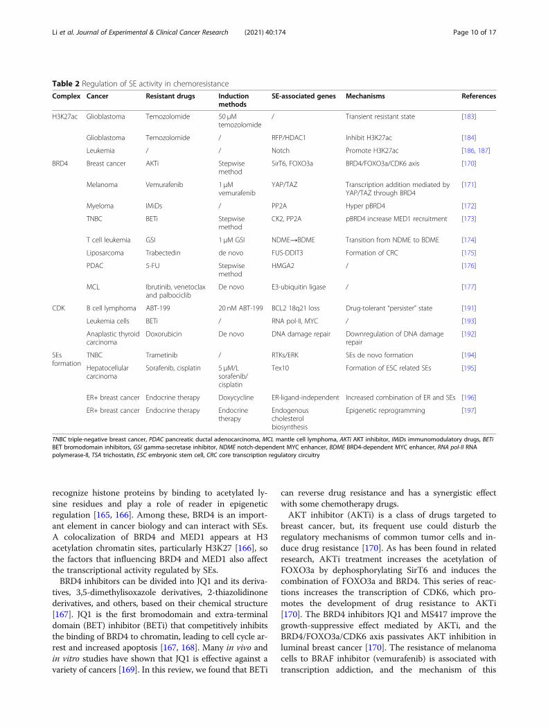
recognize histone proteins by binding to acetylated ly-sine residues and play a role of reader in epigeneticregulation [165, 166]. Among these, BRD4 is an import-ant element in cancer biology and can interact with SEs.A colocalization of BRD4 and MED1 appears at H3acetylation chromatin sites, particularly H3K27 [166], sothe factors that influencing BRD4 and MED1 also affectthe transcriptional activity regulated by SEs.BRD4 inhibitors can be divided into JQ1 and its deriva-
tives, 3,5-dimethylisoxazole derivatives, 2-thiazolidinonederivatives, and others, based on their chemical structure[167]. JQ1 is the first bromodomain and extra-terminaldomain (BET) inhibitor (BETi) that competitively inhibitsthe binding of BRD4 to chromatin, leading to cell cycle ar-rest and increased apoptosis [167, 168]. Many in vivo andin vitro studies have shown that JQ1 is effective against avariety of cancers [169]. In this review, we found that BETi
can reverse drug resistance and has a synergistic effectwith some chemotherapy drugs.AKT inhibitor (AKTi) is a class of drugs targeted to
breast cancer, but, its frequent use could disturb theregulatory mechanisms of common tumor cells and in-duce drug resistance [170]. As has been found in relatedresearch, AKTi treatment increases the acetylation ofFOXO3a by dephosphorylating SirT6 and induces thecombination of FOXO3a and BRD4. This series of reac-tions increases the transcription of CDK6, which pro-motes the development of drug resistance to AKTi[170]. The BRD4 inhibitors JQ1 and MS417 improve thegrowth-suppressive effect mediated by AKTi, and theBRD4/FOXO3a/CDK6 axis passivates AKT inhibition inluminal breast cancer [170]. The resistance of melanomacells to BRAF inhibitor (vemurafenib) is associated withtranscription addiction, and the mechanism of this
Table 2 Regulation of SE activity in chemoresistance
Complex Cancer Resistant drugs Inductionmethods
SE-associated genes Mechanisms References
H3K27ac Glioblastoma Temozolomide 50 μMtemozolomide
/ Transient resistant state [183]
Glioblastoma Temozolomide / RFP/HDAC1 Inhibit H3K27ac [184]
Leukemia / / Notch Promote H3K27ac [186, 187]
BRD4 Breast cancer AKTi Stepwisemethod
SirT6, FOXO3a BRD4/FOXO3a/CDK6 axis [170]
Melanoma Vemurafenib 1 μMvemurafenib
YAP/TAZ Transcription addition mediated byYAP/TAZ through BRD4
[171]
Myeloma IMiDs / PP2A Hyper pBRD4 [172]
TNBC BETi Stepwisemethod
CK2, PP2A pBRD4 increase MED1 recruitment [173]
T cell leukemia GSI 1 μM GSI NDME→BDME Transition from NDME to BDME [174]
Liposarcoma Trabectedin de novo FUS-DDIT3 Formation of CRC [175]
PDAC 5-FU Stepwisemethod
HMGA2 / [176]
MCL Ibrutinib, venetoclaxand palbociclib
De novo E3-ubiquitin ligase / [177]
CDK B cell lymphoma ABT-199 20 nM ABT-199 BCL2 18q21 loss Drug-tolerant “persister” state [191]
Leukemia cells BETi / RNA pol-II, MYC / [193]
Anaplastic thyroidcarcinoma
Doxorubicin De novo DNA damage repair Downregulation of DNA damagerepair
[192]
SEsformation
TNBC Trametinib / RTKs/ERK SEs de novo formation [194]
Hepatocellularcarcinoma
Sorafenib, cisplatin 5 μM/Lsorafenib/cisplatin
Tex10 Formation of ESC related SEs [195]
ER+ breast cancer Endocrine therapy Doxycycline ER-ligand-independent Increased combination of ER and SEs [196]
ER+ breast cancer Endocrine therapy Endocrinetherapy
Endogenouscholesterolbiosynthesis
Epigenetic reprogramming [197]
TNBC triple-negative breast cancer, PDAC pancreatic ductal adenocarcinoma, MCL mantle cell lymphoma, AKTi AKT inhibitor, IMiDs immunomodulatory drugs, BETiBET bromodomain inhibitors, GSI gamma-secretase inhibitor, NDME notch-dependent MYC enhancer, BDME BRD4-dependent MYC enhancer, RNA pol-II RNApolymerase-II, TSA trichostatin, ESC embryonic stem cell, CRC core transcription regulatory circuitry
Li et al. Journal of Experimental & Clinical Cancer Research (2021) 40:174 Page 10 of 17

resistance is that YAP/TAZ induces the recruitment ofSEs to BRD4 and RNA Pol-II and activates the expres-sion of growth-regulating genes [171]. In myeloma cellssensitive to immunomodulatory drugs (IMiDs), thedepletion of IKZF1/IKZF3 caused SE instability and re-duced the binding of BRD4. However, in resistant cells,the binding of BRD4 to SEs was unaffected, which couldbe attributed to the decrease in PP2A activity and the in-crease in BRD4 phosphorylation [172]. In addition, thephosphorylation of BRD4 is also related to BETi toler-ance in TNBC cells, the mechanisms for which include adecrease in PP2A activity and an increase in CK2 activityand MED1 recruitment [173]. In T cell leukemia,Notch1 could activate the expression of downstreamgenes by binding to SEs of MYC. However, in cell linesthat are resistant to a gamma-secretase inhibitor, the in-hibition of Notch1 cannot cause the downregulation ofMYC. A study by Yashiro-Ohtani et al. indicated thatthis is due to the transition of MYC SEs from Notch1-dependent (NDME) to BRD4-dependent MYC enhancer[174] (Fig. 3).Studies have shown that FUS-DDIT3 has regulatory
effects on SEs through interaction with BRD4, whichmay participate in the resistance of liposarcoma cells totrabectedin and CRC formation, and BET inhibitors caneffectively overcome this limitation in treatment [175].Furthermore, the blockage of BRD4 sensitizes 5-FU tox-icity to pancreatic ductal adenocarcinoma [176]. ARV-771, a proteolysis-targeting chimera of BET protein has
stronger activity in interfering with BET protein thanBETi, which may be promising for the overcoming ofthe resistance of MCL cells to ibrutinib, venetoclax, andpalbociclib [177].
Histone acetylationChemical modifications of DNA and histone proteins inchromatin could modulate gene expression throughchanging conformations and altering transcriptionalcomplex recruitment. Common chemical methods ofmodifying histone proteins include acetylation/deacetyla-tion and methylation/demethylation [166]. Acetylatedhistones destabilize nucleosomes, thereby increasing theaccessibility of chromatin to TFs [178, 179]. Acetylationmodification of chromatin histones is jointly regulatedby histone acetyltransferase and histone deacetylase(HDAC) enzymes; the two are in a state of dynamic bal-ance [180]. High-density H3K27ac is a sign that identi-fies SEs, which leads to a rapid response of target genesto various signals [21, 181, 182].Studies have shown that resistance is associated with
histone acetylation. Rabé et al. confirmed that the transi-ent resistance state to temozolomide in glioblastomacells is related to high levels of histone acetylation andchromatin remodeling, and the sensitive and resistantstate shows lower acetylated histone levels. The com-bined application of temozolomide and HDAC inhibitortrichostatin could prevent the transition from a transientto a resistant state [183]. Similarly, a review of the
Table 3 Reversal of chemoresistance
Target Inhibitors Cancers Resistant drugs or sensitized drugs References
BRD4 JQ1 Ovarian cancer Cisplatin [45, 119]
NSCLC TRAIL, cisplatin [142]
Breast cancer Salinomycin [121]
Breast cancer AKTi [170]
Melanoma Vemurafenib [171]
PDAC 5-FU [176]
I-BET151 MCL Ibrutinib, lenalidomide [140]
TNBC Trametinib [194]
I-BET762 NSCLC TRAIL, cisplatin [142]
OTX-015 NSCLC TRAIL, cisplatin [142]
SR2211 Pancreatic adenocarcinoma Gemcitabine [117]
ARV-771 MCL Ibrutinib, venetoclax, palbociclib [177]
MS417 Breast cancer AKTi [170]
H3K27ac TSA Glioblastoma Temozolomide [183]
CDK7 THZ1 B cell lymphoma ABT-199 [191]
CDK12 THZ531 Anaplastic thyroid carcinoma Doxorubicin [192]
NSCLC non-small cell lung cancer, PDAC pancreatic ductal adenocarcinoma, MCL mantle cell lymphoma, TNBC triple-negative breast cancer, AKTi AKT inhibitor,TRAIL tumor necrosis factor-related apoptosis-inducing ligand
Li et al. Journal of Experimental & Clinical Cancer Research (2021) 40:174 Page 11 of 17

resistance mechanism of glioblastoma cells to temozolo-mide indicated that disrupting the formation of RFP/HDAC1 complex would interfere with the function ofcis-regulatory-element, controlled by H3K27ac, and thenit would overcome the chemoresistance induced by SE-related genes [184, 185]. In leukemia and T-cell acutelymphoblastic leukemia, the tolerance of chemotherapyby cancer cells is related to the regulation of Notch1protein to H3K27 acetylation. The mutation of Notch1would suppress H3K27ac marks on SEs and disruptdownstream MYC expression, which may show how re-sistant cells maintain growth under drug pressure [186,187] (Fig. 3).
CDKsThe appearance of SEs in cancer cells leads to high tran-scription output and high transcription addiction, whichresult in stronger responses to transcriptional inhibition[188]. CDKs are an important category of protein, whichcan bind to cyclin proteins and regulate the cell cycle,playing an important role in gene transcription [189].These features make them indispensable for the regula-tion of SEs activity and overcoming chemoresistance.Studies have shown that SEs activate transcriptions areinseparable from the recruitment of CDK7-containingTFIIH (a transcription initiation complex), CDK9-containing p-TEFb (a transcription extension complex),and CDK12 [86, 97, 190]. Therefore, inhibitors that tar-get CDKs can reduce SE activity, thereby inhibiting theoccurrence and progression of cancer and reversingchemoresistance.According to research on B cell lymphoma, the emer-
gence of a drug-tolerant “persister” state is associatedwith SE remodeling in resistance to ABT-199, a targetdrug of BCL-2, and the inhibitor of CDK7 (THZ1) couldsignificantly reverse this effect [191]. One study showedthat the CDK12 inhibitor THZ531 can inhibit transcrip-tional extension and downregulate DNA damage repair,thereby increasing the sensitivity of anaplastic thyroidcarcinoma cells to doxorubicin [192]. The combinationof the BETi inhibitor and the CDK7 inhibitor leads tothe synthetic lethality in leukemia cells resistant to BETi,which is associated with the RNA pol-II activity regu-lated by SEs [193] (Fig. 3).
Other links to SE activityIn the case of drug resistance, the appearance and regu-lation of SEs are also affected by other factors. As a re-sponse to trametinib, a MEK1/2 inhibitor, adaptiveresistance takes place in TNBC as a result of de novo SEformation [194]. Tex10 upregulates ESC-related SEs insorafenib- and cisplatin-resistant cell lines, which is animportant chemoresistance mechanism for HCC [195].Studies have shown that the ER-ligand-binding domain
is mutated in ER+ breast cancer cells that are resistant toendocrine therapy, and these cells acquire ligand-independent growth. During the exploration of themechanism of this phenomenon, it was found that theinteraction between ER and SEs in the mutant cells hasincreased [196]. Furthermore, epigenetic reprogrammingfor endocrine therapy activates endogenous cholesterolbiosynthesis, which promotes the constitutive activationof ERα in drug-resistant cells [197].Moreover, inhibitors of oncogenes can both directly
influence the expression of oncogenes and blockgenome-wide oncogene enhancers and SEs activation aswell, along with downstream transcriptional signaling. Arecent study found that darolutamide, an inhibitor of theandrogen receptor, antagonizes androgen signaling byblocking enhancers and SE activation in prostate cancer[198]. The new drugs involved in SE-related oncogenes’transcriptional regulation may produce important resultsfor chemotherapeutic resistance. The remodeling of SEsin drug-resistant cells may also be related to the down-regulation of certain genes. In ovarian cancer, ISL1, alineage determinant, is downregulated when cells arecontinuously stimulated by cisplatin, mediating the in-crease in CSCs and chemoresistance induced by SE plas-ticity [199] (Fig. 3).
Perspective and summaryDue to the differing genetic/epigenetic backgrounds andheterogeneity of tumors, the efficacy of chemotherapyvaries widely across patients. Understanding the chan-ging epigenetic landscape during chemotherapy and thedynamic interaction between the genetic and epigeneticmachinery in response to chemotherapy are inevitablefor assessing the clinical efficacy of chemotherapy.Within the new frontier of epigenetic modifiers, moreand more evidence has shown the important role of SEsin tumor development and chemotherapy resistance.Epigenetic gene signatures, particularly SEs, have
attracted increased interest lately with regard to the mo-lecular subtypes of tumors and their prediction of tumorrecurrence, the prognosis of tumor patients, and chemo-therapy resistance in different cancers. Mapping the epi-genome and monitoring epi-biomarkers (such as SEs)using genome-wide analyses at clinical settings before,during, and after treatment and at relapse will helpevaluate and adjust the treatment approach and designpersonalized epigenetic therapy [11]. Despite the con-tinuous emergence of relevant research, chemotherapyresistance remains a complex process that needs to beexplored in depth. We may still need to conduct moreresearch upstream and investigate more initial mecha-nisms to clarify the reasons for the generation and regu-lation of resistance-related SEs. Furthermore, related
Li et al. Journal of Experimental & Clinical Cancer Research (2021) 40:174 Page 12 of 17

inhibitors require clinical trials to prove their effective-ness and safety for overcoming chemoresistance.
ConclusionsIn conclusion, SEs are central to the maintenance ofidentity of cancer cells and promote SE-driven-oncogenic transcriptions to which cancer cells becomehighly addicted. Chemotherapeutics induce SEs repro-gramming in cancer cells, converting a transient tran-scriptional state into a stably resistant one. Aberranttranscriptional regulation of SEs plays important roles inepigenetic mechanisms of cancer chemoresistance viathe formation of CSCs, cellular plasticity, the micro-environment, genes associated with chemoresistance,ncRNAs, and tumor immunity. This dependence on SE-driven transcription to maintain chemoresistance offersan Achilles’ heel for chemoresistance. Indeed, the inhib-ition of SE components dampens oncogenic transcrip-tion and inhibits tumor growth to ultimately achievecombined sensitization and reverse the effects of drugresistance. The research on the SEs in tumorigenesis andchemoresistance may help find new drugs to overcomechemoresistance from the bench to the bedside.
AbbreviationsTME: Tumor microenvironment; CSC: Cancer stem cells; MDRP: Multi-drugresistant protein; SE: Super-enhancer; TF: Transcription factor; ESC: Embryonicstem cell; ncRNA: non-coding RNA; tRNA: transfer RNA; lncRNA: long non-coding RNA; eRNA: enhancer RNA; seRNA: super-enhancer RNA; RNA pol-II: RNA polymerase-II; H3K27ac: Histone H3 lysine27 acetylation; CDK7: Cyclin-dependent kinase 7; BRD4: Bromodomain-containing protein 4;H3K4me1: Histone H3 lysine4 methylation; CRC: Core transcriptionalregulatory circuitry; SD: SE domain; TAD: Topologically associating domain;CTCF: CCCTC-binding factor; IDR: Intrinsically disordered region;MED1: Mediator of RNA Pol-II transcription subunit 1; TCF4: Transcriptionfactor 4; RORγ: Receptor-related orphan receptor gamma; ALDH: Aldehydedehydrogenase; EMT: Epithelial-mesenchymal transition; SCLC: Small-cell lungcancer; MCL: Mantle cell lymphoma; BCR: B cell receptor; TNBC: Triple-negative breast cancer; HCC: Hepatocellular carcinoma; TIME: Tumor immunemicroenvironment; PD-1: Programmed cell-death protein 1; PD-L1: Programmed cell death 1 ligand 1; SCC: Squamous cell carcinoma;BET: BRD and extraterminal domain; BETi: BET protein inhibitor; AKTi: AKTinhibitor; IMiDs: Immunomodulatory drugs; HDAC: Histone deacetylase
AcknowledgementsNot applicable.
Authors’ contributionsJQ and QQ had the idea for the article, GHL, TTQ, QQ and XQT performedthe literature search and data collection, GHL, JQ, QL, TTQ, and XQT draftedthe manuscript, JQ and QL critically revised the work. All authors modifiedand approved the final manuscript.
FundingThis work was supported by the National Natural Science Foundation ofChina (No. 82073944), Scientific Research Project of Hunan HealthCommission (No. 202113010170), and the Hunan Provincial Department ofFinance Grant (No. 2019–93 and 2018–92).
Availability of data and materialsAvailable.
Declarations
Ethics approval and consent to participateNot applicable.
Consent for publicationNot applicable.
Competing interestsThe authors declare that they have no competing interests.
Author details1Department of Pharmacy, the Second Xiangya Hospital, Central SouthUniversity; Institute of Clinical Pharmacy, Central South University, 139 MiddleRenmin Road, Changsha, Hunan 410011, People’s Republic of China.2Department of Pharmacy, Xiangya Hospital, Central South University,Changsha 410008, People’s Republic of China.
Received: 15 March 2021 Accepted: 5 May 2021
References1. Bray F, Ferlay J, Soerjomataram I, Siegel RL, Torre LA, Jemal A. Global cancer
statistics 2018: GLOBOCAN estimates of incidence and mortality worldwidefor 36 cancers in 185 countries. CA Cancer J Clin. 2018;68:394–424.
2. Ferlay J, Soerjomataram I, Dikshit R, Eser S, Mathers C, Rebelo M, et al.Cancer incidence and mortality worldwide: sources, methods and majorpatterns in GLOBOCAN 2012. Int J Cancer. 2015;136:E359–86.
3. The International Agency for Research on Cancer. https://www.iarc.who.int/featured-news/latest-global-cancer-data-cancer-burden-rises-to-19-3-million-new-cases-and-10-0-million-cancer-deaths-in-2020/. Accessed 15 Oct 2020.
4. Naylor EC, Desani JK, Chung PK. Targeted therapy and immunotherapy forlung Cancer. Surg Oncol Clin N Am. 2016;25:601–9.
5. Menderes G, Black J, Schwab CL, Santin AD. Immunotherapy and targetedtherapy for cervical cancer: an update. Expert Rev Anticancer Ther. 2016;16:83–98.
6. Zugazagoitia J, Guedes C, Ponce S, Ferrer I, Molina-Pinelo S, Paz-Ares L.Current challenges in Cancer treatment. Clin Ther. 2016;38:1551–66.
7. Khot VM, Salunkhe AB, Pricl S, Bauer J, Thorat ND, Townley H.Nanomedicine-driven molecular targeting, drug delivery, andtherapeutic approaches to cancer chemoresistance. Drug Discov Today.2020;26(3):724–39.
8. Vasan N, Baselga J, Hyman DM. A view on drug resistance in cancer. Nature.2019;575:299–309.
9. Wu Q, Yang Z, Nie Y, Shi Y, Fan D. Multi-drug resistance in cancerchemotherapeutics: mechanisms and lab approaches. Cancer Lett. 2014;347:159–66.
10. Li YJ, Lei YH, Yao N, Wang CR, Hu N, Ye WC, et al. Autophagy and multidrugresistance in cancer. Chin J Cancer. 2017;36:52.
11. Ponnusamy L, Mahalingaiah PKS, Singh KP. Epigenetic reprogramming andpotential application of epigenetic-modifying drugs in acquiredchemotherapeutic resistance. Adv Clin Chem. 2020;94:219–59.
12. Quagliano A, Gopalakrishnapillai A, Barwe SP. Understanding themechanisms by which epigenetic modifiers avert therapy resistance inCancer. Front Oncol. 2020;10:992.
13. Zhou Y, Sun W, Qin Z, Guo S, Kang Y, Zeng S, et al. LncRNA regulation: newfrontiers in epigenetic solutions to drug chemoresistance. BiochemPharmacol. 2020;104(10):114228.
14. Takezawa K, Okamoto I, Okamoto W, Takeda M, Sakai K, Tsukioka S, et al.Thymidylate synthase as a determinant of pemetrexed sensitivity in non-small cell lung cancer. Br J Cancer. 2011;104:1594–601.
15. Quagliano A, Gopalakrishnapillai A, Barwe SP. Epigenetic drug combinationovercomes osteoblast-induced chemoprotection in pediatric acutelymphoid leukemia. Leuk Res. 2017;56:36–43.
16. Kim K, Skora AD, Li Z, Liu Q, Tam AJ, Blosser RL, et al. Eradication ofmetastatic mouse cancers resistant to immune checkpoint blockade bysuppression of myeloid-derived cells. Proc Natl Acad Sci U S A. 2014;111:11774–9.
17. Wang W, Oguz G, Lee PL, Bao Y, Wang P, Terp MG, et al. KDM4B-regulatedunfolded protein response as a therapeutic vulnerability in PTEN-deficientbreast cancer. J Exp Med. 2018;215:2833–49.
Li et al. Journal of Experimental & Clinical Cancer Research (2021) 40:174 Page 13 of 17

18. Sengupta S, George RE. Super-enhancer-driven transcriptional dependenciesin Cancer. Trends Cancer. 2017;3:269–81.
19. Shaffer SM, Dunagin MC, Torborg SR, Torre EA, Emert B, Krepler C, et al. Rarecell variability and drug-induced reprogramming as a mode of cancer drugresistance. Nature. 2017;546:431–5.
20. Hnisz D, Abraham BJ, Lee TI, Lau A, Saint-Andre V, Sigova AA, et al. Super-enhancers in the control of cell identity and disease. Cell. 2013;155:934–47.
21. Whyte WA, Orlando DA, Hnisz D, Abraham BJ, Lin CY, Kagey MH, et al.Master transcription factors and mediator establish super-enhancers at keycell identity genes. Cell. 2013;153:307–19.
22. Niederriter AR, Varshney A, Parker SC, Martin DM. Super enhancers incancers, complex disease, and developmental disorders. Genes (Basel). 2015;6:1183–200.
23. Thandapani P. Super-enhancers in cancer. Pharmacol Ther. 2019;199:129–38.24. Gottesman MM. Mechanisms of cancer drug resistance. Annu Rev Med.
2002;53:615–27.25. Kartal-Yandim M, Adan-Gokbulut A, Baran Y. Molecular mechanisms of drug
resistance and its reversal in cancer. Crit Rev Biotechnol. 2016;36:716–26.26. Kohno K, Uchiumi T, Niina I, Wakasugi T, Igarashi T, Momii Y, et al.
Transcription factors and drug resistance. Eur J Cancer. 2005;41:2577–86.27. Nebert DW. Transcription factors and cancer: an overview. Toxicology. 2002;
181-182:131–41.28. Karamouzis MV, Gorgoulis VG, Papavassiliou AG. Transcription factors and
neoplasia: vistas in novel drug design. Clin Cancer Res. 2002;8:949–61.29. Nikolaou M, Pavlopoulou A, Georgakilas AG, Kyrodimos E. The challenge of
drug resistance in cancer treatment: a current overview. Clin Exp Metastasis.2018;35:309–18.
30. Jaffrezou JP, Chen KG, Duran GE, Kuhl JS, Sikic BI. Mutation rates andmechanisms of resistance to etoposide determined from fluctuationanalysis. J Natl Cancer Inst. 1994;86:1152–8.
31. Dumontet C, Duran GE, Steger KA, Beketic-Oreskovic L, Sikic BI. Resistancemechanisms in human sarcoma mutants derived by single-step exposure topaclitaxel (Taxol). Cancer Res. 1996;56:1091–7.
32. Buttigliero C, Tucci M, Bertaglia V, Vignani F, Bironzo P, Di Maio M, et al.Understanding and overcoming the mechanisms of primary and acquiredresistance to abiraterone and enzalutamide in castration resistant prostatecancer. Cancer Treat Rev. 2015;41:884–92.
33. Caffo O, Veccia A, Maines F, Bonetta A, Spizzo G, Galligioni E. Potential valueof rapid prostate-specific antigen decline in identifying primary resistance toabiraterone acetate and enzalutamide. Future Oncol. 2014;10:985–93.
34. Glasspool RM, Teodoridis JM, Brown R. Epigenetics as a mechanism drivingpolygenic clinical drug resistance. Br J Cancer. 2006;94:1087–92.
35. Januskeviciene I, Petrikaite V. Heterogeneity of breast cancer: theimportance of interaction between different tumor cell populations. Life Sci.2019;239:117009.
36. Horowitz M, Esakov E, Rose P, Reizes O. Signaling within the epithelialovarian cancer tumor microenvironment: the challenge of tumorheterogeneity. Ann Transl Med. 2020;8:905.
37. Mansoori B, Mohammadi A, Davudian S, Shirjang S, Baradaran B. Thedifferent mechanisms of Cancer drug resistance: a brief review. Adv PharmBull. 2017;7:339–48.
38. Housman G, Byler S, Heerboth S, Lapinska K, Longacre M, Snyder N, et al.Drug resistance in cancer: an overview. Cancers (Basel). 2014;6:1769–92.
39. Berger SL, Kouzarides T, Shiekhattar R, Shilatifard A. An operationaldefinition of epigenetics. Genes Dev. 2009;23:781–3.
40. Sharma S, Kelly TK, Jones PA. Epigenetics in cancer. Carcinogenesis. 2010;31:27–36.
41. Dawson MA, Kouzarides T. Cancer epigenetics: from mechanism to therapy.Cell. 2012;150:12–27.
42. Lao VV, Grady WM. Epigenetics and colorectal cancer. Nat Rev GastroenterolHepatol. 2011;8:686–700.
43. Brown R, Curry E, Magnani L, Wilhelm-Benartzi CS, Borley J. Poisedepigenetic states and acquired drug resistance in cancer. Nat Rev Cancer.2014;14:747–53.
44. Wilting RH, Dannenberg JH. Epigenetic mechanisms in tumorigenesis,tumor cell heterogeneity and drug resistance. Drug Resist Updat. 2012;15:21–38.
45. Shang S, Yang J, Jazaeri AA, Duval AJ, Tufan T, Lopes Fischer N, et al.Chemotherapy-induced distal enhancers drive transcriptional programs tomaintain the Chemoresistant state in ovarian Cancer. Cancer Res. 2019;79:4599–611.
46. You JS, Jones PA. Cancer genetics and epigenetics: two sides of the samecoin? Cancer Cell. 2012;22:9–20.
47. Anastasiadou E, Jacob LS, Slack FJ. Non-coding RNA networks in cancer. NatRev Cancer. 2018;18:5–18.
48. Wang KC, Chang HY. Molecular mechanisms of long noncoding RNAs. MolCell. 2011;43:904–14.
49. Mousavi K, Zare H, Dell'orso S, Grontved L, Gutierrez-Cruz G, Derfoul A, et al.eRNAs promote transcription by establishing chromatin accessibility atdefined genomic loci. Mol Cell. 2013;51:606–17.
50. Mao R, Wu Y, Ming Y, Xu Y, Wang S, Chen X, et al. Enhancer RNAs: amissing regulatory layer in gene transcription. Sci China Life Sci. 2019;62:905–12.
51. Wu M, Shen J. From super-enhancer non-coding RNA to immunecheckpoint: frameworks to functions. Front Oncol. 2019;9:1307.
52. Gomez S, Tabernacki T, Kobyra J, Roberts P, Chiappinelli KB. Combiningepigenetic and immune therapy to overcome cancer resistance. SeminCancer Biol. 2020;65:99–113.
53. Sharma SV, Lee DY, Li B, Quinlan MP, Takahashi F, Maheswaran S, et al. Achromatin-mediated reversible drug-tolerant state in cancer cellsubpopulations. Cell. 2010;141:69–80.
54. Toh TB, Lim JJ, Chow EK. Epigenetics in cancer stem cells. Mol Cancer. 2017;16:29.
55. Ding B, Lou W, Xu L, Fan W. Non-coding RNA in drug resistance ofhepatocellular carcinoma. Biosci Rep. 2018;38(5):BSR20180915.
56. Wei L, Wang X, Lv L, Zheng Y, Zhang N, Yang M. The emerging role ofnoncoding RNAs in colorectal cancer chemoresistance. Cell Oncol (Dordr).2019;42:757–68.
57. Neve B, Jonckheere N, Vincent A, Van Seuningen I. Epigenetic regulation bylncRNAs: an overview focused on UCA1 in colorectal Cancer. Cancers(Basel). 2018;10(11):440.
58. Luo YJ, Huang QM, Ren Y, Liu ZL, Xu CF, Wang H, et al. Non-coding RNA indrug resistance of gastric cancer. World J Gastrointest Oncol. 2019;11:957–70.
59. Mondal P, Natesh J, Kamal MA, Meeran SM. Non-coding RNAs in lungCancer Chemoresistance. Curr Drug Metab. 2019;20:1023–32.
60. Xiong G, Feng M, Yang G, Zheng S, Song X, Cao Z, et al. The underlyingmechanisms of non-coding RNAs in the chemoresistance of pancreaticcancer. Cancer Lett. 2017;397:94–102.
61. Ong CT, Corces VG. Enhancer function: new insights into the regulation oftissue-specific gene expression. Nat Rev Genet. 2011;12:283–93.
62. Zheng C, Liu M, Fan H. Targeting complexes of super-enhancers is apromising strategy for cancer therapy. Oncol Lett. 2020;20:2557–66.
63. Jefferson WN, Kinyamu HK, Wang T, Miranda AX, Padilla-Banks E, SuenAA, et al. Widespread enhancer activation via ERα mediates estrogenresponse in vivo during uterine development. Nucleic Acids Res. 2018;46:5487–503.
64. Ing-Simmons E, Seitan VC, Faure AJ, Flicek P, Carroll T, Dekker J, et al. Spatialenhancer clustering and regulation of enhancer-proximal genes by cohesin.Genome Res. 2015;25:504–13.
65. Furlong EEM, Levine M. Developmental enhancers and chromosometopology. Science. 2018;361:1341–5.
66. Wu C, Chen J, Liu Y, Hu X. Improved prediction of regulatory element usinghybrid abelian complexity features with DNA sequences. Int J Mol Sci. 2019;20(7):1704.
67. Poot R. The rules of successful speed dating are complex, even for super-enhancers. Cell Stem Cell. 2018;22:477–8.
68. Hah N, Benner C, Chong LW, Yu RT, Downes M, Evans RM. Inflammation-sensitive super enhancers form domains of coordinately regulated enhancerRNAs. Proc Natl Acad Sci U S A. 2015;112:E297–302.
69. Arnold PR, Wells AD, Li XC. Diversity and emerging roles of enhancer RNA inregulation of gene expression and cell fate. Front Cell Dev Biol. 2019;7:377.
70. Chen H, Liang H. A High-Resolution Map of Human Enhancer RNA LociCharacterizes Super-enhancer Activities in Cancer. Cancer Cell. 2020;38:701–715.e705.
71. Saint-André V, Federation AJ, Lin CY, Abraham BJ, Reddy J, Lee TI, et al.Models of human core transcriptional regulatory circuitries. Genome Res.2016;26:385–96.
72. Huang M, Chen Y, Yang M, Guo A, Xu Y, Xu L, et al. dbCoRC: a database ofcore transcriptional regulatory circuitries modeled by H3K27ac ChIP-seqsignals. Nucleic Acids Res. 2018;46:D71–d77.
73. Zheng H, Xie W. The role of 3D genome organization in development andcell differentiation. Nat Rev Mol Cell Biol. 2019;20:535–50.
Li et al. Journal of Experimental & Clinical Cancer Research (2021) 40:174 Page 14 of 17

74. Su JH, Zheng P, Kinrot SS, Bintu B, Zhuang X. Genome-scale imaging of the3D organization and transcriptional activity of chromatin. Cell. 2020;182:1641–59 e1626.
75. Yamagata K, Nakayamada S, Tanaka Y. Critical roles of super-enhancers inthe pathogenesis of autoimmune diseases. Inflamm Regen. 2020;40:16.
76. Cubenas-Potts C, Corces VG. Topologically associating domains: an invariantframework or a dynamic scaffold? Nucleus. 2015;6:430–4.
77. Dixon JR, Selvaraj S, Yue F, Kim A, Li Y, Shen Y, et al. Topological domains inmammalian genomes identified by analysis of chromatin interactions.Nature. 2012;485:376–80.
78. McArthur E, Capra JA. Topologically associating domain boundaries that arestable across diverse cell types are evolutionarily constrained and enrichedfor heritability. Am J Hum Genet. 2021;108:269–83.
79. Kloetgen A, Thandapani P, Ntziachristos P, Ghebrechristos Y, Nomikou S,Lazaris C, et al. Three-dimensional chromatin landscapes in T cell acutelymphoblastic leukemia. Nat Genet. 2020;52:388–400.
80. Van Bortle K, Nichols MH, Li L, Ong CT, Takenaka N, Qin ZS, et al. Insulatorfunction and topological domain border strength scale with architecturalprotein occupancy. Genome Biol. 2014;15:R82.
81. Ong CT, Corces VG. CTCF: an architectural protein bridging genometopology and function. Nat Rev Genet. 2014;15:234–46.
82. Gong Y, Lazaris C, Sakellaropoulos T, Lozano A, Kambadur P, Ntziachristos P,et al. Stratification of TAD boundaries reveals preferential insulation ofsuper-enhancers by strong boundaries. Nat Commun. 2018;9:542.
83. Sun X, Ren Z, Cun Y, Zhao C, Huang X, Zhou J, et al. Hippo-YAP signalingcontrols lineage differentiation of mouse embryonic stem cells throughmodulating the formation of super-enhancers. Nucleic Acids Res. 2020;48:7182–96.
84. Sabari BR, Dall'Agnese A, Boija A, Klein IA, Coffey EL, Shrinivas K, et al.Coactivator condensation at super-enhancers links phase separation andgene control. Science. 2018;361(6400):eaar3958.
85. Hnisz D, Shrinivas K, Young RA, Chakraborty AK, Sharp PA. A phaseseparation model for transcriptional control. Cell. 2017;169:13–23.
86. Jia Q, Chen S, Tan Y, Li Y, Tang F. Oncogenic super-enhancer formationin tumorigenesis and its molecular mechanisms. Exp Mol Med. 2020;52(5):713–23.
87. Chen D, Zhao Z, Huang Z, Chen DC, Zhu XX, Wang YZ, et al. Superenhancer inhibitors suppress MYC driven transcriptional amplification andtumor progression in osteosarcoma. Bone Res. 2018;6:11.
88. Fox S, Myers JA, Davidson C, Getman M, Kingsley PD, Frankiewicz N,et al. Hyperacetylated chromatin domains mark cell type-specific genesand suggest distinct modes of enhancer function. Nat Commun. 2020;11:4544.
89. Ohkura N, Yasumizu Y, Kitagawa Y, Tanaka A, Nakamura Y, Motooka D, et al.Regulatory T Cell-Specific Epigenomic Region Variants Are a KeyDeterminant of Susceptibility to Common Autoimmune Diseases. Immunity.2020;52:1119–1132.e1114.
90. Adam RC, Yang H, Ge Y, Infarinato NR, Gur-Cohen S, Miao Y, et al. NFItranscription factors provide chromatin access to maintain stem cell identitywhile preventing unintended lineage fate choices. Nat Cell Biol. 2020;22:640–50.
91. Bahr C, von Paleske L, Uslu VV, Remeseiro S, Takayama N, Ng SW, et al. AMyc enhancer cluster regulates normal and leukaemic haematopoietic stemcell hierarchies. Nature. 2018;553:515–20.
92. Kitagawa Y, Ohkura N, Kidani Y, Vandenbon A, Hirota K, Kawakami R, et al.Guidance of regulatory T cell development by Satb1-dependent super-enhancer establishment. Nat Immunol. 2017;18:173–83.
93. Peng XL, So KK, He L, Zhao Y, Zhou J, Li Y, et al. MyoD- and FoxO3-mediated hotspot interaction orchestrates super-enhancer activity duringmyogenic differentiation. Nucleic Acids Res. 2017;45:8785–805.
94. Alcalà-Vida R, Awada A, Boutillier AL, Merienne K. Epigenetic mechanismsunderlying enhancer modulation of neuronal identity, neuronal activity andneurodegeneration. Neurobiol Dis. 2021;147:105155.
95. Adam RC, Yang H, Rockowitz S, Larsen SB, Nikolova M, Oristian DS, et al.Pioneer factors govern super-enhancer dynamics in stem cell plasticity andlineage choice. Nature. 2015;521:366–70.
96. Boyer LA, Lee TI, Cole MF, Johnstone SE, Levine SS, Zucker JP, et al. Coretranscriptional regulatory circuitry in human embryonic stem cells. Cell.2005;122:947–56.
97. Tang F, Yang Z, Tan Y, Li Y. Super-enhancer function and its application incancer targeted therapy. NPJ Precis Oncol. 2020;4:2.
98. Khan A, Zhang X. dbSUPER: a database of super-enhancers in mouse andhuman genome. Nucleic Acids Res. 2016;44:D164–71.
99. Chen C, Zhou D, Gu Y, Wang C, Zhang M, Lin X, et al. SEA version 3.0: acomprehensive extension and update of the super-enhancer archive.Nucleic Acids Res. 2020;48:D198–d203.
100. Jiang Y, Qian F, Bai X, Liu Y, Wang Q, Ai B, et al. SEdb: a comprehensivehuman super-enhancer database. Nucleic Acids Res. 2019;47:D235–d243.
101. Zhang C, Wei S, Sun WP, Teng K, Dai MM, Wang FW, et al. Super-enhancer-driven AJUBA is activated by TCF4 and involved in epithelial-mesenchymaltransition in the progression of hepatocellular carcinoma. Theranostics.2020;10:9066–82.
102. Wang Y, Nie H, He X, Liao Z, Zhou Y, Zhou J, et al. The emerging role ofsuper enhancer-derived noncoding RNAs in human cancer. Theranostics.2020;10:11049–62.
103. Jiao W, Chen Y, Song H, Li D, Mei H, Yang F, et al. HPSE enhancer RNApromotes cancer progression through driving chromatin looping andregulating hnRNPU/p300/EGR1/HPSE axis. Oncogene. 2018;37:2728–45.
104. Ri K, Kim C, Pak C, Ri P, Om H. The KLF6 super enhancer modulatescell proliferation via MiR-1301 in human hepatoma cells. Microrna. 2020;9:64–9.
105. Peng L, Jiang B, Yuan X, Qiu Y, Peng J, Huang Y, et al. Super-enhancer-associated long noncoding RNA HCCL5 is activated by ZEB1 and promotesthe malignancy of hepatocellular carcinoma. Cancer Res. 2019;79:572–84.
106. Wang X, Zhang R, Wu S, Shen L, Ke M, Ouyang Y, et al. Super-EnhancerLncRNA LINC00162 Promotes Progression of Bladder Cancer. iScience. 2020;23:101857.
107. Ouyang Z, Li G, Zhu H, Wang J, Qi T, Qu Q, et al. Construction of a five-super-enhancer-associated-genes prognostic model for osteosarcomapatients. Front Cell Dev Biol. 2020;8:598660.
108. Qi T, Qu J, Tu C, Lu Q, Li G, Wang J, et al. Super-enhancer associated five-gene risk score model predicts overall survival in multiple myelomapatients. Front Cell Dev Biol. 2020;8:596777.
109. Zhang T, Song X, Zhang Z, Mao Q, Xia W, Xu L, et al. Aberrant super-enhancer landscape reveals core transcriptional regulatory circuitry in lungadenocarcinoma. Oncogenesis. 2020;9:92.
110. Shi X, Zheng Y, Jiang L, Zhou B, Yang W, Li L, et al. EWS-FLI1 regulates andcooperates with core regulatory circuitry in Ewing sarcoma. Nucleic AcidsRes. 2020;48:11434–51.
111. Lu B, He Y, He J, Wang L, Liu Z, Yang J, et al. Epigenetic profiling identifiesLIF as a super-enhancer-controlled regulator of stem cell-like properties inosteosarcoma. Mol Cancer Res. 2020;18:57–67.
112. Johnston MJ, Nikolic A, Ninkovic N, Guilhamon P, Cavalli FMG, Seaman S,et al. High-resolution structural genomics reveals new therapeuticvulnerabilities in glioblastoma. Genome Res. 2019;29:1211–22.
113. Mack SC, Singh I, Wang X, Hirsch R, Wu Q, Villagomez R, et al. Chromatinlandscapes reveal developmentally encoded transcriptional states thatdefine human glioblastoma. J Exp Med. 2019;216:1071–90.
114. Cheng H, Dou X, Han JD. Understanding super-enhancers. Sci China Life Sci.2016;59:277–80.
115. Shin HY. Targeting super-enhancers for disease treatment and diagnosis.Mol Cells. 2018;41:506–14.
116. Bao J, Li M, Liang S, Yang Y, Wu J, Zou Q, et al. Integrated high-throughputanalysis identifies super enhancers associated with chemoresistance inSCLC. BMC Med Genet. 2019;12:67.
117. Lytle NK, Ferguson LP, Rajbhandari N, Gilroy K, Fox RG, Deshpande A, et al.A Multiscale Map of the Stem Cell State in Pancreatic Adenocarcinoma. Cell.2019;177:572–586.e522.
118. Veschi V, Verona F, Thiele CJ. Cancer stem cells and neuroblastoma:characteristics and therapeutic targeting options. Front Endocrinol(Lausanne). 2019;10:782.
119. Yokoyama Y, Zhu H, Lee JH, Kossenkov AV, Wu SY, Wickramasinghe JM,et al. BET inhibitors suppress ALDH activity by targeting ALDH1A1 super-enhancer in ovarian Cancer. Cancer Res. 2016;76:6320–30.
120. Sharma A, Cao EY, Kumar V, Zhang X, Leong HS, Wong AML, et al. Longitudinalsingle-cell RNA sequencing of patient-derived primary cells reveals drug-inducedinfidelity in stem cell hierarchy. Nat Commun. 2018;9:4931.
121. Arfaoui A, Rioualen C, Azzoni V, Pinna G, Finetti P, Wicinski J, et al. Agenome-wide RNAi screen reveals essential therapeutic targets of breastcancer stem cells. EMBO Mol Med. 2019;11(10):e9930.
122. Yuan S, Norgard RJ, Stanger BZ. Cellular plasticity in Cancer. Cancer Discov.2019;9:837–51.
Li et al. Journal of Experimental & Clinical Cancer Research (2021) 40:174 Page 15 of 17

123. Vicente-Duenas C, Gutierrez de Diego J, Rodriguez FD, Jimenez R, CobaledaC. the role of cellular plasticity in cancer development. Curr Med Chem.2009;16:3676–85.
124. Nieto MA, Huang RY, Jackson RA, Thiery JP. Emt: 2016. Cell. 2016;166:21–45.125. Byers LA, Diao L, Wang J, Saintigny P, Girard L, Peyton M, et al. An
epithelial-mesenchymal transition gene signature predicts resistance toEGFR and PI3K inhibitors and identifies Axl as a therapeutic target forovercoming EGFR inhibitor resistance. Clin Cancer Res. 2013;19:279–90.
126. Sequist LV, Waltman BA, Dias-Santagata D, Digumarthy S, Turke AB, Fidias P,et al. Genotypic and histological evolution of lung cancers acquiringresistance to EGFR inhibitors. Sci Transl Med. 2011;3:75ra26.
127. Meacham CE, Morrison SJ. Tumour heterogeneity and cancer cell plasticity.Nature. 2013;501:328–37.
128. Yu HA, Arcila ME, Rekhtman N, Sima CS, Zakowski MF, Pao W, et al. Analysisof tumor specimens at the time of acquired resistance to EGFR-TKI therapyin 155 patients with EGFR-mutant lung cancers. Clin Cancer Res. 2013;19:2240–7.
129. Lomberk G, Blum Y, Nicolle R, Nair A, Gaonkar KS, Marisa L, et al. Distinctepigenetic landscapes underlie the pathobiology of pancreatic cancersubtypes. Nat Commun. 2018;9:1978.
130. Gerrard DL, Boyd JR, Stein GS, Jin VX, Frietze S. Disruption of broadepigenetic domains in PDAC cells by HAT inhibitors. Epigenomes. 2019;3(2):11.
131. Su Y, Subedee A, Bloushtain-Qimron N, Savova V, Krzystanek M, Li L, et al.Somatic cell fusions reveal extensive heterogeneity in basal-like breastCancer. Cell Rep. 2015;11:1549–63.
132. Biehs B, Dijkgraaf GJP, Piskol R, Alicke B, Boumahdi S, Peale F, et al. A cellidentity switch allows residual BCC to survive hedgehog pathway inhibition.Nature. 2018;562:429–33.
133. Adam RC, Yang H, Ge Y, Lien WH, Wang P, Zhao Y, et al. Temporal layeringof signaling effectors drives chromatin remodeling during hair follicle stemcell lineage progression. Cell Stem Cell. 2018;22:398–413 e397.
134. Nishida J, Momoi Y, Miyakuni K, Tamura Y, Takahashi K, Koinuma D, et al.Epigenetic remodelling shapes inflammatory renal cancer and neutrophil-dependent metastasis. Nat Cell Biol. 2020;22:465–75.
135. Noel P, Hussein S, Ng S, Antal CE, Lin W, Rodela E, et al. Triptolide targetssuper-enhancer networks in pancreatic cancer cells and cancer-associatedfibroblasts. Oncogenesis. 2020;9:100.
136. Leung VY, Gao B, Leung KK, Melhado IG, Wynn SL, Au TY, et al. SOX9governs differentiation stage-specific gene expression in growth platechondrocytes via direct concomitant transactivation and repression. PLoSGenet. 2011;7:e1002356.
137. Kadaja M, Keyes BE, Lin M, Pasolli HA, Genander M, Polak L, et al. SOX9: astem cell transcriptional regulator of secreted niche signaling factors. GenesDev. 2014;28:328–41.
138. Martini S, Bernoth K, Main H, Ortega GD, Lendahl U, Just U, et al. A criticalrole for Sox9 in notch-induced astrogliogenesis and stem cell maintenance.Stem Cells. 2013;31:741–51.
139. Nakamura Y, Hattori N, Iida N, Yamashita S, Mori A, Kimura K, et al. Targetingof super-enhancers and mutant BRAF can suppress growth of BRAF-mutantcolon cancer cells via repression of MAPK signaling pathway. Cancer Lett.2017;402:100–9.
140. Kuroda J, Tsukamoto T, Nakahata S, Morishita K, Sato R, Kanai A, et al.Genome-wide analysis of BRD4-targets reveals the therapeutic relevance ofsimultaneous targeting of BCR pathway and IKZF-MYC Axis in mantle celllymphoma. BLOOD. 2018;132(Supplement 1):4100.
141. Schafer JM, Lehmann BD, Gonzalez-Ericsson PI, Marshall CB, Beeler JS,Redman LN, et al. Targeting MYCN-expressing triple-negative breast cancerwith BET and MEK inhibitors. Sci Transl Med. 2020;12(534):eaaw8275.
142. Klingbeil O, Lesche R, Gelato KA, Haendler B, Lejeune P. Inhibition of BETbromodomain-dependent XIAP and FLIP expression sensitizes KRAS-mutated NSCLC to pro-apoptotic agents. Cell Death Dis. 2016;7:e2365.
143. Cui J, Liu J, Fan L, Zhu Y, Zhou B, Wang Y, et al. A zinc finger familyprotein, ZNF263, promotes hepatocellular carcinoma resistance toapoptosis via activation of ER stress-dependent autophagy. TranslOncol. 2020;13:100851.
144. Wei L, Wang X, Lv L, Liu J, Xing H, Song Y, et al. The emerging role ofmicroRNAs and long noncoding RNAs in drug resistance of hepatocellularcarcinoma. Mol Cancer. 2019;18:147.
145. Shi W, Zhang C, Ning Z, Hua Y, Li Y, Chen L, et al. Long non-coding RNALINC00346 promotes pancreatic cancer growth and gemcitabine resistance
by sponging miR-188-3p to derepress BRD4 expression. J Exp Clin CancerRes. 2019;38:60.
146. Suzuki HI, Young RA, Sharp PA. Super-enhancer-mediated RNA processingrevealed by integrative MicroRNA network analysis. Cell. 2017;168:1000–14e1015.
147. Jiang Y, Jiang YY, Xie JJ, Mayakonda A, Hazawa M, Chen L, et al. Co-activation of super-enhancer-driven CCAT1 by TP63 and SOX2 promotessquamous cancer progression. Nat Commun. 2018;9:3619.
148. Xu S, Wan L, Yin H, Xu H, Zheng W, Shen M, et al. Long noncoding RNALinc00152 functions as a tumor propellant in pan-Cancer. Cell PhysiolBiochem. 2017;44:2476–90.
149. Wen S, He Y, Wang L, Zhang J, Quan C, Niu Y, et al. Aberrant activation ofsuper enhancer and choline metabolism drive antiandrogen therapyresistance in prostate cancer. Oncogene. 2020;39:6556–71.
150. Nagasawa M, Tomimatsu K, Terada K, Kondo K, Miyazaki K, Miyazaki M, et al.Long non-coding RNA MANCR is a target of BET bromodomain proteinBRD4 and plays a critical role in cellular migration and invasion abilities ofprostate cancer. Biochem Biophys Res Commun. 2020;526:128–34.
151. Moreau PR, Ord T, Downes NL, Niskanen H, Bouvy-Liivrand M, Aavik E, et al.Transcriptional profiling of hypoxia-regulated non-coding RNAs in humanprimary endothelial cells. Front Cardiovasc Med. 2018;5:159.
152. Li P, Zhang X, Wang H, Wang L, Liu T, Du L, et al. MALAT1 is associatedwith poor response to Oxaliplatin-based chemotherapy in colorectal Cancerpatients and promotes Chemoresistance through EZH2. Mol Cancer Ther.2017;16:739–51.
153. Lin X, Spindler TJ, de Souza Fonseca MA, Corona RI, Seo JH, Dezem FS, et al.Super-Enhancer-Associated LncRNA UCA1 Interacts Directly with AMOT toActivate YAP Target Genes in Epithelial Ovarian Cancer. iScience. 2019;17:242–55.
154. Bonnans C, Chou J, Werb Z. Remodelling the extracellular matrix indevelopment and disease. Nat Rev Mol Cell Biol. 2014;15:786–801.
155. Sharma P, Hu-Lieskovan S, Wargo JA, Ribas A. Primary, adaptive, andacquired resistance to Cancer immunotherapy. Cell. 2017;168:707–23.
156. Vinay DS, Ryan EP, Pawelec G, Talib WH, Stagg J, Elkord E, et al. Immuneevasion in cancer: Mechanistic basis and therapeutic strategies. SeminCancer Biol. 2015;35 Suppl:S185–98.
157. Goodman A, Patel SP, Kurzrock R. PD-1-PD-L1 immune-checkpoint blockadein B-cell lymphomas. Nat Rev Clin Oncol. 2017;14:203–20.
158. Xu Y, Wu Y, Zhang S, Ma P, Jin X, Wang Z, et al. A Tumor-Specific Super-Enhancer Drives Immune Evasion by Guiding Synchronous Expression ofPD-L1 and PD-L2. Cell Rep. 2019;29:3435–3447.e3434.
159. Oldenborg PA. CD47: a cell surface glycoprotein which regulates multiplefunctions of hematopoietic cells in health and disease. ISRN Hematol. 2013;2013:614619.
160. Betancur PA, Abraham BJ, Yiu YY, Willingham SB, Khameneh F, Zarnegar M,et al. A CD47-associated super-enhancer links pro-inflammatory signalling toCD47 upregulation in breast cancer. Nat Commun. 2017;8:14802.
161. Yang H, Schramek D, Adam RC, Keyes BE, Wang P, Zheng D, et al. ETSfamily transcriptional regulators drive chromatin dynamics and malignancyin squamous cell carcinomas. Elife. 2015;4:e10870.
162. Dzobo K, Senthebane DA, Thomford NE, Rowe A, Dandara C, Parker MI. Noteveryone fits the Mold: Intratumor and Intertumor heterogeneity andinnovative Cancer drug design and development. OMICS. 2018;22:17–34.
163. Kelly AD, Issa JJ. The promise of epigenetic therapy: reprogramming thecancer epigenome. Curr Opin Genet Dev. 2017;42:68–77.
164. See YX, Wang BZ, Fullwood MJ. Chromatin interactions and regulatoryelements in Cancer: from bench to bedside. Trends Genet. 2019;35:145–58.
165. Dawson MA, Kouzarides T, Huntly BJ. Targeting epigenetic readers incancer. N Engl J Med. 2012;367:647–57.
166. Benton CB, Fiskus W, Bhalla KN. Targeting histone acetylation: readers andwriters in leukemia and Cancer. Cancer J. 2017;23:286–91.
167. Huang W, Zheng X, Yang Y, Wang X, Shen Z. An overview on smallmolecule inhibitors of BRD4. Mini Rev Med Chem. 2016;16:1403–14.
168. Zuber J, Shi J, Wang E, Rappaport AR, Herrmann H, Sison EA, et al. RNAiscreen identifies Brd4 as a therapeutic target in acute myeloid leukaemia.Nature. 2011;478:524–8.
169. Shi X, Liu C, Liu B, Chen J, Wu X, Gong W. JQ1: a novel potential therapeutictarget. Pharmazie. 2018;73:491–3.
170. Liu J, Duan Z, Guo W, Zeng L, Wu Y, Chen Y, et al. Targeting the BRD4/FOXO3a/CDK6 axis sensitizes AKT inhibition in luminal breast cancer. NatCommun. 2018;9:5200.
Li et al. Journal of Experimental & Clinical Cancer Research (2021) 40:174 Page 16 of 17

171. Zanconato F, Battilana G, Forcato M, Filippi L, Azzolin L, Manfrin A, et al.Transcriptional addiction in cancer cells is mediated by YAP/TAZ throughBRD4. Nat Med. 2018;24:1599–610.
172. Neri P, Tagoug I, Maity R, Babich J, Lockwood L, Keats JJ, et al. IKZF1regulates BRD4 retention and Recruitement at enhancer in myeloma cells:role of BRD4 in IMiDs resistance. BLOOD. 2016;128(22):358.
173. Shu S, Lin CY, He HH, Witwicki RM, Tabassum DP, Roberts JM, et al.Response and resistance to BET bromodomain inhibitors in triple-negativebreast cancer. Nature. 2016;529:413.
174. Yashiro-Ohtani Y, Wang H, Zang C, Arnett KL, Bailis W, Ho Y, et al. Long-range enhancer activity determines Myc sensitivity to notch inhibitors in Tcell leukemia. Proc Natl Acad Sci U S A. 2014;111:E4946–53.
175. Chen Y, Xu L, Mayakonda A, Huang ML, Kanojia D, Tan TZ, et al.Bromodomain and extraterminal proteins foster the core transcriptionalregulatory programs and confer vulnerability in liposarcoma. Nat Commun.2019;10:1353.
176. Sahai V, Kumar K, Knab LM, Chow CR, Raza SS, Bentrem DJ, et al. BETbromodomain inhibitors block growth of pancreatic cancer cells in three-dimensional collagen. Mol Cancer Ther. 2014;13:1907–17.
177. Sun B, Fiskus W, Qian Y, Rajapakshe K, Raina K, Coleman KG, et al. BETprotein proteolysis targeting chimera (PROTAC) exerts potent lethal activityagainst mantle cell lymphoma cells. Leukemia. 2018;32:343–52.
178. Struhl K. Histone acetylation and transcriptional regulatory mechanisms.Genes Dev. 1998;12:599–606.
179. Calo E, Wysocka J. Modification of enhancer chromatin: what, how, andwhy? Mol Cell. 2013;49:825–37.
180. Sugiura M, Sato H, Kanesaka M, Imamura Y, Sakamoto S, Ichikawa T, et al.Epigenetic modifications in prostate cancer. Int J Urol. 2020;28(2):140–9.
181. Loven J, Hoke HA, Lin CY, Lau A, Orlando DA, Vakoc CR, et al. Selectiveinhibition of tumor oncogenes by disruption of super-enhancers. Cell. 2013;153:320–34.
182. Zhang X, Wang Y, Chiang HC, Hsieh YP, Lu C, Park BH, et al. BRCA1mutations attenuate super-enhancer function and chromatin looping inhaploinsufficient human breast epithelial cells. Breast Cancer Res. 2019;21:51.
183. Rabe M, Dumont S, Alvarez-Arenas A, Janati H, Belmonte-Beitia J, Calvo GF,et al. Identification of a transient state during the acquisition oftemozolomide resistance in glioblastoma. Cell Death Dis. 2020;11:19.
184. Natsume A, Hirano M, Ranjit M, Aoki K, Wakabayashi T. Aberranttranscriptional regulation of super-enhancers by RET finger protein-histonedeacetylase 1 complex in glioblastoma: Chemoresistance to Temozolomide.Neurol Med Chir (Tokyo). 2019;59(8):293–8.
185. Yang Y, Zhao Y, Liao W, Yang J, Wu L, Zheng Z, et al. Acetylation of FoxO1activates Bim expression to induce apoptosis in response to histonedeacetylase inhibitor depsipeptide treatment. Neoplasia. 2009;11:313–24.
186. Ohtani Y, Xu S, Petrovic J, Xu L, Aster JC, Pear WS. Modular domains withina super enhancer Determine drug resistance in leukemia. BLOOD. 2015;126(23):44.
187. Pear W, Yashiro-Ohtani Y, Petrovic J, Yu S, Mercado V, Xu L, et al. Modulardomains within a super enhancer DETERMINE drug resistance in t-cell acutelymphoblastic leukemia. Exp Hematol. 2016;441:S25.
188. Galbraith MD, Bender H, Espinosa JM. Therapeutic targeting oftranscriptional cyclin-dependent kinases. Transcription. 2019;10:118–36.
189. Roskoski R Jr. Cyclin-dependent protein serine/threonine kinase inhibitors asanticancer drugs. Pharmacol Res. 2019;139:471–88.
190. Pelish HE, Liau BB, Nitulescu TA II, Poss ZC, Da Silva DH, et al. Mediatorkinase inhibition further activates super-enhancer-associated genes in AML.Nature. 2015;526:273–6.
191. Zhao X, Ren Y, Lawlor M, Shah BD, Park PMC, Lwin T, et al. BCL2 AmpliconLoss and Transcriptional Remodeling Drives ABT-199 Resistance in B CellLymphoma Models. Cancer Cell. 2019;35:752–766.e759.
192. Geng M, Yang Y, Cao X, Dang L, Zhang T, Zhang L. Targeting CDK12-mediated transcription regulation in anaplastic thyroid carcinoma. BiochemBiophys Res Commun. 2019;520:544–50.
193. Guo L, Li J, Fang S, Lee M, Zeng H, Li T, et al. Synthetic lethality ofcombinatory inhibition of BET and CDK7 in BET(i) resistant leukemia cells.BLOOD. 2018;132(Supplement 1):1354.
194. Johnson GL. Disrupting adaptive bypass pathways activated by targetedkinase inhibitors. FASEB J. 2016;301.
195. Xiang X, Deng L, Xiong R, Xiao D, Chen Z, Yang F, et al. Tex10 isupregulated and promotes cancer stem cell properties andchemoresistance in hepatocellular carcinoma. Cell Cycle. 2018;17:1310–8.
196. Jeselsohn R, Bergholz JS, Pun M, Cornwell M, Liu W, Nardone A, et al. Allele-Specific Chromatin Recruitment and Therapeutic Vulnerabilities of ESR1Activating Mutations. Cancer Cell. 2018;33:173.
197. Nguyen VTM, Barozzi I, Faronato M, Lombardo Y, Steel JH, Patel N, et al.Differential epigenetic reprogramming in response to specific endocrinetherapies promotes cholesterol biosynthesis and cellular invasion. NatCommun. 2015;6:10044.
198. Baumgart SJ, Nevedomskaya E, Lesche R, Newman R, Mumberg D, HaendlerB. Darolutamide antagonizes androgen signaling by blocking enhancer andsuper-enhancer activation. Mol Oncol. 2020;14:2022–39.
199. Ma Q, Yang F, Mackintosh C, Jayani RS, Oh S, Jin C, et al. Super-enhancerredistribution as a mechanism of broad gene dysregulation in repeatedlydrug-treated Cancer cells. Cell Rep. 2020;31:107532.
Publisher’s NoteSpringer Nature remains neutral with regard to jurisdictional claims inpublished maps and institutional affiliations.
Li et al. Journal of Experimental & Clinical Cancer Research (2021) 40:174 Page 17 of 17