sub-100-ps structural dynamics of horse heart myoglobin probed …time.kaist.ac.kr/pub/105.pdf ·...
TRANSCRIPT

Chemical Physics 442 (2014) 137–142
Contents lists available at ScienceDirect
Chemical Physics
journal homepage: www.elsevier .com/locate /chemphys
Sub-100-ps structural dynamics of horse heart myoglobin probedby time-resolved X-ray solution scattering
http://dx.doi.org/10.1016/j.chemphys.2014.03.0040301-0104/� 2014 Elsevier B.V. All rights reserved.
⇑ Corresponding author at: Department of Chemistry, KAIST, Daejeon 305-701,Republic of Korea. Tel.: +82 423502844; fax: +82 423502810.
E-mail address: [email protected] (H. Ihee).
Key Young Oang a,b, Kyung Hwan Kim a,b, Junbeom Jo a,b, Youngmin Kim a,b, Jong Goo Kim a,b,Tae Wu Kim a,b, Sunhong Jun a,b, Jeongho Kim c, Hyotcherl Ihee a,b,⇑a Center for Nanomaterials and Chemical Reactions, Institute for Basic Science (IBS), Daejeon 305-701, Republic of Koreab Department of Chemistry, Graduate School of Nanoscience & Technology (WCU), KAIST, Daejeon 305-701, Republic of Koreac Department of Chemistry, Inha University, Incheon 402-751, Republic of Korea
a r t i c l e i n f o a b s t r a c t
Article history:Available online 2 April 2014
Keywords:Time-resolved X-ray solution scatteringTime-slicingStructural dynamicsMyoglobin
Here we report sub-100-ps structural dynamics of horse heart myoglobin revealed by time-resolvedX-ray solution scattering. By applying the time-slicing scheme to the measurement and subsequentdeconvolution, we investigate the protein structural dynamics that occur faster than the X-ray temporalpulse width of synchrotrons (�100 ps). The singular value decomposition analysis of the experimentaldata suggests that two structurally distinguishable intermediates are formed within 100 ps. In particular,the global structural change occurring on the time scale of 70 ps is identified.
� 2014 Elsevier B.V. All rights reserved.
1. Introduction
Determining three-dimensional structures of intermediates in-volved in a protein transition is important for an understandingof the relationship between structure, dynamics, and function ofthe proteins. Since proteins undergo structural transitions on awide range of time scales (from sub-picoseconds to seconds) andlength scales (from sub-angstroms to tens of angstroms), charac-terization of the protein intermediates calls for an experimentaltool that has high spatiotemporal resolution. In particular, inter-mediates formed at the earliest stage of the protein transition needto be characterized because they trigger large-amplitude earth-quake in the whole protein matrix. Over the last decade, time-resolved X-ray solution scattering based on the 3rd-generationlight source (synchrotron) has revealed structural dynamics ofvarious reactions of small molecules and biological macromole-cules in solution phase [1–38]. They include diatomic and triatomicmolecules (I2, Br2, HgI2, HgBr2, and I3
�), haloalkanes (CBr4, CHI3,CH2I2, C2H4I2, and C2F4I2), organometallic compounds (Ru3(CO)12,Os3(CO)12, [Ir2(dimen)4]2+, [Fe(bpy)3]2+, cis-[Ru(bpy)2(py)2]2+ and[Pt2(P2O5H2)4]4�), nanoparticles, and protein molecules (myoglo-bin, hemoglobin, homodimeric hemoglobin, photoactive yellowprotein, cytochrome-c, and proteorhodopsin). Since all of thoseprevious studies were investigated with the time resolution of
100 ps at best, there remain many issues waiting for investigationon earlier time scale using femtosecond X-ray pulses from the 4th-generation light source (XFEL).
As a step towards this goal, here we present a time-resolved X-ray solution scattering experiment on a protein using a time-slic-ing scheme. In the time-slicing scheme described in Fig. 1, we mea-sure time-resolved X-ray solution scattering data while varying thetemporal delay between laser and X-ray pulses with fine incre-ments (10 ps) around time zero (Dt = 0 ps). Then, we deconvolutethe temporal profile of the X-ray pulse from the measured datato extract dynamics occurring earlier than the time resolution ofthe experiment (100 ps). Recently, we used this time-slicing ap-proach to investigate the reaction dynamics of I2 molecules, andvisualized geminate recombination and vibrational relaxation ofI2 molecules and accompanying solvent rearrangement [27]. In thiswork, we extend the time-slicing scheme to a protein, horse heartmyoglobin (Mb), to capture sub-100-ps structural dynamics of theprotein.
Horse heart Mb is a heme protein that can bind small-moleculeligands such as O2 and CO and has served as a prototypical modelsystem for studying protein structural dynamics [21,39–45]. So far,early-time dynamics of horse heart Mb in solution initiated by li-gand photodissociation have been studied mainly by time-resolvedoptical spectroscopies due to their superb time resolution [41–45].Although the optical spectroscopic tools have been quite successfulin identifying the time scales for the formation of intermediates in-volved in protein transitions, their signals are not directly relatedto three-dimensional structure of the protein. Alternatively,
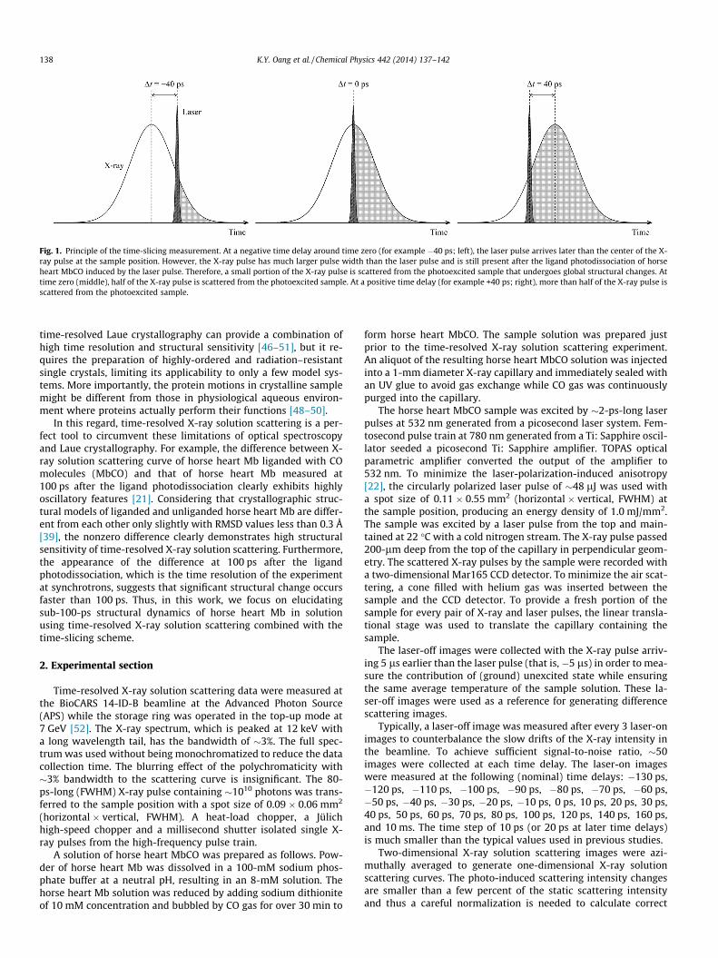
Fig. 1. Principle of the time-slicing measurement. At a negative time delay around time zero (for example �40 ps; left), the laser pulse arrives later than the center of the X-ray pulse at the sample position. However, the X-ray pulse has much larger pulse width than the laser pulse and is still present after the ligand photodissociation of horseheart MbCO induced by the laser pulse. Therefore, a small portion of the X-ray pulse is scattered from the photoexcited sample that undergoes global structural changes. Attime zero (middle), half of the X-ray pulse is scattered from the photoexcited sample. At a positive time delay (for example +40 ps; right), more than half of the X-ray pulse isscattered from the photoexcited sample.
138 K.Y. Oang et al. / Chemical Physics 442 (2014) 137–142
time-resolved Laue crystallography can provide a combination ofhigh time resolution and structural sensitivity [46–51], but it re-quires the preparation of highly-ordered and radiation–resistantsingle crystals, limiting its applicability to only a few model sys-tems. More importantly, the protein motions in crystalline samplemight be different from those in physiological aqueous environ-ment where proteins actually perform their functions [48–50].
In this regard, time-resolved X-ray solution scattering is a per-fect tool to circumvent these limitations of optical spectroscopyand Laue crystallography. For example, the difference between X-ray solution scattering curve of horse heart Mb liganded with COmolecules (MbCO) and that of horse heart Mb measured at100 ps after the ligand photodissociation clearly exhibits highlyoscillatory features [21]. Considering that crystallographic struc-tural models of liganded and unliganded horse heart Mb are differ-ent from each other only slightly with RMSD values less than 0.3 Å[39], the nonzero difference clearly demonstrates high structuralsensitivity of time-resolved X-ray solution scattering. Furthermore,the appearance of the difference at 100 ps after the ligandphotodissociation, which is the time resolution of the experimentat synchrotrons, suggests that significant structural change occursfaster than 100 ps. Thus, in this work, we focus on elucidatingsub-100-ps structural dynamics of horse heart Mb in solutionusing time-resolved X-ray solution scattering combined with thetime-slicing scheme.
2. Experimental section
Time-resolved X-ray solution scattering data were measured atthe BioCARS 14-ID-B beamline at the Advanced Photon Source(APS) while the storage ring was operated in the top-up mode at7 GeV [52]. The X-ray spectrum, which is peaked at 12 keV witha long wavelength tail, has the bandwidth of �3%. The full spec-trum was used without being monochromatized to reduce the datacollection time. The blurring effect of the polychromaticity with�3% bandwidth to the scattering curve is insignificant. The 80-ps-long (FWHM) X-ray pulse containing �1010 photons was trans-ferred to the sample position with a spot size of 0.09 � 0.06 mm2
(horizontal � vertical, FWHM). A heat-load chopper, a Jülichhigh-speed chopper and a millisecond shutter isolated single X-ray pulses from the high-frequency pulse train.
A solution of horse heart MbCO was prepared as follows. Pow-der of horse heart Mb was dissolved in a 100-mM sodium phos-phate buffer at a neutral pH, resulting in an 8-mM solution. Thehorse heart Mb solution was reduced by adding sodium dithioniteof 10 mM concentration and bubbled by CO gas for over 30 min to
form horse heart MbCO. The sample solution was prepared justprior to the time-resolved X-ray solution scattering experiment.An aliquot of the resulting horse heart MbCO solution was injectedinto a 1-mm diameter X-ray capillary and immediately sealed withan UV glue to avoid gas exchange while CO gas was continuouslypurged into the capillary.
The horse heart MbCO sample was excited by �2-ps-long laserpulses at 532 nm generated from a picosecond laser system. Fem-tosecond pulse train at 780 nm generated from a Ti: Sapphire oscil-lator seeded a picosecond Ti: Sapphire amplifier. TOPAS opticalparametric amplifier converted the output of the amplifier to532 nm. To minimize the laser-polarization-induced anisotropy[22], the circularly polarized laser pulse of �48 lJ was used witha spot size of 0.11 � 0.55 mm2 (horizontal � vertical, FWHM) atthe sample position, producing an energy density of 1.0 mJ/mm2.The sample was excited by a laser pulse from the top and main-tained at 22 �C with a cold nitrogen stream. The X-ray pulse passed200-lm deep from the top of the capillary in perpendicular geom-etry. The scattered X-ray pulses by the sample were recorded witha two-dimensional Mar165 CCD detector. To minimize the air scat-tering, a cone filled with helium gas was inserted between thesample and the CCD detector. To provide a fresh portion of thesample for every pair of X-ray and laser pulses, the linear transla-tional stage was used to translate the capillary containing thesample.
The laser-off images were collected with the X-ray pulse arriv-ing 5 ls earlier than the laser pulse (that is, �5 ls) in order to mea-sure the contribution of (ground) unexcited state while ensuringthe same average temperature of the sample solution. These la-ser-off images were used as a reference for generating differencescattering images.
Typically, a laser-off image was measured after every 3 laser-onimages to counterbalance the slow drifts of the X-ray intensity inthe beamline. To achieve sufficient signal-to-noise ratio, �50images were collected at each time delay. The laser-on imageswere measured at the following (nominal) time delays: �130 ps,�120 ps, �110 ps, �100 ps, �90 ps, �80 ps, �70 ps, �60 ps,�50 ps, �40 ps, �30 ps, �20 ps, �10 ps, 0 ps, 10 ps, 20 ps, 30 ps,40 ps, 50 ps, 60 ps, 70 ps, 80 ps, 100 ps, 120 ps, 140 ps, 160 ps,and 10 ms. The time step of 10 ps (or 20 ps at later time delays)is much smaller than the typical values used in previous studies.
Two-dimensional X-ray solution scattering images were azi-muthally averaged to generate one-dimensional X-ray solutionscattering curves. The photo-induced scattering intensity changesare smaller than a few percent of the static scattering intensityand thus a careful normalization is needed to calculate correct

K.Y. Oang et al. / Chemical Physics 442 (2014) 137–142 139
photo-induced scattering intensity difference. As a normalizationreference, we used the isosbestic point of the water scatteringcurves with respect to temperature change (q = 2.07 �1) so thatthe difference scattering intensity at this q value is zero. By takingthe differences between the laser-on scattering curves measured atpositive time delays and the reference scattering curve measuredat �5 ls (laser-off), the time-resolved difference X-ray solutionscattering curves, qDS(q, t) was obtained. The difference scatteringcurves, qDS(q, t), following photoexcitation of horse heart Mb solu-tion are shown in Fig. 2.
3. Results and discussion
To determine (1) how many structurally distinguishable inter-mediates of horse heart Mb are formed in the time range of ourinterest and (2) how fast each intermediate is formed, we per-formed the singular value decomposition (SVD) analysis of theexperimental data in the q range of 0.15–1.0 Å�1 and the timerange from �130 ps to 160 ps. Detailed description of the SVDanalysis can be found in our previous publications [21,24–26]. Inbrief, from the time-resolved X-ray solution scattering curves ac-quired at various time delays, we produced an nq � nt matrix Aexp(-q, t), where nq is the number of q points in the scattering curve at agiven time-delay point and nt is the number of time delay points.For the data matrix Aexp(q, t) measured in this work, nq and nt are416 and 26, respectively. Then, we decomposed the matrix Aexp(q,t) into three matrices (Uexp(q), Sexp, and Vexp(t)) while obeying therelationship:
Aexpðq; tÞ ¼ UexpðqÞSexpVexpðtÞT ð1Þ
where Uexp(q) is a nq � nt matrix whose column vectors correspondto time-independent q spectra of Aexp(q, t) (left singular vectors;lSVs), Sexp is a nt � nt diagonal matrix whose diagonal elements cor-respond to singular values of Aexp(q, t), and Vexp(t) is a nt � nt matrix
Fig. 2. Time-resolved difference X-ray solution scattering curves, qDS(q, t),measured for a solution sample of horse heart MbCO after the ligand photodisso-ciation. The (nominal) time delay after the photodissociation is indicated aboveeach curve. Experimental data (black) are compared with theoretical data (red). Seethe text for details. (For interpretation of the references to colour in this figurelegend, the reader is referred to the web version of this article.)
whose column vectors correspond to amplitude changes of Uexp(q)as a function of time (right singular vectors; rSVs). The matricesUexp(q) and Vexp(t) obey the following relationship:
UexpðqÞUexpðqÞT ¼ VexpðtÞVexpðtÞT ¼ Int ð2Þ
where Int is the identity matrix. Since the singular values of Sexp areordered so that s1 P s2 P . . . P snt > 0, both lSVs and rSVs onmore left have larger contributions to the data. Accordingly, wecan extract time-independent scattering intensity components fromthe lSVs and time-dependent change of their amplitudes from therSVs.
The singular values and autocorrelation values of the singularvectors shown in Fig. 3 suggest that only the first two singularcomponents are significant enough to represent the experimentaldata, while the contributions from the third singular componentand beyond are insignificant. In other words, only two structurallydistinguishable intermediates are formed in the time range inves-tigated in this work. Then, we extracted the relaxation times ofrSVs to determine the structural dynamics of Mb. If the temporalwidth of X-ray pulse is shorter than the time scale of the dynamicsof our interest, the relaxation times of rSVs can be determined sim-ply by globally fitting the rSVs with multiple exponentials. How-ever, the X-ray pulse duration in this work is longer than orcomparable to the dynamics of our interest, and therefore we needto deconvolute the temporal profile of the X-ray pulse from themeasured data in order to determine the relaxation times accu-rately.In theory, our experimental data, Aexp(q, t), presented in thiswork can be expressed as a convolution [27]:
Aexpðq; tÞ ¼ Ainstðq; t � t0Þ � IX�rayðt � t0Þ ð3Þ
where Ainst(q, t) is nq � nt matrix containing the instantaneous re-sponse of the solution sample, IX-ray(t) is the temporal intensity pro-file of X-ray pulse, and t0 is the actual time-zero value of themeasurement. According to Eq. (1), the following relationship alsoholds:
VexpðtÞ ¼ Vinstðt � t0Þ � IX�rayðt � t0Þ ð4Þ
where Vinst(t) is a nt � nt matrix whose column vectors correspondto the amplitude changes of Uexp(q) as a function of time (right sin-gular vectors; rSVs) in case that an infinitely short X-ray pulse isused. Because only the first two singular components contributesignificantly to the data, we defined new matrices (U0exp(q), S0exp,and V0exp(t)) to remove the contributions of insignificant singularcomponents. U0exp(q) is a nq � 2 matrix containing only the firsttwo left singular vectors of Uexp(q), S0exp, is a 2 � 2 matrix containingonly the first two singular values of Sexp, and V0exp(t) is a 2 � nt ma-trix containing only the first two right singular vectors of Vexp(t). Weused only these matrices to extract the structural dynamics of theprotein transition. From Eq. (4), we can obtain the followingrelationship:
V0expðtÞ ¼ V0instðt � t0Þ � IX�rayðt � t0Þ ð5Þ
where V0 inst(t) is an 2 � nt matrix containing only the first two col-umn vectors of Vinst(t). We can model the column vectors of V0 inst(t)by a sum of multiple exponentials sharing common relaxationtimes. Then, we optimized the coefficients and the relaxation timesof the exponentials as well as the actual time zero, t0, by minimizingthe discrepancy (i.e. chi-square value) between V0exp(t) and the con-volution of V0inst(t�t0) and IX-ray(t�t0). However, standard deviationsfor V0exp(t), which are needed for the optimization described above,are not available from our experimental data and thus we insteadused the following method to optimize V0inst(t). According to Eqs.(1) and (5), the following relationship holds:
Aexpðq; tÞ ¼ U0expðqÞS0exp V0instðt� t0Þ � IX�rayðt� t0Þ� �T ð6Þ
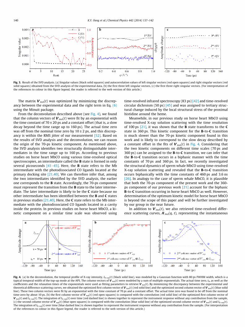
Fig. 3. Result of the SVD analysis. (a) Singular values (black solid squares) and autocorrelation values of left singular vectors (red open squares) and right singular vectors (redsolid squares) obtained from the SVD analysis of the experimental data, (b) the first three left singular vectors, (c) the first three right singular vectors. (For interpretation ofthe references to colour in this figure legend, the reader is referred to the web version of this article.)
140 K.Y. Oang et al. / Chemical Physics 442 (2014) 137–142
The matrix V0inst(t) was optimized by minimizing the discrep-ancy between the experimental data and the right term in Eq. (6)using the Minuit package.
From the deconvolution described above (see Fig. 4), we foundthat the column vectors of V0inst(t) were fit by an exponential withthe time constant of 70 ± 20 ps and a constant offset (that is, a slowdecay beyond the time range up to 160 ps). The actual time zerowas off from the nominal time zero by 10 ± 2 ps, and this discrep-ancy is within the RMS jitter of our measurement [52]. Based onthe results of SVD analysis and the deconvolution, we can reasonthe origin of the 70-ps kinetic component. As mentioned above,the SVD analysis identifies two structurally distinguishable inter-mediates in the time range up to 160 ps. According to previousstudies on horse heart MbCO using various time-resolved opticalspectroscopies, an intermediate called the B state is formed in onlyseveral picoseconds [41–45]. Here, the B state refers to the Mbintermediate with the photodissociated CO ligands located at theprimary docking site [21,40]. We can therefore infer that, amongthe two intermediates identified by the SVD analysis, the earlierone corresponds to the B state. Accordingly, the 70-ps componentmust represent the transition from the B state to the later interme-diate. The later intermediate is likely to be the C state because noother intermediate has been identified between the B and C statesin previous studies [21,40]. Here, the C state refers to the Mb inter-mediate with the photodissociated CO ligands located in a cavityinside the protein. In previous studies on horse heart MbCO, a ki-netic component on a similar time scale was observed using
Fig. 4. (a) In the deconvolution, the temporal profile of X-ray intensity, IX-ray(t) (black sotypical temporal width of the top-up mode at the APS. The column vectors of V0 inst(t) wercoefficients and the relaxation times of the exponentials were used as fitting parametertheoretical difference scattering curves, we obtained the optimized first column vector ofline). These two column vectors were fit by an exponential with the time constant of 70time-zero by about 10 ps, (b) the first column vector of V0exp(t) (red open squares) is comV0 inst(t) and IX-ray(t). The integration of IX-ray(t) over time (red dashed line) is shown togeth(c) the second column vector of V0exp(t) (blue open squares) is compared with the convolThe integration of IX-ray(t) over time (blue dashed line) is shown together to represent theof the references to colour in this figure legend, the reader is referred to the web versio
time-resolved infrared spectroscopy (83 ps) [42] and time-resolvedcircular dichroism (50 ps) [45] and was assigned to tertiary struc-tural change induced by the local structural stress of the proximalhistidine around the heme.
Meanwhile, in our previous study on horse heart MbCO usingtime-resolved X-ray solution scattering with the time resolutionof 100 ps [21], it was shown that the B state transforms to the Cstate in 360 ps. This kinetic component for the B-to-C transitionis much slower than the 70-ps kinetic component found in thiswork and is likely to correspond to the slow decay described bya constant offset in the fits of V0inst(t) in Fig. 4. Considering thatthe two kinetic components on different time scales (70 ps and360 ps) can be assigned to the B-to-C transition, we can infer thatthe B-to-C transition occurs in a biphasic manner with the timeconstants of 70 ps and 360 ps. In fact, we recently investigatedthe structural dynamics of sperm whale MbCO using time-resolvedX-ray solution scattering and revealed that the B-to-C transitionoccurs biphasically with the time constants of 460 ps and 3.6 ns[26]. In analogy to the case of sperm whale MbCO, it is plausiblethat both the 70-ps component of the present work and the 360-ps component of our previous work [21] account for the biphasicB-to-C transition occurring in horse heart MbCO as well. However,determination of the optimum kinetic model for horse heart MbCOis beyond the scope of this paper and will be further investigatedby our group in the near future.
In addition to V0inst(t), we also retrieved time-resolved differ-ence scattering curves, A0inst(q, t), representing the instantaneous
lid line), was modeled by a Gaussian function with 80 ps FWHM width, which is ae modeled by a sum of multiple exponentials. The actual time zero, t0, as well as thes to retrieve V0 inst(t). By minimizing the discrepancy between the experimental andV0 inst(t) (red solid line) and the optimized second column vector of V0 inst(t) (blue solidps and a constant offset. The actual time zero was found to be off from the nominalpared with the convolution (red solid line) of the optimized first column vector ofer to represent the instrument response without any contribution from the sample,
ution (blue solid line) of the optimized second column vector of V0 inst(t) and IX-ray(t).instrument response without any contribution from the sample. (For interpretation
n of this article.)
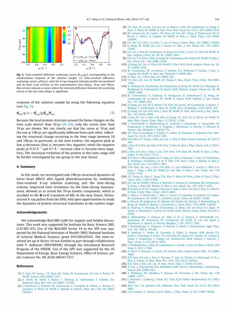
Fig. 5. Time-resolved difference scattering curves, A0 inst(q,t), corresponding to theinstantaneous response of the solution sample. (a) Time-resolved differencescattering curves, qDS(q,t), with the X-ray temporal intensity profile deconvolutedand (b) their cross sections at two representative time delays, 10 ps and 100 ps.Blue arrows indicate q values where the intensity difference between the scatteringcurves at the two time delays is significant.
K.Y. Oang et al. / Chemical Physics 442 (2014) 137–142 141
response of the solution sample by using the following equation(see Fig. 5):
A0instðq; tÞ ¼ U0expðqÞS0expV0inst ð7Þ
Because the local protein structure around the heme changes on thetime scale shorter than 10 ps [41–45], only the curves later than10 ps are shown. We can clearly see that the curve at 10 ps andthe one at 100 ps are significantly different from each other, reflect-ing the structural change occurring in the time range between 10and 100 ps. In particular, as the time evolves, the negative peak atlow q decreases (that is, becomes less negative) while the negativepeaks at 0.35 Å�1 and 0.7 Å�1 increase (that is, become more nega-tive). The structural evolution of the protein in this time range willbe further investigated by our group in the near future.
4. Summary
In this work, we investigated sub-100-ps structural dynamics ofhorse heart MbCO after ligand photodissociation by combiningtime-resolved X-ray solution scattering and the time-slicingscheme. Improved time resolution via the time-slicing measure-ment allowed us to reveal the 70-ps kinetic component, which isascribed to the B-to-C transition of Mb. In the near future, femto-second X-ray pulses from the XFEL will open opportunities to studythe dynamics of protein structural transitions at the earliest stage.
Acknowledgements
We acknowledge BioCARS staffs for support and helpful discus-sions. This work was supported by Institute for Basic Science (IBS)[CA1401-01]. Use of the BioCARS Sector 14 at the APS was sup-ported by the National Institutes of Health (NIH) National Instituteof General Medical Sciences grant P41GM103543. The time-re-solved set up at Sector 14 was funded in part through collaborationwith P. Anfinrud (NIH/NIDDK) through the Intramural ResearchProgram of the NIDDK. Use of the APS was supported by the USDepartment of Energy, Basic Energy Sciences, Office of Science, un-der Contract No. DE-AC02-06CH11357.
References
[1] H. Ihee, M. Lorenc, T.K. Kim, Q.Y. Kong, M. Cammarata, J.H. Lee, S. Bratos, M.Wulff, Science 309 (2005) 1223.
[2] A. Plech, M. Wulff, S. Bratos, F. Mirloup, R. Vuilleumier, F. Schotte, P.A.Anfinrud, Phys. Rev. Lett. 92 (2004) 125505.
[3] J. Davidsson, J. Poulsen, M. Cammarata, P. Georgiou, R. Wouts, G. Katona, F.Jacobson, A. Plech, M. Wulff, G. Nyman, R. Neutze, Phys. Rev. Lett. 94 (2005)245503.
[4] T.K. Kim, M. Lorenc, J.H. Lee, M. Lo Russo, J. Kim, M. Cammarata, Q. Kong, S.Noel, A. Plech, M. Wulff, H. Ihee, Proc. Natl. Acad. Sci. U.S.A. 103 (2006) 9410.
[5] M. Cammarata, M. Lorenc, T.K. Kim, J.H. Lee, Q.Y. Kong, E. Pontecorvo, M. LoRusso, G. Schiro, A. Cupane, M. Wulff, H. Ihee, J. Chem. Phys. 124 (2006)124504.
[6] J.H. Lee, K.H. Kim, T.K. Kim, Y. Lee, H. Ihee, J. Chem. Phys. 125 (2006) 174504.[7] Q. Kong, M. Wulff, J.H. Lee, S. Bratos, H. Ihee, J. Am. Chem. Soc. 129 (2007)
13584.[8] J.H. Lee, J. Kim, M. Cammarata, Q. Kong, K.H. Kim, J. Choi, T.K. Kim, M. Wulff, H.
Ihee, Angew. Chem. Int. Ed. 47 (2008) 1047.[9] J.H. Lee, T.K. Kim, J. Kim, Q. Kong, M. Cammarata, M. Lorenc, M. Wulff, H. Ihee, J.
Am. Chem. Soc. 130 (2008) 5834.[10] Q. Kong, J.H. Lee, A. Plech, M. Wulff, H. Ihee, M.H. Koch, Angew. Chem. Int. Ed.
47 (2008) 5550.[11] M. Cammarata, M. Levantino, F. Schotte, P.A. Anfinrud, F. Ewald, J. Choi, A.
Cupane, M. Wulff, H. Ihee, Nat. Methods 5 (2008) 881.[12] H. Ihee, Acc. Chem. Res. 42 (2009) 356.[13] T.K. Kim, J.H. Lee, M. Wulff, Q.Y. Kong, H. Ihee, Chem. Phys. Chem. 10 (2009)
1958.[14] K. Haldrup, M. Christensen, M. Cammarata, Q. Kong, M. Wulff, S.O. Mariager, K.
Bechgaard, R. Feidenhans’l, N. Harrit, M.M. Nielsen, Angew. Chem. Int. Ed. 48(2009) 4180.
[15] M. Christensen, K. Haldrup, K. Bechgaard, R. Feidenhans’l, Q. Kong, M.Cammarata, M. Lo Russo, M. Wulff, N. Harrit, M.M. Nielsen, J. Am. Chem.Soc. 131 (2009) 502.
[16] Q. Kong, J.H. Lee, M. Lo Russo, T.K. Kim, M. Lorenc, M. Cammarata, S. Bratos, T.Buslaps, V. Honkimaki, H. Ihee, M. Wulff, Acta. Crystallogr. A 66 (2010) 252.
[17] Q. Kong, J.H. Lee, K.H. Kim, J. Kim, M. Wulff, H. Ihee, M.H. Koch, J. Am. Chem.Soc. 132 (2010) 2600.
[18] S. Jun, J.H. Lee, J. Kim, K.H. Kim, Q. Kong, T.K. Kim, M. Lo Russo, M. Wulff, H.Ihee, Phys. Chem. Chem. Phys. 12 (2010) 11536.
[19] S. Westenhoff, E. Malmerberg, D. Arnlund, L. Johansson, E. Nazarenko, M.Cammarata, J. Davidsson, V. Chaptal, J. Abramson, G. Katona, A. Menzel, R.Neutze, Nat. Methods 7 (2010) 775.
[20] H.S. Cho, N. Dashdorj, F. Schotte, T. Graber, R. Henning, P. Anfinrud, Proc. Natl.Acad. Sci. U.S.A. 107 (2010) 7281.
[21] K.H. Kim, K.Y. Oang, J. Kim, J.H. Lee, Y. Kim, H. Ihee, Chem. Commun. 47 (2011)289.
[22] J. Kim, K.H. Kim, J.G. Kim, T.W. Kim, Y. Kim, H. Ihee, J. Phys. Chem. Lett. 2 (2011)350.
[23] J. Kim, J.H. Lee, J. Kim, S. Jun, K.H. Kim, T.W. Kim, M. Wulff, H. Ihee, J. Phys.Chem. A 116 (2012) 2713.
[24] K.H. Kim, S. Muniyappan, K.Y. Oang, J.G. Kim, S. Nozawa, T. Sato, S.Y. Koshihara,R. Henning, I. Kosheleva, H. Ki, Y. Kim, T.W. Kim, J. Kim, S. Adachi, H. Ihee, J.Am. Chem. Soc. 134 (2012) 7001.
[25] T.W. Kim, J.H. Lee, J. Choi, K.H. Kim, L.J. van Wilderen, L. Guerin, Y. Kim, Y.O.Jung, C. Yang, J. Kim, M. Wulff, J.J. van Thor, H. Ihee, J. Am. Chem. Soc. 134(2012) 3145.
[26] K.Y. Oang, J.G. Kim, C. Yang, T.W. Kim, Y. Kim, K.H. Kim, J. Kim, H. Ihee, J. Phys.Chem. Lett. 5 (2014) 804.
[27] J.H. Lee, M. Wulff, S. Bratos, J. Petersen, L. Guerin, J.C. Leicknam, M. Carnmarata,Q. Kong, J. Kim, K.B. Moller, H. Ihee, J. Am. Chem. Soc. 135 (2013) 3255.
[28] K.H. Kim, H. Ki, K.Y. Oang, S. Nozawa, T. Sato, J. Kim, T.K. Kim, J. Kim, S.I. Adachi,H. Ihee, Chem. Phys. Chem. (2013) 3687.
[29] L. Salassa, E. Borfecchia, T. Ruiu, C. Garino, D. Gianolio, R. Gobetto, P.J. Sadler,M. Cammarata, M. Wulff, C. Lamberti, Inorg. Chem. 49 (2010) 11240.
[30] J. Vincent, M. Andersson, M. Eklund, A.B. Wohri, M. Odelius, E. Malmerberg, Q.Kong, M. Wulff, R. Neutze, J. Davidsson, J. Chem. Phys. 130 (2009) 154502.
[31] K. Haldrup, T. Harlang, M. Christensen, A. Dohn, T.B. van Driel, K.S. Kjaer, N.Harrit, J. Vibenholt, L. Guerin, M. Wulff, M.M. Nielsen, Inorg. Chem. 50 (2011)9329.
[32] E. Malmerberg, Z. Omran, J.S. Hub, X. Li, G. Katona, S. Westenhoff, L.C.Johansson, M. Andersson, M. Cammarata, M. Wulff, D. van der Spoel, J.Davidsson, A. Specht, R. Neutze, Biophys. J. 101 (2011) 1345.
[33] S. Ibrahimkutty, P. Wagener, A. Menzel, A. Plech, S. Barcikowski, Appl. Phys.Lett. 101 (2012) 103104.
[34] K. Haldrup, G. Vanko, W. Gawelda, A. Galler, G. Doumy, A.M. March, E.P.Kanter, A. Bordage, A. Dohn, T.B. van Driel, K.S. Kjaer, H.T. Lemke, S.E. Canton, J.Uhlig, V. Sundstrom, L. Young, S.H. Southworth, M.M. Nielsen, C. Bressler, J.Phys. Chem. A 116 (2012) 9878.
[35] S. Ibrahimkutty, J. Kim, M. Cammarata, F. Ewald, J. Choi, H. Ihee, A. Plech, ACSNano 5 (2011) 3788.
[36] A. Plech, V. Kotaidis, A. Siems, M. Sztucki, Phys. Chem. Chem. Phys. 10 (2008)3888.
[37] K.H. Kim, J.H. Lee, J. Kim, S. Nozawa, T. Sato, A. Tomita, K. Ichiyanagi, H. Ki, J.Kim, S. Adachi, H. Ihee, Phys. Rev. Lett. 110 (2013) 165505.
[38] K.H. Kim, J. Kim, J.H. Lee, H. Ihee, Struct. Dyn. 1 (2014) 011301.[39] K. Chu, J. Vojtchovsky, B.H. McMahon, R.M. Sweet, J. Berendzen, I. Schlichting,
Nature 403 (2000) 921.[40] Y. Nishihara, M. Sakakura, Y. Kimura, M. Terazima, J. Am. Chem. Soc. 126
(2004) 11877.[41] L. Richard, L. Genberg, J. Deak, H.L. Chiu, R.J.D. Miller, Biochemistry 31 (1992)
10703.[42] M.H. Lim, T.A. Jackson, P.A. Anfinrud, Proc. Natl. Acad. Sci. U.S.A. 90 (1993)
5801.[43] G.D. Goodno, V. Astinov, R.J.D. Miller, J. Phys. Chem. A 103 (1999) 10630.

142 K.Y. Oang et al. / Chemical Physics 442 (2014) 137–142
[44] Y. Mizutani, T. Kitagawa, J. Phys. Chem. B 105 (2001) 10992.[45] T. Dartigalongue, F. Hache, Chem. Phys. Lett. 415 (2005) 313.[46] Y.O. Jung, J.H. Lee, J. Kim, M. Schmidt, K. Moffat, V. Srajer, H. Ihee, Nat. Chem. 5
(2013) 212.[47] F. Schotte, M.H. Lim, T.A. Jackson, A.V. Smirnov, J. Soman, J.S. Olson, G.N.
Phillips, M. Wulff, P.A. Anfinrud, Science 300 (2003) 1944.[48] H. Ihee, S. Rajagopal, V. Srajer, R. Pahl, S. Anderson, M. Schmidt, F. Schotte, P.A.
Anfinrud, M. Wulff, K. Moffat, Proc. Natl. Acad. Sci. U.S.A. 102 (2005) 7145.[49] J.E. Knapp, R. Pahl, V. Srajer, W.E. Royer Jr., Proc. Natl. Acad. Sci. U.S.A. 103
(2006) 7649.
[50] J.E. Knapp, R. Pahl, J. Cohen, J.C. Nichols, K. Schulten, Q.H. Gibson, V. Srajer, W.E.Royer Jr., Structure 17 (2009) 1494.
[51] F. Schotte, H.S. Cho, J. Soman, M. Wulff, J.S. Olson, P.A. Anfinrud, Chem. Phys.422 (2013) 98.
[52] T. Graber, S. Anderson, H. Brewer, Y.S. Chen, H.S. Cho, N. Dashdorj, R.W.Henning, I. Kosheleva, G. Macha, M. Meron, R. Pahl, Z. Ren, S. Ruan, F. Schotte,V.S. Rajer, P.J. Viccaro, F. Westferro, P. Anfinrud, K. Moffat, J. SynchrotronRadiat. 18 (2011) 658.