aging of the nervous system: structural changes chapters 7, 8, 9 ps timiras chapters 7, 8, 9 ps...
Post on 21-Dec-2015
218 views
TRANSCRIPT
Aging of the Aging of the Nervous System:Nervous System:
Structural ChangesStructural Changes
Chapters 7, 8, 9Chapters 7, 8, 9
PS TimirasPS TimirasChapters 7, 8, 9Chapters 7, 8, 9
PS TimirasPS Timiras
Brain Plasticity and Brain Plasticity and CNS Regenerative PotentialCNS Regenerative Potential
Brain Plasticity and Brain Plasticity and CNS Regenerative PotentialCNS Regenerative Potential
From the beginning of the 20th Century until From the beginning of the 20th Century until the 1990s, it was stated that neurons DID NOT the 1990s, it was stated that neurons DID NOT proliferate.proliferate.
The fact that they COULD NOT proliferate did The fact that they COULD NOT proliferate did not exclude the not exclude the possibilitypossibility of proliferation of proliferation under “specific conditions.”under “specific conditions.”
In fact, the CNS has a considerable In fact, the CNS has a considerable regenerative potential depending on the special regenerative potential depending on the special conditions of the neuronal environmentconditions of the neuronal environment..
Neurons that may proliferate into Neurons that may proliferate into adulthood include:adulthood include:
Neurons that may proliferate into Neurons that may proliferate into adulthood include:adulthood include:
Progenitor “precursor” neurons lining the Progenitor “precursor” neurons lining the cerebral ventriculescerebral ventricules
Neurons in the hippocampusNeurons in the hippocampus
Neurons usually “dormant” with potential Neurons usually “dormant” with potential for neuron and glia proliferationfor neuron and glia proliferation
Neuroglia (astrocytes, oligodentrocytes) Neuroglia (astrocytes, oligodentrocytes) and microglia (immune cells) with the and microglia (immune cells) with the ability to perpetually self renew and ability to perpetually self renew and produce the three types of neural cellsproduce the three types of neural cells
Regenerative potential depends Regenerative potential depends on changes in on changes in whole body whole body and and
neural microenvironmentneural microenvironment
Whole body changes:Whole body changes: Physical exercisePhysical exercise Appropriate nutritionAppropriate nutrition Good circulationGood circulation EducationEducation StressStress othersothers
• Neural Neural microenvironment microenvironment changes:changes:
–Brain metabolism (oxygen Brain metabolism (oxygen consumption, free radicals, consumption, free radicals, circulatory changes)circulatory changes)–Hormonal changes Hormonal changes (estrogens, growth factors, (estrogens, growth factors, others)others)–othersothers

Major Function of the Major Function of the Nervous SystemNervous System
The major function of the CNS is to The major function of the CNS is to communicate & to connect:communicate & to connect:
•with other CNS cellswith other CNS cells
•with peripheral tissues (outside CNS)with peripheral tissues (outside CNS)
•with the external environment with the external environment (including physical and social (including physical and social
environments)environments)
This communication regulates:This communication regulates:•MobilityMobility•Sensory informationSensory information•Cognition Cognition •Affect and moodAffect and mood•Functions of whole-body systemsFunctions of whole-body systems
Major Function of Major Function of the Nervous System, IIthe Nervous System, II
Cogito Ergo Sum
“ I think, therefore I am”-René Descartes (1596-1650)
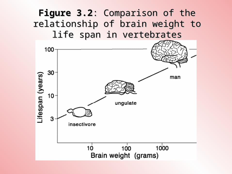
Figure 3.2Figure 3.2: Comparison of the : Comparison of the relationship of brain weight to relationship of brain weight to
life span in vertebrateslife span in vertebrates
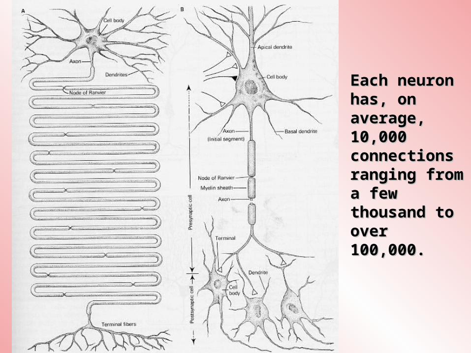
Each neuron Each neuron has, on has, on average, average, 10,000 10,000 connections connections ranging from ranging from a few a few thousand to thousand to over over 100,000.100,000.
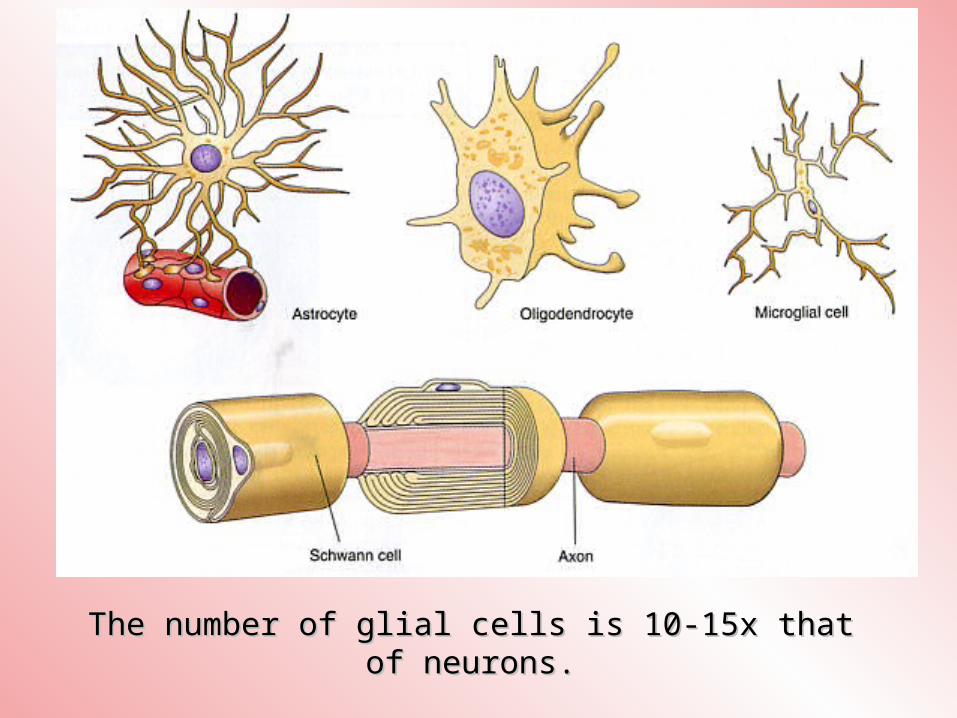
The number of glial cells is 10-15x that The number of glial cells is 10-15x that of neurons.of neurons.

Fig. 7-4Fig. 7-4: “Denudation” of the neurons. Changes in : “Denudation” of the neurons. Changes in pyramidal neurons of the aging human cerebral pyramidal neurons of the aging human cerebral
cortexcortex

In normal aging, the loss of In normal aging, the loss of neurons is moderate & occurs neurons is moderate & occurs in specific brain areas:in specific brain areas:
Locus ceruleus (catecholaminergic Locus ceruleus (catecholaminergic neurons)neurons)
Substantia nigra (dopaminergic Substantia nigra (dopaminergic neurons)neurons)
Nucleus basalis of Meynert Nucleus basalis of Meynert (cholinergic neurons) (cholinergic neurons)
Hippocampus (cholinergic neurons) Hippocampus (cholinergic neurons)
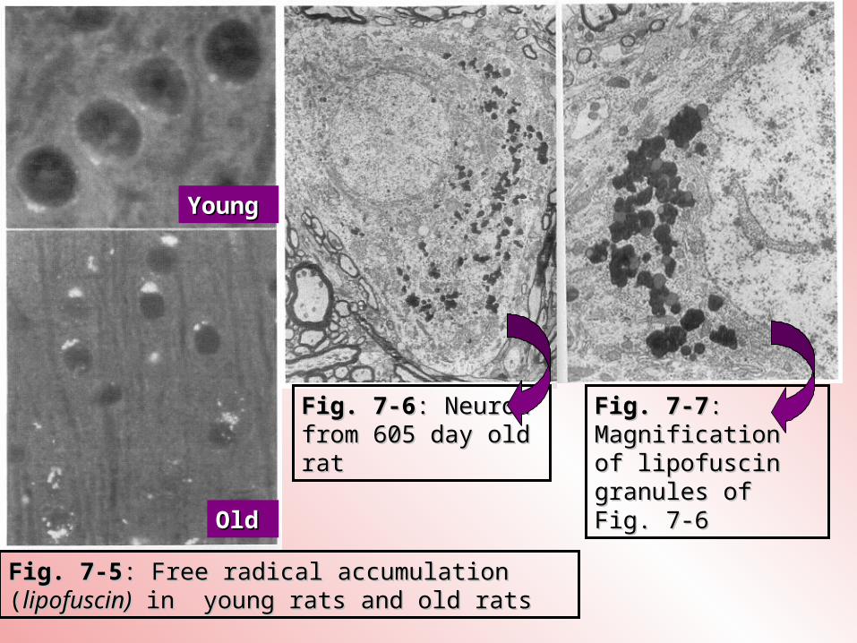
Fig. 7-5Fig. 7-5: Free radical accumulation : Free radical accumulation ((lipofuscin)lipofuscin) in young rats and old in young rats and old rats rats
Fig. 7-6Fig. 7-6: Neuron : Neuron from 605 day old from 605 day old ratrat
Fig. 7-7Fig. 7-7: : Magnification Magnification of lipofuscin of lipofuscin granules of granules of Fig. 7-6Fig. 7-6
YoungYoung
OldOld
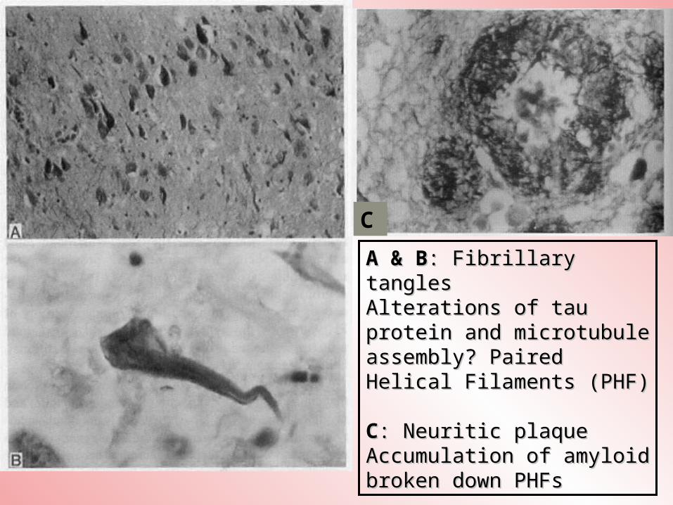
C
A & BA & B: Fibrillary : Fibrillary tanglestanglesAlterations of tau Alterations of tau protein and microtubule protein and microtubule assembly? Paired assembly? Paired Helical Filaments (PHF)Helical Filaments (PHF)
CC: Neuritic plaque: Neuritic plaqueAccumulation of amyloid Accumulation of amyloid broken down PHFsbroken down PHFs
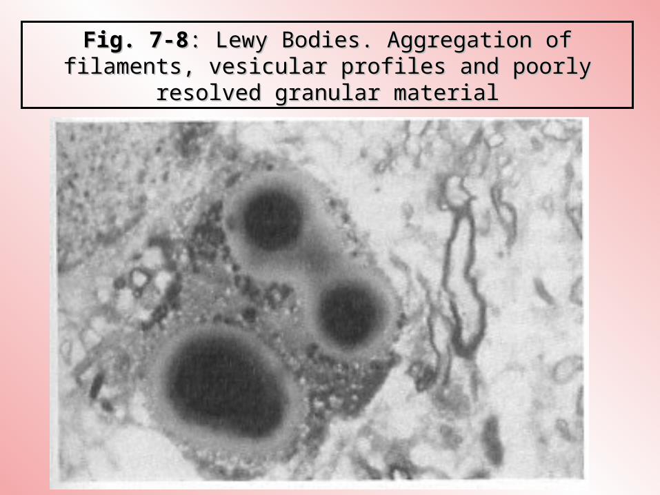
Fig. 7-8Fig. 7-8: Lewy Bodies. Aggregation of : Lewy Bodies. Aggregation of filaments, vesicular profiles and poorly filaments, vesicular profiles and poorly
resolved granular materialresolved granular material
Pathological and Cellular Pathological and Cellular Changes with Normal AgingChanges with Normal AgingPathological and Cellular Pathological and Cellular Changes with Normal AgingChanges with Normal Aging
Increased intracellular deposits Increased intracellular deposits of lipofuscinof lipofuscin
Intracellular formation of PHFsIntracellular formation of PHFs Accumulation of amyloid deposits Accumulation of amyloid deposits in the neuritic plaques and in the neuritic plaques and surrounding the cerebral blood surrounding the cerebral blood vesselsvessels
Accumulation of Lewy bodiesAccumulation of Lewy bodies Cell death (apoptosis, necrosis)Cell death (apoptosis, necrosis)
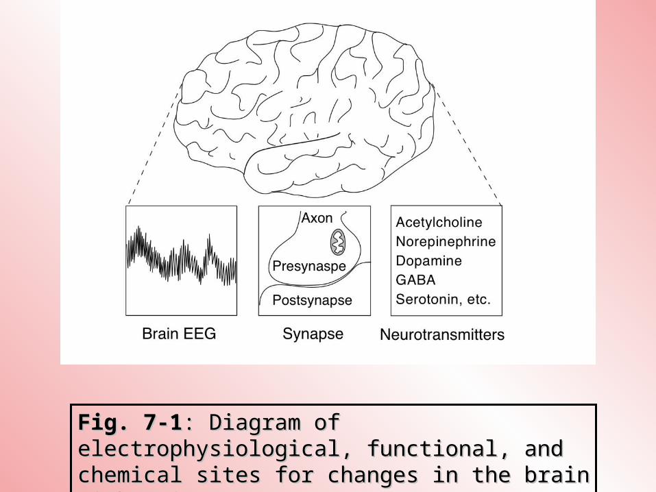
Fig. 7-1Fig. 7-1: Diagram of : Diagram of electrophysiological, functional, and electrophysiological, functional, and chemical sites for changes in the brain chemical sites for changes in the brain with agingwith aging
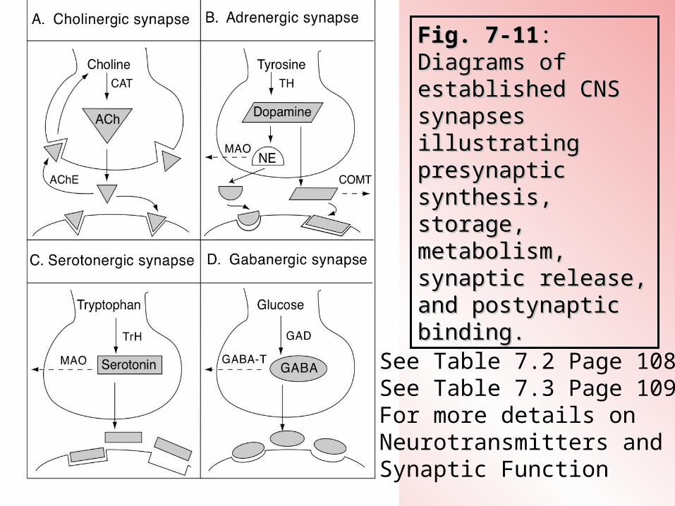
Fig. 7-11Fig. 7-11: : Diagrams of Diagrams of established CNS established CNS synapses synapses illustrating illustrating presynaptic presynaptic synthesis, synthesis, storage, storage, metabolism, metabolism, synaptic release, synaptic release, and postynaptic and postynaptic binding.binding.
See Table 7.2 Page 108See Table 7.3 Page 109For more details on Neurotransmitters and Synaptic Function
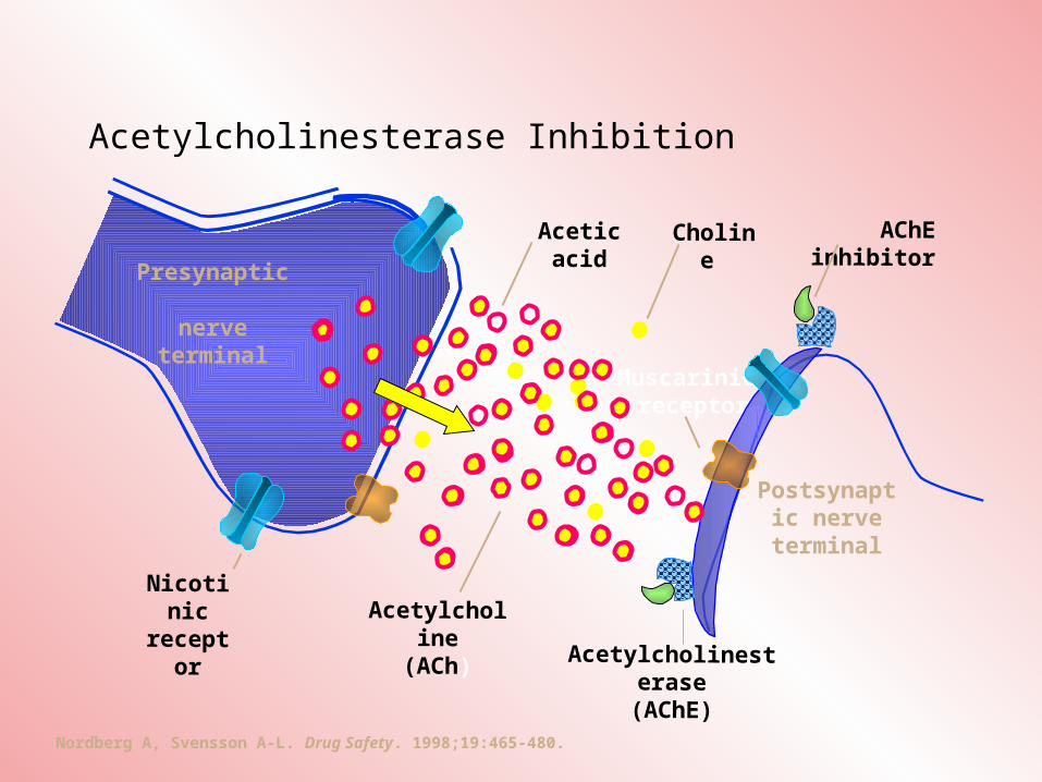
Acetylcholinesterase Inhibition
Nordberg A, Svensson A-L. Drug Safety. 1998;19:465-480.
Postsynaptic nerve
terminal
Nicotinic
receptor
Presynaptic nerve terminal
Muscarinic receptor
Acetylcholine(ACh)
Acetic acid Choline
Acetylcholinesterase
(AChE)
AChE inhibitor