structure-based inhibitor design for cdk2, a cell cycle ... inhibitor design for cdk2, a cell cycle...
TRANSCRIPT

Pure & Appl. Chern., Vol. 70, No. 3, pp. 555-565, 1998. Printed in Great Britain. (B 1998 IUPAC
Structure-based inhibitor design for CDK2, a cell cycle controlling protein kinase
Sung-Hou Kim, Department of Chemistry and E. 0. Lawrence Berkeley National Laboratory, University of California, Berkeley, California 94720 U.S.A.
Abstract: The central role of cyclin-dependent kinases (CDKs) in cell cycle regulation makes them a promising target for discovering small inhibitory molecules that can modify the degree of cell proliferation. The three- dimensional structure of CDKZ provides a structural foundation for understanding the mechanisms of activation and inhibition af CDKZ and for the discovery of the inhibitors. In this presentation five structures of human CDK2 are summarized: apo protein, ATP complex, olomoucine complex, isopentenyladenine complex, and flavopiridol complex. This structural information is used for the design of a small, directed combinatorial library.
INTRODUCTION Cell cycle progression is tightly controlled by the activity of cyclin-dependent kinases (CDKs) (refs. 1-3). CDKs remain inactive by themselves, and activation requires binding to cyclins, a diverse family of proteins whose levels oscillate during the cell cycle, and phosphorylation by CDK-activating kinase (CAK) on a specific threonine residue (refs. 4, 5 ) . In addition to the positive regulatory proteins such as cyclins and CAK, many negative regulatory proteins (CDK inhibitory proteins, CKIs) have been discovered (refs. 6-8) such as p16 (ref. 9), p21 (refs. 10-13), and p28 (ref. 14). Since deregulation of cyclins and/or alteration or absence of CKIs have been associated with many cancers, there is strong interest in chemical inhibitors of CDKs that could play an important role in the discovery of a new family of anti-tumor agents or anti-proliferative drugs.
Since ATP is the authentic cofactor of CDK2 it can be considered as a "pseudo-lead compound" for discovery of CDK2 inhibitors. However there are two m&jor concerns: adenine-containing compounds are common ligands for many enzymes in cells, thus, any adenine derivatives may inhibit many enzymes in the cells: second, any compounds with highly charged groups such as phosphates in ATP will prevent uptake by the cells. Our studies provide the structural basis for overcoming the two difficulties by appropriate modification of a common base such as adenine to endow specificity and cell uptake of the derivatives. The results summarized here are obtained in collaboration with the laboratories of David Morgan at the University of California, San Francisco, Peter Schultz at the University of California, Berkeley, Laurent Meijer of CNRS-Roscoff in France, and Peter Worland and Edward Sausville of NIH.
MATERIALS AND METHODS
Human CDK2 was prepared as described in Ref. 15. Briefly, Sf9 insect cells were infected with baculovirus containinga human CDK2 gene. The supernatant of cell lysate was loaded over a DEAE Sepharose column followed by an S-Sepharose column. The flow-through was loaded onto an ATP-affinity column and eluted by NaCl salt gradient. The purified protein is fully functional in that it binds to cyclin A, the resulting complex can be fully activated when incubated with partially purified human CAK, and phosphorylated CDKZ-cyclin A complex can phosphorylate histon H 1.
Olomoucine was obtained from L. Meijer of CNRS, Roscoff, France, and isopentenyladenine and ATP were purchased from Sigma (St. Louis, Missouri). Des-chloro-flavopiridol and flavopiridol are derived by chemical synthesis from a parent structure obtained from Dysoxylum binectariferum (ref. 16), a plant native to India. Both were provided by Dr. Peter Worland now at Mitotix (Cambridge, Massachusetts) who obtained it from Dr. Harald Sedlacek, Behringwerke, Marburg, Germany. The chemical structures of ATP and the inhibitors discussed are shown in Fig. 1.
555

556
b H bH
S.-H. KIM AND E. 0. LAWRENCE
OH
Fig. 1. Chemical structures of four ligands of CDK2 discussed in this paper: (top left) ATP, (bottom left) olomoucine, (top right) isopentenyladenine, and (bottom right) des-chloro- flavopiridol.
s a n g and co -crystals Recombinant CDK2 was crystallized as below (ref. 17). A CDK2 solution was concentrated to 10 mg/ml by dialysis against 20 mM Hepes buffer (pH 7.4) and 1 mM EDTA. Sitting drops were equilibrated by vapor diffusion at 4°C against reservoirs containing 200 mM Hepes buffer at pH 7.4. Diamond- and wedge-shaped crystals appeared after two to four days. A gradual increase of the Hepes buffer concentration in the reservoirs (up to 800 mM) produced crystals with average dimensions of about 0.2 mm x 0.3 mm x 0.3 mm. After CDK2 crystals had formed, small amounts of powdered chemical inhibitors were added to the crystallization drops. All inhibitor complexes were obtained by this soaking procedure (refs. 18, 19), except for the olomoucine complex, which was obtained by co-crystallization of a precomplexed solution of CDKZ and olomoucine. Crystals were soaked usually for 48 hours before data collection. Crystallization of CDKZ apoprotein and co-crystallization of CDKZ and olomoucine were performed using the sparse matrix method (ref. 20).
X-rav -hit shdki Relevant crystallographic parameters and refinement parameters are given in Table 1. X-ray diffraction data were collected using a Rigaku RAXIS-I1 imaging plate area detector and processed with the RAXIS data processing software. The crystal structure of the apoprotein (ref. 17) was determined by the multiple isomorphous replacement method, and ATP complex structure as well as all the inhibitor complex structures (refs. 17, 18, 19) were solved by the molecular replacement method using the apo structure as the probe. All the structures were refined using X- PLOR 3.0 (ref. 22). The refinement statistics are given also in Table 1.
Domain structure and rpnformation of CDKZ The CDK2 structure is almost identical in CDK-apoenzyme (ref. 17), ATP complex (ref. 17), olomoucine complex (ref. 18), isopentenyladenine complex (ref. 18), des-chloro-flavopiridol (ref. 19), and flavopiridol complex (unpublished), and very similar to that in CDK2-cyclin A complex (ref. 23), except in five regions (see below). The enzyme is folded into the bilobal structure typical for most of protein kinases, with the smaller N-terminal domain consisting predominantly of P-sheet structure and the larger C-terminal domain consisting primarily of a-helices (Fig. 2). There are no significant differences in the domain orientations between the ligand-enzyme complexes and the apoenzyme. In all cases except CDK2-cyclin A complex (ref. 23), electron density is weak in two regions in the enzyme, spanning residues 36-47 which links the N-terminal
RESULTS
0 1998 IUPAC, Pure and Applied Chemistry70.555-565

P a 0
P
b
P 2
8
TA
BL
E 1
. C
ryst
allo
grap
hic p
aram
eter
s and
ref
inem
ent s
tatis
tics.
APO
Spac
e gro
up
m12
121
Cel
l dim
ensi
ons (A
) a=
73.1
2 b=
72.7
2 ~
34
.25
U
niqu
e re
flect
ions
27
.034
C
ompl
eten
ess o
f dat
a (A
) -1.
8 98
.6%
Rsym
* (%
I 6.
1 R
esol
utio
n (A
) 8.
00-1
.8
Rva l
ue *
0.18
R
f/ee t
0.25
B
aver
ageW
2)
31
Dev
iatio
ns o
bser
ved:
rm
sd fr
om id
eal b
ond
leng
th (A
) 0.0
1 1
rmsd
from
idea
l bon
d an
gle
(")
1.61
N
umbe
r of w
ater
mol
ecul
es
180
1.9-1.
8 97
.1%
ATP
n1
2121
a=
72.8
2 b=
72.6
6 ~=
54.0
7 22
,636
(3,
019)
-1.
9
6.7
8.00
-1.8
0.
18
0.27
38
0.01
2 1.
78
108
Olo
mou
cine
m
1212
1 a=
73.7
7 b=
72.5
5 c=
54.0
6 15
,044
-2.
5 99
.0%
2.5
-2.2
97.0
%
8.4
8.00
-2.2
0.
19
0.27
31
0.01
3 1.
70
76
Isop
ente
nyla
deni
ne
a=73
.25
m12
121
b=72
.5 1
c=
54.0
3 25
.37
1 -2.
0 96
.4%
2.0
-1.8
84.0
%
8.3 8.00
-1.8
0.
20
0.27
30
Des
-chl
orof
lavo
pirid
o1
n121
21
a=7 1
.30
b=72
.03
~=53
.70
1 1,4
30
-2.0
91.7
%
4.9 8.G
2.3
3
20.3
28
.8
32.3
5
0.01
1
0.01
2 1.
76
1.7
99
84
*Rva
[ue=
lOO
~lF
~
- Fcl
/x(F
o), th
e sum
s bei
ng ta
ken
over
all
refle
ctio
ns w
ith F
/s(F
) > 2
cuto
ff.
tRfi
ee =
Rva
lue
for 1
0% o
f the
dat
a, w
hich
wer
e no
t inc
lude
d du
ring
crys
tallo
grap
hic r
efin
emen
t. 'B
aver
age
= A
vera
ge B
val
ues
for a
ll no
n-hy
drog
en at
oms.

33N3UMW .O ‘3 aNV WII ‘H-’S

Structure of CDKZ 559
2 r ';=\ 1 Nucleophilic attack
Fig. 3. Schematic drawing of the five regions that have different conformations between (a) CDK- ATP binary complex, and (b) CDK2-ATP-cyclin A ternary complex structures. They are: triphosphate of ATP, P-loop, cyclin-binding helix, alp transition box, and T-loop.
0 1998 IUPAC, Pure and Applied Chernistry70,555-565
560 S:H. KIM AND E. 0. LAWRENCE
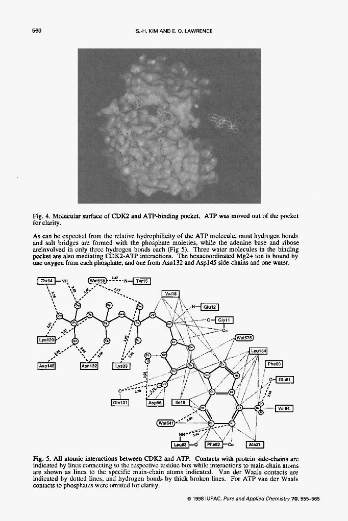
Fig. 4. Molecular surface of CDKZ and ATP-binding pocket. ATF’ was moved out of the pocket for clarity.
As can be expected from the relative hydrophilicity of the ATP molecule, most hydrogen bonds and salt bridges are formed with the phosphate moieties, while the adenine base and ribose areinvolved in only three hydrogen bonds each (Fig 5). Three water molecules in the binding pocket are also mediating CDKZ-ATP interactions. The hexacoordinated Mg2+ ion is bound by one oxygen from each phosphate, and one from Asn132 and Asp145 side-chains and one water.
Fig. 5. All atomic interactions between CDK2 and ATP. Contacts with protein side-chains are indicated by lines connecting to the respective residue box while interactions to main-chain atoms are shown as lincs to the specific main-chain atoms indicated. Van der Waals contacts are indicated by dotted lines, and hydrogen bonds by thick broken lines. For ATP van der Waals contacts to phosphates were omitted for clarity.
0 1998 IUPAC, Pure andApplied Chemistry70. 555-565

Structure of CDK2 561
tors: olomorrcrne and rso-ne- Kinase assay of adenine derivatives revealed that two adenine derivatives have inhibitory properties of CDC2 and CDK2 with IC50 of about 10 mM range: olomoucine inhibits cell cycle kinases (except CDK4) quite specifically, and isopentenyladenine (IPA) inhibits both cell cycle kinases and non-cell cycle kinases.(ref. 23). The crystal structures of CDKZ-IPA complex and CDKZ-olomoucine complex have been determined to high resolution (1.8 8, and 2.2 8, respectively), with good stereochemistry (Table 1). As in the apoenzyme and in the CDKZ-ATP complex, electron density is weak for two regions in the enzyme spanning residues 37-43 of interdomain connector and 153-163 of the T-loop containing the activating Thrl6O phosphorylation site (ref. 17).
Both P A and olomoucine are adenine derivatives. Although the purine rings bind roughly in the same area of the binding cleft as the adenine ring of ATP, the relative orientation of each purine ring with respect to the protein is different for all three ligands (ATP. IPA. and olomoucin) as shown in Fig. 6. This is probably due to the different size of the suhstituent groups of the purine
+ + I
Fig. 6. A w i e d view (it' the supcrporition of ATP (red). olomou;in (green). des-chloro- IlJvupiridol (purple). and isopcntcnyladeninc (hluc) in the ATP-hiding piickct of CDK2
in the three ligandr: the N6 amino group of the adenine ring is replxcd by rn iwpcntenylamino group in IPA and hy a hulky henzylamino gruup in olomoucinc. The complete atomic interaction ktwccn CDK2 and oldmuucine is shuwn in Fig. 7, and that hetwccn CDK2 3nd IPA in Fig. 8.
. . ~ . c t , l ~ , r " - ~ Previms studies have shown that flavopiridol. a novel flavonoid which is not an ddminc derivxive. can inhibit g r w t h of h x s t anJ lung carcinuma cell l i n s and can inhihit CDK activity at thc s m e nanomolar cuncentritiiin range of the inhihitor (ref. 16). To understmd how Ilwupiridol ana1og.i bind hi the A I P puckct. we determined th.: X-ray structure iif CDK2 in complex witn the 11avopit.idol and des-chloro-flxvopiridol (DFP. Fig. I). and compaxd i t with the X-my structures of CDKi-ATP cumplex (ref. 17) and other inhibitor complexes of CDK2 (ref. 18). Fl~vop)ridol hinds to CDKZ in the same mmncr ils DFP.
DFP hinds in the Alp-hinding pockct. with th: hcnmpyran ring wcupying approximately the same region as the purine ring of ATP (Fig. 6). The two ring systems overlap in thu s m e p h e , but the knmpyran is rotatcd ahuut 60' relative 1 0 the adenine in ATP, measured as the angle between the carbon-carbon bonds j i n i n g the two cyclcs in henzupyran and adenine rings, rcspectivdy. In this oricntatim. the 0 5 hydroxyl and the 01 of the inhihitor are close td the position of the N6 aniiio group and N I in adeninc, respccti\*cly. The piperidinyl ring pmiilly occupies the a-phosphate pocket and is assigned to a chair conformation (although a boat conformation c m not be ruled out in the curmnt rcsulutiun map). The d:&l inleractions hetwL.cn DFP and CDKZ arc. shdwn in Fig. 9.
Differences in wutein side-chain confurmatinns in lhe hindine _. oocka In contrast to the tinding that cyclin A changes very littlc un CDK2 hinding (ref. 21). the confortnation of CDK2 2ppears to be much more adaptahlc as manifcst-.d hy i k changes on hinding of ATP, olomuucin. IPA and DFP. 3s well as cyclin A (refs. 17. 18. 19. 21). To t inJ out whethcr any inliihitor or ATP binding to CDK2 induces changes in side-chxin contormation in the
@ 1998 IVPAC, Pure and Applied Chemistry70.55b565

562 S.-H. KIM AND E. 0. LAWRENCE
Fig. 7. All atomic interactions between CDK2 and olomoucine. The rest of the legend is the same as that of Fig. 5. All ligand contacts are shown.
Fig. 8. All atomic interactions between CDK2 and isopentenyladenine. The rest of the legend is the same as that of Fig. 5. All ligand contacts are shown.
0 1998 IUPAC, Pure and Applied Chemistry 70,555-565

Structure of CDK2 563
Fig. 9. All atomic interactions between CDK2 and des-chloro-flavopiridol. The rest of the legend is the same as that of Fig. 5. All ligand contacts are shown.
binding pocket, we compared the binding pockets of CDK2-ligand complexes and CDKZ apo protein after a superposition of the enzymes based on their Ca atoms. This superposition revealed the significant movement of few side-chains. In addition to the P-loop, four residues change their side chain conformations most: Lys33, Q131, IlelO, and N132. Lys33 and its equivalent Lys72 in cyclic AMP dependent protein kinase are important for ATP binding. In all ligana bound complexes the side chain of this residue has moved away to accommodate the ligands except IPA complex, where the side chain conformation is the same as that of apoenzyme presumably because of the small size of the ligand. In the CDKZATP complex, Lys33 forms salt bridges with the a phosphate of ATP and Asp145 another residue involved in ATP-Mg2+ binding (ref. 17).
The side-chain of Gln131 is directed away from the binding pocket in the ATP and IPA complex. In the olomoucine complex, however, the side-chain points into the binding pocket and forms van der Waals contacts and a hydrogen bond with the hydroxyethylamino group of.olomoucine. IlelO and Asn132 make smaller conformational changes on the ligand binding. In summary, the observed side-chain differences in the binding pocket seem to contribute to an improved fit between protein and ligand molecules.
Structure-based cornbinatorigd-lbxy Using the crystal structure of the complex between CDK2 and olomoucine we have chosen three sites of adenine for introducing various chemical groups in our combinatorial libraries. Peter Schultz’s laboratory at University of California-Berkeley synthesized three classes of small combinatorial libraries using three different sites of adenine to conjugate to the solid plastic pins in 8 x 12 arrays (ref. 26). In v i m assay for the inhibition of CDKZ activities were measured using histone H1 as a substrate. Compared to olomoucine, which has IC50 of about 10 pM under our assay condition, the best inhibitors identified from the three libraries have about three orders of magnitude greater potency ( IC 50 of about 15 nM) than olomoucine (manuscript in preparation).
0 1998 IUPAC, Pure and Applied Chernistry70,555-565

564 S.-H. KIM AND E. 0. LAWRENCE
DISCUSSION The high-resolution X-ray structures of human CDKZ apoenzyme and ATP complex as well as CDK2-cyclin A-ATP complex provide the structural basis for understanding the mechanism of activation of CDK2. Furthermore, the structures of the complexes between CDKZ with four inhibitors described here explain the specificity and potency of these inhibitors compared to other related derivatives, and suggest structure-based drug design and combinatorial library design that may enhance the chance of discovering better and improved inhibitors of CDK2 and other CDKs. Several interesting lessons were learned from these structural studies:
(a) The ATP-binding pocket of CDK2 has the surprising capacity to bind varieties of flat aromatic moieties with some unpredictable conformational changes of residues in the pocket. This may be related to the relatively weak binding of the ligands (Kd -pM).
(b) Knowing the orientation of adenine of ATP in the ATP pocket did not allow us to predict how the adenine moiety of olomoucine and IPA would bind to CDK2. Experimental determination of IPA and olomoucine complexes made us realize that any "rational drug design" based on adenine position of ATP would have completely missed in predicting olomoucine and IPA as potential inhibitors.
(c) Specificity of each inhibitor for CDK2 over other kinase comes from the interaction between substitution groups outside of the aromatic scaffold and the peripheral surface of the ATP pocket. This observation dispels the conventional wisdom that "the common base such as adenine cannot be a good scaffold for inhibitor design because there are many proteins in cells that bind adenine derivatives." Our results clearly demonstrate that specificity can be created by appropriate modification of a common scaffold molecule.
(d) The knowledge of the structures of CDK2 complexes with the three inhibitors suggests a few new scaffolds to consider in structure-based drug design and combinatorial library design.
(e) Structure-based design of combinatorial library is a highly effective way of discovering improved inhibitors (refs. 24,25).
(0 Structure-based combinatorial library design is an effective way to improve the potency of an inhibitor.
REFERENCES 1. C. Norbury and P. Nurse. Annu. Rev. Biockm. 61,441470 (1992). 2. F. Fang and J. W. Newport. Cell 66,731-742 (1991). 3. T. Hunt. Curr. Opin. Cell Biol. 1,274-286 (1989). 4. D. Desk, Y. Gu and D. 0. Morgan. Mol. Biol. Cell 3,571-582 (1992). 5. Y. Gu, J. Rosenblatt and D. 0. Morgan. EMBO J. 11,39954005 (1992). 6. H. E. Richardson, C. S . Stueland, J. Thomas, P. Russel and S. I. Reed. Genes Dev. 4, 1332-1344 (1990). 7. M. Semno, G. J. Hannon and D. Beach. Nuture 366,704-707 (1993). 8. M. Peter and I. Herskowitz. Cell 79,181-184 (1994). 9. T. Nobori, K. Miura, D. J. Wu, A. Lois, K. Takabayashi and D. A. Carson. Nuture 368,753-756 (1994). 10. Y. Gu, C. W. Turck and D. 0. Morgan. Nuture 366,707-710 (1993). 11. Y. Xiong, G. J. Hannon, H. Zhang, D. Casso, R. Kobayashi and D. Beach. Nature 366,701-704 (1993). 12. J. W. Harper, G. R. A M , N. Wei, K. Keyomarsi and S. J. Elledge. Cell 75,805-816 (1993). 13. V. Dulic, W. K. Kaufinann, S. J. Wilson, T. D. Tisty, E. Lees, J. W. Harper and S . J. Elledge. Cell 76,1013-1023
14. L. Hengst, V. Dulic, J. M. Slingerkind, E. Lees and S. I. Reed. Proc. Natl. Acad. Sci., USA 91,5291-5295 (1994). 15. H. L. De Bondt, J. Rosenblatt, 1. Jancuik, H. D. Jones, D. 0. Morgan and S.-H. Kim. Nature 363,595 (1993). 16. M. D. Losiewicz, A. C. Bradley, G. Kaur, E. A. Sausville and P. J. Worland. Biochem. Biophys. Res. Conunun.
(1994).
201.589-595 (1994): G. Kaur, M. Stetler-Stevenson, S . Seber, P. Wordland, H. Sedlacek, C. Myers, J. Czech, R. Naik and E. Sausville. J. Nutl. Cuncer Inst. 84, 1736-1740 (1992).
(1993).
Structure, Function, and Genetics 22, 378-391 (1995).
press, Proc. Nutl. Acud Sci., USA, (1996).
17. H. L. De. Bondt, J. Rosenblatt, J. Jancarik, H. D. Jones, D. 0. Morgan and S.-H. Kim. Nature 363,59542
18. U. Schulze-Gahmen, J. Brandsen, H. D. Jones, D. 0. Morgan, L. Meijer, J. Ve:cly and S.-H. Kim. Proteins:
19. W. F . Azevedo Jr., H.-J. Mueller-Dieckmann, U. Schulze-Gahmen, P. J. Worland, E. Sausville and S.-H. Kim. In
0 1998 IUPAC, Pure and Applied Chemistry 70,555-565

Structure of CDK2 565
20. J. Jancarik and S . 3 . Kim. J. Appl. Ctyst. 24,409411 (1991). 21. P. D. Jeffrey, A. A. Russo, K. Polyak, E. Gibbs, J. Hurwitz, J. Massague and N. P. Pavletich. Nature 376,313-
22. A. T. Briinger. X-PLOR, a System f o r Crystdlogruphy and NMR,Version 3.0. Yale University Press, New Haven,
23. J. Vesely, L. Havlicek, M. Strand, J. J. Blow, A. Donella-Deana, L. Pinna, D. S. Letham, J.-Y. Kato, L.
24. T. C. Norman, N. S. Gray, J. T. Koh and P. G. Schultz. 1. A m Chem SOC. 118,7430-7431 (19%). 25. N. S. Gray, S. Kwon and P. G. Schultz. Tet. Letter 38, 1161-1164 (1997). 26. H.M. Geyson, R.H. Meloen, and S.J. Barteling. Proc. Nufl. Acud. Sci. U.S.A. 81,3998-4002 (1984).
320 (1995).
CT (1991).
Detrivaud, S. Leclerc and L. Meijer. Eur. J. Biochem 224,771-786 (1994).
0 1998 IUPAC, Pure and Applied Chemistry 70,555-565