structurally distinct bacterial tbc-like gaps link arf ... · structurally distinct bacterial...
TRANSCRIPT

Structurally Distinct Bacterial TBC-likeGAPs Link Arf GTPase to Rab1Inactivation to Counteract Host DefensesNa Dong,1,3 Yongqun Zhu,2,3,* Qiuhe Lu,1 Liyan Hu,1 Yuqing Zheng,1 and Feng Shao1,*1National Institute of Biological Sciences, Beijing 102206, China2Life Sciences Institute, Zhejiang University, Hangzhou, Zhejiang 310058, China3These authors contributed equally to this work
*Correspondence: [email protected] (F.S.), [email protected] (Y.Z.)http://dx.doi.org/10.1016/j.cell.2012.06.050
SUMMARY
Rab GTPases are frequent targets of vacuole-livingbacterial pathogens for appropriate trafficking ofthe vacuole. Herewe discover that bacterial effectorsincluding VirA from nonvacuole Shigella flexneri andEspG from extracellular Enteropathogenic Escheri-chia coli (EPEC) harbor TBC-like dual-finger motifsand exhibits potent RabGAP activities. Specificinactivation of Rab1 by VirA/EspG disrupts ER-to-Golgi trafficking. S. flexneri intracellular persistencerequires VirA TBC-like GAP activity that mediatesbacterial escape from autophagy-mediated hostdefense. Rab1 inactivation by EspG severely blockshost secretory pathway, resulting in inhibited inter-leukin-8 secretion from infected cells. Crystal struc-tures of VirA/EspG-Rab1-GDP-aluminum fluoridecomplexes highlight TBC-like catalytic role for thearginine and glutamine finger residues and reveal a3D architecture distinct from that of the TBC domain.Structure of Arf6-EspG-Rab1 ternary complex illus-trates a pathogenic signaling complex that rewireshost Arf signaling to Rab1 inactivation. Structuraldistinctions of VirA/EspG further predict a possibleextensive presence of TBC-like RabGAP effectorsin counteracting various host defenses.
INTRODUCTION
RabGTPases, the largest family of small guanosine triphosphate
(GTP)-binding proteins, regulate multiple steps in eukaryotic
vesicle trafficking including vesicle budding, vesicle tethering,
and membrane fusion (Hutagalung and Novick, 2011; Pfeffer,
2007; Stenmark, 2009). In humans, a total of �63 Rab family
members are localized to distinct membrane compartments to
ensure correct trafficking of different cargos (Pfeffer and Aiva-
zian, 2004). Rabs function as molecular switches that cycle
between GTP-bound ‘‘on’’ and guanosine diphosphate (GDP)-
bound ‘‘off’’ conformational states. Rab-GTP recruits and acti-
vates specific downstream effectors. The exchange of GDP for
GTP in Rabs is catalyzed by guanine nucleotide exchange factor,
whereas GTP hydrolysis is facilitated by GTPase-activating
protein (GAP) (Barr and Lambright, 2010; Nottingham and
Pfeffer, 2009). Most known GAPs for Rab GTPases share
a homologous catalytic domain of �200 residues, termed the
TBC (Tre-2/Bub2/Cdc16) domain (Barr and Lambright, 2010;
Strom et al., 1993). The TBC domain harbors no sequence simi-
larity to GAPs for Ras and Rho families of GTPases, but similarly
employs a conserved arginine residue, termed the arginine
finger, to catalyze GTP hydrolysis (Albert et al., 1999; Rak
et al., 2000). Structural studies of yeast Gyp1p reveal a unique
catalytic glutamine residue (‘‘glutamine finger’’) in the TBC-
domain GAPs (Pan et al., 2006).
Manipulation of RabGTPase function is often used by intracel-
lular bacterial pathogens to modulate the endocytic and lyso-
somal pathways, usually for inhibiting phagosome maturation
(Brumell and Scidmore, 2007; Salcedo and Holden, 2005).
Shigella flexneri, a causative agent of bacillary dysentery,
invades epithelial cells, escapes from the vacuole, resists auto-
phagy-mediated defense, multiplies in the host cytoplasm, and
spreads from cell to cell (Ashida et al., 2011). Critical to many
aspects of S. flexneri pathogenesis is a conserved type III secre-
tion apparatus that translocates a handful of effector proteins
into host cells. VirA, one of the earliest identified such effectors,
is essential for S. flexneri intracellular persistence (Uchiya et al.,
1995). The intracellular survival defects of S. flexneri DvirA
mutant can be complemented by the homologous type III effec-
tors EspG and EspG2 from enteropathogenic and enterohae-
morrhagic Escherichia coli (EPEC and EHEC) that harbor
�20% sequence identity to VirA (Elliott et al., 2001). VirA was
originally proposed to sever host microtubules by using
a YopT-like (Shao et al., 2002) cysteine protease activity (Yosh-
ida et al., 2006), but this viewwas challenged by recent structural
studies (Davis et al., 2008; Germane et al., 2008; Germane and
Spiller, 2011; Selyunin et al., 2011). EPEC is an extracellular
pathogen with a pathogenesis life cycle distinct from S. flexneri.
EspG is required for bacterial colonization and virulence in
a mouse model of EPEC infection (Hardwidge et al., 2005), but
its virulence function in cell culture infection has not been
Cell 150, 1029–1041, August 31, 2012 ª2012 Elsevier Inc. 1029

defined. Thus, the molecular mechanism and host target of the
VirA/EspG family as well as their roles in Shigella and EPEC
infection remain largely unknown.
Here, we discover that both VirA and EspG are indeed GAP
proteins that efficiently catalyzes GTP hydrolysis in Rab1, and
their expression in host mammalian cells causes disruption of
Rab1-mediated ER-to-Golgi trafficking. Crystal structures of
VirA/EspG-Rab1-GDP-aluminum fluoride, approximating the
GTP-hydrolysis transition-state intermediate, reveal a TBC-like
dual-finger catalytic mechanism, but an overall 3D architecture
completely different from that of host TBC domain. VirA TBC-
like GAP activity mediates suppression of host autophagy,
contributing to S. flexneri intracellular persistence. Rab1 inacti-
vation by EspG severely blocks host general secretory pathway
during EPEC infection, including secretion of interleukin-8 (IL-8).
Crystal structure of the Arf6-EspG-Rab1 ternary complex further
demonstrate that EspG is targeted to Arf-containing vesicles and
this synthetic pathogenic signaling complex rewires host Arf
signaling to Rab1 inactivation.
RESULTS
Identification of TBC-GAP-like Dual-Catalytic-FingerMotif in the VirA/EspG Family that Are Important forShigella Intracellular PersistenceExpression of VirA is lethal to Saccharomyces cerevisiae (Sla-
gowski et al., 2008), suggesting a conserved function in the yeast
model. Nearly all the residues conserved in VirA, EspG, and
EspG2 were mutated individually or in-combination to alanine
(Figure S1 available online), and the mutants were each ex-
pressed in yeast. Among a total of 46 residues analyzed, three
residues (Asp-185, Arg-188, and Gln-280) were found to be
required for VirA to block yeast growth (Figure 1A). Mutations
of any of the other 43 residues, except for a structurally important
tyrosine residue (Davis et al., 2008; Germane et al., 2008), did not
affect VirA toxicity in yeast. In S. flexneri-infected mouse embry-
onic fibroblast (MEF) cells, expression of wild-type VirA in the
DvirA strain of S. flexneri could fully rescue the survival defect
of the mutant bacteria (Figure 1B). Consistent with what was
observed in yeast, VirA-RQ mutant (R188K/Q280A) was inactive
in supporting intracellular survival ofS. flexneri (Figure 1B). These
analyses establish the importance of Arg-188 and Gln-280 for
the physiological function of VirA during S. flexneri infection.
Notably, substitution of Arg-188 even into the related lysine
residue completely abolished the yeast toxicity of VirA (Fig-
ure 1A). This reminded us the catalytic arginine finger residue
in GAP proteins, which is nonreplaceable by a lysine residue.
We then manually inspected the sequence motifs in different
classes of GAP proteins and found that Asp-185, Arg-188, Gln-
280, and their adjacent sequences in the VirA/EspG family
showed a certain degree of similarity to the signature motifs of
TBC-domain GAPs for Rab GTPases (Pan et al., 2006) (Fig-
ure 1C). TBC GAP contains a unique glutamine-finger motif
(YxQ) in addition to the catalytic arginine-finger motif (IxxDxxR);
Arg-188 in 182VWHDIYR188 and Gln-280 in 278FWQ280 in VirA
appeared to feature the two catalytic residues in TBCGAPs (Fig-
ure 1C). Moreover, the aspartate residue in the arginine finger
motif in Gyp1p (IxxDxxR) has been shown to be essential for
1030 Cell 150, 1029–1041, August 31, 2012 ª2012 Elsevier Inc.
Gyp1p GAP activity (Pan et al., 2006), and the equivalent Asp-
185 in VirA was also found to be required for its cytotoxicity in
yeast. These function-directed sequence and structural (see
below) analyses implicate that VirA and EspG may function as
a GAP for host Rab GTPases.
VirA Harbors a Potent TBC-like GAPActivity that Prefersto Target Rab1To test the above hypothesis, we investigated the ability of re-
combinant VirA protein to accelerate GTP hydrolysis. Among
a panel of 30 mammalian Rab GTPases assayed, VirA showed
a highest GAP activity toward Rab1 (Figure 1D). Several other
Rabs including Rab6, Rab33, Rab35, and Rab37 could also
serve as substrates of VirA with catalytic efficiencies (kcat/Km)
ranging from one-twentieth to one-fourth of that on Rab1. VirA
displayed no detectable GAP activity on other families of GTP-
binding proteins such as RhoA and tubulin (data not shown).
The catalytic efficiency of VirA (kcat/Km, 3.18 3 105 M�1S�1)
on Rab1 is even higher than those of the canonical TBC-domain
GAP Gyp1p on Rab1 and Rab33 (Pan et al., 2006) (Figure 2A).
Importantly, the R188K and Q280A mutants of VirA were
completely inactive in catalyzing GTP hydrolysis in Rab1 (Fig-
ure 2B). Thus, VirA is a bona fide GAP that prefers to target
host Rab1 in vitro.
To establish which Rab is the potential cellular target of VirA,
we examined cellular localization of Rabs that showed a relative
activity greater than one-sixtieth of that of Rab1, including Rab1,
Rab37, Rab30, Rab6, Rab35, Rab38, Rab22, Rab11, Rab19,
Rab34, Rab13, and Rab33, in cells expressing VirA or its
R188K mutant. Rab1, Rab30, and Rab37 were found to be
perfectly colocalized with VirA R188K mutant (Figures S2A–
S2C), whereas colocalization with other Rabs was either not
observed or at least insignificant (Figures S2D–S2L). Notably,
when wild-type VirA was coexpressed, the localization pattern
of Rab1 and only Rab1 underwent a drastic change, from a peri-
nuclear enriched compartment to a completely diffused pattern
that lost the colocalization with VirA (Figure S2A). This finding
indicates an inactivation of cellular Rab1 by the GAP activity of
VirA. In contrast to Rab1, Rab30, and Rab37 remained colocal-
ized with wild-type VirA despite that their localizations became
slightly fragmented into punctate structures (Figures S2B and
S2C), suggesting that Rab30 and Rab37 are not directly inacti-
vated by VirA. The change of Rab30/Rab37 distribution pattern
is probably an indirect consequence of VirA inactivation of other
endogenous Rabs, most likely Rab1. Different from the ubiqui-
tous expression of Rab1, Rab37 is a master cell-specific Rab
(Masuda et al., 2000) and expression of Rab30 is also largely
restricted in the bone marrow-derived haemopoietic system
(http://biogps.gnf.org). Genetic inactivation of Ypt1p, the yeast
counterpart of Rab1, is lethal to yeast cells (De Antoni et al.,
2002), reminiscent to the effect of VirA expression (Figure 1A).
These analyses strongly suggest that Rab1 is a preferred target
of VirA GAP activity in host epithelial cells.
Rab1 Inactivation by Ectopically Expressed VirADisrupts ER-to-Golgi TraffickingEndogenous Rab1 is mainly located at ER exit sites, the pre-
Golgi intermediate compartment as well as the cis-Golgi, and
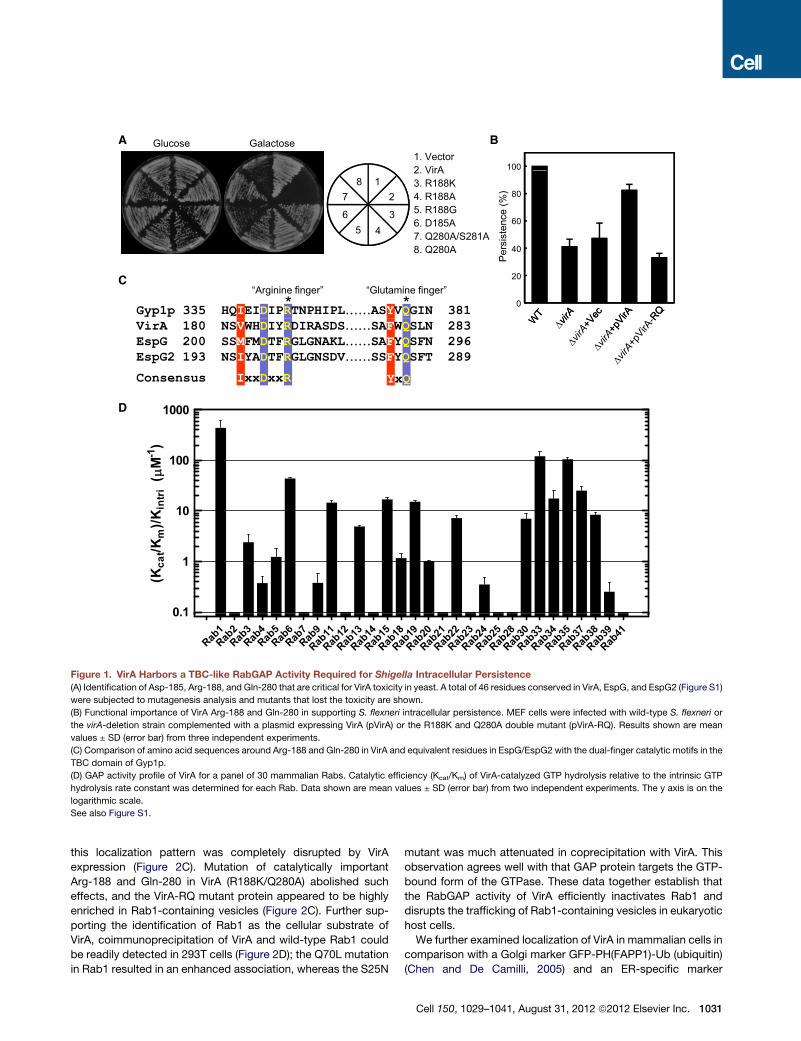
A
C
D
B
Figure 1. VirA Harbors a TBC-like RabGAP Activity Required for Shigella Intracellular Persistence
(A) Identification of Asp-185, Arg-188, and Gln-280 that are critical for VirA toxicity in yeast. A total of 46 residues conserved in VirA, EspG, and EspG2 (Figure S1)
were subjected to mutagenesis analysis and mutants that lost the toxicity are shown.
(B) Functional importance of VirA Arg-188 and Gln-280 in supporting S. flexneri intracellular persistence. MEF cells were infected with wild-type S. flexneri or
the virA-deletion strain complemented with a plasmid expressing VirA (pVirA) or the R188K and Q280A double mutant (pVirA-RQ). Results shown are mean
values ± SD (error bar) from three independent experiments.
(C) Comparison of amino acid sequences around Arg-188 and Gln-280 in VirA and equivalent residues in EspG/EspG2 with the dual-finger catalytic motifs in the
TBC domain of Gyp1p.
(D) GAP activity profile of VirA for a panel of 30 mammalian Rabs. Catalytic efficiency (Kcat/Km) of VirA-catalyzed GTP hydrolysis relative to the intrinsic GTP
hydrolysis rate constant was determined for each Rab. Data shown are mean values ± SD (error bar) from two independent experiments. The y axis is on the
logarithmic scale.
See also Figure S1.
this localization pattern was completely disrupted by VirA
expression (Figure 2C). Mutation of catalytically important
Arg-188 and Gln-280 in VirA (R188K/Q280A) abolished such
effects, and the VirA-RQ mutant protein appeared to be highly
enriched in Rab1-containing vesicles (Figure 2C). Further sup-
porting the identification of Rab1 as the cellular substrate of
VirA, coimmunoprecipitation of VirA and wild-type Rab1 could
be readily detected in 293T cells (Figure 2D); the Q70L mutation
in Rab1 resulted in an enhanced association, whereas the S25N
mutant was much attenuated in coprecipitation with VirA. This
observation agrees well with that GAP protein targets the GTP-
bound form of the GTPase. These data together establish that
the RabGAP activity of VirA efficiently inactivates Rab1 and
disrupts the trafficking of Rab1-containing vesicles in eukaryotic
host cells.
We further examined localization of VirA in mammalian cells in
comparison with a Golgi marker GFP-PH(FAPP1)-Ub (ubiquitin)
(Chen and De Camilli, 2005) and an ER-specific marker
Cell 150, 1029–1041, August 31, 2012 ª2012 Elsevier Inc. 1031
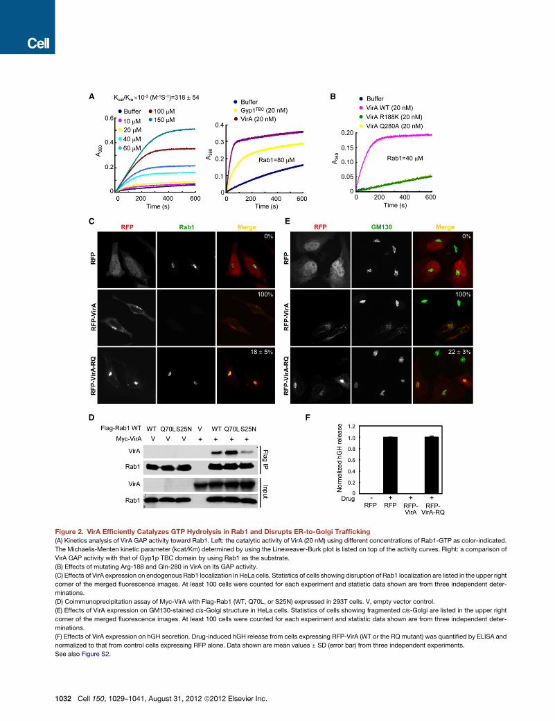
Figure 2. VirA Efficiently Catalyzes GTP Hydrolysis in Rab1 and Disrupts ER-to-Golgi Trafficking
(A) Kinetics analysis of VirA GAP activity toward Rab1. Left: the catalytic activity of VirA (20 nM) using different concentrations of Rab1-GTP as color-indicated.
The Michaelis-Menten kinetic parameter (kcat/Km) determined by using the Lineweaver-Burk plot is listed on top of the activity curves. Right: a comparison of
VirA GAP activity with that of Gyp1p TBC domain by using Rab1 as the substrate.
(B) Effects of mutating Arg-188 and Gln-280 in VirA on its GAP activity.
(C) Effects of VirA expression on endogenous Rab1 localization in HeLa cells. Statistics of cells showing disruption of Rab1 localization are listed in the upper right
corner of the merged fluorescence images. At least 100 cells were counted for each experiment and statistic data shown are from three independent deter-
minations.
(D) Coimmunoprecipitation assay of Myc-VirA with Flag-Rab1 (WT, Q70L, or S25N) expressed in 293T cells. V, empty vector control.
(E) Effects of VirA expression on GM130-stained cis-Golgi structure in HeLa cells. Statistics of cells showing fragmented cis-Golgi are listed in the upper right
corner of the merged fluorescence images. At least 100 cells were counted for each experiment and statistic data shown are from three independent deter-
minations.
(F) Effects of VirA expression on hGH secretion. Drug-induced hGH release from cells expressing RFP-VirA (WT or the RQ mutant) was quantified by ELISA and
normalized to that from control cells expressing RFP alone. Data shown are mean values ± SD (error bar) from three independent experiments.
See also Figure S2.
1032 Cell 150, 1029–1041, August 31, 2012 ª2012 Elsevier Inc.

calreticulin. Immunostaining of endogenous calreticulin showed
a typical ER morphology that apparently did not overlap with the
distribution pattern of VirA or VirA-RQ mutant (Figure S2M). In
contrast, both VirA and VirA-RQ were perfectly colocalized
with GFP-PH(FAPP1)-Ub (Figure S2N). The Golgi apparatus
stained by GFP-PH(FAPP1)-Ub was severely fragmented by
the GAP activity of VirA (Figure S2N). Further confirming this
observation, the cis-Golgi structure stained by GM130, a subunit
of a tethering complex on cis-Golgi and also a Rab1 effector
(Moyer et al., 2001; Weide et al., 2001), was found to be
completely fragmented by expression of wild-type VirA but not
the GAP-inactive VirA-RQ mutant (Figure 2E). Localization of
VirA was highly enriched in GM130-labeled membrane structure
even after the organelle had been disorganized by VirA expres-
sion. Consistent with the functional importance of the Golgi for
extracellular secretion of secretory proteins, pharmacologically
induced secretion of human growth hormone (hGH) (Rivera
et al., 2000) was completely blocked in cells expressing wild-
type VirA but not the GAP-inactive VirA-RQ mutant (Figure 2F).
Thus, Rab1 inactivation by the TBC-like GAP activity of VirA
blocks ER-to-Golgi trafficking, leading to functional destruction
of the cis-Golgi structure in VirA-transfected cells.
VirA Inactivation of Rab1 Counteracts Autophagy-Mediated Host DefenseWhereas exploring whether disruption of the cis-Golgi structure
by VirA plays a role in S. flexneri intracellular survival, we noted
that intracellular S. flexneri caused severe Golgi fragmentation
in a VirA-independent manner (data not shown), suggesting
that the virulence function of VirA is not associated with its
activity of disrupting ER-to-Golgi trafficking. One key strategy
employed by S. flexneri for intracellular survival is to suppress
and escape from autophagy-mediated host defense (Ogawa
et al., 2005). Recent studies have established a role of Rab1
and functional ER exit sites in autophagosome formation (Zop-
pino et al., 2010); in particular, the activity of Rab1 is required
for antibacterial autophagy in Salmonella-infected host cells
(Huang et al., 2011). We then examined autophagosome forma-
tion associated with S. flexneri DvirA mutant in comparison with
the parental wild-type strain. Consistent with that reported previ-
ously, wild-type S. flexneri was largely resistant to decoration by
GFP-LC3 and could escape from autophagy (Figure 3A). In
contrast, the DvirA mutant bacteria were often surrounded by
GFP-LC3 (Figure 3A), which was also positive for the lysosomal
marker LAMP-2 (Figure S3). Similar results were obtained when
endogenous LC3 was stained by a LC3-specific antibody (data
not shown). This suggests that deletion of VirA impairs the ability
of the bacteria to counteract autophagy-mediated host defense.
Supporting this idea, complementation of the DvirA strain with
a plasmid expressing wild-type VirA, but not the GAP-inactive
VirA-RQ mutant, reversed bacteria-induced autophagosome
and autolysosome formation (Figure 3A and Figure S3). Statis-
tical data showed that loss of VirA TBC-like activity rendered
a significant number of more than 20% of infected cells showing
autophagosome formation around intracellular S. flexneri (Fig-
ure 3B), which explains the defective intracellular persistence
observed with the VirA deletion and GAP activity-deficient
mutant S. flexneri (Figure 1B). We further investigated whether
inhibiting Rab1 activity or disrupting the cis-Golgi structure
from the host side could suppress the increased autophagy
induced by loss of VirA TBC-like GAP activity. Expression of
RFP-Rab1 S25N, the dominant-negative GDP-bound form of
Rab1, but not wild-type RFP-Rab1 or RFP alone, completely
blocked autophagosome formation around S. flexneri DvirA as
well asDvirA complementedwith the VirA-RQmutant (Figure 3C).
In contrast, cis-Golgi fragmentation induced by RNAi knock-
down of GM130 had no effects on S. flexneri DvirA-induced
autophagosome formation (data not shown). This result confirms
that it is the Rab1 activity but not its function in ER-to-Golgi
transport that is required for Rab1 function in antibacterial
autophagy (Huang et al., 2011). Thus, inactivation of Rab1 by
VirA TBC-like GAP activity plays an important role in Shigella
suppression of host autophagy, thereby contributing to bacterial
intracellular survival.
Crystal Structure of VirA-Rab1 Complex Approximatingthe GTP Hydrolysis Transition-State IntermediateTo better understand and define the structural basis for VirA GAP
activity, we crystallized the VirA-Rab1-GDP-aluminum fluoride
(AlF3) complex and solved the structure to 3.2 A (Figure 4A and
Table S1). AlF3 is commonly used to stabilize the GDP-bound
GTPase in complex with its GAP, which approximates the tran-
sition-state intermediate in GTP hydrolysis. The structure of
VirA in the Rab1 complex is almost identical to the previously
determined apo-structures (Davis et al., 2008; Germane et al.,
2008) (Figure S4A), consisting of well-separated N-terminal
and C-terminal domains. In the Rab1-binding C-terminal
domain, i.e., the GAP domain, the central six-stranded b sheet
is bound by one helix (a4) from the concave surface and seven
helices (a5-11) from the convex surface. A large rectangular
groove is formed on the convex surface of the GAP domain by
a5, a10, the central region of the b sheet, and the linking loops
(Figure 4A and Figure S4B). The two putative catalytic finger resi-
dues Arg-188 (in a5) and Gln-280 (in the linking loop between a9
and a10) are located in the rectangular groove and protrude to
bind AlF3 and GDP (Figure 4A and Figure S4B). The rectangular
groove in VirA GAP domain provides the surface for contacting
substrate Rab1.
Superimposition of VirA-Rab1-GDP-AlF3 complex structure
onto that of Gyp1p-Rab33-GDP-AlF3 complex (Pan et al.,
2006) reveals that Rab1 adopts the same conformation as
Rab33 and also uses switch I and II regions for contacting VirA
(Figure 4B). The rectangular groove-mediated Rab1 binding in
VirA also applies to Gyp1p that harbors a similar groove for
Rab33 binding (Figure 4B and Figures S4B and S4C). Notably,
a5 in VirA and aG5 in Gyp1p that supply and position the arginine
finger residues are well aligned (Figure 4B). Indeed, the two cata-
lytic finger residues Arg-188 and Gln-280 in VirA are located in
the same position in the groove and adopt the same conforma-
tion as the corresponding Arg-343 and Gln-378 residues in
Gyp1p TBC domain.
The stimulated annealing omit map shows a clear electron
density for GDP, Mg2+, AlF3 and a putative nucleophilic water
(Figure 4C), allowing for modeling these ligands in the VirA-
Rab1 complex structure (Figure 4D). AlF3 occupies the position
of g-phosphate in the GTP-bound state, and its aluminum ion
Cell 150, 1029–1041, August 31, 2012 ª2012 Elsevier Inc. 1033

B
(%)
25
A GFP-LC3 MergeShigella
d ce
lls w
ith L
C3-
posi
tive Shigella
(
5
25
15
10
20WT
+ Vec
C
Infe
cted
0∆virA +pVirA
WT ∆virA+Vec
∆virA +pVirA-RQ
sitiv
e Shigella
(%)
25
20
RFPRFP-Rab1RFP-Rab1 S25N
∆virA
∆virA
+pVirA
Infe
cted
cel
ls w
ith L
C3-
pos
0
5
15
10
∆virA +pVirA
WT ∆virA +Vec
∆virA +pVirA-RQ∆v
irA+pVirA-RQ
∆
Figure 3. VirA TBC-like GAP Activity Is Required for Shigella Escape from Autophagy
(A and B) Loss of VirA TBC-like GAP activity induces autophagy of Shigella. HeLa cells expressing GFP-LC3 were infected with wild-type S. flexneri or the DvirA
mutant strain complemented with a plasmid expressing VirA (pVirA) or the R188K and Q280A double mutant (pVirA-RQ). Bacteria were stained by anti-Shigella
antibody (red) (A). Shown in (B) are percentages of infected cells containing LC3-positive S. flexneri. Approximately 200 infected cells were examined for each
infection experiment and data are represented as mean values ± SD (error bar) from three independent experiments.
(C) Blocking of VirA deletion-induced Shigella autophagy by dominant-negative Rab1. HeLa cells expressing GFP-LC3 and RFP or RFP-Rab1a (WT or the
S25N dominant-negative mutant) were infected with indicated S. flexneri strains. Cells were analyzed by confocal microscopy and statistic data are obtained as
those in (A) and (B). Data are represented as mean values ± SD (error bar) from three independent experiments.
See also Figure S3.
is additionally coordinated by an axial oxygen from GDP as
well as the putative nucleophilic water. The guanidine group in
Arg-188 has bidentate hydrogen-bonding/ionic interactions
with the oxygen from the b-phosphate of GDP and an equatorial
fluoride ion (Figure 4D). All these chemical features and
geometries resemble those observed with other GAP-GTPase-
GDP-AlF3 complexes, including the Gyp1p-Rab33-GDP-AlF3complex (Pan et al., 2006; Rittinger et al., 1997; Scheffzek
et al., 1997). In the Gyp1p-Rab33 complex, the catalytic gluta-
mine finger residue mediates bipartite polar interactions with
the equatorial fluoride ion and the putative nucleophilic water
(Pan et al., 2006), and this is also the case for Gln-280 in VirA
(Figure 4D). Also similarly to that in the Gyp1p-Rab33-GDP-
AlF3 structure (but different from those in other GTPase-
GDP-AlF3 complexes), Gln-70 in Rab1 does not act as the
cis-glutamine residue involved in GTP hydrolysis and instead
has hydrogen-bonding interactions with Thr-177 and Asn-180
in VirA. Thus, the conformational and chemical similarities
between Arg-188/Gln-280 in VirA and the arginine/glutamine
1034 Cell 150, 1029–1041, August 31, 2012 ª2012 Elsevier Inc.
fingers in Gyp1p TBC domain provide the structural basis for
VirA TBC-like dual-finger mechanism of catalyzing GTP hydro-
lysis in Rab1.
Contrasting to the strikingly similar catalytic mechanism and
underlying structural basis between VirA and the TBC domain,
the a/b-fold GAP domain in VirA exhibits no overall structural
similarity to Gyp1p TBC domain that instead is all helical (Fig-
ure 4B). In fact, except for the helix that supplies the arginine
finger, no other secondary structural elements can be superim-
posed from structures of VirA and Gyp1p TBC domain. The
Rab1-binding rectangular groove in VirA, compared with that in
Gyp1 TBC domain, bears a relatively larger binding interface,
within which distinct detailed interactions are involved in con-
tacting the two switch regions in Rab1 (Figure S4D). Moreover,
the secondary structural contexts for the glutamine finger
residue are completely different in VirA and Gyp1 TBC domain.
These structural divergences clearly distinguish the bacterial
RabGAP from the host TBC-domain RabGAPs despite a similar
catalytic mechanism.

Figure 4. Crystal Structure of VirA-Rab1-GDP-AlF3 Complex and Comparison with Gyp1p TBC Domain in Complex with Rab33-GDP-AlF3
(A) Overall structure of VirA-Rab1-GDP-AlF3 complex. The two switch regions of Rab1 are colored and labeled in blue and dark orange. GDP, AlF3 and the two
putative catalytic residues R188 and Q280 of VirA are shown as sticks in yellow, cyan, and purple, respectively. Mg2+ and the nucleophilic water are shown as
green and red spheres, respectively.
(B) Superimposition of VirA-Rab1-GDP-AlF3 complex structure with that of Gyp1-Rab33-GDP- AlF3 complex (PDB ID code 2G77) by using Rab1 and Rab33 as
the reference. The dual-finger catalytic residues (R343 and Q378 in Gyp1p, R188, and Q280 in VirA) are shown as sticks.
(C) Sigma-A weighted Fo-Fc simulated annealing omit map for the modeled GDP, AlF3, Mg2+ and a putative nucleophilic water in the VirA-Rab1 complex
(contoured at 3.0 s).
(D) Comparison of the arginine and glutamine fingers and interactions in the AlF3-binding site in the VirA-Rab1 complex (left) with those in the Gyp1p-Rab33
complex (right, PDB ID code 2G77). The catalytic arginine and glutamine residues, GDP, AlF3 and other interacting residues are shown as sticks. Mg2+ and the
nucleophilic water are shown as green and red spheres, respectively. The hydrogen/salt bonds are indicated by dash lines.
See also Figure S4.
EspG Exhibits a Similar TBC-like GAP Activity andDisrupts Rab1-Mediated ER-to-Golgi TraffickingEspG/EspG2 only share �20% sequence identity with VirA
(Figure S1), but adopts a similar 3D-fold to VirA as revealed
by crystal structures of EspG that others (Germane and Spiller,
2011; Selyunin et al., 2011) and we determined (Figure S5A and
Table S1). EspG shares the arginine and glutamine finger motifs
of VirA (Figure 1C), and the two putative catalytic residues
(Arg-208 and Gln-293) are structurally equivalent to Arg-188
and Gln-280 in VirA (Figures S5A and S5B). Purified EspG
showed a potent GAP activity toward Rab1 with a catalytic
efficiency (kcat/Km, 2.35 3 105 M�1S�1) slightly lower than
that of VirA on Rab1 (kcat/Km, 3.18 3 105 M�1S�1) (Figure 5A).
Mutation of either Arg-208 or Gln-293 completely abolished
EspG GAP activity (Figure 5A). EspG also showed a highest
GAP activity toward Rab1 among the panel of 30 mammalian
Rabs assayed (Figure S5C). Interestingly, the substrate spec-
trum of EspG appeared to be narrower than that of VirA
(Figure S5C and Figure 1D). Also, similar to expression of
VirA, expression of EspG in mammalian host cells caused
severe disruption of the Golgi structure (Clements et al.,
2011; Selyunin et al., 2011), whereas the catalytically inactive
EspG-RQ mutant (R208K and Q293A double mutant) was
significantly attenuated in causing such effects (Figure S5D).
Golgi fragmentation induced by EspG was less severe than
that by VirA, agreeing with its relative lower GAP activity.
EspG expression resulted in disruption of endogenous Rab1
localization, requiring its TBC-like GAP activity (Figure S5E).
These results suggest that EspG uses a TBC-like GAP activity
to interfere with the function of host Rab GTPases in organizing
membrane trafficking. Identification of the TBC-like GAP
activity of EspG is not only consistent with its high structural
homology to VirA but also explains the functional similarity
between the two effectors.
Cell 150, 1029–1041, August 31, 2012 ª2012 Elsevier Inc. 1035

A
B
C
D
Figure 5. EPEC Type III Effector EspG also Functions as a TBC-like GAP to Inactivate Rab1 and Blocks Cytokine Secretion during Infection
(A) In vitro GAP assay of EspG and its arginine (R208) and glutamine (Q293) mutants using Rab1 as the substrate. The Michaelis-Menten kinetic parameter (kcat/
Km) determined by using the Lineweaver-Burk plot is listed on top of the activity curves.
(B) Disruption of endogenous Rab1 localization by EspG in EPEC-infected HeLa cells. Actin straining in the lower panels marks EPEC-infected cells (the bacteria
can induce actin clusters underneath the attachment sites).
(C) Assay of EspG disruption of host secretory pathway in EPEC-infected HeLa cells. Drug-induced hGH release from infected cells was quantified by ELISA and
normalized to that from uninfected cells. Results shown are mean values ± and SD (error bar) from three independent experiments.
(D) Effects of EspG on IL-8 secretion from EPEC-infected HeLa cells. IL-8 release in response to TNFa stimulation was measured by standard ELISA assay.
Results shown are mean values ± and SD (error bar) from two independent experiments.
See also Figure S5.
1036 Cell 150, 1029–1041, August 31, 2012 ª2012 Elsevier Inc.

Figure 6. Crystal Structure of EspG-Rab1-GDP-AlF3 Complex
(A) Overall structure of EspG-Rab1-GDP-AlF3 complex.
(B) Sigma-A weighted Fo-Fc simulated annealing omit map for the modeled GDP, AlF3, Mg2+ and a putative nucleophilic water in the EspG-Rab1 complex
(contoured at 3.0 s).
(C) The arginine and glutamine fingers and interactions in the AlF3-binding site in the EspG-Rab1-GDP-AlF3 complex. Orientation and presentation of the structure
are similar to those in Figure 4D.
See also Figure S6.
Inactivation of Rab1 by EspG Blocks of the GeneralSecretory Pathway and Inhibits IL-8 Secretion in EPEC-Infected CellsWe then investigated EspG inactivation of Rab1 and disruption
of ER-to-Golgi trafficking in EPEC-infected cells. Immunofluo-
rescence staining showed a largely diffused localization of
endogenous Rab1 in EPEC-infected HeLa cells; genetic deletion
of both espG and espG2 restored the normal localization pattern
of Rab1 (Figure 5B). Complementation of the deletion strain with
wild-type EspG, but not EspG-RQ mutant, resulted in a Rab1
distribution similar to that observed with wild-type EPEC infec-
tion (Figure 5B). These demonstrate that that the GAP activity
of EspG can also inactivate Rab1and interfere with Rab1-medi-
ated trafficking during bacterial infection.
Different from intracellular S. flexneri, EPEC infection by itself
did not cause Golgi fragmentation, suggesting that EspG disrup-
tion of Rab1-mediated cis-Golgi traffickingmight be of functional
importance for bacterial virulence. In fact, HeLa cells infected
with wild-type EPEC strain showed significantly attenuated
hGH reporter secretion; ablation of espG and espG2 resulted
in a completely normal hGH release (Figure 5C). Expression of
wild-type EspG in the deletion strain regained the phenotype of
blocking the general secretory pathway, whereas the GAP-inac-
tive EspG-RQ mutant (R208K and Q293A double mutant) failed
to do so (Figure 5C). Interestingly, expression of wild-type VirA,
but not theGAP-inactive VirA-RQmutant, in espG/espG2 double
deletion EPEC strain, also resulted in a rescue effect (Figure 5C),
providing a further support to the previously observed inter-
changeable function of VirA and EspG/EspG2 during Shigella
infection (Elliott et al., 2001).
A functional secretory pathway is responsible secretion of
many cytokines, chemokines, and antimicrobial peptides that
are important for host defense. IL-8 secretion is a hallmark of
many enteric bacterial infections including Shigella and EPEC.
Consistent with that reported previously, wild-type EPEC infec-
tion drastically inhibited IL-8 production upon TNFa stimulation.
Notably, deletion of espG and espG2 from EPEC resulted in
a significantly higher level (>3-fold) of IL-8 secretion (Figure 5D).
As EPEC also secretes other effectors to block NF-kB-mediated
IL-8 transcription, IL-8 secretion observed with espG/espG2
strain was still lower than that in uninfected cells. Elevated IL-8
secretion in the double deletion strain could be rescued by
expression of wild-type EspG but not the GAP-inactive EspG-
RQmutant (Figure 5D). These data suggest that the GAP activity
of EspG, different from that of VirA in S. flexneri, contributes to
suppression of host cytokine secretion as a result of disruption
of Rab1-mediated cis-Golgi trafficking. This finding provides
one plausible explanation why extracellular pathogens like
EPEC have evolved virulence effectors that specifically target
host vesicle trafficking pathways.
Structure of EspG-Rab1-GDP-AlF3 ComplexWe also crystallized the EspG-Rab1-GDP-AlF3 complex and
determined the 2.80 A structure of this transition-state-
mimicking complex (Figure 6A and Table S1). Structural compar-
ison revealed little conformational changes of EspG upon Rab1
binding (Figure S6A). As expected, the EspG-Rab1 complex
adopts an overall architecture highly similar to the VirA-Rab1
complex. Superimposition of the two complex structures shows
that the corresponding open rectangular groove in EspGbinds to
Cell 150, 1029–1041, August 31, 2012 ª2012 Elsevier Inc. 1037

Figure 7. Crystal Structure of a Ternary
Complex of Arf6-EspG-Rab1
(A) Overall structure of the Arf6-EspG-Rab1
complex. Arf6 is bound with GTP; GDP and AlF3used to stabilize the EspG-Rab1 complex are also
present in the ternary complex structure. All three
molecules are shown in cartoons, and presenta-
tion in the right panel is rotated by 180� from that in
the left panel.
(B) Superimposition of Arf6-EspG-Rab1 ternary
complex with EspG-Rab1 binary complex.
(C) Superimposition of the Arf6-EspG-Rab1
ternary complex with the Arf6-EspG binary
complex (PDB ID code 3PCR).
Rab1 in a VirA-like manner (Figures S6B and S6C). A clear elec-
tron density for GDP, Mg2+, AlF3 and a putative nucleophilic
water was also identified in the stimulated annealing omit map
(Figure 6B). The putative catalytic finger residues Arg-208 and
Gln-293 in EspG, located in equivalent secondary-structure
contexts, are engaged in several catalytic interactions with
GDP, AlF3, and the putative nucleophilic water in an almost iden-
tical fashion to those observed with Arg-188 and Gln-280 in the
VirA-Rab1-GDP-AlF3 complex (Figure 6C). Thus, EspG is a struc-
tural homolog of VirA and its GAP activity employs the same
TBC-like dual-finger catalytic mechanism despite the fact that
the two effectors appear to impact the virulence of the cognate
bacteria in a different way.
Structure of an Arf6-EspG-Rab1Ternary ComplexShows a Synthetic Arf-EspG-Rab1 Signaling RouteA recent study identifies small GTPases Arf1/Arf6 as host binding
partners of EspG (Selyunin et al., 2011). The switch I region in
Arf6-GTP is mainly responsible for EspG binding and this inter-
action is required for EspG to cause Golgi disruption in the trans-
fection assay. Our infection assay further established the physi-
ological relevance of this binding as the Arf-binding deficient
EspG E392R mutant was unable to complement the deletion
strain for disrupting the general secretory pathway (Figure 5C),
perturbing endogenous Rab1 localization (Figure 5B) as well as
inhibiting IL-8 secretion (data not shown). We also succeeded
in solving a 4.1 A crystal structure of Arf6-EspG-Rab1 complex
(Figure 7A and Figure S7). In the ternary complex, EspG and
1038 Cell 150, 1029–1041, August 31, 2012 ª2012 Elsevier Inc.
Rab1 adopt the same conformation and
bear a GDP-AlF3-mediated interface
similar to the binary EspG-Rab1 complex
(Figure 7B). Meanwhile, the GTP-bound
Arf6 contacts a small surface in EspG in
a manner similar to that in the previously
determined Arf6-EspG complex structure
(Figure 7C). Notably, EspG rotates about
10� from its position in the binary complex
and adopts a slight more open presenta-
tion for Rab1 binding in the ternary
complex, which results from the confor-
mational flexibility of its Arf6-contacting
loops. This structure variation, if not
a result of crystal packing, might be of
functional significance given that both Rab1 and Arf proteins
have constrained presentation with their carboxyl and amino
terminus, respectively, anchored to membrane vesicles.
The ternary complex structure clearly demonstrates that
EspG uses nonoverlapping surface area to contact Arf6 and
Rab1. In fact, Arf6 binding had little effects on the RabGAP
activity of EspG in vitro (data not shown). Given that loss of a
single Arf protein causes no defects in any step of mem-
brane transport in HeLa cells including ER-to-Golgi trafficking
(Volpicelli-Daley et al., 2005), binding to Arf1/6 likely plays
a role in targeting EspG to the right membrane location, where
it can physically access Rab1 and catalyze GTP hydrolysis.
The two GTPases contact the same side of the EspG molecule,
which spatially allows for their lipid-modified termini to be
anchored to the same membrane vesicle. Thus, EspG simulta-
neously hijacks two small GTPases in ER-to-Golgi membrane
trafficking with Arf1/6 mainly for membrane targeting and
Rab1 for catalytic inactivation, creating a synthetic signaling
route of Arf-GTP/EspG/catalytic inactivation of Rab1. This
unique mode of action shall be beneficial for the bacteria to
secure the disruption of ER-to-Golgi trafficking as EspG
binding also prevents Arf from regular nucleotide cycling (Selyu-
nin et al., 2011).
Given that VirA harbors a largely similar surface region despite
the absence of the Arf6-binding residues (Selyunin et al., 2011),
our model suggests a reasonable possibility that VirA might use
the same surface to bind Arf1 or another host protein for correct
membrane localization. Thus, the VirA/EspG family of bacterial

RabGAP is further distinguished from host TBC-domain GAPs in
their modes of specific membrane targeting.
DISCUSSION
In this study, we establish that the VirA/EspG family of type III
effectors from S. flexneri and EPEC harbors unexpected TBC-
like RabGAP activities with Rab1 as the preferred host target
in vitro and in vivo. For S. flexneri that is largely free-living inside
host cytoplasm, inactivation of Rab1 by VirA contributes to
bacterial intracellular survival by counteracting autophagy-medi-
ated host defense. The extracellular EPEC exploits the same
TBC-like GAP activity of EspG to inactivate Rab1 and block
the general secretory pathway, particularly secretion of proin-
flammatory cytokine IL-8, which can enhance bacterial survival
in host animals. Thus, the VirA/EspG family of RabGAP effectors
shall stimulate our thinking of a new virulence mechanism of tar-
geting host vesicle trafficking system by diverse pathogens like
S. flexneri and EPEC.
The TBC-like GAP Activity Might Be Widely Used inRegulating Membrane TraffickingVirA and EspG share a limited degree of sequence identity, and
both proteins bear neither sequence homology nor 3D-fold simi-
larity to eukaryotic TBC-domain GAP. This represents an excel-
lent example of convergent evolution of bacterial virulence
stressed by host environment. The TBC-like RabGAP activity
of the VirA/EspG family is prone to escape from simple and
structural homology searches, particularly when no functional
insights are available. Therefore, it is not unexpected that
other virulence factors with TBC-like GAP activity might be
widely present in bacterial pathogens, particularly those having
intracellular life cycle. For example, LepB, a type IV effector
from intracellular Legionella pneumophila, has been shown to
possess a Rab1-specificGAP activity with an undefined catalytic
mechanism (Ingmundson et al., 2007). It will be interesting to test
whether LepB also employs a TBC-like dual-finger catalytic
mechanism. Moreover, the possibility also exists that TBC-like
RabGAP activity might be used by other unidentified eukaryotic
proteins that bear primary sequence and 3D folds different from
the ones already known and also defined here.
A Unique Mode of Manipulating Eukaryotic Signalingby EspG and Synthetic Interplay with Host ActivitiesThe mechanism of action of EspG or possibly the VirA/EspG
family is also unique in that it targets another Arf GTPase mainly
for achieving specific membrane localization, which generates
a synthetic pathogenic signaling complex of Arf-EspG-Rab1.
Given that EspG specifically recognizes the GTP-bound form
of Arf and also because targeting a single Arf protein is unlikely
sufficient for disruption of any host function, EspG can be re-
garded as a unique Arf ‘‘effector’’ coming from the bacterial
kingdom for pathogenesis, which rewires host Arf signaling to
Rab1 inactivation. Interestingly, Gyp1p is also targeted to the
Golgi through its N-terminal region-mediated binding to another
Rab protein Ypt32p in yeast cells (Rivera-Molina and Novick,
2009). EspG can interact with GM130, a Rab1 effector (Clements
et al., 2011). If this interaction is not mediated through Rab1,
binding to GM130 could potentially serve as another means for
EspG to ensure correct membrane targeting. EspG has also
been shown to bind to a 12-residue peptide from PAK, and
this binding, though promotes the kinase activity of PAK, has
no role in EspG disruption of the Golgi structure (Germane and
Spiller, 2011; Selyunin et al., 2011). Interestingly, the PAK
peptide occupies part of the Rab1-binding surface in EspG. A
possible hypothesis is that PAK binding may serve as a host
defense mechanism to counteract the deleterious pathogenic
effect of the Arf-EspG-Rab1 complex. Alternatively, PAK might
be another host target of EspG, which results in perturbation of
other host signaling that remains to be defined.
EXPERIMENTAL PROCEDURES
Fluorescence Microscopy and Immunoprecipitation
For fluorescence staining, HeLa cells cultured on coverslips were fixed with
4% paraformaldehyde for 10 min at room temperature (RT), washed with
PBS, and then permeabilized for 10 min in PBS containing 0.5% Triton
X-100. Cells were then blocked with 1% BSA for 30 min followed by blotting
with first and second antibodies for at least 1 hr each. Images were acquired
by using a Zeiss LSM510META laser scanning confocalmicroscopy. Immuno-
precipitation was carried out as previously described (Dong et al., 2010).
Recombinant Proteins and the GAP Assay
E. coli BL21 (DE3) strain (Novagen) was used as the host for all recombinant
expression. Protein expression was induced for 16 hr at 16�C or 22�C with
1 mM isopropyl-b-D-thiogalactopyranoside. See Extended Experimental
Procedures for detailed description of recombinant protein expression and
purification. The GAP assay using the EnzChek Phosphate Assay Kit (Invitro-
gen) and determination of kinetics were carried out by strictly following the
previously described procedure (Pan et al., 2006). The absorbance at
360 nm was monitored on a UV-2450 spectrometer (Shimadzu).
Crystallization and Data Collection
Rab1-GDP was prepared by adding 10-fold molar excess of GDP into purified
Rab1 protein and incubated in a buffer containing 50 mM Tris-HCl (pH 8.0),
150 mM NaCl, 5 mM MgCl2, and 2 mM DTT for 30 min at RT. VirA was
mixed with Rab1-GDP (molar ratio, 1:1) in a buffer containing 50 mM Tris-
HCl (pH 8.0), 150 mM NaCl, 20 mM NaF, 2 mM AlCl3, 5 mM MgCl2, and
2 mM DTT, and incubated at 4�C overnight to obtain the VirA-Rab1-GDP-
AlF3 complex. The EspG-Rab1-GDP-AlF3 complex was obtained by using
the same method. Both VirA-Rab1 and EspG-Rab1 complexes were concen-
trated to 20 mg/ml for crystallization. See Extended Experimental Procedures
for detailed description of crystallization and data collection of EspG, EspG/
VirA-Rab1 complexes and Arf6- EspG-Rab1 ternary complex.
Structural Determination and Refinement
The VirA-Rab1 complex structure was solved by molecular replacement using
VirA (Protein Data Bank [PDB] ID code: 3EB8) and Rab1 (PDB ID code: 2FOL)
structures as the searching models. EspG structure was solved by Se-SAD
using anomalous diffraction data of Se-Met EspG crystal. The EspG-Rab1
complex structures were solved by molecular replacement by using our native
EspG structure and Rab1model from the VirA-Rab1 complex as the searching
models. The Arf6-EspG-Rab1 complex structure was also solved bymolecular
replacement using our EspG-Rab1 complex and Arf6 structure from EspG-
Arf6 complex (PDB ID code: 3PCR) as the searching models. All structural
pictures were drawn in PyMol (http://www.delanoscientific.com/). Statistics
of data collection and refinement are listed in Table S1. See Extended Exper-
imental Procedures for detailed description of structural determination and
refinement.
hGH Release and IL-8 Secretion
hGH trafficking assay was performed as described previously (Rivera et al.,
2000). See Extended Experimental Procedures for detailed description of
Cell 150, 1029–1041, August 31, 2012 ª2012 Elsevier Inc. 1039

the assay protocol. For IL-8 secretion, HeLa cells cultured in 24-well plates
were infected with wild-type or indicated derivatives of EPEC E2348/69 for
4 hr and then incubated with fresh medium supplemented with 50 mg/ml
gentamicin and 20 ng/ml TNF for 24 hr. The supernatant was collected after
centrifugation, and quantification of IL-8 released into the supernatant was
performed with an IL-8 ELISA kit (Invitrogen) by following the manufacturer’s
instruction.
Bacterial Manipulation and Intracellular Persistence
Nalidixic acid-resistant S. flexneri 2457T strain and EPEC strain E2348/68
strain were cultured in Tryptic soy broth (TSB) and LB broth, respectively.
The DvirA S. flexneri and the DespG/espG2 EPEC strain were constructed
by using the suicide vector pCVD442 (Donnenberg and Kaper, 1991). Intracel-
lular persistence of S. flexneri was assessed as previously described (Elliott
et al., 2001). See Extended Experimental Procedures for detailed description
of bacterial manipulation, infection, and intracellular persistence assay.
Shigella-Induced Autophagosome Formation
HeLa cells (3 3 104/well on 24-well plate formats) were transfected with
pEGFP-LC3 alone or together with pCS2-RFP, pCS2-RFP-Rab1a, or pCS2-
RFP-Rab1a S25N. Eighteen hours later, transfected cells were infected with
S. flexneri 2457T wild-type ([WT] pBAD24), DvirA (pBAD24), DvirA (pBAD24-
VirA), or DvirA (pBAD24-VirA-RQ) at a MOI of 100. After a 10 min centrifuge
(8003 g), infected cells were incubated at 37�C for 1 hr to allow for bacterial
invasion and then washed with PBS for three times before fresh medium con-
taining 200 mg/ml of gentamycin was added to kill the extracellular bacteria.
Cells were further incubated at 37�C for another 2 hr before being subjected
to immunofluorescence staining and confocal laser scanning microscopy
analysis. Intracellular bacteria were stained with an anti-Shigella antibody,
and the lysosome was marked by anti-LAMP-2 staining.
ACCESSION NUMBERS
Coordinates and structure factors for EspG, the VirA-Rab1-GDP-AlF3
complex, the EspG-Rab1-GDP-AlF3 complex, and Arf6-EspG-Rab1 complex
have been deposited with the PDB under the ID codes 4FMA, 4FMB, 4FMC
and 4FMD, and 4FME, respectively.
SUPPLEMENTAL INFORMATION
Supplemental Information includes Extended Experimental Procedures, seven
figures, and one table and can be found with this article online at http://dx.doi.
org/10.1016/j.cell.2012.06.050.
ACKNOWLEDGMENTS
We thank Dr. David G. Lambright for kindly providing bacterial expression
constructs for 30 Rabs. We thank the staff at the KEK Synchrotron Facility
(Tsukuba, Japan) and Argonne Advanced Photon Source (Chicago, IL) for
assistances with data collection. We also thank Yan Zhou, Jieling Yang, Yin
Xu, and Jianjin Shi for technical assistance. The research was supported in
part by an International Early Career Scientist grant from the Howard Hughes
Medical Institute to F.S. Y.Z. was supported by new faculty start-up funds
from Zhejiang University. This work was also supported by the National
Basic Research Program of China (973 Programs 2010CB835400 and
2012CB518700) to F.S.
Received: October 26, 2011
Revised: February 15, 2012
Accepted: June 29, 2012
Published: August 30, 2012
REFERENCES
Albert, S., Will, E., and Gallwitz, D. (1999). Identification of the catalytic
domains and their functionally critical arginine residues of two yeast
1040 Cell 150, 1029–1041, August 31, 2012 ª2012 Elsevier Inc.
GTPase-activating proteins specific for Ypt/Rab transport GTPases. EMBO
J. 18, 5216–5225.
Ashida, H., Ogawa, M., Kim, M., Suzuki, S., Sanada, T., Punginelli, C., Mimuro,
H., and Sasakawa, C. (2011). Shigella deploy multiple countermeasures
against host innate immune responses. Curr. Opin. Microbiol. 14, 16–23.
Barr, F., and Lambright, D.G. (2010). Rab GEFs and GAPs. Curr. Opin. Cell
Biol. 22, 461–470.
Brumell, J.H., and Scidmore, M.A. (2007). Manipulation of rabGTPase function
by intracellular bacterial pathogens. Microbiol. Mol. Biol. Rev. 71, 636–652.
Chen, H., and De Camilli, P. (2005). The association of epsin with ubiquitinated
cargo along the endocytic pathway is negatively regulated by its interaction
with clathrin. Proc. Natl. Acad. Sci. USA 102, 2766–2771.
Clements, A., Smollett, K., Lee, S.F., Hartland, E.L., Lowe, M., and Frankel, G.
(2011). EspG of enteropathogenic and enterohemorrhagic E. coli binds the
Golgi matrix protein GM130 and disrupts the Golgi structure and function.
Cell. Microbiol. 13, 1429–1439.
Davis, J., Wang, J., Tropea, J.E., Zhang, D., Dauter, Z., Waugh, D.S., and
Wlodawer, A. (2008). Novel fold of VirA, a type III secretion system effector
protein from Shigella flexneri. Protein Sci. 17, 2167–2173.
De Antoni, A., Schmitzova, J., Trepte, H.H., Gallwitz, D., and Albert, S. (2002).
Significance of GTP hydrolysis in Ypt1p-regulated endoplasmic reticulum to
Golgi transport revealed by the analysis of two novel Ypt1-GAPs. J. Biol.
Chem. 277, 41023–41031.
Dong, N., Liu, L., and Shao, F. (2010). A bacterial effector targets host DH-PH
domain RhoGEFs and antagonizes macrophage phagocytosis. EMBO J. 29,
1363–1376.
Donnenberg, M.S., and Kaper, J.B. (1991). Construction of an eae deletion
mutant of enteropathogenic Escherichia coli by using a positive-selection
suicide vector. Infect. Immun. 59, 4310–4317.
Elliott, S.J., Krejany, E.O., Mellies, J.L., Robins-Browne, R.M., Sasakawa, C.,
and Kaper, J.B. (2001). EspG, a novel type III system-secreted protein from
enteropathogenic Escherichia coli with similarities to VirA of Shigella flexneri.
Infect. Immun. 69, 4027–4033.
Germane, K.L., and Spiller, B.W. (2011). Structural and functional studies
indicate that the EPEC effector, EspG, directly binds p21-activated kinase.
Biochemistry 50, 917–919.
Germane, K.L., Ohi, R., Goldberg, M.B., and Spiller, B.W. (2008). Structural
and functional studies indicate that Shigella VirA is not a protease and does
not directly destabilize microtubules. Biochemistry 47, 10241–10243.
Hardwidge, P.R., Deng, W., Vallance, B.A., Rodriguez-Escudero, I., Cid, V.J.,
Molina, M., and Finlay, B.B. (2005). Modulation of host cytoskeleton function
by the enteropathogenic Escherichia coli and Citrobacter rodentium effector
protein EspG. Infect. Immun. 73, 2586–2594.
Huang, J., Birmingham, C.L., Shahnazari, S., Shiu, J., Zheng, Y.T., Smith, A.C.,
Campellone, K.G., Heo, W.D., Gruenheid, S., Meyer, T., et al. (2011). Antibac-
terial autophagy occurs at PI(3)P-enriched domains of the endoplasmic retic-
ulum and requires Rab1 GTPase. Autophagy 7, 17–26.
Hutagalung, A.H., and Novick, P.J. (2011). Role of Rab GTPases in membrane
traffic and cell physiology. Physiol. Rev. 91, 119–149.
Ingmundson, A., Delprato, A., Lambright, D.G., and Roy, C.R. (2007). Legion-
ella pneumophila proteins that regulate Rab1 membrane cycling. Nature 450,
365–369.
Masuda, E.S., Luo, Y., Young, C., Shen, M., Rossi, A.B., Huang, B.C., Yu, S.,
Bennett, M.K., Payan, D.G., and Scheller, R.H. (2000). Rab37 is a novel mast
cell specific GTPase localized to secretory granules. FEBS Lett. 470, 61–64.
Moyer, B.D., Allan, B.B., and Balch, W.E. (2001). Rab1 interaction with
a GM130 effector complex regulates COPII vesicle cis—Golgi tethering. Traffic
2, 268–276.
Nottingham, R.M., and Pfeffer, S.R. (2009). Defining the boundaries: RabGEFs
and GAPs. Proc. Natl. Acad. Sci. USA 106, 14185–14186.
Ogawa, M., Yoshimori, T., Suzuki, T., Sagara, H., Mizushima, N., and
Sasakawa, C. (2005). Escape of intracellular Shigella from autophagy. Science
307, 727–731.
Pan, X., Eathiraj, S., Munson, M., and Lambright, D.G. (2006). TBC-domain
GAPs for Rab GTPases accelerate GTP hydrolysis by a dual-finger mecha-
nism. Nature 442, 303–306.
Pfeffer, S.R. (2007). Unsolved mysteries in membrane traffic. Annu. Rev.
Biochem. 76, 629–645.
Pfeffer, S., and Aivazian, D. (2004). Targeting Rab GTPases to distinct
membrane compartments. Nat. Rev. Mol. Cell Biol. 5, 886–896.
Rak, A., Fedorov, R., Alexandrov, K., Albert, S., Goody, R.S., Gallwitz, D., and
Scheidig, A.J. (2000). Crystal structure of the GAP domain of Gyp1p: first
insights into interaction with Ypt/Rab proteins. EMBO J. 19, 5105–5113.
Rittinger, K., Walker, P.A., Eccleston, J.F., Smerdon, S.J., and Gamblin, S.J.
(1997). Structure at 1.65 A of RhoA and its GTPase-activating protein in
complex with a transition-state analogue. Nature 389, 758–762.
Rivera, V.M., Wang, X., Wardwell, S., Courage, N.L., Volchuk, A., Keenan, T.,
Holt, D.A., Gilman, M., Orci, L., Cerasoli, F., Jr., et al. (2000). Regulation of
protein secretion through controlled aggregation in the endoplasmic reticulum.
Science 287, 826–830.
Rivera-Molina, F.E., and Novick, P.J. (2009). A Rab GAP cascade defines the
boundary between two Rab GTPases on the secretory pathway. Proc. Natl.
Acad. Sci. USA 106, 14408–14413.
Salcedo, S.P., and Holden, D.W. (2005). Bacterial interactions with the eukary-
otic secretory pathway. Curr. Opin. Microbiol. 8, 92–98.
Scheffzek, K., Ahmadian, M.R., Kabsch, W., Wiesmuller, L., Lautwein, A.,
Schmitz, F., and Wittinghofer, A. (1997). The Ras-RasGAP complex: structural
basis for GTPase activation and its loss in oncogenic Ras mutants. Science
277, 333–338.
Selyunin, A.S., Sutton, S.E., Weigele, B.A., Reddick, L.E., Orchard, R.C.,
Bresson, S.M., Tomchick, D.R., and Alto, N.M. (2011). The assembly of
a GTPase-kinase signalling complex by a bacterial catalytic scaffold. Nature
469, 107–111.
Shao, F., Merritt, P.M., Bao, Z., Innes, R.W., and Dixon, J.E. (2002). A Yersinia
effector and a Pseudomonas avirulence protein define a family of cysteine
proteases functioning in bacterial pathogenesis. Cell 109, 575–588.
Slagowski, N.L., Kramer, R.W., Morrison, M.F., LaBaer, J., and Lesser, C.F.
(2008). A functional genomic yeast screen to identify pathogenic bacterial
proteins. PLoS Pathog. 4, e9.
Stenmark, H. (2009). Rab GTPases as coordinators of vesicle traffic. Nat. Rev.
Mol. Cell Biol. 10, 513–525.
Strom, M., Vollmer, P., Tan, T.J., and Gallwitz, D. (1993). A yeast GTPase-
activating protein that interacts specifically with a member of the Ypt/Rab
family. Nature 361, 736–739.
Uchiya, K., Tobe, T., Komatsu, K., Suzuki, T., Watarai, M., Fukuda, I., Yoshi-
kawa, M., and Sasakawa, C. (1995). Identification of a novel virulence gene,
virA, on the large plasmid of Shigella, involved in invasion and intercellular
spreading. Mol. Microbiol. 17, 241–250.
Volpicelli-Daley, L.A., Li, Y., Zhang, C.J., and Kahn, R.A. (2005). Isoform-selec-
tive effects of the depletion of ADP-ribosylation factors 1-5 on membrane
traffic. Mol. Biol. Cell 16, 4495–4508.
Weide, T., Bayer, M., Koster, M., Siebrasse, J.P., Peters, R., and Barnekow, A.
(2001). The Golgi matrix protein GM130: a specific interacting partner of the
small GTPase rab1b. EMBO Rep. 2, 336–341.
Yoshida, S., Handa, Y., Suzuki, T., Ogawa, M., Suzuki, M., Tamai, A., Abe, A.,
Katayama, E., and Sasakawa, C. (2006). Microtubule-severing activity of
Shigella is pivotal for intercellular spreading. Science 314, 985–989.
Zoppino, F.C., Militello, R.D., Slavin, I., Alvarez, C., and Colombo, M.I. (2010).
Autophagosome formation depends on the small GTPase Rab1 and functional
ER exit sites. Traffic 11, 1246–1261.
Cell 150, 1029–1041, August 31, 2012 ª2012 Elsevier Inc. 1041