structural cell wall proteins - plantphysiol.org · structural cell wall proteins ... of the plant...
TRANSCRIPT

Plant Physiol. (1993) 101: 1127-1 130
Structural Cell Wall Proteins
Beat Keller*
Department of Plant Breeding, Swiss Federal Research Station for Agronomy, Reckenholzstrasse 191, CH-8046 Zurich, Switzerland
Biological structures as diverse as skin, hair, spider webs, silk cocoons, connective tissue, and some alga1 cell walls share a common property: they consist mainly of structural proteins. Some of these proteins have been studied in detail and have contributed greatly to our understanding of protein structure. Silk fibroin, for example, is the model protein for the 0-pleated sheet secondary structure, collagen for a triple- stranded helix. The amino acids Gly and Pro (or Hyp) are frequently found to be major constituents of these structural proteins and, therefore, appear to be well suited as building blocks for structural proteins.
Not surprisingly, plant cell walls, the major structural part of the plant cell, also contain structural proteins. The primary amino acid sequences of many of these proteins have been deduced from cDNA and genomic sequences, but less is known about the precise localization and developmental expression pattern of these proteins. However, it has become clear that both the synthesis and the cross-linking of these wall proteins are under strict developmental control. In ad- dition, their synthesis and cross-linking can be environmen- tally induced by a number of triggers. Despite our growing knowledge about these proteins, their biological function is still largely a matter of speculation. Three major classes of structural wall proteins have been recognized to date: exten- sins, PRPs, and GRPs. Other wall proteins that have been described might also have a structural role, but less is known about these proteins and this discussion will focus on the well-characterized wall proteins.
HRGPs OR EXTENSINS
Except for certain algae that have cell walls made almost exclusively of proteins (for a review, see Adair, 1988), cell walls in plants contain only relatively small amounts of protein. Primary walls contain more protein than secondary walls. The best-known structural wall proteins are the exten- sins, or HRGPs, which are characterized by the repeated pentapeptide sequence Ser-(Hyp)., (for reviews, see Cassab and Varner, 1988; Showalter and Varner, 1989). Their struc- ture consists of an extended polyproline I1 helix. Most Pro's in these proteins are hydroxylated to give Hyp and are then O-glycosylated with Ara. Similarly, the Ser is often O-substi- tuted with Gal. Extensins are generally also rich in Lys, making them basic proteins, possibly interacting with acidic pectic blocks in the cell wall. The abundant Tyr residues
* Fax 41-1-377-7201. 1127
might be involved in isodityrosine cross-links that have been proposed to be responsible for the observed insolubilization of HRGPs in cell walls. In the monocot maize, similar proteins with slightly different motifs such as Ser-Hyp-Lys-Pro-Hyp (Kieliszewski et al., 1990) have been described, revealing an evolutionary related motif. Extensins with a Ser-(Hyp)4 motif have not only been described in dicots and monocots but also in a gymnosperm (Fong et al., 1992).
The study of different extensins has demonstrated that a specific extensin is synthesized in only one or a few cell types in a plant. A soybean extensin was found in sclerenchyma and in hour-glass cells in the seedcoat (Cassab and Varner, 1987). A gene encoding a tobacco extensin was specifically expressed in one or two cell layers in emerging lateral roots (Keller and Lamb, 1989). These cell layers were localized at the tip of a newly formed lateral root that is mechanically breaking through the root cortex. Deposition of extensin might strengthen their walls. Genes encoding other tobacco extensin or extensin-like proteins show pistil-specific expres- sion (De S. Goldmann et al., 1992). The cell-type-specific expression of extensin genes suggests that the proteins are functionally important parts of a particular cell wall.
The expression of extensins is not only developmentally regulated but also induced after pathogen attack. In bean infected with the funga1 pathogen Colletotrichum lindemu- thianum, in situ hybridization with a specific extensin cDNA was performed (Templeton et al., 1990). In an incompatible reaction, the gene was induced immediately after infection in epidermal and cortical cells adjacent to the infection site. In a compatible interaction, HRGP was induced slowly after infection and not only adjacent to the site of infection. These results suggest that HRGP is an important component of the localized hypersensitive resistance mechanism.
At the ultrastructural level, an extensin has been localized in carrot storage roots and was found to be uniformly distrib- uted across the primary cell wall but was absent from the middle lamella (Stafstrom and Staehelin, 1988). There is also evidence that extensin cannot cross the middle lamella sep- arating the walls of adjacent cells, suggesting that extensin is made by each cell itself (Stafstrom and Staehelin, 1988). In a different study at the ultrastructural level, increased deposi- tion of HRGP was observed at the interface of pea root cells interacting with mycorrhizal fungi (Bonfante-Fasolo et al., 1991).
Abbreviations: GRP, glycine-rich protein; HRGP, hydroxyproline- rich glycoprotein; PRP, proline-rich protein.
www.plantphysiol.orgon June 6, 2018 - Published by Downloaded from Copyright © 1993 American Society of Plant Biologists. All rights reserved.
1128 Keller Plant Physiol. Vol. 101, 1993
PRPs
PRPs can also contain some Hyp (H/PRP), but they do nothave the Ser-(Hyp)4 repeat of the extensins. The distinctionbetween extensins and PRPs is somewhat artificial and hasbecome less clear with the increasing number of proteinsanalyzed and primary sequences available (Kieliszewski etal., 1992a). Both groups of proteins should be considered asmembers of a large superfamily of related proteins. Many ofthe H/PRPs have a pentapeptide repeat of the form Pro-Pro-Val-X-Lys. Such proteins may contain structural oddities suchas six consecutive His's (Sheng et al., 1991) or peptide pal-indromes (Kieliszewski et al., 1992b). H/PRPs have beenfound in both monocots and dicots. Their sequences andexpression patterns are best characterized in soybean (Wyattet al., 1992) and maize (Jose-Estanyol et al., 1992). Threesoybean PRPs were localized immunologically in the lightmicroscope (Marcus et al., 1991) and gene expression patternswere analyzed by in situ hybridization (Wyatt et al., 1992).
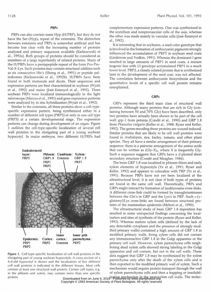
Similar to the extensins, all these proteins show a cell-type-specific expression pattern, being synthesized either in anumber of different cell types (PRP2) or only in one cell type(PRPS) at a certain developmental stage. The expressionpatterns can change during development of an organ. Figure1 outlines the cell-type-specific localization of several cellwall proteins in the elongating part of a young soybeanhypocotyl. In maize embryos, two different H/PRPs had
Endodermoidlayer:PRP3
PhloemGRP1.8PRP1
Cambium: Xylem:Extensin GRP 1.8
PRP1
Epidermis:PRP1PRP2
Cortex:PRP2
I Odercortex:Extensin
Inner pithparenchyma:PRP2
Figure 1. Cell-type-specific localization of cell wall proteins in theelongating part of young soybean hypocotyls. A cross-section of a4-d-old hypocotyl is shown and the localization of five differentcell wall proteins is indicated in the different tissues. Most cellscontain at least one structural wall protein. Certain cell types, e.g.in the phloem and xylem, may contain more than one specificprotein.
complementary expression patterns. One was synthesized inthe scutellum and nonprovascular cells of the axis, whereasthe other was made mainly in vascular cells (Jose-Estanyol etal., 1992).
It is interesting that in soybeans, a seed color genotype thatis involved in the formation of anthocyanin pigments stronglyinfluenced the accumulation of PRP1 in soybean seed coats(Lindstrom and Vodkin, 1991). Whereas the dominant / generesulted in large amounts of PRP1 in seed coats, a mutantisogenic line with i/i genotype accumulated PRP1 to a muchlower level. PRP2, a closely related protein that is synthesizedlater in the development of the seed coat, was not affected.The correlation between anthocyanin biosynthesis and thequantitative levels of a specific cell wall protein remainsunexplained.
GRPs
GRPs represent the third main class of structural wallproteins. Although many proteins that are rich in Gly (con-taining between 50 and 70% Gly) have been described, onlytwo proteins have actually been shown to be part of the cellwall: grp-1 from petunia (Condit et al., 1990) and GRP 1.8from Phaseolus vulgaris (Keller et al., 1988; Ryser and Keller,1992). The genes encoding these proteins are wound induced.Similar proteins that are likely to be cell wall proteins werefound in Arabidopsis, rice, barley, tomato, and other plantspecies. They all have a similar arrangement of their primarysequence: there is a pairwise arrangement of the amino acidsthat can be written as (Gly-X)n, where X is frequently Gly.Such a sequence suggests that GRPs have a ^-pleated sheetsecondary structure (Condit and Meagher, 1986).
The bean GRP 1.8 was localized in phloem fibers and earlyxylem elements of hypocotyls (Ye et al., 1991; Ryser andKeller, 1992) and appears to colocalize with PRP (Ye et al.,1991). Because PRPs have not yet been localized at theultrastructural level, it is not clear if both types of proteinsare found in the same cell wall. Theoretically, PRPs andGRPs might interact by formation of isodityrosine cross-links.A different cross-link could be formed by transglutaminasesbetween the Gin's in GRP and the Lys's in PRP. Such e-(y-glutamyl)Lys cross-links are found between structural pro-teins of the mammalian epidermis (Mehrel et al., 1990).
The ultrastructural study of bean GRP 1.8 deposition hasresulted in some unexpected findings concerning the local-ization and sites of synthesis of the protein (Ryser and Keller,1992). Whereas mature xylem cells (defined by the lack ofany detectable cytoplasm and the presence of strongly mod-ified primary walls) contained a high amount of GRP 1.8 inmodified primary walls, living xylem cells did not containany immunoreactive GRP 1.8 in the Golgi apparatus or theprimary cell wall. However, xylem parenchyma cells neigh-boring dead xylem cells showed strong labeling in the Golgiapparatus and cell corners, but not in the cell walls. Thesedata suggest that GRP 1.8 may be synthesized by the xylemparenchyma only after the death of the xylem cells and isthen exported to the modified primary wall of xylem. Such amechanism would require protein transport through the wallof xylem parenchyma cells and then a trapping or insolubil-ization mechanism in the protoxylem cell walls. The molec- www.plantphysiol.orgon June 6, 2018 - Published by Downloaded from
Copyright © 1993 American Society of Plant Biologists. All rights reserved.
Cell Wall Proteins 1129
ular details of such a mechanism are not clear, but the analysis of interactions of GRP 1.8 with other cell wall components should provide some of the answers.
PRPs AND RAPID CROSS-LINKINC MECHANISMS
Soybean PRP2 was localized in cortical cells and in the vascular tissue of hypocotyls and in inner integuments of the seed coat (Wyatt et al., 1992). The same protein was found to be rapidly insolubilized in the wall after elicitor treatment of soybean cell cultures (Bradley et al., 1992). This oxidative cross-linking into the wall structure was mediated by H202, initiated within 2 min, and completed after 10 min. The same cross-linking was observed in wounded bean hypocotyls close to the wound sites. Insolubilization of PRP2 also oc- curred during normal development; in mature regions of hypocotyls, PRP was insoluble but immunologically detect- able in the wall.
In addition, in stem-petiole junctions that are subjected to mechanical stress, PRP was insolubilized, whereas the same protein was still soluble in internodes. The developmental and stress-controlled insolubilization of PRP2 demonstrates the involvement of cell wall proteins both in development and in defense. The observed temporal and spatial separation of PRP2 mRNA synthesis and the site of cross-linking of the protein indicates that control of cross-linking, in addition to transcriptional control, represents an important mechanism of regulation of cell wall properties by proteins.
FUTURE RESEARCH DIRECTIONS AND CONCLUDING REMARKS
Many GRPs/PRPs are known only as amino acid sequences derived from cloned genes. They contain amino-terminal signal peptides but have not yet been shown to be cell wall proteins. Thus, localization, ultimately at the ultrastructural level, is necessary to know which proteins are present in the wall of a specific cell type and to know the precise localization within these walls. Such information will be essential to formulate a working hypothesis about the function of cell wall proteins during development and stress. There is good evidence that wall proteins can strengthen a cell wall. The very rapid cross-linking (Bradley et al., 1992) and the highly specific expression of an extensin during lateral root devel- opment (Keller and Lamb, 1989) suggest a mechanical strengthening of cell walls by proteins. An additional role was suggested for PRP2 from soybean that is localized in the middle lamella and the intercellular spaces of the cortex (Marcus et al., 1991). It was suggested that this protein acts to "cement" these cells.
The synthesis and active function of cell wall proteins can be regulated both at the transcriptional and the cross-linking level. Transcriptional control can be very complex, as was recently described for the bean GRP 1.8 (Keller and Baum- gartner, 1991). It is likely that wall proteins provide some functional properties to the wall that carbohydrates and polyphenolic polymers cannot. Elasticity and strength might be mediated by a GRP, similar to the GRPs that make up the strong but flexible spider silk (Lewis, 1992). In analogy to a major mammalian epidermal skin protein in the highly cor-
nified, insoluble envelope (55% Gly, Mehrel et al., 1990), GRPs might be involved in sealing off some walls. Immu- nolabeling of modified primary walls for GRP antigen gave a very dense reaction (Ryser and Keller, 1992), suggesting that there is enough protein for such a function. Proteins might also act as scaffolds for the deposition of other mole- cules, and the defined spacing of Tyr residues would control cross-linking and the "pore-size" of the wall. In addition, the genetically defined and precise length of such molecules might be important in a network of other macromolecules with no precise mechanism for length determination.
Little is known about the protein secondary structure of GRPs and PRPs as well as about their interaction with other wall polymers. Biochemical studies as well as the fate of mutated cell wall proteins in the wall will hopefully reveal more details about such interactions. Structural information and a better knowledge of the physical properties of isolated GRPs and PRPs will contibute to a working hypothesis on the biological function of such proteins. Future studies on cell wall proteins will certainly lead to new and surprising concepts of cell wall structure and the role of structural proteins.
ACKNOWLEDCMENT
I would like to thank Ueli Ryser and Jiirg Schmid for critical reading of the manuscript.
Received October 19, 1992; accepted December 15, 1992. Copyright Clearance Center: 0032-0889/93/101/1127/04.
LITERATURE CITED
Adair WS (1988) Organization and in vitro assembly of the Chlam- ydomonas reinhartdtii cell wall. Zn JE Vamer, ed, Self-Assembling Architecture. Alan L Liss, New York, pp 25-41
Bonfante-Fasolo P, Tamagnone L, Peretto R, Esqueree-Tugaye MT, Mazau D, Mosiniak M, Vian B (1991) Immunocytochemical location of hydroxyproline rich glycoproteins at the interface be- tween a mycorrhizal fungus and its host plants. Protoplasma 165:
Bradley DJ, Kjellbom P, Lamb CJ (1992) Elicitor- and wound- induced oxidative cross-linking of a proline-rich plant cell wall protein: a novel, rapid defense response. Cell70 21-30
Cassab GI, Varner JE (1987) Immunocytolocalization of extensin in developing soybean seedcoat by immunogold-silver staining and by tissue printing on nitrocellulose paper. J Cell Biol 1 0 5
Cassab GI, Varner JE (1988) Cell wall proteins. Annu Rev Plant Physiol Plant Mo1 Biol 3 9 321-353
Condit CM, McLean BC, Meagher RB (1990) Characterization of the expression of the petunia glycine-rich protein-1 gene product. Plant Physiol93 596-602
Condit CM, Meagher RB (1986) A gene encoding a novel glycine- rich structural protein of petunia. Nature 323: 178-181
De S Goldman MH, Pezzotti M, Seurinck J, Mariani C (1992) Developmental expression of tobacco pistil-specific genes encoding novel extensin-like proteins. Plant Cell 4 1041-1051
Fong C, Kieliszewski MJ, de Zacks R, Leykam JF, Lamport DTA (1992) A gymnosperm extensin contains the serine-tetrahydroxy- proline motif. Plant Physiol 9 9 548-552
Jose-Estanyol M, Ruiz-Avila L, Puigdomènech P (1992) A maize embryo-specific gene encodes a proline-rich and hydrophobic protein. Plant Cell4 413-423
Keller B, Baumgartner C (1991) Vascular-specific expression of the bean GRP 1.8 gene is negatively regulated. Plant Cell 3
127-138
2581-2588
1051-1061
www.plantphysiol.orgon June 6, 2018 - Published by Downloaded from Copyright © 1993 American Society of Plant Biologists. All rights reserved.

1130 Keller Plant Physiol. Vol. 101, 1993
Keller B, Lamb CJ (1989) Specific expression of a novel cell wall hydroxyproline-rich glycoprotein gene in lateral root initiation. Genes Dev 3 1639-1646
Keller B, Sauer N, Lamb CJ (1988) Glycine-rich cell wall proteins in bean: gene structure and association of the protein with the vascular system. EMBO J 7: 3625-3633
Kieliszewski M, de Zacks R, Leykam JF, Lamport DTA (1992a) A repetitive proline-rich protein from the gymnosperm Douglas fir is a hydroxyproline-rich glycoprotein. Plant Physiol98: 9 19-926
Kieliszewski MJ, Kamyab A, Leykam JF, Lamport DTA (1992b) A histidine-rich extensin from Zea mays is an arabinogalactan protein. Plant Physiol99 538-547
Kieliszewski MJ, Leykam JF, Lamport DTA (1990) Structure of the threonine-rich extensin from Zen mays. Plant Physiol92 316-326
Lewis RV (1992) Spider silk: the unraveling of a mystery. Accounts Chem Res 2 5 392-398
Lindstrom JT, Vodkin LO (1991) A soybean cell wall protein is affected by seed color genotype. Plant Cell 3: 561-571
Marcus A, Greenberg J, Averyhart-Fullard V (1991) Repetitive proline-rich proteins in the extracellular matrix of the plant cell. Physiol Plant 81: 273-279
Mehrel T, Hohl D, Rothnagel JA, Longley MA, Bundman D, Cheng C, Lichti U, Bisher ME, Steven AC, Steinert PM, Yuspa
SH, Roop DR (1990) Identification of a major keratinocyte cell envelope protein, loricrin. Cell61: 1103-1 112
Ryser U, Keller B (1992) Ultrastructural localization of a bean glycine-rich protein in unlignified primary wall of protoxylem cells. Plant Cell4: 773-783
Sheng J, D’Ovidio R, Mehdy MC (1991) Negative and positive regulation of a novel proline-rich protein mRNA by funga1 elicitor and wounding. Plant J 1: 345-354
Showalter AM, Varner JE (1989) Plant hydroxyproline-rich glyco- proteins. In PK Stumpf, EE Conn, eds, Biochemistry of Plants, Vol 15. Academic Press, New York, pp 485-520
Stafstrom JP, Staehelin LA (1988) Antibody localization of extensin in cell walls of carrot storage roots. Planta 174 321-332
Templeton MD, Dixon RA, Lamb CJ, Lawton MA (1990) Hydrox- yproline-rich glycoprotein transcripts exhibit different spatial pat- tems of accumulation in compatible and incompatible interactions between Phaseolus vulgaris and Colletotrichum lindemuthianum. Plant Physiol94 1265-1269
Wyatt RE, Nagao RT, Key JL (1992) Pattems of soybean proline- rich protein gene expression. Plant Cell4 99-110
Ye Z-H, Song Y-R, Marcus A, Varner JE (1991) Comparative localization of three classes of cell wall proteins. Plant J 1: 175-183
www.plantphysiol.orgon June 6, 2018 - Published by Downloaded from Copyright © 1993 American Society of Plant Biologists. All rights reserved.