gtpase-activating proteins for cdc42 - eukaryotic cell
TRANSCRIPT
EUKARYOTIC CELL, June 2002, p. 469–480 Vol. 1, No. 31535-9778/02/$04.00�0 DOI: 10.1128/EC.1.3.469–480.2002Copyright © 2002, American Society for Microbiology. All Rights Reserved.
GTPase-Activating Proteins for Cdc42Gregory R. Smith,† Scott A. Givan,‡ Paul Cullen, and George F. Sprague Jr.*
Institute of Molecular Biology, University of Oregon, Eugene, Oregon 97403-1229
Received 10 December 2001/Accepted 20 March 2002
The Rho-type GTPase, Cdc42, has been implicated in a variety of functions in the yeast life cycle, includingseptin organization for cytokinesis, pheromone response, and haploid invasive growth. A group of proteinscalled GTPase-activating proteins (GAPs) catalyze the hydrolysis of GTP to GDP, thereby inactivating Cdc42.At the time this study began, there was one known GAP, Bem3, and one putative GAP, Rga1, for Cdc42. Weidentified another putative GAP for Cdc42 and named it Rga2 (Rho GTPase-activating protein 2). Weconfirmed by genetic and biochemical criteria that Rga1, Rga2, and Bem3 act as GAPs for Cdc42. A detailedcharacterization of Rga1, Rga2, and Bem3 suggested that they regulate different subsets of Cdc42 function. Inparticular, deletion of the individual GAPs conferred different phenotypes. For example, deletion of RGA1, butnot RGA2 or BEM3, caused hyperinvasive growth. Furthermore, overproduction or loss of Rga1 and Rga2, butnot Bem3, affected the two-hybrid interaction of Cdc42 with Ste20, a p21-activated kinase (PAK) kinaserequired for haploid invasive growth. These results suggest Rga1, and possibly Rga2, facilitate the interactionof Cdc42 with Ste20 to mediate signaling in the haploid invasive growth pathway. Deletion of BEM3 resultedin cells with severe morphological defects not observed in rga1� or rga2� strains. These data suggest that Bem3and, to a lesser extent, Rga1 and Rga2 facilitate the role of Cdc42 in septin organization. Thus, it appears thatthe GAPs play a role in modulating specific aspects of Cdc42 function. Alternatively, the different phenotypescould reflect quantitative rather than qualitative differences in GAP activity in the mutant strains.
In the yeast Saccharomyces cerevisiae, the Cdc42 protein (33)is a member of the Rho subfamily of the Ras superfamily ofGTPases, which act as molecular switches and regulate manycellular processes (1, 34). Like all GTPases, Cdc42 can exist intwo states, a GTP-bound, active state and a GDP-bound, in-active state. The cycling of Cdc42 between these states is con-trolled by two sets of proteins: guanine nucleotide exchangefactors (GEFs) and GTPase-activating proteins (GAPs). Thereis one GEF for Cdc42 in yeast, called Cdc24 (62, 67, 69). At thetime this study began, there was one protein with demon-strated biochemical GAP activity for Cdc42, Bem3 (68), andone protein with potential GAP activity based on the presenceof a 170-amino-acid GAP domain, Rga1 (14, 63). AlthoughBem3 and Rga1 are fairly similar in their GAP domains, theyare divergent over the remainder of their protein sequences.Rga1 contains two tandem Lin-11, Isl-1, Mec-3 (LIM) domains(23, 35, 63, 66). LIM domains bind zinc ions and are thought tomediate protein-protein interactions (2, 18, 45). In contrast,Bem3 contains a Pleckstrin homology (PH) domain. PH do-mains are thought to play roles in membrane localization andprotein-protein interactions (40).
Cdc42 is required for polarization of the actin cytoskeleton,for cytokinesis, and for morphological changes that accompanyactivation of some signal transduction pathways, specificallythe pheromone response pathway and the filamentous growthpathway (for review, see reference 32). Several proteins, aside
from Cdc24, Rga1, and Bem3, that bind to Cdc42 have beenidentified and are presumed to be effectors of various Cdc42functions. These proteins include Ste20 and Cla4, two proteinkinases (17, 61), and Gic1 and Gic2, two proteins of similarsequence but as-yet-undefined biochemical activity (7, 13).Ste20 is required for operation of the pheromone and filamen-tation pathways (38, 53, 56), Cla4 plays ill-defined roles inseptin organization and cytokinesis (4, 9, 17, 41), and togetherGic1 and Gic2 are required to polarize the actin cytoskeleton(5).
The finding that there is more than one potential GAP forCdc42 raises the possibility that each GAP may regulate Cdc42activity with respect to only a subset of its biological roles. Toexplore this possibility, we sought to identify the full comple-ment of GAPs for Cdc42 and examine the phenotypic conse-quences of the absence of one or more of these GAPs. Here wereport the identification of a third potential GAP, Rga2, whichhas strong sequence similarity to Rga1. We demonstrate thatRga1 and Rga2 have biochemical GAP activity for Cdc42.Finally, we provide evidence that strains lacking Rga1, Rga2,and Bem3 display distinct phenotypes, supporting the idea thatthey influence Cdc42 activity in different biological settings.
MATERIALS AND METHODS
Strains, plasmids, and microbiological techniques. The yeast strains used arelisted in Table 1. Gene deletions were constructed by PCR and confirmed byPCR and phenotyping when applicable. In all cases, the entire coding region wasreplaced with the indicated marker. Yeast and bacterial strains were propagatedusing standard methods (8, 57, 59). Yeast extract-peptone-dextrose and syntheticdextrose media were prepared as previously described (58). Yeast transforma-tions were performed using modifications of the LiOAc method (12, 24). Bac-terial transformations, DNA preparations, and plasmid constructions were per-formed by standard methods (59). Growth capabilities at various temperatureswere measured by spotting equal volumes of serial dilutions of mid-log phasecultures to agar plates and incubating at the indicated temperatures.
* Corresponding author. Mailing address: Institute of MolecularBiology, University of Oregon, Eugene, OR 97403-1229. Phone: (541)346-5883. Fax: (541) 346-4854. E-mail: [email protected].
† Present address: Department of Biomolecular Chemistry, Univer-sity of Wisconsin, Madison, WI 53706.
‡ Present address: Center for Gene Research and Biotechnology,Oregon State University, Corvallis, OR 97331.
469
on January 13, 2019 by guesthttp://ec.asm
.org/D
ownloaded from

Two-hybrid analysis. Two-hybrid analysis (22) of interactions between thepanel of GTPases and Rga1, Rga2, and Bem3 was performed with strain EGY40(26). To assess �-galactosidase activity, strain EGY40 containing the LexAop-lacZ reporter plasmid pSH18-34 (26) was transformed with a pEG202-derivedLexA DNA-binding domain (DBD) plasmid and a pJG4-5-derived Gal4 activa-tion domain (AD) plasmid. At least three separate isolates were grown tomid-log phase in rich medium containing 2% galactose and quantitated as pre-viously described (63). Most of the LexA DBD fusions in pEG202 and Gal4 ADfusions in pJG4-5 were previously described (63). pJG4-5-RGA2 was constructedusing gap repair (42) of the entire RGA2 coding region and EcoRI-cut pJG4-5(26). pEG202-RGA2 was constructed by ligating an XhoI-EcoRI fragment ofpJG4-5-RGA2 into XhoI-EcoRI-cut pEG202 (26). The expression of two-hybridconstructs was confirmed by Western blotting techniques (59) using �-LexADBD and �-Gal4 AD antibodies, respectively (52). �-Galactosidase assays wereused to determine the relative strength of the interactions and were performedessentially as described previously (29).
Additional two-hybrid analysis was performed with the PJ69-4A strain (30).pGS38 and pGS39 were constructed by ligating the EcoRI-XhoI fragments frompEG202-CDC42G12V,C188S and pEG202-CDC42Q61L,C188S (63), respectively, intothe Gal4 activation domain fusion vector pGAD-C(2) (30), which was cut withEcoRI and SalI. pSL2682 contains a truncation of STE20 (encoding amino acids1 to 565) cloned into pOBD. CYH2 (64) was a kind gift from Megan Keniry. Theexpression of these fusion proteins was confirmed by Western blot analysis using
a �-Gal4 DBD monoclonal antibody (52). pGS40 was constructed by ligating aKpnI-HindIII fragment from YEp13-RGA1 containing the coding region and1,500 nucleotides upstream of RGA1 to KpnI-HindIII-cut YEp352 (28). pGS41was constructed by ligating a SacI-XhoI fragment from YEp13-RGA2 containingthe coding region and 800 nucleotides upstream of RGA2 to SacI-XhoI-cutYEp352. pGS42 was constructed by ligating a SacI-PstI fragment from YEp13-BEM3 containing the coding region and 800 nucleotides upstream of BEM3 toSacI-PstI cut YEp352. �-Galactosidase assays were used to determine the rela-tive strength of the interactions and were performed essentially as describedpreviously (29).
Protein purification. The GTPases Cdc42 and Rsr1 (Bud1) were purified asglutathione S-transferase (GST) fusion proteins using hybrid genes present inthe plasmid pGEX-5X-1 (47). pGEX-5X-1-Cdc42C188S and pGEX-5X-1-Cdc42Q61L,C188S were gifts from David Mitchell, and pGEX-Rsr1 was kindlyprovided by Hay-Oak Park (49). Escherichia coli cells (DH5�) were grown tomid-log phase, protein expression was induced with 1 mM IPTG (Sigma) for 2 h,and cells were lysed by two passages through a French press. The lysate wascentrifuged at 5,000 rpm in an SS-34 Sorvall rotor for 10 min to pellet unbrokencells and cellular debris. Swollen glutathione agarose beads were added to thesupernatant, incubated at 4°C for 2 h, washed, and frozen at �80°C in GTPasestorage buffer (20 mM Tris-HCl [pH 7.5], 1 mM dithiothreitol [DTT], 100 �MGTP, 1� protease inhibitors cocktail [Roche], and 15% glycerol).
The GAP domains of Rga1, Rga2, and Bem3 were purified as maltose bindingprotein (MBP) fusions. These fusions were expressed from plasmids based onpMAL-c2 (25). pGS53 was constructed by addition of SalI and HindIII sites tothe ends of an RGA1 PCR product using a plasmid containing the RGA1 codingregion as a template. This PCR product was digested and ligated to SalI-HindIII-cut pMAL-c2. pGS54 was constructed by addition of SalI and PstI sites to theends of an RGA2 PCR product by using a plasmid containing the RGA2 codingregion as a template. This PCR product was digested and ligated to SalI-PstI-cutpMAL-c2. pGS55 was constructed by addition of EcoRI and SalI sites to the endsof a BEM3 PCR product by using a plasmid containing the BEM3 coding regionas a template. This PCR product was digested and ligated to EcoRI-SalI-cutpMAL-c2. Bacterial protein expression and cell lysis were performed essentiallyas described above for the GST fusions, except proteins were purified using anamylose column (44) and eluted with Column Buffer II (20 mM Tris-HCl [pH7.4], 1 mM EDTA, 200 mM NaCl, 10 mM maltose). In all cases, standard sodiumdodecyl sulfate-polyacrylamide gel electrophoresis separation and Coomasiestaining confirmed protein expression and purification (59).
Biochemistry. Purified GST, GST-Cdc42C188S, GST-Cdc42Q61L,C188S, or GST-Rsr1 was incubated with �2 �Ci of [�-32P]GTP or [�-32P]GTP (New EnglandNuclear Life Sciences) at room temperature for 10 min in reaction buffer (20mM Tris-HCl [pH 7.5], 1 mM DTT, 100 �M GTP). Then, 2 nmol of GTP wasadded to each reaction mixture. Each reaction mixture contained one of thefollowing: 25.6 pmol of GST-Cdc42 (C188S), 21.3 pmol of GST-Cdc42 (Q61L,C188S), or 71.6 pmol of GST-Rsr1. As a result, 5.7% of the GST-Cdc42 (C188S)bound [�-32P]GTP, 5.9% of the GST-Cdc42 (C188S) bound [�-32P]GTP, and7.6% of the GST-Rsr1 bound [�-32P]GTP. Glutathione agarose beads boundwith a [�-32P]GTP- or [�-32P]GTP-loaded GTPase were added to micro-spincolumns (Bio-Rad) and washed six times with 250 �l of wash buffer (20 mMTris-HCl [pH 7.5], 1 mM DTT, 5 mM MgCl2). The beads were resuspended,removed from the column, and added to �2 �g (28.6 pmol) of MBP, MBP-Rga1,MBP-Rga2, or MBP-Bem3. The mixtures were incubated at room temperaturefor the indicated time, added to a micro-spin column, spun, and washed. Then,100 �l of elution buffer (1% sodium dodecyl sulfate–20 mM EDTA) was addedto the column and incubated at 65°C for 15 min to denature the protein. Thesamples were centrifuged to collect the released nucleotides. The reaction buffer,wash buffer, and eluate were counted in a scintillation counter. The ratio ofbound to released counts was used to determine GAP activity.
Microscopy. Standard microscopic techniques were used and cells were exam-ined using a 100� objective. Methods for staining with rhodamine phalloidin tovisualize F-actin, Calcofluor to visualize chitin deposits in bud scars, and DAPI(4,6-diamidino-2-phenylindole) for visualization of DNA were performed es-sentially as previously described (51). Septins were visualized by immunofluo-rescence using a �-Cdc3 antibody (36; purified by April Goehring) followed byfluorescence microscopy.
FUS1-lacZ assays. Cells containing a FUS1-lacZ reporter construct integratedat mfa2 (6) were grown to mid-log phase at 30°C in yeast extract-peptone-dextrose medium. �-Galactosidase assays were performed as described previ-ously (31).
Invasive growth assays. The plate-washing assay was performed as previouslydescribed (56). The single cell assay was also performed essentially as previouslydescribed (16). For some experiments, cells were scraped from plates using 4 ml
TABLE 1. Yeast strains
Strain Genotype Source orreference
DJTD2-16D MAT� cdc42-1ts ura3 trpl leu2 his4 gal2 45aYEF24H MAT� cdc24-Hts ura3 trpl leu2 his4 ade2 34EGY40 MAT� ura3-52 his3 trpl leu2 26PJ69-4A MATa GALI-HIS3 GAL2-ADE8 GAL7-
lacZ leu2 ura3 his3 gal4 gal8030
�1278B MAT� ura3 G. FinkYAG512 �1278B except his3::ura3- A. GoehringYGS45 YEF24H except rga1::HIS3 This studyYGS46 YEF24H except rga2::HIS3 This studyYGS47 YEF24H except bem3::HIS3 This studyYGS156 PJ69-4A except rga1::URA3 This studyYGS157 PJ69-4A except rga2::URA3 This studyYGS158 PJ69-4A except bem3::URA3 This studySY2002 MATa FUS1::HIS3 his3 mfa2-1::FUS1-
lacZ adel leu2 trpl ura3Lab strain
YGS2 SY2002 except rga1::URA3 This studyYGS7 SY2002 except rga2::TRP1 This studyYGS50 SY2002 except bem3::TRP1 This studyYGS72 SY2002 except rga1::URA3 rga2::TRP1 This studyYGS51 SY2002 except rga1::URA3 bem3::TRP1 This studyYGS56 SY2002 except rga2::TRP1 bem3::TRP1 This studyYGS57 SY2002 except rga1::URA3 rga2::TRP1
bem3::TRP1This study
YGS58 SY2002 except ste4::LEU2 This studyYGS59 YGS2 except ste4::LEU2 This studyYGS60 YGS7 except ste4::LEU2 This studyYGS61 YGS50 except ste4::LEU2 This studyYGS62 YGS51 except ste4::LEU2 This studyYGS63 YGS72 except ste4::LEU2 This studyYGS64 YGS56 except ste4::LEU2 This studyYGS65 YGS57 except ste4::LEU2 This studyYGS281 YAG512 except rga1::HIS3 This studyYGS328 YAG512 except rga2::HIS3 This studyYGS329 YAG512 except bem3::HIS3 This studyYGS105 MATa mfa2-1::FUS1-lacZ his3 leu2
trp1 ura3 ade1This study
YGS286 YGS105 except ste20::URA3 This studyYGS351 YGS286 except rga1::HIS3 This studyYGS352 YGS286 except rga2::HIS3 This studyYGS353 YGS286 except bem3::HIS3 This studyIDY22 MAT�cla4::TRP1 mfa2-1::FUS1-lacZ
leu2 ura3 ade2 ade3 lys2D. Mitchell
YGS80 IDY22 except rga1::URA3 This studyYGS81 IDY22 except rga2::URA3 This studyYGS82 IDY22 except bem3::LEU2 This study
470 SMITH ET AL. EUKARYOT. CELL
on January 13, 2019 by guesthttp://ec.asm
.org/D
ownloaded from

of distilled water, concentrated by centrifugation, resuspended in 20 �l of water,and visualized by microscopy. For other experiments, a coverslip was placeddirectly on the agar medium and cells were visualized by microscopy at 100�.
RESULTS
Rga2 is putative Rho-GTPase-activating protein for Cdc42.In an effort to find putative GAPs for Cdc42, we performed ahomology search of the Saccharomyces Genome Database(http://genome-www.stanford.edu/Saccharomyces/) by usingthe BLAST algorithm. Searching for proteins similar to Rga1identified seven proteins that contained a potential Rho-GAPmotif: Ydr379w, Bem3, Lrg1, Rgd1, Bem2, Sac7, and Bag7(Fig. 1A). Many of these proteins have been previously de-scribed as GAPs for a variety of Rho-type GTPases. Bem3 wasshown to be a GAP for Cdc42 (68). Rgd1 was demonstrated tohave GAP activity for Rho3 and Rho4, but not for Cdc42 (3,20). The putative Rho-GAP, Lrg1, acts as a GAP for Rho1during 1,3-�-glucan synthesis (65). Bem2 was shown to be aGAP for Rho1, but not for Cdc42 (67, 68). Sac7 is thought toact as a GAP for Rho1 (60). Since Rgd1, Lrg1, Bem2, Sac7,and Bag7 have been at least partially characterized as beinginvolved in other processes, they were not pursued as putativeGAPs for Cdc42.
One of the open reading frames identified, YDR379W,showed a high degree of similarity to RGA1 across the entireprotein sequence and was renamed RGA2 for Rho-GTPase-activating protein 2. RGA2 encodes a 1,009-amino-acid proteinthat contains two tandem LIM domains and a Rho-GAP do-main (Fig. 1B). Rga1 and Rga2 share particularly high levels ofidentity in these regions. Rga2 also has sequence similarity toBem3 over the GAP domain, but Bem3 does not contain LIMdomains (Fig. 1B). Due to its high sequence similarity to Rga1and, to a lesser extent, to Bem3, Rga2 was designated a puta-tive GAP for Cdc42p.
Genetic support for Rga2 as a GAP for Cdc42. To investi-gate the possibility that Rga2 is indeed a GAP for Cdc42, wefirst asked whether RGA2 showed the same genetic interac-tions with CDC42 and CDC24 as do RGA1 and BEM3 (63). Wereasoned that an increase in GAP activity for Cdc42 shouldreduce the amount of active Cdc42 in the cell, thereby loweringthe restrictive temperature of a temperature-sensitive CDC42mutant. Consistent with this reasoning, overexpression ofRGA2 lowered the restrictive temperature of a cdc42-1 strain(data not shown), as was observed for RGA1 and BEM3 (63).Conversely, we expected that a reduction in GAP activitywould reduce the requirement for Cdc24 (GEF) activity. In
FIG. 1. Protein sequence alignments of Rho-GAPs in yeast. (A) Protein alignment of putative GAP motifs for Rho-type GTPases in the yeastS. cerevisiae. Consensus residues are shaded for residues shared by seven or eight of the proteins and unshaded for residues shared by four to sixof the proteins. ��, 61 omitted residues for Sac7. (B) A graphical representation of the known and putative GAPs for Cdc42. The numberscorrespond to amino acid positions in the protein sequence. I, identity; S, similarity; LIM, tandem LIM domains; PH, a pleckstrin homologydomain; Rho-GAP, consensus GAP motif as shown in panel A.
VOL. 1, 2002 GAPs FOR Cdc42 471
on January 13, 2019 by guesthttp://ec.asm
.org/D
ownloaded from

accord with this expectation, deletion of RGA2 or BEM3 in-creased the restrictive temperature of a cdc24-H strain (datanot shown), just as previously shown for RGA1 (63). Thus,genetic evidence supports the hypothesis that Rga2, as well asRga1 and Bem3, are GAPs for Cdc42.
As a second test of the possibility that Rga2 is a GAP forCdc42, we investigated the two-hybrid interactions betweenRga2 and known GTPases of the Rho subfamily. Rga2 inter-acted with activated forms (G12V and Q61L) but not an inac-tive form (D118A) of Cdc42, as had previously been seen forRga1 and Bem3 (63; Table 2). Rga2 failed to interact withRho1, Rho2, Rho3, or Rho4, including activated versions ofthese proteins that could not be prenylated (data not shown).In addition, Rga1 and Rga2, but not Bem3, interacted with theGTPase, Rsr1, in both its wild-type and activated (G12V)forms (Table 2). This interaction pattern is distinct from thepattern seen for GTPases and their GAPs: GAPs usually in-teract preferentially with the active form of the GTPase. Rsr1is a Ras-type GTPase involved in bud site selection and hasbeen shown to interact with Cdc24 (49). Together, these ex-periments support the hypothesis that Rga1, Rga2, and Bem3are all GAPs for Cdc42 and also suggest that Rga1 and Rga2may have a connection with Rsr1.
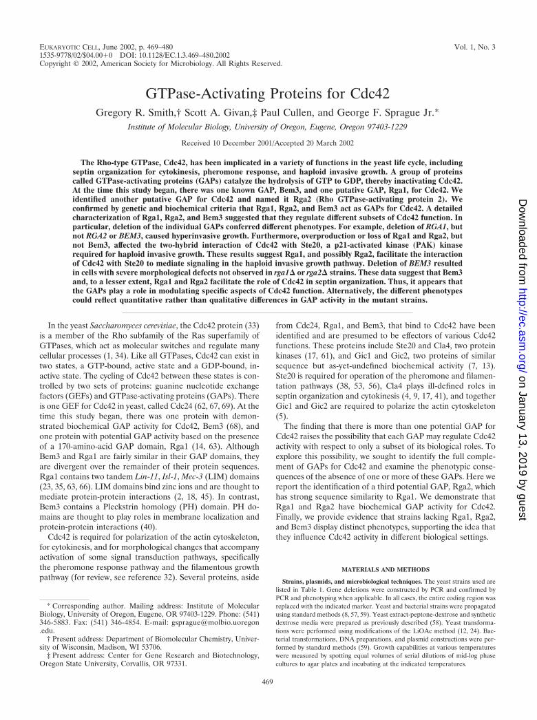
Rga1 and Rga2 have in vitro biochemical GAP activity. Toexpand on these genetic studies, we sought to determinewhether Rga1 and Rga2 have biochemical GAP activity towardCdc42; that is, we asked whether they accelerate GTP hydro-lysis by Cdc42. A fusion protein containing the Bem3 GAPdomain joined to GST was previously shown to catalyze thehydrolysis of GTP to GDP when the nucleotide was bound toCdc42 (68). GST-Cdc42C188S was purified using glutathioneagarose beads and bound to [�-32P]GTP (see Materials andMethods), and the GAP domains of Rga1, Rga2, and Bem3were purified as MBP fusions. When mixed with the[�-32P]GTP-loaded GST-Cdc42C188S, all three GAP domainscatalyzed the hydrolysis of GTP to GDP (Fig. 2A). After only2.5 min, the GAP domains of Rga1 and Rga2 had catalyzed the
hydrolysis of over 94% of the GTP on Cdc42 (summary ofmultiple data sets). In contrast, incubation with MBP aloneresulted in hydrolysis of less than 20% of the GTP after 2.5min. Incubation with the Bem3 GAP domain resulted in hy-drolysis of �75% of the GTP bound to Cdc42 after 2.5 min.Thus, it appears that Rga1, Rga2, and Bem3 all catalyze GTPhydrolysis on Cdc42. However, Rga1 and Rga2 may do somore efficiently than Bem3.
To ensure that the loss of counts from the beads was due toGTP hydrolysis and not simply dissociation of the bound nu-cleoside triphosphate, purified Cdc42C188S was bound to[�-32P]GTP instead of [�-32P]GTP and then incubated with theGAPs. If Rga1, Rga2, and Bem3 were to cause dissociation ofGTP rather than hydrolysis to GDP, [�-32P]GTP would bereleased in this experiment, resulting in a loss of bound radio-activity. In fact, very little radioactivity was released, indicatingthat [�-32P]GTP did not dissociate from Cdc42C188S when in-cubated with the GAP domains of Rga1, Rga2, or Bem3 (Fig.2B). The GAP assay was also performed with GST-Cdc42Q61L,C188S. Since the Q61L substitution locks Cdc42 inits GTP-bound, active state (70), this mutant should not becapable of GTP hydrolysis. Indeed, incubation with Rga1,Rga2, or Bem3 did not cause hydrolysis of [�-32P]GTP whenbound to Cdc42Q61L, C188S (Fig. 2C). These results show thatthe GAP domains of Rga1, Rga2, and Bem3 catalyze thehydrolysis of GTP by Cdc42.
Motivated by the two-hybrid results discussed above (Table2), biochemical GAP assays were performed to ask whetherthe GAP domain of Rga1, Rga2, and Bem3 could accelerateGTP hydrolysis by the bud-site selection GTPase, Rsr1. Thepresence of the Rga1, Rga2, or Bem3 GAP domains did notcatalyze the hydrolysis of GTP bound to GST-Rsr1 (Fig. 2D).Thus, Rga1, Rga2, and Bem3 are GAPs for Cdc42 and, basedon two-hybrid and biochemical evidence, it seems unlikely thatthey act as GAPs for Rsr1, Rho1, Rho2, Rho3, or Rho4.
Deletion of GAPs for Cdc42 alters cell morphology. Why arethere three GAPs for Cdc42? One possibility is that Rga1,Rga2, and Bem3 are responsible for regulating distinct subsetsof Cdc42 function. To explore this possibility, we examined thephenotypes conferred by deletion of RGA1, RGA2, and BEM3,singly and in combination. We first observed cellular morphol-ogy, which, for ease of quantification, was divided into fiveclasses: normal, elongated, peanut, fingered, and misshapen.Elongated cells were defined as cells whose length is greaterthan 1.5 times but less than three times, their width. Peanutcells appeared as two round or elongated cells with a smoothconnection between them, reminiscent of two newly fused mat-ing cells. Cells of the fingered class had multiple growth pro-jections or had a length greater than three times their width.Misshapen cells were defined as cells of aberrant morphologythat did not fit in any of the other classes. As previously re-ported (63), some rga1 cells displayed an elongated cell mor-phology (40% versus 9% for wild type; Table 3). No morpho-logical aberrations were observed for rga2 cells. Interestingly,a small percentage of the bem3 cells (8% versus �3% for thewild type; Table 3) displayed severe morphological defects(peanut, fingered, and misshapen cells). The combination ofrga1 and bem3 deletions resulted in a high percentage(60%) of cells with severe morphological defects. Deletion of
TABLE 2. Two-hybrid interactions of Rga1, Rga2, andBem3, with Cdc42 and Rsr1a
Plasmid
Level of interaction with products of:
Originalplasmid
Rga1hybrid
Rga2hybrid
Bem3hybrid
pEG202 6 6 4 6pEG202-CDC42 1 2 1 1pEG202-CDC42C188S 18 135 4 5pEG202-CDC42G12V,C188S 143 2,576 1,327 1,303pEG202-CDC42Q6IL,C188S 137 2,442 1,399 1,324pEG202-CDC42D118A,C188S 1 1 1 1pEG202-RSR1 1 96 55 1pEG202-RSR1G12V 1 84 56 1
pJG4-5 9 1 1 1pJG4-5-CDC42 6 1 1 2pJG4-5-CDC42C188S 3 1 1 1pJG4-5-CDC42G12V,C188S 2 100 10 6pJG4-5-CDC42Q61L,C188S 2 109 8 6
a Products of plasmid pEG202 and its constructs interacted with products ofpJG4-5 and its constructs and vice versa. �-Galactosidase activity was deter-mined as described in Materials and Methods. The reported values are given inMiller units and are an average of three independent determinations.
472 SMITH ET AL. EUKARYOT. CELL
on January 13, 2019 by guesthttp://ec.asm
.org/D
ownloaded from
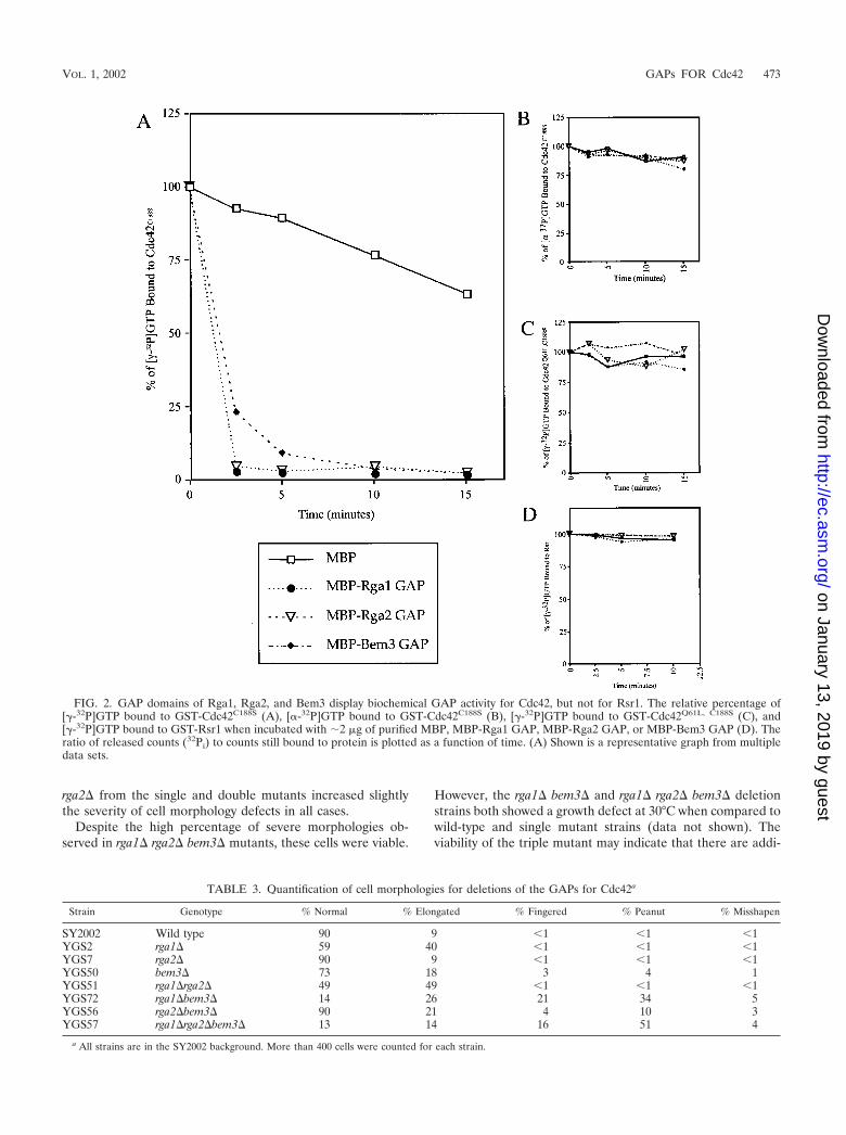
rga2 from the single and double mutants increased slightlythe severity of cell morphology defects in all cases.
Despite the high percentage of severe morphologies ob-served in rga1 rga2 bem3 mutants, these cells were viable.
However, the rga1 bem3 and rga1 rga2 bem3 deletionstrains both showed a growth defect at 30°C when compared towild-type and single mutant strains (data not shown). Theviability of the triple mutant may indicate that there are addi-
FIG. 2. GAP domains of Rga1, Rga2, and Bem3 display biochemical GAP activity for Cdc42, but not for Rsr1. The relative percentage of[�-32P]GTP bound to GST-Cdc42C188S (A), [�-32P]GTP bound to GST-Cdc42C188S (B), [�-32P]GTP bound to GST-Cdc42Q61L, C188S (C), and[�-32P]GTP bound to GST-Rsr1 when incubated with �2 �g of purified MBP, MBP-Rga1 GAP, MBP-Rga2 GAP, or MBP-Bem3 GAP (D). Theratio of released counts (32Pi) to counts still bound to protein is plotted as a function of time. (A) Shown is a representative graph from multipledata sets.
TABLE 3. Quantification of cell morphologies for deletions of the GAPs for Cdc42a
Strain Genotype % Normal % Elongated % Fingered % Peanut % Misshapen
SY2002 Wild type 90 9 �1 �1 �1YGS2 rga1 59 40 �1 �1 �1YGS7 rga2 90 9 �1 �1 �1YGS50 bem3 73 18 3 4 1YGS51 rga1rga2 49 49 �1 �1 �1YGS72 rga1bem3 14 26 21 34 5YGS56 rga2bem3 90 21 4 10 3YGS57 rga1rga2bem3 13 14 16 51 4
a All strains are in the SY2002 background. More than 400 cells were counted for each strain.
VOL. 1, 2002 GAPs FOR Cdc42 473
on January 13, 2019 by guesthttp://ec.asm
.org/D
ownloaded from
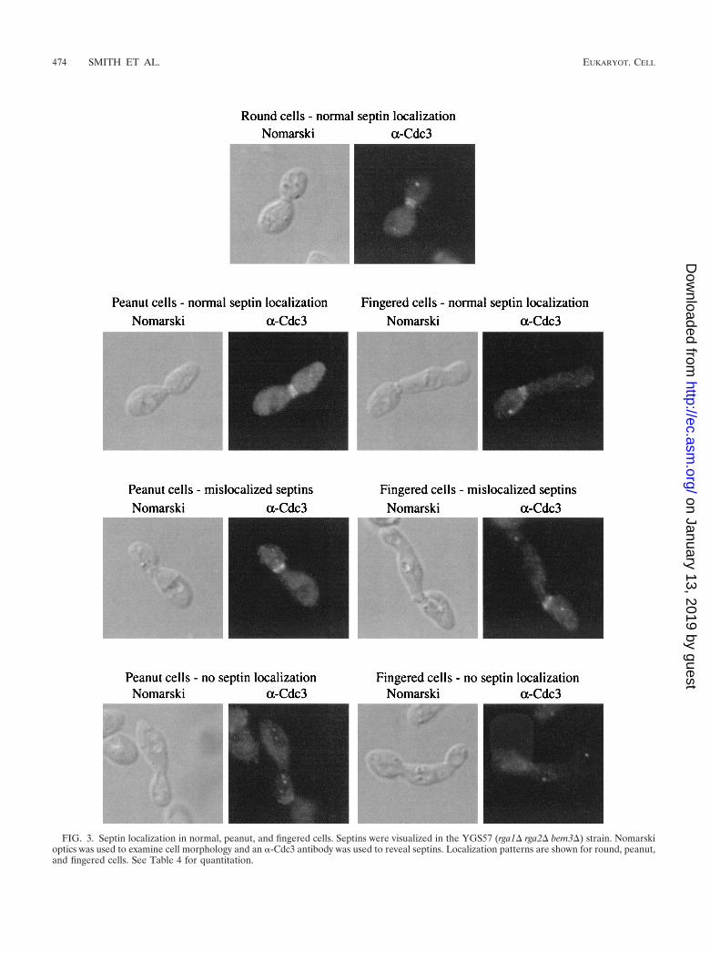
FIG. 3. Septin localization in normal, peanut, and fingered cells. Septins were visualized in the YGS57 (rga1 rga2 bem3) strain. Nomarskioptics was used to examine cell morphology and an �-Cdc3 antibody was used to reveal septins. Localization patterns are shown for round, peanut,and fingered cells. See Table 4 for quantitation.
474 SMITH ET AL. EUKARYOT. CELL
on January 13, 2019 by guesthttp://ec.asm
.org/D
ownloaded from

tional proteins with GAP activity for Cdc42 or that the inher-ent GTPase activity of Cdc42 is sufficient for viability (27, 67).
The localization of actin, chitin, and septin rings was exam-ined in peanut and fingered cells. Such cells had defects inchitin localization as revealed by Calcofluor staining. In par-ticular, these cells showed diffuse staining around the periph-ery in contrast to the rings of staining seen at previous bud sitesin normal cells (data not shown). Immunofluorescence micros-copy using the �-Cdc3 antibody to highlight septins revealeddefects in the peanut and fingered cells (Fig. 3). Septins nor-mally localize as a double ring at the bud neck (19; for review,see reference 21; Fig. 3). However, the septins rings wereimproperly localized in some peanut and fingered cells (Fig. 3).For cells with large buds, 43% of peanut cells and 68% offingered cells failed to properly localize their septins rings,compared to only 4% of round cells in the same strain (Table4). In most of the cells displaying mislocalized septins, a singleseptin ring had formed on one side of the bud neck. Becausemany of these cells had not yet undergone nuclear division, themother cell could be identified by the presence of the onlynucleus. In all cases, this single septin ring was located on thedaughter side of the bud neck (data not shown). Finally, nodefects in actin localization were detected in the GAP mutants(data not shown). Thus, peanut and fingered cells were foundto have defects in the organization of septins (Fig. 3; Table 4)and chitin (data not shown), but not actin (data not shown).
Bud site selection defects in GAP deletion strains. Wild-typehaploid cells are round and bud in an axial pattern in whicheach new bud is adjacent to the site of the previous bud (10,11). Using Calcofluor to visualize bud scars, we observed non-axial budding patterns for rga1 cells. In toto, 78% of cells ina rga1 deletion strain displayed a nonaxial budding pattern(Table 5; see also reference 63): 51% of the rga1 cells had a
bipolar pattern in which buds were seen at both poles of thecell and 27% of cells showed a random budding pattern withbud scars at either or both poles and in the middle of the cell(Table 5). In contrast, rga2, bem3, and rga2 bem3 cellsexhibited an axial pattern with all of the bud scars at one pole(Table 5). The rga1 rga2 double mutant cells had a buddingpattern similar to that of rga1 cells (Table 5). Budding pat-terns for rga1 bem3 and rga1 rga2 bem3 cells could notbe accurately determined due to the aberrant cell morpholo-gies and diffuse chitin staining (see above). Thus, Rga1 has adistinct function in bud site selection that is not shared by Rga2or Bem3.
Activation of FUS1-lacZ reporter in GAP deletion strains.RGA1 was first identified in a screen for negative regulators ofthe pheromone response pathway (63). It was shown that ste4rga1 cells activated a FUS1-lacZ reporter construct (63). Wedetermined that deletion of RGA2 or BEM3 did not causeactivation of the FUS1-lacZ reporter (Table 6). Thus, therga1 deletion appears to be unique in its ability to affectFUS1-lacZ expression. Deletion of RGA2 in a rga1 straincaused a slight increase in FUS1-lacZ expression (38.9 forste4 rga1 versus 45.5 for ste4 rga1 rga2). However, de-letion of all three GAP genes caused an eightfold increase inexpression (Table 6). Thus, although Rga1 has a primary rolein controlling the activation of FUS1-lacZ, Rga2, and Bem3must also have a negative regulatory role, albeit a modest one.
Roles of Rga1, Rga2, and Bem3 in haploid invasive growth.We also examined the roles of Rga1, Rga2, and Bem3 inhaploid invasive growth, a process involving Cdc42 (46). Theseexperiments were done with the �1278b strain (provided by G.Fink), a strain that exhibits more vigorous agar invasion thanother lab strains. Two techniques were used to assess filamen-tous growth: the plate-washing assay (56), which assesses agarinvasion, and the single-cell assay (16), which provides detailedinformation on cell morphology and budding patterns. Bythe plate-washing assay, rga1 cells displayed hyperinvasivegrowth, whereas rga2 and bem3 cells invaded the agar at alevel similar to wild-type cells (Fig. 4). On glucose-rich me-dium, rga1 mutant cells showed a propensity to bud at thedistal pole (17%), whereas bem3 (2% distal), rga2 (1%), andwild-type (1%) cells did not (Fig. 4b). The rga1 mutant also hadan elongated cell morphology in glucose-rich medium; 45% ofthe cells were elongated compared to less than 1% for wild-type and the rga2 mutant. The bem3 mutant also had elongatedcells (18%) on this medium, but the cells were not as elongated
TABLE 4. Quantification of septin localizationa
Strain Cell type % with normallocalization
% with mislo-calization
% with nolocalization
SY2002 Round 99 �1 �1YGS57 Round 96 2 2YGS57 Peanut 57 34 9YGS57 Fingered 33 36 32
a Septin staining was performed as described in the Materials and Methods.Only cells with large buds were counted. More than 200 cells were counted foreach class.
TABLE 5. Budding patterns of GAP deletion strainsa
Strain Genotype % Axial % Bipolar % Random
SY2002 Wild type 89 8 3YGS2 rga1 22 51 27YGS7 rga2 90 6 3YGS50 bem3 90 7 3YGS51 rga1rga2 14 64 22YGS72 rga1bem3 NDb ND NDYGS56 rga2bem3 94 4 3YGS57 rga1rga2bem3 ND ND ND
a Bud scars were visualized using Calcofluor as described in Materials andMethods. Only cells with three of more scars were counted. More than 200 cellswere counted for each strain.
b ND, not determined. Accurate counts could not be determined due to diffusechitin staining and abnormal cell morphologies
TABLE 6. Activation of FUS1-lacZ by deletion of GAPs for Cdc42
Strain Genotypea FUS1-lacZ expressionb
YGS58 ste4 1.2 0.1YGS59 ste4rga1 38.9 2.5YGS60 ste4rga2 1.8 0.5YGS61 ste4bem3 1.3 0.1YGS62 ste4rga1rga2 45.5 2.8YGS63 ste4rga1bem3 30.3 3.7YGS64 ste4rga2bem3 3.9 1.2YGS65 ste4rga1rga2bem3 396.1 20.5
a All strains are in the SY2002 background.b �-Galactosidase activity was determined as described in Materials and Meth-
ods. The reported values are given in 100� Miller units and are averages( standard deviation) of three determinations.
VOL. 1, 2002 GAPs FOR Cdc42 475
on January 13, 2019 by guesthttp://ec.asm
.org/D
ownloaded from
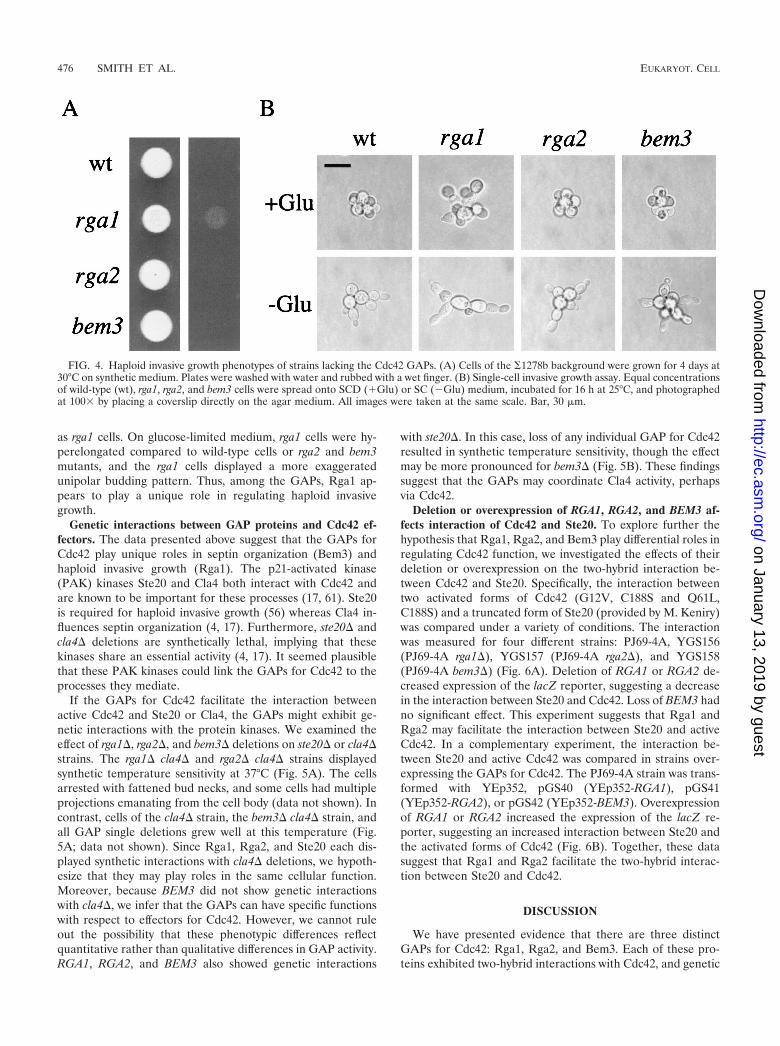
as rga1 cells. On glucose-limited medium, rga1 cells were hy-perelongated compared to wild-type cells or rga2 and bem3mutants, and the rga1 cells displayed a more exaggeratedunipolar budding pattern. Thus, among the GAPs, Rga1 ap-pears to play a unique role in regulating haploid invasivegrowth.
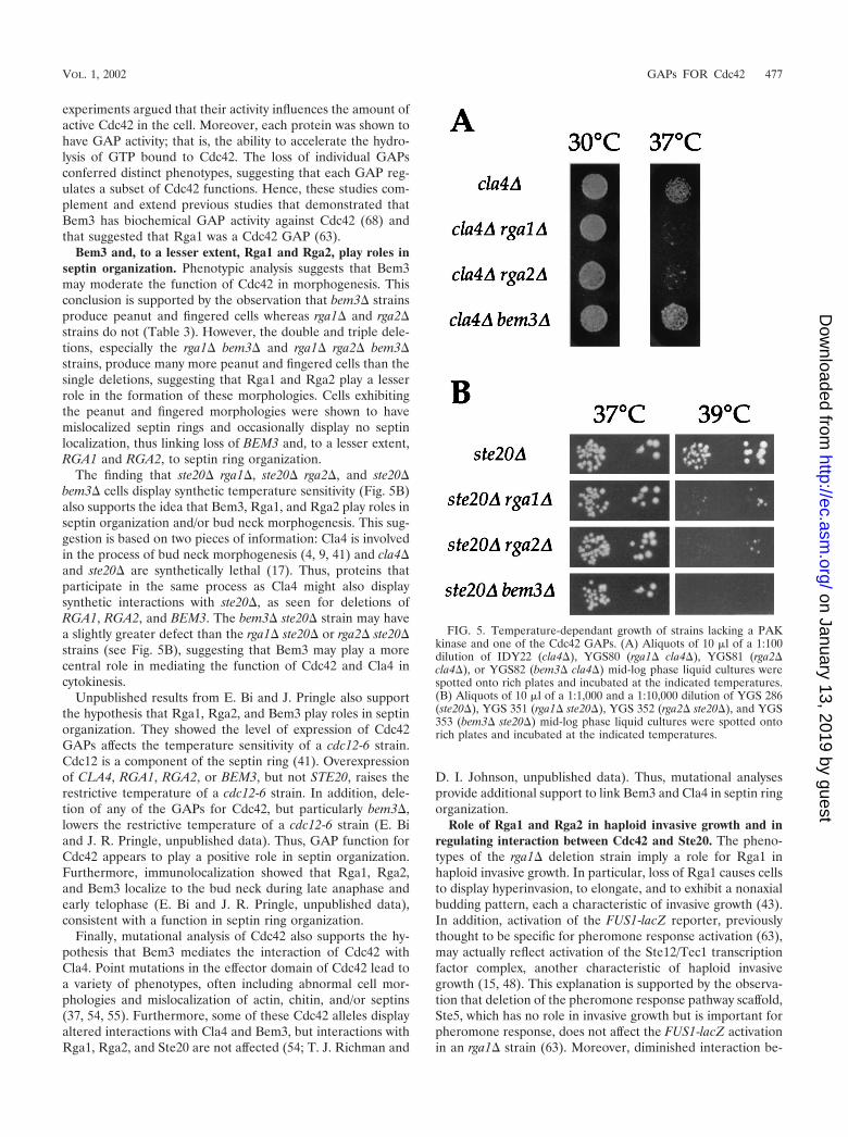
Genetic interactions between GAP proteins and Cdc42 ef-fectors. The data presented above suggest that the GAPs forCdc42 play unique roles in septin organization (Bem3) andhaploid invasive growth (Rga1). The p21-activated kinase(PAK) kinases Ste20 and Cla4 both interact with Cdc42 andare known to be important for these processes (17, 61). Ste20is required for haploid invasive growth (56) whereas Cla4 in-fluences septin organization (4, 17). Furthermore, ste20 andcla4 deletions are synthetically lethal, implying that thesekinases share an essential activity (4, 17). It seemed plausiblethat these PAK kinases could link the GAPs for Cdc42 to theprocesses they mediate.
If the GAPs for Cdc42 facilitate the interaction betweenactive Cdc42 and Ste20 or Cla4, the GAPs might exhibit ge-netic interactions with the protein kinases. We examined theeffect of rga1, rga2, and bem3 deletions on ste20 or cla4strains. The rga1 cla4 and rga2 cla4 strains displayedsynthetic temperature sensitivity at 37°C (Fig. 5A). The cellsarrested with fattened bud necks, and some cells had multipleprojections emanating from the cell body (data not shown). Incontrast, cells of the cla4 strain, the bem3 cla4 strain, andall GAP single deletions grew well at this temperature (Fig.5A; data not shown). Since Rga1, Rga2, and Ste20 each dis-played synthetic interactions with cla4 deletions, we hypoth-esize that they may play roles in the same cellular function.Moreover, because BEM3 did not show genetic interactionswith cla4, we infer that the GAPs can have specific functionswith respect to effectors for Cdc42. However, we cannot ruleout the possibility that these phenotypic differences reflectquantitative rather than qualitative differences in GAP activity.RGA1, RGA2, and BEM3 also showed genetic interactions
with ste20. In this case, loss of any individual GAP for Cdc42resulted in synthetic temperature sensitivity, though the effectmay be more pronounced for bem3 (Fig. 5B). These findingssuggest that the GAPs may coordinate Cla4 activity, perhapsvia Cdc42.
Deletion or overexpression of RGA1, RGA2, and BEM3 af-fects interaction of Cdc42 and Ste20. To explore further thehypothesis that Rga1, Rga2, and Bem3 play differential roles inregulating Cdc42 function, we investigated the effects of theirdeletion or overexpression on the two-hybrid interaction be-tween Cdc42 and Ste20. Specifically, the interaction betweentwo activated forms of Cdc42 (G12V, C188S and Q61L,C188S) and a truncated form of Ste20 (provided by M. Keniry)was compared under a variety of conditions. The interactionwas measured for four different strains: PJ69-4A, YGS156(PJ69-4A rga1), YGS157 (PJ69-4A rga2), and YGS158(PJ69-4A bem3) (Fig. 6A). Deletion of RGA1 or RGA2 de-creased expression of the lacZ reporter, suggesting a decreasein the interaction between Ste20 and Cdc42. Loss of BEM3 hadno significant effect. This experiment suggests that Rga1 andRga2 may facilitate the interaction between Ste20 and activeCdc42. In a complementary experiment, the interaction be-tween Ste20 and active Cdc42 was compared in strains over-expressing the GAPs for Cdc42. The PJ69-4A strain was trans-formed with YEp352, pGS40 (YEp352-RGA1), pGS41(YEp352-RGA2), or pGS42 (YEp352-BEM3). Overexpressionof RGA1 or RGA2 increased the expression of the lacZ re-porter, suggesting an increased interaction between Ste20 andthe activated forms of Cdc42 (Fig. 6B). Together, these datasuggest that Rga1 and Rga2 facilitate the two-hybrid interac-tion between Ste20 and Cdc42.
DISCUSSION
We have presented evidence that there are three distinctGAPs for Cdc42: Rga1, Rga2, and Bem3. Each of these pro-teins exhibited two-hybrid interactions with Cdc42, and genetic
FIG. 4. Haploid invasive growth phenotypes of strains lacking the Cdc42 GAPs. (A) Cells of the �1278b background were grown for 4 days at30°C on synthetic medium. Plates were washed with water and rubbed with a wet finger. (B) Single-cell invasive growth assay. Equal concentrationsof wild-type (wt), rga1, rga2, and bem3 cells were spread onto SCD (�Glu) or SC (�Glu) medium, incubated for 16 h at 25°C, and photographedat 100� by placing a coverslip directly on the agar medium. All images were taken at the same scale. Bar, 30 �m.
476 SMITH ET AL. EUKARYOT. CELL
on January 13, 2019 by guesthttp://ec.asm
.org/D
ownloaded from
experiments argued that their activity influences the amount ofactive Cdc42 in the cell. Moreover, each protein was shown tohave GAP activity; that is, the ability to accelerate the hydro-lysis of GTP bound to Cdc42. The loss of individual GAPsconferred distinct phenotypes, suggesting that each GAP reg-ulates a subset of Cdc42 functions. Hence, these studies com-plement and extend previous studies that demonstrated thatBem3 has biochemical GAP activity against Cdc42 (68) andthat suggested that Rga1 was a Cdc42 GAP (63).
Bem3 and, to a lesser extent, Rga1 and Rga2, play roles inseptin organization. Phenotypic analysis suggests that Bem3may moderate the function of Cdc42 in morphogenesis. Thisconclusion is supported by the observation that bem3 strainsproduce peanut and fingered cells whereas rga1 and rga2strains do not (Table 3). However, the double and triple dele-tions, especially the rga1 bem3 and rga1 rga2 bem3strains, produce many more peanut and fingered cells than thesingle deletions, suggesting that Rga1 and Rga2 play a lesserrole in the formation of these morphologies. Cells exhibitingthe peanut and fingered morphologies were shown to havemislocalized septin rings and occasionally display no septinlocalization, thus linking loss of BEM3 and, to a lesser extent,RGA1 and RGA2, to septin ring organization.
The finding that ste20 rga1, ste20 rga2, and ste20bem3 cells display synthetic temperature sensitivity (Fig. 5B)also supports the idea that Bem3, Rga1, and Rga2 play roles inseptin organization and/or bud neck morphogenesis. This sug-gestion is based on two pieces of information: Cla4 is involvedin the process of bud neck morphogenesis (4, 9, 41) and cla4and ste20 are synthetically lethal (17). Thus, proteins thatparticipate in the same process as Cla4 might also displaysynthetic interactions with ste20, as seen for deletions ofRGA1, RGA2, and BEM3. The bem3 ste20 strain may havea slightly greater defect than the rga1 ste20 or rga2 ste20strains (see Fig. 5B), suggesting that Bem3 may play a morecentral role in mediating the function of Cdc42 and Cla4 incytokinesis.
Unpublished results from E. Bi and J. Pringle also supportthe hypothesis that Rga1, Rga2, and Bem3 play roles in septinorganization. They showed the level of expression of Cdc42GAPs affects the temperature sensitivity of a cdc12-6 strain.Cdc12 is a component of the septin ring (41). Overexpressionof CLA4, RGA1, RGA2, or BEM3, but not STE20, raises therestrictive temperature of a cdc12-6 strain. In addition, dele-tion of any of the GAPs for Cdc42, but particularly bem3,lowers the restrictive temperature of a cdc12-6 strain (E. Biand J. R. Pringle, unpublished data). Thus, GAP function forCdc42 appears to play a positive role in septin organization.Furthermore, immunolocalization showed that Rga1, Rga2,and Bem3 localize to the bud neck during late anaphase andearly telophase (E. Bi and J. R. Pringle, unpublished data),consistent with a function in septin ring organization.
Finally, mutational analysis of Cdc42 also supports the hy-pothesis that Bem3 mediates the interaction of Cdc42 withCla4. Point mutations in the effector domain of Cdc42 lead toa variety of phenotypes, often including abnormal cell mor-phologies and mislocalization of actin, chitin, and/or septins(37, 54, 55). Furthermore, some of these Cdc42 alleles displayaltered interactions with Cla4 and Bem3, but interactions withRga1, Rga2, and Ste20 are not affected (54; T. J. Richman and
D. I. Johnson, unpublished data). Thus, mutational analysesprovide additional support to link Bem3 and Cla4 in septin ringorganization.
Role of Rga1 and Rga2 in haploid invasive growth and inregulating interaction between Cdc42 and Ste20. The pheno-types of the rga1 deletion strain imply a role for Rga1 inhaploid invasive growth. In particular, loss of Rga1 causes cellsto display hyperinvasion, to elongate, and to exhibit a nonaxialbudding pattern, each a characteristic of invasive growth (43).In addition, activation of the FUS1-lacZ reporter, previouslythought to be specific for pheromone response activation (63),may actually reflect activation of the Ste12/Tec1 transcriptionfactor complex, another characteristic of haploid invasivegrowth (15, 48). This explanation is supported by the observa-tion that deletion of the pheromone response pathway scaffold,Ste5, which has no role in invasive growth but is important forpheromone response, does not affect the FUS1-lacZ activationin an rga1 strain (63). Moreover, diminished interaction be-
FIG. 5. Temperature-dependant growth of strains lacking a PAKkinase and one of the Cdc42 GAPs. (A) Aliquots of 10 �l of a 1:100dilution of IDY22 (cla4), YGS80 (rga1 cla4), YGS81 (rga2cla4), or YGS82 (bem3 cla4) mid-log phase liquid cultures werespotted onto rich plates and incubated at the indicated temperatures.(B) Aliquots of 10 �l of a 1:1,000 and a 1:10,000 dilution of YGS 286(ste20), YGS 351 (rga1 ste20), YGS 352 (rga2 ste20), and YGS353 (bem3 ste20) mid-log phase liquid cultures were spotted ontorich plates and incubated at the indicated temperatures.
VOL. 1, 2002 GAPs FOR Cdc42 477
on January 13, 2019 by guesthttp://ec.asm
.org/D
ownloaded from

tween Ste20 and active Cdc42 had little effect on pheromoneresponse pathway signaling and mating but did result in de-creased invasive growth (39, 50). Together, these findings sup-port the idea that Rga1 affects haploid invasive growth, possi-bly by mediating the interaction of active Cdc42 with Ste20.
A role for Rga1 in mediating the interaction between Cdc42and Ste20 is also supported by the synthetic temperature sen-sitivity of rga1 and cla4 mutations (Fig. 5). Since cla4 andste20 are synthetically lethal (17), it would follow that a pro-tein involved in the same process as Ste20 might also displaysynthetic interactions with cla4. Interestingly, rga2 cla4cells, but not bem3 cla4 cells, also display synthetic temper-ature sensitivity, suggesting that Rga2 might also play a role inmediating the interaction between Cdc42 and Ste20. Thesephenotypic differences may reflect different quantitative con-tributions to overall GAP activity by Rga1, Rga2, and Bem3.However, the phenotypic differences could also indicate thatthe GAPs have qualitatively different roles in orchestratingCdc42 activity. Consistent with the proposed role of Rga1 andRga2 as facilitators of the interaction between Cdc42 andSte20, deletion of RGA1 or RGA2 decreased the two-hybridinteraction between activated forms of Cdc42 with Ste20,whereas RGA1 or RGA2 overexpression increased this inter-action. These experiments suggest that Rga1 and Rga2 facili-tate the interaction between Cdc42 and Ste20. This functionwould appear to be distinct from the GAP activity of Rga1 andRga2 since these two-hybrid experiments were performed withversions of Cdc42 locked in the GTP-bound, active form (70).How do Rga1 and Rga2 facilitate this interaction? Given thatRga1 and Rga2 have not been observed to interact with Ste20by two-hybrid analysis (63; data not shown), perhaps these twoGAPs do not directly promote an interaction between Ste20and Cdc42. Rather, perhaps Rga1 and Rga2 prevent Cdc42from interacting with other effectors or perhaps they recruitsome unidentified protein that aids in this process.
ACKNOWLEDGMENTS
We thank April Goehring, Megan Keniry, Hay-Oak Park, PhilJames, Brian Stevenson, Betsy Ferguson, Dave Mitchell, Erica Gole-mis, Gerald Fink, and Doug Johnson for providing advice, strains,and/or plasmids. Thanks to Erfei Bi, John Pringle, Tammy Richman,and Doug Johnson for sharing results before publication. We alsothank David Rivers, Hilary Kemp, Jesse Dillon, and Phil Kinsey forhelpful comments and suggestions.
This work was supported by research (GM-30027 to G.F.S) andtraining (GM-07413 to G.R.S., HD-07348 to S.A.G., and GM-19888 toP.C.) grants from the U.S. Public Health Service and by a fellowshipfrom the American Heart Association (AHA1206352) to P.C.
REFERENCES
1. Adams, A. E., D. I. Johnson, R. M. Longnecker, B. F. Sloat, and J. R. Pringle.1990. CDC42 and CDC43, two additional genes involved in budding and theestablishment of cell polarity in the yeast Saccharomyces cerevisiae. J. CellBiol. 111:131–142.
2. Archer, V. E., J. Breton, I. Sanchez-Garcia, H. Osada, A. Forster, A. J.Thomson, and T. H. Rabbitts. 1994. Cysteine-rich LIM domains of LIM-homeodomain and LIM-only proteins contain zinc but not iron. Proc. Natl.Acad. Sci. USA 91:316–320.
3. Barthe, C., G. de Bettignies, O. Louvet, M. F. Peypouquet, C. Morel, F.Doignon, and M. Crouzet. 1998. First characterization of the gene RGD1 inthe yeast Saccharomyces cerevisiae. C. R. Acad. Sci. Ser. III 321:453–462.
4. Benton, B. K., A. Tinkelenberg, I. Gonzalez, and F. R. Cross. 1997. Cla4p, aSaccharomyces cerevisiae Cdc42p-activated kinase involved in cytokinesis, isactivated at mitosis. Mol. Cell. Biol. 17:5067–5076.
5. Bi, E., J. B. Chiavetta, H. Chen, G. C. Chen, C. S. Chan, and J. R. Pringle.2000. Identification of novel, evolutionarily conserved Cdc42p-interacting
FIG. 6. Effects of deletion or overexpression of Cdc42 GAPs ontwo-hybrid interaction between Ste20 and activated versions of Cdc42.(A) The interaction of Ste20 residues 1 to 565 fused to the Gal4 DBD(pSL2682) with three versions of the Gal4 transcription activationdomain—GAD itself and GAD fused either to Cdc42G12V,C188S
(pGS38) or to Cdc42Q61L,C188S (pGS39)—in four separate strains werecompared: PJ69-4A, YGS156 (rga1), YGS157 (rga2), and YGS158(bem3). Expression of the lacZ reporter in three separate isolates wasmeasured. (B) The same two-hybrid interactions investigated abovewere compared for strain PJ69-4A transformed with four differentplasmids: YEp352, pGS40 (YEp352-RGA1), pGS41 (YEp352-RGA2),and pGS42 (YEp352-BEM3). The expression of the lacZ reporter wasmeasured for three separate isolates in four independent trials. Thecombined normalized data were graphed.
478 SMITH ET AL. EUKARYOT. CELL
on January 13, 2019 by guesthttp://ec.asm
.org/D
ownloaded from
proteins and of redundant pathways linking Cdc24p and Cdc42p to actinpolarization in yeast. Mol. Biol. Cell. 11:773–793.
6. Boone, C., N. G. Davis, and G. F. Sprague, Jr. 1993. Mutations that alter thethird cytoplasmic loop of the a-factor receptor lead to a constitutive andhypersensitive phenotype. Proc. Natl. Acad. Sci. USA 90:9921–9925.
7. Brown, J. L., M. Jaquenoud, M. P. Gulli, J. Chant, and M. Peter. 1997. NovelCdc42-binding proteins Gic1 and Gic2 control cell polarity in yeast. GenesDev. 11:2972–2982.
8. Burke, D., D. Dawson, and T. Stearns. 2000. Methods in yeast genetics. ColdSpring Harbor Laboratory Press, Plainview, N.Y.
9. Carroll, C. W., R. Altman, D. Schieltz, J. R. Yates, and D. Kellogg. 1998. Theseptins are required for the mitosis-specific activation of the Gin4 kinase.J. Cell Biol. 143:709–717.
10. Chant, J., and I. Herskowitz. 1991. Genetic control of bud site selection inyeast by a set of gene products that constitute a morphogenetic pathway. Cell65:1203–1212.
11. Chant, J., M. Mischke, E. Mitchell, I. Herskowitz, and J. R. Pringle. 1995.Role of Bud3p in producing the axial budding pattern of yeast. J. Cell Biol.129:767–778.
12. Chen, D. C., B. C. Yang, and T. T. Kuo. 1992. One-step transformation ofyeast in stationary phase. Curr. Genet. 21:83–84.
13. Chen, G. C., Y. J. Kim, and C. S. Chan. 1997. The Cdc42 GTPase-associatedproteins Gic1 and Gic2 are required for polarized cell growth in Saccharo-myces cerevisiae. Genes Dev. 11:2958–2971.
14. Chen, G. C., L. Zheng, and C. S. Chan. 1996. The LIM domain-containingDbm1 GTPase-activating protein is required for normal cellular morpho-genesis in Saccharomyces cerevisiae. Mol. Cell. Biol. 16:1376–1390.
15. Cullen, P. J., J. Schultz, J. Horecka, B. J. Stevenson, Y. Jigami, and G. F.Sprague, Jr. 2000. Defects in protein glycosylation cause SHO1-dependentactivation of a STE12 signaling pathway in yeast. Genetics 155:1005–1018.
16. Cullen, P. J., and G. F. Sprague, Jr. 2000. Glucose depletion causes haploidinvasive growth in yeast. Proc. Natl. Acad. Sci. USA 97:13619–13624.
17. Cvrckova, F., C. De Virgilio, E. Manser, J. R. Pringle, and K. Nasmyth. 1995.Ste20-like protein kinases are required for normal localization of cell growthand for cytokinesis in budding yeast. Genes Dev. 9:1817–1830.
18. Dawid, I. B., J. J. Breen, and R. Toyama. 1998. LIM domains: multiple rolesas adapters and functional modifiers in protein interactions. Trends Genet.14:156–162.
19. DeMarini, D. J., A. E. Adams, H. Fares, C. De Virgilio, G. Valle, J. S.Chuang, and J. R. Pringle. 1997. A septin-based hierarchy of proteins re-quired for localized deposition of chitin in the Saccharomyces cerevisiae cellwall. J. Cell Biol. 139:75–93.
20. Doignon, F., C. Weinachter, O. Roumanie, and M. Crouzet. 1999. The yeastRgd1p is a GTPase activating protein of the Rho3 and Rho4 proteins. FEBSLett. 459:458–462.
21. Field, C. M., and D. Kellogg. 1999. Septins: cytoskeletal polymers or signal-ling GTPases? Trends Cell Biol. 9:387–394.
22. Fields, S., and O. Song. 1989. A novel genetic system to detect protein-protein interactions. Nature 340:245–246.
23. Freyd, G., S. K. Kim, and H. R. Horvitz. 1990. Novel cysteine-rich motif andhomeodomain in the product of the Caenorhabditis elegans cell lineage genelin-11. Nature 344:876–879.
24. Gietz, R. D., R. H. Schiestl, A. R. Willems, and R. A. Woods. 1995. Studieson the transformation of intact yeast cells by the LiAc/SS-DNA/PEG pro-cedure. Yeast 11:355–360.
25. Guan, C., P. Li, P. D. Riggs, and H. Inouye. 1987. Vectors that facilitate theexpression and purification of foreign peptides in Escherichia coli by fusionto maltose-binding protein. Gene 67:21–30.
26. Gyuris, J., E. Golemis, H. Chertkov, and R. Brent. 1993. Cdi1, a human G1and S phase protein phosphatase that associates with Cdk2. Cell 75:791–803.
27. Hart, M. J., K. Shinjo, A. Hall, T. Evans, and R. A. Cerione. 1991. Identi-fication of the human platelet GTPase activating protein for the CDC42Hsprotein. J. Biol. Chem. 266:20840–20848.
28. Hill, J. E., A. M. Myers, T. J. Koerner, and A. Tzagoloff. 1986. Yeast/E. colishuttle vectors with multiple unique restriction sites. Yeast 2:163–167.
29. Ijzerman, M. M., J. O. Falkinham III, and C. Hagedorn. 1993. A liquid,colorimetric presence-absence coliphage detection method. J. Virol. Meth-ods 45:229–233.
30. James, P., J. Halladay, and E. A. Craig. 1996. Genomic libraries and a hoststrain designed for highly efficient two-hybrid selection in yeast. Genetics144:1425–1436.
31. Jarvis, E. E., D. C. Hagen, and G. F. Sprague, Jr. 1988. Identification of aDNA segment that is necessary and sufficient for �-specific gene control inSaccharomyces cerevisiae: implications for regulation of �-specific and a-spe-cific genes. Mol. Cell. Biol. 8:309–320.
32. Johnson, D. I. 1999. Cdc42: an essential Rho-type GTPase controlling eu-karyotic cell polarity. Microbiol. Mol. Biol. Rev. 63:54–105.
33. Johnson, D. I., C. W. Jacobs, J. R. Pringle, L. C. Robinson, G. F. Carle, andM. V. Olson. 1987. Mapping of the Saccharomyces cerevisiae CDC3, CDC25,and CDC42 genes to chromosome XII by chromosome blotting and tetradanalysis. Yeast 3:243–253.
34. Johnson, D. I., and J. R. Pringle. 1990. Molecular characterization of
CDC42, a Saccharomyces cerevisiae gene involved in the development of cellpolarity. J. Cell Biol. 111:143–152.
35. Karlsson, O., S. Thor, T. Norberg, H. Ohlsson, and T. Edlund. 1990. Insulingene enhancer binding protein Isl-1 is a member of a novel class of proteinscontaining both a homeo- and a Cys-His domain. Nature 344:879–882.
36. Kim, H. B., B. K. Haarer, and J. R. Pringle. 1991. Cellular morphogenesis inthe Saccharomyces cerevisiae cell cycle: localization of the CDC3 gene prod-uct and the timing of events at the budding site. J. Cell Biol. 112:535–544.
37. Kozminski, K. G., A. J. Chen, A. A. Rodal, and D. G. Drubin. 2000. Functionsand functional domains of the GTPase Cdc42p. Mol. Biol. Cell 11:339–354.
38. Leberer, E., D. Dignard, D. Harcus, D. Y. Thomas, and M. Whiteway. 1992.The protein kinase homologue Ste20p is required to link the yeast phero-mone response G-protein beta gamma subunits to downstream signallingcomponents. EMBO J. 11:4815–4824.
39. Leberer, E., C. Wu, T. Leeuw, A. Fourest-Lieuvin, J. E. Segall, and D. Y.Thomas. 1997. Functional characterization of the Cdc42p binding domain ofyeast Ste20p protein kinase. EMBO J. 16:83–97.
40. Liu, X., H. Wang, M. Eberstadt, A. Schnuchel, E. T. Olejniczak, R. P.Meadows, J. M. Schkeryantz, D. A. Janowick, J. E. Harlan, E. A. Harris,D. E. Staunton, and S. W. Fesik. 1998. NMR structure and mutagenesis ofthe N-terminal Dbl homology domain of the nucleotide exchange factorTrio. Cell 95:269–277.
41. Longtine, M. S., H. Fares, and J. R. Pringle. 1998. Role of the yeast Gin4pprotein kinase in septin assembly and the relationship between septin as-sembly and septin function. J. Cell Biol. 143:719–736.
42. Ma, H., S. Kunes, P. J. Schatz, and D. Botstein. 1987. Plasmid constructionby homologous recombination in yeast. Gene 58:201–216.
43. Madhani, H. D., and G. R. Fink. 1997. Combinatorial control required forthe specificity of yeast MAPK signaling. Science 275:1314–1317.
44. Maina, C. V., P. D. Riggs, A. G. I. Grandea, B. E. Slatko, L. S. Moran, J. A.Tagliamonte, L. A. McReynolds, and C. Guan. 1988. A vector to express andpurify foreign proteins in Escherichia coli by fusion to, and separation form,maltose-binding protein. Gene 74:365–373.
45. Michelsen, J. W., K. L. Schmeichel, M. C. Beckerle, and D. R. Winge. 1993.The LIM motif defines a specific zinc-binding protein domain. Proc. Natl.Acad. Sci. USA 90:4404–4408.
45a.Miller, P. J., and D. I. Johnson. 1997. Characterization of the Saccharomycescerevvisiae cdc42-1 allele and new temperature-conditional-lethal cdc42 al-leles. Yeast 13:561–572.
46. Mosch, H. U., T. Kohler, and G. H. Braus. 2001. Different domains of theessential GTPase Cdc42p required for growth and development of Saccha-romyces cerevisiae. Mol. Cell. Biol. 21:235–248.
47. Nilsson, B., L. Abrahmsen, and M. Uhlen. 1985. Immobilization and purifi-cation of enzymes with staphylococcal protein A gene fusion vectors. EMBOJ. 4:1075–1080.
48. O’Rourke, S. M., and I. Herskowitz. 1998. The Hog1 MAPK prevents crosstalk between the HOG and pheromone response MAPK pathways in Sac-charomyces cerevisiae. Genes Dev. 12:2874–2886.
49. Park, H. O., E. Bi, J. R. Pringle, and I. Herskowitz. 1997. Two active statesof the Ras-related Bud1/Rsr1 protein bind to different effectors to determineyeast cell polarity. Proc. Natl. Acad. Sci. USA 94:4463–4468.
50. Peter, M., A. M. Neiman, H. O. Park, M. van Lohuizen, and I. Herskowitz.1996. Functional analysis of the interaction between the small GTP bindingprotein Cdc42 and the Ste20 protein kinase in yeast. EMBO J. 15:7046–7059.
51. Pringle, J. R., R. A. Preston, A. E. Adams, T. Stearns, D. G. Drubin, B. K.Haarer, and E. W. Jones. 1989. Fluorescence microscopy methods for yeast.Methods Cell Biol. 31:357–435.
52. Printen, J. A., and G. F. Sprague, Jr. 1994. Protein-protein interactions inthe yeast pheromone response pathway: Ste5p interacts with all members ofthe MAP kinase cascade. Genetics 138:609–619.
53. Ramer, S. W., and R. W. Davis. 1993. A dominant truncation allele identifiesa gene, STE20, that encodes a putative protein kinase necessary for matingin Saccharomyces cerevisiae. Proc. Natl. Acad. Sci. USA 90:452–456.
54. Richman, T. J., and D. I. Johnson. 2000. Saccharomyces cerevisiae Cdc42pGTPase is involved in preventing the recurrence of bud emergence duringthe cell cycle. Mol. Cell. Biol. 20:8548–8559.
55. Richman, T. J., M. M. Sawyer, and D. I. Johnson. 1999. The Cdc42p GTPaseis involved in a G2/M morphogenetic checkpoint regulating the apical-iso-tropic switch and nuclear division in yeast. J. Biol. Chem. 274:16861–16870.
56. Roberts, R. L., and G. R. Fink. 1994. Elements of a single MAP kinasecascade in Saccharomyces cerevisiae mediate two developmental programs inthe same cell type: mating and invasive growth. Genes Dev. 8:2974–2985.
57. Rose, A. B., and J. R. Broach. 1990. Propagation and expression of clonedgenes in yeast: 2-microns circle-based vectors. Methods Enzymol. 185:234–279.
58. Rose, M. D., F. Winston, and P. Hieter. 1990. Methods in yeast genetics.Cold Spring Harbor Laboratory Press, Plainview, N.Y.
59. Sambrook, J., E. F. Fritsch, and T. Maniatis. 1989. Molecular cloning: alaboratory manual, 2nd ed. Cold Spring Harbor Laboratory Press, ColdSpring Harbor, N.Y.
60. Schmidt, A., M. Bickle, T. Beck, and M. N. Hall. 1997. The yeast phospha-
VOL. 1, 2002 GAPs FOR Cdc42 479
on January 13, 2019 by guesthttp://ec.asm
.org/D
ownloaded from
tidylinositol kinase homolog TOR2 activates RHO1 and RHO2 via the ex-change factor ROM2. Cell 88:531–542.
61. Simon, M. N., C. De Virgilio, B. Souza, J. R. Pringle, A. Abo, and S. I. Reed.1995. Role for the Rho-family GTPase Cdc42 in yeast mating-pheromonesignal pathway. Nature 376:702–705.
62. Sloat, B. F., A. Adams, and J. R. Pringle. 1981. Roles of the CDC24 geneproduct in cellular morphogenesis during the Saccharomyces cerevisiae cellcycle. J. Cell Biol. 89:395–405.
63. Stevenson, B. J., B. Ferguson, C. De Virgilio, E. Bi, J. R. Pringle, G. Am-merer, and G. F. Sprague, Jr. 1995. Mutation of RGA1, which encodes aputative GTPase-activating protein for the polarity-establishment proteinCdc42p, activates the pheromone-response pathway in the yeast Saccharo-myces cerevisiae. Genes Dev. 9:2949–2963.
64. Uetz, P., L. Giot, G. Cagney, T. A. Mansfield, R. S. Judson, J. R. Knight, D.Lockshon, V. Narayan, M. Srinivasan, P. Pochart, A. Qureshi-Emili, Y. Li,B. Godwin, D. Conover, T. Kalbfleisch, G. Vijayadamodar, M. Yang, M.Johnston, S. Fields, and J. M. Rothberg. 2000. A comprehensive analysis ofprotein-protein interactions in Saccharomyces cerevisiae. Nature 403:623–627.
65. Watanabe, D., M. Abe, and Y. Ohya. 2001. Yeast Lrg1p acts as a specializedRhoGAP regulating 1,3-beta-glucan synthesis. Yeast 18:943–951.
66. Way, J. C., and M. Chalfie. 1988. mec-3, a homeobox-containing gene thatspecifies differentiation of the touch receptor neurons in C. elegans. Cell54:5–16.
67. Zheng, Y., R. Cerione, and A. Bender. 1994. Control of the yeast bud-siteassembly GTPase Cdc42. J. Biol. Chem. 269:2369–2372.
68. Zheng, Y., M. J. Hart, K. Shinjo, T. Evans, A. Bender, and R. A. Cerione.1993. Biochemical comparisons of the Saccharomyces cerevisiae Bem2 andBem3 proteins. Delineation of a limit Cdc42 GTPase-activating proteindomain. J. Biol. Chem. 268:24629–24634.
69. Ziman, M., and D. I. Johnson. 1994. Genetic evidence for a functionalinteraction between Saccharomyces cerevisiae CDC24 and CDC42. Yeast10:463–474.
70. Ziman, M., J. M. O’Brien, L. A. Ouellette, W. R. Church, and D. I. Johnson.1991. Mutational analysis of CDC42Sc, a Saccharomyces cerevisiae gene thatencodes a putative GTP-binding protein involved in the control of cellpolarity. Mol. Cell. Biol. 11:3537–3544.
480 SMITH ET AL. EUKARYOT. CELL
on January 13, 2019 by guesthttp://ec.asm
.org/D
ownloaded from