structural analysis of ph, w19h, mouse
TRANSCRIPT

Proc. Nati. Acad. Sci. USAVol. 91, pp. 7237-7241, July 1994Genetics
Structural analysis of chromosomal rearrangements associated withthe developmental mutations Ph, W19H, and Rw on mousechromosome 5
(developmental genetics/physical maps/receptor tyrouine kinase)
DEBORAH L. NAGLE*, PATRICIA MARTIN-DELEONt, RICHARD B. HOUGH*, AND MAJA BUtAN*t*Department of Psychiatry, University of Pennsylvania, Philadelphia, PA 19104; and tSchool of Life and Health Sciences, University of Delaware,Newark, DE 19716
Communicated by R. L. Brinster, March 24, 1994
ABSTRACT We are studying the chromosomal structureof three developmental mutations, dint spotting (W),patch (Ph), and rump white (Rw) on mouse chromosome 5.These mutations are clustered in a region containing threegenes encoding tyrosine kinase receptors (Kit, PdJfyu, andFRkl). Using probes for these genes and for a closely linkedlocus, D5Mn125, we esta hed a high-resolution physical mapcovering r2.8 Mb. The entire chromosomal segment mappedin this study is deleted in the W19H mutation. The map i testhe position of the Ph deletion, which encompasses not morethan 400 kb around and including the Pdgfra gene. The mapalso places the distal breakpoint of theRw inversion to a limitedchromosomal segment between Kit and Pdgfi. In light of thestructure of the Ph-W-Rw region, we interpret the previouslypublished complementtion analyses as indicating that thepigmentation defect inRw/+ heterozygotes could be due to thedisruption ofKit and/or Pdefia regulatory sequences, whereasthe gene(s) responsible for the recessive lethality of Rw/Rwembryos is not closely linked to the Ph and W loci and mapspromaily to the W19H deletion. The structural analysis ofchromosomal rearrangements associated with W19H, Ph, andRw combined with the high-resolution physical mapping pointsthe way toward the definition of these mutations in molecularterms and Isolation of homologous genes on human chromo-some 4.
Three mutations on mouse chromosome 5, dominant spotting(W), patch (Ph), and rump white (Rw), were historicallydescribed as a gene triplet on the basis of their close linkageand similar mutant phenotypes (1). Molecular analysis ofthese three loci has progressed rapidly due to the findings thatWandPh are associated with mutations and/or chromosomalrearrangements of genes encoding receptor tyrosine kinases(RTKs) (2). RTKs are known to have an important role in theregulation of growth and differentiation (3).There are multiple alleles of W, which vary in their degree
of severity and in their pleiotropic effects on melanogenesis,hematopoiesis, and germ cell development (4). Analysis ofthe protooncogene c-kit (a RTK) in several independentlyidentified W alleles demonstrated that the W mutant pheno-type is due to mutations that affect either the structure of theKit receptor or the level and spatial distribution of Kitexpression (5-10). More recently, it has been shown that theproduct ofthe Si locus on mouse chromosome 10 encodes theligand for the Kit receptor (reviewed in ref. 2).The Ph mutation is associated with a dominant white
spotting phenotype characterized by a depigmented beltaround the abdomen (1, 11). Ph/Ph homozygotes die aroundmidgestation and, prior to their death, embryos display
several morphological abnormalities (e.g., small size, undif-ferentiated somites, abnormal heart, wavy neural tube) (11-13). The Ph locus encodes the platelet-derived growth factorreceptor a subunit, Pdgfra, a member ofthe RTKgene family(14, 15). It is not known, however, whether the mutantphenotype ofPh/Ph embryos is due solely to the deletion ofPdgfra or to the deletion of an additional closely linkedgene(s).The W19H allele is a deletional mutation spanning 2-7
centimorgans (cM), encompassing both the Ph and W loci,and a recessive lethal (1), which has yet to be defined at amolecular level (15-17). The hypopigmentation associatedwith the W19H mutation in mice is strikingly similar to theobserved depigmentation in humans with the piebald trait.This developmental defect is caused by a deletion of thehomologous portion of human chromosome 4 containing theKit and Pdgfra genes (18-20).The Rw mutation is characterized by heterozygous spot-
ting of the posterior trunk; the embryonic lethality ofRw/Rwmice occurs during midgestation as a result of unknowncauses (1). Although a candidate gene for the Rw mutationhas not been identified, it has been shown recently that thisirradiation-induced mutation is associated with an inversioninvolving the proximal one-third of the chromosome. Thedistal breakpoint of the inversion has been mapped by in situanalysis to the chromosomal segment spanning 4cM betweenthe Kit and DSBucl loci, while the proximal breakpoint mapscentromeric to the engrailed 2 (En2) locus (21).The cluster of RTKs (class III subfamily) located in the
central portion of mouse chromosome 5 includes at leastthree members: Kit, Pdgfra, and FMki (22-24). Previously,Kit and Pdgfra have been linked genetically and physically toone another (14, 15, 25). FikI, an endothelial cell-specificRTK, has been genetically linked to the Kit locus (24).
In an attempt to determine the molecular basis of the Ph,Rw, and W19H mutations and to assess the role of the clusterof RTKs on mouse chromosome 5 in development, we haveestablished a high-resolution physical map covering 2.8Mb ofthat region on the wild-type (C57BL/6J) chromosome. Inaddition, we have determined the approximate map positionof the breakpoints of the Ph deletion and placed the distalbreakpoint oftheRw inversion on the physical map. The mapindicates close proximity (within 200 kb) of sequences dis-rupted by the Ph and Rw chromosomal anomalies. We haveutilized the wealth of previously published genetic data on
Abbreviations: RTK, receptor tyrosine kinase; cM, centimorgan(s);YAC, yeast artificial chromosome; PFGE, pulsed-field gel electro-phoresis; FISH, fluorescence in situ hybridization; FITC, fluores-cein isothiocyanate; PI, propidium iodide; DAPI, diaminophenylin-dole.tTo whom reprint requests should be addressed at: Department ofPsychiatry, University of Pennsylvania, 111A CRB, 422 CurieBoulevard, Philadelphia, PA 19104-6141.
7237
The publication costs of this article were defrayed in part by page chargepayment. This article must therefore be hereby marked "advertisement"in accordance with 18 U.S.C. §1734 solely to indicate this fact.
Dow
nloa
ded
by g
uest
on
Nov
embe
r 3,
202
1
Proc. Nati. Acad. Sci. USA 91 (1994)
Ph+/+Rw and Rw+/+Wl9H compound heterozygotes tosuggest that the embryonic lethality of Rw/Rw embryos isprobably due to the disruption of a gene at the proximalbreakpoint of this inversion, near the centromeric portion ofmouse chromosome 5.
MATERIALS AND METHODSMice. Mouse inbred strains (C57BL/6J, C3H/HeJ, and
Mus spretus) and mutations (W19H, Ph) were obtained fromThe Jackson Laboratory. Rw mice were provided by BruceCattanach (Medical Research Council Radiobiology Unit,Harwell, U.K.). Mice used for deletion mapping were the F1progeny from the following crosses: Wl9H/C57BL/6J x M.spretus and Ph/C57BL/6J x M. spretus.DNA Probes. The DNA probes used in this study are a 0.6-
kb HindIII fragment ofthe fetal liver kinase 1 gene (Flkl) (24);a 1.2-kb EcoRI/Sph I fragment of the c-kit protooncogenecDNA (Kit) (5' portion of the gene) and a 0.9-kb HindIIIfragment of Kit (3' portion of the gene) (P. Dubreuil and A.Bernstein, personal communication); a 1.8-kb EcoRI/HindIII fragment of the platelet-derived growth factor recep-tor a cDNA (Pdgfra) (5' portion) and a 2-kb EcoRI/HindIIIfiagment of the Pdgfra (3' portion of the gene) (26); a 0.4-kbEcoRI/HincII fragment of an anonymous brain cDNA(DSMnl25) (17); a 0.5-kb EcoRI/HindIII fragment (DSBuc2)corresponding to the right end and a 0.4-kb EcoRI/Hae IIIfragment (DSBuc3) corresponding to the left end of a Pdgfrayeast artificial chromosome (YAC) (ICRFy902GO8127) (thispaper); and a 0.6-kb Pst I fragment ofthe randomDNA probe(DSBucl) (ref. 21; J. DeLoia, M.B., and D. Solter, unpub-lished data).
Analysis by Pulsed-Field Gel Electrophoresis (PFGE). Meth-ods for PFGE, including DNA preparation in agarose blocksand restriction analysis, have been described (27). DNAseparated by PFGE was transferred to Hybond N+ (Amer-sham) membrane by capillary blotting in denaturation buffer(28), UV crosslinked (autocrosslink set up on Stratalinker,Stratagene), and hybridized as described (27).Iolaton of YAC Clone. A YAC clone for Pdgfra was
obtained by screening high-density robot spotted filters con-taining a mouse (C3H/He) YAC library (29), generouslyprovided by H. Lehrach (Imperial Cancer Research Fund,London). After a rescreen, DNA from a positive clone wasisolated by using published protocols (30). YAC ends wereisolated by the inverse PCR procedure (31).
Fluorescence in Situ Hybridization (FISH) Analysis. Mitoticspreads were prepared from spleen lymphocytes of Rwheterozygotes by using a modification of the proceduredescribed by Sawyer et al. (32). YAC DNA used for in situhybridization was isolated by separating yeast DNA byPFGE. Purified YAC DNA was biotin-labeled by randompriming. The hybridization probe was prepared by mixing 400ng ofbiotinylated YAC DNA with 6.25 ,gg ofmouse genomicDNA and 12.5 ,g of salmon sperm DNA in hybridizationbuffer (Hybrisol VII; Oncor). It was applied to denaturedchromosomal DNA on slides, which were incubated for 16 hrat 3rC. Slides were washed five times for 5 min each at 42°Cin 50% formamide/2x standard saline citrate (SSC) with the
last wash in 0.1x SSC. Detection and amplification of thelabeled probe were performed using fluorescein isothiocya-nate (FITC)-avidin and anti-avidin (Oncor detection kit) andslides were stained in 1.5 pg ofpropidium iodide (PI) per ml,1.5 pg of diaminophenylindole (DAPI) per ml, and 1 mg ofp-phenylenediamine per ml. A triple bandpass filter forDAPI/FITC/rhodamine was used for chromosome identifi-cation and a dual-wavelength filter cube (FITC and rhoda-mine) was used for signal visualization.
RESULTSPhydcal Map. In our attempt to analyze the chromosomal
structure and rearrangements associated with the Ph, W19H,and Rw mutations, we made use of the available molecularprobes to establish a long-range restriction map of the cor-responding chromosomal region on the wild-type chromo-some. The gene probes, Fikl, Kit, and Pdgfra, and ananonymous cDNA, DSMnl25, the first molecular markermapped within the W19H deletion (17), were used in PFGEanalysis of C57BL/6J DNA. In addition, we used end clones(DSBuc2 and DSBuc3) of a Pdgfra-containing YAC. Allmolecular probes were sequentially hybridized to filterscontaining splenocyte DNA digested withBssHl, Mlu I, NotI, and combinations of these enzymes. Analysis of thehybridization patterns and fragment sizes (Table 1) allowedthe construction ofa long-range restriction map spanning 2.8Mb (Fig. 1A). The established order of loci is DSMnl2S-D5Buc2-Pdgfra-Kit-Flkl. Although the map spans 2.8 Mb,the three homologous RTK genes, Pdgfra-Kit-Flkl, mapwithin 1 Mb.
Deletion Mapping of Ph and W19H Chromosomes. Twomutations included in this study, Ph and W19H, have beenassociated with deletions of genetic material (5, 14-17). Todetermine the positions of the deletion breakpoints on thephysical map, we used the same molecular probes includedin the PFGE analysis and an additional locus, DSBucl,located 4 cM proximal to Kit (21). These probes wereanalyzed by Southern blot hybridization in DNA isolatedfrom F1 progeny of the following interspecific crosses: Ph/C57BL/6J x M. spretus and WI9H/C57BL/6J x M. spretus.Since the tested molecular probes detected restriction frag-
ment length variants between M. spretus and Mus musculus(carrying the Ph and W19H deletions), it was possible todistinguish the two parental chromosomes and determinewhether any of these sequences are deleted on the Ph and/orW19H chromosome (data not shown). The results of deletionmapping are summarized in Fig. 1B. The DSBucl locus mapsoutside the W19H and Ph deletions; DSMnl2S, and Kit, andFiki are deleted in W19H but not on the Ph chromosome,whereas Pdgfra is deleted on the W19H and Ph chromosomes.One end clone, DSBuc3, maps within the Ph deletion, whilethe other clone, DSBuc2, maps outside the deletion.FISH Mapping of Pdgfra on the Rw/C57BL/6J Chromo-
some. Previous work has demonstrated that the distal break-point of the inversion associated with the Rw mutation mapsto a 4-cM region between DSBucl, a locus within the inver-sion, and Kit, which is located outside the inversion (21). Todefine the location of the distal inversion breakpoint we used
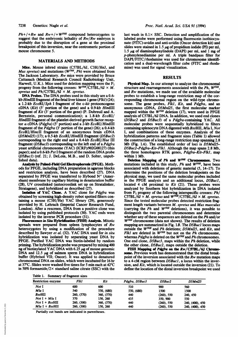
Table 1. Summary of fragment sizesRestriction enzyme FMk) Kit Pdgfra, DSBuc3 DSBuc2 DSMnl2SNot I 600 600 510 900 560Mlu I 540 550, (680) 550, (680) 1500 1500BssHII 260, (570) 360, (570) 440 (260), 550 240, 450Not I + Mlu I 370 150, 260 435 350, 900 550Not I + BssHII 260, (300) 360, (570) 440 (260), 550 240, (400), 450MIu I + BssHII 260, (300) 150, 260 440 (260), 550 240, (400), 450Partially cut bands are indicated in parentheses.
7238 Genetics: Nagle et al.
Dow
nloa
ded
by g
uest
on
Nov
embe
r 3,
202
1

Proc. Natl. Acad. Sci. USA 91 (1994) 7239
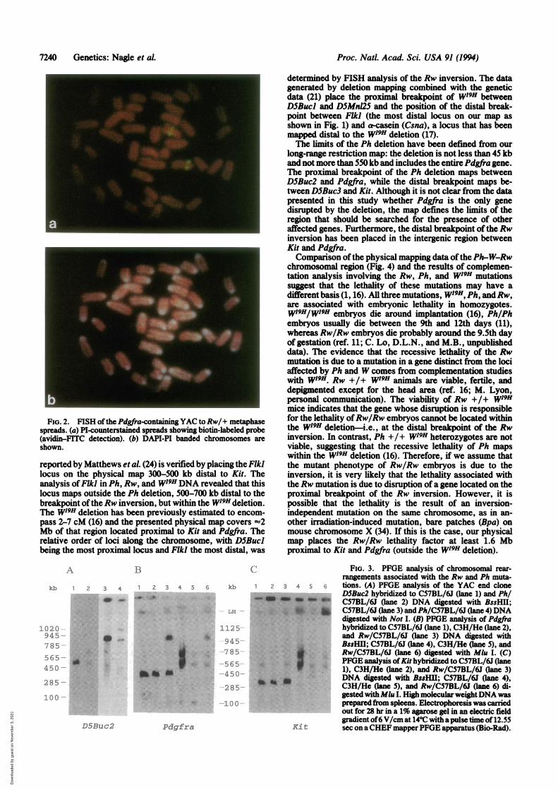
FISH analysis ofRw/C57BL/6J metaphase spreads and thePdgfra YAC as a probe. Fig. 2 shows that the position of thesignal for the Pdgfra gene differs on the two homologues ofchromosome 5. On one chromosome, the hybridization signalis confined to the central portion of the chromosome, corre-sponding to the location on the wild-type chromosome (21),whereas on the other chromosome it is located near thecentromeric heterochromatin. Since it has been previouslyshown by FISH analysis that Kit maps to the same locationon both homologues (21), we can strongly argue that thebreakpoint of the Rw inversion must be located between Kitand Pdgfra.PFGE Analysis ofPh/C57BL/6J andRw/C57BL/6J Chro-
mosomes. The physical mapping data demonstrate that theKit and Pdgfra genes are located on a Mlu I DNA fragmentof 550 kb and that they share an additional partially digestedMlu I fragment of 680 kb (Table 1; Fig. 1A; ref. 15).Stephenson et al. (14) demonstrated that Pdgfra is deleted inthe Ph mutation and that Kit genomic sequences are notdisrupted in Ph, thus defining the position of the distalbreakpoint of the Ph deletion as lying between Pdgfra andKit. Here, we demonstrate by FISH analysis that the samechromosomal region is associated with the distal breakpointof the Rw inversion (Fig. 2; ref. 21). Conventional Southernblot analysis ofgenomic sequences surrounding the Kit genein DNA from Ph/C57BL/6J and Rw/C57BL/6J indicated noalterations (data not shown), nor were there any alterationsof Rw/C57BL/6J DNA detected with the Pdgfra probe.However, chromosomal rearrangements were detected byPFGE analysis of large fragments containing the two genes.We mapped the proximal breakpoint of the Ph deletion by
PFGE of heterozygous (Ph/C57BL/6J) DNA. Molecularprobes from the W-Ph-Rw region were hybridized toC57BL/6J and Ph/C57BL/6J DNA digested with BssHII,Mlu I, Not I, and combinations of these enzymes. The YACend, DSBuc2, shown to map outside the Ph deletion bydeletion mapping (Fig. 1B), detects all C57BL/6J-specificfragments and additional, larger fragments ofDNA digested
A Scale
YAC
Loci
PFGEmap
B
with Not 1 (920 kb) and BssHll (640 kb), indicating that thelarge genomic fiagments recognized by this probe are alteredby the Ph mutation (Fig. 3A). Based on the PFGE map, weestimate that D5Buc2 maps 50 kb proximal to the Ph deletion(Fig. 1A).
Similarly, we were interested in identifying the position ofthe distal breakpoint of the Rw inversion. High molecularweight DNA isolated from C57BL/6J, C3H/HeJ, and Rw/C57BL/6JDNA was analyzed byPFGE using Kit andPdgfraprobes (Fig. 3 B and C). The Kit cDNA probe detected analtered fragment in Mlu digested Rw/C57BL/6J DNA. ThePdgfra cDNA clone detected altered DNA fragments in MluI-, BssHI1-, and Not digested Rw/C57BL/6J DNA. Sincethe Mlu I bands shared by Kit and Pdgfra are altereddifferently inRwDNA, we have further confirmation that theRw/C57BL/6J inversion breakpoint maps between Kit andPdgfra (Fig. 1; Fig. 3 B and C). The FISH mapping of aPdgfra YAC (8127) to Rw/C57BL/6J metaphase spreadssuggests that sequences contained within the YAC, covering=450 kb, map within the Rw inversion (Fig. 2). Based onthese data, the Rw breakpoint can be placed to the chromo-somal segment between the distal YAC end clone, DSBuc3,and Kit.
DISCUSSIONWe have established a high-resolution physical map of theregion surrounding three members of the class III subfamilyofRTKs (Kit, Pdgfra, and FikI) (Fig. 1). This map indicatesthe position and extent of chromosomal rearrangementsassociated with the Rw, Ph, and WJ9H mutations, which haveprofound developmental defects raning from mild pigmen-tation anomalies in heterozygotes to embryonic lethality inhomozygous mice.The long-range restriction map spans 2.8 Mb and indicates
the position of the Kit, Pdgfra, and Flkl genes and ananonymous mouse brain cDNA clone, DSMnl25. The closegenetic linkage between the Flkl and the Kit/Pdgfra cluster
kb 0 400 800 1200 1600 2000 2400 2800
Y8127
Buc 1 D5MnI25 Buc2 Pdgfra Buc3 Kit Flkl- - * - _
B (B) B B BB B (B) B BSSHIIM (M) M M MyL
N N) | N N N Noti
-7/A-Chromosomalrearrangements
~-P4 Rw inversion
...........i Ph deletion
-// ............*.*.-.-.-.-.-.-. -- -. - - - - W 19Hdeletion
FIG. 1. Physical map of the W-Ph-Rw chromosomal region. (A) Long-range restriction map of C57BL/6J DNA constructed with the datain Table 1. Map indicates sites for the enzymes Not I (N), Mlu I (M), and BssHUl (B). Partially cut sites are shown in parentheses. Scale (kb)is shown above the map. Positions ofprobes are indicated by solid rectangles above the map (longer solid boxes indicate the limits ofuncertaintyofprobe locations). D5Bucl, DSBuc2, and DSBuc3 loci are indicated as Bucl, Buc2, and Buc3. Hatched box indicates position ofthe YAC cloneY8127. (B) Chromosomal rearrangements associated with Ph, WI9H, andRw chromosomes determined in this study and in previously publishedreports, including the deletion ofPdgfra in the Ph mutation (12-1S); deletion ofKit, DSMnl25, and Pdgfra in WI9H (5, 6, 14, 15, 17); and inversionofthe DSBucl toEn2 region on theRw chromosome (21). In this study, the extent ofthe Ph and W'9Hchromosomal abnormalities was determinedby a combination of deletion mapping and PFGE analysis. Dotted lines indicate deleted segments; dashed lines indicate limits of uncertaintyof breakpoint locations. Position of the distal breakpoint of the Rw inversion (arrow) was determined by FISH mapping and PFGE analysis,as illustrated in Figs. 2 and 3. Position of DSBucl on the physical map is inferred from genetic data (21), deletion mapping, and hybridizationof D5Bucl and D5MnI25 to a Not I fragment of the same size (-3500 kb).
Genetics: Nagle et al.
Dow
nloa
ded
by g
uest
on
Nov
embe
r 3,
202
1
Proc. Nati. Acad. Sci. USA 91 (1994)
FIG. 2. FISH ofthe Pdgfra-containing YAC toRw/+ metaphasespreads. (a) PI-counterstained spreads showing biotin-labeled probe(avidin-FITC detection). (b) DAPI-PI banded chromosomes areshown.
reported by Matthews et al. (24) is verified by placing the Fikilocus on the physical map 300-500 kb distal to Kit. Theanalysis ofFPkH in Ph, Rw, and W19HDNA revealed that thislocus maps outside the Ph deletion, 500-700 kb distal to thebreakpoint oftheRw inversion, but within the W19H deletion.The W19H deletion has been previously estimated to encom-pass 2-7 cM (16) and the presented physical map covers %2Mb of that region located proximal to Kit and Pdgfra. Therelative order of loci along the chromosome, with DSBuclbeing the most proximal locus and Flkl the most distal, was
determined by FISH analysis of the Rw inversion. The datagenerated by deletion mapping combined with the geneticdata (21) place the proximal breakpoint of W19H betweenD5Bucl and DSMnI25 and the position of the distal break-point between Fiki (the most distal locus on our map asshown in Fig. 1) and a-casein (Csna), a locus that has beenmapped distal to the WI9H deletion (17).The limits of the Ph deletion have been defined from our
long-range restriction map: the deletion is not less than 45 kband not more than 550 kb and includes the entire Pdgfra gene.The proximal breakpoint of the Ph deletion maps betweenDSBuc2 and Pdgfra, while the distal breakpoint maps be-tween DSBuc3 and Kit. Although it is not clear from the datapresented in this study whether Pdgfra is the only genedisrupted by the deletion, the map defines the limits of theregion that should be searched for the presence of otheraffected genes. Furthermore, the distal breakpoint ofthe Rwinversion has been placed in the intergenic region betweenKit and Pdgfra.Comparison ofthe physical mapping data ofthe Ph-W-Rw
chromosomal region (Fig. 4) and the results of complemen-tation analysis involving the Rw, Ph, and W19H mutationssuggest that the lethality of these mutations may have adifferent basis (1, 16). All three mutations, W19H, Ph. andRw,are associated with embryonic lethality in homozygotes.W19H/W19H embryos die around implantation (16), Ph/Phembryos usually die between the 9th and 12th days (11),whereas Rw/Rw embryos die probably around the 9.5th dayof gestation (ref. 11; C. Lo, D.L.N., and M.B., unpublisheddata). The evidence that the recessive lethality of the Rwmutation is due to a mutation in a gene distinct from the lociaffected by Ph and W comes from complementation studieswith W19H. Rw +/+ WJ9H animals are viable, fertile, anddepigmented except for the head area (ref. 16; M. Lyon,personal communication). The viability of Rw +/+ W"9Hmice indicates that the gene whose disruption is responsiblefor the lethality ofRw/Rw embryos cannot be located withinthe W19H deletion-i.e., at the distal breakpoint of the Rwinversion. In contrast, Ph +/+ W19H heterozygotes are notviable, suggesting that the recessive lethality of Ph mapswithin the W19H deletion (16). Therefore, if we assume thatthe mutant phenotype of Rw/Rw embryos is due to theinversion, it is very likely that the lethality associated withthe Rw mutation is due to disruption of a gene located on theproximal breakpoint of the Rw inversion. However, it ispossible that the lethality is the result of an inversion-independent mutation on the same chromosome, as in an-other irradiation-induced mutation, bare patches (Bpa) onmouse chromosome X (34). If this is the case, our physicalmap places the Rw/Rw lethality factor at least 1.6 Mbproximal to Kit and Pdgfra (outside the W19H deletion).
A 13
kb 1 2 3 4 1 2 3 4 5 6
45
-S.. z I-
4 5
:85
F. .I
C
kb 1 2 3 4 5 6
^ 4W -
1L .,.. --
db ~~94 S*
.~~~~~8
.sf.:. 565
S. 4550
2285S
D5Buc2 Pdgfra
FiG. 3. PFGE analysis of chromosomal rear-rangements associated with the Rw and Ph muta-tions. (A) PFGE analysis of the YAC end cloneD5Buc2 hybridized to C57BL/6J (lane 1) and Ph/C57BL/6J (lane 2) DNA digested with BssHll;CS7BL/6J (lane 3) and Ph/C57BL/6J (lane 4) DNAdigested with Not I. (B) PFGE analysis of Pdgfrahybridized to C57BL/6J (lane 1), C3H/He (lane 2),and Rw/C57BL/6J (lane 3) DNA digested withBssHll; C57BL/6J (lane 4), C3H/He (lane 5), andRw/C57BL/6J (lane 6) digested with Mlu I. (C)PFGE analysis ofKit hybridized to CS7BL/6J (lane1), C3H/He (lane 2), and Rw/CS7BL/6J (lane 3)DNA digested with BssHIU; CS7BL/6J (lane 4),C3H/He (lane 5), and Rw/CS7BL/6J (lane 6) di-gested withMlu I. High molecular weightDNA wasprepaed ffrom spleens. Electrophoresis was carriedout for 28 hr in a 1% agarose gel in an electric fieldgradient of6 V/cm at 14'C with a pulse time of 12.55sec on aCHEF mapperPFGE apparatus (Bio-Rad).
7240 Genetics: Nagle et aL
, 'I.11-1
.ot,
Ki t
Dow
nloa
ded
by g
uest
on
Nov
embe
r 3,
202
1

Proc. Nati. Acad. Sci. USA 91 (1994) 7241
Ph W79H Rw wt Human Institutes of Health Grant HD 28410 (to M.B.), a Fellowship in IVDrug Abuse Research Center T32-DA07241 (to D.L.N.), a Predoc-homology toral Fellowship from the National Institutes of Health Cellular andMolecular Biology Training Grant (GM07229-19) (to R.B.H.), and
PgY I 1gY1,PgY1
'' National Science Foundation Grant MCB 9210351 (to P.M.-D.).__pgyl _ pgy1 ¶ Pgyl -_pgyl 7q21
D5Buc1. Pdgfra
Kit
4q12
FIG. 4. Summary of analysis of chromosomal organization of theW-Ph-Rw region on wild-type and mutant chromosomes. Deducedmap positions of chromosome 5 loci on the wild-type chromosome(wt) and the mutant chromosomes Ph, W19H, and Rw based on datapresented in this study and by others (5, 6, 14, 15, 17, 21). Bracketsindicate approximate positions of the breakpoints; however, theextent of the deletions is not accurately represented. Arrow indicatesposition of the distal breakpoint of the Rw inversion. Positions ofhomologous segments in the human genome (33) are indicated on theright.
Rw +/+ Ph double heterozygotes are viable although thepigmentation defect in these double mutants is more pro-nounced than either mutation alone (1). This observationoffers further support for the conclusion that the recessivelethalities of Rw and Ph are due to mutations in differentgenes. However, it does not exclude the possibility that thedominant coat color defect in Ph and Rw is due to thedisruption of Kit or Pdgfra regulatory sequences. Recentobservations by Duttlinger et al. (10) indicate that the Wshmutation, which is associated with a pigmentation defect butlacks other pleiotropic defects (macrocytic anemia and ste-rility) present in the majority of W alleles, is probably adeletion that removes control elements associated with theKit gene. Based on our data, these same regulatory elementscould be disrupted by both the distal breakpoint of the Phdeletion and the distal breakpoint of the Rw inversion.
Since it is known that at least two homologous human RTKgenes (KIT and PDGFRA) are closely linked on humanchromosome 4 and that their deletion is associated with acomparable pigmentation defect in humans (18-20), thestructural analysis of the W-Ph-Rw region will serve as auseful guide in comparative mapping efforts (Fig. 4). Inaddition, the proximal breakpoint of the Rw inversion islocated in the segment of synteny conservation betweenmouse chromosome 5 and human chromosome 7 (33). There-fore, if the Rw lethality factor proves to be located at theproximal breakpoint of the Rw inversion, we predict theexistence of a developmentally important gene causing lateembryonic lethality in the homologous region on humanchromosome 7 (q21 or q36).
We thank D. Solter in whose laboratory these studies wereinitiated; B. Cattanach for Rw mice; A. Bernstein, M. Mercola, I.Lemischka, and M. MacDonald for molecular probes; L. Stubbs, Z.Larin, and H. Lehrach for a Pdgfra-containing YAC clone; studentswho over the years contributed to this project; D. Stephenson, V.Chapman, S. Poethig, and P. Nolan for helpful discussions andcomments on the manuscript; M. F. Lyon for communicating un-published observations. These studies were supported by National
1. Searle, A. G. & Truslove, G. M. (1970) Genet. Res. 15, 227-235.2. Reith, A. D. & Bernstein, A. (1991) in Genome Analysis: Genes and
Phenotypes, eds. Davis, K. E. & Tilghman, S. M. (Cold SpringHarbor Lab. Press, Plainview, NY), Vol. 3, pp. 105-133.
3. Pawson, T. & Bernstein, A. (1990) Trends Genet. 6, 350-356.4. Silvers, W. K. (1979) The Coat Colors of Mice (Springer, New
York), pp. 206-241.5. Chabot, B., Stephenson, D. A., Chapman, V. M., Besmer, P. &
Bernstein, A. (1988) Nature (London) 335, 88-89.6. Geissler, E. N., Ryan, M. A. & Housman, D. E. (1988) Cell 55,
185-192.7. Tan, J. C., Nocka, K., Ray, P., Traktman, P. & Besmer, P. (1990)
Science 247, 209-212.8. Nocka, K., Majumder, S., Chabot, B., Ray, P., Cervone, M.,
Bernstein, A. & Besmer, P. (1989) Genes Dev. 3, 816-826.9. Reith, A. D., Rottapel, R., Giddens, E., Brady, C., Forrester, L. &
Bernstein, A. (1990) Genes Dev. 4, 390-400.10. Duttlinger, R., Manova, K., Chu, T. Y., Gyssler, C., Zelenetz,
A. D., Bachvarova, R. F. & Besmer, P. (1993) Development 118,705-717.
11. Grdneberg, H. & Truslove, G. M. (1960) Genet. Res. 1, 69-90.12. Orr-Urtreger, A., Bedford, M. T., Do, M.-S., Eisenbach, L. &
Lonai, P. (1992) Development 11S, 289-303.13. Schatteman, G. C., Morrison-Graham, K., Van Koppen, A.,
Weston, J. A. & Bowen-Pope, D. F. (1992) Development 115,123-131.
14. Stephenson, D. A., Mercola, M., Anderson, E., Wang, C., Stiles,C. D., Bowen-Pope, D. F. & Chapman, V. M. (1991) Proc. Natl.Acad. Sci. USA 88, 6-10.
15. Smith, E. A., Seldin, M. F., Martinez, L., Watson, M. L., Choud-hury, G. G., Lalley, P. A., Pierce, J., Aaronson, S., Barker, J.,Naylor, S. L. & Sakaguchi, A. Y. (1991) Proc. Natd. Acad. Sci.USA 88, 4811-4815.
16. Lyon, M. F., Glenister, P. H., Loutit, J. F., Evans, E. P. & Peters,J. (1984) Genet. Res. 44, 161-168.
17. Geissler, E. N., Cheng, S. V., Gusella, J. F. & Housman, D. E.(1988) Proc. NatI. Acad. Sci. USA 85, 9635-99.
18. Fleischman, R. A., Saltman, D. L., Stastay, V. & Zneimer, S.(1991) Proc. NatI. Acad. Sci. USA 88, 10885-10889.
19. Spritz, R. A., Droetto, S. & Fukushima, Y. (1992) Am. J. Med.Genet. 44, 492-495.
20. Fleischman, R. A. (1993) Trends Genet. 9, 285-290.21. Stephenson, D. A., Lee, K.-h., Nagle, D. L., Yen, C.-H., Morrow,
A., Miller, D., Chapman, V. M. & Bucan, M. (1994) Mamm.Genome, in press.
22. Besmer, P., Murphy, J. E., George, P. C., Qiu, F., Bergold, P. J.,Lederman, L., Snyder, H. W., Jr., Brodeur, D., Zuckerman, E. E.& Hardy, W. D. (1986) Nature (London) 32, 415-421.
23. Matsui, T., Heidaran, M., Miki, T., Popescu, N., La Rochelle, W.,Kraus, M., Pierce, J. & Aaronson, S. (1989) Science 243, 800-804.
24. Matthews, W., Jordan, C. T., Gavin, M., Jenkins,N. A., Copeland,N. G. & Lemischka, I. R. (1991) Proc. NatI. Acad. Sci. USA 88,9026-9030.
25. Hsieh, C.-L., Navankasattusas, S., Escobedo, J. A., Williams,L. T. & Francke, U. (1991) Cytogenet. Cell Genet. 56, 160-163.
26. Wang, C., Kelly, J., Bowen-Pope, D. F. & Stiles, C. D. (1990) Mol.Cell. Biol. 10, 6781-6784.
27. Herrmann, B. G., Barlow, D. P. & Lehrach, H. (1987) Cell 48,813-825.
28. Herrmann, B. G., Bucan, M., Mains, P. E., Frischauf, A.-M.,Silver, L. M. & Lehrach, H. (1986 Cell 44, 469-476.
29. Lamn, Z., Monaco, A. P. & Lehrach, H. (1991) Proc. Natd. Acad.Sci. USA 88, 4123-4127.
30. Green, E. D. & Olson, M. V. (1990) Proc. NatI. Acad. Sci. USA 87,1213-1217.
31. Arveiler, B. & Porteous, D. J. (1991) Technique 3, 24-28.32. Sawyer, J. R., Moore, M. M. & Hozier, J. C. (1987) Chromosoma
95, 350-358.33. Nadeau, J. H., Davisson, M. T., Doolittle, D. P., Grant, P., Hill-
yard, A. L., Kosowsky, M. R. & Roderick, T. H. (1992) Mamm.Genome 3, 480-536.
34. Evans, E. P. & Phillips, R. J. S. (1975) Nature (London) 256,40-41.
+ En2 + En2x I + En2 7q36
* DSBucIof. Kit
I;: D5BucI t En2Kitt
Genetics: Nagle et A
Dow
nloa
ded
by g
uest
on
Nov
embe
r 3,
202
1