streptococci. morphology and identification gram-positive cocci arranged in chains or pairs. most...
TRANSCRIPT
Streptococci

Morphology and Identification
Gram-positive cocci arranged in chains or pairs.Most group A, B, and C strains produce capsules. Most strains grow as discoid colonies, 1-2 mm in diameter.Catalase-negative.Grow better in media enriched with blood or tissue fluid.
Most are facultative a
naerobic and some ar
e capnophilic. For mo
st species growth and
hemolysis are aided b
y incubation in 10% C
O2.

Hemolysis
-hemolysis: incomplete lysis of RBC with the formation
of green pigment.
-hemolysis: complete hemolysis
No hemolysis
Lancefield classification: a serologic classification (A to
V)
Biochemical reactions are used for species that can not b
e classified into the Lancefield classification (nongroupab
le), e.g. viridans streptococci.
Classification
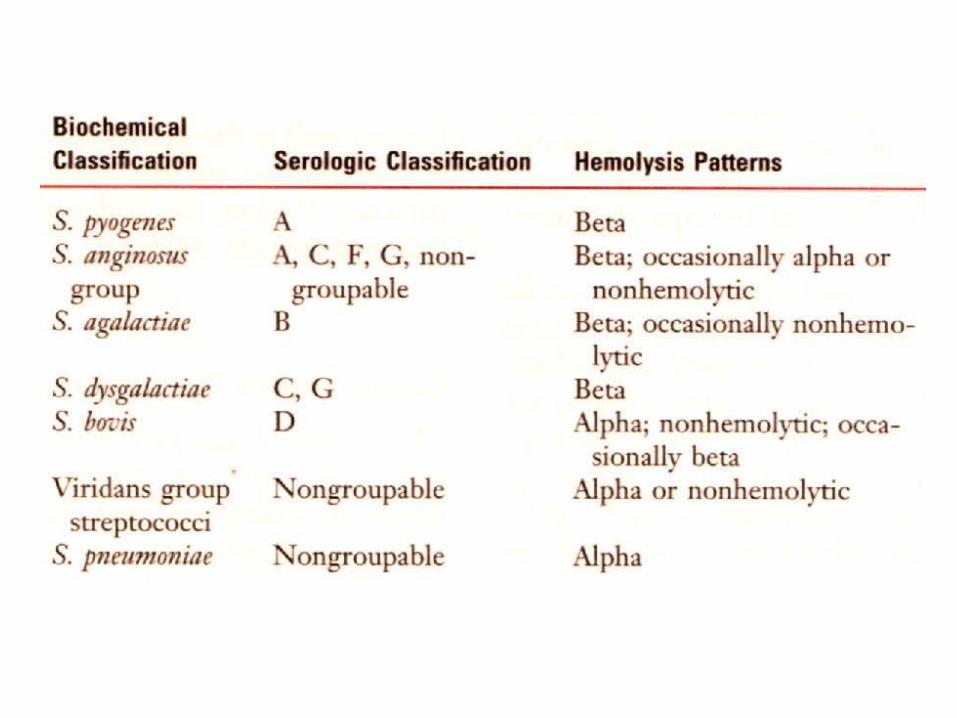

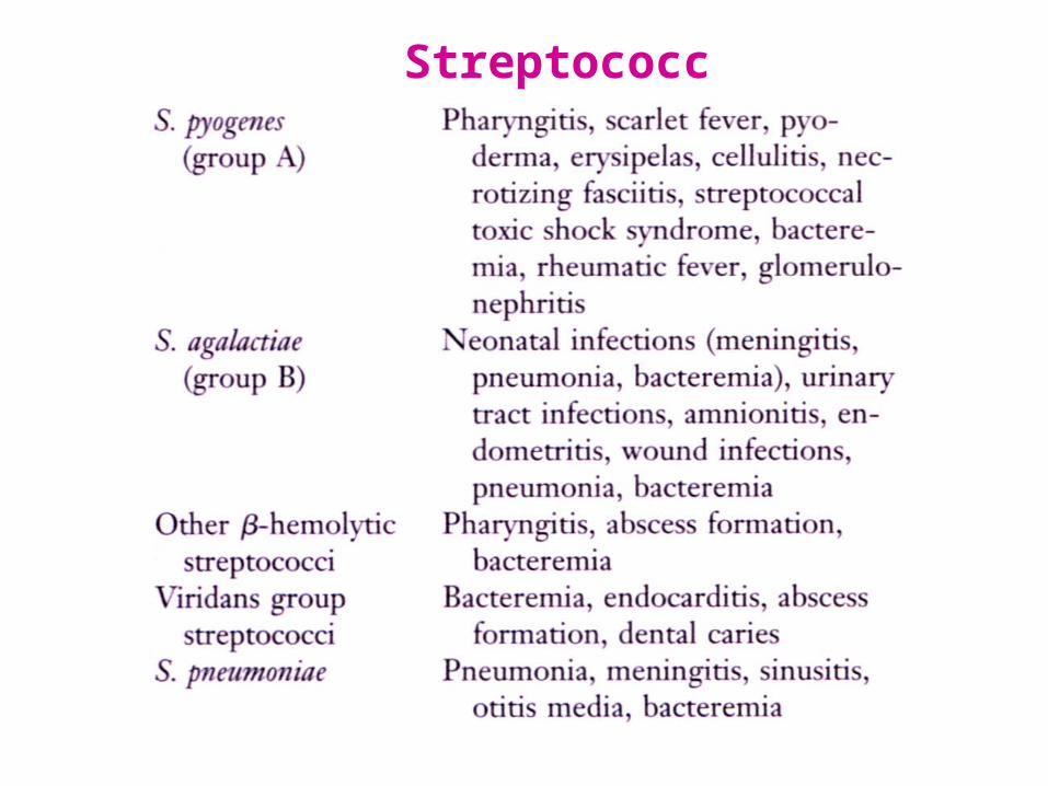
Streptococcus pyogenes
Capsule: antiphagocytosis. The capsule of group A streptococci is composed of hyaluronic acid.
Group-specific cell wall antigen (Lancefield group A)
Carbohydrate
A dimer of N-acetylglucosamine and rhamnose.
M protein
T protein: type-specific; function unknown.
M-like proteins: binds IgM, IgG and 2-macroglobulin; interfere with phagocytosis.
Lipoteichoic acid: binds to epithelial cells.
Protein F: a major adhesin of S. pyogenes, binding with fibronectin.

Adherence to the epithelial cells;
>10 adhesion molecules
invasion into the epithelial cells;
mediated by M protein and protein F
important for persistent infections and invasion into deep tissues
avoiding opsonization and phagocytosis;
M protein, M-like proteins, and C5a peptidase
producing enzymes and toxins
Pathogenesis (via invasiveness and production of toxins)
Streptococcus pyogenes
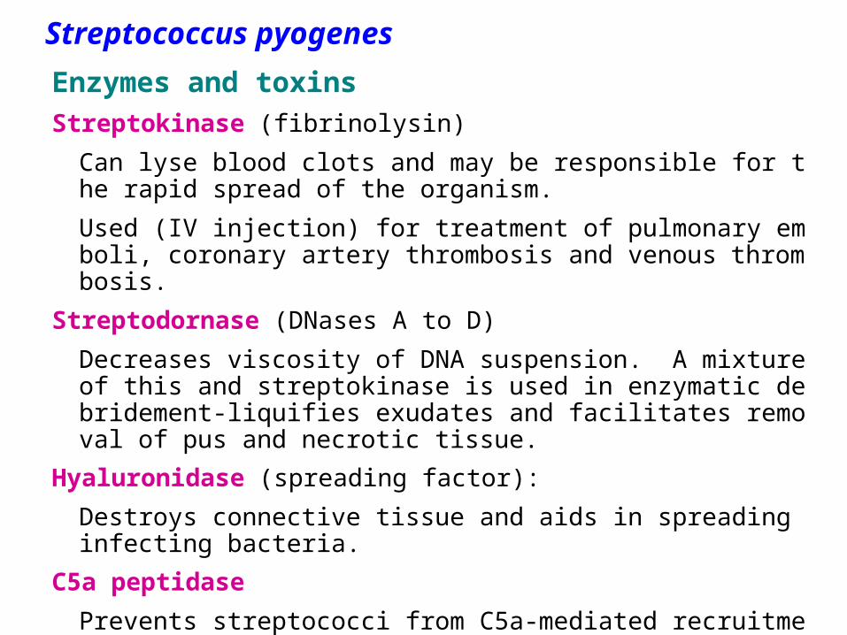
Enzymes and toxinsStreptokinase (fibrinolysin)
Can lyse blood clots and may be responsible for the rapid spread of the organism.
Used (IV injection) for treatment of pulmonary emboli, coronary artery thrombosis and venous thrombosis.
Streptodornase (DNases A to D)
Decreases viscosity of DNA suspension. A mixture of this and streptokinase is used in enzymatic debridement-liquifies exudates and facilitates removal of pus and necrotic tissue.
Hyaluronidase (spreading factor):
Destroys connective tissue and aids in spreading infecting bacteria.
C5a peptidase
Prevents streptococci from C5a-mediated recruitment and activation of phagocytes, and is important for survival of S. pyogenes in tissue and blood.
Streptococcus pyogenes

Streptococcal pyrogenic exotoxins (Spe)
Produced by both the scarlet fever strains and new invasive S. pyogenes strains.
More than four serologically distinct toxins (SpeA, B, C and F).
They are superantigens (except for SpeB, which is a cysteine protease) and may exhibit the following biological activities:
Enhances release of proinflammatory cytokines (pyrogenicity)
causes skin rash
Immunosuppression
Spe is associated with streptococcal toxic shock syndrome or other invasive S. pyogenes diseases.
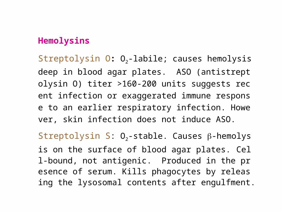
Hemolysins
Streptolysin O: O2-labile; causes hemolysis deep in blood
agar plates. ASO (antistreptolysin O) titer >160-200 units
suggests recent infection or exaggerated immune respons
e to an earlier respiratory infection. However, skin infection
does not induce ASO.
Streptolysin S: O2-stable. Causes -hemolysis on the surf
ace of blood agar plates. Cell-bound, not antigenic. Produced in the presence of serum. Kills phagocytes by releasing the lysosomal contents after engulfment.

EpidemiologyS. pyogenes can transiently colonize the oropharynx and skin.
Diseases are caused by recently acquired strains that can establish an infection of the pharynx or skin.
S. pyogenes causes pharyngitis mainly in children of 5 to 15 years old.
The pathogen is spread mainly by respiratory droplets.
Crowding increases the opportunity for the pathogen to spread, particularly during the winter months.
Soft tissue infections are preceded by skin colonization and the organisms are introduced into the superficial or deep tissue through a break in the skin.
Streptococcus pyogenes
Clinical Diseases
1. Local infection with S. pyogenes
Streptococcal sore throat (pharyngitis), and scarlet fever.
Streptococcal pyoderma (impetigo, local infection of supe
rficial layers of skin).
Strains that cause skin infections are different from those
that cause pharyngitis.
Streptococcus pyogenes
2. Invasion by S. pyogenes
Invasion from respiratory tract: otitis media, sinusitis, pneumonia, meningitis, osteomyelitis, and arthritis.
Invasion from skin: erysipelas, cellulitis, and necrotizing fasciitis. Diffuse and rapidly spreading infection that extends along lymphatic pathways with only minimal local suppuration.
Sepsis (streptococcal toxic shock syndrome, STSS): the organism is introduced into the subcutaneous tissue through a break in the skin cellulitis necrotizing fasciitis systemic toxicity, multiple organ failure, and death (mortality > 40%).

3. Poststreptococcal diseases (occurs 1-4 weeks after acut
e S. pyogenes infection, hypersensitivity responses)
Rheumatic fever: most commonly preceded by infection of the
respiratory tract. Inflammation of heart (pancarditis), joints, bloo
d vessels, and subcutaneous tissue. Results from cross reactivi
ty of anti-M protein Ab and the human heart tissue.
Acute glomerulonephritis: preceded by infection of the skin
(more commonly) or the respiratory tract. Symptoms: edema, h
ypertension, hematuria, and proteinuria. Initiated by Ag-Ab co
mplexes on the glomerular basement membrane.
* Rheumatic fever can be reactivated by recurrent streptococca
l infections, whereas nephritis does not.

S. agalactiae (group B, -hemolytic, contains type-specific capsular polysaccharides which is the most important virulence factor and can induce protective antibodies; may colonize at lower gastrointestinal tract and genitourinary tract)
Neonatal sepsis or meningitis
Early-onset (during the first week of life): infection acquired in utero or at birth. Pneumonia is common in addition to meningitis.
Late-onset (older infants): infection acquired from an exogenous source.
(Premature infants are at greater risk.)
Infection of pregnant women
Urinary tract infections, amnionitis, endometritis, and wound infections
Infection in men and nonpregnant women
Patients are generally older and have underlying conditions.
Bacteremia, pneumonia, bone and joint infections, skin and soft tissue infections. Mortality is higher.

Viridans streptococci (-hemolytic or nonhemolytic, most are nongroupable; they, except for S. suis, are divided into 5 subgroups based on the specific diseases they cause)
These streptococci colonize the oropharynx, GI tract, and GU tract; rarely on the skin surface.
Diseases:
Subacute endocarditis (group: Mitis)
Intra-abdominal infections (group: Anginosus)
Dental caries (group: Mutans)
Cariogenicity of S. mutans is related to its ability to synthesize glucan from fermentable carbohydrates (e.g. sucrose) as well as to modify glucan in promoting increased adhesiveness.
S. pneumoniae

Laboratory DiagnosisSmears: useful for soft tissue infections or pyoderma, but not for r
espiratory infections.
Antigen detection tests: commercial kits for rapid detection of group A streptococcal antigen from throat swabs.
Detection of group A streptococci by molecular methods: PCR assay for pharyngeal specimens.
Culture: Specimens are cultured on blood agar plates in air. Antibiotics may be added to inhibit growth of contaminating bacteria.
Identification: serological and biochemical tests.
Antibody detectionASO titration for respiratory infections.Anti-DNase B and antihyaluronidase titration for skin infections.Antistreptokinase; anti-M type-specific antibodies.

Identification of Gram-positive cocci
None

CAMP testChristie R, Atkins NE, and Munch-Peterson E. 1944. A note on a lytic phenomenon
shown by group B streptococci. Aust. J. Exp. Biol. Med. Sci. 22:197-200

Treatment
All S. pyogenes are sensitive to penicillin G.
Effective doses of penicillin or erythromycin for 10 days c
an prevent poststreptococcal diseases.
Drainage and aggressive surgical debridement must be p
romptly initiated in patients with serious soft tissue infecti
ons.
Group B streptococci are also susceptible to penicillin G.
Antibiotic sensitivity test is helpful for treatment of bacteri
al endocarditis.
Most streptococci are normal flora of the human body.
Source of S. pyogenes and S. agalactiae is a person harboring these organisms (carrier).
Control:
1. Prompt eradication of streptococci from early infections.
2. Prophylactic antibiotic treatment for rheumatic fever patients.
3. Eradication of S. pyogenes from carriers.
4. Dust control, ventilation, air filtration, UV irradiation and aerosol mists are of doubtful efficacy.
5. Intrapartum penicillin to mother at risk of giving birth to an infant with invasive group B disease.
Prevention and Control

S. pneumoniaeMorphology and Physiology
Gram-positive lancet-shaped diplococci for typical organisms.
-hemolytic (pneumolysin is similar to streptolysin O).
Form small round colonies on the plate, at first dome-shaped and later developing a central plateau with an elevated rim.
Autolysis is enhanced in bile salt.
Growth is enhanced by 5-10% CO2.
Capsular polysaccharide:
type-specific, 90 types.
Smooth (capsular polysaccharide-
producing) vs. rough colonies
*Quellung reaction (for rapid
identification or typing of the bacteria)

Pathogenesis and Immunity
S. pneumoniae
Pneumococci produce disease through their ability to multiply in the tissues (invasiveness). Virulence factors: capsule, cell wall polysaccharide, phosphocholine, pneumolysin, IgA protease, etc.
40-70% of humans are at sometimes carrier of virulent pneumococci. Normal respiratory tract has natural resistance to the pneumococcus. Major host defense mechanisms: ciliated cells of respiratory tract and spleen. Loss of natural resistance may be due to:
1. Abnormalities of the respiratory tract (e.g. viral RT infections).
2. Alcohol or drug intoxication; abnormal circulatory dynamics.
3. Patients undergone renal transplant; chronic renal diseases.
4. Malnutrition, general debility, sickle cell anemia, hyposplenism or splenectomy, nephrosis or complement deficiency.
5. Young children and the elderly.

Sudden onset with fever, chills and sharp chest pain. Bloody, rusty sputum. Empyema (mostly caused by type 3) is a rare but significant complication.
Complications caused by spreading of pneumococci to other org
ans: sinusitis, middle ear infection, meningitis, endocarditis, septic arthritis.
Clinical diseases
S. pneumoniae
Pneumococcal pneumonia develops when the bacteria multiply rapidly in the alveolar space after aspiration. The affected area is generally localized in the lower lobes of the lungs (lobar pneumonia). Children and the elderly can have a more generalized bronchopneumonia. Resolution occurs when specific anticapsular antibodies develop.

S. pneumoniae
Laboratory diagnosis Examination of sputum
Stained smears of sputum: a rapid diagnosis.
Quellung test with multivalent anticapsular antibodies.
Culture
Specimen: sputum, aspirates from sinus or middle ear, CSF.
cultured on blood agar plate in 5-10% CO2.
Identification: bile solubility, optochin sensitivity, etc. for differentiation from other -hemolytic streptococci. Additional biochemical, serologic or molecular diagnostic tests for a definitive identification.
Antigen detection: detect pneumococcal C polysaccharide (teichoic acid; type-specific) in urine (bacteremic) or CSF (meningitis).

S. pneumoniae
Treatment, Prevention, and Control
Healthy carriers are the source of dissemination. In the development of illness, predisposing factors are more important than exposure to the bacteria.
Vaccination of high-risk population (too old, too young, and people losing natural resistance) with vaccines containing multiple capsular polysaccharide types.
7-valent conjugate vaccine for infants <2 years.
23-valent vaccine for older children and adults.
Penicillins are the drugs of choice. However, strains resistant to penicillin and other antibiotics are common nowadays.

Enterococci(E. faecalis, E. faecium)
Form large colonies on blood plate; m
ost are nonhemolytic.
Microscopic morphology is similar to
S. pneumoniae.
Resistant to 6.5% NaCl, 0.1% methyl
blue and grow in bile-esculin agar. M
ore resistant to antibiotics than the str
eptococci.
Colonize the large intestine of human
s and animals. An opportunist.
Physiological properties are similar to the streptococci.

One of the leading causes of nosocomial infections. Urinary tract (UTI), peritoneum (peritonitis) and heart tissue (endocarditis- a severe complication) are involved most often.
Particularly common in patients with intravascular or urinary catheters, and in hospitalized patients with prolonged broad-spectrum antibiotic treatment.
Intra-abdominal abscess and wound infections: generally polymicrobial.
Many strains are completely resistant to all conventional antibiotics. Vancomycin-resistant strains have been isolated (first reported in England and France in 1987).
Enterococci can be differentiated by simple biochemical tests (e.g., resistant to optochin and bile, hydrolyze PYR, etc.)
EnterococciClinical Diseases
Laboratory Diagnosis

EnterococciTreatment, Prevention, and ControlResistance in enterococci to aminoglycosides and vancomycin is mediated by plasmids and can be transferred to other bacteria.
Combined antibiotic therapy: an aminoglycoside and a cell-wall-active antibiotic.
New antibiotics have been developed for treatment of enterococci resistant to both ampicillin and vancomycin.
It is difficult to prevent and control enterococcal infections.
Control: careful restriction of antibiotic treatment and appropriate infection-control practices (isolation of infected patients; use of gowns and gloves by anyone in contact of patients.)


M protein
Forms hair-like projections (fimbriae) from the cell membrane.
Major virulence factor of S. pyogenes.
Enhances degradation of C3b via binding with factor H, and phagocytosis by PMNs is prevented.
Promotes adherence to epithelial cells.
Induces type-specific protective immunity (>100 serotypes).
Back

ErysipelasBack

High risk population for STSS: patients with HIV infection, cancer, d
iabetes mellitus, heart or pulmonary disease, varicella-zoster virus i
nfection, and intravenous drug abusers and alcoholic.
Back
STSS

S. pneumoniae virulence factors

Back