stomach regional anatomy
DESCRIPTION
About the content of the stomachTRANSCRIPT

Dr. Nurzarina Abdul Rahman Faculty of Medicine and Health Sciences
USIM
بسم اهللا الرحمن الرحيم
¨ At the end of the lecture, the student should be able to:
¨ describe the surface marking of the stomach ¨ identify the parts of the stomach and name of its borders ¨ describe the relation of the stomach to the peritoneal
cavity and the name of the peritoneal folds ligament attach to the organ
¨ describe the relation of the stomach to the other organs including the structures present at the stomach bed
¨ mention the blood supply of the stomach

¨ Muscular bag forming the widest and most distensible part of digestive tract
¨ Connected above to lower end of the oesophagus and below to duodenum
¨ Function – reservoir of food and prepares food for digestion

¨ Developmentally related to the foregut
¨ Position : upper left quadrant - epigastrium, left hypochondrium and umbilical region
¨ Lies under cover of left costal margin and ribs
¨ Shape : J-shape (empty) or pyriform (partially distended)
¨ Capacity : 1000-1500 ml

¨ 25 cm long
¨ Relatively fixed at both ends
¨ Upper end ¡ cardio-oesophageal
junction ¡ left of mid line (T10) ¡ 40 cm from incisor teeth
¨ Lower end ¡ pylorus ¡ right of the mid line (L1)
L1
Midline of the abdomen
T10

4 parts ¡ Cardia ¡ Fundus ¡ Body ¡ Pylorus
Surfaces and borders ¨ 2 ends : cardiac end and
pyloric end ¨ 2 borders : right lesser
curvature and left greater curvature
¨ 2 surfaces : anterior and posterior
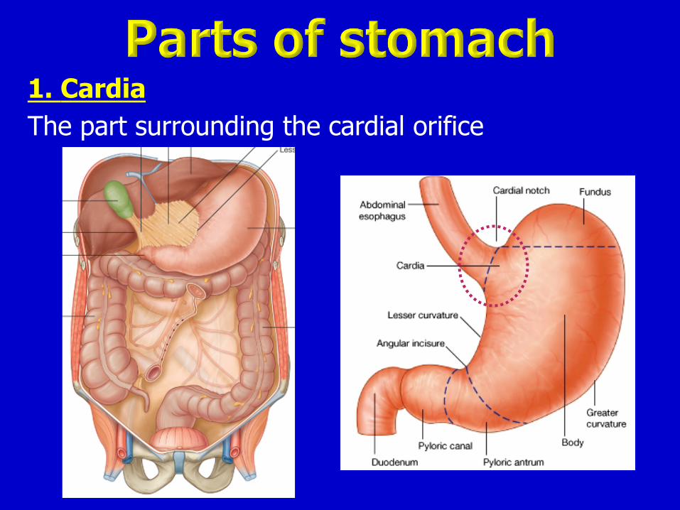
1. Cardia The part surrounding the cardial orifice
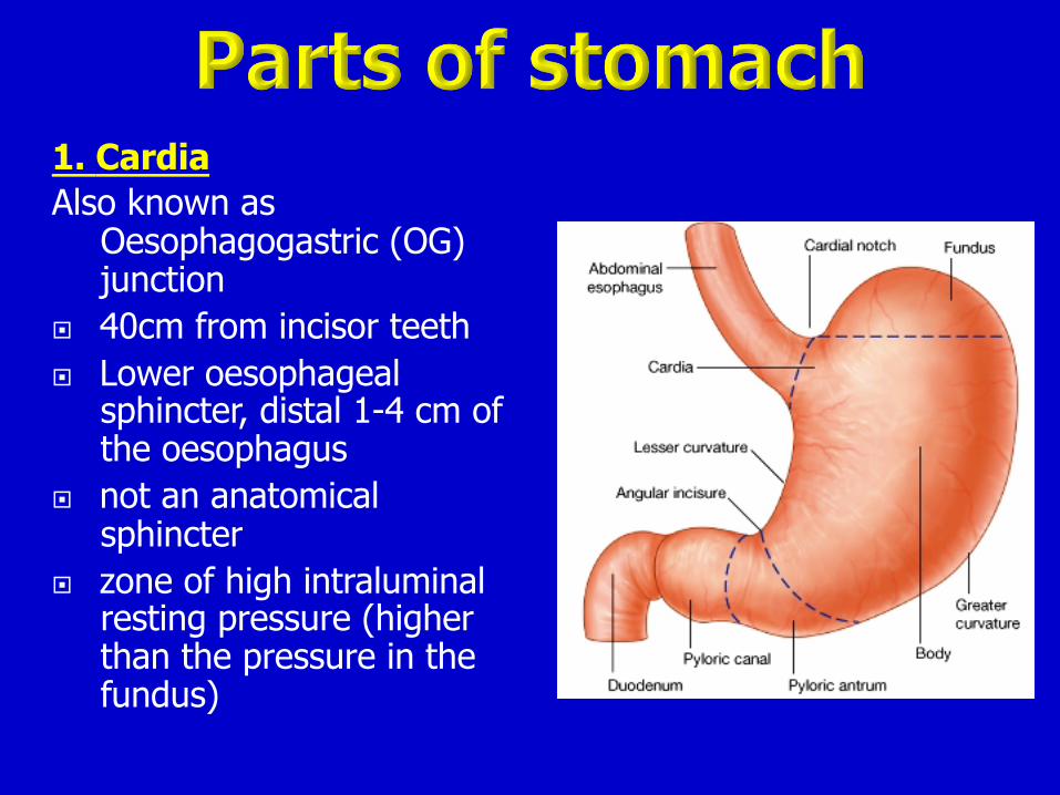
1. Cardia Also known as
Oesophagogastric (OG) junction
¨ 40cm from incisor teeth ¨ Lower oesophageal
sphincter, distal 1-4 cm of the oesophagus
¨ not an anatomical sphincter
¨ zone of high intraluminal resting pressure (higher than the pressure in the fundus)

2. Fundus Dilated superior part, related to left dome of diaphragm Above level of cardia Superior part reaches level of left 5th Intercostal space Cardial notch – between esophagus and fundus

3. Body Major part between fundus
and pylorus From fundus to incisura
angularis Incisura angularis
separates body from the pyloric part
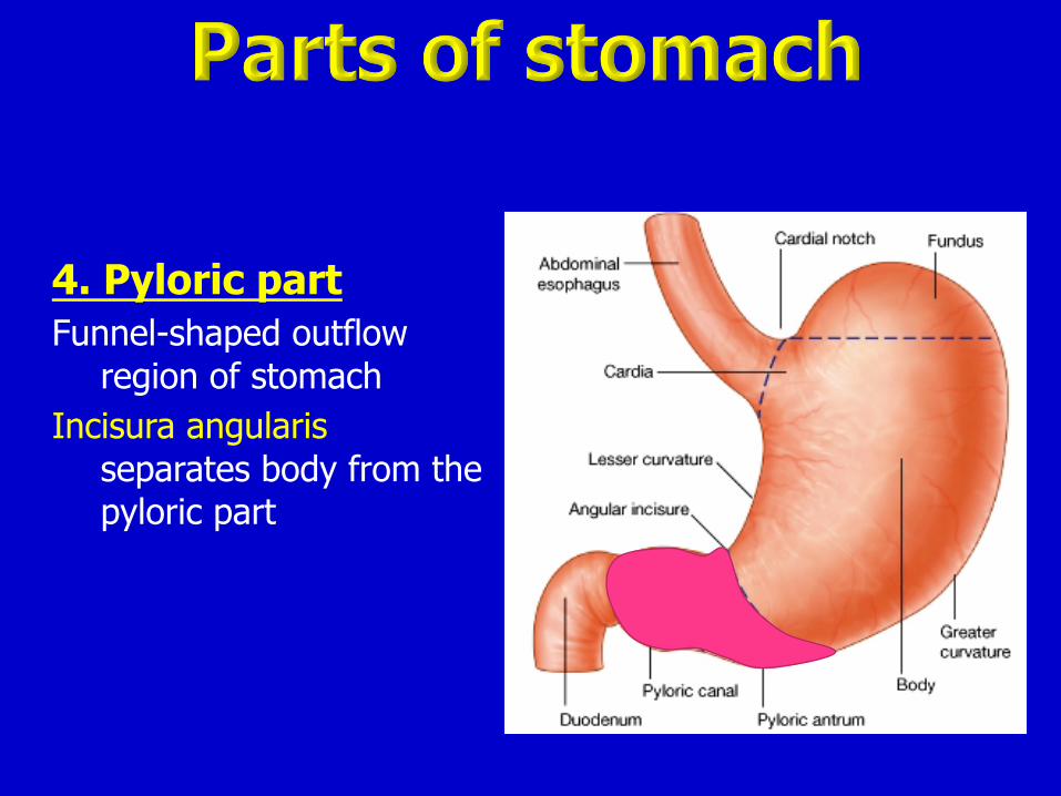
4. Pyloric part Funnel-shaped outflow
region of stomach Incisura angularis
separates body from the pyloric part

4. Pyloric part (pyloros = gateguard) Three parts:
Pyloric part
Pyloric antrum Pyloric canal
Pyloric sphinter Duodenum
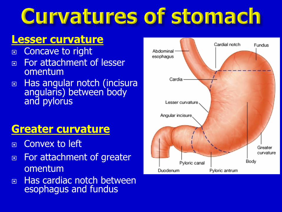
Lesser curvature ¨ Concave to right ¨ For attachment of lesser
omentum ¨ Has angular notch (incisura
angularis) between body and pylorus
Greater curvature ¨ Convex to left ¨ For attachment of greater
omentum ¨ Has cardiac notch between
esophagus and fundus

Stomach is completely covered by peritoneum
(intraperitoneum organ) .

Area where the stomach is not covered with peritoneum :
¨ A bare area at the back of cardial orifice
¨ Where blood vessels run along its curvatures
Stomach has peritoneal folds: ¨ lesser omentum ¨ greater omentum ¨ gastrophrenic ligaments ¨ gastro- splenic ligaments

Gastrophrenic ligament ¨ from the fundus to the
diaphragm Gastrosplenic ligament ¨ from the greater curvature to the hilum of spleen Greater omentum ¨ from the greater curvature
to the transverse colon (gastro-colic ligament) Lesser omentum ¨ from lesser curvature to
the liver porta hepatis and fissure for ligamentum venosum
(gastro-hepatic ligament)

Anteriorly related to: ¨ diaphragm ¨ left lobe of the liver ¨ anterior abdominal
wall
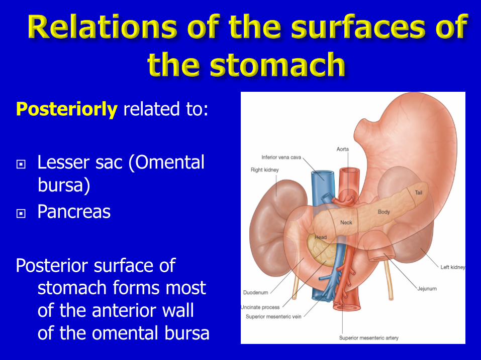
Posteriorly related to: ¨ Lesser sac (Omental
bursa) ¨ Pancreas Posterior surface of
stomach forms most of the anterior wall of the omental bursa
Anterior
Posterior

Posteriorly related to: ¨ Lesser sac (Omental
bursa) ¨ Pancreas Posterior surface of
stomach forms most of the anterior wall of the omental bursa
Anterior
Posterior

¨ Definition : the bed on which the stomach rests on SUPINE position
¨ Structures at the stomach bed form the posterior wall of Lesser sac (omental bursa)
¨ Structures at the stomach bed THAT are separated from the stomach by the cavity of lesser sac

¨ Structures at the stomach bed THAT are separated from the stomach by the cavity of lesser sac:
(From superior to inferior) 1. left crus of diaphram - related to the bare area at the
back of the cardiac end of the stomach 2. spleen – separated from stomach by cavity of greater
sac 3. left suprarenal gland 4. left kidney 5. splenic artery along upper border of PANCREAS 6. pancreas 7. left colic flexure 8. transverse mesocolon and colon


From coeliac artery A) along lesser curvature -
two arteries 1. left gastric artery and 2. right gastric artery B) along the greater
curvature - three arteries 1. short gastric arteries from
splenic artery to fundus 2. left gastro-epiploic artery
(from the splenic artery) 3. right gastro-epiploic artery
(from the branches of hepatic artery)

¨ the veins accompany the arteries
¨ empty into portal circulation
B A
C
D
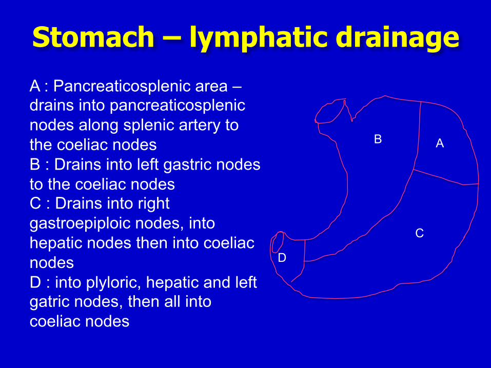
Stomach – lymphatic drainage
A : Pancreaticosplenic area – drains into pancreaticosplenic nodes along splenic artery to the coeliac nodes B : Drains into left gastric nodes to the coeliac nodes C : Drains into right gastroepiploic nodes, into hepatic nodes then into coeliac nodes D : into plyloric, hepatic and left gatric nodes, then all into coeliac nodes


¨ anterior ulcers perforate ¨ posterior ulcer erode (GIT bleeding) or
penetrate ¡ gastric ulcer erodes pancreas, splenic artery

Protrusion of part of stomach (cardia and part of fundus) into the mediastinum through the oesophageal hiatus of the diaphragm
Occurs in people after middle age (due to weakening of the muscular part of diaphragm)