stimulation of cadmium uptake by estradiol in the kidney of male rats treated with cadmium
TRANSCRIPT
Biochemical Pharmacoiogy. Vol. 37. NO. 16, pp. 3091-3096, 1988 Printed in Great Britain.
@X6-295&‘88 S3.00 + 0.M) @1988. Pergamon press plc
STIMULATION OF CADMIUM UPTAKE BY ESTRADIOL IN THE KIDNEY OF MALE RATS TREATED WITH CADMIUM
SHOJI NISHIYAMA,*~ SATOMI ONOSAKA,+ TAKAHIRO TAGUCHI,~ YUUKO KONISHI,(J KEIICHI TANAKA* and HIDEO KINEBUCHI~
Department of t ~n~ronment~ and Occupational Health, 8 Anatomy and 11 Medical Research Laboratory, Kochi Medical School, Nankoku, Kochi 781-51, Japan, and $ Faculty of Nutrition,
Kobe-Gakuin University, Ikawadani, Nishiku, Kobe 673, Japan
(Received 11 January 1988; accepted 26 February 1988)
Abstract-The present study was carried out to analyze the sex differences in the retention of Cd in rats treated with a small amount of Cd, and its mechanisms. Cd and Zn concentrations in the kidney and liver of female rats treated with 28 nmoi Cd or 1 nmole Zn were significantly higher than those in mafe rats. Pretreatment with estradiol (1.8 ,umol/kg of b.w., twice a day, 6 consecutive days) increased the Cd and Zn con~ntrations in the kidney of male rats treated with Cd or Zn. Incubation of MDCK cells with 10e5 M estradiol, IO+ M stilboestrol and lO+ M progesterone caused a significant increase in Cd uptake. These results suggest that endogenous female sex hormones may play a role in a higher concentration of Cd and Zn in the kidney of female rats than that in male rats. The basal level of metallothionein (MT) in the liver and kidney of control female rats was within the same range as that in the control male rats. Cd and Zn accumulations caused by pretreatment with estradiol in the kidney of male rats treated with Cd or Zn were so low (cd: 38 ppb, Zn: 1.0 ppb) as to be probably unable to induce the synthesis of MT. An increase in the concentration of Cd in the cultured renal cells occurred 1 hr after treatment with estradiol and Cd. Pretreatment with estradiol alone also resulted in a m~fi~tion of the concentration of Na and K, which cannot be bound to MT. Together, all of the above findings suggest that estradiol directly increases the a~cum~ation of Cd into the renal cells without inducing the synthesis of MT.
Japanese women accumulate larger amounts of cad- mium (Cd) than men 111. In experimental animals, it has been shown that the renal and hepatic con- centrations of Cd are higher in female rats than in male rats after long-term oral admi~stration of Cd [2]. The biological half-life of Cd2+ in the liver is about 1.3-1.4 times longer in the female rat than in the male rat. However, the factors and the mech- anisms causing the sex differences in Cd metabolism remain to be determined.
The major part of Cd accumulated in the body is found in the liver and kidney and, in these organs, more than 80% of Cd is bound to the metallo- protein, metallot~onein (MT) [3]. Currently it seems to be accepted that MT plays a major role in homeostasis of metals [3]. Recently we have reported that a female sex hormone, estradiol,fi affects the biosynthesis of zinc-thionein (Zn-MT) [4]. We also demonstrated that pretreatment with estradiol increases the MT concentration in the kidney and liver of male rats treated with Cd, though tes- tosterone could not increase the MT levels [5]. A positive correfation between dose-related increases in hepatic MT concentration and Cd2+ or Zn2+ has been observed [6]. These findings suggest that female
* To whom all correspondence should be addressed. 1 Abbreviations used: estradiol, 1,3,5(10)-estratriene-
3,17,3-diol; estrone, 1,3,5-estratrien-3-ol-17-one; stilbo- estrol, (E)~,4’-(~,2~iethyl-l,Z-ethene~yl)bisphenol; tes- tosterone, ~7~hydro~androst4~n-3-one; progesterone, p~gn-4-ene-3,20-dione; Dbc-AMP, ~but~l-~clic AMP.
sex hormones can induce the synthesis of MT in the liver and kidney, which in turn results in a higher concentration of MT and retention of Cd2+ in female rats than in male rats.
The present study was carried out to analyze the sex differences in the retention of Cd in rats treated with a small amount of Cd, and its mechanisms. The data presented in this report suggest that estradiol directly influences the accumulation of Cd into the renal cells without the need for increased synthesis of MT. Endogenous female hormones may play a role in a higher concentration of Cd and Zn in the kidney of the female rats than in the male rats.
MATERIALS AND METHODS
Animals. Twelve-week-old male and female STD- Wistar rats, weighing 250 g and 180 g respectively on average, were purchased from the Shizuoka Labora- tory Animal Center and maintained on a commercial diet (Clea Co., Tokyo CE-2). They received water ad iibit~~. They were housed in a temperature- and light-controlled room as previously reported [7]_
Cd and Zn injection in male and female rats. Twenty-one male and female rats were divided into two groups and received an intravenous injection of CdC12 at a dose of 28 nmol/kg body weight (b.w.). The injected solution of Cd (0.1 ml) contained labeled 109CdC12 at a final concentration of 50 @i/ ml (specific activity 2.6mCi/mg, New England Nuclear). The injection schedule of 65ZnClz was the same as that described above for treatment with
3091
3092 S. NISHIYAMA et al.
CdQ. The injected solution contained 0St_1Ci of 65ZnC12 in a volume of 0.1 ml (specific activity 7.5 mCi/mg, New England Nuclear). The total dose of Zn equalled 1 nmole of Zn2+. Twenty-one male and female rats placed in metabolic cages were sac- rificed at 4 (N = 7), 10 (N = 7) and 24 hr (N = 7) after treatment. The animals were not starved over- night before being sacrificed. Urine was collected over a 24-hr period. The liver, kidney, brain, heart, lung, spleen, pancreas and serum of the male and female rats treated with Cd or Zn were removed and vveighed. Tissues were directly analyzed by y- counting.
Cd and estrudiol injection. Thirty male rats were divided into six groups: control rats (group l), rats treated with Cd (group 2), estradiol-treated rats (group 3), rats treated with Cd and estradiol (group 4), Zn-treated rats (group 5) and rats treated with Zn and estradiol (group 6). The rats of groups 2 (N = 5) and 4 (N = 5) respectively received an intra- venous injection of CdCl* at a dose of 28nmol/kg of body weight (b.w.), after pretreatment with 40% ethanol ( 1.8 ,umol P
roup 2) or estradiol (group 4) at a dose of kg b.w. subcutaneously twice a day for six
consecutive days. The rats of group 2 were predosed with 40% ethanol because estradiol was dissolved in 40% ethanol. The rats of group 1 (N = 5) and 3 (N = 5) respectively received an injection of 0.1 ml of physiological saline, after pretreatment with 40% ethanol (group 1) or estradiol (group 3) at a dose of 1.8 mol/kg b.w. subcutaneously twice a day for six consecutive days. The rats of groups 5 (N = 5) and 6 (N = 5) respectively received an intravenous injec- tion of 0.5 PCi of 65ZnClz (1 nmole as Zn2+), after pretreatment with 40% ethanol (group 5) or estradiol (group 6) at a dose of 1.8hmol/kg b.w. sub- cutaneously twice a day for six consecutive days. After 24 hr of treatment with Cd or Zn the rats placed in a metabolic cage were sacrificed. The injected solution of Cd (0.1 ml) or Zn (0.1 ml) contained labeled ro9CdC12 or @ZnC12 at a final concentration of 5O,&i/ml and 5 @&‘ml respectively (specific activity 26 mCi/mg of Cd, 7.5 mCi/mg of Zn: New England Nuclear). The animals were not starved overnight before being sacrificed. They were quickly decapitated between 0900 and 1100 hr.
C~romat~gra~h~~ procedure. The liver and whole kidney stored at -80” were homogenized in ice-cold 20 mM Tris-HCl buffer (pH 8.0) by bubbling with Nz gas. The supernatant was obtained by centrifuging at 105,OOOg for 60 min as previously reported [4]. The supernatant fraction was quickly applied to a column (2.6 x 100 cm) packed with Sephadex G-75 gel. The sample was eluted from the column with 20 mM Tris-HCI (pH 8.0) at a rate of 36 ml/hr. A fraction (3ml) was collected and analyzed by y- counting.
Assay of metallothionein. The livers and kidneys from the control and treated rats of both sexes were removed and frozen quickly in iiquid Nz. MT con- centration in the kidney and liver was determined by the Cd saturation-hemolysate(Cd-hem) method as previously reported [8].
Metal concentrations in the kidney, liver and heart. The kidney and liver of the rats treated with estradiol alone or 40% ethanol were removed on day 7,
weighed and frozen quickly in liquid Nz. The Na, K and Zn levels in the kidney and liver were assayed with atomic absorption spectrophotometry as pre- viously reported [9],
Culture of Madly-Dar~y canine kidney ~~D~~) ceils. MDCK (NBL-2) cells were obtained from Flow Lab. Inc (ATCC No. Ccl-34). Upon reaching con- fluency, the cells were incubated in a culture dish (multi-well plate 6 wells, 30mm) containing Dulbecco’s modified Eagle medium (DMEM) sup- plemented with 10% fetal calf serum in a humidified 5% CO,/95% air mixture at 37”.
Hormones and Cd treatment in MDCK celis, The cultured cells were washed with serum-free medium prior to treatment with hormones. The test hormones were added to serum-free fresh medium containing 5 @i/ml of 109CdC12 (specific activity 2.3 mCi/mg) or 0.5 @i/ml of 65ZnCl, (specific activity, 7.5 mCi/ mg) and the cells were incubated for 1-24 hr. The cells were harvested, washed with ice-cold 20mM Tris-HCl buffer (pH 8.0) containing 10e6 M CdClr or 10m6 M ZnClz at the final concentration and homo- genized in the same buffer without addition of CdClr or ZnClz. Ethanol (0.1%) was added to control medium at the final concentration because the test hormones except for Dbc-AMP were dissolved in 0.1% ethanol. The morphology of the cultured cells treated with the metals and hormones was observed by phase-contrast microscopy.
E ect ,&i P
of estrudiol on effiux of Cd. Five mllo9 CdCl* was added to the fresh serum free
medium and the cultures were incubated for 4 hr, washed briefly with fresh serum-free medium, and incubated in medium with or without 10FSM estradiol for 30 min to 24 hr. Medium with or without 10e5 M estradiol and cells were harvested and ana- lyzed by y-counting.
Statistical anu~ys~. The data were evaluated by Student’s t-test.
RESULTS
Cd and Zn concentrations in the organs
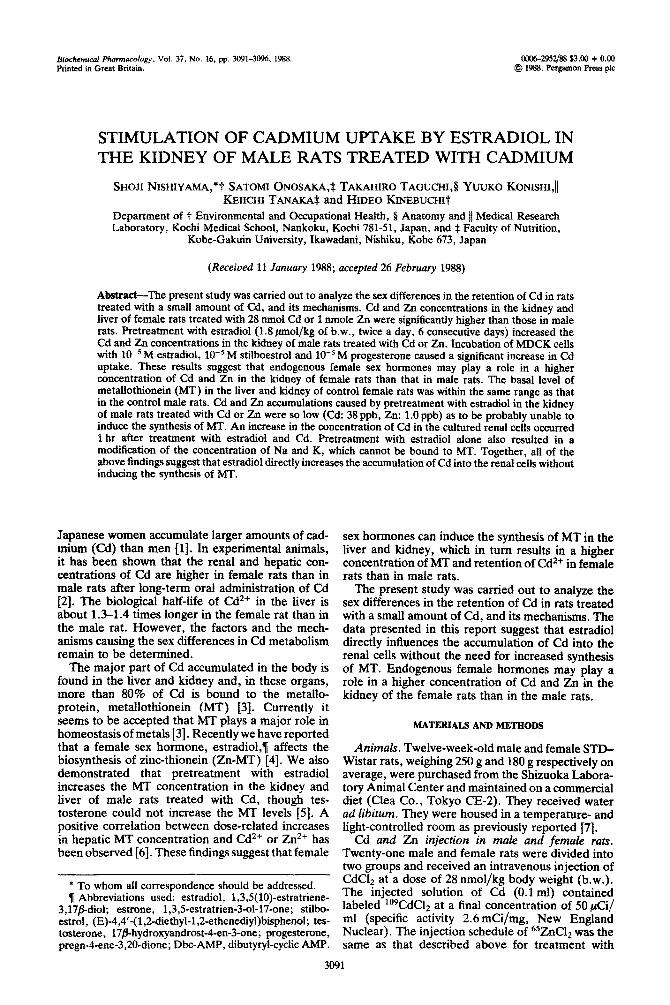
Cd concentration in the liver of the female rats treated with 28 nmol Cd was significantly higher than that in the male rats after 4 and 10 hr, but returned to the levels of the male rats up to 24 hr (Fig. la). Cd concentration in the kidney and pancreas of the female rats treated with Cd was also significantly higher than that in the male rats after 10 hr. The pattern of Zn uptake in the liver and kidney of the female rats was simitar to that of the Cd uptake (Fig. lb). The gel filtration profile of the supernatant of the liver from the female and male rats treated with Cd at 4 hr showed a large peak of radioactivity cor- responding to the peak of Cd-thionein (Cd-MT) (Fig. 2). Cd-MT concentration in the liver of the female rats 4 hr after the Cd treatment was sig- nificantly higher than the corresponding values in the male rats (Fig. 2). No significant difference in the basal level of MT in the kidney and liver between the control female and male rats was observed (Fig. 3).
Stimulation of cadmium uptake by estradiol in kidney of male rats 3093
(a)
4 10 24
Hour
tb)
4 10 24
Hour
Fig. l(a) Cd concentration in the hver, kidney and pancreas of male and female rats treated with Cd. The symbols represent the Cd concentration in the liver of male (0) and female rats (O), kidney of male (0) and-female rats (m), pancreas of male (A) and female rats (A) treated with Cd. Significantly different from the values in male rats treated with Cd at P < 0.01 (*). Each value is expressed as ng Cd per g of wet tissue and X rt SE of seven animals. Male and female rats received an intravenous injection of CdClz at a dose of 28 mnol per kg body weight. (b) Zn concentration in the liver and kidney of male and female rats treated with Zn. The symbols represent the Zn concentration in liver of male (0) and female rats (O), kidney of male (A) and female rats (A) treated with 65ZnClz (1 nmol as Zt?). Significantly different from the values in male rats treated with Zn at P < 0.01 (*). Each value is expressed as ng Zn
per g of wet tissue and X f SE of seven animals.
6.0
k ; P I.8 2.0
li;
0 10 20 40
Fraction Number
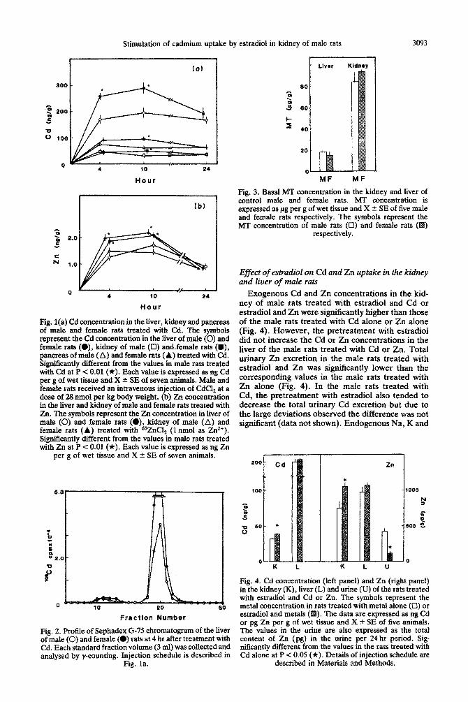
Fig. 2. Profile of Sephadex G-75 chromatogram of the liver of male (0) and female (0) rats at 4 hr after treatment with Cd. Each standard fraction volume (3 ml) was collected and analysed by y-counting. Injection schedule is described in
Fig. la.
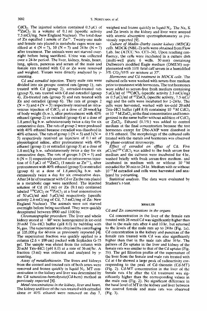
MF MF
Fig. 3. Basal MT concentration in the kidney and liver of control male and female rats. MT concentration is expressed as fig per g of wet tissue and X & SE of five male and female rats respectively. The symbols represent the MT concentration of male rata (0) and female rats (a)
respectively.
Effect of estrudi~l on Cd and Zn uptake in the kidney and liver of male ruts
Exogenous Cd and Zn concentrations in the kid- ney of male rats treated with estradiol and Cd or estradiol and 2% were significantly higher than those of the male rats treated with Cd alone or Zn alone (Fig. 4). However, the pretreatment with estradioi did not increase the Cd or Zn concentrations in the liver of the male rats treated with Cd or Zn. Total urinary Zn excretion in the male rats treated with estradioi and Zn was significantly lower than the co~esponding values in the male rats treated with Zn alone (Fig. 4). In the male rats treated with Cd, the pretreatment with estradiol also tended to decrease the total urinary Cd excretion but due to the large deviations observed the difference was not significant (data not shown). Endogenous Na, K and
0 K L
0 K L U
Fig. 4. Cd ~ncen~ation (left panel) and Zn (right paneI) in the kidney(K), liver(L) and urine (U) of the rats treated with estradiol and Cd or Zn. The symbols represent the metal concentration in rats treated with metal alone (U) or estradiol and metals (a). The data are expressed as ng Cd or pg Zn per g of wet tissue and X * SE of five animals. The values in the urine are also expressed as the total content of Zn (pg) in the urine per 24 hr period. Sig nificantly different from the values in the rats treated with Cd alone at P < 0.05 (*). Details of injection schedule are
described in Materials and Methods.
3094 S. NZSHIYAMA et al.
Table 1. Concentration of sodium (Na), potassium (K), zinc (Zn) and calcium (Ca) in the kidney and liver of male
rats treated with estradiol
Kidney Liver
Control Estradiol
Na
K
1.57 + 0.05 0.67 t 0.01 1.76 t 0.04* o&4 r 0.03+
Control Estradiol
3.19 f 0.09 3.69 t 0.03 2.85 2 0.06* 4.10 + 0.10*
Zn Control 17.5 + 0.6 31.0 f 0.6 Estradiol 20.5 * 0.2’ 36.6 ? 1.9”
The data expressed as ng (Zn) or mg (Na,K) per g of wet tissue and X t SE of five animafs.
* Significantly different from the control values at P < 0.05. The control or estradiol-treated rats received an injection of 0.1 ml of physiolo~cal saline after pre- treatment with 40% ethanol or e&radio1 at a dose of 1.8 pmole/kg b.w. subcutaneously twice a day for six con- secutive days.
Zn concentrations in the kidney and liver of the male rats treated with estradiol alone were significantly higher than those in the control male rats except for the K concentration in the kidney (Tabte 1).
Effect of hormones on Cd uptake in MDCK cells
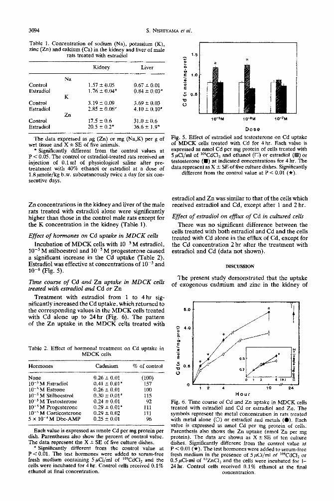
Incubation of MDCK cells with 10ms M estradiol, 10m5 M stilboestrol and 10Vs M progesterone caused a significant increase in the Cd uptake (Table 2). Estradiol was effective at concentrations of 10F5 and low6 (Fig. 5).
Time course of Cd and Zn uptake in MDCK cells treated with estradiol and Cd or Zn
Treatment with estradiol from 1 to 4 hr sig- nificantly increased the Cd uptake, which returned to the corresponding values in the MDCK cells treated with Cd alone up to 24 hr (Fig. 6). The pattern of the Zn uptake in the MDCK cells treated with
Table 2. Effect of hormonaf treatment on Cd uptake in MDCK cells
Hormones Cadmium % of control
None 0.26 t 0.01 10e5 M Estradiol 0.41 a 0.01* ‘:!? lows M Estrone 0.26 -e 0.01 100 10e5 M Stilboestrol 0.30 t 0.01* 115 10m5 M Testosterone 0.24 ‘- 0.01 92 10m5 M Progesterone 0.29 r O.Ol* 111 10s5 M Corticosterone 0.29 ” 0.02 111 5 x W4 M Dbc-AMP 0.25 ? 0.01 96
Each value is expressed as nmole Cd per mg protein per dish. Parentheses also show the percent of control value. The data represent the X -+ SE of five culture dishes.
* Significantly different from the control value at P < 0.01. The test hormones were added to serum-free fresh medium containing S&i/ml of i09CdC12 and the cells were incubated for 4 hr. Control cells received 0.1% ethanol at final concentration.
lo-5u 1 o-%I
Dose
1 o-%4
Fig. 5. Effect of estradioi and testosterone on Cd uptake of MDCK cells treated with Cd for 4 hr. Each value is expressed as nmol Cd per mg protein of cells treated with 5 &i/ml of ‘WdC12 and ethanol (0) or estradiol (B) or testosterone (a) at indicated concentrations for 4 hr. The data represent as X t SE of five culture dishes. Significantly
different from the control value at P < 0.01 (*).
estradiol and Zn was similar to that of the cells which received estradiol and Cd, except after 1 and 2 hr.
Effect of es~adiol on ebb of Cd in cu~~red cells
There was no significant difference between the cells treated with both estradiol and Cd and the cells treated with Cd alone in the efflux of Cd, except for the Cd con~ntration 2 hr after the treatment with estradiol and Cd (data not shown).
DISCUSSION
The present study demonstrated that the uptake of exogenous cadmium and zinc in the kidney of
6*o I,.----:
0 1 2 4 10 24
Hour
Fig. 6. Time course of Cd and Zn uptake in MDCK cells treated with estradiol and Cd or estradiol and Zn. The symbols represent the metal concentration in rats treated with metal alone (0) or estradiol and metals (0). Each vahte is expressed as nmol Cd per mg protein of cells. Parenthesis also shows the Zn uptake (nmol Zn per mg protein). The data are shown as X + SE of ten culture dishes. Significantly different from the control value at P < 0.01 (*). The test hormones were added to serum-free fresh medium in the presence of 5 &i/ml of ‘09CdC12 or 0.5 &i-ml of 65ZnC12 and the cells were incubated for l- 24 hr. Control cells received 0.1% ethanol at the final
concentration.
Stimulation of cadmium uptake by estradiol in kidney of male rats 3095
female rats which received a small amount of Cd or Zn was higher than that in male rats. Some workers have reported that renal and hepatic concentrations of Cd are higher in the female rats than in the male rats after both a single and long-term oral administration of Cd [Z, lo]. According to the for- mula A = a,&(1 - epb3, the biological half life of Cd2+ in the liver is about 1.3-1.4 times longer in female rats than in male rats [2]. Several mechanisms have been proposed for the observed sex differences. Kello et al. [lo] have suggested that the sex dif- ferences in the retention of Cd are presumably caused by changes in the absorption of the metals, since the elimination rate of Cd six days after its oral ad~nistration (when all exogenous fecal Cd has been excreted) seems to be inde~ndent of ho~onal factors. In the present study, the stimulation of Cd and Zn uptake by treatment with estradiol in the kidney in viva and in cultured renal cells suggests that estradiol directly stimulates the uptake of Cd and Zn in the renal cells. However, there was no evidence of the stimulation of Cd and Zn uptake by the treatment with a male sex hormone, testosterone, in the kidney in uivo and in cultured renal cells. Therefore, endogenous female sex hormones may play a role in a higher concentration of Cd and Zn in the kidney of the female rats than that in the male rats. However, the mechanisms by which the treatment with estradiol stimulated the uptake of the Cd and Zn in the kidney remain to be determined.
It is currently recognized that MT plays a major role in the homeostasis of the essential metals 131. Cousins have suggested that the Zn metabolism is regulated by the synthesis of intestinal and hepatic MT which controls the eftlux of the cation from the mucosal cells and its uptake from the blood by the liver respectively [ll]. In the present study, the basal level of MT, presumably in the ZnMT form in the liver and kidney of control female rats, was within the same range as that in the control male rats. However, when a small amount of Cd was injected into the female and male rats, the concentrations of Cd2+ and Cd-MT in the liver of the female rats were higher than those in the male rats, and all of the increased concentra~on of Cd2+ was bound to MT. We have already reported that a female sex hormone, estradiol, can induce the synthesis of Zn- MT in the kidney and liver of male rats [4]. The Znz+ and Cd*+ concentrations in the liver of animals treated with Zn or Cd at an appropriate dose increase with a concomitant increase in hepatic MT [6]. Therefore, we postulate that the Zn-MT synthesis in the kidney was stimulated by the pretreatment with estradiol which facilitates the uptake of Cd and Zn into the renal cells. It is well known that a critical concentration of approximately 3 ppm for Cd and 30ppm for Zn for 2 hr at least are required for the initiation of the synthesis of MT in the liver of the rats in response to the injection of an appropriate dose of Cd or Zn [12,13]. In the present study, Cd and Zn accumulations caused by pretreatment with estradiol in the kidney of male rats treated with Cd and Zn were so low (Cd: 38 ppb, Zn: 1.0 ppb) that they were probably unable to induce the synthesis of MT. The uptake of Cd in the cultured renal cells 1 hr after the treatment with estradiol and Cd significantly
increased though a modification of the effhrx of Cd was not observed, suggesting that the stimulation of Cd uptake occurred prior to the initiation of the synthesis of MT. The pretreatment with estradiol alone also resulted in a modification of the con- centration of Na and K, which cannot be bound to MT. Together, all of the above findings suggest that the synthesis of MT in the kidney which was stimu- lated by the pretreatment with estradiol did not facilitate the uptake of Cd and Zn into the renal cells. Estradiol may directly increase the accumulation of Cd into the renal cells without inducing the synthesis of MT. It is noteworthy that the uptake of 64Cu by rat hepatocytes is not increased when the MT levels are increased by injection of Zn into the rats 1141. There are now arguments against MTplaying a major role in the uptake of the essential metals in the liver and intestine [3].
The data presented in this report suggest that estradiol directly influences the accumulation of Cd into the renal cells without the need for increased synthesis of MT. Endogenous female sex hormones may play a role in higher concentrations of Cd and Zn in the kidney of female rats than those in male rats.
Ac&~ow~e~ge~e~~-We thank Professor Hiroshi Saito of Nagasaki University for his advice regarding the sex dif- ferences in cadmium pollution. We also thank Professor Akihiko Irimajiri of Kochi Medical School for supplying MDCK cells. We thank Dr Jacqueline Bartman of Tropical Agriculture Research Center for correcting the manuscript. This study was supported in part by a grant from the Ministry of Education, Science and Culture of Japan (No. 61770359).
RlWERENCEs
1. Sumino K, Hayakawa K, Shibata T and Kitamura S, Heavy metals in normal Japanese tissues. Arch Environ Health 30: 487-494, 1975.
2. Stonard MD and Webb M, Influence of dietary cad- mium on the dist~bution of the essential metals copper, zinc and iron in tissue of the rat. C~em-RioI Interact 15: 349-363, 1976.
3. Webb M and Cain K, Functions of met~o~ionein. Biochem F~ur~co~ 31: 137-142.1982.
4. Nishiyama S, Taguchi T and Onosaka S, Induction of zinc-thionein by estradiol and protective effects on inorganic mercury-induced renai toxicity. Biochem Pharmacol36: 3387-3391, 1987.
5. Nishiy~a S, Taguchi T and Onosaka S, Protective effects of estradiol on mercu~-induced renal toxicitv in castrated male rats. Jpn J t&g 42: 141, 1987. *
6. Probst GS, Bousauet WF and Miva T. Correlation . ‘
of hepatic ‘metalldthionein concentrations with acute cadmium toxicity in the mouse. Toxic01 Appl Phar- macof 39: 61-49, 1977.
7. Nishiyama S and Nakamura K, Stimulation of adrenal DNA synthesis in cadmium-treated male rats. Toxicol Appl Pharmacol74: 33’7-344, 1984.
8. Onosaka S and Cherian G, The induced synthesis of met~lo~ionein in various tissues of rat in response to metals. I. Effect of repeated injection of cadmium salts. Toxicology 22: 91-101, 1981.
9. Nishiyama S, Nakamura K and Konishi Y, Effect of selenium on blood pressure, urinary sodium excretion and plasma aldosterone in cadmium-treated male rats. Arch Toxicol59: 36.5-370, 1987.
3096 S. NISHIYAMA et al.
10. Kello M, Dekanic D and Kostrial K, Influence of sex 13. Squibb KS and Cousins RJ, Control of cadmium bind- and dietary calcium on intestinal cadmium absorption ing protein synthesis in rat liver. Environ Physiol Bio- in rats. Arch Environ Health 34: 30-33, 1979. them 4: 24-30, 1974.
11. Cousins RJ, Regulatory aspects of zinc metabolism in 14. Bremner I and Davies NT, Studies on the appearance liver and intestine. Nutr Reu 37: 97-103, 1979. of a hepatic copper-binding protein in normal and zinc-
12. Kobayashi S, Imano M and Kimura M, Turnover deficient rats. Br J Nutr 36: 101-112, 1976. metallothionein in mammalian cells. In: Biological Roles of Metallothionein (Ed. Foulkes), pp. 305-322. Elsevier, New York, 1982.