estradiol valerate in sprague dawley rats. development of
TRANSCRIPT

Page 1/24
Effect of 150 kHz Electromagnetic Radiation on theDevelopment of Polycystic Ovaries Induced byEstradiol Valerate in Sprague Dawley Rats.Stephanie Mohammed ( [email protected] )
The University of the West Indies Saint Augustine Campus: The University of the West Indies at StAugustine https://orcid.org/0000-0002-3458-3991Venkatesan Sundaram
The University of the West Indies Saint Augustine Campus: The University of the West Indies at StAugustineNikolay Zyuzikov
The University of the West Indies
Research
Keywords: 150 kHz Electromagnetic Radiation, Estradiol Valerate, Polycystic Ovary Model
Posted Date: December 11th, 2020
DOI: https://doi.org/10.21203/rs.3.rs-125234/v1
License: This work is licensed under a Creative Commons Attribution 4.0 International License. Read Full License
Version of Record: A version of this preprint was published on February 5th, 2021. See the publishedversion at https://doi.org/10.1186/s13048-021-00774-4.

Page 2/24
AbstractBackground: Polycystic ovary syndrome (PCOS) is the most common complex endocrine disorderaffecting approximately 2-20% of reproductive aged females. Tumour Treating Fields (100-300 kHz) is arecent innovative, non-invasive therapeutic approach to cancer therapy. This frequency as an alternativetherapy for the management of polycystic ovaries has not yet been explored.
Objectives: To explore the effect of full-body exposure of 150 kHz Electromagnetic Radiation (EMR), onthe development of polycystic ovaries in an estradiol valerate-induced PCOs rats’ model.
Method: Twenty-one female adult rats were divided into three groups (n=7 each): control, EstradiolValerate (EV) and EV + EMR groups. The EV + EMR group was subjected to full body exposure at 150 kHzEMR continuously for eight consecutive weeks. Estradiol valerate was administered orally to inducepolycystic ovaries in EV and EV+EMR groups. Body and ovarian weights were recorded and analysed. Theregularity of the estrous cycle was assessed in all three groups. The histological study of ovarian tissuewas carried out by haematoxylin and eosin staining. The serum concentration levels of LuteinizingHormone (LH), Follicle-Stimulating Hormone (FSH) and testosterone were measured using the ELISAmethod.
Results: The body and ovary weights did not differ signi�cantly between the EV and EV + EMR groups.The estrous cycle was found to be irregular in both the EV and EV + EMR groups. Ovarian histologyrevealed near normal morphology with little or no degenerative and morphological changes in developingfollicles in the exposed group. Histometrical analysis showed an increased number of developing folliclesand a signi�cant reduction in the number and size of follicular cysts (p < 0.05) in the EV+EMR group.Hormonal analysis revealed no signi�cant difference in the testosterone and FSH levels between theEV+EMR and EV groups. However, the LH, LH/FSH ratio decreased signi�cantly in the EV+EMR groupcompares to the EV group.
Conclusion: The 150 kHz EMR appear to have little or no degenerative and morphological changes in thedeveloping follicles, an increased number of typical developing follicles and a signi�cant reduction in thenumber and size of the follicular cysts (p < 0.05).
Subject Classi�cation codes: Electromagnetic Radiation, Polycystic Ovary
IntroductionPolycystic ovary syndrome (PCOS) is recognized as the most common complex endocrine disorderaffecting approximately 2–20% of reproductive aged females (Ding et al., 2017). This conditionmanifests polycystic ovaries, hyperandrogenism, androgenic alopecia, hirsutism, acne, menstrualirregularity, anovulation or oligo-amenorrhea, miscarriage, and infertility (Sirmans and Pate, 2014).Symptoms such as unwanted hair growth and hormonal changes can negatively affect the emotionalcharacter and subsequently may result in depression and anxiety (Blay et al., 2016; Mohammed and

Page 3/24
Nayak, 2017). Women with PCOS are more susceptible to several chronic conditions including obesity,dyslipidaemia, hypertension, heart disease, and type 2 diabetes mellitus (T2DM) (Orio et al., 2016). Thede�nite aetiology of PCOS remains largely unknown. However, complex interactions between genetic,behavioural, and environmental factors play critical roles in the development of PCOS and subsequenttherapeutic options (Kakoly et al., 2019). Present treatment options focus on controlling the associatedsigns and symptoms. Therefore, the search for more e�cacious, affordable treatment options is a topicof interest for the management of PCO and its subsequent complications.
Electromagnetic radiation (EMR) consists of electromagnetic waves, which are synchronized oscillationsof both electric and magnetic �elds that travel through a vacuum at the speed of light. These waveswhich are constantly emitted from the natural environment, as well as from everyday appliances,frequently in�uence the human body. The effect of this type of energy waves on living tissues may exertvarious effects on their ability to function, although the mechanisms conditioning this phenomenon havenot been fully understood. The effects of the EMR on the reproductive system is categorized ashazardous, neutral or bene�cial (Wdowiak et al., 2017). The results of reproductive studies con�rming abene�cial effect of electromagnetic waves evoke hope for the need of these inventions in the treatment ofPCO.
Currently, the Intermediate Frequency (300 Hz to 10 MHz) range has offered controversial outlook on thetherapeutic use of this range of frequency. Con�rmingly the range of (100 kHz – 300 kHz) known asTumor Treating Fields (TTF) has provided substantial evidence for a more positive advancement in the�eld of oncology. Tumor Treating Field is an innovative, non-invasive and advanced therapeutic approachto various cancer therapies. This particular frequency disrupts mitosis and selectively kills rapidlydividing cancer cells by delivering continuous (over 18 hours per day) low intensity, intermediatefrequency, alternating electric �elds (100 kHz – 300 kHz) to the tumor site (Stupp et al., 2017). TumorTreating Fields have been found very effective in the treatment of several cancers including Glioblastomamultiforme and ovarian cancers (Mun et al. 2018; Kinzel et al., 2019) in a preclinical setting.
It is postulated that the follicular disruption of PCOS is 2-fold (Dewailly et al., 2003). First, the earlyfollicular growth is extreme and second, the selection of one follicle from this increased pool of growingfollicles for further maturation to a dominant follicle is arrested. It remains unknown whether the primarydefects lie within the theca, granulosa or oocyte but it is presumed a consequence of intra-ovarianhyperandrogenism. Previous reports of TTFs on the action of abnormally proliferating cells, thereforeevokes interest for exploring the effect of this frequency during the follicular development of polycysticovaries.
The optimal frequency of TTFs for antimitotic effect varies by cancer type, and can be adjusted formaximal anticancer effect. In a preclinical setting, 150 kHz TTFs was found to be effective for pancreaticcancer, Non-Small-Cell lung carcinoma (NSCLC), brain metastasis from NSCLC and mesotheliomatreatment when combined with chemotherapy (Mun et al., 2018). Currently, 200 kHz is being explored forovarian cancers in the same setting (Vergote et al., 2019). Therefore, this current study is designed to test

Page 4/24
the effect of 150 kHz Electromagnetic radiation (EMR) during the development of Estradiaol Valverateinduced polycystic ovaries in rats.
Materials And MethodsAnimals and Experimental Design
A total of twenty-one (21) adult female Sprague Dawley (SD) rats (12-15 weeks-old) weighing 250-300gwere procured from the Lab Animal Facility at the School of Veterinary Medicine for the study. Theanimals were placed in ventilated metal cages with the dimensions of 40 × 24 × 14 cm (2 rats per cage)with paper bedding material in a pathogen free room with a temperature of 24 ± 2°C, humidity of 50%–60% and 12-hour light/dark cycle. The rats were fed with a standard pellet diet and water ad libitum. Therats were allowed to acclimatize to laboratory condition for 7 days.
The animals were divided randomly into three groups (n=7 each): control group, Estradiol Valerate Group(EV) and EV + EMR group. The EV +EMR group was subjected to full body exposure of EMR at 150 kHzcontinuously (except for about one hour per week that was needed for changing the cages) for eightconsecutive weeks (Ahmadi,. et al 2016). Polycystic ovarian condition were induced in the EV and EV +EMR groups of animals by administering commercially available estradiol valerate tablets at a single oraldosage of 4 mg per animal on the �rst and 14th day of experiment as reported by (Brawer et al., 1986) toensure the EV activity was maintained for the development of PCO. The control and EV group was keptunder similar conditions without EMR. The Campus Research Ethics Committee of the University of theWest Indies approved the protocols for animal experimentation (CEC 310/09/17).
Exposure Device
The animals were kept in uniform electromagnetic �eld with frequency 150 kHz and Amplitude voltage of12V. The electric signal was produced by Kenwood AG-203A oscillator 10 Hz-1 MHz with maximumoutcome intensity. The �eld was generated by two parallel electrodes made of cardboard covered byaluminium foil. The electrodes were placed at opposite cage walls. The distance between electrodes of40 cm was determined by the cage size, so, the amplitude �eld strength was 0.3 V/cm. The intensity ofthe �eld in the cages was measured by broadband (100 kHz-6 GHz) radiation meter Airmed Narda NBM-550. The control group of animals were in the same room and to reduce leaking radiation, the control andEV group cages were surrounded by foil on cardboards from all 4 sides. The intensity of �eld was 50-80µW/cm2 inside experimental cages and 20-50 nW/cm2 in the control and EV cages. The overall room hadan exposure of 0-100 nW/cm2. Thus the intensity of electromagnetic �eld in the irradiation cages wasmore than 1000 times higher in comparison to the control/sham exposed cages which was due to thegeneration of an EM �eld by the oscillator. During the electromagnetic �eld intensity measurements, allmobile phones were placed away. The device was the only source for emitting the desired EMR frequency.Geometry and positions of cages, electrodes and oscillator were not changed during the experiment. TheEMR level was monitored weekly to ensure consistent levels of exposure to each cage and to each group.

Page 5/24
Assessment of estrous cycle
All animals were assessed for the regularity of the estrous cycle by exfoliative vaginal cytology beforeand throughout the experiment. The animals with three consecutive normal estrous cycles alone wereused for the study. The oestrus cycle was assessed by vaginal swab method. The vaginal smears weretaken early in the morning daily at the same time to reduce variability and to ensure evaluators wereaware of inherent variations. Cotton tipped swabs moistened with phosphate buffered saline wereinserted into the vaginal cavity to obtain exfoliative cells. The cells were directly smeared onto clearmicroscope glass slides with pre-labelled identi�cation numbers. The slides were immediately stainedwith Methylene blue and left to air dry (Mohammed and Sundaram, 2018). After 10 minutes, vaginalcytology was analysed to determine the stage of the estrous cycle with the aid of an Olympus BX51system microscope. The different stages of the estrous cycle were identi�ed by exfoliative cytology asseen in (Figure 1.0). These consisted of: Proestrous - predominance of small nucleated cells; estrous -predominance of irregularly shaped epithelial cells with invisible nucleus; Metestrous - mixture ofnucleated, corni�ed and neutrophils and Diestrous - predominance of neutrophils. The persistent vaginalcorni�cation is a sign of PCO development and animals with these cytology is con�rmed as PCO(S)animals
Hormonal Analysis
At the end of the exposure period, the animals were weighed and sedated with ketamine hydrochloride ata dosage rate of 80 mg/kg intraperitoneally. Once the rats were sedated, they were put under deepanaesthesia by administering pentobarbital sodium at a dosage rate of 40 mg/kg intraperitoneally. Oncethe anaesthetic had taken effect, 5 ml of blood was collected using a standard terminal cardiac punctureprotocol. Immediately after collection of blood, the animals were euthanized by overdosing withpentobarbital sodium at a dosage rate of 120 mg/kg intraperitoneally. The blood samples werecentrifuged 1500 rpm for 10 mins at 4°C and serum was separated. The serum samples were then storedat -80°C until testing. A testosterone ELISA kit (ab 108666, Abcam), sensitive to 0.07 ng/ml, was used tomeasure the levels of testosterone in the serum. ELISA Assays for Luteinising Hormone (LH) (cat no.ENZKIT 107, Enzo Life Sciences) and Follicle Stimulating Hormone (FSH) (cat no. LS-F38636, LifespanBiosciences, NC) were used to estimate levels of LH and FSH.
Histological Analysis
The ovaries were dissected out, weighed and �xed in 10% buffered neutral formalin and processed furtherby routine histological procedure. Sections were cut at 3 - 5 μm thickness using a rotary microtome(Thermo Shandon Finesse ME). The slides were stained with Haematoxylin and Eosin (H&E) usingstandard protocol and analysed with aid of the Olympus BX51 system microscope. All follicles wereclassi�ed as either normal or atretic. Follicles with intact oocytes surrounded by layers of completegranulosa cells were considered as normal. While, atretic follicles presented with vacuolization andpyknotic nuclei within the granulosa cells and also some occasional shrinkage of oocytes.Photomicrographs were then taken with the help of an Olympus DP71 microscope digital camera.

Page 6/24
Histomorphometric Analysis
The ovarian tissues that were stained with haematoxylin and eosin (H&E) were also used forhistomorphometry. Follicles were assigned four groups based on their developmental stage: (1)primordial follicles (oocytes of follicles surrounded by a layer of squamous or �attened granulosa cells);(2) primary follicles (oocytes surrounded by a single layer of cuboidal granulosa cells); (3)preantral/secondary follicles (oocytes surrounded by more than one layer of cuboidal granulosa cellswith no antrum); and (4) antral follicles (oocytes surrounded by more than one layer of cuboidalgranulosa cells with a visible antrum). A quantitative assessment was made by counting the number offollicles in each section of the ovary. Follicles with visible oocytes in the nuclei was counted three timesand averaged (Taye� et al., 2015). The number of corpora lutea (CL) were also counted.
Statistical Analysis
Data was analysed with the use of IBM SPSS Statistics V21 (Armonk, New York, USA) software.Descriptive statistics were calculated for all animals used in the experiment. The mean and standarddeviation were calculated among the categorical groups using ANOVA. Statistical signi�cance was set atp < 0.05.
ResultsEffect on body and ovary weight
The body weights measured at the end of the experiment revealed that the mean and standard deviationhad reduced signi�cantly when animals were given EV regardless of exposure. However, there was nosigni�cant difference among the EV group and the exposure group with regards to body weight.Additionally the weight of the left and right ovary did not vary signi�cantly among all three groups (Table1).
Effect on estrous Cycle
All three groups showed normal estrous cycle prior to the experiment. During the experiment, the normalestrous cycle of 4-5 days with all four phases was observed in the control group, whereas it was disruptedin EV induced group with a dominant estrous stage (many corni�ed cells). The EV+ EMR showed lesscorni�cation stages with improved estrous cycle than the EV group (Figure 2).
Effect on Histological structure of Ovary
The ovarian follicles at different stages of development were normal and intact for the control group. Thepreantral and antral follicles revealed signs of degeneration, including cell pyknosis, thin granulosa cellslayer, numerous cystic follicles, thickened theca layer, distorted zona pellucida and cumulus oophorousand blood congestion and reduced number of CL in EV group rats. The EV + EMR group showed little
Page 7/24
signs of distortion from the antral follicle to the mature follicle. Follicles at various stages were observedfor this group (almost similar to the control), with a smaller number of cysts present (Figure 3).
Histomorphometrical Analysis
The histomorphometric analysis of ovarian follicles in the control, EV and EV+EMR groups are presentedin (Figure 4). In the EV group, a signi�cant decline was observed in the number of preandral follicleswhereas, the number of antral follicle and cystic follicle increased in number compared with control andEV+EMR groups. The number of atretic follicles did not show any signi�cant difference among thegroups. In EV+EMR group, the number of ovarian follicles at the different stages of development wereclosely similar to the control group. The number of corpus luteum was lower in the EV group and highestin the EV + EMR among all three groups. The mode number of follicular cysts per ovary in the EV groupwas higher than all groups as each rats presented with at least 2 cysts with inner diameter > 40mm. Tworats in the control group were observed to have cysts with inner diameter < 40mm as seen in (Figure 5).The EV + EMR group had an average of 1 follicular cyst per animal with inner diameter < 30mm andsome had no visible follicular cysts.
Effect on Gonadotrophic hormones
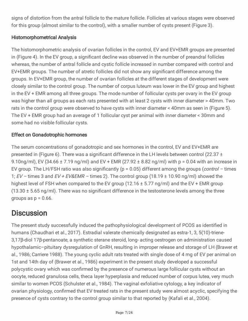
The serum concentrations of gonadotropic and sex hormones in the control, EV and EV+EMR arepresented in (Figure 6). There was a signi�cant difference in the LH levels between control (22.37 ±9.10ng/ml), EV (34.66 ± 7.19 ng/ml) and EV + EMR (27.92 ± 8.82 ng/ml) with p = 0.04 with an increase inEV group. The LH/FSH ratio was also signi�cantly (p = 0.05) different among the groups (control – times1; EV – times 3 and EV + EV&EMR – times 2). The control group (18.19 ± 10.90 ng/ml) showed thehighest level of FSH when compared to the EV group (12.16 ± 5.77 ng/ml) and the EV + EMR group(13.30 ± 5.65 ng/ml). There was no signi�cant difference in the testosterone levels among the threegroups as p = 0.66.
DiscussionThe present study successfully induced the pathophysiological development of PCOS as identi�ed inhumans (Chaudhari et al., 2017). Estradial valerate chemically designated as estra-1, 3, 5(10)-triene-3,17β-diol 17β-pentanoate, a synthetic sterane steroid, long- acting oestrogen on administration causedhypothalamic–pituitary dysregulation of GnRH, resulting in improper release and storage of LH (Brawer etal., 1986; Carriere 1988). The young cyclic adult rats treated with single dose of 4 mg of EV per animal on1st and 14th day of (Brawer et al., 1986) experiment in the present study developed a successfulpolycystic ovary which was con�rmed by the presence of numerous large follicular cysts without anoocyte, reduced granulosa cells, theca layer hyperplasia and reduced number of corpus lutea, very muchsimilar to women PCOS (Schulster et al., 1984). The vaginal exfoliative cytology, a key indicator ofovarian physiology, con�rmed that EV treated rats in the present study were almost acyclic, specifying thepresence of cysts contrary to the control group similar to that reported by (Kafali et al., 2004).

Page 8/24
The EV + EMR exposed groups in the present study exhibited several positive effects such as, slightlylower body weight but not by a signi�cant amount, improved reproductive cycle, usual morphology ofdeveloping follicles, increased number of typical developing follicles, reduction in the mean number anddiameter of the follicular cysts than the PCOS rats (EV group) and closely similar to the control group.The most important �nding in the study is the reduction in the number of follicular cysts present peranimal. The highest number of cysts in the EV + EMR group was only two cysts per animal and some hadnone. This revealed that the EMR might have reduced the formation of the cysts.
The present study revealed that overall follicular dynamics was less disrupted in the group exposed toEMR and the observations were very close to the control group. This is because the folliculardevelopmental staging from primordial to secondary was observed to be present. The follicular celldifferentiation into granulosa cells and thecal cells with little to no distortion and vacuolisation whencompared with EV group was also evident. However, a lot of research has focused on the harmful effectsof EMFs on the granulosa cells of the oocytes. Apoptosis of these cells was reported in many articles dueto EMFs (Roshangar et al., 2014). An increase in the number of macrophages and autophagy vacuoles ingranulosa cell layers were identi�able with transmission electron microscopy from research conducted onthe effects of EMFs on female rats. The study also revealed liquid drops in the luteal and theca cells(Roshnagar and Rad, 2004).
Another research showed an increase in macrophages in the corpora lutea and growing follicles withEMFs exposure. Assumption is made that the process of apoptosis in the ovaries is increased with EMFsexposure. The destruction of ovarian cortical tissue, luminal epithelium, glandular epithelium, and stromalcells in the uterus and fallopian tubes are believed to result from the process of apoptosis from EMFexposure (Rad et al., 2001). However, the 150 kHz EMR exposure in the present study did not show any ofthe above changes in the ovarian tissue.
The hormone progesterone is produced by the corpus luteum. This hormone is responsible for the controlof the reproductive cycle and in return the preparation of the uterus for implantation if conceptionhappens (Stocco et al., 2007). The reduced number of corpus luteum demonstrated an anovulatory statein the EV group that makes chances of conception minimal (Palomba et al., 2017). The EV + EMRexposed group showed a higher number of corpus lutea than the EV group and the reproductive cyclewas an improved one in this group than the EV group. Morphological atresia was evident in all threegroups. No major comparison was observed as follicular atresia is considered an active cellular process.Yet, the susceptibility to programmed cell death at various stages during follicular development remainsunde�ned (Roshangar et al., 2014).
A key factor in hormone function changes and causes of infertility symptoms in females are the result ofneuroendocrine changes caused by the impact of EMFs on females (Nelson et al., 1996). The decrease innumber preandral follicles in the PCOS ovaries cause the overproduction of androgens that impedes withnormal follicular maturation process (Rezvanfar et al., 2012). But the present study did not showsigni�cant elevation in the testosterone level among the all groups. In the present study, the FSH

Page 9/24
concentrations did not alter but LH concentrations increased in rats with PCOS, thus the maturation offollicles was impaired and multi-sized cystic follicles were formed. The LH/FSH ratio in EV group and EV + EMR group were also signi�cantly higher than the control group. The group exposed to EMR showed anincrease in the LH and LH/FSH ratio, which is contrary to the reports in a DHEA-induced PCOS rat model(Francou et al., 2008; Rencher et al., 2018). Generally, a high frequency in gonadotropin-releasinghormone (GnRH) pulses in the hypothalamus leads to LH secretion from the pituitary. In this case,increased levels can also be from accelerated GnRH activity, increased responsiveness to GnRH ordecreased sensitivity of the hypothalamus received via negative feedback from sex steroids (Teharani etal., 2014).
Overall, the reduction in cystic formation from exposure to NIR can be a possible avenue for furtherresearch. The possible mechanism on which this 150 kHz works can be linked to some of the Bio-Electromagnetic Principles. One major principle being the sensitivity of receptor e�ciency on the surfaceof target cells to signal transduction (Neil, 2002) but cannot be con�rmed in this experiment.
As alternating electric �elds of intermediate frequency and low intensity, the TTFields have been reportedto slow down the growth of tumor cells while having no obvious bioeffects on normal cells (Kirson et al.,2007; Davies et al., 2013; Stupp et al., 2017). However, this frequency has never been examined during thedevelopment of non-cancerous conditions such as PCO. Hence, the consistency of the present resultscannot be con�rmed without results of the mechanistic studies involved in cancer cell lines in thisfrequency.
The effect of EMR on cells can be direct as shown by previous experiments on glioma cell lines (Lukas etal., 2019) but it is unlikely because the same changes in all different layers of different types of cells inovaries were observed in the present study. It can be speculated that there is an indirect effect either bythe in�uence of EMR to cell receptors or the effect on the hypothalamus and signalling via certainhormones. Since the present study is focussed on the effect of 150 kHz on estrous cycle, ovarianhistology and serum levels of gonadotrophic hormones, which is not strong enough to come to a solidconclusion. So, further investigations are required to assess the effect of EMR on follicuogenesis of PCOdevelopment by investigating the follicular ultrastructure and immunohistochemical characterisation ofsurface receptors of the granulosa and thecal cells and a detailed study on the Hypothalamo-hypopysio-gonal axis.
ConclusionThe 150 kHz EMR appears to have a positive effect like improved reproductive cycle, reversal to usualmorphology of developing follicles, increased number of typical developing follicles, reduction in themean number and diameter of the follicular cysts. However, a more detailed study, which includes thelimitations as highlighted.
Abbreviations

Page 10/24
CL - Corpora lutea
DHEA – Dehydroepiandrosterone
ELISA – Enzyme-linked Immunosorbent Assay
EM – Electromagnetic
EMF – Electromagnetic Frequency
EMR – Electromagnetic Radiation
EV – Estradiol Valverate
FSH – Follicle Stimulating Hormone
GnRH – Gonadotropin-releasing hormone
H & E – Haematoxylin and Eosin
LH – Luteinizing Hormone
NIR – Non-ionizing Radiation
PCO – Polycystic ovary
PCOS – Polycystic Ovary Syndrome
SD – Sprague Dawley
DeclarationsEthics
The Campus Research Ethics Committee of the University of the West Indies approved the protocols foranimal experimentation (CEC 310/09/17).
Consent for publication
All author consent for publication of this manuscript
Availability of data and materials
All data is available for this experiment. It will not be released because there are other phases of thisexperiment in progress.
Competing interests

Page 11/24
All authors declare no con�ict of interest.
Funding
The authors thanks to School of Graduate Studies and Research, The University of the West Indies, St.Augustine, Trinidad and Tobago for the �nancial support through Campus Research and PublicationFund (CRP.5.NOV19.60)
Authors contribution
SM led the design, conceived the study, performed the experimental work, vaginal cytology,morphometrical analysis and hormone estimation. Compiled and analysed all the results and wrote theoriginal manuscript. VS performed histology, supervised and edited the manuscript. NZ conceived thestudy, performed induction of EMR system and overall supervision of the experiment and edited themanuscript. All authors read and approved the �nal manuscript.
Acknowledgments
The authors wish to express sincere thanks to The Department of Physics, and The School of VeterinaryMedicine, The University of the West Indies for the support rendered throughout the project. The authorsalso acknowledge the services offered by Dr. Jenelle Johnson, in-charge of Lab Animal Facility at Schoolof Veterinary Medicine for this study. Authors also acknowledge the laboratory help from Lester Gikes,Samuel Rampersad and Gerald Chadoo.
Author’s information
Statistical Analysis: Stephanie Mohammed. Email: [email protected]
Data Sharing Statement: All data generated and analysed during this study are included in this publishedarticle
ReferencesAhmadi SS, Khaki AA, Ainehci N, Alihemmatic A, Khatooni AA, Khaki A, et al. Effect of non-ionizingelectromagnetic �eld on the alteration of ovarian follicles in rats. Electronic physician. 2016;8(3):2168.
Blay Sl, Aguiar JVA, Passos IC. Polycystic ovary syndrome and mental disorders: a systematic review andexplatory meta-analysis. Neuropsychiatr Dis Treat. 2016;12:2895.
Brawer JR, Munoz M, Farookhi R. Development of the Polycystic ovarian Condition (PCO) in the EstradiolValverate-Treated Rat. Biol Reprod. 1986;35:647–55.
Carriere PD, Brawer JR, Farookhi R. Pituitary gonadrtropin-releasing hormone receptor content in rats withpolycystic ovaries. Biol Reprod. 1988;38:562–7.

Page 12/24
Chaudhari NK, Nampoothiri LP. Neurotransmitter in a testosterone propionate-induced polycystic ovarian.Horm Mol Biol Clin Investig. 2017;29(2):71–7.
Davies AM, Winberg U, Palti Y. Tumor treating �elds: a new frontier in cancer therapy. Annals of the NewYork Academy of Science. 2013;1291(1):86–95.
Dewailly D, Cortet-Rudelli C, Decanter C. The Polycystic Ovary Syndrome: Reproductive aspects. In WassJAH, Shalet SM, editors. The Oxford Textbook of Endocrinology. Oxford University Press, Oxford UK2003:1135–1143.
Ding T, Hardiman PJ, Petersen I, Wang F-F, Qu F, Baio G. The prevalence of polycystic ovary syndrome inreproductive-aged women of different ethnicity: a systematic review and meta-analysis. Oncotarget.2017;8:9635.
Francou MM, Durdos M, Salvetli RN, Bravalle C, Rey F, Ortega HH. Characterization of Pituitary CellPopulations in Rats with Induced Polycystic Ovaries. Cell Tissues Organs. 2008;188:310–9.
Kakoly AS, Earnest A, Teede HJ, Moran LJ, Joham AE. The Impact of Obesity on the Incidence of Type 2Diabetes Among Women with Polycystic Ovary Syndrome. Diabetes Care. 2019.https://doi.org/10.2337/dc18-1738.
Kafali H, Iriadam M, Ozardali I, Demir N. Letrozole-induced polycystic ovaries in the rat: a new model forcystic ovarian disease. Arch Med Res. 2004;35(2):103–8.
Kinzel A, Ambrogi M, Varshaver M, Kirson ED. Tumor Treating Fields for Glioblastoma Treatment: PatientSatisfaction and Compliance with the Secondary-Generation Optune System. Clinical Medicine Insights.Oncology 2019:13.
Kirson ED, Dbaly V, Tovarys F, Vymazal J, Soustiel JF, Itzhaki A, et al. Alternating electric �elds arrest cellproliferation in animal tumor models and human brain tumors. Proc Acad Sci USA. 2007;104(24):10152–7.
Lukas B, Almke B, Meshksar S, Dierks A, Majernik GH, Krauss JK, et al. Tumor Treating Fields(TTFields):Investigations on the mechanism of action by electromagnet exposure of cells intelophase/cytokinesis. Sci Rep. 2019;9:7362.
Mohammed SB, Nayak BS. Polycystic Ovarian Syndrome Trend in a Nutshell. International Journal ofWomen’s Health Reproduction Sciences. 2017;5(3):153–7.
Mohammed SB, Sundaram V. Comparative Study of Metachromatic Staining Methods in Assessing theExfoliative Cell Types During Oestrous Cycle in Sprague-Dawley Laboratory Rats. Int J Morphol.2018;36(3):962–8.

Page 13/24
Mun EJ, Babiker HM, Weinberg U, Kirson ED, Van Hoff DD. Tumor-treating �elds: a fourth modality incancer treatment. Clin Cancer Res. 2017;24:266–75.
Neil C. Criticism of health assessment in the ICNIRP guidelines for radiofrequency and microwaveradiation (100 kHz – 300 kHz). Lincoln: New Zeeland Lincoln University; 2002.
Nelson JF, Karelus K, Bergman MD, Felicio LS. Neuroendocrine involvement in aging: evidence fromstudies of reproductive ageing and caloric restriction. Neurobiol Aging. 1995;16(5):837–43.
Orio F, Muscogiuri G, Nese C, Palomba S, Savastano S, Tafuri D, et al. Obesity, type 2 diabetes mellitusand cardiovascular disease risk: an update in the management of polycystic ovary syndrome. Eur JObstet Gynecol Reprod Biol. 2016;207:214–4.
Palomba S, Daolio J, La Sala BG. Oocyte competence in women with polycystic ovary. Trends EndocrinolMetab. 2017;28(3):186–98.
Rencher FS, Ozbek KS, Eraldemir C, Zehra S, Tugba K, Ceylan S, et al. Effect of resveratrol and metforminon ovarian ultrastructure in PCOS: an experimental study. Journal of Ovarian Research. 2018;11:55.
Rezvanfar M, Ahmadi A, Saadi HS, Baeeri M, Abdollahi M. Mechanistic links betweenoxidative/nitrosative stress and tumor necrosis factor alpha in letrozole-induced murine polycystic ovary:biochemical and pathological evidence for bene�cial effect of pioglitazone. Hum Exp toxicol.2012;31(9):887–97.
Roshanhar L, Hamdi BA, Khaki AA, Soleimani Rad J, Soleimani Rad S. Effect of low frequencyelectromagnetic �eld exposure on oocyte differentiation and follicular development. Adv Biomed Res.2014;3:76.
Roshangar L, Rad SJ. Electron microscopic study of folliculogenesis after electromagnetic �eld exposure.Journal of Reproduction Infertility. 2004;5(4):299–307.
Schulster A, Farookhi R, Brawer JR. Polycystic ovarian condition in estradiol valverate-treated rats:spontaneous changes in characteristic endocrine features. Biol Reprod. 1984;31:587–93.
Sirmans SM, Pate KA. Epidemiology, diagnosis and management of polycystic ovary syndrome. ClinEpidemiol. 2014;6:1.
Rad SJ, Rowshangar L, Karimi K. The effect of Electromagnetic �eld on Fallopian Tube. IFFS 2001Selected Free Communication. International Proceedings Division; Melbourne. November 2001:25–30.
Stocco C, Telleria C, Gibori G. The molecular control of Corpus luteum formation, function and regression.Endocr Rev. 2007;28(1):117–49.

Page 14/24
Stupp R, Taphoorn M, Driven L, Taillibert S, Chen HT, Paek SSH, et al. Tumor Treating Fields (TT�elds)- ANovel Cancer Treatment Modality: Translating Preclinical Evidence and Engineering into a Survial Bene�twith Delayed Decline in Quality of Life. International Journal of Radiation Oncology.Biology. Physics2017;99(5).
Taye� NH, Gavami M, Aknarzadeh A, Beheshti R, Mohammednejad D, Abedelahi A. Preservation of mouseovarian tissue follicle morphology and ultra-structure after vitrifying in biotechnological protocols. JOvarian R. 2015;8:7.
Teharni FR, Noroozzadeti M, Zahediasl S, Piryaei A, Azizi F. Introducing a rat model of prenatal androgen-induced polycystic ovary syndrome in adulthood. Exp Physiol. 2014;99(5):792–801.
Vergote I, Copeland L, Monk B, Coleman R, Cibula D, Sehouli J, et al. P154 Tumor Treating �elds (200 kHz)concomitant with weekly paclitaxel for platinum-resistant ovarian cancer: phase 3 INNOVATIVE/ENGOT-OV50 study 2019.
Wdowiak A, Mazurek P, Wdowiak A, Bojar I. Effect of electromagnetic waves on human reproduction. AnnAgric Environ Med. 2017;24(1):13–8.
TablesTable 1. The effect of 150 kHz EMR on the body and ovarian weights (n=7).
Parameter Control EV EV + EMR p
Body weight/g 391.19 ± 51.28 283.27 ± 33.33 281.80 ± 23.46 0.14
Weight of left ovary/g
0.07 ± 0.02
0.08 ± 0.01
0.07 ± 0.03
0.74
Weight of right ovary/g 0.06 ± 0.02 0.07 ± 0.01 0.06 ± 0.02 0.40
*The mean difference is signi�cant at the p = 0.05 con�dence interval
Figures

Page 15/24
Figure 1
Exfoliative cytology during the estrous cycle. (a) Proestrous stage shows small nucleated cells. (b)Estrous stage shows corni�ed cells. (c) Metestrous stage shows nucleated, corni�ed and neutrophil cells.(d) Diestrous stage shows neutrophils.

Page 16/24
Figure 1
Exfoliative cytology during the estrous cycle. (a) Proestrous stage shows small nucleated cells. (b)Estrous stage shows corni�ed cells. (c) Metestrous stage shows nucleated, corni�ed and neutrophil cells.(d) Diestrous stage shows neutrophils.

Page 17/24
Figure 2
Representation of estrous cycle of EV and EV + EMR group for 46 days.

Page 18/24
Figure 2
Representation of estrous cycle of EV and EV + EMR group for 46 days.
Figure 3
Photomicrographs of different ovarian follicles in control, EV and EV + EMR groups. Thephotomicrograph showing the nests of primordial follicles in the Control( A), EV(b) and EV + EMR ( c)

Page 19/24
groups. The unilaminar primary follicles in the Control (d), EV (e) and EV + EMR (f) groups. Themultilaminar primary follicles in the Control (g), EV (h) and EV + EMR (i) groups. The antral follicles in theControl (j), EV (k) with marked distorted granulosa and theca layer cells greater than the EV + EMR (l)groups. The matured follicle in the control (m) and EV+EMR (o) groups with less distortion of thegranulosa cells. The cystic follicle (n) in the EV group with thin layer of granulosa cells. The cross sectionof the ovary of the Control (p), EV (q) and EV + EMR (r) groups. o- oocytes; f: follicular cells; g granulosacells; t-thecal cells; a; a-antrum; c-cysts cl; corpus luteum gf; Gra�an follicle.

Page 20/24
Figure 3
Photomicrographs of different ovarian follicles in control, EV and EV + EMR groups. Thephotomicrograph showing the nests of primordial follicles in the Control( A), EV(b) and EV + EMR ( c)groups. The unilaminar primary follicles in the Control (d), EV (e) and EV + EMR (f) groups. Themultilaminar primary follicles in the Control (g), EV (h) and EV + EMR (i) groups. The antral follicles in theControl (j), EV (k) with marked distorted granulosa and theca layer cells greater than the EV + EMR (l)groups. The matured follicle in the control (m) and EV+EMR (o) groups with less distortion of thegranulosa cells. The cystic follicle (n) in the EV group with thin layer of granulosa cells. The cross sectionof the ovary of the Control (p), EV (q) and EV + EMR (r) groups. o- oocytes; f: follicular cells; g granulosacells; t-thecal cells; a; a-antrum; c-cysts cl; corpus luteum gf; Gra�an follicle.
Figure 4
Effect of 150 kHz EMR in the follicular development in rats

Page 21/24
Figure 4
Effect of 150 kHz EMR in the follicular development in rats
Figure 5

Page 22/24
Effect of 150 kHz EMR on the distribution and size of follicular cysts in rats.
Figure 5
Effect of 150 kHz EMR on the distribution and size of follicular cysts in rats.

Page 23/24
Figure 6
Effect of 150kHz EMR on serum levels of gonadotrophic hormones in the rats.

Page 24/24
Figure 6
Effect of 150kHz EMR on serum levels of gonadotrophic hormones in the rats.