stereo microscope - · pdf file · 2015-02-19stereo microscope part 3: ... figure...
TRANSCRIPT

66
Stereo
Microscopy
Stereo Microscope Part 3: Common Main Objective Stereo Microscopes 3rd Edition
R. Jordan Kreindler (USA)
Figure 56. Woven fabric pattern through Zeiss "Opton" CMO microscope

67
Stereo
Microscopy
___________________________________________________________________
This paper is the third in a multi- part series on stereo microscopes, including their history,
design, and, applications. The paper concludes with considerations for potential stereo
microscope buyers. In this Part Common Main Objective (CMO) microscopes are discussed.
______________________________________________________________________________
Figure 57. From the Zeiss brochure: Microscopy from Carl Zeiss. Stemi DR, Stemi DV4, Stemi 2000 Stereomicroscopes, Courtesy, and with permission of Carl Zeiss Microscopy, LLC. A comparison of
Greenough and CMO (Telescope) design microscopes

68
Stereo
Microscopy
The Common Main Objective (CMO) Stereo Microscope
A comparison of the Greenough and CMO (called telescope design by Zeiss) designs is shown in Fig. 57. In CMO designs there are two distinct light paths through both halves of a relatively large common objective lens. This design can have the benefit of long working distances. The modern common main objective (CMO) concept, although this designation appeared later, appears to have been developed by Carl Zeiss, Jena 1938-1941 (Michel, 1962). Dr. Kurt Michel worked on the Praepariermikroskop "Citoplast" (Preparation and dissection microscope “Citoplast”) from its inception. He joined Zeiss' Microscope Department in 1934, and became its Vice Head in 1938. When first developed, the Citoplast was to be priced at 816 RM (Zeiss, 1942). However, in response to the German government's direction, commercial manufacturing of the Citoplast was delayed until after WW II, i.e., until 1946 (Zeiss, 1946). Zeiss' East German R&D department received a Citoplast on October 3, 1946, and another went to Moscow. According to Zeiss' internal documents, most shipments went to Russia. [Author: These were probably supplied to state run agencies.] In private email received from Berndt-Joachim Lau of Carl Zeiss (Lau, 2012), by the author, Herr Lau explained Zeiss' situation at the end of WW II as follows,
In June 1945 the US Army took the action "We take the brain" before Jena was given to the Russians. At October 24, 1945 the Russian Army ordered the demounting of Zeiss factory and the deportation of ca. 800 Zeiss people to Soviet Union.
The Russians viewed the movement of Zeiss factories, manufacturing equipment, and personal as appropriate reparations for their WW II costs. However, this was not the view shared by many of the affected Zeiss personnel. Some of Zeiss' relocated resources were used to establish the Kiev camera works, makers of the Zeiss Ikon and Contax. As noted above, the U.S. Army occupied Jena before the partition of Germany. They moved many of Zeiss, Jena's leading technicians, and upper management, to the Contessa plant in Stuttgart, West Germany. In 1947 this was the core team that formed the Zeiss-Opton Optische Werke Oberkochen GmbH in Oberkochen, West Germany. The first modern CMO was developed at Carl Zeiss, Jena decades before the first American CMO, the AO Cycloptic. The first Zeiss CMO microscopes appeared commercially in East Germany as the Zeiss "Citoplast", Fig. 58, and in West Germany, Figs. 59 and 60, under the Opton label.

69
Stereo
Microscopy
In his book Armin Herman (Hermann, 1991) mentions that the first West German Zeiss-Opton
Stereomicroscope was given in a small ceremony on January 23, 1949 to Professor Bauersfeld
on the occasion of his 70th birthday.
The Opton CMO version was manufactured by the Opton-Optische Werstatte Oberkochen GmbH Zeiss factory at Oberkochen, West Germany. For a period, versions of this CMO microscope were made by both East and West German Zeiss companies. The Zeiss Opton-branded CMO was taller, wider, and heavier than most previous stereo microscopes. At 12 pounds, it weighed almost 50% more than the c. 1929 B&L Greenough microscope shown in Fig. 25. Production of the West German Opton version, ceased in 1954, when the name of the West German Zeiss company was changed to Carl Zeiss, about three years before the introduction of AO's Cycloptic (Orlowski, 2012), (Zeiss, undated), (Walker, 2011). An Opton-style CMO microscope continued in production by the West German Zeiss company until 1959, under the Carl Zeiss label. (Schulze, 2011, 2012). At least until the 1980s, Zeiss West Germany appears to have used CMO designs in their stereo microscopes, except for models 01, 02, I, Ib, III, and the D-series.

70
Stereo
Microscopy
Figure 58. from East German Citoplast brochure, date unknown Courtesy, and with permission, of Carl Zeiss
Microscopy, LLC

71
Stereo
Microscopy
Figure 59. From West German Opton brochure (translated to English), date unknown, Courtesy, and with permission, of Carl Zeiss Microscopy, LLC

72
Stereo
Microscopy
Figure 60. Zeiss Opton-branded CMO Microscope

73
Stereo
Microscopy
Early model Zeiss CMO microscopes continued to be manufactured for some years. Sander (1994) in his interesting and informative Centennial Essay mentions that "many [Citoplast stereo microscopes] must still be in service", and examples of early East and West German Zeiss CMO microscopes are often seen for sale on eBay. The Citoplast morphed, sometime in the 1950/60s, to the Zeiss SM-XX (sometimes, perhaps inappropriately, called the SM 20), with only changes from the earlier design. The Citoplast and its derivative microscopes were popular in Eastern Bloc countries and China. The SM-XX started its life finished in black, sometime later its color was changed to off-white. Two separate Zeiss companies had developed in the aftermath of the Second World War. So, CMOs were manufactured by both Zeiss East and West German companies. Most East German models were made about 54 miles (about an 1-1/2 hour drive) from Berlin in Brandenburg, Germany in the city of Rathenow by the Optische Werke Rathenow. Rathenow had contained the only spectacle manufacturing facility in the GDR. It was integrated with Zeiss as the Kombinat VEB Carl Zeiss Jena after the close of WWII, possibly c. 1970. [As an aside, after the disintegration of the Soviet Union, a location near Rathenow was found to contain the graves of Adolph Hilter and some of his associates.]
Shortly after the fall of the Berlin wall in November 1989, the two separate Zeiss companies began talks and reunited in the early 1990s after East Germany's first free elections. After the reunification of the East and West German Zeiss companies, the unified company introduced the first of the Stemi Greenough, stereo microscope, models. After Unification Kombinat VEB Carl Zeiss Jena became Zeiss Jena, Gesellschaft mit beschränkter Haftung (GmbH). In 1991 after a division of functional assets, the new microscopy group Carl Zeiss AG (Oberkochen) reunited the previous Zeiss company's microscopy groups and relocated back to Jena, Germany along with its planetarium group. The new combined Zeiss company designed a new logo combining at its top a portion of the square used by the West German company in its last logo before unification, and at the bottom the curve from the logo used by the East German company.

74
Stereo
Microscopy
Common Main Objective (CMO) and Greenough Pros and Cons Fig. 61 provides examples of Greenough and CMO microscopes, although not to scale. The Zeiss CMO is taller than the Leitz Greenough.
Figure 61. Greenough and CMO stereo microscopes
Zeiss CMO design Leitz Greenough design

75
Stereo
Microscopy
Each design has its strengths and weaknesses The Greenough design suffers, slightly, from "keystone" distortion, i.e., distortion due to the viewing angles involved. An exaggerated version of keystone distortion is produced when a projector is aimed at a steep angle to a wall so that the projected image has the shape of the "keystone" in an arch, that is a trapezoidal shape. This can result in focus distortion, where some portions of the image are slightly out of focus and may cause eyestrain for some users after extended viewing periods.
CMO stereomicroscopes have parallel paths, i.e., for practical purposes the two eyes view images at right angles to the object plane, and so do not suffer from keystoning. Viewing objects with the eyes focused at infinity can, perhaps, be less stressful with continuous use. However, quality CMOs are expensive to make, are usually heavier, and have some optical problems of their own, see below.
We usually see nearby objects with both eyes, at an angle, and not images in parallel. If we interpret the parallel images as coming into the eyes at an angle, this results in "perspective distortion", as the central portion of the object appears thicker than it actually is. For non-photographic use the CMO has two minor problems: (1) we are used to processing parallel paths for objects at infinity, not for nearby objects, so our minds normally process parallel images from nearby objects inappropriately, and (2) there is some degradation of images that pass through the edges of a lens. The first issue is not a problem in photography through the microscope where images are two dimensional, and the second problem can be minimized in trinocular photomicrography by using the objective lens so its optical center coincides with the, usually, single light path used for photography. Zeiss helped minimized the perception problems inherent in the CMO's design with their development of multi-element, non-APO, PLS objectives.
76
Stereo
Microscopy
AO's Cycloptic Microscope: The first American CMO American Optical (AO), hoped to dominate stereo microscope sales in the late 1950s, when they achieved a major landmark in American stereo microscope development. They brought out the Cycloptic microscope (Phillips, 2011, 2012), (NikonU, undated), the first American CMOs. The AO Cycloptic was designed on the Zeiss model. As with the Zeiss Citoplast, the microscope received images for both eyepieces through a bottom main objective, large enough to easily support two light paths. Fig. 62 shows one of the basic versions of the AO Cycloptic microscope with a Galilean, also called telescope, changer (AO refers to this as a "Magni-Changer").

77
Stereo
Microscopy
Figure 62. AO Cycloptic basic model with Galilean telescope changer ("AO Magni-Changer")

78
Stereo
Microscopy
Fig. 63 shows the common main objective (CMO) from the microscope in Fig. 62. However, AO CMOs were often sold with additional lens attachments to provide further magnification options. Fig. 64 shows a common main objective with an attached 0.75x auxiliary lens (from AO model 59F-T1) to reduce magnification slightly, to allow more "in context" views of some objects.
CMOs are similar to the earlier Riddell-Stephenson design, in terms of a single objective. However, for CMOs this objective is considerably larger, compared to smaller objectives found in the earlier stereo microscopes. These CMOs, with their low magnifications, offered a much greater working distance than was possible with a standard compound microscope.
In spite of the relatively minor optical flaws inherent in CMO instruments, AO's Cycloptic microscopes, although expensive, "led the pack", and were for a time the royalty of stereo microscopes. From the number of Cycloptic microscopes still widely available on the used market today, the sales of these instruments, at least in Western countries, were much larger than those of the Zeiss Citoplast. This was, perhaps, owing to the residual negative feelings by US consumers toward German products after the close of WW II.
Figure 64. AO Cycloptic "Common Main Objective" with 2/3X No. 267 achromatic aux. lens-attachment
Figure 63. Common Main Objective from the object facing side

79
Stereo
Microscopy
AO's CMO microscopes are unusual relative to their Greenoughs, not only for their single main objectives but for their use of rotating cylindrical drum assemblies, ("AO's Magni-Changer"), see Galilean drum discussion below. AO drums have unique and clearly distinguishable end markings. These markings allow this microscopes to be identified at a distance, even when somewhat obscured, as in some movies or TV shows. These Galilean drums are located above the CMO to make magnification changes easier. In their CMO microscopes both Zeiss and AO used multiple pairs of lenses contained in a single drum to speed magnification changes, as opposed to the alternate approach, as in some Greenough-style microscopes, of exchanging objectives. AO used single housing magnification changers of their own design in their Greenough microscopes, before the Cycloptic was introduced. Their Greenough microscope designs were derived from the original Zeiss designs as, apparently, was their Cycloptic. Thus, primacy in two, perhaps most basic, areas of modern stereo microscope development belongs to Zeiss, although these developments were often capitalized on by other companies as well. Freely copying the microscope design of others was common practice over a considerable time. (Kreindler and Goren, May 2011). The AO Magni-Changer (Galilean drum - see below) pictured in Fig 65, contains four clear openings, in opposite pairs, and two telescopes. The clear openings allow for "straight through" images. The dual paired telescopes have four lens groups each. The telescopes can be rotated into the optical path in opposite orientations. This allows, as with stand-alone telescopes, for the magnification or diminution of images. The drum provides five magnification options. One for the "see-thru" openings, the same in either forward or backward orientations, four additional magnifications using the two telescopes on the drum, in either front or back orientations.
Figure 65. AO Cycloptic stereo microscope drum, offering five magnifications with two telescopes
and one "see-thru" opening

80
Stereo
Microscopy
This type of magnification changer where the same components are used but reversed to obtain different magnifications, is often referred to as a "Galilean drum", as here, as the drum actually contains small Galilean telescopes. These telescopes are frequently composed of plano-convex and bi-concave lenses. Galilean telescopes provide erect images. The Cycloptic, with its Galilean drum and distinctive external markings to show magnification choices has a unique appearance, and has been used in various US TV shows including, possibly the most popular TV drama series of its time, CSI where it was used by Supervisor Dr. Gil Grissom, one of the show's lead characters. Apparently, most of AO Cycloptic's screw-in objectives were apochromatic, although with such low magnifications this was probably not as difficult to design as it would have been for a small high magnification compound microscope objective. As often happens when one company's technology introduction negatively impacts sales of another, the impacted company develops its own improvements. In response to AO's Cycloptic microscopes, Bausch and Lomb (B&L), c. 1959, see Part 2b, came out with their StereoZoom microscopes. Which, instead of providing a single or various fixed focus magnifications, provided continuous magnification zooming. StereoZoom microscopes used mirrors instead of Porro prisms, thus reducing both weight and cost. It was not until a few years later, 1961 and the Olympus SZ microscope, that zoom stereo microscopes were introduced by Japanese companies.

81
Stereo
Microscopy
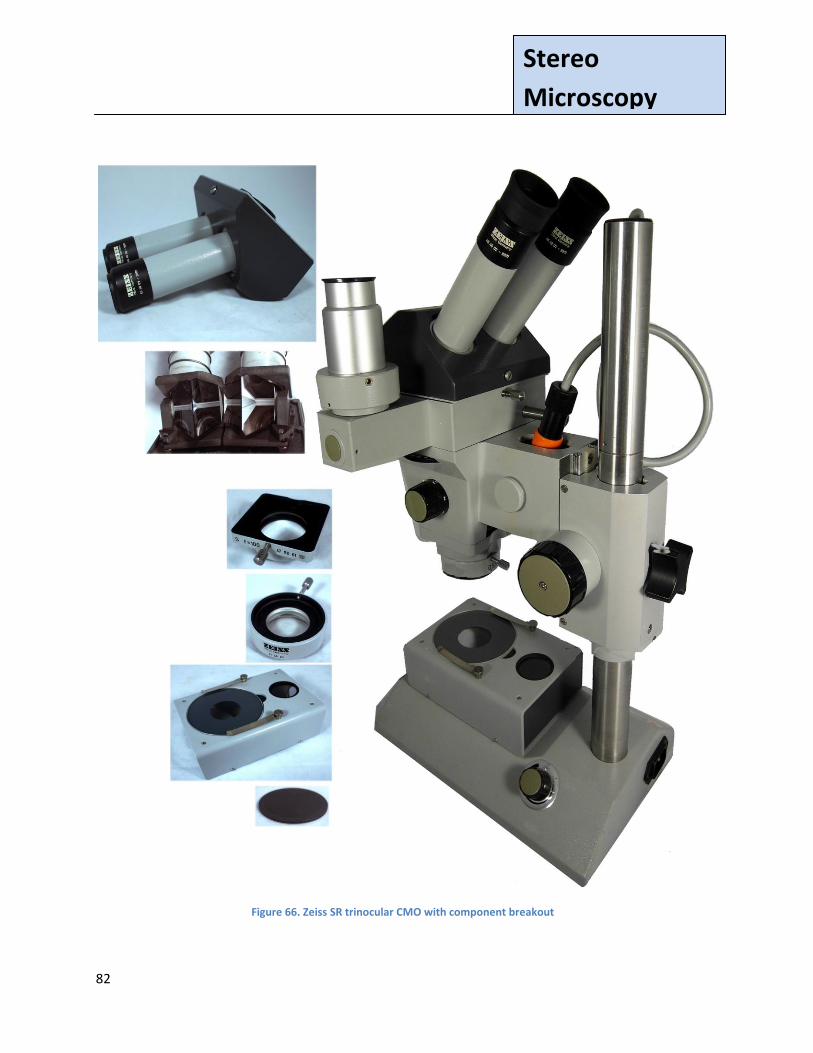
Zeiss SR CMO Stereo Microscope: The Anatomy of a CMO
This Figure provides a breakout of some of the main components of this CMO, from the top down. This breakout is shown to left of the microscope in Fig. 66. (1) The eyepiece assembly (2) The prisms contained in the eyepiece assembly that refract the light at an angle appropriate to allow easier viewing; here the eyepiece's cover has been removed to allow the prisms to be seen (3) The common main objective (CMO), mounted in a square assembly (4) A 0.5x auxiliary lens to allow more of the object to be seen and to increase the working distance (the distance from the object to the objective). Here, the distance between the object and the objective with the 0.5x auxiliary lens attached is significantly increased to 8 inches. (5) The optional transmitted light base, to allow the normally incident light to be reflected up under the stage's glass plate (6) The reversible, black on one side, white on the other, stage plate is present but is hidden by the optional transmitted light base in the picture of the microscope. Note: The red cylinder at the middle right contains the microscope's illumination. Its brightness control (rheostat) can be seen on the rear left-side (as seen from the eyepiece side) of the base. The microscope provides a locking fixture below the focusing assembly, to prevent the instrument from inadvertently falling into the base. The Zeiss SR is about 22 inches tall in working position (with 0.5x auxiliary lens attached, but without the optional transmitted light assembly), compared to AO's Cycloptic's height in working position of 12 inches (without auxiliary lens). The SR with optional accessories attached, but without a camera weighs approximately 16 pounds 1 ounce, compared to the AO Cycloptic's 8 pounds 5 ounces. That is, the Zeiss SR is a relatively large and heavy instrument.
82
Stereo
Microscopy
Figure 66. Zeiss SR trinocular CMO with component breakout

83
Stereo
Microscopy
Fig. 67 shows the hind wing of a
Swallowtail butterfly (Family:
Papilionidae), as seen through
the Zeiss SR CMO shown in Fig.
66. The full display of the wing
shows the value of having a 0.5x
auxiliary lens mounted, when a
larger "in context" view is called
for.
[Author's note: The AO Cycloptic
could also be purchased with a
1/2x apochromatic objective, AO
No. 266, which provided an 8-
inch working distance. This
working distance is similar to the
working distance on Zeiss' SR
microscope with a 0.5x auxiliary
lens.]
Figure 67. Hind wing of Swallowtail butterfly, seen through a Zeiss SR CMO with Zeiss 0.5x auxiliary lens

84
Stereo
Microscopy
Additional pictures through this Zeiss CMO stereo microscope are shown in Figs. 68-72. Figs. 68
and 69 present details from Hong Kong $10 bills, and Fig. 70 from a US $10 bill. These three
figures demonstrate the difference in engraving quality between Hong Kong and US currencies.
In Hong Kong three commercial (private) banks were authorized by the government to print
bank notes [the Bank of China, Standard Chartered Bank, and the Hong Kong and Shanghai
Banking Corporation (HSBC)]. [Author's aside: The Bank of China headquarters were next to the
author's Hong Kong flat.] The bills shown here were printed by HSBC
In the US only the federal government has the authority to print bank notes. [Author's aside:
The dollar sign '$' used for HK and US currency derives from an earlier symbol used to represent
the peso and not, as described in Ayn Rand's fictional novel Atlas Shrugged, from the letters 'U'
and 'S' written one on top of the other.]
Fig. 71 shows a small piece of orthoclase (or potassium) feldspar with smoky quartz inclusions
from Ontario, Canada. Fig. 72 is a Rhynchonella Fossil, c. 70 million years ago, found in
Morocco, North Africa.

85
Stereo
Microscopy
Figure 68. Detail from a Hong Kong $10 bill printed by the HSBC

86
Stereo
Microscopy
Figure 69. Detail from shield on Hong Kong HSBC $10 bill

87
Stereo
Microscopy
Figure 70. Federal Reserve seal on US $10 bill

88
Stereo
Microscopy
Figure 71. Orthoclase Feldspar

89
Stereo
Microscopy
Figure 72. Rhynchonella, an articulate brachiopod, filter feeder, marine invertebrate fossil, c. 100 million years old

90
Stereo
Microscopy
Combined References and End Notes (This list includes references/notes for the full paper. However, additional references may be added in later Parts)
Allen, R. M., (1940) The Microscope. Boston: D. Van Nostrand Company, Inc., p87. Bryant, Dr. Mark L., (2012) The author's thanks to Dr. Bryant and his staff for permission to photograph their Topcon slit lamp. Bausch & Lomb Optical Co, (1929) Microscopes & Accessories: Photomicrographic and Micro-Projection Apparatus Microtomes . Colorimeters Optical Measuring Instruments and Refractometers. Bausch & Lomb New York, p 81. Blocker (2012) Blocker History of Medicine, http://ar.utmb.edu/ar/Library/BlockerHistoryofMedicineCollection/BlockerHistoryofMedicineArtifacts/MicroscopeCollection/MicroscopesMakersandTheirInstruments/MicroscopeSwift/tabid/877/Default.aspx
Carpenter, William (with revisions by Rev. W. H. Dallinger) , (1901) The Microscope and Its Revelations.
Eighth Edition. Philadelphia: P. Blakiston's Son & Company, p 96.
Cherubin, d'Orleans. Père, (1677) La Dioptrique Oculaire ou La vision parfait ou le concours des deux axes de la vision en un seul point de l'objet , Paris: S. Mabre-Cramoisy del Cerro, Manual (2012) The author's thanks to Dr. del Cerro for his kindness in reviewing the
section on ophthalmology, and his helpful suggestions. However, all content is the sole
responsibility of the author.
Doherty, Glenn (2012) The author's thanks to Mr. Doherty, Support Representative, Carl Zeiss Microscopy, LLC for his help in identifying start and end manufacturing dates for some Zeiss stereomicroscopes.
Davis, George E., F.R.M. S. (1882) Practical Microscopy. London: David Bogue Encyclopaedia Britannica, (1910) A Dictionary of Arts, Sciences, Literature and General Information, 11th Edition, Volume 3, Binocular Instrument. New York, p 950.

91
Stereo
Microscopy
Ferraglio, Paul L. (2008) The Riddell-Stephenson Binocular Microscope. The Journal of the Microscope
Historical Society. Volume 16.
The author's thanks to Dr. Ferraglio, a leading authority on Prof. Riddell's microscope
and its successors. Dr. Ferraglio was kind enough to provide the author with reprints
of his papers, as well as helpful comments on an earlier version of this paper. However,
all content here is the sole responsibility of the author.
Ford, Brian (1973) The Optical Microscope Manual. Past and Present Uses and Techniques. New York: Crane, Russet & Company, Inc. Goren, Yuval The author's thanks to Dr. Goren for the many discussions we've had on historical
microscopes, and his emphasis on the importance of setting microscopes in their historical
context.
Gubas, Lawrence J. (2008) A Survey of Zeiss Microscopes 1846-1945. Las Vegas: Graphics 2000. This
book provides additional color photographs of a Model XV and its storage on page 253.
It can be highly recommended for its detailed and excpetional discussions of Zeiss microscopes.
Gubas, Lawrence J. (private correspondence, 2012) The author's thanks to Mr. Gubas for information on Zeiss instruments and employees, and pointers to Zeiss materials.
Hagan, Kevin (private correspondence, 2011) Thanks to Mr. Hagan of ALA industries Limited, Valparaiso, Indiana for providing a Contamikit brochure and PDF of the Instruction Manual. Hermann, Armin Nur Der Name War Geb lieben: Die absenteuerliche Geschichte der Firma Carl Zeiss
Stuttgart: Deutsche Verlag-Anstalt, 1991, p. 37
Journal of the Society of Arts, Vol XXXIV, (November 1886). London: George Bell and Sons, for the Society of Arts, Fig. 16, p 1014.
Kreindler, R.J. and Yuval Goren (March 2011),
Comparison of the Swift FM-31 Portable Field Microscope and an FM-31 Clone, Micscape, Figs.
11, 12, and 13.
Kreindler, R.J. and Yuval Goren (May 2011), Baker's Traveller's Microscope, Micscape
Kreindler, R.J. and Yuval Goren (November 2011), The TWX-1 Folded-Optics Microscope, Micscape
92
Stereo
Microscopy
Kreindler, R. J. (2012) The author worked in Silicon Valley for a number of years and saw the
extensive use, and occasional abuse, stereo microscopes in high-tech companies were
subjected to.
Lau, Berndt-Joachin (2012) The author 's thanks to Herr Lau of Carl Zeiss Microscopy GmbH for his
information on early Zeiss stereomicroscopes, Zeiss GDR microscopes, and Zeiss' situation in
Germany after WWII. His extended employment at Zeiss and his personal recollections
and pointers to Zeiss references have been of truly immeasurable assistance to the author.
Maertin, Rainer (2012) www.photosrsenal.com. The author's thanks for his permission to use the
photo of the Brewster type stereo viewer.
Mappes, Timo (2005) The First Commercial Comparison Microscope, made after Wilhelm Thörner by
W. & H. Seibert, Wetzlar. The Journal of the Microscope Historical Society. Volume 13, No. 2.
Mappes, Timo (2005-2006) Museum optischer Instrumente, http://www.musoptin.com/seibert_15368.html
Moe, Harald, (2004) The Story of the Microscope. Denmark: Rhodes International Science and Art Publishers with the Collaboration of The Royal Microscopical Society, p. 176. Nikon Microscopy U (undated) Introduction to Stereomicroscopy states, "The first modern stereomicroscope was introduced in the United States by the American Optical Company in 1957. Named the Cycloptic, this breakthrough design...". Although this was a landmark in American stereomicroscopes, the common objective concept was first used by Riddell in 1850s, and a common large objective was later implemented by Zeiss in their Citoplast, considerably before the Cycloptic was introduced. NYMS (1957) The author's thanks to the NYMS for permission to reprint the advertisement from their 1957 Newsletter (See Pollinger, 1957) Orlowski, Kristen and Dr. Michael Zölffel (private correspondence, 2012)
- The author's thanks to both Kristen Orlowski, Product Marketing Manager, Light Microscopes, Carl Zeiss Microscopy, LLC and Dr. Michael Zölffel, Carl Zeiss MicroImaging Gmb, Jena, Germany for information and materials they provided regarding Zeiss history. Ozment, Randall R. (2012) The author's thanks to Dr. Ozment for permission to photograph his Haag- Streit slit lamp, and for his explanation of its use in clinical practice.
93
Stereo
Microscopy
Phillips, Jay. (private correspondence, 2011, 2012) Provided a copy of Zeiss' catalog Mikroskope für Wissenschaft und Technologie (Prob. 1951). Pollinger, Mel. (1957) The author's thanks to Mr. Pollinger, Editor NYMS Newsletter for permission to reprint the advertisement from The New York Microscopical Society (NYMS) Newsletter of 1957
(See NYMS, 1957) Phillips, Jay. (private correspondence, (2011, 2012) Provided a copy of Zeiss' catalog "Mikroskope für Wissenschaft und Technologie" (Prob. 1951). Purtle, Helen R. (Second Edition), (1987 reprint) The Billings Microscope Collection. Second Edition. Washington, D.C.: Armed Forces Institute of Pathology, p 228, Figure 458 (Catalog number: M- 030.00541, AFIP accession number: 518,969, MIS photograph: 73-3899) Riemer, Marvin F., (1962) Microscope and the World of Science. New York: SCOPE Instrument Corp. RMS (1898) Journal of the Royal Microscopical Society, Volume 18, pp 469-471 Sander, Klaus. (1994) An American in Paris and the origins of the stereomicroscope. Institut für Biologie I (Zoologie). Freiburg, Germany: Springer-Verlag Schulze, Fritz , (2011, 2012) The author's thanks to Mr. Schulze, former head of the Historical Microscopical Society of Canada for his extensive knowledge of Zeiss microscopes which he kindly shared, and our extended exchanges on stereo microscopes.
Schwabe, Ms. Marte (2012) The author's thanks to Ms. Schabe, Assistant to Dr. Wimmer, Carl . Zeiss Archiv for her assistance (see Wimmer below). Schwidefsky, Kurt,( 1950) Grundriss der Photogrammetrie, Verlag für Wissenschaft und Fachbuch: 1950 (Reference from Fritz Schulze). Stanley, Jay (2012) The author's thanks for permission to use photos from his web site Classic Optics. Wade Nicolas , (1998) A Natural History of Vision. Cambridge, Mass: MIT press,p 301. Waldsmith, John (1991) Stereo Views: An Illustrated History and Price Guide. Wallace-Homestead Book Company: Radnor, Pennsylvania.

94
Stereo
Microscopy
Walker, David (undated) This is a short no frills introduction to stereo microscopes. http://www.microscopy-uk.org.uk/dww/novice/choice3.htm
Walker, David (July 2012) Product review: A 144 LED ring light for the stereo microscope (typical model
YK-B144T), July 2012, Micscape
Wheatstone, Charles. (1838) Contributions to the Physiology of Vision.—Part the First. On some remarkable, and hitherto unobserved, Phenomena of Binocular Vision, June 21, 1838 Wise, F. C., Francis Edmund Jury Ockenden, P. K.Sartory, (1950) The binocular microscope: its development, illumination and manipulation. (Quekett Microscopical Club Monograph) London: Williams & Norgate Wimmer, Wolfgang. The author's thanks to Dr. Wimmer's office at the Carl Zeiss Archiv Jena, Germany for their help. Zeiss, (Microscopy, LLC, MicroImaging Gmb, Jena)
- Zeiss (1934) Zeiss 1934 catalog, English version - Zeiss (1937) Zeiss catalog - Zeiss (1951) Mikroskope für Wissenschaft und Technologie Catalog - Zeiss (1984) Catalog 41-603-e - Zeiss(1984-GDR) GSM Stereo Microscopes Publication # 30-735-1 - Zeiss (Undated) Citoplast brochure, East Germany - Zeiss (Undated GDR-2) GSM GSZ Stereomicroscopes - Zeiss (Undated History) - Two Zeiss Factories in Germany, http://corporate.zeiss.com/history/en_de/corporate-history /at-a-glance.html#inpagetabs-4 [The extended extract is available at the Zeiss site. It is reproduced with permission of Wolfgang Mühlfriedel and Edith Hellmuth (1996), from a publication of the Regional Center for Political Education, Thuringia] - Zeiss (Undated) Opton catalog,, West Germany - Zeiss (Undated) Stemi DR, Stemi DV4, Stemi Stereomicroscopes brochure Zölffel, Michael (2012) see Orlowski above

95
Stereo
Microscopy
Published in the online magazine Micscape, September 2012,
http://www.microscopy-uk.org.uk/mag/artsep12/jk-stereo3.pdf
www.microscopy-uk.org.uk.
Please report any Web problems or offer general comments to the Micscape Editor. Micscape is the on-line monthly magazine of the Microscopy UK web
site at Microscopy-UK