stem cell therapy: a new treatment for burns?
TRANSCRIPT

Pharmaceuticals 2011, 4, 1355-1380; doi:10.3390/ph4101355
Pharmaceuticals ISSN 1424-8247
www.mdpi.com/journal/pharmaceuticals
Review
Stem Cell Therapy: A New Treatment for Burns?
Anna Arno 1,2, Alexandra H. Smith 1, Patrick H. Blit 1, Mohammed Al Shehab 1,
Gerd G. Gauglitz 3 and Marc G. Jeschke 1,*
1 Ross Tilley Burn Centre, Sunnybrook Health Sciences Centre, University of Toronto,
2075 Bayview Avenue, Toronto, Ontario M4N 3M5, Canada 2 Plastic Surgery Department and Burn Unit, Vall d’Hebron University Hospital, Autonomous
University of Barcelona, Passeig de la Vall d’Hebron 119-129, 08035, Barcelona, Spain 3 Department of Dermatology and Allergology, Ludwig Maximilians University,
Geschwister-Scholl-Platz 1, 80539, Munich, Germany
* Author to whom correspondence should be addressed: E-Mail: [email protected];
Tel.: +1-416-480-6703; Fax: +1-416-480-6763.
Received: 12 July 2011; in revised form: 21 September 2011 / Accepted: 10 October 2011 /
Published: 21 October 2011
Abstract: Stem cell therapy has emerged as a promising new approach in almost every
medicine specialty. This vast, heterogeneous family of cells are now both naturally
(embryonic and adult stem cells) or artificially obtained (induced pluripotent stem cells or
iPSCs) and their fates have become increasingly controllable, thanks to ongoing research in
this passionate new field. We are at the beginning of a new era in medicine, with multiple
applications for stem cell therapy, not only as a monotherapy, but also as an adjunct to other
strategies, such as organ transplantation or standard drug treatment. Regrettably, serious
preclinical concerns remain and differentiation, cell fusion, senescence and signalling
crosstalk with growth factors and biomaterials are still challenges for this promising
multidisciplinary therapeutic modality. Severe burns have several indications for stem cell
therapy, including enhancement of wound healing, replacement of damaged skin and perfect
skin regeneration – incorporating skin appendages and reduced fibrosis –, as well as systemic
effects, such as inflammation, hypermetabolism and immunosuppression. The aim of this
review is to describe well established characteristics of stem cells and to delineate new
advances in the stem cell field, in the context of burn injury and wound healing.
Keywords: stem cells; burn; tissue engineering; regenerative medicine
OPEN ACCESS
Pharmaceuticals 2011, 4
1356
1. Introduction
Burn injury is a devastating trauma with systemic consequences. Although survival rates are
increasing, burn injury remains a great challenge in the field of cutaneous wound healing. Major burn
patients lack enough skin to cover their burns and the currently used cutaneous substitutes and cultured
epithelial autografts (CEA) are still neither efficient nor effective solutions [1,2]. Transplanted skin
from donors is currently not an option due to rejection; however, augmenting immunotolerance via
stem cell therapy may overcome this problem. Regenerative medicine using stem cells (SC) is an
efficient, low-morbidity and high-quality therapy for skin coverage in burns, mainly due to the
regeneration of skin appendages [3] and the minimal risk of hypertrophic scarring [4]. Furthermore,
stem cells may be able to address the other systemic effects of burn injury, such as hypermetabolism
and inflammation [5]. Further research is needed to analyze long-term effects of SCs and unravel the
optimal method of administration (when and with which matrix).
2. Skin Regeneration
Skin maintains homeostasis by temperature regulation via hair follicles, sweat glands and dermal
capillaries, and by lubrication via sebaceous glands. Burn injury represents a cellular stress in the skin.
Normal adult skin repair is slow, with high risk of infection and hypertrophic scarring [3]. Epidermal
keratinocytes form a scar without cutaneous appendages, such as hair follicles, sweat or sebaceous
glands (Table 1). The regenerated epidermis is thin and has fewer and flatter epidermal ridges.
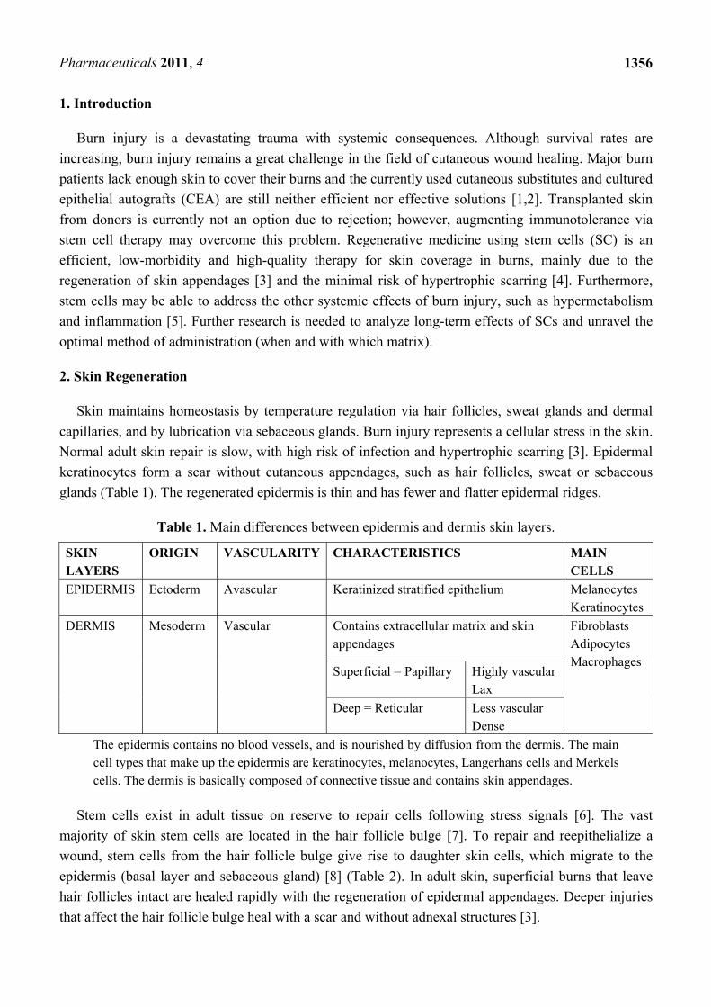
Table 1. Main differences between epidermis and dermis skin layers.
SKIN LAYERS
ORIGIN VASCULARITY CHARACTERISTICS MAIN CELLS
EPIDERMIS Ectoderm Avascular Keratinized stratified epithelium Melanocytes Keratinocytes
DERMIS Mesoderm Vascular Contains extracellular matrix and skin appendages
Fibroblasts Adipocytes Macrophages
Superficial = Papillary Highly vascular Lax
Deep = Reticular Less vascular Dense
The epidermis contains no blood vessels, and is nourished by diffusion from the dermis. The main cell types that make up the epidermis are keratinocytes, melanocytes, Langerhans cells and Merkels cells. The dermis is basically composed of connective tissue and contains skin appendages.
Stem cells exist in adult tissue on reserve to repair cells following stress signals [6]. The vast
majority of skin stem cells are located in the hair follicle bulge [7]. To repair and reepithelialize a
wound, stem cells from the hair follicle bulge give rise to daughter skin cells, which migrate to the
epidermis (basal layer and sebaceous gland) [8] (Table 2). In adult skin, superficial burns that leave
hair follicles intact are healed rapidly with the regeneration of epidermal appendages. Deeper injuries
that affect the hair follicle bulge heal with a scar and without adnexal structures [3].

Pharmaceuticals 2011, 4
1357
Table 2. Types of cutaneous stem cells.
Epidermal Dermal Sebaceous Hair follicle
Sweat glands
Melanocytes MSC Neural Endothelial
Interfollicular Hair Bulge
Cutaneous stem cells include epidermal stem cells (interfollicular and bulge stem cells), dermal stem cells, sebaceous stem cells, hair follicle stem cells, sweat gland stem cells, melanocyte stem cells, mesenchymal stem cells, neural stem cells and endothelial stem cells. The more abundant skin stem cells are the epidermal hair bulge stem cells. Only a small fraction of stem cells, the interfollicular stem cells, reside in the basal layer of the interfollicular epidermis. These stem cells maintain adult skin homeostasis and hair regeneration, but they also participate in the repair of the epidermis after trauma.
Regenerative medicine aims to not only accelerate reepithelialization after burn injury, but also to
reconstruct functional skin with sweat glands, hair follicles and dermal capillaries. These goals might be
achieved by stem cell therapy. Approaches to stem cell therapy include local recruitment of endogenous
SCs or SC transplantation (often in vitro modified), either of which can be combined with gene therapy
or tissue engineering. Tissue engineering is an experimental procedure that combines cellular biology,
engineering and medicine to develop three-dimensional tissues and restore function [9].
3. Stem Cells Definition and Classification
3.1. Stem Cells Definition
Stem cells (SCs) are defined by two main characteristics: their capacity of prolonged self-renewal
(proliferation) and multilineage differentiation (asymmetric replication) [3,10]. These characteristics are
more pronounced in younger sources [11]. By asymmetric replication, after every cell division, one cell
retains its self-renewing capacity, while the other (transit-amplifying or TA cell) enters a differentiation
pathway and joins a mature non-dividing population [12]. When an unspecialized stem cell differentiates, it
assumes characteristics of a specific tissue [13]. SCs are pluri-, multi- or unipotent [14]. The zygote is the
only mammalian cell capable of producing all cells and tissues of an organism and thus is considered
totipotent [15]. Pluripotent cells produce cells and tissues belonging to all three germ layers—
Ectoderm, mesoderm and endoderm [16]. Multipotent cells produce more than one cell lineage, within
a closely related family of cells. Unipotent cells only differentiate into a single cell phenotype [17].
Plasticity describes the phenomenon whereby SCs from one tissue produce cell types of a completely
different tissue [18]. SCs can remain undifferentiated, in which state there is risk of uncontrolled
proliferation and tumor formation [11]. SCs have a slow-cycling nature in vivo, high proliferative
potential and participate in tissue regeneration and repair, during both fetal development and adult
wound healing [19].
3.2. Stem Cells Classification
When classified by origin, there are two types of stem cells: embryonic (ESC) and non-embryonic
stem cells. The latter are also referred to as adult (ASC) or somatic stem cells (however, SCs derived
from the placenta or umbilical cord are also considered ASCs) [20,21]. Embryonic germ cells are

Pharmaceuticals 2011, 4
1358
derived from the primitive gonadal ridges of the developing embryo or fetus (6-9 weeks gestation in
the humans) and have many of the pluripotential properties of ESCs [22].
3.2.1. Embryonic Stem Cells (ESCs)
ESCs are pluripotent stem cells derived from the inner cell mass of an early stage embryo known as
blastocyst [10] and give rise to all cells of the three embryonic germ layers: endoderm, mesoderm
and ectoderm. Human Embryonic Stem Cells (hESCs) are derived from excess developing
pre-implantation embryos (5 day-old embryos, 4-8 day-old morula, or inner cell mass of blastocysts)
that have usually been fertilized in vitro at a fertilization clinic. ESCs can be propagated indefinitely in
an undifferentiated state [23] in vitro using either feeder layers or extracellular stimuli (e.g., cytokines
or growth factors) [24]. Derivation of human embryonic cell lines is controversial because it requires
destruction of an embryo [11], may develop teratocarcinomas (tumours composed of tissues from all
three germ layers [25]), are immunogenic and show genetic instability in vitro [15]. Accordingly, adult
stem cell research is currently favoured [6,26,27]. Apart from being used in regenerative medicine,
ESCs may be used to perform developmental, genetic (through knock-out technology) and
pharmacological research. hESC-based in vitro studies of drug toxicity have proven to be an accurate
alternative to experimental animal models [28].
ESCs are capable of unlimited expansion in vitro and are considered an immortal epiblast derivative
with a checkpoint in differentiation that enables their expansion as undifferentiated colonies, which
can be instructed to maintain this phenotype indefinitely [23].
3.2.2. Adult Stem Cells (ASCs)
SC clinical studies have increased during the past two decades in almost every field of medicine;
including, haemato/immunotherapies [29-32], diabetes mellitus [33], chronic degenerative illnesses
(e.g., in the field of rheumatology) [34-37], cardiovascular diseases [38], multiple sclerosis and other
neuropathies [39], corneal repair [40] and wound healing [41].
Adult stem cells (ASCs) were discovered in the mid-1950s; they are found in low abundance in
almost all adult tissues and in high abundance in the umbilical cord [10]. They are found in special
regulatory niches as self-renewing progenitor cells that are able to produce one or more specialized cell
types. ASCs are usually considered to be tissue specific, self-renewing populations of cells, which can
differentiate into cell types associated with the organ system in which they reside [42].
Slowly replicating and bromodeoxyuridine-label-retaining, ASCs are under strict regulatory control
of their mobilization and differentiation [23]. ASCs are less potent (usually only uni- or multipotent)
and have less differentiation potential than ESCs. Distinct from ESCs, ASCs are not capable of
unlimited expansion in vitro. ASC potency and plasticity is still in contention, though [11,22,43].
ASCs include mesenchymal stem cells (MSCs), hematopoietic stem cells (HSCs), epithelial and
neural stem cells, and others. HSCs and MSCs originate in the bone marrow (MSCs may have
additional origins, as we will describe later) and differentiate into endothelium, liver, bone, muscle,
skin or others [11].
Epidermal and dermal SCs are also multipotent. It has been suggested that they not only contribute
to skin production, but can also be stimulated into neural and osteogenic lineages [44,45].

Pharmaceuticals 2011, 4
1359
3.2.3. Induced Pluripotent Stem Cells (iPSCs)
Induced pluripotent stem cells (iPSCs) are artificially derived from non-pluripotent cells, typically
adult somatic cells (mostly fibroblasts of murine or human origin), most frequently by epigenetic
reprogramming and also by nuclear transfer or cell division [46]. Expression of transcription
factors characteristic for undifferentiated embryonic stem cells is induced; for example, OCT4 (also
known as POU5F1, being the most important one), SOX2, c-MYC, KLF4, Lin28 and/or
NANOG [6,10,15,47,48] (Figure 1). Transcription factors or cell markers are the key mediators of
cellular identity [49]. Direct reprogramming (also referred to as transdifferentiation) describes ectopic
expression of defined transcription factors, a very slow and inefficient process that may limit the quality
of resulting iPSCs [50]. For example, fibroblasts can be reprogrammed into neurons, cardiomyocytes and
blood-cell progenitors [51-53]. Small molecules may improve the reprogramming efficiency but increase
its tumorigenicity [54]. Elements that influence reprogramming include the respective donor cell type,
the transcription or reprogramming factors utilized, the mode of delivery (e.g., virus, RNA, etc.) and the
culture conditions, all of which depend on the purpose of the process [46].
Figure 1. Methods of production of induced pluripotent stem cells. Reproduced from [10]
with permission from Rightslink.
iPSCs are generated from adult somatic cells through somatic cell nuclear transfer (SCNT), cell fusion or direct reprogramming, by ectopic expression of defined transcription factors. iPSCs are pluripotent stem cell lines suitable for disease modeling, autologous cell therapy, drug screening and basic research.
Broadly, we could imagine iPSCs as “artificial ESCs”. iPSCs represent stable lines of embryonic-like
pluripotent stem cells [55]. In contrast to ESCs, human iPSCs can be derived from the patient to be
treated (for autologous cell therapy), reducing the risk of HLA mismatching and immune rejection [56].
iPSCs may also be used for establishing in vitro disease models, drug or toxicity screening, and basic
gene research [46]. iPSCs represent a widely available, non-controversial, non-restricted and practically
infinite source of pluripotent cells. Nonetheless, they still share with classic ESCs the critical

Pharmaceuticals 2011, 4
1360
disadvantage of malignancy transformation [10]; although, integrative delivery systems with consequent
deletion seems to lower the risks of iPSCs oncogenesis [54]. A multitude of protocols for iPSCs
generation have been developed in recent years, spanning across different mouse and human donor
populations and varying in the number, identity and delivery of the reprogramming factors [57-60].
4. Adult Mesenchymal Stem Cells
Ideally, stem cells for regenerative medicine should be abundantly available, accessible by a
minimally invasive procedure and then safely and effectively transplanted to either an autologous or
allogeneic host [6]. As previously mentioned, tumorigenicity and ethical considerations have impeded
the widespread clinical use of ESCs [11]. Instead, most regenerative medicine research is focused on
iPSCs and ASCs; in particular, adult mesenchymal stem cells (MSCs) [61,62].
MSCs are derived mainly from bone marrow and adipose tissue [63,64], and to a lesser extent,
placenta [65], amniotic fluid [61], umbilical cord [66], dental pulp [67], tendon [68], trabecular bone [69]
and synovia [70]. Actually, MSCs may reside in all post-natal tissues. Bone marrow (where they were
first identified) and adipose tissue are the main sources of MSCs for cell therapy, due to high
expansion potential and reproducible isolation protocols [71]. CD34+ hematopoietic stem cells are the
most widely studied and represent the only currently clinically approved stem cell [12,71].
Human mesenchymal stem cells (hMSCs) are characterized by three criteria: (1) plastic-adherent
under culture; (2) capacity to differentiate into at least three mesenchymal lineages: bone, fat and
cartilage; (3) express cell markers CD73, CD90, CD105 and negative for CD11b, CD14, CD34, CD45
and HLA-DR [62,72].
MSCs release various cytokines and growth factors that influence the microenvironment by either
modulating the host immune response or stimulating resident cells [73], conferring anti-fibrotic,
anti-apoptotic, pro-angiogenic and immunosuppressive properties [74,75]. Mediators involved in
MSC-mediated immunomodulation include interferon-, toll like receptors, tumor necrosis factor-,
interleukin (IL)-1, IL-1, indoleamine 2,3-dioxygenase, leukemia inhibitory factor, HLAG5, IL-10,
transforming growth factor (TGF)-1, hepatocyte growth factor, heme oxygenase1, IL6, IL-1 receptor
antagonist (IL-1RA) and prostaglandin E2 [71,75-80]. MSCs also stimulate the proliferation of other
progenitor cell populations within target organs to promote endogenous repair [77].
MSCs have a great potential in tissue engineering and may serve to treat chronic inflammatory and
degenerative disorders due to their immunosuppressive properties [75,81]. They are currently being
tested in several clinical trials for osteoarthritis, osteogenesis imperfecta, articular cartilage defects,
osteonecrosis and bone fracture [82,83]. They are considered ‘immunoprivileged’ by many researchers
and may permit allo-transplantation without immunosuppressive therapy, which would become
particularly useful in treating acute injuries [84,85]. Having said that, animal studies have shown that
intramyocardial injection of MSCs may differentiate into encapsulated structures with calcifications
and ossifications, raising the possibility of malignant transformation [77].

Pharmaceuticals 2011, 4
1361
5. Main Sources of Adult Stem Cells
5.1. Bone Marrow-Derived Stem Cells (BMSCS)
Bone marrow has been the primary source of mesenchymal stem cells; however, bone marrow
collection is invasive and MSC isolation is inefficient (<0.05%) [86,87]. BMSCs are able to undergo cell
fusion, a natural process of mingling of genetic material that modifies gene expression patterns [88]. Cell
fusion is implicated in regeneration, normal development, immune response and tissue formation and
plays a prominent role in stem cell plasticity [89]. Indeed, cell fusion can modify the gene program and
govern cell fate, transforming the cell into a more immature state to achieve a regenerative function [90].
Haematopoietic stem cell transplantation is the first and most widely available stem cell therapy [91].
hMSCs derived from bone marrow are capable of differentiating into epithelial cells of the liver, lung,
gastrointestinal tract and skin [92].
Autologous bone-marrow derived cultured hMSCs have been applied to wounds, using a
specialized fibrin spray system; this approach is currently being performed and supposed to be safe and
valid for topical cell administration, although with no solid data to support its validity [93].
5.2. Adipose Tissue-Derived Stem Cells (ADSCS)
Adipose-derived stem/stromal cells (ADSCs) are multipotent somatic stem cells that can
differentiate into several lineages, including adipose cells, chondrocytes, osteoblasts, endothelial cells,
epithelium, cardiomyocytes and neuronal cells [94]. The existence of stem cells within adipose tissue
was reported for the first time in 2001 [95]. They are often described as processed lipoaspirate cells
(PLA), preadipocytes, or adipose stem cells, although the international Fat Applied Technology
Society recommends the term “ADSCs” [6]. ADSCs express mesenchymal cell-specific markers and
molecular markers typical for the embryonic stem cell phenotype: OCT4, Nanog and Sox2 [96,97].
ADSCs are heterogeneous, differing depending on their anatomical regions and depending on their
type (white or brown) [98].
Current research suggests that they may actually be pluripotent and form cell types of all three germ
layers [99]. ADSCs represent a promising source of adult mesenchymal stem cells, mainly because
isolation is less invasive and more efficient [6,71]. Aspirated fat is in plentiful supply in many plastic
surgery procedures—e.g., liposuction and liposculpture—and precursor cells can be purified to obtain
the ADSC-rich stromal vascular fraction (SVF) [100]. The SVF is a heterogeneous mixture containing
endothelial cells, preadipocytes, fibroblasts, vascular cells, macrophages, and numerous mesenchymal
stem cells [6] and is now studied as a supplement to free fat transfer in order to increase yield [101].
Expansion of ADSC populations in culture can yield 100 to 1,000 times more progenitor cells than
isolation from bone marrow [102].
Besides being one of the richest sources of ASCs in the human body, adipose tissue is also an
endocrine organ that secretes numerous hormones, growth factors and cytokines, that support wound
healing and other functions such as leptin, epidermal growth factor, tumor necrosis factor-, basic
fibroblast growth factor, keratinocyte growth factor, transforming growth factor-1 (TGF-1), vascular
endothelial growth factor, hepatocyte growth factor, interleukin (IL)-6, IL-7, IL-8, IL-11, IL-12,
macrophage-colony stimulation factor, platelet-derived growth factor, brain-derived neurotrophic

Pharmaceuticals 2011, 4
1362
factor, granulocyte colony stimulating factor and leukaemia inhibitory factor [6,102]. These paracrine
influences are of great importance in many stem cell therapies, creating a favourable environment for
development of more functional cells and tissue repair, through promotion of neovascularization,
endogene repair mechanisms and regulation of immune responses [77,103].
ADSCs represent a highly efficient source of iPSCs [104]. They may be used to test drug toxicity,
reducing the need for animals. They may also offer an important tool for cell-based gene therapy in the
field of wound healing, because ADSCs can efficiently (above 60%) be transducted with vectors [12,105].
Various clinical trials have shown the regenerative capability of adipose-derived stem cells in
subspecialties of medical fields such as plastic surgery (for breast reconstruction and aesthetic
lipofilling), general surgery (to heal chronic fistulas in Crohn’s disease), orthopedic surgery, oral and
maxillofacial surgery (to stimulate bone repair in calvarial defects) and cardiology (ischemic heart
disease and acute myocardial infarct) [77,106,107]. With the risk of malignancy transformation and
heterogeneity, however, there is still not enough evidence to encourage wide and safe use of ADSCs
and further research is required [108-110]. Future studies to optimize the differentiation of ADSCs into
deficit cells may unlock their therapeutic potential in regenerative medicine.
All in all, ADSCs hold great promise for use in tissue repair and regeneration, due to their availability,
pro-angiogenic an anti-apoptotic factor secretion, immunomodulatory effects and multilineage
differentiation, becoming one of the most popular adult stem cells currently explored [6,77,100,103,111].
5.3. Umbilical Cord (Blood) Derived Stem Cells
Umbilical cord (UC) and cord blood-derived stem cells remain the world’s largest potential source
of stem cells, considering the global birth rate of around 135 million per year [86]. The umbilical cord
represents a well known source of endothelial progenitor cells [112]. Umbilical cord blood contains
haematopoietic as well as non-haematopoietic stem cells, these latter also named as CBEs (Cord Blood
Embryonic-like stem cells) [113,114]. CBEs have been shown to differentiate into neural,
hepatobiliary, pancreatic-like precursors and potentially others [115,116].
Human umbilical cord blood is a rich source of hemopoietic stem cells for clinical application and
may be one of the largest sources of stem cells with naive immune status [12,101].
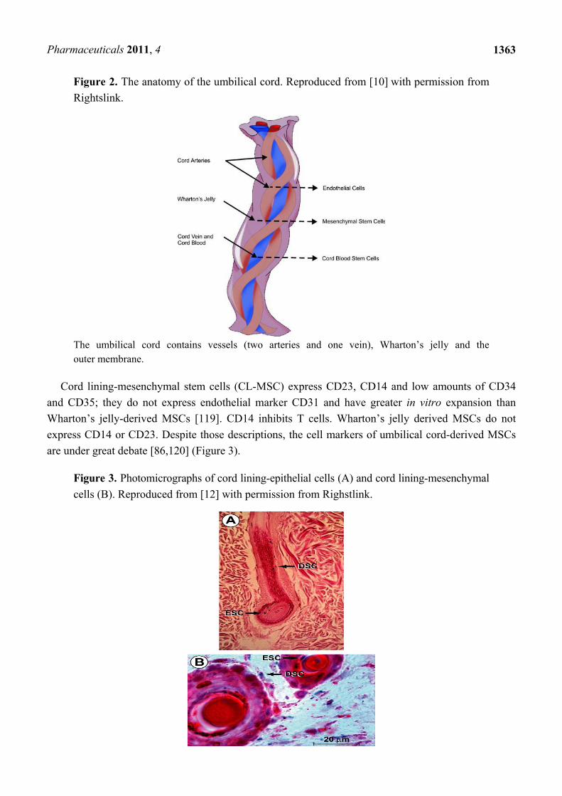
From innermost to outermost, the layers within the UC include the vessels, Wharton’s jelly or matrix,
and amniotic membrane or cord-lining, epithelium or subamnion [86]. Wharton’s jelly gives rise to
MSCs and has lower cell density, yet allows us to quickly isolate many cells [117]. A single piece of
5–10 mm3 of Wharton’s jelly has the potential to yield up to 1 billion MSCs in 30 days; bearing in mind
that the average UC measures 50 cm [15], it undoubtedly represents a rich source of SCs. Human
umbilical cord perivascular cells are nearly identical to Wharton’s jelly MSCs [118] (Figure 2).
Amniotic membrane and cord-lining are sometimes interchangeable words referring to the UC
membrane in general, or referring to different cell types. The outer membrane of the UC is an
extremely rich source of stem cells for burn resurfacing [12]. The cord lining gives rise to multipotent
epithelial stem cells (CL-epithelial stem cells) [86].
Pharmaceuticals 2011, 4
1363
Figure 2. The anatomy of the umbilical cord. Reproduced from [10] with permission from
Rightslink.
The umbilical cord contains vessels (two arteries and one vein), Wharton’s jelly and the outer membrane.
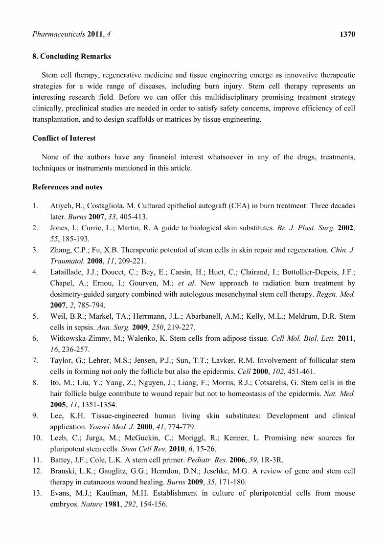
Cord lining-mesenchymal stem cells (CL-MSC) express CD23, CD14 and low amounts of CD34
and CD35; they do not express endothelial marker CD31 and have greater in vitro expansion than
Wharton’s jelly-derived MSCs [119]. CD14 inhibits T cells. Wharton’s jelly derived MSCs do not
express CD14 or CD23. Despite those descriptions, the cell markers of umbilical cord-derived MSCs
are under great debate [86,120] (Figure 3).
Figure 3. Photomicrographs of cord lining-epithelial cells (A) and cord lining-mesenchymal
cells (B). Reproduced from [12] with permission from Righstlink.

Pharmaceuticals 2011, 4
1364
Generally, umbilical cord-derived MSCs can differentiate into bone, skin, endothelium, hepatocyte,
neural lineages and others. Amniotic membrane-derived MSCs specifically can differentiate into bone,
cartilage and fat [12,121].
Regarding hematologic diseases, the immaturity of umbilical cord blood (UCB) cells is associated
with low immunogenicity, which reduces their graft-versus-host reactivity compared to adult-derived
bone marrow grafts [122]. On the other hand, umbilical cord blood supplies multipotent stem cells at a
rate 30% lower than that achieved from adult bone marrow [101]. Umbilical cord blood was
introduced as an alternative source of allogeneic HSCs after the success of cord transplantation in a
child with Fanconi’s anemia. Both cord blood transplants and matched unrelated bone marrow
transplants share similar disease-free survival and transplant-related mortality [91]. Further research
will need to clarify when allogeneic cord blood transplantation is best indicated [124].
With respect to burns and skin wound healing, umbilical cord and amniotic membrane may emerge
as new promising sources of “off-the-shelf” cell-engineered skin [86]. Moreover, co-administration of
several types of stem cells may elicit synergistic benefits [77], suggesting the use of both epithelial and
mesenchymal stem cells.
5.4. Hair-Follicle Stem Cells
Hair follicles are a promising source of easily accessible multi (or pluri) potent stem cells which are
non-oncogenic and carry no ethical concerns, in contrast to embryonic stem cells or induced
pluripotent stem cells. In fact, many researchers consider hair follicles to be the most promising source
of multipotent stem cells [19,124].
Hair follicle pluripotent stem cells of the scalp are positive for nestin and the embryonic stem cell
transcription factors Nanog and Oct4. These cells can differentiate into neurons, smooth muscle cells
and melanocytes [125]. The hair follicle bulge area contains nestin-negative, K15-positive cells; these
cells can differentiate into keratinocytes, neurons, glial cells and smooth muscle cells [126].
Human hair follicle stem cells promote nerve repair or the functional recovery of injured peripheral
nerve and spinal cord [127].
Hair follicle bulge stem cells give rise to both hair follicle cells and epidermal cells. The hair
follicle stem cells form epidermal stem cells only when the epidermis is wounded or stressed [128].
Bulge stem cells respond rapidly to epidermal wounding by generating short-lived TA cells
responsible for acute wound repair [129] (Figure 4). Intense research is devoted to this promising
source of stem-cell therapy to improve wound healing.
6. Directing Cell Fate for Regenerative Medicine
Regenerative medicine or cell-replacement therapy aims to treat human diseases caused by deficits
in quality or quantity of particular cells, restoring damaged tissues in addition to alleviating the related
symptoms. These diseases include neurodegenerative disorders, diabetes, liver and cardiovascular
diseases, blindness, deafness, burns, and many others [130,131].

Pharmaceuticals 2011, 4
1365
Figure 4. Hair follicle bulge and multipotent stem cells. Reproduced from [19] with
permission from Wolters Kluwer.
Schematic representation of the cutaneous epithelium and the cell lineages derived from multi-potent stem cells in the hair follicle bulge. Under steady-state conditions, the stem cells are quiescent. At the beginning of the hair cycle when the lower follicle and matrix is regenerated, the stem cells in the bulge proliferate to give rise to new hair follicle keratinocytes. Under wounding conditions, the stem cells produce daughter cells that migrate from the niche to re-populate the basal layer of the epidermis, and the sebaceous gland.
To accomplish its goal, regenerative medicine should be able to not only create these cells, but also
to deliver them to patients. To create them, we can direct the cell fate of already available cells (ideally
the patient’s own cells, although age and comorbidities impair stem cell functionality [12,77]).
There are two main approaches to direct cell fate: (1) through directed differentiation, whereby
cultured pluripotent stem cells (e.g., ESCs or iPSCs) follow several steps as they would in vivo or
during embryonic development; or (2) via reprogramming or transdifferentiation, whereby a
differentiated cell is converted directly into the cell of interest without proceeding through a
pluripotent intermediate, most often by transcription factors [50,132-134].
Differentiation is performed in vitro by treating cells with recombinant growth factors (e.g., TGF-
superfamily, WNT and fibroblast growth factors, combined with co-culture systems), or with small
molecules, which are homogenous, stable chemical compounds that are non-immunogenic and cheaper
than proteins [135,136]. In addition, differentiation can be accomplished through spontaneously with
embryoid bodies or floating clumps of cells [50].

Pharmaceuticals 2011, 4
1366
Unfortunately, current protocols are still inefficient [137,138], but solutions for directed cell fate
and transdifferentiation are under continuous investigation.
7. Stem Cells and Burns
Cell therapy has been used to treat burns since the introduction of composite epithelial autografts
(CEA) by Green in 1975, evolving to dermal substitutes, later on to dermal-epidermal bio-engineered
cultured skin substitutes and eventually to stem cells [139]. Stem cell therapy (allogeneic MSCs, iPSCs
or immunomodulatory TCells) after burn injury emerges as a promising treatment strategy, not only
for wound healing, but also to treat systemic effects of burn trauma, the hypermetabolic response,
inflammation (e.g., inflammatory-related diseases, such as acute lung injury/respiratory distress
syndrome), and immunosuppression [5,140]. Stem cell therapy may offer an alternative to large volume
resuscitation and be an adjunct to lung-protective ventilation strategies after severe burn injury [141].
Paracrine mechanisms and growth factor secretion, rather than post-engraftment differentiation and
proliferation, seem to predominate in therapeutic effects of MSCs [71,139]. In vivo, MSCs attenuate
proinflammatory cytokine release and nitric oxide production while upregulating the anti-inflammatory
cytokines TGF-, IL-10 and IL-12 [5]. MSCs also exhibit antiapoptotic, immunosuppressive and
antifibrotic effects [62].
For the treatment of acute and chronic non-healing wounds (not burn related), combined gene
delivery with stem cell therapy appears promising [12]. Gene therapy involves the insertion of a gene
into recipient cells by viral transfection, naked DNA application, high pressure injection or liposomal
vectors [142]. Sequential growth factor gene therapy delivers a cocktail of growth factor genes at
strategic time points of wound healing [12,143].
To enhance the therapeutic response after stem cell treatment in burn patients, intense tissue
engineering with the development of 3D scaffolds or matrices is of vital importance, as well as
improved preconditioning cell treatments and optimized culture conditions [77].
7.1. The Challenge of Stem Cell Delivery in Burn Patients: A Link between Scaffolds and Wound Healing
Stem cell administration for burn patients still remains a challenge of intense research [140].
Intravenous infusion and local application of MSCs have been described in the clinical setting [144]. The
concomitant use of acellular matrices or scaffolds is strongly recommended in order to increase cell
homing, differentiation, mobilization and adhesion, eventually improving wound healing [144,145].
An ideal method for the effective administration of stem cells for burn patients has not yet been
elucidated. Further comparison of the local and systemic effects in burn patients associated with each
route of stem cell delivery needs to be performed. There is still not enough evidence in terms of
analyzing systemic or local effects of stem cell delivery in burn patients, regarding different possible
routes of administration. We still do not know exactly which percentage of locally delivered cells in a
burn wound exerts its effects in wound healing and which, if any, affects the main circulation and has
systemic effects, regarding the inflammatory and hypermetabolic response after a major burn. On the
other hand, the efficiency of non-topical administration (such as intrapulmonary infused cells primarily
to treat adult respiratory distress syndrome) for wound healing is unclear. To help clarify this, reporter

Pharmaceuticals 2011, 4
1367
gene imaging allows for stem cell tracking in vivo. Briefly, positron emission tomography is used to
detect cell markers for assessment of the viability and location of stem cells after transplantation [146].
If we focus on wound healing, application of cells to the burn wound could be performed, either by
the bedside as a non-invasive procedure, or in the operating room, immediately after debridement. The
cells should be transferred on a matrix, scaffold or dermal substitute. One method is to first spray cells
onto the wound with fibrin sealant [93] and afterwards cover with a dermal substitute, skin graft or
film. The cover over the cells acts not only as a temporary dressing, but also to theoretically enhance
cell paracrine signaling and homing of the cells, improving wound healing [147]. Although the use of
cell administration using spray technologies is currently being performed in the clinical setting, there
are no conclusive data to support its validity as a matrix vehicle.
Cutaneous wound healing requires the well-orchestrated integration of cell migration and
proliferation, as well as extracellular matrix deposition, angiogenesis, and remodelling [12]. Delays in
burn wound closure worsen a patient’s susceptibility to infection, prolong pain, increase the total number
of operative procedures, increase the incidence of hypertrophic scarring, and lengthen hospital stays.
Stem cell therapies in wound care may lessen these morbidities [140]. Specifically, the burn wound has
unique characteristics that have to be considered when designing a clinical trial for stem cell therapy
applications: it is an ischemic wound, with an altered pH and temperature, prone to infection and
development of chronic sequelae such as non-healing ulcers (sometimes even with malignant
conversion) and hypertrophic scarring [148,149]. Furthermore, a major burn represents a handicap, with
uncovered wounds open to air, which require frequent operations and dressing changes, and with long
periods of immobilized hospital stay, which involve frequent position changes and physiotherapy, to
avoid pressure sores, enhance rehabilitation and improve overall prognosis. This dynamic paradigm
popularized the use of polymeric films for repair and closure of wounds. These films are semipermeable,
microperforated and transparent materials that create an accelerated healing environment while avoiding
dehydration, trauma and infection over the injury [150]. Additionally, radiofrequency applied to
wound-contacting nano-engineered polymeric films (iron oxide-coated biodegradable nanoparticles
and/or fibers -NPFs-) has been used to debride the wound. This may represent a novel burn treatment
method, once stronger scientific evidence is available [151].
Several studies support the use of “intelligent matrices”, natural acellular matrices such as a porcine
dermal acellular matrix accompanied by NPFs coated with monoclonal antibodies (e.g., anti-CD44)
and loaded with specific growth factors, cytokines and antibiotics. The growth factors and cytokines
improve the homing of autologous circulating MSCs, often elevated in patients with large burns [152],
and also of delivered stem cells in general [144]. Chemoattractants for BM-MSC include hepatocyte
growth factor, basic fibroblast growth factor and CXCL5, for instance [153]. Indeed, recruiting
endogenous stem cells to the site of injury represents an alternative to transplantation; however, direct
application of stem cells appears advantageous over diffusible growth factor administration. Stem cells
can interact with their environment and release multiple wound healing factors [12]. Unfortunately,
tissue-engineered dermal equivalents lack blood vessels and may act as possible barriers against
nutrients necessary for keratinocyte or stem cell survival on top of such composites [144]. The time
required to reperfuse the skin substitute increases the duration of ischemia, enhancing the risk of
infection and graft destruction. One approach to minimize ischemia is through the use of BM-MSCs,
which secrete VEGF [153]. On the other hand, VEGF could have detrimental effects in scarring [149].

Pharmaceuticals 2011, 4
1368
Furthermore, in severe burn trauma with sepsis, bone marrow suppression has been described.
Ichoka et al. investigated the effect of a BM-MSC impregnated collagen matrix on wound healing in a
microcirculatory mouse model and observed significant increases in angiogenesis [153]. Other models
of delayed wound healing in diabetes also report stem cells stimulating wound healing. Fiorina et al.
showed that bone-marrow progenitor cells BM/PCs mobilize to the site of injury during diabetic
wound repair in db/db mice, where increased levels of stem cells corresponded to higher levels of
wound reepithelialization [154].
Ha et al. showed that transplantation of BMSCs transfected with an adenovirus hepatic growth
factor gene can accelerate wound repair in diabetic rats [155]. Tark et al. showed that human
CB-MSCs improved wound healing, when applied to diabetic mice, probably due to TGF- [156].
Promising preclinical results with stem cells in wound healing are encouraging further research. In
2006, a patient bearing a diabetic foot was treated with a combination of autologous BM-MSCs and
autologous fibroblasts on a biodegradable collagen membrane. Wound size decreased and vascularity
increased [144]. Currently, nanoengineered multifunctional acellular biologic scaffolds, films and
wound dressings emerge as delivery vehicles for stem cells in burn wounds. Although many types of
tissue-regenerating stem cells may be used clinically, adding endometrial stem cells to MSCs may
improve the vascularisation of these tissue engineered-constructs and significantly improve the
outcomes of severely burned patients [144,145].
7.2. Legislation, Biosafety and Biotechnology Industry in Cell Therapy for Burn Care
Stem cell research is in its early stages of development and the market is therefore still immature.
Approximately 1.1 million people affected by burns and other wounds in the U.S. would benefit from
cell therapy products [157]. Although the preliminary results achieved to date raise great expectations,
many pharmaceutical companies are reluctant to enter this market. To date, the most profitable strategy
has been the signing of agreements between big pharmaceutical and other small biotechnology
companies whose activity is entirely devoted to cell therapy and regenerative medicine. In addition,
many stem cell-derived products are developed at universities and basic research institutions, where
preclinical studies are also conducted.
Cell therapy, gene therapy and tissue engineering are considered ‘advanced therapy products’
(ATPs). As such, they should follow a regulatory framework to ensure patient accessibility and
governmental assistance. An effective regulation implies scientific reality and objectivity, as well as
flexibility to adapt as technology changes [159].
The U.S. Food and Drug Administration (F.D.A.) defines somatic cell therapy as the administration of
autologous, allogeneic or xenogeneic non-germ cells excluding blood products for transfusion, which
have been manipulated, processed, propagated, expanded, selected ex vivo, or drug-treated [160]. Cell
therapy products are considered drugs, so they follow the same regulations, requiring a strict control of
manipulation and facilities. Cell therapy products should adhere to the Current Good Manufacturing
Practices, including quality control and quality assurance programs, which establish minimum quality
requirements for management, staff, equipment, documentation, production, quality control,
contracting-out, claims, product recall, and self-inspection [159]. The key points of the current FDA
regulation for cell therapy products include: demonstrations of preclinical safety and efficacy; no risk for

Pharmaceuticals 2011, 4
1369
donors of transmission of infectious or genetic diseases; no risk for recipients of contamination or other
adverse effects of cells or sample processing; specific and detailed determination of the type of cells
forming the product and what are their exact purity and potency; in vivo safety and efficacy of the
product [161]. Cell therapy products must be carefully described, stating whether autologous, allogeneic
or xenogeneic cells are administered. According to the U.S. F.D.A., human cells are considered
xenogeneic cells provided there has been any ex vivo contact with living non-human cells, tissues or
organs [160]. It should also be detailed whether cells have been manipulated together with biomaterials,
growth factors or serum, which is common in burn applications. Indeed, the biomaterial itself may have a
more important role than the cell product [159]. Biomaterials for cell therapy should be biocompatible to
prevent immune rejection or necrosis, biodegradable and assimilable without causing an inflammatory
response, and have certain structural and mechanical properties [162]. Whether natural or artificial,
biomaterial type and use is related to the route of administration of cell therapy protocols, implantation or
injection. In the latter, which are simpler, biomaterials are usually in a hydrogel state, forming a
hydrophilic polymer network, as occurs in polyethylene oxide, polyvinyl alcohol, polyacrylic acid,
agarose, alginate, collagen and hyaluronic acid. Control of the biomaterials’ porous structure is very
important for increasing their efficacy in tissue regeneration [159,162].
Regarding the production process, a detailed description must be given of all procedures related to
product quality in the Standard Operating Procedures, as for conventional medical products. The
purity, safety, functionality and identity criteria used for conventional drugs must be met.
Development of techniques for cell identification within a mixed cell culture population and for
follow-up of transplanted cells will also be essential to ascertain the potential in vivo invasive
processes and to ensure biosafety [159]. The production process must occur in a highly aseptic
environment with comprehensive controls of both raw materials and handlers; it has to be reproducible
and validated. Facilities where products are handled, packaged and stored should be designed and
organized according to the Good Manufacturing Practice for Pharmaceutical Manufacturers (G.M.P.)
guidelines [163]. Production and distribution should be controlled by the relevant local or national
authorities based on the International Conference on Harmonization of Pharmaceuticals for Human
Use, which standardized the potential interpretations and applications of the corresponding
recommendations [164]. It is of paramount importance to prevent potential contamination, both
microbiological and by endotoxins, due to defects in environmental conditions, handlers, culture
containers or raw materials, or cross contamination with other products prepared at the same
production plant. The number of technical staff should be the minimum required and should be trained
in hygiene measures required for manipulation in a clean room [159].
In summary, aspects to be regulated mainly include control of development, manufacturing and
quality using release and stability tests; non-clinical aspects such as the need for studies on
biodistribution, cell viability and proliferation, differentiation levels and rates, and duration of in vivo
function; and clinical aspects such as special dose characteristics, stratification risk and specific
pharmacovigilance and traceability issues [159-161].
Since new stem cell-based therapies develop very fast, the regulatory framework must also adapt,
although legislation may be expected to change more slowly.
Pharmaceuticals 2011, 4
1370
8. Concluding Remarks
Stem cell therapy, regenerative medicine and tissue engineering emerge as innovative therapeutic
strategies for a wide range of diseases, including burn injury. Stem cell therapy represents an
interesting research field. Before we can offer this multidisciplinary promising treatment strategy
clinically, preclinical studies are needed in order to satisfy safety concerns, improve efficiency of cell
transplantation, and to design scaffolds or matrices by tissue engineering.
Conflict of Interest
None of the authors have any financial interest whatsoever in any of the drugs, treatments,
techniques or instruments mentioned in this article.
References and notes
1. Atiyeh, B.; Costagliola, M. Cultured epithelial autograft (CEA) in burn treatment: Three decades
later. Burns 2007, 33, 405-413.
2. Jones, I.; Currie, L.; Martin, R. A guide to biological skin substitutes. Br. J. Plast. Surg. 2002,
55, 185-193.
3. Zhang, C.P.; Fu, X.B. Therapeutic potential of stem cells in skin repair and regeneration. Chin. J.
Traumatol. 2008, 11, 209-221.
4. Lataillade, J.J.; Doucet, C.; Bey, E.; Carsin, H.; Huet, C.; Clairand, I.; Bottollier-Depois, J.F.;
Chapel, A.; Ernou, I.; Gourven, M.; et al. New approach to radiation burn treatment by
dosimetry-guided surgery combined with autologous mesenchymal stem cell therapy. Regen. Med.
2007, 2, 785-794.
5. Weil, B.R.; Markel, TA.; Herrmann, J.L.; Abarbanell, A.M.; Kelly, M.L.; Meldrum, D.R. Stem
cells in sepsis. Ann. Surg. 2009, 250, 219-227.
6. Witkowska-Zimny, M.; Walenko, K. Stem cells from adipose tissue. Cell Mol. Biol. Lett. 2011,
16, 236-257.
7. Taylor, G.; Lehrer, M.S.; Jensen, P.J.; Sun, T.T.; Lavker, R.M. Involvement of follicular stem
cells in forming not only the follicle but also the epidermis. Cell 2000, 102, 451-461.
8. Ito, M.; Liu, Y.; Yang, Z.; Nguyen, J.; Liang, F.; Morris, R.J.; Cotsarelis, G. Stem cells in the
hair follicle bulge contribute to wound repair but not to homeostasis of the epidermis. Nat. Med.
2005, 11, 1351-1354.
9. Lee, K.H. Tissue-engineered human living skin substitutes: Development and clinical
application. Yonsei Med. J. 2000, 41, 774-779.
10. Leeb, C.; Jurga, M.; McGuckin, C.; Moriggl, R.; Kenner, L. Promising new sources for
pluripotent stem cells. Stem Cell Rev. 2010, 6, 15-26.
11. Battey, J.F.; Cole, L.K. A stem cell primer. Pediatr. Res. 2006, 59, 1R-3R.
12. Branski, L.K.; Gauglitz, G.G.; Herndon, D.N.; Jeschke, M.G. A review of gene and stem cell
therapy in cutaneous wound healing. Burns 2009, 35, 171-180.
13. Evans, M.J.; Kaufman, M.H. Establishment in culture of pluripotential cells from mouse
embryos. Nature 1981, 292, 154-156.

Pharmaceuticals 2011, 4
1371
14. Thomson, J.A.; Itskovitz-Eldor, J.; Shapiro, S.S.; Waknitz, M.A.; Swiergiel, J.J.; Marshall, V.S.;
Jones, J.M. Embryonic stem cell lines derived from human blastocysts. Science 1998, 282,
1145-1147.
15. Forraz, N.; McGuckin, C.P. The umbilical cord: a rich and ethical stem cell source to advance
regenerative medicine. Cell Prolif. 2011, 44, 60-69.
16. Lin, G.; Xu, R.H. Progresses and challenges in optimization of human pluripotent stem cell
culture. Curr. Stem Cell Res. Ther. 2010, 5, 207-214.
17. Parekkadan, B.; Milwid, J.M. Mesenchymal stem cells as therapeutics. Annu. Rev. Biomed. Eng.
2010, 12, 87-117.
18. Hadjantonakis, A.; Papaioannou, V. The stem cells of early embryos. Differentiation 2001, 68,
159-166.
19. Roh, C.; Lyle, S. Cutaneous stem cells and wound healing. Pediatr. Res. 2006, 59, 100R-103R.
20. Vats, A.; Tolley, N.S.; Polak, J.M.; Buttery, L.D. Stem cells: Sources and applications.
Clin. Otolaryngol. Allied Sci. 2002, 27, 227-232.
21. Bishop, A.E.; Buttery, L.D.; Polak, J.M. Embryonic stem cells. J. Pathol. 2002, 197, 424-429.
22. Jiang, Y.; Jahagirdar, B.N.; Reinhardt, R.L.; Schwartz, R.E.; Keene, C.D.; Ortiz-Gonzalez, X.R.;
Reyes, M.; Lenvik, T.; Lund, T.; Blackstad, M.; et al. Pluripotency of mesenchymal stem cells
derived from adult marrow. Nature 2002, 418, 41-49.
23. Trounson, A. The production and directed differentiation of human embryonic stem cells.
Endocr. Rev. 2006, 27, 208-219.
24. Amit, M.; Carpenter, M.K.; Inokuma, M.S.; Chiu, C.P.; Harris, C.P.; Waknitz, M.A.;
Itskovitz-Eldor, J.; Thomson, J.A. Clonally derived human embryonic stem cell lines maintain
pluripotency and proliferative potential for prolonged periods of culture. Dev. Biol. 2000, 227,
271-278.
25. Erdö, F.; Bührle, C.; Blunk, J.; Hoehn, M.; Xia, Y.; Fleischmann, B.; Föcking, M.; Küstermann, E.;
Kolossov, E.; Hescheler, J.; et al. Host-dependent tumorigenesis of embryonic stem cell
transplantation in experimental stroke. J. Cereb. Blood Flow Metab. 2003, 23, 780-785.
26. Watt, F.M. Stem cell fate and patterning in mammalian epidermis. Curr. Opin. Genet. Dev. 2001,
11, 410-417.
27. Blau, H.M.; Brazelton, T.R.; Weimann, J.M. The evolving concept of a stem cell: Entity or
function? Cell 2001, 105, 829-841.
28. Sartipy, P; Bjorquist, P; Strehl, R; Hyllner, J. The application of human embryonic stem cell
technologies to drug discovery. Drug Discov. Today 2007, 12, 688-699.
29. Ende, M.; Ende, N. Hematopoietic transplantation by means of fetal (cord) blood. A new
method. Va. Med. Mon. (1918) 1972, 99, 276-280.
30. Ziegner, U.H.; Ochs, H.D.; Schanen, C.; Feig, S.A.; Seyama, K.; Futatani, T.; Gross, T.;
Wakim, M.; Roberts, R.L.; Rawlings, D.J.; et al. Unrelated umbilical cord stem cell
transplantation for X-linked immunodeficiencies. J. Pediatr. 2001, 138, 570-573.
31. Voltarelli, J.C.; Couri, C.E.; Stracieri, A.B.; Oliveira, M.C.; Moraes, D.A.; Pieroni, F.;
Coutinho, M.; Malmegrim, K.C.; Foss-Freitas, M.C.; Simoes, B.P.; et al. Autologous
nonmyeloablative hematopoietic stem cell transplantation in newly diagnosed type 1 diabetes
mellitus. JAMA 2007, 297, 1568-1576.
Pharmaceuticals 2011, 4
1372
32. Burt, R.K.; Traynor, A.; Statkute, L.; Barr, W.G.; Rosa, R.; Schroeder, J.; Verda, L.;
Krosnjar, N.; Quigley, K.; Yaung, K.; et al. Non-myeloablative hematopoietic stem cell
transplantation for systemic lupus erythematosus. JAMA 2006, 295, 527-535.
33. Zhou, Q; Brown, J; Kanarek, A; Rajagopal, J.; Melton, D.A. In vivo reprogramming of adult
pancreatic exocrine cells to beta-cells. Nature 2008, 455, 627-632.
34. Burt, R.K.; Oyama, Y.; Verda, L.; Quigley, K.; Brush, M.; Yaung, K.; Statkute, L.; Traynor, A.;
Barr, W.G. Induction of remission of severe and refractory rheumatoid arthritis by allogeneic
mixed chimerism. Arthritis Rheum. 2004, 50, 2466-2470.
35. Wang, W; Li, B; Yang, J; Xin, L.; Li, Y.; Yin, H.; Qi, Y.; Jiang, Y.; Ouyang, H.; Gao, C. The
restoration of full-thickness cartilage defects with BMSCs and TGF-beta 1 loaded PLGA/fibrin
gel constructs. Biomaterials 2010, 31, 8964-8973.
36. Undale, A.H.; Westendorf, J.J.; Yaszemski, M.J.; Khosla, S. Mesenchymal stem cells for bone
repair and metabolic bone diseases. Mayo Clin. Proc. 2009, 84, 893-902.
37. Horwitz, E.M.; Gordon, P.L.; Koo, W.K.; Marx, J.C.; Neel, M.D.; McNall, R.Y.; Muul, L.;
Hofmann, T. Isolated allogeneic bone marrow-derived mesenchymal cells engraft and stimulate
growth in children with osteogenesis imperfecta: Implications for cell therapy of bone.
Proc. Natl. Acad. Sci. USA 2002, 99, 8932-8937.
38. Blocklet, D.; Toungouz, M.; Berkenboom, G.; Lambermont, M.; Unger, P.; Preumont, N.;
Stoupel, E.; Egrise, D.; Degaute, J.P.; Goldman, M.; et al. Myocardial homing of nonmobilized
peripheral-blood CD34+ cells after intracoronary injection. Stem Cells 2006, 24, 333-336.
39. Soldner, F.; Hockemeyer, D.; Beard, C.; Gao, Q.; Bell, G.W.; Cook, E.G.; Hargus, G.; Blak, A.;
Cooper, O.; Mitalipova, M.; et al. Parkinson’s disease patient-derived induced pluripotent stem
cells free of viral reprogramming factors. Cell 2009, 136, 964-977.
40. Rama, P.; Matuska, S.; Paganoni, G.; Spinelli, A.; de Luca, M.; Pellegrini, G. Limbal stem-cell
therapy and long-term corneal regeneration. N. Engl. J. Med. 2010, 363, 147-155.
41. Badiavas, E.V.; Falanga, V. Treatment of chronic wounds with bone marrow-derived cells.
Arch. Dermatol. 2003, 139, 510-516.
42. Gomillion, C.T.; Burg, K.J. Stem cells and adipose tissue engineering. Biomaterials 2006, 27,
6052-6063.
43. Herzog, E.L.; Chai, I.; Krause, D.S. Plasticity of marrow-derived stem cells. Blood 2003, 102,
3483-3493.
44. Hunt, D.P.; Morris, P.N.; Sterling, J.; Anderson, J.A.; Joannides, A.; Jahoda, C.; Compston, A.;
Chandran, S. A highly enriched niche of precursor cells with neuronal and glial potential within
the hair follicle dermal papilla of adult skin. Stem Cells 2008, 26, 163-172.
45. Jahoda, C.A.; Whitehouse, J.; Reynolds, A.J.; Hole, N. Hair follicle dermal cells differentiate
into adipogenic and osteogenic lineages. Exp. Dermatol. 2003, 12, 849-859.
46. Gonzalez, F.; Boue, S.; Izpisua Belmonte, J.C. Methods for making induced pluripotent stem
cells: Reprogramming a la carte. Nat. Rev. Genet. 2011, 12, 231-242.
47. Maherali, N.; Sridharan, R.; Xie, W.; Utikal, J.; Eminli, S.; Arnold, K.; Stadtfeld, M.;
Yachechko, R.; Tchieu, J.; Jaenisch, R.; et al. Directly reprogrammed fibroblasts show global
epigenetic remodeling and widespread tissue contribution. Cell Stem Cell 2007, 1, 55-70.

Pharmaceuticals 2011, 4
1373
48. Wernig, M.; Meissner, A.; Foreman, R.; Brambrink, T.; Ku, M.; Hochedlinger, K.;
Bernstein, B.E.; Jaenisch, R. In vitro reprogramming of fibroblasts into a pluripotent ES-cell-like
state. Nature 2007, 448, 318-324.
49. Zhou, Q.; Melton, D.A. Extreme makeover: Converting one cell into another. Cell Stem Cell
2008, 3, 382-388.
50. Cohen, D.E.; Melton, D. Turning straw into gold: Directing cell fate for regenerative medicine.
Nat. Rev. Genet. 2011, 12, 243-252.
51. Vierbuchen, T.; Ostermeier, A.; Pang, Z.P.; Kokubu, Y.; Südhof, T.C.; Wernig, M. Direct
conversion of fibroblasts to functional neurons by defined factors. Nature 2010, 463, 1035-1041.
52. Ieda, M.; Fu, J.D.; Delgado-Olquin, P.; Hayashi, Y.; Bruneau, B.G.; Srivastava, D. Direct
reprogramming of fibroblasts into functional cardiomyocytes by defined factors. Cell 2010, 142,
375-386.
53. Szabo, E.; Rampalli, S.; Risueño, R.M.; Schnerch, A.; Mitchell, R.; Fiebig-Comyn, A.;
Levadoux-Martin, M.; Bhatia, M. Direct conversion of human fibroblasts to multilineage blood
progenitors. Nature 2010, 468, 521-526.
54. Feng, B.; Heng, J.C.; NQ, H.H. Molecules that promote or enhance reprogramming of somatic
cells to induced pluripotent stem cells. Cell Stem Cell 2009, 4, 301-312.
55. Takahashi, K; Yamanaka, S. Induction of pluripotent stem cells from mouse embryonic and adult
fibroblast cultures by defined factors. Cell 2006, 126, 663-676.
56. Kimbrel, E.A.; Lu, S.J. Potential clinical applications for human pluripotent stem cell-derived
blood components. Stem Cells Int. 2011, 2011, doi:10.4061/2011/273076.
57. Aasen, T.; Garreta, E.; Consiglio, A.; Gonzalez, F.; Vassena, R.; Bilic, J.; Pekarik, V.;
Tiscornia, G.; Edel, M.; Boue, S.; et al. Efficient and rapid generation of induced pluripotent
stem cells from human keratinocytes. Nat. Biotechnol. 2008, 26, 1276-1284.
58. Giorgetti, A.; Montserrat, N.; Aasen, T.; Gonzalez, F.; Rodriguez-Piza, I.; Vassena, R.; Raya, A.;
Boue, S.; Barrero, M.J.; Corbella, B.A.; et al. Generation of induced pluripotent stem cells from
human cord blood using OCT4 and SOX2. Cell Stem Cell 2009, 5, 353-357.
59. Yu, J.; Vodyanic, M.A.; Smuga-Otto, K.; Antosiewicz-Bourget, J.; Frane, J.L.; Tian, S.; Nie, J.;
Jonsdottir, G.A.; Ruotti, V.; Stewart, R.; et al. Induced pluripotent stem cell lines derived from
human somatic cells. Science 2007, 318, 1917-1920.
60. Stadtfeld, M.; Brennand, K.; Hochedlinger, K. Reprogramming of pancreatic cells into induced
pluripotent stem cells. Curr. Biol. 2008, 18, 890-894.
61. Huang, G.T.; Gronthos, S.; Shi, S. Mesenchymal stem cells derived from dental tissues vs. those
from other sources: Their biology and role in regenerative medicine. J. Dent. Res. 2009, 88,
792-806.
62. Uccelli, A.; Moretta, L.; Pistoia, V. Mesenchymal stem cells in health and disease. Nat. Rev.
Immunol. 2008, 8, 726-736.
63. Friedenstein, A.J.; Gorskaja, J.F.; Kulagina, N.N. Fibroblast precursors in normal and irradiated
mouse hematopoietic organs. Exp. Hematol. 1976, 4, 267-274.
64. Zuk, P.A.; Zhu, M.; Ashjian, P.; de Ugarte, D.A.; Huang, J.I.; Mizuno, H.; Alfonso, Z.C.;
Fraser, J.K.; Benhaim, P.; Hedrick, M.H. Human adipose tissue is a source of multipotent stem
cells. Mol. Biol. Cell 2002, 13, 4279-4295.

Pharmaceuticals 2011, 4
1374
65. Fukuchi, Y.; Nakajima, H.; Sugiyama, D.; Hirose, I.; Kitamura, T.; Tsuji, K. Human
placenta-derived cells have mesenchymal stem/progenitor cell potential. Stem Cells 2004, 22,
649-658.
66. Romanov, Y.A.; Svintsitskaya, V.A.; Smirnow, V.N. Searching for alternative sources of
postnatal human mesenchymal stem cells: Candidate MSC-like cells from umbilical cord. Stem
Cells 2003, 21, 105-110.
67. Gronthos, S; Mankani, M.; Brahim, J.; Robey, P.G.; Shi, S. Postnatal human dental pulp stem
cells (DPSCs) in vitro and in vivo. Proc. Natl. Acad. Sci. USA 2000, 97, 13625-13630.
68. Bi, Y.; Ehirchiou, D.; Kilts, T.M.; Inkson, C.A.; Embree, M.C.; Sonoyama, W.; Li, L.; Leet, A.I.;
Seo, B.M.; Zhang, L.; et al. Identification of tendon stem/progenitor cells and the role of the
extracellular matrix in their niche. Nat. Med. 2007, 13, 1219-1227.
69. Nöth, U.; Osyczka, A.M.; Tuli, R.; Hickok, N.J.; Danielson, K.G.; Tuan, R.S. Multilineage
mesenchymal differentiation potential of human trabecular bone-derived cells. J. Orthop. Res.
2002, 20, 1060-1069.
70. de Bari, C.; Dell’Accio, F.; Tylzanowski, P.; Luyten, F.P. Multipotent mesenchymal stem cells
from adult human synovial membrane. Arthritis Rheum. 2001, 44, 1928-1942.
71. Maumus, M.; Guerit, D.; Toupet, K.; Jorgensen, C.; Noël, D. Mesenchymal stem cell-based
therapies in regenerative medicine: Applications in rheumatology. Stem Cell Res. Ther. 2011, 2,
14.
72. Bühring, H.J.; Battula, V.L.; Treml, S.; Schewe, B.; Kanz, L.; Vogel, W. Novel markers for the
prospective isolation of human MSC. Ann. NY Acad. Sci. 2007, 1106, 262-271.
73. Fong, E.L.; Chan, C.K.; Goodman, S.B. Stem cell homing in musculoskeletal injury.
Biomaterials 2011, 32, 395-409.
74. Tögel, F.; Cohen, A.; Zhang, P.; Yang, Y.; Hu, Z.; Westenfelder, C. Autologous and allogeneic
marrow stromal cells are safe and effective for the treatment of acute kidney injury. Stem Cells
Dev. 2009, 18, 475-485.
75. Ghannam, S.; Bouffi, C.; Djouad, F.; Jorgensen, C.; Noël, D. Immunosuppression by
mesenchymal stem cells: Mechanisms and clinical applications. Stem Cell Res. Ther. 2010, 1, 2.
76. Krampera, M.; Cosmi, L.; Angeli, R.; Pasini, A.; Liotta, F.; Andreini, A.; Santarlasci, V.;
Mazzinghi, B.; Pizzolo, G.; Vinante, F.; et al. Role for interferon-gamma in the
immunomodulatory activity of human bone marrow mesenchymal stem cells. Stem Cells 2006,
24, 386-398.
77. Davis, D.R.; Stewart, D.J. Autologous cell therapy for cardiac repair. Expert Opin. Biol. Ther.
2011, 11, 489-508.
78. Aggarwal, S.; Pittenger, M.F. Human mesenchymal stem cells modulate allogeneic immune cell
responses. Blood 2005, 105, 1815-1822.
79. Meisel, R.; Zibert, A.; Laryea, M.; Göbel, U.; Däubener, W.; Dilloo, D. Human bone marrow
stromal cells inhibit allogeneic T-cell responses by indoleamine 2,3-dioxygenase mediated
tryptophan degradation. Blood 2004, 103, 4619-4621.
80. Morandi, F.; Raffaghello, L.; Bianchi, G.; Meloni, F.; Salis, A.; Millo, E.; Ferrone, S.; Barnaba, V.;
Pistoia, V. Immunogenicity of human mesenchymal stem cells in HLA-class-1-restricted T-cell
responses against viral or tumor-associated antigens. Stem Cells 2008, 1275-1287.

Pharmaceuticals 2011, 4
1375
81. Bartholomew, A.; Sturgeon, C.; Siatskas, M.; Ferrer, K.; McIntosh, K.; Patil, S.; Hardy, W.;
Devine, S.; Ucker, D.; Deans, R.; et al. Mesenchymal stem cells suppress lymphocyte
proliferation in vitro and prolong skin graft survival in vivo. Exp. Hematol. 2002, 30, 42-48.
82. Horwitz, E.M.; Prockop, D.J.; Fitzpatrick, L.A.; Koo, W.W.; Gordon, P.L.; Neel, M.;
Sussman, M.; Orchard, P.; Marx, J.C.; Pyeritz, R.E.; et al. Transplantability and therapeutic
effects of bone marrow-derived msenchymal cells in children with osteogenesis imperfecta.
Nat. Med. 1999, 5, 309-313.
83. Prockop, D.J.; Olson, S.D. Clinical trials with adult stem/progenitor cells for tissue repair. Let’s
not overlook some essential precautions. Blood 2007, 109, 3147-3151.
84. Frank, M.H.; Sayegh, M.H. Immunomodulatory functions of mesenchymal stem cells. Lancet
2004, 363, 1411-1412.
85. Le Blanc, K. Immunomodulatory effects of fetal and adult mesenchymal stem cells. Cytotherapy
2003, 5, 485-489.
86. Kita, K.; Gauglitz, G.G.; Phan, T.T.; Herndon, D.N.; Jeschke, M.G. Isolation and
characterization of mesenchymal stem cells from the sub-amniotic human umbilical cord lining
membrane. Stem Cells Dev. 2010, 19, 491-502.
87. Pittenger, M.F.; Mackay, A.M.; Beck, S.C.; Jaiswal, R.K.; Douglas, R.; Mosca, J.D.;
Moorman, M.A.; Simonetti, D.W.; Craig, S.; Marshak, D.R. Multilineage potential of adult
human mesenchymal stem cells. Science 1999, 284, 143-147.
88. Alvarez-Dolado, M.; Martinez-Losa, M. Cell fusion and tissue regeneration. Adv. Exp. Med. Biol.
2011, 713, 161-175.
89. Yamanaka, S.; Blau, H.M. Nuclear reprogramming of a pluripotent state by three approaches.
Nature 2010, 465, 704-712.
90. Lluis, F.; Cosma, M.P. Cell-fusion-mediated somatic-cell reprogramming: A mechanism for
tissue regeneration. J. Cell Physiol. 2010, 223, 6-13.
91. Wagner, J.E.; Gluckman, E. Umbilical cord blood transplantation: The first 20 years.
Semin. Hematol. 2010, 47, 3-12.
92. Cha, J.; Falanga, V. Stem cells in cutaneous wound healing. Clin. Dermatol. 2007, 25, 73-78.
93. Falanga, V.; Iwamoto, S.; Chartier, M.; Yufit, T.; Butmarc, J.; Kouttab, N.; Shrayer, D.;
Carson, P. Autologous bone marrow-derived cultured mesenchymal stem cells delivered in a
fibrin spray accelerate healing in murine and human cutaneous wounds. Tissue Eng. 2007, 13,
1299-1312.
94. Dawn, B.; Bolli, R. Adult bone marrow-derived stem cells: Regenerative potential, plasticity and
tissue commitment. Basic Res. Cardiol. 2005, 100, 495-503.
95. Zuk, P.A.; Zhu, M.; Mizuno, H.; Huang, J.; Futrell, J.W.; Katz, A.J.; Benhaim, P.; Lorenz, H.P.;
Hedrick, M.H. Multilineage cells from human adipose tissue: Implications for cell-based
therapies. Tissue Eng. 2001, 7, 211-218.
96. Rodda, D.J.; Chew, J.L.; Lim, L.H.; Loh, Y.H.; Wang, B.; Ng, H.H.; Robson, P. Transcriptional
regulation of nanog by oct4 and sox2. J. Biol. Chem. 2005, 280, 24731-24737.

Pharmaceuticals 2011, 4
1376
97. de Ugarte, D.A.; Alfonso, Z.; Zuk, P.A.; Elbarbary, A.; Zhu, M.; Ashjian, P.; Benhaim, P.;
Hedrick, M.H.; Fraser, J.K. Differential expression of stem cell mobilization-associated
molecules on multi-lineage cells from adipose tissue and bone marrow. Immunol. Lett. 2003, 89,
267-270.
98. Prunet-Marcassus, B.; Cousin, B.; Caton, D.; Andre, M.; Penicaud, L.; Casteilla, L. From
heterogeneity to plasticity in adipose tissues: Site-specific differences. Exp. Cell. Res. 2006, 312,
727-736.
99. Zuk, P.A. The adipose-derived stem cell: Looking back and looking ahead. Mol. Biol. Cell 2010,
21, 1783-1787.
100. Gimble, J.M.; Katz, A.J.; Bunnell, B.A. Adipose-derived stem cells for regenerative medicine.
Circ. Res. 2007, 100, 1249-1260.
101. Wilson, A.; Butler, P.E.; Seifalian, A.M. Adipose-derived stem cells for clinical applications: A
review. Cell Prolif. 2011, 44, 86-98.
102. Utsunomiya, T.; Shimada, M.; Imura, S.; Morine, Y.; Ikemoto, T.; Mori, H.; Hanaoka, J.;
Iwahashi, S.; Saito, Y.; Iwaguro, H. Human adipose-derived stem cells: Potential clinical
applications in surgery. Surg. Today 2011, 41, 18-23.
103. Ogawa, R. The importance of adipose-derived stem cells and vascularized tissue regeneration in
the field of tissue transplantation. Curr. Stem Cell Res. Ther. 2006, 1, 13-20.
104. Sun, N; Panetta, N.J.; Gupta, D.M.; Wilson, K.D.; Lee, A.; Jia, F.; Hu, S.; Cherry, A.M.;
Robbins, R.C.; Longaker, M.T.; et al. Feeder-free derivation of induced pluripotent stem cells
from adult human adipose stem cells. Proc. Natl. Acad. Sci. USA 2009, 106, 15720-15725.
105. Liu, H.; Chu, Y.; Lou, G. Fiber-modified adenovirus can mediate human adipose tissue-derived
mesenchymal stem cell-based anti-angiogenic gene therapy. Biotechnol. Lett. 2010, 32,
1181-1188.
106. Tobita, M.; Orbay, H.; Mizuno, H. Adipose-derived stem cells: Current findings and future
perspectives. Discov. Med. 2011, 11, 160-170.
107. Clinicaltrials. Available online: http://www.clinicaltrials.gov/ct2/results?term=adipose+derived+cells
(Accessed on 5 June 2011).
108. Locke, M.; Feisst, V.; Dunbar, P.R. Concise review: Human adipose-derived stem cells:
Separating promise from clinical need. Stem Cells 2011, 29, 404-411.
109. Yoshimura, K.; Sato, K.; Aoi, N.; Kurita, M.; Hirohi, T.; Harii, K. Cell-assisted lipotransfer for
cosmetic breast augmentation: Supportive use of adipose-derived stem/stromal cells. Aesthetic
Plast. Surg. 2008, 32, 48-55.
110. Bel, A.; Planat-Bernard, V.; Saito, A.; Bonnevie, L.; Bellamy, V.; Sabbah, L.; Bellabas, L.;
Brinon, B.; Vanneaux, V.; Pradeau, P.; et al. Composite cell sheets: A further step toward safe
and effective myocardial regeneration by cardiac progenitors derived from embryonic stem cells.
Circulation 2010, 122, S118-S123.
111. Casteilla, L.; Planat-Benard, V.; Laharrague, P.; Cousin, B. Adipose-derived stromal cells: Their
identity and uses in clinical trials, an update. World J. Stem Cells 2011, 3, 25-33.
112. Knudtzon, S. In vitro growth of granulocytic colonies from circulating cells in human cord blood.
Blood 1974, 43, 357-361.

Pharmaceuticals 2011, 4
1377
113. Broxmeyer, H.E.; Douglas, G.W.; Hangoc, G.; Cooper, S.; Bard, J.; English, D.; Arny, M.;
Thomas, L.; Boyse, E.A. Human umbilical cord blood as a potential source of transplantable
hematopoietic stem/progenitor cells. Proc. Natl. Acad. Sci. USA 1989, 86, 3828-3832.
114. McGuckin, C.P.; Pearce, D.; Forraz, N.; Tooze, J.A.; Watt, S.M.; Pettengell, R. Multiparametric
analysis of immature cell populations in umbilical cord blood and bone marrow. Eur. J.
Haematol. 2003, 71, 341-350.
115. McGuckin, C.; Forraz, N.; Baradez, M.O.; Basford, C.; Dickinson, A.M.; Navran, S.;
Hartgerink, J.D. Embryonic-like stem cells from umbilical cord blood and potential for neural
modeling. Acta Neurobiol. Exp. (Wars) 2006, 66, 321-329.
116. Denner, L.; Bodenburg, Y.; Zhao, J.G.; Howe, M.; Cappo, J.; Tilton, R.G.; Copland, J.A.;
Forraz, N.; McGuckin, C.; Urban, R. Directed engineering of umbilical cord blood stem cells to
produce C-peptide and insulin. Cell Prolif. 2007, 40, 367-380.
117. Sobolewski, K.; Malkowski, A.; Barikowski, E.; Jaworski, S. Wharton’s jelly as a reservoir of
peptide growth factors. Placenta 2005, 26, 747-752.
118. Majore, I.; Moretti, P.; Hass, R.; Kasper, C. Identification of subpopulations in mesenchymal
stem cell-like cultures from human umbilical cord. Cell Commun. Signal. 2009, 7, 6.
119. Ishige, I.; Nagamura-Inoue, T.; Honda, M.J.; Harnprasopwat, R.; Kido, M.; Sugimoto, M.;
Nakauchi, H.; Tojo, A. Comparison of mesenchymal stem cells derived from arterial, venous,
and Wharton’s jelly explants of human umbilical cord. Int. J. Hematol. 2009, 90, 261-269.
120. Wu, K.H.; Zhou, B.; Lu, S.H.; Feng, B.; Yang, S.G.; Du, W.T.; Gu, D.S.; Han, Z.C.; Liu, Y.L.
In vitro and in vivo differentiation of human umbilical cord derived stem cells into endothelial
cells. J. Cell Biochem. 2007, 100, 608-616.
121. Harris, D.T. Non-haematological uses of cord blood stem cells. Br. J. Haematol. 2009, 147,
177-184.
122. Zhao, Y.; Wang, H.; Mazzone, T. Identification of stem cells from human umbilical cord blood
with embryonic and hematopoeitic characteristics. Exp. Cell Res. 2006, 312, 2454-2464.
123. Doan, P.L.; Chao, N.J. Advances in cord blood transplants in adults. F1000 Med. Rep. 2010, 2, 12.
124. Chunmeng, S.; Tianmin, C. Skin: A promising reservoir for adult stem cell populations.
Med. Hypotheses 2004, 62, 683-688.
125. Amoh, Y.; Li, L.; Yang, M.; Moossa, A.R.; Katsuoka, K.; Penman, S.; Hoffman, R.M.
Multipotent nestin-positive, keratin-negative hair follicle bulge stem cells can form neurons.
Proc. Natl. Acad. Sci. USA 2005, 102, 5530-5534.
126. Amoh, Y.; Katsuoka, K.; Hoffman, R.M. The advantages of hair follicle pluripotent stem cells
over embryonic stem cells and induced pluripotent stem cells for regenerative medicine.
J. Dermatol. Sci. 2010, 60, 131-137.
127. Amoh, Y.; Kanoh, M.; Niyama, S.; Hamada, Y.; Kawahara, K.; Sato, Y.; Hoffman, R.M.;
Katsuoka, K. Human hair follicle pluripotent stem (HFPS) cells promote regeneration of
peripheral-nerve injury: An alternative to ES and iPS cells. J. Cell Biochem. 2009, 107,
1016-1020.
128. Oshima, H.; Rochat, A.; Kedzia, C.; Kobayashi, K.; Barrandon, Y. Morpohogenesis and renewal
of hair follicles from adult multipotent stem cells. Cell 2001, 104, 233-245.

Pharmaceuticals 2011, 4
1378
129. Langton, A.K.; Herrick, S.E.; Headon, D.J. An extended epidermal response heals cutaneous
wounds in the absence of a hair follicle stem cell contribution. J. Invest. Dermatol. 2008, 128,
1311-1318.
130. Lengner, C.J. iPS cell technology in regenerative medicine. Ann. NY Acad. Sci. 2010, 1192, 38-44.
131. Wilmut, I.; Sullivan, G.; Chambers, I. The evolving biology of cell reprogramming.
Philos. Trans. R. Soc. Lond. B Biol. Sci. 2011, 366, 2183-2197.
132. Moustakas, A.; Heldin, C.H. The regulation of TGF signal transduction. Development 2009,
136, 3699-3714.
133. Rao, T.P.; Kühl, M. An update overview on Wnt signaling pathways: A prelude for more.
Circ. Res. 2010, 106, 1798-1806.
134. Turner, N.; Grose, R. Fibroblast growth factor signalling: From development to cancer. Nat. Rev.
Cancer 2010, 10, 116-129.
135. Ding, S.; Schultz, P.G. A role for chemistry in stem cell biology. Nat. Biotechnol. 2004, 22,
833-840.
136. Rubin, L.L. Stem cells and drug discovery: The beginning of a new era? Cell 2008, 132,
549-552.
137. Oldershaw, R.A.; Baxter, M.A.; Lowe, E.T.; Bates, N.; Grady, L.M.; Soncin, F.; Brison, D.R.;
Hardingham, T.E.; Kimber, S.J. Directed differentiation of human embryonic stem cells toward
chondrocytes. Nat. Biotechnol. 2010, 28, 1187-1194.
138. Chen, S.; Borowiak, M.; Fox, J.L.; Maehr, R.; Osafune, K.; Davidow, L.; Lam, K.; Peng, L.F.;
Schreiber, S.L.; Rubin, L.L.; et al. A small molecule that directs differentiation of human ESCs
into the pancreatic lineage. Nat. Chem. Biol. 2009, 5, 258-265.
139. Leclerc, T.; Thepenier, P.J.; Bey, E.; Peltzer, J.; Trouillas, M.; Duhamel, P.; Bargues, L.;
Prat, M.; Bonderriter, M.; Lataillade, J.J. Cell therapy of burns. Cell Prolif. 2011. 44, 48-54.
140. Butler, K.L.; Goverman, J.; Ma, H.; Fischman, A.; Yu, Y.M.; Bilodeau, M.; Rad, A.M.;
Bonab, A.A.; Tompkins, R.G.; Fagan, S.P. Stem cells and burns: Review and therapeutic
implications. J. Burn Care Res. 2010, 31, 874-881.
141. Dancey, D.R.; Hayes, J.; Gomez, M.; Schouten, D.; Fish, J.; Peters, W.; Slutsky, A.S.;
Stewart, T.E. ARDS in patients with thermal injury. Intens. Care Med. 1999, 25, 1231-1236.
142. Khavari, P.A.; Rollman, O.; Vahlquist, A. Cutaneous gene transfer for skin and systemic
diseases. J. Int. Med. 2002, 252, 1-10.
143. Sprugel, K.H.; McPherson, J.M.; Clowes, A.W.; Ross, R. Effects of growth factors in vivo. I.
Cell ingrowth into porous subcutaneous chambers. Am. J. Pathol. 1987, 129, 601-613.
144. Drago, H.; Marin, G.H.; Sturla, F.; Roque, G.; Martire, K.; Diaz Aquino, V.; Lamonega, R.;
Gardiner, C.; Ichim, T.; Riordan, N.; et al. The next generation of burns treatment: Intelligent
films and matrix, controlled enzymatic debridement, and adult stem cells. Transplant. Proc.
2010, 42, 345-349.
145. Mansilla, E.; Spretz, R.; Larsen, G.; Nuñez, L.; Drago, H.; Sturla, F.; Marin, G.H.; Roque, G.;
Martire, K.; Diaz Aquino, V.; et al. Outstanding survival and regeneration process by the use of
intelligent acellular dermal matrices and mesenchymal stem cells in a burn pig model.
Transplant. Proc. 2010, 42, 4275-4278.

Pharmaceuticals 2011, 4
1379
146. Willmann, J.K.; Paulmurugan, R.; Rodriguez-Porcel, M.; Stein, W.; Brinton, T.J.;
Connolly, A.J.; Nielsen, C.H.; Lutz, A.M.; Lyons, J.; Ikeno, F.; et al. Imaging gene expression in
human mesenchymal stem cells: From small to large animals. Radiology 2009, 252, 117-127.
147. Bannasch, H.; Unterberg, T.; Föhn, M.; Weyand, B.; Horch, R.E.; Stark, G.B. Burns: Cultured
keratinocytes in fibrin with decellularised dermis close porcine full-thickness wounds in a single
step. Burns 2008, 34, 1015-1021.
148. Kaufman, T.; Neuman, R.A.; Weinberg, A. Is postburn dermal ischaemia enhanced by oxygen
free radicals? Burns 1989, 15, 291-294.
149. van der Veer, W.M.; Bloemen, M.C.; Ulrico, M.M.; Molema, G.; van Zuijlen, P.P.;
Middelkoop, E.; Niessen, F.B. Potential cellular and molecular causes of hypertrophic scar
formation. Burns 2009, 35, 15-29.
150. Chaby, G.; Senet, P.; Vaneau, M.; Martel, P.; Guillaume, J.C.; Meaume, S.; Teot, L.;
Debure, C.; Dompmartin, A.; Bachelet, H.; et al. Dressings for acute and chronic wounds: A
systematic review. Arch. Dermatol. 2007, 143, 1297-1304.
151. Shinkai, M.; Ueda, K.; Ohtsu, S.; Honda, H.; Kohri, K.; Kobayashi, T. Effect of functional
magnetic particles on radiofrequency capacitive heating: An in vivo study. Jpn. J. Cancer Res.
2002, 93, 103-108.
152. Mansilla, E.; Marin, G.H.; Drago, H.; Sturla, F.; Salas, E.; Gardiner, C.; Bossi, S.; Lamonega, R.;
Guzman, A.; Nuñez, A.; et al. Bloodstream cells phenotypically identical to human mesenchymal
bone marrow stem cells circulate in large amounts under the influence of acute large skin
damage: New evidence for their use in regenerative medicine. Transplant. Proc. 2006, 38,
967-969.
153. Nedeau, A.E.; Bauer, R.J.; Gallagher, K.; Chen, H.; Liu, Z.J.; Velazquez, O.C. A CXCL5- and
bFGF-dependent effect of PDGF-B-activaded fibroblasts in promoting trafficking and
differentiation of bone marrow-derived mesenchymal stem cells. Exp. Cell Res. 2008, 314,
2176-2186.
154. Fiorina, P.; Pietramaggiori, G.; Scherer, S.S.; Jurewicz, M.; Mathews, J.C.; Vergani, A.;
Thomas, G.; Orsenigo, E.; Staudacher, C.; La Rosa, S.; et al. The mobilization and effect of
endogenous bone marrow progenitor cells in diabetic wound healing. Cell Transplant 2010, 19,
1369-1381.
155. Ha, X.; Yin, Q.; Dong, F.; Jia, Q.; Lv, T. Study on bone marrow mesenchymal stem cells
transfected with adenovirus hepatocyte growth factor gene promoting wounds repair in diabetic
rats. Zhongguo Xiu Fu Chong Jian Wai Ke Za Zhi 2010, 24, 1520-1524.
156. Tark, K.C.; Hong, J.W.; Kim, Y.S.; Hahn, S.B.; Lee, W.J.; Lew, D.H. Effects of human cord
blood mesenchymal stem cells on cutaneous wound healing in leprdb mice. Ann. Plast. Surg.
2010, 65, 565-572.
157. Advanced Cell Technology. Available online: http://www.advancedcell.com/potential-markets
(Accessed on 10 August 2011).
158. Smith, D. Commercialization challenges associated with induced pluripotent stem cell-based
products. Regen. Med. 2010, 5, 593-603.
159. Liras, A. Future research and therapeutic applications of human stem cells: General, regulatory,
and bioethical aspects. J. Transl. Med. 2010, 8, 131-146.

Pharmaceuticals 2011, 4
1380
160. FDA. Proposed Approach to Regulation of Cellular and Tissue-Based Products. Available online:
http://www.fda.gov/downloads/BiologicasBloodVaccines/GuidanceComplianceRegulatoryInforma
tion/Guidances/Tissue/UCM062601.pdf (Accessed on 26 July 2011).
161. Halme, D.G.; Kessler, D.A. FDA regulation of stem cell-based therapies. N. Engl. J. Med. 2006,
355, 1730-1735.
162. Metcalfe, A.D.; Ferguson, M.W. Tissue engineering of replacement skin: The crossroads of
biomaterials, wound healing, embryonic development, stem cells and regeneration. J. R. Soc.
Interface 2007, 4, 413-437.
163. Good manufacturing practices for pharmaceutical manufacturers. Available online:
http://www.gmp-quality.com/index.html (Accessed on 8 September 2011).
164. The international conference on harmonisation of technical requirements for registration of
pharmaceuticals for human use. Available online: http://www.ich.org/fileadmin/Public_Web_
Site/ICH_Products/Guidelines/Efficacy/E6_R1/Step4/E6_R1__Guideline.pdf (Accessed on 15
August 2011).
© 2011 by the authors; licensee MDPI, Basel, Switzerland. This article is an open access article
distributed under the terms and conditions of the Creative Commons Attribution license
(http://creativecommons.org/licenses/by/3.0/).