spring 2014 exam 1 omsi clis. mosby’s alkaline phosphatase * assigned pages wrong normally 25-160...
TRANSCRIPT

Spring 2014 exam 1 OMSI CLIs

Mosby’s
Alkaline Phosphatase *assigned pages wrong
• Normally 25-160 IU/L• Increased: • Liver disease • Biliary obstruction
• ALP in cells (Kupffer) lining biliary collecting system • Excreted in the bile• Increased in :
• Extra- and intra-hepatic obstructive biliary disease • Cirrhosis
• Bone tumors• Healing fracture• Hyperparathyroidism• Hyperthyroidism
• Decreased: • Malnutrition• Excessive vitamin D intake, • Pernicious anemia, • Zinc deficiency

Alkaline phosphatase• Used to detect and monitor diseases of the liver or bone• Test explanation
• Highest concentration found in• Liver• Biliary tract epithelium• Bone• Intestinal mucosa • Placenta
Liver disease• Hepatic tumors, hepatotoxic drugs, and hepatitis increase levels
• Most sensitive test to detect tumor metastasis to the liver
New bone growth elevates ALP• Osteoblastic metastatic (breast, prostate) tumors• Paget, healing fractures, RA, hyperparathyroidism, and normal growing bones are
sources of elevated ALP• ALP1 is liver origin and is heat stable• ALP2 is inactivated by heat and is bone
• ALP 5’ nucleotidase are elevated in diseases of the liver

ALP• Interfering factors:
• Recent ingestion of a meal can increase levels
• Young children with rapid bone growth
• Compounds that Increase ALP: • Albumin made from placental tissue, allopurinol, antibiotics, azathioprine, colchicine,
fluorides, indomethacin, isoniazid, methotrexate, methyldopa, nicotinic acid, phenothiazine, probenecid, tetracycline, and verapamil
• Compounds that Decrease ALP: • Arsencials, cyanides, fluorides, nitrofurantoin, oxalates, and zinc salts
Serum amylase p. 60-62 • Ordered frequently to:
• Detect and monitor the clinical course of pancreatitis • When a patient presents with acute abdominal pain
• Test explanation• Most specific for pancreatitis
• Amylase is normally secreted from pancreatic acinar cells into pancreatic duct and into duodenum
• Aids in the digestion of carbs• Damage to acinar or obstruction of duct by carcinoma or gallstones causes
outpouring into intrapancreatic lymph and free peritoneum• Abnormal levels rise within 12 hours of the onset of the disease• It is rapidly excreted by kidneys• Persistence = pathology• Non-pancreatic diseases that can elevate:
• Can be elevated for bowel perforation, penetrating peptic ulcer, duodenal obstruction, salivary gland infection, ectopic pregnancies, severe diabetic ketoacidosis
• Patients with chronic pancreatic necrosis due to tumor or massive hemorrhage may cause low amylase levels

Serum amylase• Interfering factors
• Serum lipidemia factitiously decreases amylase• IV dextrose lowers amylase• Aminosalicylic acid, aspirin, azathioprine, corticosteroids, dexamethasone, ethyl alcohol, glucocorticoids, iodine
containing contrast medium, loop diuretics, methyldopa, narcotic analgesics, oral contraceptives, prednisone
• Increased levels• Acute pancreatitis, chronic relapsing pancreatitis, penetrating
peptic ulcer into the pancreas• GI disease• Acute cholecystitis• Parotiditis (mumps)• Ruptured ectopic pregnancy• Renal failure• Diabetic ketoacidosis• Pulmonary infarction• After endoscopic retrograde pancreatography

Antinuclear antibody (ANA) p. 90-92
• Used to diagnose systemic lupus erthematosus (SLE) and other autoimmune disease• Drug-induced SLE• Scleroderma• Rheumatoid Arthritis• Sjogren syndrome• Dermatomyotosis• Polyarteritis
• ANA is a group of protein antibodies that react against cellular nuclear material
• Normal findings negative at 1:40 dilution. • Used to rule out SLE, negative results probably not SLE.
Erythrocyte sedimentation rate (ESR) p. 234-235
• Non-specific test used to detect illnesses associated with acute and chronic infection, inflammation, advanced neoplasm, and tissue necrosis or infarction
• Routine test for patient with vague symptoms• ESR lags behind other indicators early in an infection. May stay elevated
longer in the convalescent stage of a disease or infection. • Especially helpful for inflammatory autoimmune disease• Measure rate at which RBC settle in saline solution or plasma per unity
time• RBC will settle faster with illness due to increased plasma proteins
(fibrogen)• Westergren Method• Male up to 15 mm/hr• Female up to 20 mm/hr• Child up to 10 mm/hr• New born 0-2 mm/hr

GGT p. 259-260• Sensitive to hepatobiliary disease, also an indicator pf heavy and chronic
alcohol use• Test explanation
• Enzyme participates in the transfer of amino acids and peptides across the cell membrane
• Highest concentrations found in liver and biliary tract• Smaller concentrations found in kidney, spleen, heart, intestine, brain, and
prostate gland• Detect liver cell dysfunction highly accurate in indicating even slightest degree of
cholestasis• Detects biliary obstruction, cholangitis, or cholecystitis• Parallels elevation of ALP but more sensitive• Not increased in bone disease• Elevated in 75% of patients that chronically drink• Elevated with MI
• Interfering factors• May decrease late in pregnancy• Drugs that increase: alcohol, phenobarbitol, and phenytoin• Drugs that decrease: clofibrate and oral contraceptives

Gliadin antibodiesAnti-gliadin IgA/IgG; Anti-endomysium IgA; Anti-tissue transglutaminase IgA p. 263-265 • Endomysial IgA, gliadin IgA, tissue transglutaminase TG-ab• Diagnose celiac disease and sprue by identifying ab to gliadin
and gluten in affected patients• Crohn, colitis, and severe lactose intolerance may increase
levels• Test explanation
Gladin and Gluten are found in wheat products. Patients cannot tolerate ingestion of gliadin and gluten which are
toxic to intestinal mucosa• Patients experience severe malabsorptive symptoms• Gliadin and gluten cause direct mucosal damage and Ig appear
in gut mucosa and in serum
Growth Hormone (aka. GH,
Somatotropin hormone) p. 283-285• Used to evaluate:• Dwarfism, adolescents with short stature, delayed sexual maturity, or other
growth deficiencies. (decreased)• Gigantism (kids) , or Acromegaly [occurs after closer of long bones].
(increased)• Screening for pituitary hypofunction.
• Produced in acidophil cells in anterior pituitary• GH is secreted during sleep, exercise, and ingestion of protein and in
response to hypoglycemia (if glucose levels are high, GH should be low, oral glucose tolerance test used to see if levels suppressed) • Secretion is episodic GH should be drawn 60-90 minutes after deep sleep. Or
after strenuous exercise for 30 minutes• GH plays a role increasing protein synthesis, increasing breakdown of fatty
acids in adipose tissue, and increasing the blood glucose level. • Insulin-like growth factor-1 (IGF-1) or somatomedin C provide a more accurate
reflection for the mean plasma concentration of GH. • A person with normal IGF-1 almost never has acromegaly.

Lactose tolerance test *assigned pages wrong
• Used to diagnose lactose intolerance caused by lactase insufficiency, intestinal malabsorption, maldigestion, or bacterial overgrowth in small intestine. In enterogenous diarrhea (lactose broken down but not absorbed due to damaged gut)
• Test explanation:• Glucose plasma will not rise after the ingestion and the small bowel is
flooded with a high lactose load• Bacterial catabolism occurs in the intestine creates flatus and
hydrogen• Symptoms include flatulence, abdominal cramping, bloating, diarrhea,
and failure to thrive in infants• Lactose load is given and if lactase is absent then the serum glucose
will not rise• Given glucose tolerance test to isolate lack of lactase• Hydrogen Breath test in which expelled air is analyzed for hydrogen
content (goes up) for when bacteria are exposed to undigested food

Rheumatoid factor (RF) p. 471-472• Negative <60 units/mL• Used in the diagnosis of RA• RA: • morning stiffness for 6 weeks• pain in at least one joint• swelling in at least 1 joint• symmetric bilateral joint swelling,• presence of subcutaneous nodules• radiographic changes
• Abnormal IgG made in synovial joints, act as “antigens” • IgG and IgM along with Fc attack abnormal IgG• Immune complexes are activated and joint destruction begins
RF• Tests mainly for identification of IgM (Reactive IgM and
sometimes IgG and IgA make up Rheumatoid Factor)• Approximately 80% of pts with RA have positive RF titers• Must be found in greater than 1:80 dilution• SLE may also give false positive (dilution usually less than 1:80)• Other autoimmune dzs, tuberculosis, chronic hepatitis, infectious
mononucleosis and subacute bacterial endocarditis may give false reading
• Does not disappear in remission, ANA does• False negatives 20% of time, so negative test not used to rule
out RA.
Somatomedin C (aka insulin like
growth factor-1, IGF-1) p.483-484• Used to screen:• Patients with growth hormone deficiency, Pituitary insufficiency, acromegaly.
• Levels depend on GH levels• Somatomedins stimulate somatostatin and should feedback to decrease pituitary.
• Test explanation:• GHRH (from hypothalamus) -> GH (from anterior pituitary) -> Somatomedin
C/IGF-1 (mostly from liver)• GH secretion varies widely throughout the day. • Insulin-like growth factor-1 (IGF-1) provides a more accurate reflection for the
mean plasma concentration of GH. • Not used to ddx GH deficiency (additional testing needed)
• A person with normal IGF-1 almost never has acromegaly.
IGF-1
• Increased:• Gigantism• Acromegaly• Stress• Major sugery• Hypoglycemia• Starvation • Deep-sleep state• Exercise
•Decreased:• GH deficiency (more
tests to ddx)• Pituitary insufficiency• Dwarfism• Laron type dwarfism
(GH receptor insensitivity)• Hyperglycemia• Hypothryoidism • Etc…

Free Thyroxine Index p 512-513• Evaluate thyroid function• Corrects for changes in thyroid hormone binding serum proteins that can affect T4• Diagnose hypothyroidism and hyperthyroidism esp. in patients with abnormal thyroxin-binding
globulin or evaluation during pregnancy (TBGs go up).• Measures the amount of free thyroxine T4 which is only 1% unbound goes into cells and is activated • Not affected by thyroxin-binding globulin (TBG ) abnormalities so it correlates more closely to
hormonal status than total T4 and T3 • If TBG is increased, the T3 uptake decreases and corrects for the increased T4 association TBG
proteins.• If TBG is normal and T4 is elevated, FT4 will be elevated indicating true hyperthyroidism• Low FT4 indicates hypothyroidism• Increased levels:
• primary hyperthyroidism, • acute thyroiditis, • facticious hyperthyroidism, • struma ovarii
• Decreased: • hypothyroidism,• pituitary insufficiency,• Hypothalamic failure• iodine insufficiency

Total Thyroxine p. 15-516• Diagnose thyroid function and to monitor replacement and suppressive therapy• Measures T4, both free and protein bound
• T4 is 90% of secreted hormone from thyroid. • Nearly all T3 and T4 are bound by serum proteins (eg TBG, albumin)• TRH (hypothalamus) -> TSH (Pituitary) -> Thyroid hormones
• TSH stimulates thyroid to secrete thyroid hormone • High levels of hormone inhibit TRH
• High levels indicate hyperthyroid, low is hypothyroid• TBG affects results (When T4 is bound it is not metabolically active, so
increased binding causes increased secretion of hormones, without metabolic abnormalities)
• Interfering factors:• increased after iodinated contrast x-ray, pregnancy causes increased levels, amphetamines, clofibrate, estrogens, heroin,
iodinated contrast media, iodine, methadone, and oral contraceptives increase• Decrease levels:
• anabolic steroids, androgens, anti-inflammatory drugs, antithyroid drugs, barbituates, furosemide, nonsteroidal lithium phenytoin, propranolol, propylthiouracil

Total Thyroxine
High:• Primary Hyperthyroidism• Acute thyroiditis,• Familial dysalbuminemic
hyperthyroxemia*• facticious hyperthyroidism,• Struma ovarii, • TBG increase *
*=difference from Free T4 test
Low:• Hypothyroidism• pituitary insufficiency• hypothalamic failure• protein malnutrition and
other protein depleted states*• iodine insufficiency• non-thyroid illness

Uric Acid, blood p. 536-537• Used to evaluate gout or recurrent urinary calculus (Kidney
stones).• Test explanation: • Uric acid is a waste product of purine catabolism, made primarily
by liver. • 75% of uric acid is excreted by the kidneys, 25% by intestinal tract• Uric acid is poorly soluble and with elevations (hyperuricemia)
crystals can from in kidney’s or ureters or synovium of joints (esp. distal lower extremity (Gout). Soft tissue deposition are called tophi.
• Causes of hyperurcemia can be overproduction (eg tumor lysis syndrome in chemotherapy, enzyme deficiencies) or decreased excretion (e.g. kidney failure). Many cases are idiopathic

Uric AcidIncreased• Increased production
• Increased ingestion of purines (foods such as liver, breads, kidney, anchovies)
• Genetic inborn error in purine metabolism• Metastatic cancer• Multiple myeloma• Leukemias• Cancer Chemotherapy• Hemolysis• Rhabdomyelysis
• Decreased excretion• Idiopathic• Chronic renal disease• Acidosis• Hypothryroidism• Alcoholism• Shock or chronic blood volume depletion
states
Decreased• Wilsons disease• Faconi syndrome• Lead poisoning• Yellow atrophy of the liver

Sigmoidoscopy p. 654-656• Direct visualization of rectum and sigmoid colon (lower GI is difficult to visualize on
radiography)• Recommended for patients with a change in bowel habits or obvious or occult
blood or abdominal pain• Routine screening for ppl over 50 every 3-5 years in no colorectal cancer risk.• During procedure can remove found polyps, reduce volvulus, obliterate
hemorrhoids• Poor bowel prep may obscure visualization, rectal bleeding may obstruct lens• Contraindications:
• Pts who are uncooperative, with diverticulitis (risk of causing rupture), painful anorectal conditions, severe GI bleeding or suspected perforation
• Detect:• colorectal cancer, • colorectal polyps, • ulcerative proctitis (Ulcerative colitis frequently involves rectum charateristic, not
found in crohns), • pseudomembranous colitis, • intestinal ischemia (ischemia first causes darkening in the mucosa)
Arthrocentesis p 673-674
• Normal findings: • Synovial fluid – clear and straw colored with few WBCs, no crystals, and
good mucin clot
• Indications: • Ddx joint infection, arthritis, crystal-induced arthritis (gout and
pseudogout), synovitis, or neoplasms involving the joint• Monitor chronic arthritic dzs, inject steroids • Can be performed on any major joint (examples: knee, shoulder,
hip, elbow, wrist or ankle)• Adding acetic acid to aspirated joint fluid, should clot• Poor clot quality in in presence of inflammatory disease.• If bleeding has occurred into the joint, it may clot spontaneously,
but this is abnormal.

Arthrocentesis p 673-674• Septic Arthritis:
• Resulting from either penetrating trauma or blood-borne infection (during bacteremia) • Joint is usually red, warm, swollen, and painful
• Reduced glucose, increased WBCs, increased Protein, Increased lactate. • Gram stain and culture
• Osteoarthritis:• Non gouty crystals or other degenerative changes can cause chronic and acute flare up.
• Synovitis:• Inflammatory or infectious
• Neoplasm:• Protein levels elevated, microcopy may reveal malignant cells
• Joint effusion:• Fluid in the joint, fluid analyzed to determine source of swelling
• Systemic lupus erythematous, Rheumatoid Arthritis:• Autoimmune or collagen-vascular dzs can be ass with immunogenic arthritis
• Reduced complement level, increased WBCs, increased protein
• Gout, pseudogout:• Cystral-induced arthritis with urate crystals or calcium pyrophosphate crystals are deposited into joint-
surrounding structures and joint surface cartilage. • Inflammation (up WBCs in synovial fluid)
• Trauma:• Joint effusion or bleeding into joint may occur

Cardiac Nuclear Scanning p. 831-834• Evaluates myocardial ejection fraction, cardiac flow, cardiac
muscle function, coronary perfusion. Sometimes used in conjunction with stress testing.• Screening of adults for past and recent infarction• Evaluation of ventricular function in patients with myocardial
disease or in patients receiving cardiotoxic drugs• Evaluation of patients with chest pain and unclear EKG results
(pts with certain medications, bundle branch blocks, ventricular hypertrophy)
• Evaluation of myocardial perfusion before and after therapy (examples - bypass, or stents)

Cardiac Nuclear Scanning• Non-invasive, detects left ventricular muscle function and coronary
artery distribution• Compounds used most often • Technetium-99m pertechnetate, (ischemic and infarcted area does not
take up material shows up dark, profusion scan)• thallium-201, (ischemic and infarcted area does not take up material
shows up dark, profusion scan)• Technetium-99m pyrophosphate (binds to calcium in a recent infarcted
area (hotspot), myocardial infraction scan, very useful in delayed diagnoses of MI 5-10days before visit. Ischemic areas do not wash out material, show up as hotspots)
• IV admin and radiation placed over heart produces an image of the heart that can be recorded
• Cardiac flow studies inject into jugular or anticubital vein and uses first pass through heart. Very useful in suspected ventricular septal defects.
Quantitative fecal/stool fat p. 893-895• Confirm diagnosis of steatorrhea, when patient has large, greasy, and foul-smelling
stools• Total output of fecal fat per 24 hours in a 3-day stool collection provides the most
reliable measurements.• Abnormally high fat content confirms diagnosis• Fat retention coefficient is used in infants and children. Coefficient should be at least
95%. Increases in fecal fat:
• Cystic Fibrosis: • Children with CF have obstructed pancreatic ducts so they cannot be expelled into the
intestine • Any condition that causes malabsorption (sprue, Crohns, Whipple, gallstones,
tumor, duct obstructions)• Short gut: causes higher fecal fat• Enemas and laxatives may increase fat• Barium and fiber laxatives decrease • Increased: CF, malabsorption due to celiac, sprue, whipple, crohns or radiation
enteritis, short gut

Stool for occult blood p. 898-901• Screening for colorectal cancer• Test explanation
• Tumors of the intestine grow into the lumen and are subjected to repeat trauma by the fecal stream
• The friable neovascular tumor ulcerates and bleeds• Guaiac chemistry (most common) performed on the stool to detect blood
peroxidase-like activity of hgb, which catalyzes reaction of peroxide and a chromogen forming ortholidine, producing a blue color.
• OB can be detected by immunochemical methods called fecal immunochemical test (FIT) or immunochemical fecal occult blood test, these are not affected by red meats or plants like Guaiac, but may fail to recognize upper GI blood
• DNA stool sample test is twice as sensitive as guaiac for colorectal precancerous, benign or malignant tumors because some polyps don’t shed blood
• Benign, malignant GI tumors, ulcers, inflammatory bowel disease, arteriovenous malformations, diverticulosis, hematobilia all cause OB
• Also Hemorrhoids and swallowed blood result in OB

Stool hemoccult• Interfering factors
• Bleeding gums following dental procedure or disease• Animal hemoglobin of ingested animal meat• Peroxidase rich vegetables (turnips, horseradish, artichokes, mushrooms, radishes, broccoli, bean
sprouts, cauliflower, oranges, bananas, cantaloupes, grapes)• Anticoags, aspirin, colchicine, iron, nonsteroidal antiarthritics, and steroids• drugs that instigate peroxidation reaction Boric acid, bromides, colchicine, iodine, iron, rauwolfia• Vitamin C inhibits peroxidation reaction causing false negatives
• Results and significance: Can detect occult blood with as little as 5mL lost per day• GI tumor and polyps• Peptic disease (esophagitis, gastritis, and ulceration)• Varices (from portal hypertension)• IBD (Ulcerative colitis, Crohn disease)• Ischemic bowel disease• GI trauma or surgery• Hemorrhoids and other anorectal problems

Urine amylase p. 953-954• Normal value up to 5000 somogyi units• Used to assist in making the diagnosis of pancreatitis although other
nonpancreatic diseases can cause elevated urine amylase levels• Levels rise later than blood amylase levels• Several days after the onset of disease serum may be normal but
urine levels are significantly elevated, useful for detecting pancreatitis late in the disease course
• Test explanation:• Kidneys clear amylase, disorders that affect pancreas cause increased
amylase levels in urine• Serum levels rise transiently after resolution of acute phase of disease,
urine levels remain elevated 5-7 days after onset• Not specific for disorders: parotiditis, cholecystitis, perforated bowel,
peptic ulcer, ectopic pregnancy and renal disease• See Serum amylase for test result significance

Toxicology p. 995-997• Toxicology is used to evaluate for drugs of abuse, overdose, or poisoning
Prescribed drugs (and alochol) Include:AcetaminophenAcoholAmobarbitalButabarbitalCarbon dioxideGlutethimideLeadLithiumMeprobamatePheobarbitalPhenytoinSalicylate (aspirin)AmphetamineDextroamphetamineMethamphetaminePhenmetrazine
Toxicology• Drugs last in urine longer than in blood• Commonly abused drugs
• Marijuana • THC in urine. Most labs detect carboxy-THC and use 100ng/mL as cut off. Detectable in urine 1
hour afterwards and persist for 1-3 days afterwards. • Editorial comment: THC can linger on average up to 10 days for a casual user, two to four
weeks for a people who use marijuana often, and more than a month for people who use more frequently.
• Cocaine (including crack)• Metabolite benzoylecgonine detectable in urine 1-4 hours afterwards and for 2-3days
• Phencyclidine (PCP)• PCP or one of its metabolites detectable in urine 6-18 hours after use and lasts as long as 3 days.
• Amphetamines (esp. Meth)• In urine about 3 hours after use and last for 1-2 days. Note that OTC cold remedies and weight
loss products often contain amphetamine analogs. • Morphine and other narcotic alkaloids
• Enter urine in glucuronide conjugated forms 2 hours after use and for 2-3 days. Some OTC cough syrup can produce positive results.
• Barbituates• Detected in blood ,urine ,or gastric contents by direct immunoassay
Toxicology• Common toxins• Lead
• Delta-ALA accumulates in blood and urine• Other heavy metals
• Mercury, Arsenic, Bismuth, and Antimony can be identified in urine
• Abuse or use of nonprescription drugs: Urine most useful• Heave metals and lead poisoning: Blood, urine, CSF, Tissue
specimens• Suicide attempts: Determination of toxic levels of drugs is
much more accurately determined with blood tests, although urine may also be used.

Barium enema p.1047-1052• Visualize colon, distal small bowel, and appendix (BE, Lower GI x-ray series)• Indicated when:
• Abdominal pain• Obvious or occult blood in the stools• IBD• Suspected cancer• Volvulus • Obstruction
• Determines presence of polyps, tumors, and diverticula also anatomic abnormalities• Assess filling of the appendix - failure to fill means appendicitis• Affects diseases of ileum like Crohn, IBS, and fistulas• Air contrast can be sufflated to increase accuracy –esp. helpful with small polyps• Contraindicated:
• with suspected perforation • non cooperative – pts must consciously hold barium in side rectum• Megacolon – barium may worsen condition
• May cause perforation, fecal impaction• Residual stool, spasm, and old barium may affect study

Barium enemaDetect:• Malignant tumor – evident as filling defect “apple core” appearance.• Polyps – round filling defects, however stool can create same effect• Diverticula – outpouchings. Diverticulitis is inflammation of these defects in the wall and may
show narrowing• Inflammatory bowel disease - evident as narrowing of colon
• Ulcerative colitis – may produce cobblestone-like patterns a result of inflammation surrounding the colon (do not confuse with cobblestone appearance inside lumen!)
• Crohn disease- areas devoid of contrast are classic finding. Rectum is usually involved in crohn, but spared in Ulcerative colitis. Fistulas may be evident
• Colonic stenosis secondary to ischemia – “non-apple core” like narrowing • Perforated colon – leakage of contrast. Most common cause is cancer or diverticulitis• Colonic fistula– leakage to another organ (example : urinary bladder)• Appendicitis- lack of filling, 30%-60% of normal appendixes do not fill. • Extrinsic compression of colon from extracolonic tumor or absecess – convexity• Malrotation of gut- congenital abnormality cecum usually in RLQ, appears in LUQ• Colon Volvulus – cut off of flow• Intussusception – flow of barium stops at the tip of the intussuceptum. • Hernia – seen inside gut lumen outside abdomen

Bone Densitometry p 1055-1057• Findings:
• Normal = <1 SD below normal• Osteopenia = 1.0-2.5 SD below normal• Osteoporosis = >2.5 SD below normal
• DEXA = Dual Emission X-ray Absorptiometry• Dual photons used in x-ray spectrum, can measure density of bones• [THIS WAS A BONUS QUESTION LAST YEAR]
• Important causes of reduced bone density• Postmenopausal women, esp. with early menopause• Hyperparathyroidism• Chronic renal insufficiency (vitamin D is activated in kidney and phosphate levels regulated)• GI malabsorption (Vit D is fat soluble, calcium not absorbed)• Anorexia• Certain cancers• Corticosteroid use longer than 3 months• Certain endocrinopathies (eg Cushing syndrome)• Chronic Heparin therapy• Chronic immobility

CBC• Measures RBC• Hemoglobin• Hematocrit• RBC Indices• WBC count• Blood smear• Platelet count• Mean platelet volume

CBC• Mean corpuscular volume ( MCV)• Average volume or size of a single RBC• Divide hematocrit by total RBC count• Large: folic acid or B12 deficiency• Small: iron deficient anemia or thalassemia
• RBC• # circulating RBC• Normal life span 120 days• Lysed and extracted from circulation by spleen

CBC• Mean corpuscular hemoglobin• Measure of average weight of hemoglobin within RBC
• Mean corpuscular hemoglobin concentration• Average concentration or % of hemoglobin within RBC
• RBC distribution width• Indicates variation of size of RBC• Important in classifying anemias

CBC• Blood smear• Information concerning drugs and diseases that affect RBCs and
WBCs• Examines RBC, platelet, and WBC
• White count• Neutrophils, basophils, eosinophils, monocytes, lymphocytes

CBC• Platelet count• Number of platelets formed in bone marrow of megakaryocytes• Adult/child 150,000-400,000• Newborn/ premature infant: 100,000-300,000• Infant 200,000-475,000
• Mean platelet volume• Measure volume of large number of platelets to evaluate platelet
disorders especially thrombocytopenia
Cause of Increased Differentials
• Basophils: Leukemia, s/p spleenectomy• Eosnophils: Allergies, asthma, parasites• Lymphocytes: Viral infections, leukemia• Monocytes: Bacterial infections, protozoan infections,
ulcerative colitis• Neutophils: Bacterial infection, noninfectious tissue damage,
metabolic disorders

Chem-7• Sodium• Chloride • Potassium• Bicarbonate or Carbon dioxide• BUN• Creatinine• Glucose

BMP vs. CMP
• BMP/Chem-7:• Sodium• Chloride• Potassium• CO2/Bicarbonate• BUN• Creatinine• Glucose
• CMP/Chem-12:• Same as BMP plus:
• AST• ALT• Albumin• Bilirubin• Alkaline Phosphatase

Sodium (Na)• Normally 125-145 mmol/l• Collect in red top tube• Increased: Diabetes inspidius, exessive sweating, Cushing’s
syndrome• Decreased: Excess body water (CHF, renal failure, small cell
lung cancer, brain disorders), hypothyroidism, vomiting, diarrhea, pancreatitis
Chloride (Cl)• Normally 97-107 mEq/L• Collect in tiger top tube• Increased: Diarrhea, hyperalimentation• Decreased: Vomiting, renal disease, diabetic ketoacidosis

Potassium (K)• Normally 3.5-5 mEq/L• Collect in red or tiger top tube• Hemolysis may falsely elevate level• Increased: Renal failure, Addison’s disease, dehydration, ACE
inhibitors, Spironolactone• Decreased: Diuretics, NG suctioning, vomiting, diarrhea,
metabolic alkalosis
Carbon Dixoide (CO2)
• Normally 23-29 mmol/L• Collect in tiger tube top; don’t expose to air• CO2 excreted into blood as bicarbonate• Increased: COPD, severe vomiting• Decreased: Starvation, diabetic ketoacidosis, diarrhea,
dehydration

Blood Urea Nitrogen• Normally 5-20 mg/dl• Collect in tiger top tube• Increased: Renal failure, CHF, aminoglycosides• Decreased: Starvation, liver failure• BUN:Creatinine >20 suggests dehydration• BUN:Creatinine >30 suggests GI bleed

Creatinine• Normally <1.1 mg/dl• Collect in tiger or red top tube• Measures blood flow through kidneys• Increased: Renal failure, false positive seen in diabetic
ketoacidosis• Decreased: Muscle wasting, liver disease

Glucose• Normally 80-140 mg/dl• Collect in red or tiger top tube• Slight increase normal with aging• Increased: DM, Cushing’s syndrome, pancreatitis, thiazide
diuretics• Decreased: Liver disease, malnutrition, sepsis, endocrine
tumors

Arterial blood gases• Monitor patients on ventilators, monitor critically ill
nonventilator patients, establish preoperative baseline parameters, and regulate electrolyte therapy
• pH –log[H+]• Acids normally found in blood: carbonic, dietary, lactic and
ketoacids• Elevated indicates alkalosis• Decreased indicates acidosis

Blood gases• PCO2• Measure of partial pressure of carbon dioxide in the blood• Measure of ventilation• 10% free floating in plasma, 90% carried by RBCs• Respiratory component of acid-base determination• Co2 and pH are inversely proportional

Blood gases• HCO3- or CO2 content• Measure of the metabolic component of the acid-base
equilibrium• Regulated by the kidney• Directly proportional to pH• In alkalosis kidneys excrete more into the urine to lower pH
• PO2• Pressure of oxygen dissolved in plasma• Indirect measure of O2 content• Determines effectiveness of oxygen therapy• Determines the force of oxygen to diffuse across the pulmonary
alveoli membrane
Blood gases• Oxygen saturation• Percentage of hemoglobin saturated with oxygen• As PO2 decreases so does saturation of hemoglobin
• Oxygen content• The amount of oxygen in the blood• Nearly all of it is bound to hemoglobin
• Base excess/deficit• Amount of • anions in the blood, bicarbonate being the largest• Also hemoglobin, proteins, phosphates• Negative base excess indicates acidosis, positive alkalosis
Urinalysis• Bilirubin: Jaundice, hepatitis, fecal contamination of sample• Blood: Stones, BPH, infection, Foley cath• Glucose: DM, pancreatitis, steroids• Ketones: Starvation, high fat diet, diabetic ketoacidosis,
vomiting, diarrhea, aspirin overdose• Leukoesterase: UTI• Leukoesterase plus nitrates: 75% of UTI• Neither LE or nitrates: 92% not UTI
• Protein: Renal failure, CHF
• Glucose, Ketones, Leukoesterase and bilirubin in urine is never normal

urinalysis
• Normal: • Appearance: clear• Color: amber yellow• Odor: aromatic• pH 4.6-8• Protein 0-8 mg/dL• Nitrites none• Crystals none• Bilirubin none• Urobilinogen 0.1-1• Casts none• Glucose none• WBC 0-4 low power field• RBC <2• RBC casts none• Specific gravity 1.005-1.030• Leukocyte esterase negative• Ketones none
Ua• Indications: used as part of routine diagnostic and screening
evaluations can give info on kidneys, performed on essentially everyone
• Test explanation: divided one is sent to UA the other half is cultured
• Lab exam:• Color: clear, cloudy indicates WBC, RBC or bacteria, color
indicates concentration of urine and varies specific gravity, abnormal color may indicate bleeding from kidney (dark red) bleeding from lower UT (bright red)
• Dark yellow indicates urobilinogen or bilirubin• Pseudomonas could cause green urine

UA• Odor:
• Diabetics have strong, sweet smell of acetone• UTI causes foul odor• Fecal odor could be a fistula
• pH• Alkaline indicates alkalemia, bacteria, UTI or citrus fruits or
vegetables, common after eating• Acidic urine: dehydration, high meats and cranberries,• Alkaline causes calcium carbonate, phosphate and magnesium
phosphate stones
UA• Protein
• Indicates if glomerular membrane is intact like in glomerularnephritis, protein then seeps into urine and can lead to hypoproteinemia which decreases capillary oncotic pressure causing edema called nephrotic syndrome
• Proteinuria indicates renal disease or preeclampsia • Indicates complications of DM, glomerularnephritis, amyloidosis, multiple
myeloma• Specific gravity
• High indicates concentrated urine• Low is dilute urine• Weight of urine compared to that of pure water• Chronic diseases associated with low specific gravity measure of hydration
status• Dehydration causes it to be really high
• Leukocyte esterase• Positive indicates UTI

UA• Nitrites
• Screening for UTI, bacteria produce reducase converting nitrates to nitrites
• Ketones• Poorly controlled diabetes and hyperglycemia, massive fatty acid
catabolism• Bilirubin and urobilinogen
• Conjugated bilirubin is water soluble, indicates disease affecting bilirubin affecting bilirubin metabolism after conjugation or defects in excretion indicate previously suspected liver disease, gallstones, or drug toxicity
• Crystals• Indicate renal stone formation is imminent• Can be with high serum uric acid levels• Parathyroid causes high phosphate and calcium crystals
UA• Casts
• Rectangular clumps that form renal distal and collecting tubules, pH must be acidic and urine concentrated• Hyaline
• Conglomerations of protein, proteinuria• Cellular
• Conglomerations of degenerated cells• Granular
• After exercise and renal disease, result from disintegration of cellular material into granular particles within a WBC
• Fatty• Some diseases epithelial cells desquamate into renal tubule fatty droplets become free oval fat bodies or
incorporated into proteins associate with nephrotic syndrome or nephrosis, fatty emboli or bone fractures• Waxy
• Cell, hyaline, renal failure or further degeneration of granular casts, associated with chronic renal disease and renal failure or diabetic nephropathy, malignant hypertension, and glomerularnephritis
• Epithelial• Shed from bladder from tumor, infection, or polyps• Tubule epithelial casts indicate glomerulonephritis
• WBC• Five or more indicate UTI involving bladder, kidneys, or both• Inflammatory nephritis, glomerulonephritis • pyelonephritis
• RBC• Bladder, urethral, and ureteral disease, tumor, trauma stones infection, glomerloneprhitis, renal infarct,
goodpasture, vasculitis, sickle cell, interstitial nephritis, tubular necrosis, pyelonephritis
Lipoproteins• Lipoproteins Should be collected after a 12-14 hour fast.• Measured and classified by their density.• Interfering Factors: smoking and alcohol ingestion decrease HDL, binge
eating alter lipoproteins, HDL values are age and sex-dependent, HDL values (similar to cholesterol) decrease for 3 months post-MI, elevated HDL in hypothyroid, high triglyceride levels make LDL calculations inaccurate.
• General Categories:• Chylomicrons-carry TAGs from the intestine to liver, skeletal muscle, adipose tissue• VLDLs- carry newly synthesized TAGs from liver adipose tissue. VLDLs are the
predominant carriers of triglycerides. To a lesser degree, VLDLs are also associated with increased risk of CAD because they can be converted to LDL by lipoprotein lipase in skeletal muscle.
• IDLs- intermediates between VLDLs and LDLs, not detectable in blood• LDLs- carry cholesterol from liver cells of the body. “bad cholesterol”• HDLs- collects cholesterol from the body’s tissues and brings it back to the liver,
protective effect against heart disease. Out of the 5 subclasses of HDL, only 2b is cardioprotective

Lipoproteins, pp. 356-361 • Risk for Coronary Heart Disease Based on Ratio of Cholesterol to HDL• High levels of LDLs are atherogenic…target levels vary according to risk
profile of patient (see p. 359). LDL= total cholesterol- ((TGs/5)-HDL). SGGE divides LDL into 7 classes based on particle size. IIIa and IIIb are the most commonly elevated forms, IVa and IVb are associated with aggressive arterial plaques (nearly all patients with IVa and IVb levels greater than 10% of total LDL have a cardiovascular events within months!)
• LDL patterns have been identified to assess risk of CAD:• (LDLs can be lowered with diet, exercise, and statins)• LDL Pattern A: mostly large LDL particles, no increased risk for coronary artery
disease (CAD)• LDL Pattern B: mostly small LDL particles associated with increased risk for CAD• Intermediate pattern: small and large LDL molecules, carries an intermediate risk.
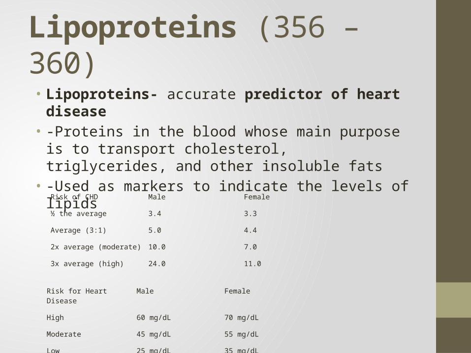
Lipoproteins (356 – 360)• Lipoproteins- accurate predictor of heart disease• -Proteins in the blood whose main purpose is to transport
cholesterol, triglycerides, and other insoluble fats• -Used as markers to indicate the levels of lipids
Risk for Heart Disease Male Female
High 60 mg/dL 70 mg/dL
Moderate 45 mg/dL 55 mg/dL
Low 25 mg/dL 35 mg/dL
Risk of CHD Male Female
½ the average 3.4 3.3
Average (3:1) 5.0 4.4
2x average (moderate) 10.0 7.0
3x average (high) 24.0 11.0

Drugs

Drug Uses Side effects Contraindications Therapeutic considerations
Alendronate Class: BisphosphonateMech: Decreases bone reabsorption by osteoclasts; blocks a step in the mevalonate pathwayIndications: • Osteoporosis
prevention and treatment
• Paget’s disease
• Jaw osteonecrosis in cancer patients
• Cessation of bone remodeling
• Gastroesophageal pain
• Delayed gastric emptying• Inability to sit up for 30
minutes after taking drug• hypocalcemia
• Extended skeletal effects,
• unclear how to define overdose
• IV dose corrects hypercalcemia in days
• all secreted by kidney
Calcitonin (Salmon)
Mech: binds to and activates a G-protein coupled receptor on osteoclasts to decrease resorptive activityIndications:• Hypercalcemia• Paget’s disease• Postmenopausal
osteoporosis
• Flushing• Nausea• Diarrhea• Tachyphylaxis
• Hypersensitivity • Nasal spray or subcutaneous
• Subcutaneous lowers blood calcium over hours
Raloxifene Class: Selective estrogen receptor modulator (SERMs)Mech: Estrogen receptor agonist in bone, estrogen receptor antagonist against endometrium and breastIndication: • Osteoporosis prevention
and treatment
• Retinal vascular occlusion
• Venous thromboembolism
• Pulmonary embolism• Hot flashes• Leg cramps
• Pregnancy• History or presence of
venous thromboembolism
• Decreases breast cancer incidence

Drug Uses Side effects Contraindications Therapeutic considerations
Enalapril Class: ACE InhibitorsMech: Decreases conversion of angiotensin (AT) I to AT II, which decreases vasoconstriction of arterioles, aldosterone synthesis, renal proximal tubule NaCl reabsorption, and ADH release; also inhibit degradation of bradykinin, which increases vasodilationIndications:• Hypertension• heart failure• diabetic nephropathy• MI
• Angioedema (more frequent in black patients)
• Agranulocytosis• Neutropenia• Cough,• Edema• Hypotension• Rash• Gynecomastia• Hyperkalemia• Proteinuria
• History of angioedema
• Bilateral renal artery stenosis
• Renal failure• Pregnancy
• Ester prodrug activated in plasma
• Bradykinin causes cough and edema; angioedema can be potentially life-threatening
• Delays progression of cardiac contractile dysfunction in HF and after MI; delay diabetic neuropathy
• Co-admin with allopurinol may predispose to hypersensitivity rxn including Steven Johnson syndrome
Amlodipine (Dihydropyridine)
Class: Calcium channel blockerMech: calcium channel blocker Indication:• Exertional angina• Unstable angina• Coronary spasm• Hypertension• Hypertrophic
cardiomyopathy• Pre-eclampsia
• Increased angina,• Rare MI• Palpitations• Peripheral edema• Flushing• Constipation• Heartburn• Dizziness
• Preexisting hypotension
• Arteriolar dilation greater than venous
• High vascular to cardiac selectivity
• Less depression of myocardial contractility, minimal effects on nodal conduction
• Higher bioavailability, longer time to peak plasma concentration, and slower hepatic metabolism

Drug Uses Side effects Contraindications Therapeutic considerations
Naproxen Propionic acid (NSAID) Mech: See Ibuprofinmild to moderate pain, fever, osteoarthritis, RA, dysmenorrhea, gout
pseudoporphyria, See Ibuprofen • Longer half life,• 20x more potent
than ibuprofen, • causes fewer GI
adverse affects
Ketorolac Acetic acid (NSAID)
See Ibuprofen
See Ibuprofen See Ibuprofen Analgesia in postsurgical patients, used for no more than 3-5 days
FeSO4 Iron supplement for anemia due to blood loss and iron insufficiency
Stomach upset or pain, constipation, diarrhea, nausea or vomiting
Diabetes, infants, may upset ulcers, not to be take with thalassemias, may irritate IBS
Drug Uses Side effects Contraindications Therapeutic considerations
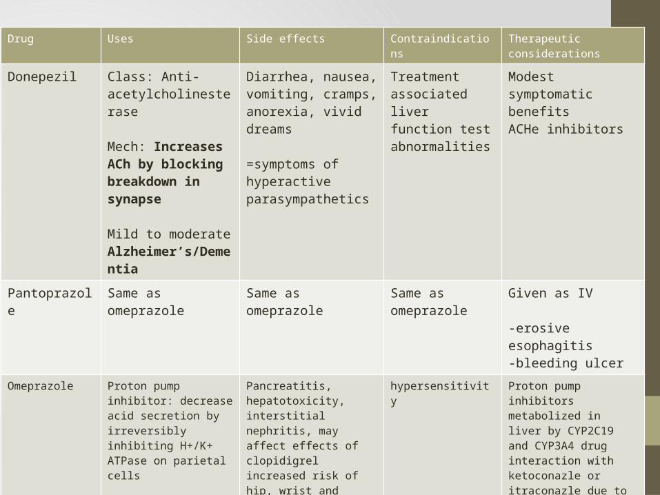
Donepezil Class: Anti-acetylcholinesterase
Mech: Increases ACh by blocking breakdown in synapse
Mild to moderate Alzheimer’s/Dementia
Diarrhea, nausea, vomiting, cramps, anorexia, vivid dreams
=symptoms of hyperactive parasympathetics
Treatment associated liver function test abnormalities
Modest symptomatic benefitsACHe inhibitors
Pantoprazole Same as omeprazole Same as omeprazole Same as omeprazole
Given as IV
-erosive esophagitis-bleeding ulcer
Omeprazole Proton pump inhibitor: decrease acid secretion by irreversibly inhibiting H+/K+ ATPase on parietal cells
Peptic ulcer disease, GERD, erosive esophagitis, gastic acid hypersecretionh. Pylori GI tract infection
Pancreatitis, hepatotoxicity, interstitial nephritis, may affect effects of clopidigrel increased risk of hip, wrist and spine fracture, hospital acquired pneumonia, and enteric infections including clostridium difficile, salmonella, E. coli, headache, rash, GI discomfort, diarrhea, anorexia, asthenia, back pain
hypersensitivity Proton pump inhibitors metabolized in liver by CYP2C19 and CYP3A4 drug interaction with ketoconazle or itraconazle due to acid environment needed to absorb azole drugs
Drug Uses Side effects Contraindications Therapeutic considerations
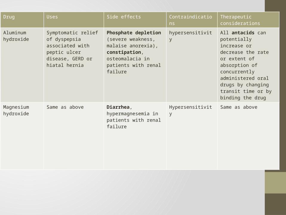
Aluminum hydroxide
Symptomatic relief of dyspepsia associated with peptic ulcer disease, GERD or hiatal hernia
Phosphate depletion (severe weakness, malaise anorexia), constipation, osteomalacia in patients with renal failure
hypersensitivity All antacids can potentially increase or decrease the rate or extent of absorption of concurrently administered oral drugs by changing transit time or by binding the drug
Magnesium hydroxide
Same as above Diarrhea, hypermagnesemia in patients with renal failure
Hypersensitivity Same as above

Drug Uses Side effects Contraindications Therapeutic considerations
levothyroxine Hormone, T4, for hypothyroidism, myxedema comaReplaces missing hormone
Hyperthyroidism, osteopenia, pseudotumor cerebri, seizure, myocardial infarction
Acute MI, uncorrected adrenal cortical insufficiencyUntreated thyrotoxicosis
Cholestyramine and sodium polystyrene sulfonate decrease absorption of synthetic thyroid hormoneRifampin and phenytoin increase metabolismT4 desirable because of its longer half life
Hydrocortisone • Corticosteroid • Replacement therapy
for primary and secondary adrenal insufficiency
• Reduces inflammation
Cushing syndrome, reduces bone density with chronic use
Fungal infection
Verapamil (Phenylalkylamine)
Class: Calcium channel blockerMech: block voltage-gated L-type calcium channels & prevent influx of calcium that promotes actin-myosin cross-bridge formationIndications: • Prinzmetal or variant
angina or chronic stable angina
• Hypertension• A fib or flutter, paroxysmal
SVT
• Rare cardiac arrhythmia• AV block• Bradyarrhythmia• Exacerbation of heart failure• Peripheral edema• Syncope• Gingival hyperplasia• Dizziness
• IV is contraindicated in patients with ventricular tachycardia and patients receiving IV beta blockers
• Sick sinus syndrome or 2nd or 3rd AV block
• SVT associated with bypass tract
• Left ventricular failure• Hypotension• Acute MI
• Low ratio of vascular to cardiac selectivity
• Depresses both SA and AV node conduction velocity
• Raises serum carbamazepine levels which may cause toxicity
• Avoid using with beta blockers
• Greater suppressive effect on cardiac contractility

Drug Uses Side effects Contraindications Therapeutic considerations
Acetominophen Class: NSAIDMech Weak inhibitor of peripheral cyclooxygenases; predominant effect may be inhibition of cyclooxygenase-3 (COX-3) in the CNSIndications: FeverMild to moderate pain
Hepatotoxicity, nephrotoxicity (rare)Rash, hypothermia
Hypersensitivity to acetaminophen
• Although acetaminophen has analgesic and antipyretic effects similar to aspirin, the anti -inflammatory effect of acetaminophen is insignificant because of its weak inhibition of peripheral cyclooxygenases
• Acetaminophen overdose is a leading cause of hepatic failure
• Antidote for acetaminophen overdose is N-acetylcysteine
Ibuprofen Class: Propionic acids:Mech: Inhibit cyclooxygenase-l (COX-I) and cyclooxygenase-2 (COX-2), decreasing the biosynthesis of downstream eicosanoids and thereby limiting the inflammatory responseIndications: • Mild to moderate pain• Fever• Osteoarthritis, rheumatoid
arthritis• Dysmenorrhea• Gout
Gastrointestinal hemorrhage, ulceration, perforation;nephrotoxicity; Stevens-Johnson syndrome;Gastrointestinal disturbance, tinnitus
Gastrointestinal or intracranialbleedingCoagulation defectsAsthma, urticaria, or allergic-typereactions after taking NSAIDS,due to risk of severe, even fatal,anaphylactic reactionsSignificant renal insufficiency
N-AcetylcystineMech: Supplies cysteine to replenish glutathioneIndications: • Acetominophen Overdose
ANTIDOTE FOR ACETOMINAPHEN OD

Drug Uses Side effects Contraindications Therapeutic considerations
Celecoxib
Only COX-2 inhibitor still on market!
Class: COX-2 inhibitor (NSAID)Mech: Selectively inhibits COX-2 Indications: • Osteoarthritis, rheumatoid• arthritis in adults, and• ankylosing spondylitis• Primary dysmenorrhea• Acute pain in adults• Familial adenomatous
polyposis
Myocardial infarction, ischemic stroke, heart failure;gastrointestinal bleeding, ulceration, perforation;renal papillary necrosis; exacerbation of asthmaGastrointestinal disturbance, peripheral edema
Hypersensitivity to sulfonamidesHypersensitivity to celecoxibAsthma, urticaria, or allergic-typereactions after taking NSAIDs,due to risk of severe, even fatal,anaphylactic reactionsPain associated with coronaryartery bypass graft surgery
• Decreases efficacy of ACE inhibitors
• Incidence of gastropathy and nephropathy may be less than that associated with NSAIDs, but may still be significant
• Valdecoxib and rofecoxib recently withdrawn from U.S. market due to possible increase in cardiovascular mortality
Etomidate Class: carboxylated imidazole Mech appears to facilitate GABA-minergic neurotransmission by increasing the number of available GABA receptors, possibly by displacing endogenous inhibitors of GABA binding Indications: IV anesthesia intubation
Can cause adrenal insufficiency, can cause seizure activity
Labor / Imminent Delivery
Septic shock
Loperamide
OTC- Imodium
Class: Opioid agonistMech: activates u-recptor in GI tract, specifically in the myenteric plexus, reduces muscle tone and decreases parastalis Indications: • Diarrhea
• dry mouth• dizziness• drowsiness• vomiting• stomach pain, discomfort,
or distention (enlargement)• constipation• fatigue-anticholenergic effects (turns off parasympathetics)
Under two years of age
Paralytic ileus
Drug Uses Side effects Contraindications Therapeutic considerations
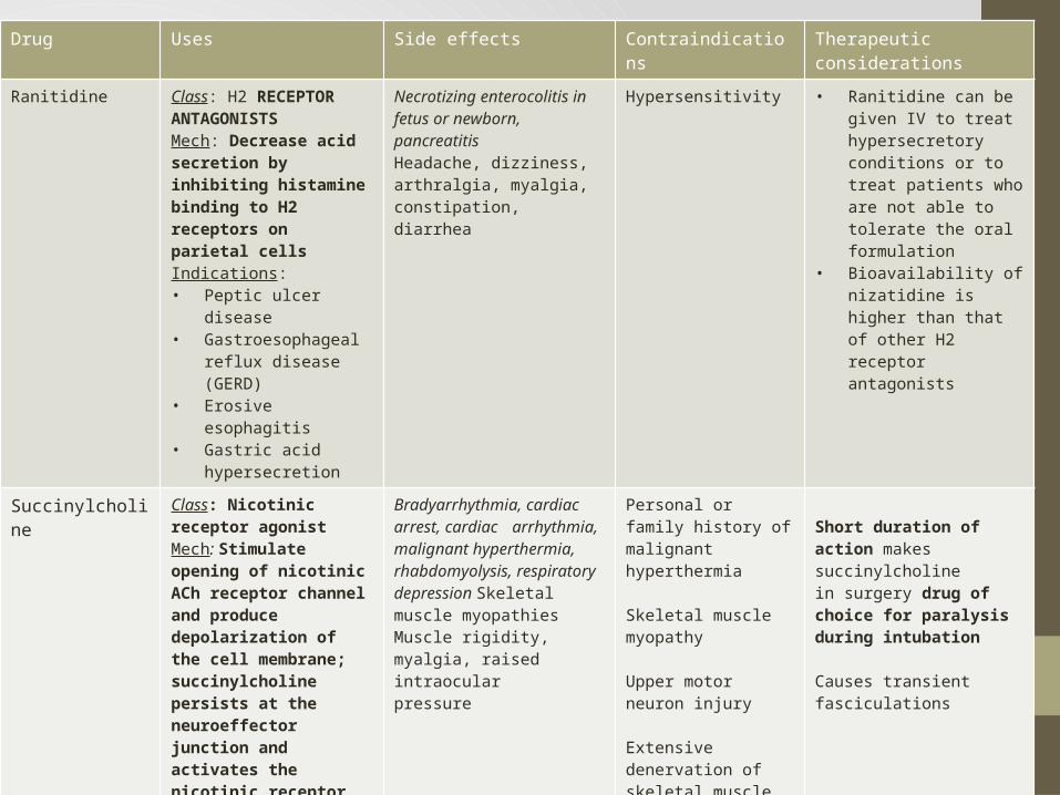
Ranitidine Class: H2 RECEPTOR ANTAGONISTSMech: Decrease acid secretion by inhibiting histamine binding to H2 receptors on parietal cellsIndications: • Peptic ulcer disease• Gastroesophageal reflux
disease (GERD)• Erosive esophagitis• Gastric acid
hypersecretion
Necrotizing enterocolitis in fetus or newborn,pancreatitisHeadache, dizziness, arthralgia, myalgia,constipation, diarrhea
Hypersensitivity • Ranitidine can be given IV to treat hypersecretory conditions or to treat patients who are not able to tolerate the oral formulation
• Bioavailability of nizatidine is higher than that of other H2 receptor antagonists
Succinylcholine Class: Nicotinic receptor agonistMech: Stimulate opening of nicotinic ACh receptor channel and produce depolarization of the cell membrane; succinylcholine persists at the neuroeffector junction and activates the nicotinic receptor channels continuously,which results in inactivation of voltage-gated sodium channels so that they cannot open to support further action potentials (sometimes called "depolarizing blockade‘)Indications: • Induction of
neuromuscular blockade in surgery
• intubation
Bradyarrhythmia, cardiac arrest, cardiac arrhythmia, malignant hyperthermia, rhabdomyolysis, respiratory depression Skeletal muscle myopathies Muscle rigidity, myalgia, raised intraocular pressure
Personal or family history of malignant hyperthermia
Skeletal muscle myopathy
Upper motor neuron injury
Extensive denervation of skeletal muscle
Short duration of action makes succinylcholinein surgery drug of choice for paralysis during intubation
Causes transient fasciculations