sports knee injury orthosports effusions mri orthosports€¦ · effusions mri orthosports...
TRANSCRIPT
Sporting Knee Effusions and MRI
Sports Knee InjuryEffusions
MRI
www.orthosports.com.auOrthosports
O
rthosports
Orthosports
Orthosports
Orthosports
Orthosports
Sporting Knee Effusions and MRI
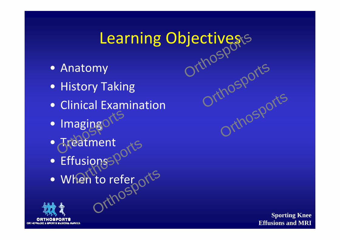
Learning Objectives
• Anatomy• History Taking• Clinical Examination• Imaging• Treatment• Effusions• When to refer
Orthosports
O
rthosports
Orthosports
Orthosports
Orthosports
Orthosports
Sporting Knee Effusions and MRI
Anatomy
Orthosports
O
rthosports
Orthosports
Orthosports
Orthosports
Orthosports
Sporting Knee Effusions and MRI
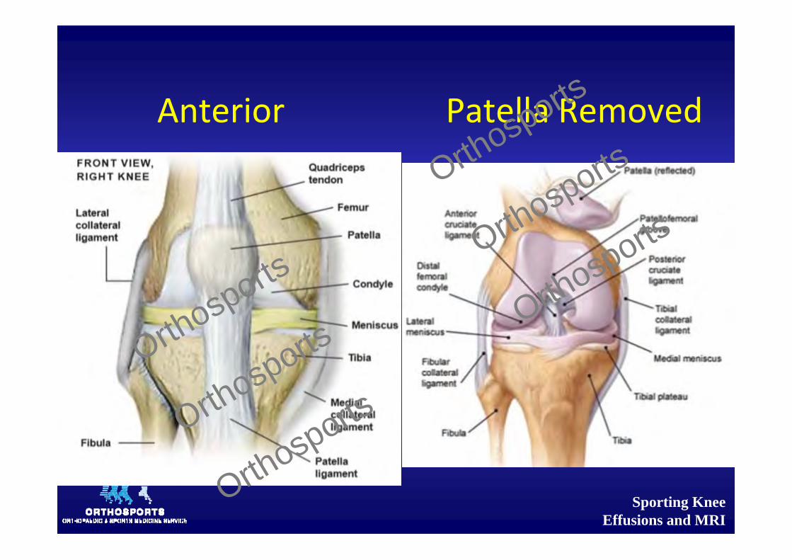
Anterior Patella Removed
Orthosports
O
rthosports
Orthosports
Orthosports
Orthosports
Orthosports
Sporting Knee Effusions and MRI
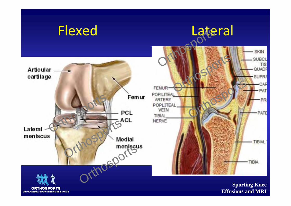
Flexed Lateral
Orthosports
O
rthosports
Orthosports
Orthosports
Orthosports
Orthosports
Sporting Knee Effusions and MRI
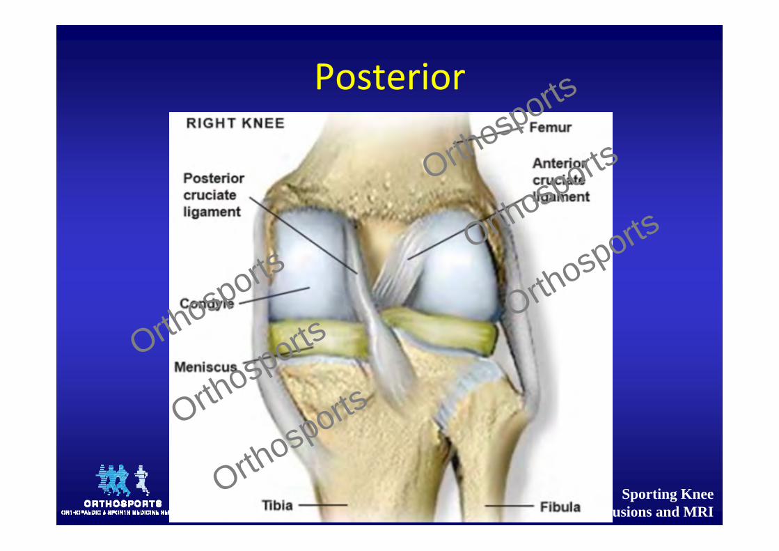
Posterior
Orthosports
O
rthosports
Orthosports
Orthosports
Orthosports
Orthosports
Sporting Knee Effusions and MRI
Chondral Cartilage
Orthosports
O
rthosports
Orthosports
Orthosports
Orthosports
Orthosports
Sporting Knee Effusions and MRI
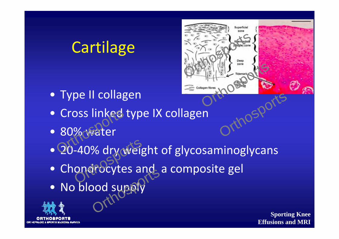
Cartilage
• Type II collagen• Cross linked type IX collagen• 80% water• 20‐40% dry weight of glycosaminoglycans• Chondrocytes and a composite gel• No blood supply
Orthosports
O
rthosports
Orthosports
Orthosports
Orthosports
Orthosports
Sporting Knee Effusions and MRI
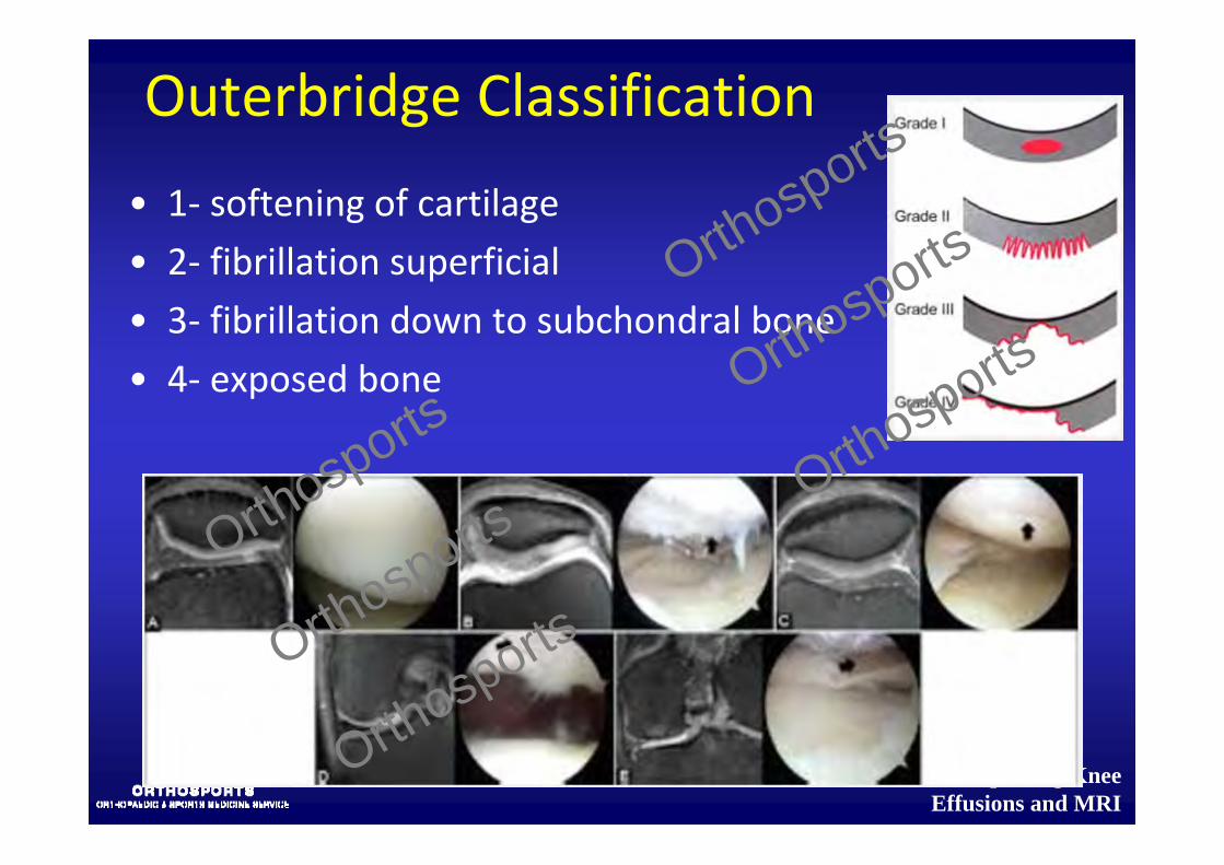
Outerbridge Classification
• 1‐ softening of cartilage• 2‐ fibrillation superficial• 3‐ fibrillation down to subchondral bone• 4‐ exposed bone
Orthosports
O
rthosports
Orthosports
Orthosports
Orthosports
Orthosports
Sporting Knee Effusions and MRI
Meniscus
Orthosports
O
rthosports
Orthosports
Orthosports
Orthosports
Orthosports
Sporting Knee Effusions and MRI
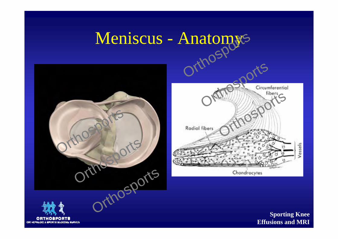
Meniscus - Anatomy
Orthosports
O
rthosports
Orthosports
Orthosports
Orthosports
Orthosports
Sporting Knee Effusions and MRI
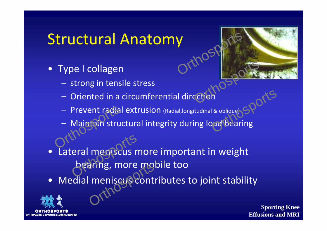
Structural Anatomy
• Type I collagen– strong in tensile stress– Oriented in a circumferential direction– Prevent radial extrusion (Radial,longitudinal & oblique)– Maintain structural integrity during load bearing
• Lateral meniscus more important in weight bearing, more mobile too
• Medial meniscus contributes to joint stability
Orthosports
O
rthosports
Orthosports
Orthosports
Orthosports
Orthosports
Sporting Knee Effusions and MRI
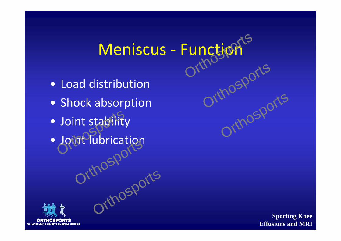
Meniscus ‐ Function
• Load distribution• Shock absorption• Joint stability• Joint lubrication
Orthosports
O
rthosports
Orthosports
Orthosports
Orthosports
Orthosports
Sporting Knee Effusions and MRI
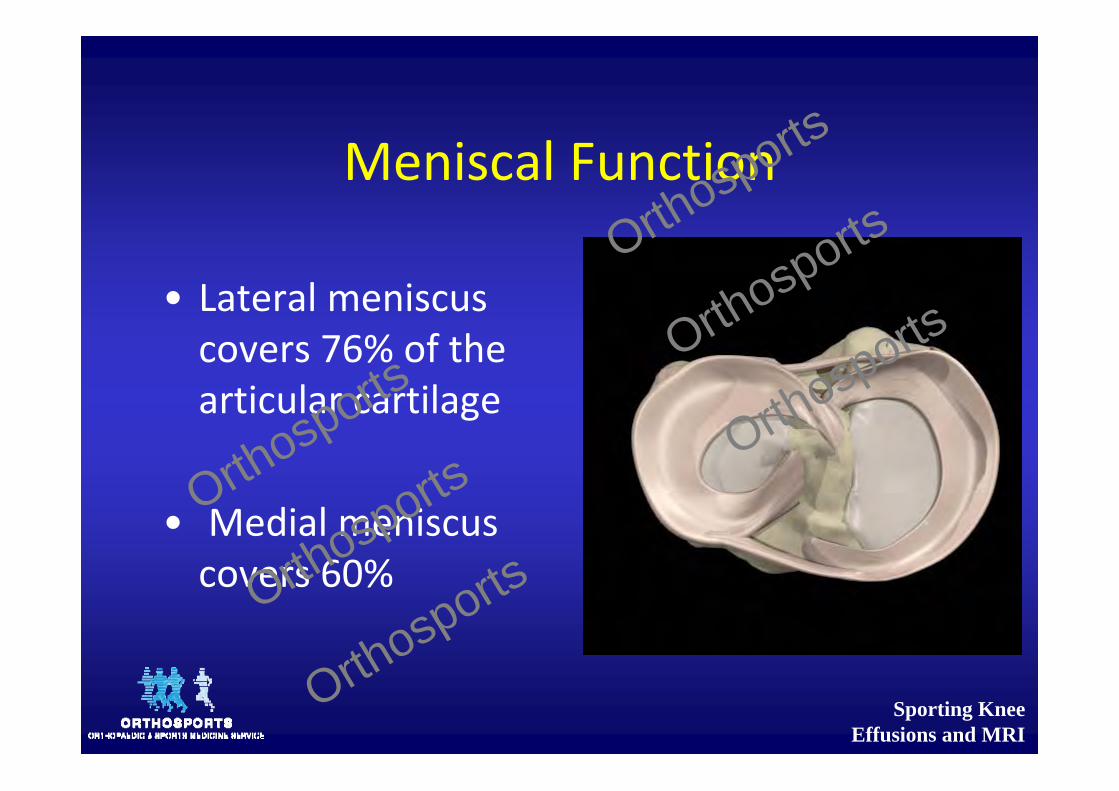
Meniscal Function
• Lateral meniscus covers 76% of the articular cartilage
• Medial meniscus covers 60%
Orthosports
O
rthosports
Orthosports
Orthosports
Orthosports
Orthosports
Sporting Knee Effusions and MRI
Meniscal Function
• Load transmission– 45 – 50% load transmitted to menisci in extension– 85% load transmitted to menisci in flexion– Medial side, MM and MTP share load– Lateral side, LM takes 80% load
Orthosports
O
rthosports
Orthosports
Orthosports
Orthosports
Orthosports
Sporting Knee Effusions and MRI
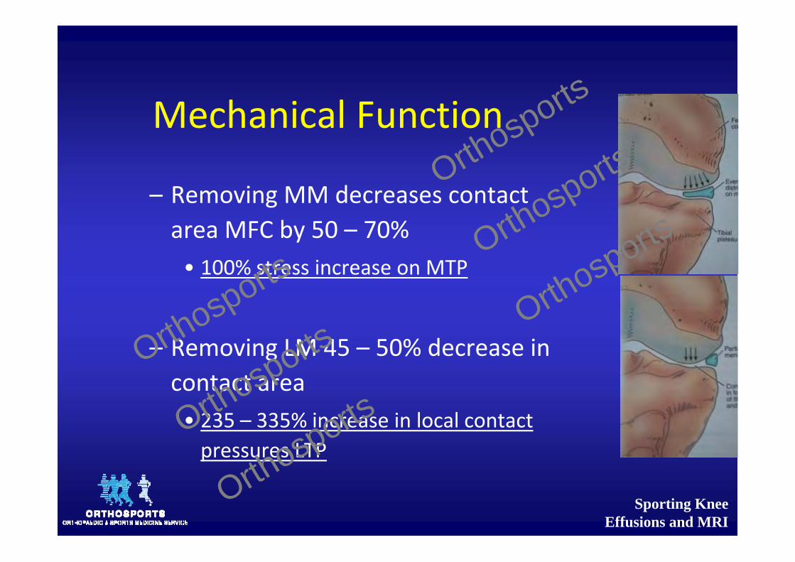
Mechanical Function
– Removing MM decreases contact area MFC by 50 – 70%• 100% stress increase on MTP
– Removing LM 45 – 50% decrease in contact area• 235 – 335% increase in local contact pressures LTP
Orthosports
O
rthosports
Orthosports
Orthosports
Orthosports
Orthosports
Sporting Knee Effusions and MRI
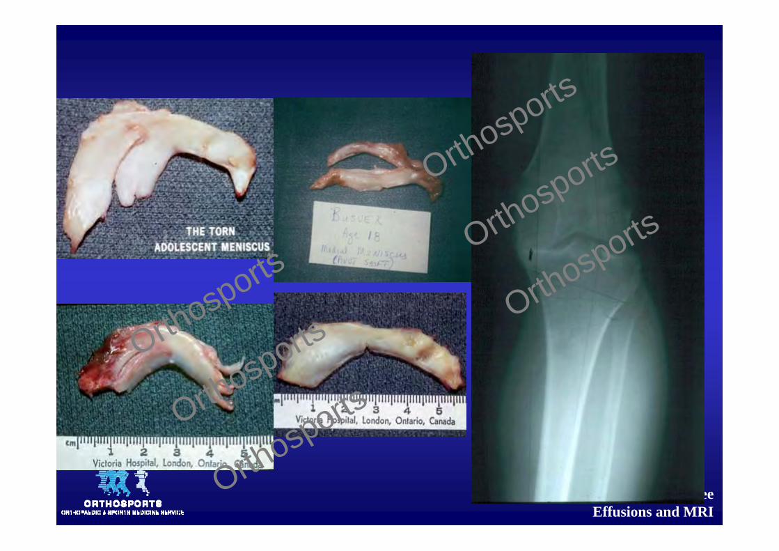
Swelling and Meniscus Tears
• Isolated meniscal tears do not cause recurrent swelling
• If the knee is swollen there is almost certainly chondral damageOrthosports
Orthosports
Orthosports
Orthosports
Orthosports
Orthosports
Sporting Knee Effusions and MRI
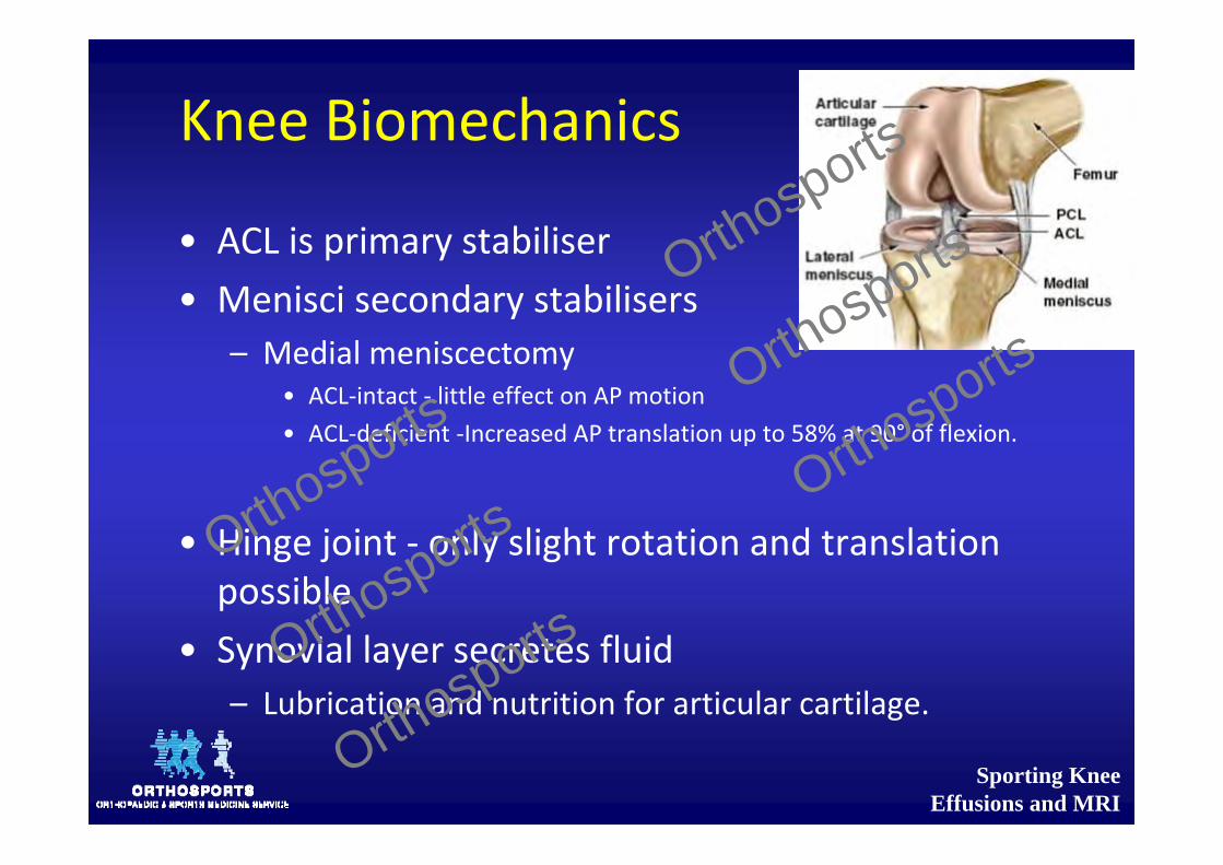
Knee Biomechanics
• ACL is primary stabiliser• Menisci secondary stabilisers
– Medial meniscectomy• ACL‐intact ‐ little effect on AP motion• ACL‐deficient ‐Increased AP translation up to 58% at 90° of flexion.
• Hinge joint ‐ only slight rotation and translation possible
• Synovial layer secretes fluid– Lubrication and nutrition for articular cartilage.
Orthosports
O
rthosports
Orthosports
Orthosports
Orthosports
Orthosports
Sporting Knee Effusions and MRI
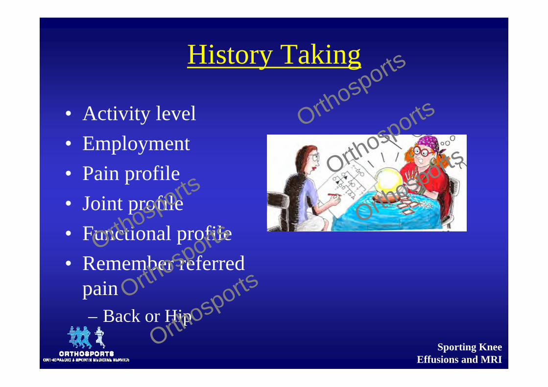
History Taking
• Activity level• Employment• Pain profile• Joint profile• Functional profile• Remember referred
pain– Back or Hip
Orthosports
O
rthosports
Orthosports
Orthosports
Orthosports
Orthosports
Sporting Knee Effusions and MRI
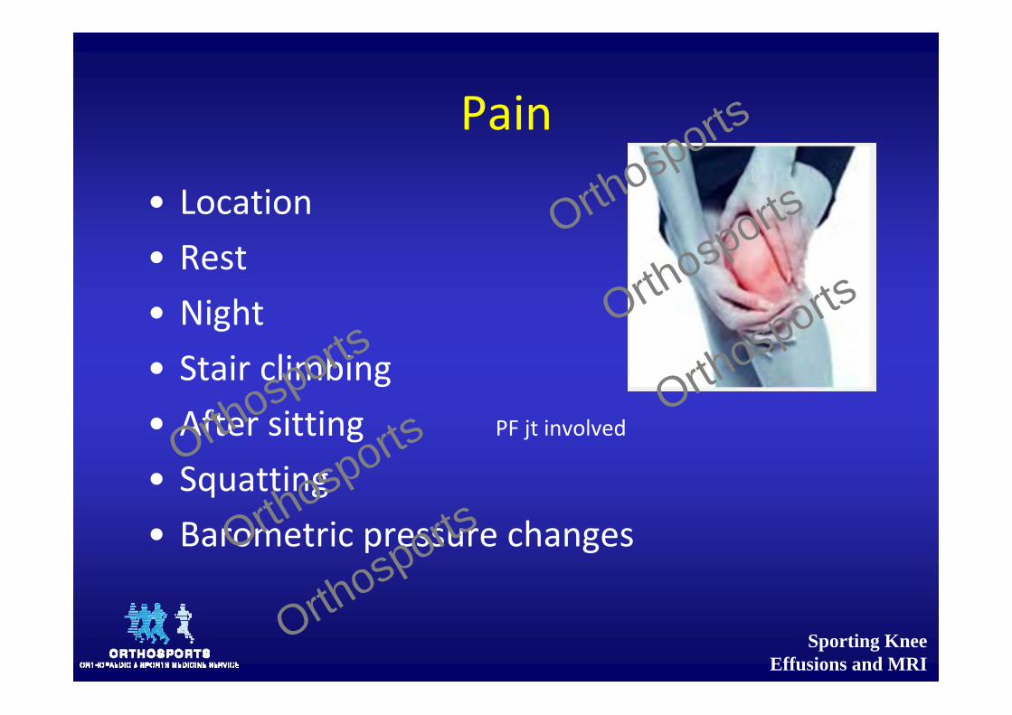
Pain
• Location• Rest • Night• Stair climbing• After sitting PF jt involved• Squatting• Barometric pressure changes
Orthosports
O
rthosports
Orthosports
Orthosports
Orthosports
Orthosports
Sporting Knee Effusions and MRI
Symptoms
• Swelling• Catching• Instability• Onset of symptoms
• Response to prior treatment
Orthosports
O
rthosports
Orthosports
Orthosports
Orthosports
Orthosports
Sporting Knee Effusions and MRI
Giving way
Primary Instability• Joint actually gives way
– Ligament deficiency– Not painful but knee hurts afterwards /swells
– Repeated giving way leads to arthritis.
Secondary Instability • Pain within the joint.
– Quads relax involuntarily– Leg buckles– Sensation of giving way– Remove the pain = no giving way
– Meniscal tear, loose body, arthritis, or synovitis.
Orthosports
O
rthosports
Orthosports
Orthosports
Orthosports
Orthosports
Sporting Knee Effusions and MRI
Giving way
Primary Instability• Surgery required to fix the problem– ACL– PCL– LCL– PLC– PFJ
Secondary Instability • Generally not causing further damage
• Patella subluxing can be felt as giving way
• Giving way often non‐specific– Loose bodies, patellar chondromalacia, and quads weakness
Orthosports
O
rthosports
Orthosports
Orthosports
Orthosports
Orthosports
Sporting Knee Effusions and MRI
Patella
Orthosports
O
rthosports
Orthosports
Orthosports
Orthosports
Orthosports
Sporting Knee Effusions and MRI
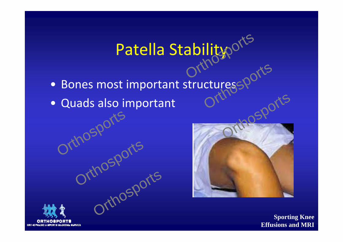
Patella Stability
• Bones most important structures• Quads also important
Orthosports
O
rthosports
Orthosports
Orthosports
Orthosports
Orthosports
Sporting Knee Effusions and MRI
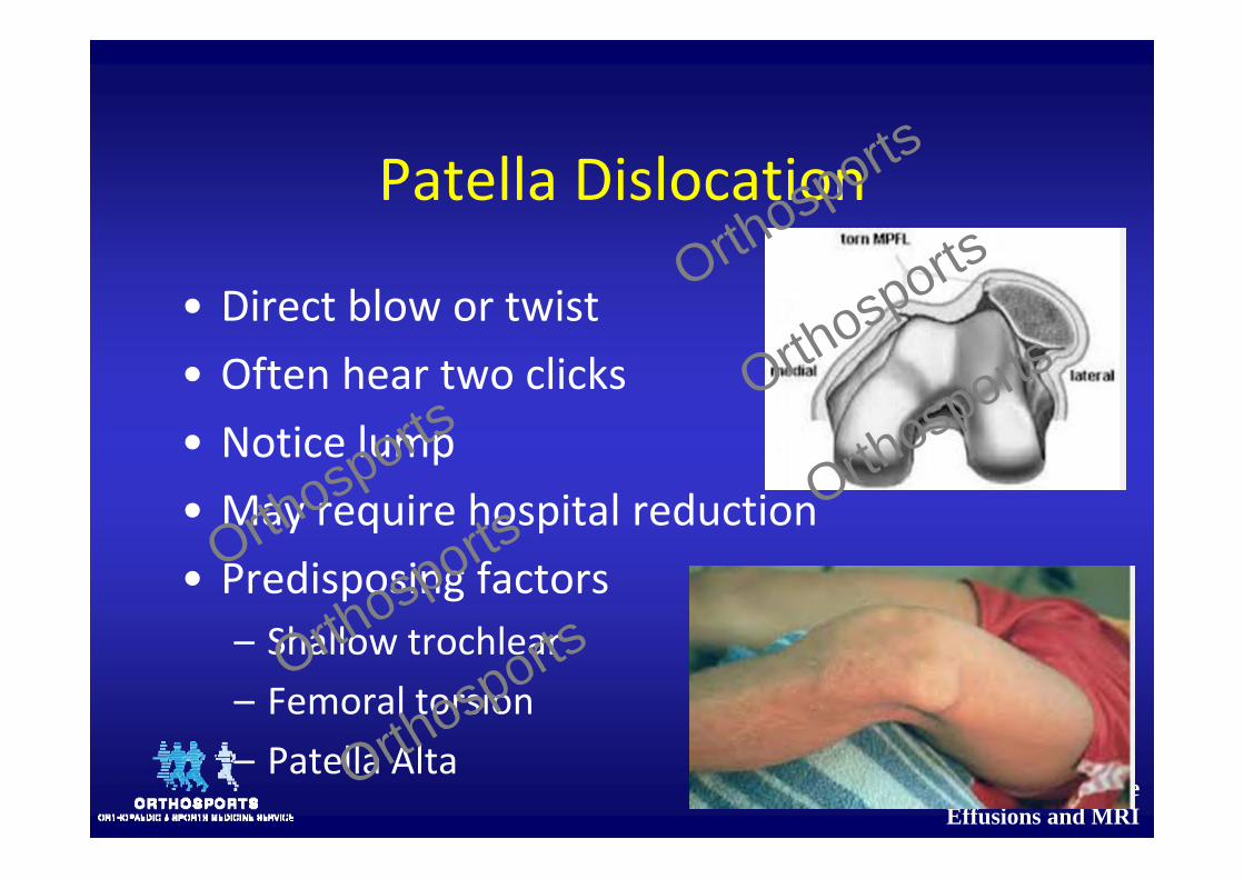
Patella Dislocation
• Direct blow or twist• Often hear two clicks• Notice lump• May require hospital reduction• Predisposing factors
– Shallow trochlear– Femoral torsion– Patella Alta
Orthosports
O
rthosports
Orthosports
Orthosports
Orthosports
Orthosports
Sporting Knee Effusions and MRI
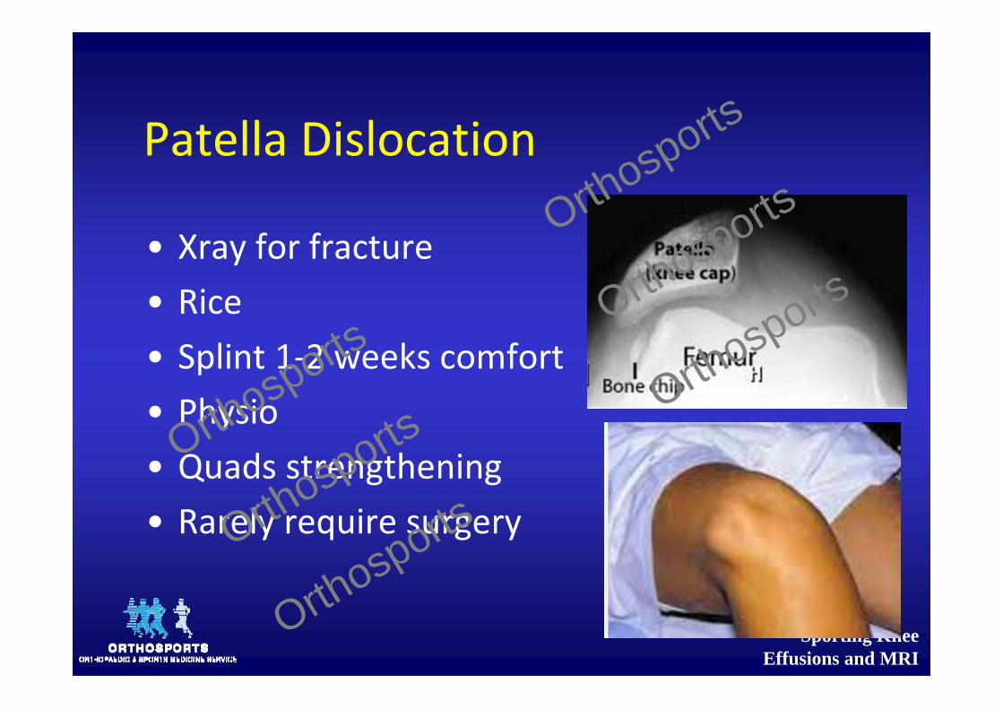
Patella Dislocation
• Xray for fracture• Rice• Splint 1‐2 weeks comfort• Physio• Quads strengthening• Rarely require surgery
Orthosports
O
rthosports
Orthosports
Orthosports
Orthosports
Orthosports
Sporting Knee Effusions and MRI
Patella Dislocation ‐ Earlier Referral
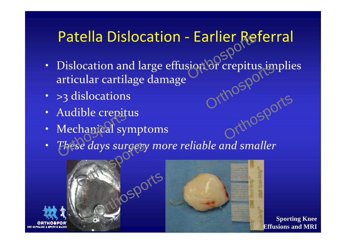
• Dislocation and large effusion or crepitus implies articular cartilage damage
• >3 dislocations• Audible crepitus• Mechanical symptoms• These days surgery more reliable and smallerOrthosports
Orthosports
Orthosports
Orthosports
Orthosports
Orthosports
Sporting Knee Effusions and MRI
Extensor Mechanism
Orthosports
O
rthosports
Orthosports
Orthosports
Orthosports
Orthosports
Sporting Knee Effusions and MRI
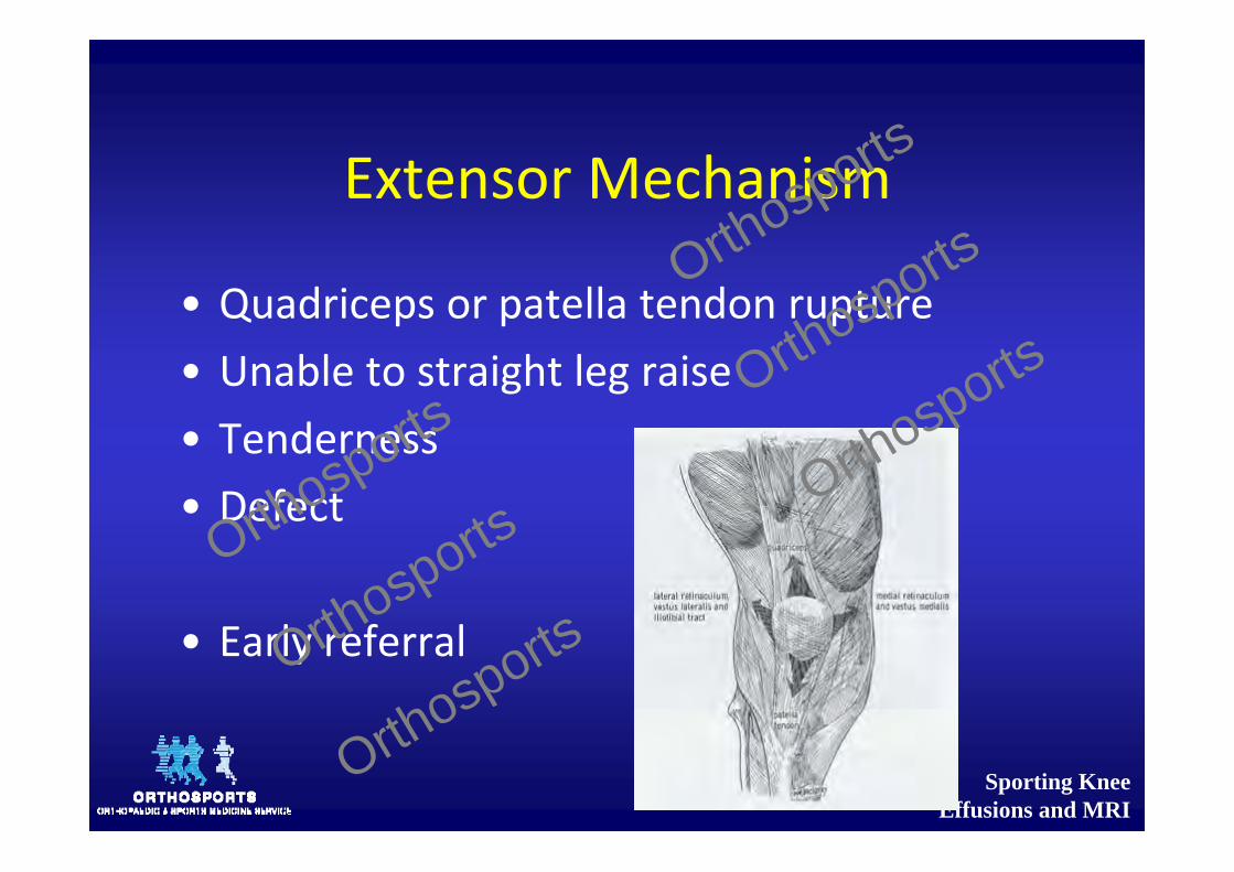
Extensor Mechanism
• Quadriceps or patella tendon rupture• Unable to straight leg raise• Tenderness• Defect
• Early referral
Orthosports
O
rthosports
Orthosports
Orthosports
Orthosports
Orthosports
Sporting Knee Effusions and MRI
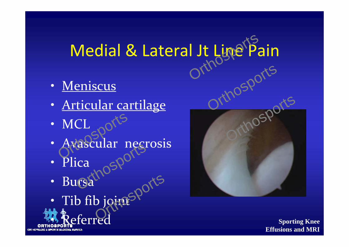
Medial & Lateral Jt Line Pain
• Meniscus• Articular cartilage• MCL• Avascular necrosis• Plica• Bursa• Tib fib joint• Referred
Orthosports
O
rthosports
Orthosports
Orthosports
Orthosports
Orthosports
Sporting Knee Effusions and MRI
Anterior Knee Pain
• Chondromalacia• Subluxation• Maltracking• Traumatic• Non specific
– (see overuse later)
Orthosports
O
rthosports
Orthosports
Orthosports
Orthosports
Orthosports

Sporting Knee Effusions and MRI
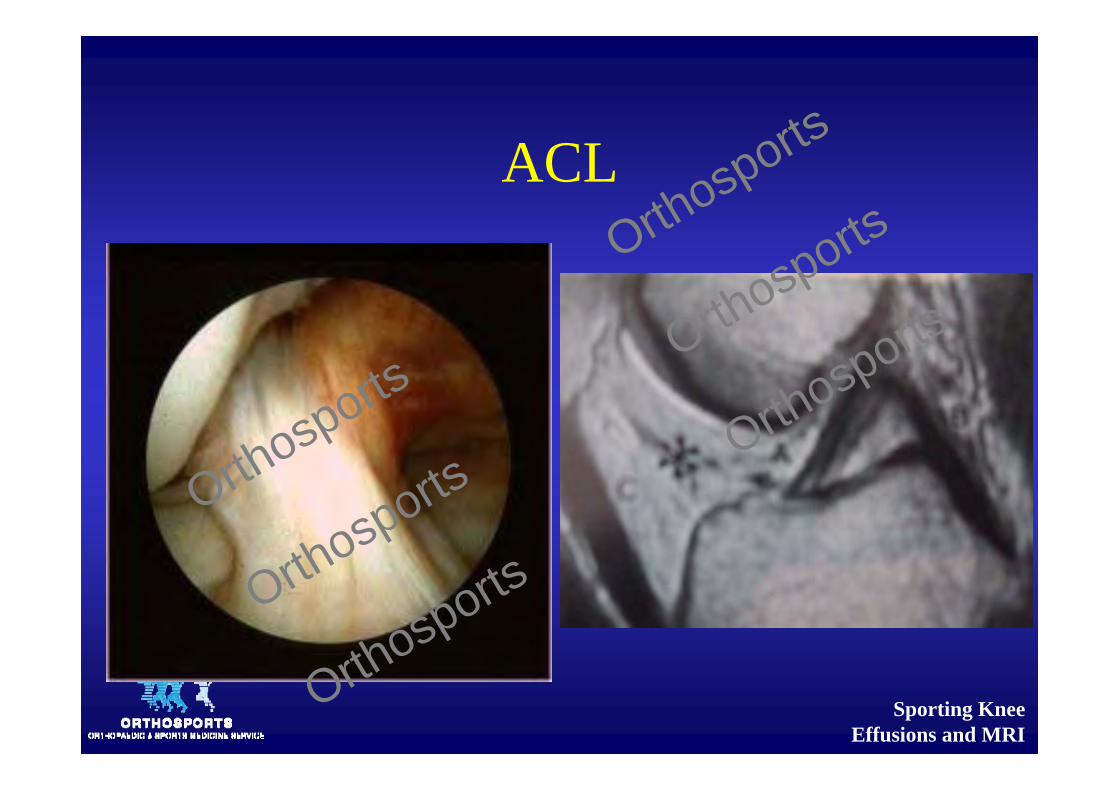
Anterior Cruciate Ligament
Orthosports
O
rthosports
Orthosports
Orthosports
Orthosports
Orthosports
Sporting Knee Effusions and MRI
• Controls 90% of stability to anterior displacement of the tibia
• Varus, valgus, rotational restraint• Anteromedial bundle tightens in flexion
and the posterolateral becomes lax
ACL
Orthosports
O
rthosports
Orthosports
Orthosports
Orthosports
Orthosports
Sporting Knee Effusions and MRI
ACL ‐ HISTORY
• Usually twisting injury• Older patients often no trauma• Swelling several hours later or next day• Pain• Clicking• Locking• Giving way• Swelling‐effusions only 50% cases
Orthosports
O
rthosports
Orthosports
Orthosports
Orthosports
Orthosports
Sporting Knee Effusions and MRI
ACL
• Best time to examine is immediate• Worst is 3‐7 days• No need for urgent referral• Not everyone needs surgery• No harm at all in watching the older patients to see if they have instabilityOrthosports
Orthosports
Orthosports
Orthosports
Orthosports
Orthosports
Sporting Knee Effusions and MRI
Refer Earlier If
• Competitive sports• Articular surface damage particularly patellofemoral and medial compartment
• Medial meniscus loss• Heavy people who are more likely to stretch secondary restraints
• Varus alignment• Younger
Orthosports
O
rthosports
Orthosports
Orthosports
Orthosports
Orthosports
Sporting Knee Effusions and MRI
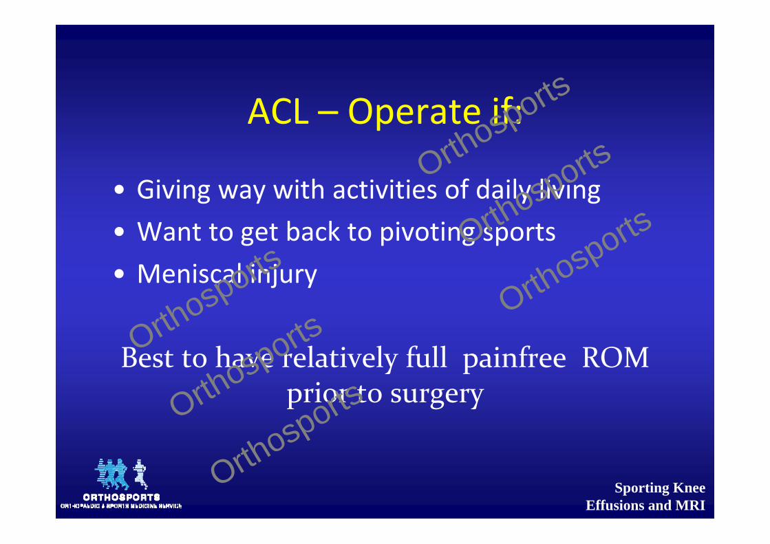
ACL – Operate if:
• Giving way with activities of daily living• Want to get back to pivoting sports• Meniscal injury
Best to have relatively full painfree ROM prior to surgery
Orthosports
O
rthosports
Orthosports
Orthosports
Orthosports
Orthosports
Sporting Knee Effusions and MRI
ACL
Orthosports
O
rthosports
Orthosports
Orthosports
Orthosports
Orthosports
Sporting Knee Effusions and MRI
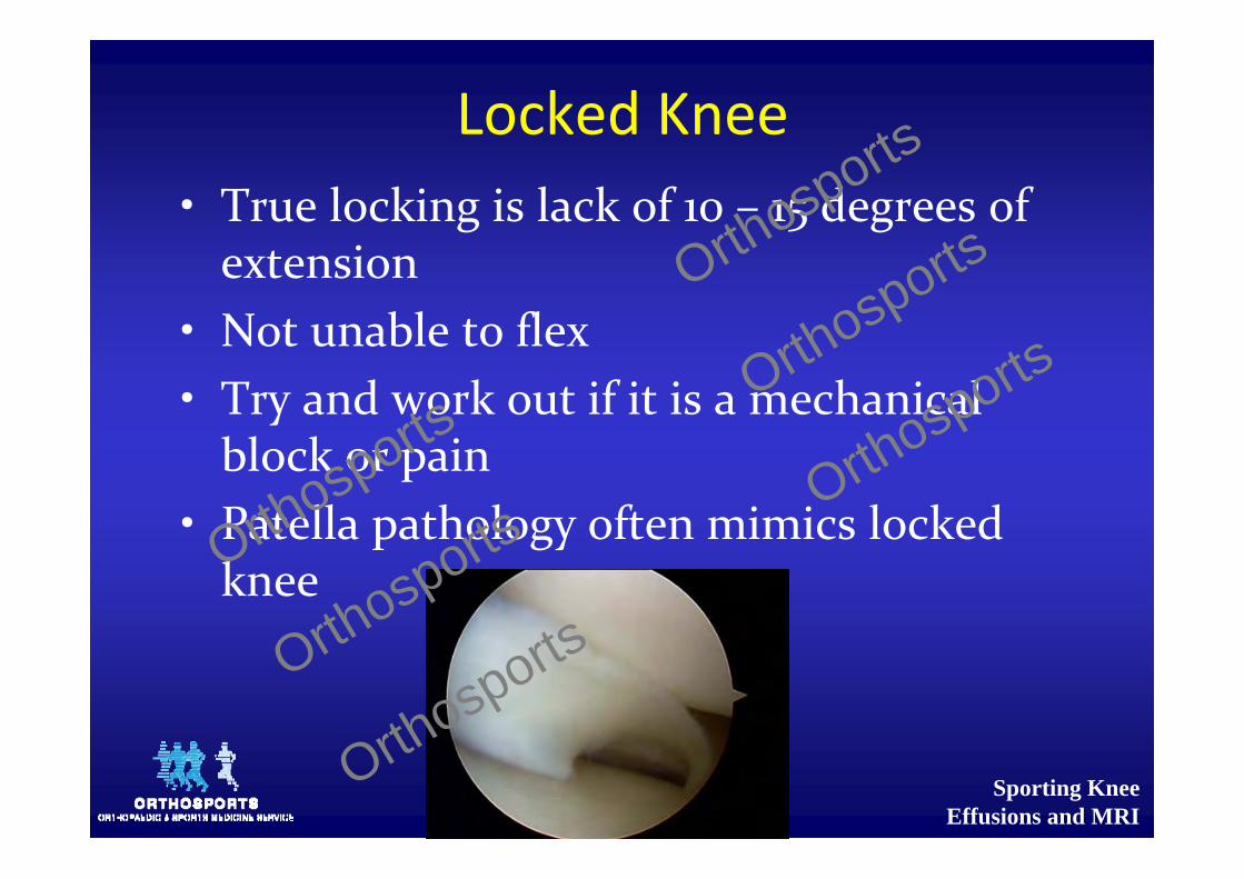
Locked Knee• True locking is lack of 10 – 15 degrees of extension
• Not unable to flex• Try and work out if it is a mechanical block or pain
• Patella pathology often mimics locked kneeOrthosports
Orthosports
Orthosports
Orthosports
Orthosports
Orthosports
Sporting Knee Effusions and MRI
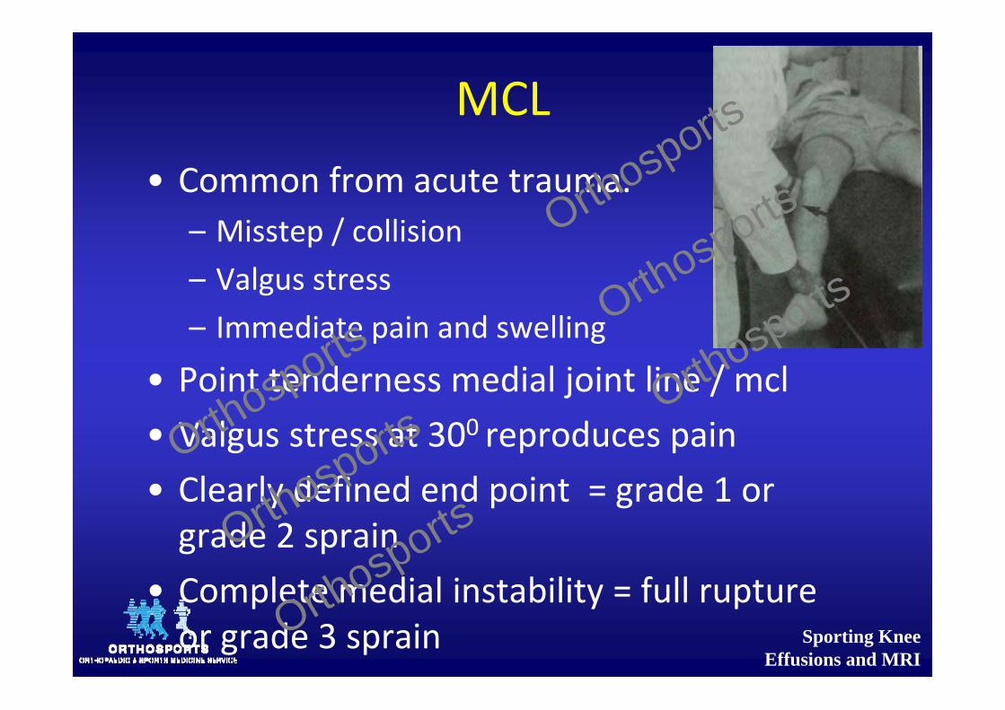
MCL• Common from acute trauma.
– Misstep / collision– Valgus stress– Immediate pain and swelling
• Point tenderness medial joint line / mcl• Valgus stress at 300 reproduces pain• Clearly defined end point = grade 1 or grade 2 sprain
• Complete medial instability = full rupture or grade 3 sprain
Orthosports
O
rthosports
Orthosports
Orthosports
Orthosports
Orthosports
Sporting Knee Effusions and MRI
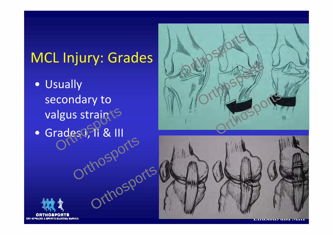
MCL Injury: Grades
• Usually secondary to valgus strain
• Grades I, II & IIIOrthosports
Orthosports
Orthosports
Orthosports
Orthosports
Orthosports
Sporting Knee Effusions and MRI
LCL
• LCL injury <<< MCL• Varus stress to the knee
– Runner plants one foot and turns toward the ipsilateral knee.
• Acute onset of lateral knee pain / stop the activity
• Point tenderness at lateral joint line.• Instability or pain occurs with varus stress testing of the knee at 30 degrees.
Orthosports
O
rthosports
Orthosports
Orthosports
Orthosports
Orthosports
Sporting Knee Effusions and MRI
Overuse Syndromes• Lateral Knee Pain
– Aggravated by activity• Running downhill and climbing stairs.• Excessive friction between the iliotibial band and the lateral femoral condyle
• Commonly occurs in runners and cyclists,
• Tightness of the iliotibial band, excessive foot pronation, genu varum, and tibial torsion
Orthosports
O
rthosports
Orthosports
Orthosports
Orthosports
Orthosports
Sporting Knee Effusions and MRI
Overuse Syndromes (cont)
• PF pain syndrome (chondromalacia patellae)
– Vague history of mild to moderate pain– After prolonged sitting
• Almost always tight hamstrings
• Treatment– Physiotherapy to stretch the hamstrings and unload the patellofemoral joint
Orthosports
O
rthosports
Orthosports
Orthosports
Orthosports
Orthosports

Sporting Knee Effusions and MRI
Children
Orthosports
O
rthosports
Orthosports
Orthosports
Orthosports
Orthosports
Sporting Knee Effusions and MRI
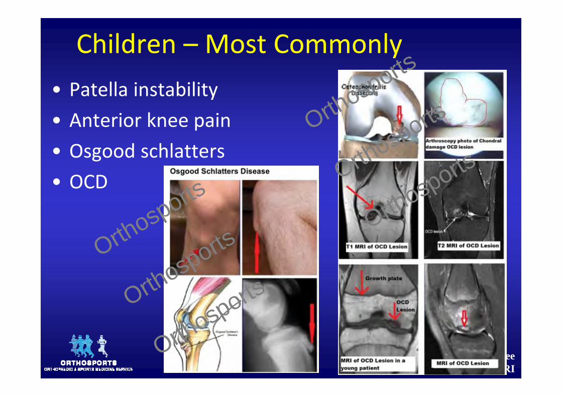
Children – Most Commonly• Patella instability• Anterior knee pain• Osgood schlatters• OCD
Orthosports
O
rthosports
Orthosports
Orthosports
Orthosports
Orthosports
Sporting Knee Effusions and MRI
Children ‐ Don’t miss these
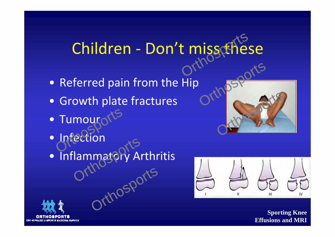
• Referred pain from the Hip• Growth plate fractures• Tumour• Infection• Inflammatory ArthritisOrthosports
Orthosports
Orthosports
Orthosports
Orthosports
Orthosports
Sporting Knee Effusions and MRI
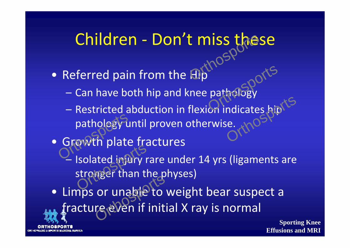
Children ‐ Don’t miss these
• Referred pain from the Hip– Can have both hip and knee pathology– Restricted abduction in flexion indicates hip pathology until proven otherwise.
• Growth plate fractures– Isolated injury rare under 14 yrs (ligaments are stronger than the physes)
• Limps or unable to weight bear suspect a fracture even if initial X ray is normal
Orthosports
O
rthosports
Orthosports
Orthosports
Orthosports
Orthosports
Sporting Knee Effusions and MRI
Kids continued
• Tumour• Present with pain, swelling or pathological fracture• If symptoms and signs are atypical think of this
• Infection (same as adult)• Inflammatory ArthritisOrthosports
Orthosports
Orthosports
Orthosports
Orthosports
Orthosports
Sporting Knee Effusions and MRI
Clinical Examination
Orthosports
O
rthosports
Orthosports
Orthosports
Orthosports
Orthosports
Sporting Knee Effusions and MRI
Clinical Examination
• Remove socks and expose thighs• Try standing and walking• Examine the good leg first
LOOK – FEEL ‐MOVE
Orthosports
O
rthosports
Orthosports
Orthosports
Orthosports
Orthosports
Sporting Knee Effusions and MRI
Functional Anatomy / Assessment
• Gait• Alignment• Range of Motion• Hip
• Knee• Ankle/Foot
Orthosports
O
rthosports
Orthosports
Orthosports
Orthosports
Orthosports
Sporting Knee Effusions and MRI
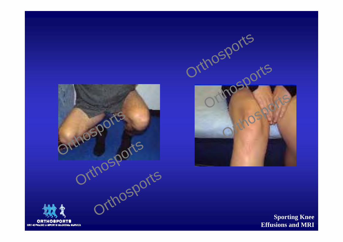
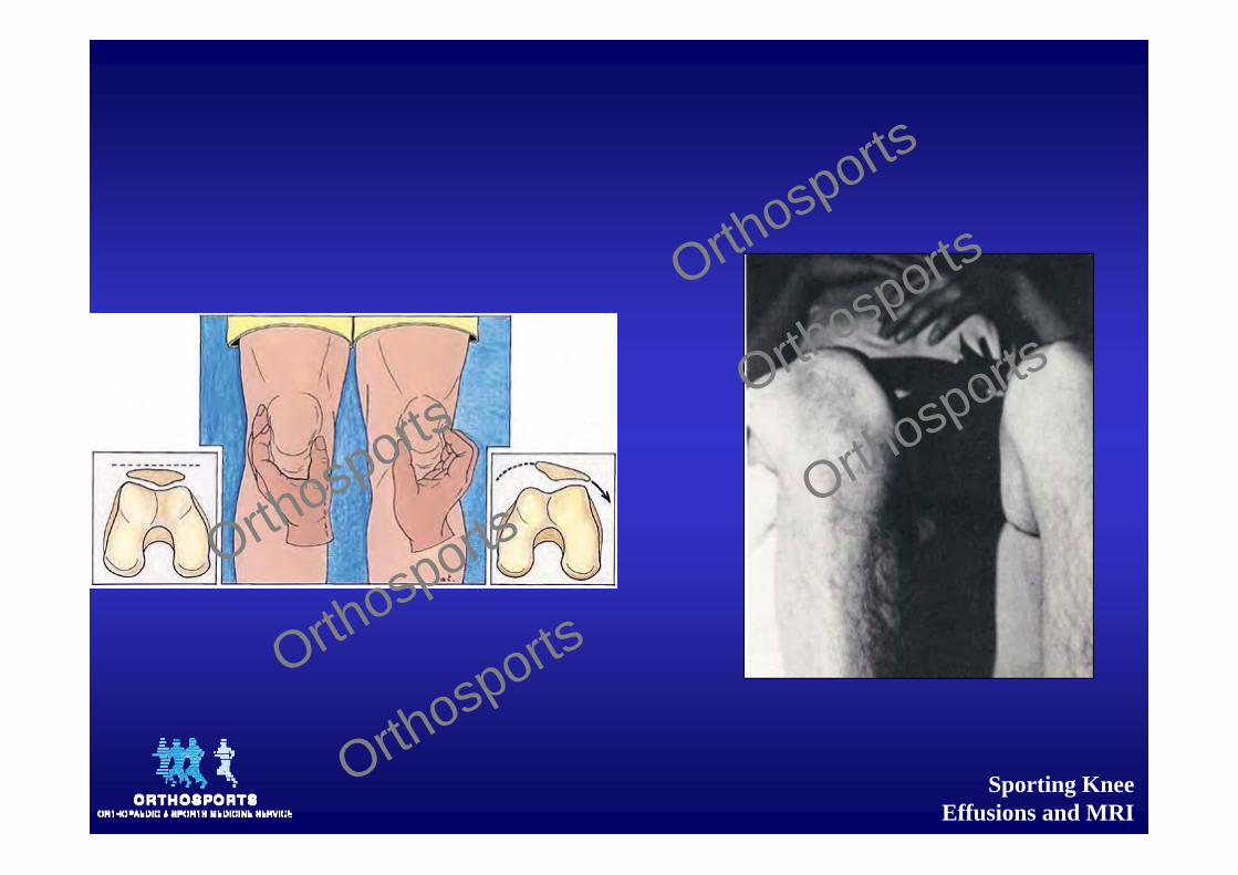
Clinical Assessment
– Body habitus– Gait – antalgic, thrust, stiff etc– Swelling– Scars– Muscle Wasting– Tenderness– Instability– Neurovascular status
Orthosports
O
rthosports
Orthosports
Orthosports
Orthosports
Orthosports
Sporting Knee Effusions and MRI
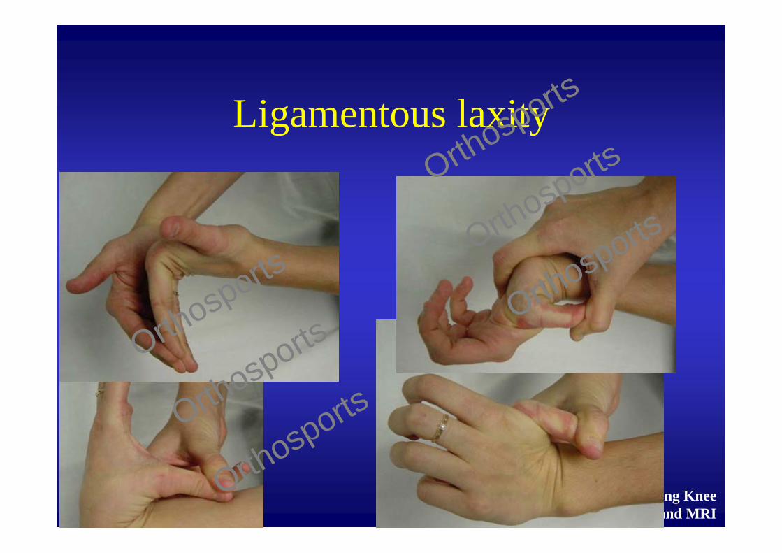
Ligamentous laxity
Orthosports
O
rthosports
Orthosports
Orthosports
Orthosports
Orthosports
Sporting Knee Effusions and MRI
Orthosports
O
rthosports
Orthosports
Orthosports
Orthosports
Orthosports
Sporting Knee Effusions and MRI
Orthosports
O
rthosports
Orthosports
Orthosports
Orthosports
Orthosports
Sporting Knee Effusions and MRI
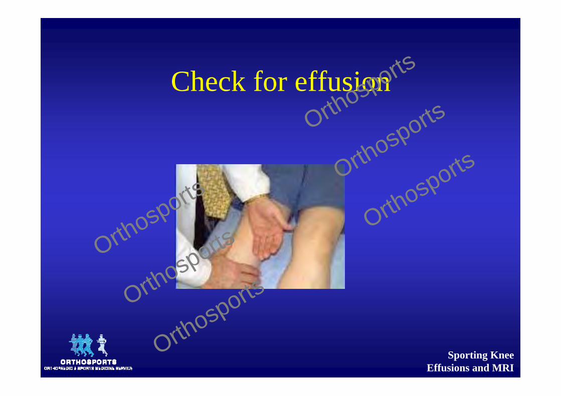
Check for effusion
Orthosports
O
rthosports
Orthosports
Orthosports
Orthosports
Orthosports
Sporting Knee Effusions and MRI
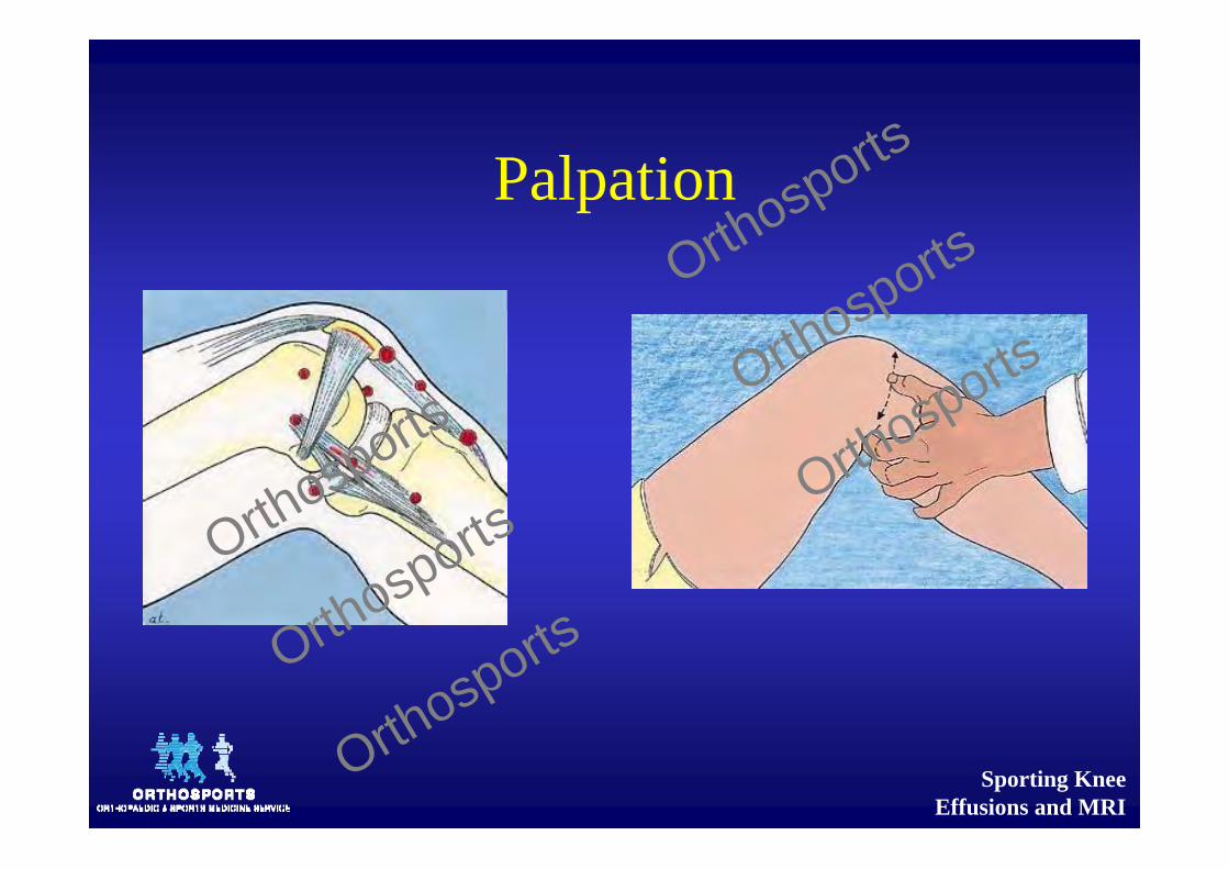
Palpation
Orthosports
O
rthosports
Orthosports
Orthosports
Orthosports
Orthosports
Sporting Knee Effusions and MRI
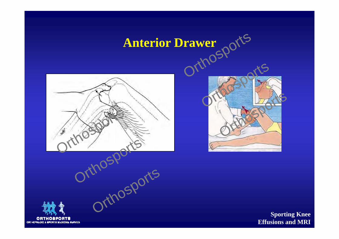
Anterior Drawer
Orthosports
O
rthosports
Orthosports
Orthosports
Orthosports
Orthosports
Sporting Knee Effusions and MRI
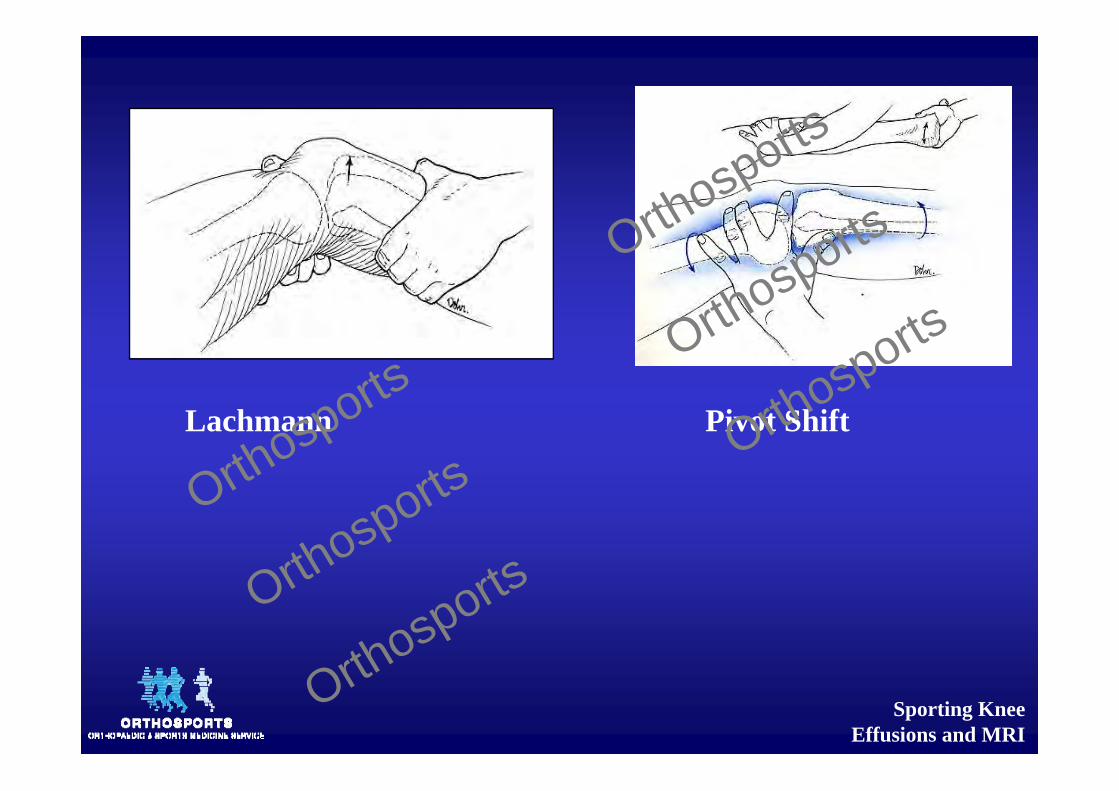
Lachmann Pivot Shift
Orthosports
O
rthosports
Orthosports
Orthosports
Orthosports
Orthosports
Sporting Knee Effusions and MRI
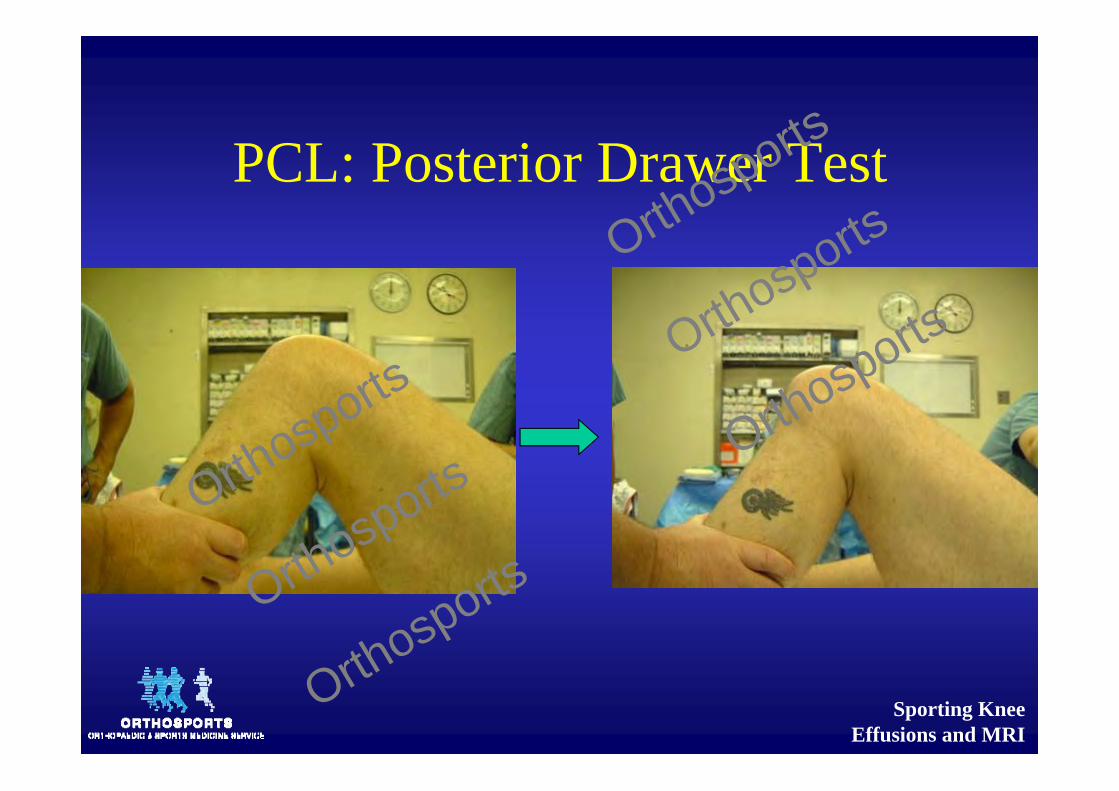
PCL: Posterior Drawer Test
Orthosports
O
rthosports
Orthosports
Orthosports
Orthosports
Orthosports

Sporting Knee Effusions and MRI
Orthosports
O
rthosports
Orthosports
Orthosports
Orthosports
Orthosports
Sporting Knee Effusions and MRI
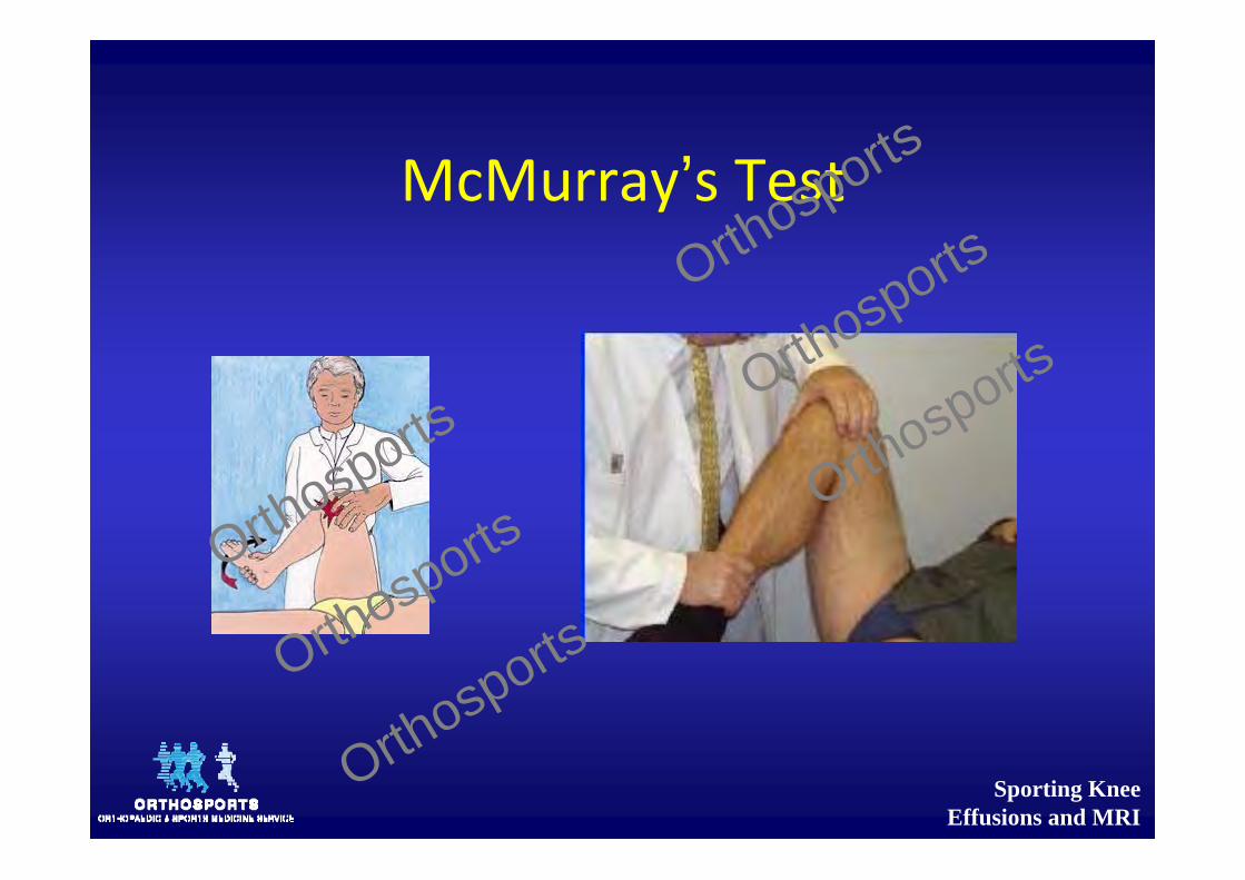
McMurray’s Test
Orthosports
O
rthosports
Orthosports
Orthosports
Orthosports
Orthosports
Sporting Knee Effusions and MRI
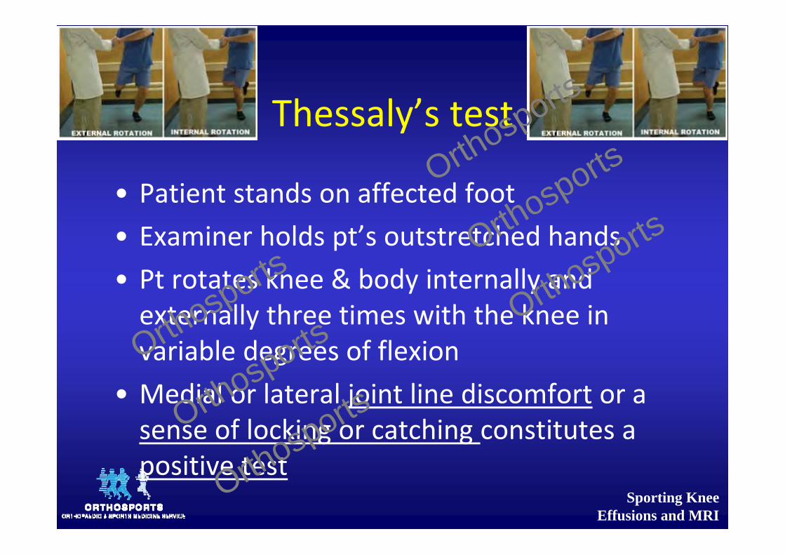
Thessaly’s test
• Patient stands on affected foot• Examiner holds pt’s outstretched hands• Pt rotates knee & body internally and externally three times with the knee in variable degrees of flexion
• Medial or lateral joint line discomfort or a sense of locking or catching constitutes a positive test
Orthosports
O
rthosports
Orthosports
Orthosports
Orthosports
Orthosports
Sporting Knee Effusions and MRI
Orthosports
O
rthosports
Orthosports
Orthosports
Orthosports
Orthosports
Sporting Knee Effusions and MRI
Med Meniscus Clinical Findings
• Joint line tenderness– Medial in cross leg position– Lateral at 300 flexion
• Pain on forced flexion• McMurray’s / Thessaly’s Test• Loss of extension• Clunking of meniscus
Orthosports
O
rthosports
Orthosports
Orthosports
Orthosports
Orthosports
Sporting Knee Effusions and MRI
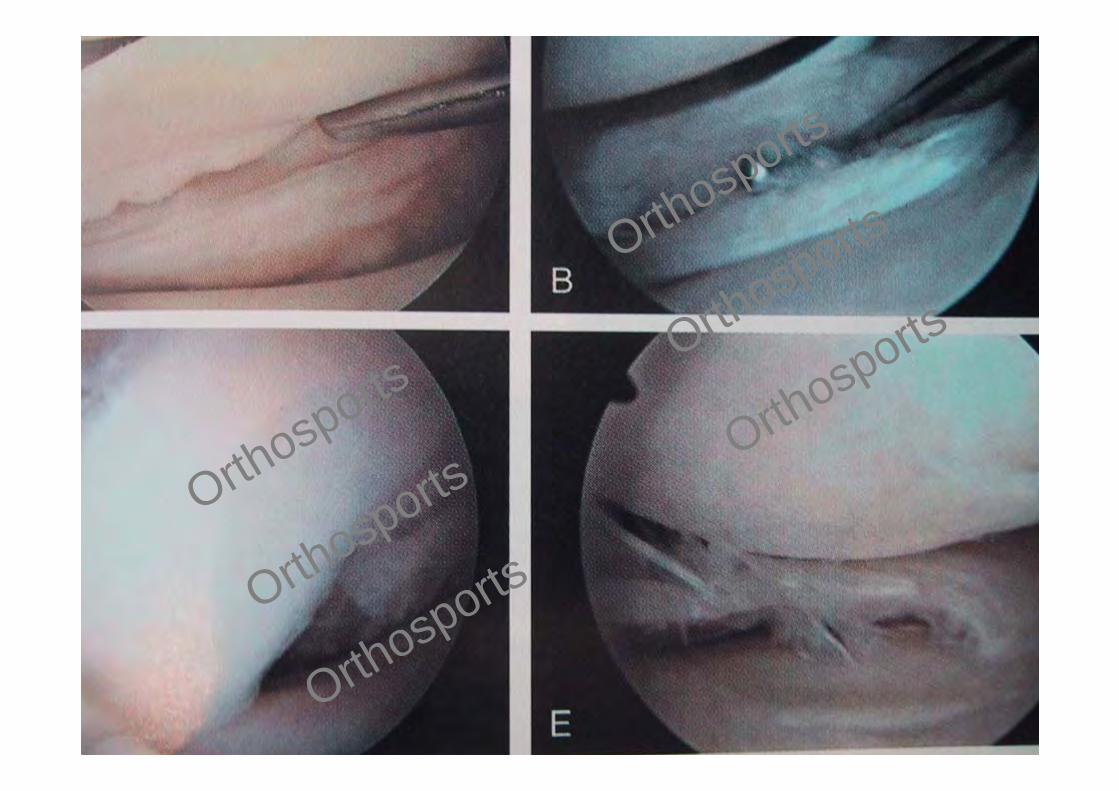
Meniscal Injuries
Orthosports
O
rthosports
Orthosports
Orthosports
Orthosports
Orthosports
Sporting Knee Effusions and MRI
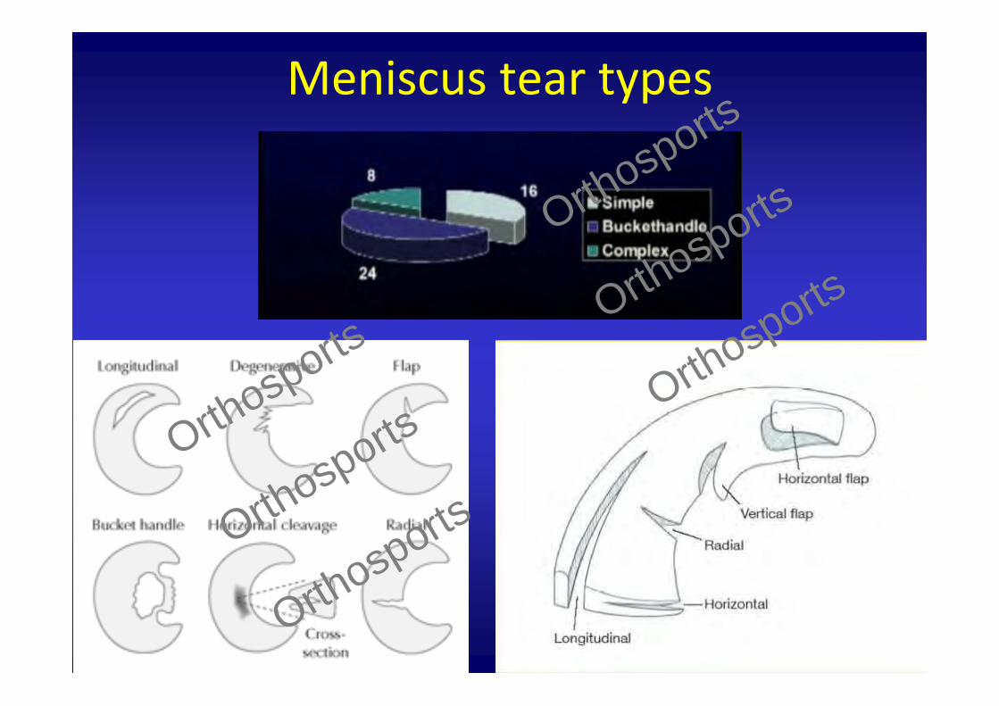
Meniscus tear types
Orthosports
O
rthosports
Orthosports
Orthosports
Orthosports
Orthosports
Sporting Knee Effusions and MRI
Meniscal tears• Younger patients are more likely to have an acute traumatic event as the cause of their meniscal pathology
• Acute ACL injury– Lat > Med
• Chronic ACL – Med > Lat
Orthosports
O
rthosports
Orthosports
Orthosports
Orthosports
Orthosports
Sporting Knee Effusions and MRI
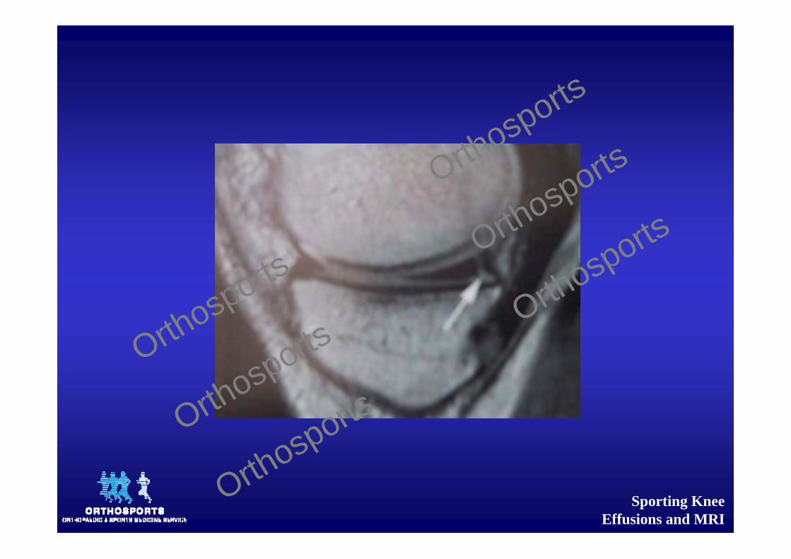
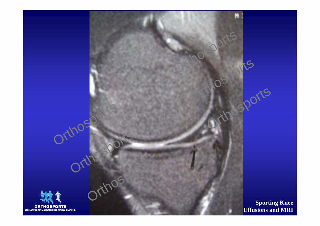
Meniscal Tears ‐ Diagnosis
• Diagnosis made from a careful history• The onset of symptoms and mechanism of injury are often clues to the diagnosis– Twisting injury– Hyperflexion
• Acute pain and swelling.
– Pain when kneeling or standing from sitting
Orthosports
O
rthosports
Orthosports
Orthosports
Orthosports
Orthosports
Sporting Knee Effusions and MRI
Meniscal tears ‐ Diagnosis
• Locking /catching– Also from chondral injury or patellofemoralchondrosis
• Loss of motion / mechanical block to extension– displaced bucket handle meniscal tear (or a loose body)
– Usually requires acute surgical treatment. It can also be caused by a loose body though.
Orthosports
O
rthosports
Orthosports
Orthosports
Orthosports
Orthosports
Sporting Knee Effusions and MRI
Degenerative Tears
• Older patients (>40 years)– Atraumatic chronic mild joint swelling– Joint line pain– Mechanical symptoms– Often associated with some chondral damage.
• Try to reproduce Snaps, clicks, catches or jerks when examining
Orthosports
O
rthosports
Orthosports
Orthosports
Orthosports
Orthosports
Sporting Knee Effusions and MRI
Orthosports
O
rthosports
Orthosports
Orthosports
Orthosports
Orthosports
Sporting Knee Effusions and MRI
Orthosports
O
rthosports
Orthosports
Orthosports
Orthosports
Orthosports
Sporting Knee Effusions and MRI
Orthosports
O
rthosports
Orthosports
Orthosports
Orthosports
Orthosports
Sporting Knee Effusions and MRI
Orthosports
O
rthosports
Orthosports
Orthosports
Orthosports
Orthosports
Sporting Knee Effusions and MRI
Orthosports
O
rthosports
Orthosports
Orthosports
Orthosports
Orthosports
Sporting Knee Effusions and MRI
Effusion
Orthosports
O
rthosports
Orthosports
Orthosports
Orthosports
Orthosports
Sporting Knee Effusions and MRI
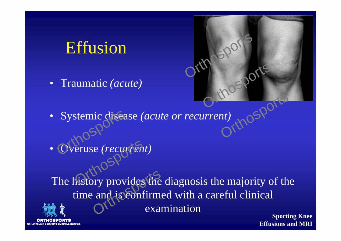
Effusion
• Traumatic (acute)
• Systemic disease (acute or recurrent)
• Overuse (recurrent)
The history provides the diagnosis the majority of the time and is confirmed with a careful clinical
examination
Orthosports
O
rthosports
Orthosports
Orthosports
Orthosports
Orthosports
Sporting Knee Effusions and MRI
Spontaneous Swelling
• Often the first sign of arthritis• Tumour or infection
– Systemic symptoms • fevers or chills, intravenous drug use, sexual contact, night pain or weight loss
• monoarticular arthritis with joint redness, swelling, pain and loss of motion
– Infiltrative disorders such as gout and pseudogout– sometimes the only way to differentiate between them is with a joint aspiration
– Most common joint involved in both benign and malignant tumors.
Orthosports
O
rthosports
Orthosports
Orthosports
Orthosports
Orthosports
Sporting Knee Effusions and MRI
Effusion
• Rapid onset (<2 hrs) large, tense effusion– Lig rupture or fracture
• Slower onset (24 ‐ 36 hrs) mild to mod– Meniscal injury / lig sprain/tear / infection
• Isolated meniscal tears do not always cause swelling and tend to indicate some chondral damage
Orthosports
O
rthosports
Orthosports
Orthosports
Orthosports
Orthosports
Sporting Knee Effusions and MRI
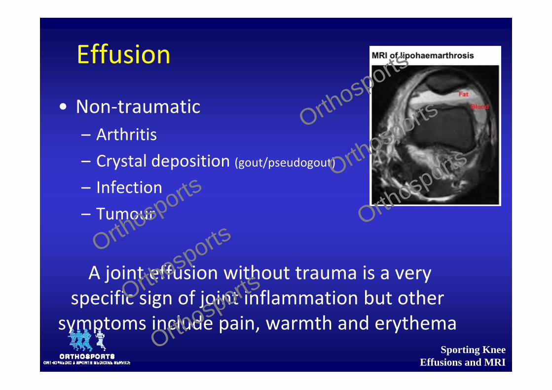
Effusion
• Non‐traumatic– Arthritis– Crystal deposition (gout/pseudogout)– Infection– Tumour
A joint effusion without trauma is a very specific sign of joint inflammation but other
symptoms include pain, warmth and erythema
Orthosports
O
rthosports
Orthosports
Orthosports
Orthosports
Orthosports
Sporting Knee Effusions and MRI
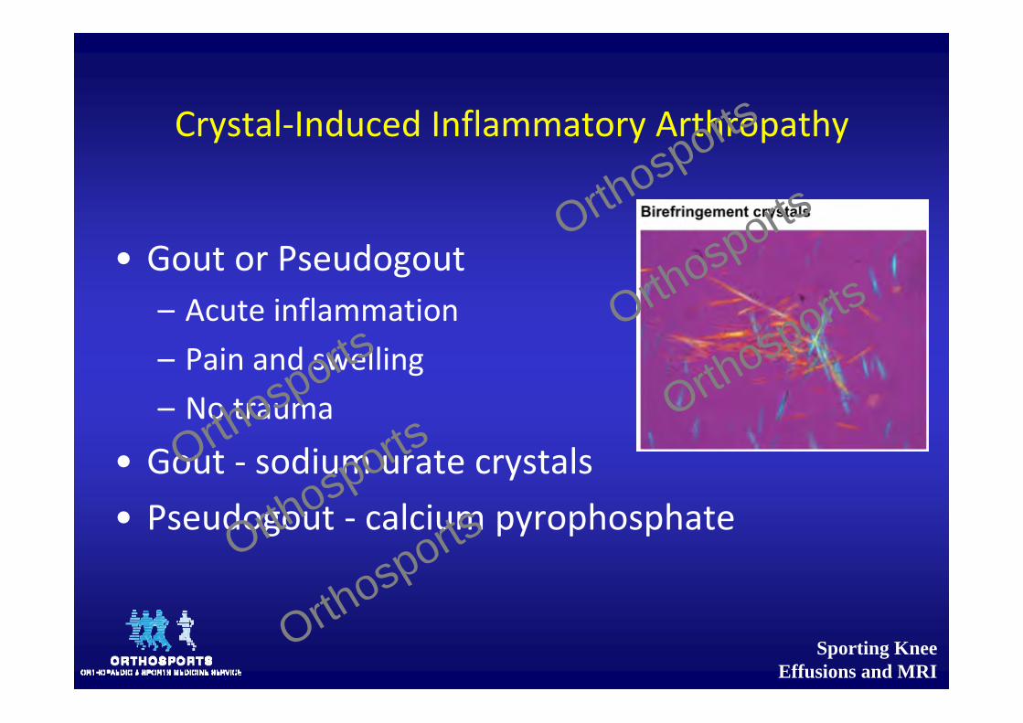
Crystal‐Induced Inflammatory Arthropathy
• Gout or Pseudogout– Acute inflammation– Pain and swelling– No trauma
• Gout ‐ sodium urate crystals • Pseudogout ‐ calcium pyrophosphate
Orthosports
O
rthosports
Orthosports
Orthosports
Orthosports
Orthosports
Sporting Knee Effusions and MRI
Infection
• Sudden onset of pain and swelling• No history of trauma• Warm, swollen, exquisitely tender• Slight motion causes intense pain• Any age
• Immunocompromised– (diabetes, alcoholism, AIDS, or corticosteroid therapy)
Orthosports
O
rthosports
Orthosports
Orthosports
Orthosports
Orthosports
Sporting Knee Effusions and MRI
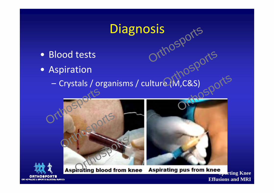
Diagnosis
• Blood tests• Aspiration
– Crystals / organisms / culture (M,C&S)
Orthosports
O
rthosports
Orthosports
Orthosports
Orthosports
Orthosports
Sporting Knee Effusions and MRI
Blood Tests
• Spontaneous effusion• With normal x‐ray:
– FBC, EUC, LFTs, ESR, CRP, ANA, Rh Factor, Anti CCP, Serum Immunoglobulins and HLA B27.
Orthosports
O
rthosports
Orthosports
Orthosports
Orthosports
Orthosports
Sporting Knee Effusions and MRI
Is it infected?
• Elevated blood WBC, ESR and CRP– Remember Fungi, TB and Lyme disease
• Fluid cell counts of 50‐100x109/L suggestive of infection
• Crystal‐induced arthritis can present in a similar fashion as an infection
• Sodium urate crystals precipitate in the knee joint and cause an intense inflammatory response
Orthosports
O
rthosports
Orthosports
Orthosports
Orthosports
Orthosports
Sporting Knee Effusions and MRI
Is it infection or crystals?
• Slightly cloudy synovial fluid– WBC count 2 ‐ 75 × 109/L– Polarized‐light microscopy– Negatively birefringent rods with gout– Positively birefringent rhomboids with pseudogout.
• The presence of crystals does not rule out an infection, as the two may co‐exist– No Abs unless infection proven
Orthosports
O
rthosports
Orthosports
Orthosports
Orthosports
Orthosports
Sporting Knee Effusions and MRI
Rheumatic disease (Inflammatory)
• Synovial Fluid– WBC count 2 ‐ 50 × 109/L suggest an inflammatory process
Rheumatology referral within 6 weeks is recommended for patients in whom inflammatory arthritis is suspected
Orthosports
O
rthosports
Orthosports
Orthosports
Orthosports
Orthosports
Sporting Knee Effusions and MRI
Treatment
• General measures to relieve knee pain and swelling– Splinting, assisted weight bearing, ice packs, and NSAID’s
– No AB’s until specimens taken– No HC if suspect infection
• Arthroscopy is rare with acute swelling of the knee without trauma
Orthosports
O
rthosports
Orthosports
Orthosports
Orthosports
Orthosports
Sporting Knee Effusions and MRI
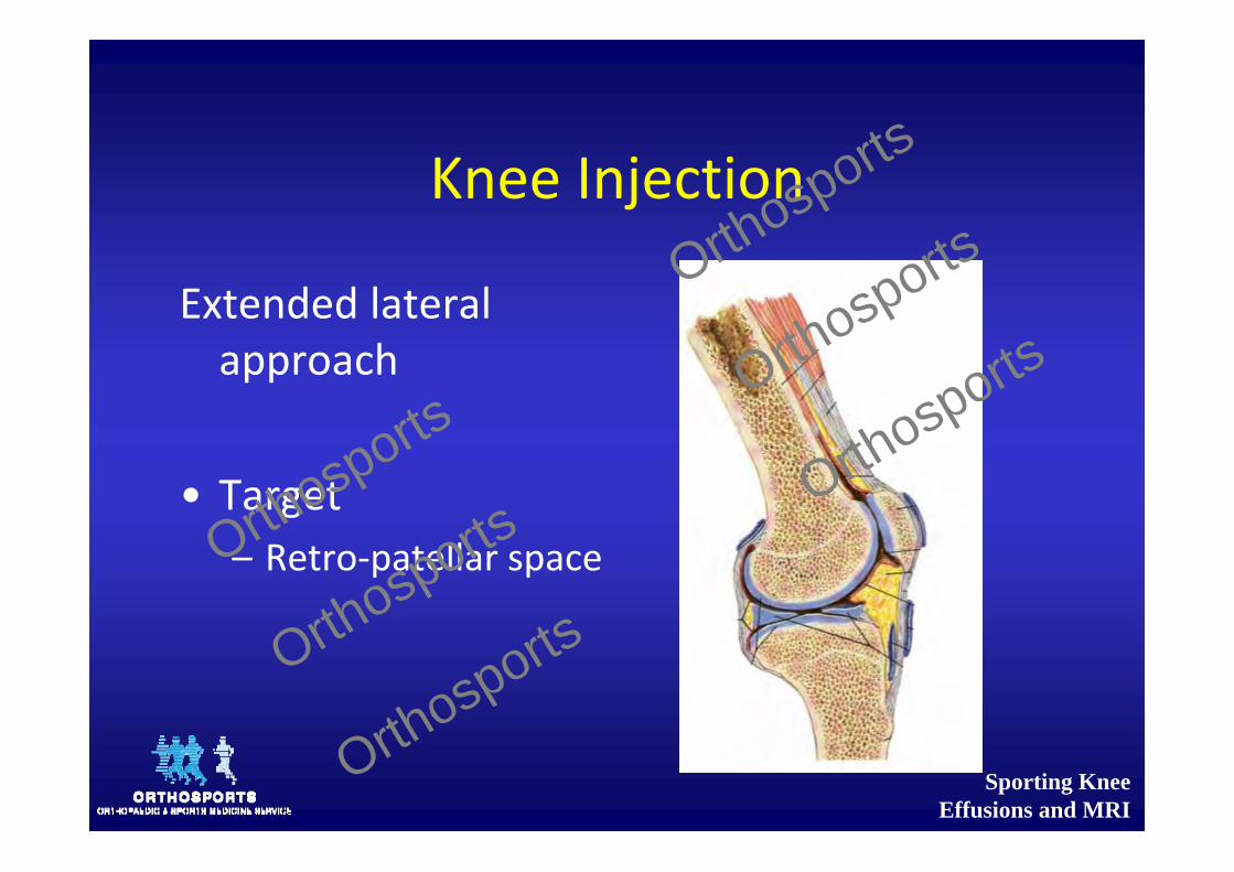
Knee Injection
Extended lateral approach
• Target – Retro‐patellar space Orthosports
Orthosports
Orthosports
Orthosports
Orthosports
Orthosports
Sporting Knee Effusions and MRI
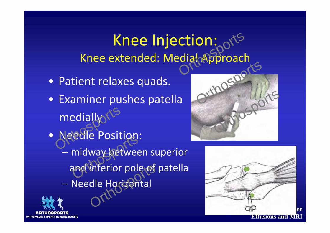
Knee Injection:Knee extended: Medial Approach
• Patient relaxes quads.• Examiner pushes patella medially
• Needle Position:– midway between superior and inferior pole of patella
– Needle Horizontal
Orthosports
O
rthosports
Orthosports
Orthosports
Orthosports
Orthosports
Sporting Knee Effusions and MRI
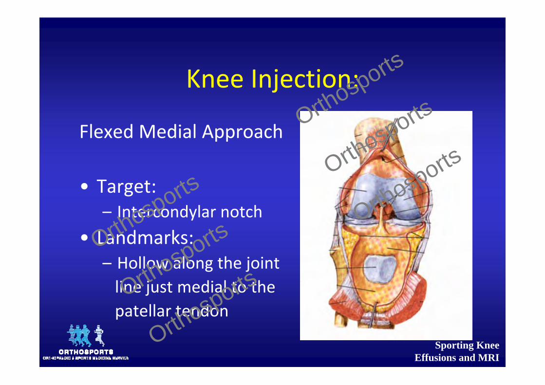
Knee Injection:
Flexed Medial Approach
• Target:– Intercondylar notch
• Landmarks:– Hollow along the joint line just medial to the patellar tendon
Orthosports
O
rthosports
Orthosports
Orthosports
Orthosports
Orthosports
Sporting Knee Effusions and MRI
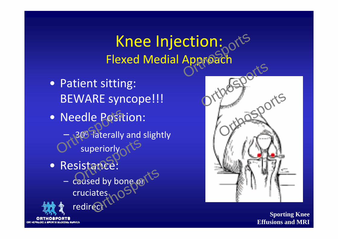
Knee Injection:Flexed Medial Approach
• Patient sitting: BEWARE syncope!!!
• Needle Position:– 300 laterally and slightly
superiorly
• Resistance:– caused by bone or cruciatesredirect
Orthosports
O
rthosports
Orthosports
Orthosports
Orthosports
Orthosports
Sporting Knee Effusions and MRI
Orthosports
O
rthosports
Orthosports
Orthosports
Orthosports
Orthosports
Sporting Knee Effusions and MRI
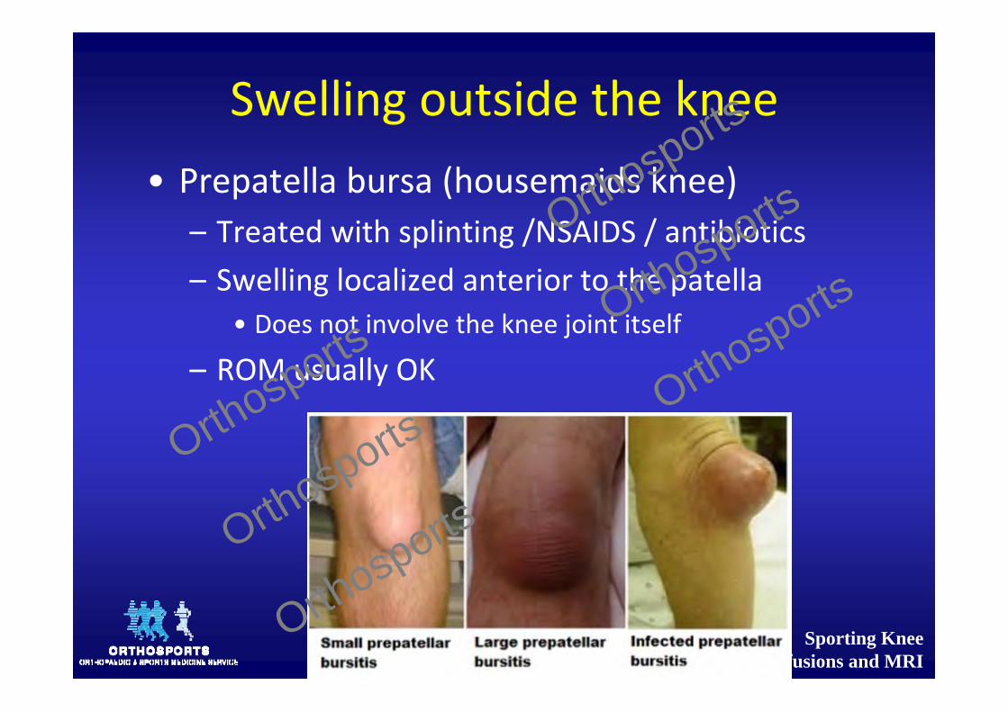
Swelling outside the knee• Prepatella bursa (housemaids knee)
– Treated with splinting /NSAIDS / antibiotics– Swelling localized anterior to the patella
• Does not involve the knee joint itself
– ROM usually OK
Orthosports
O
rthosports
Orthosports
Orthosports
Orthosports
Orthosports
Sporting Knee Effusions and MRI
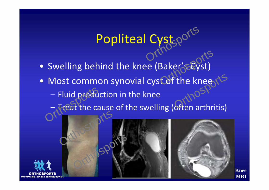
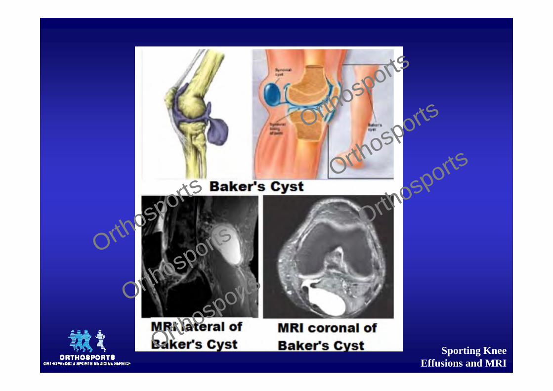
Popliteal Cyst
• Swelling behind the knee (Baker’s Cyst)• Most common synovial cyst of the knee
– Fluid production in the knee– Treat the cause of the swelling (often arthritis)Orthosports
Orthosports
Orthosports
Orthosports
Orthosports
Orthosports

Sporting Knee Effusions and MRI
Popliteal Cyst
• Origin – posteromedial ‐ Gastrocnemius / Semimembranous bursa
– Insidious onset of mild to moderate pain posteriorly
– Usually symptomatic when very large or rupture
– Rupture ‐ quite severe calf pain and swelling and difficulty walking may look like a DVT.
Orthosports
O
rthosports
Orthosports
Orthosports
Orthosports
Orthosports
Sporting Knee Effusions and MRI
Orthosports
O
rthosports
Orthosports
Orthosports
Orthosports
Orthosports
Sporting Knee Effusions and MRI
Post Knee Pain DDx
• Arterial popliteal aneurysm• Adipose tissue• Tumour• DVT
Orthosports
O
rthosports
Orthosports
Orthosports
Orthosports
Orthosports
Sporting Knee Effusions and MRI
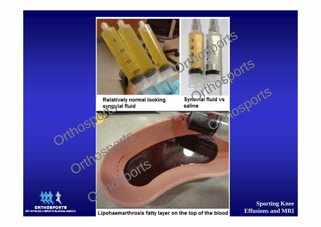
Traumatic Injury
• Can’t WB = fracture• Pop/giving = ACL tear• Pain with twisting, kneeling or standing from
sitting = meniscal injury• Isolated meniscal tears do not cause swelling
– Chondral injuries do
• Haemarthrosis becomes less bloodstained and more serous in appearance with time
Orthosports
O
rthosports
Orthosports
Orthosports
Orthosports
Orthosports
Sporting Knee Effusions and MRI
Who gets an Xray?
Orthosports
O
rthosports
Orthosports
Orthosports
Orthosports
Orthosports
Sporting Knee Effusions and MRI
Ottawa knee rules• After acute knee injury knee x‐rays are indicated if any of the following criteria present:– aged 55 years or over– tenderness at the head of the fibula– isolated tenderness of the patella– inability to flex knee to 90 degrees– inability to bear weight (defined as an inability to take four steps, ie. two steps on each leg, regardless of limping) immediately and at presentation
Orthosports
O
rthosports
Orthosports
Orthosports
Orthosports
Orthosports
Sporting Knee Effusions and MRI
Ottawa knee rules
• Majority of acute knee injuries are soft tissue injuries not identifiable on plain radiographs.
• A normal looking knee X‐ray after acute trauma does not exclude a fracture– Tibial plateau fractures, Segond fractures Salter‐Harris type 1 fractures are easily missed if not complemented with clinical findings
– Follow up should be recommended if symptoms persist.
Orthosports
O
rthosports
Orthosports
Orthosports
Orthosports
Orthosports
Sporting Knee Effusions and MRI
Investigation
• Xray• Xray• Xray• Xray • XrayOrthosports
Orthosports
Orthosports
Orthosports
Orthosports
Orthosports
Sporting Knee Effusions and MRI
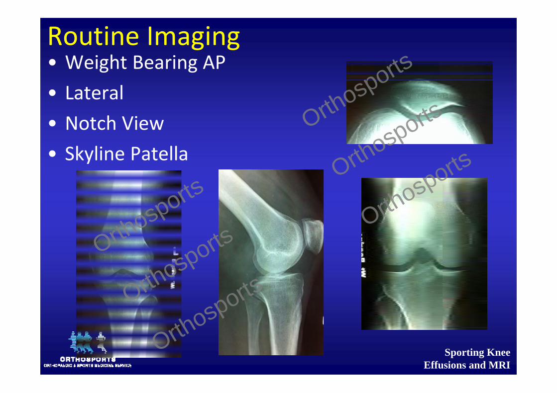
Routine Imaging• Weight Bearing AP• Lateral• Notch View• Skyline Patella
Orthosports
O
rthosports
Orthosports
Orthosports
Orthosports
Orthosports
Sporting Knee Effusions and MRI
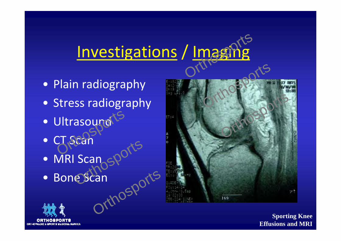
Investigations / Imaging
• Plain radiography• Stress radiography• Ultrasound• CT Scan• MRI Scan• Bone Scan
Orthosports
O
rthosports
Orthosports
Orthosports
Orthosports
Orthosports
Sporting Knee Effusions and MRI
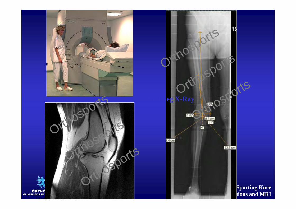
™Trademark of Smith & Nephew.
MRI Scan Full Leg X-Ray
Orthosports
O
rthosports
Orthosports
Orthosports
Orthosports
Orthosports
Sporting Knee Effusions and MRI
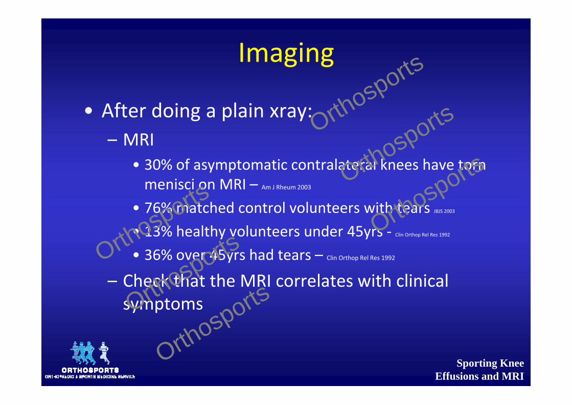
Imaging
• After doing a plain xray:– MRI
• 30% of asymptomatic contralateral knees have torn menisci on MRI – Am J Rheum 2003
• 76% matched control volunteers with tears JBJS 2003• 13% healthy volunteers under 45yrs ‐ Clin Orthop Rel Res 1992
• 36% over 45yrs had tears – Clin Orthop Rel Res 1992
– Check that the MRI correlates with clinical symptoms
Orthosports
O
rthosports
Orthosports
Orthosports
Orthosports
Orthosports
Sporting Knee Effusions and MRI
MRI
• Noninvasive nature• Multiple planes• No ionizing radiation• See other structures within the joint• Relatively high cost• Overcalls pathology• Not all magnets and reports equal
Orthosports
O
rthosports
Orthosports
Orthosports
Orthosports
Orthosports
Sporting Knee Effusions and MRI
MRI• Accuracy >95%
• Unfortunately being used as the first investigation for a painful knee
• Common to see meniscal tear & chondral damage• Weight bearing xrays show arthritis, which is actually what the patient needs treatment for
– Normal clinical exam = MRI only 5% chance of showing a meniscal tear
– Asymptomatic patients: • <45 yrs old, 13% have a meniscal tear• >45 years old, 36% have a meniscal tear
Orthosports
O
rthosports
Orthosports
Orthosports
Orthosports
Orthosports
Sporting Knee Effusions and MRI
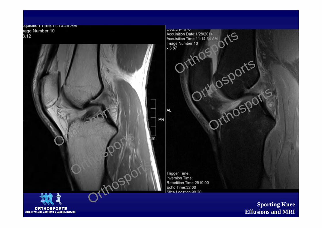
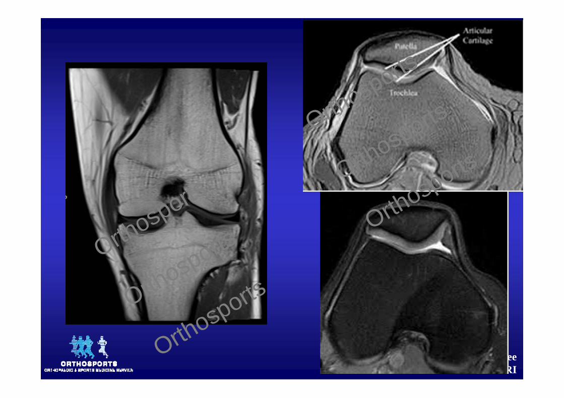
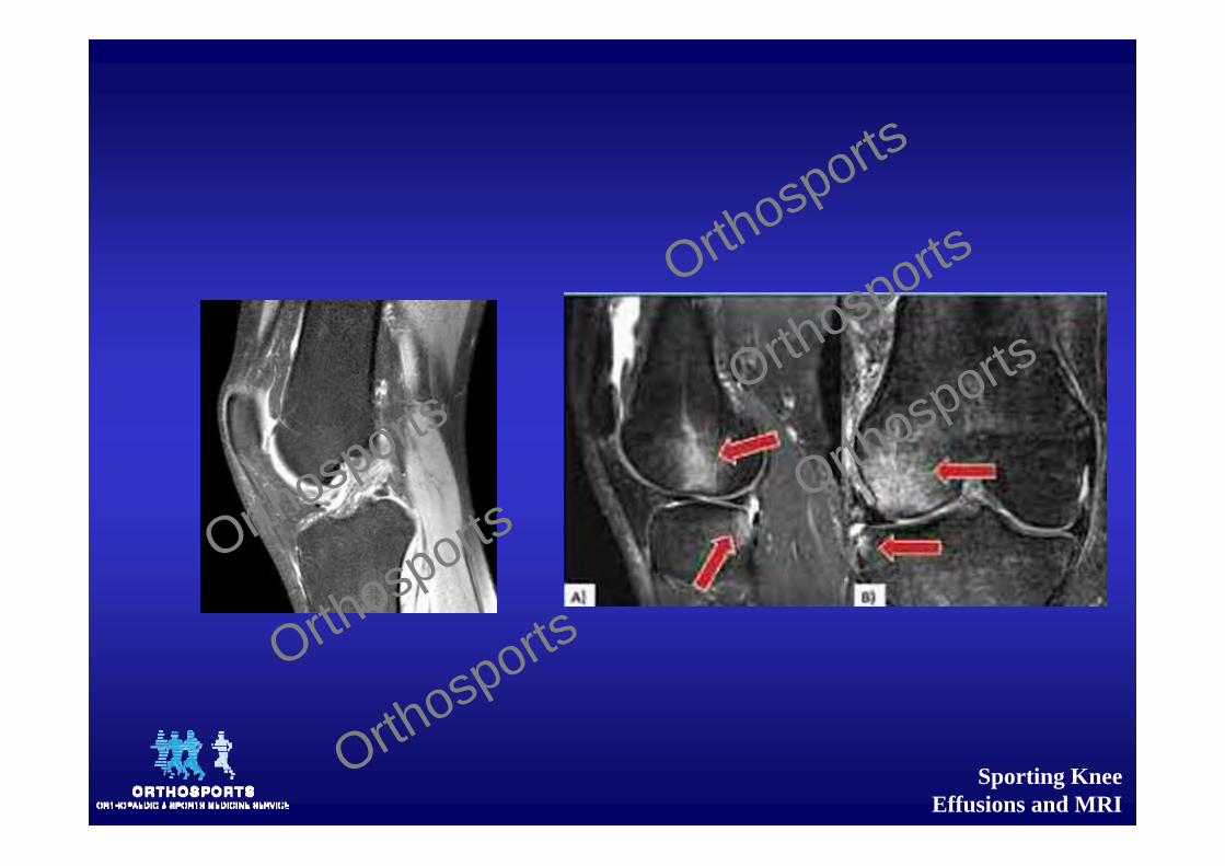
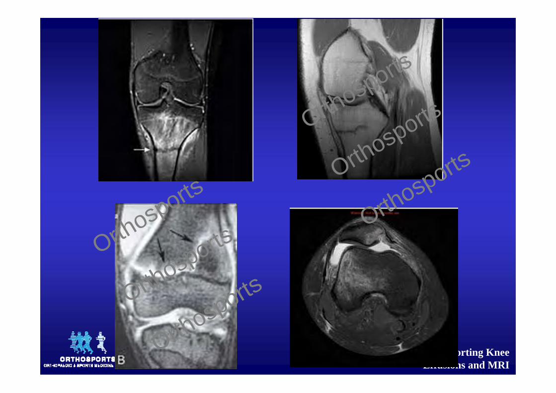
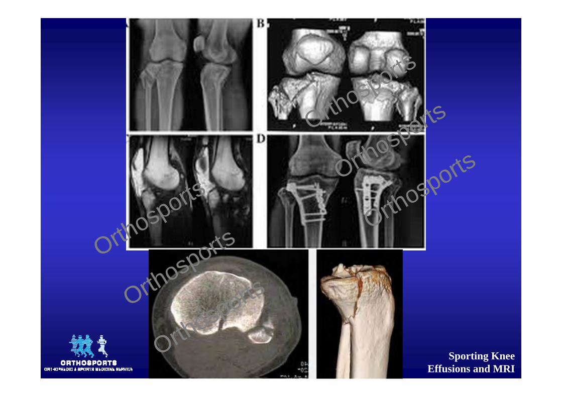
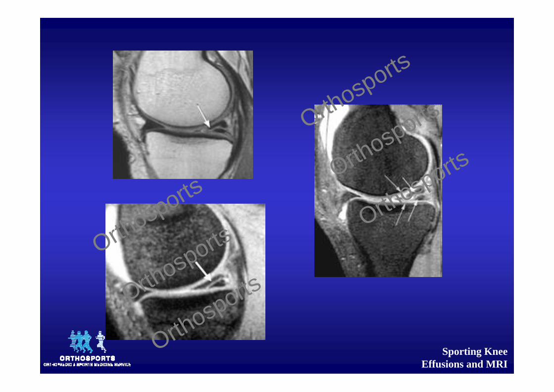
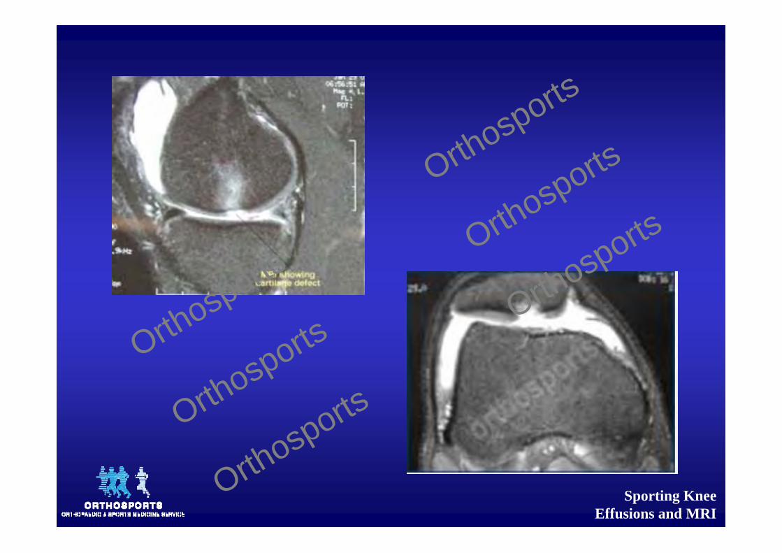
Reading an MRI
• T1• T2• Gadolinium
Orthosports
O
rthosports
Orthosports
Orthosports
Orthosports
Orthosports
Sporting Knee Effusions and MRI
Orthosports
O
rthosports
Orthosports
Orthosports
Orthosports
Orthosports
Sporting Knee Effusions and MRI
Orthosports
O
rthosports
Orthosports
Orthosports
Orthosports
Orthosports
Sporting Knee Effusions and MRI
Orthosports
O
rthosports
Orthosports
Orthosports
Orthosports
Orthosports
Sporting Knee Effusions and MRI
Orthosports
O
rthosports
Orthosports
Orthosports
Orthosports
Orthosports
Sporting Knee Effusions and MRI
Orthosports
O
rthosports
Orthosports
Orthosports
Orthosports
Orthosports
Sporting Knee Effusions and MRI
Orthosports
O
rthosports
Orthosports
Orthosports
Orthosports
Orthosports
Sporting Knee Effusions and MRI
Orthosports
O
rthosports
Orthosports
Orthosports
Orthosports
Orthosports
Sporting Knee Effusions and MRI
Orthosports
O
rthosports
Orthosports
Orthosports
Orthosports
Orthosports
Sporting Knee Effusions and MRI
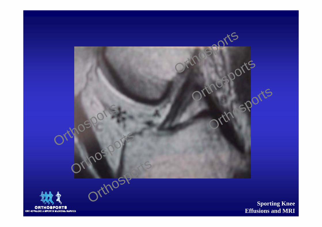
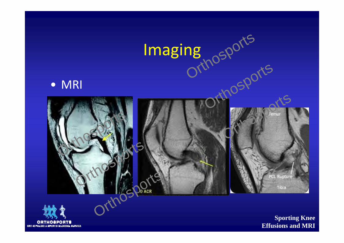
Imaging
• MRI
Orthosports
O
rthosports
Orthosports
Orthosports
Orthosports
Orthosports
Sporting Knee Effusions and MRI
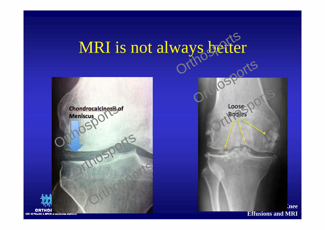
MRI is not always better
Orthosports
O
rthosports
Orthosports
Orthosports
Orthosports
Orthosports
Sporting Knee Effusions and MRI
Arthritis vs Meniscal Tear
• Clinical exam less reliable in these pts– Different, less acute mechanism of injury– Numerous other possible degenerative causes contributing to their intra‐articular knee pain
– Very high incidence of meniscal tears on MRI scanning with OA
– The decision as to whether or not to operate is often difficult
Orthosports
O
rthosports
Orthosports
Orthosports
Orthosports
Orthosports
Sporting Knee Effusions and MRI
Arthritis vs Meniscal Tear
– A meniscal tear can lead to knee OA, but knee OA can also lead to a spontaneous meniscal tear
– A degenerative meniscal lesion often suggests early‐stage knee OA
– Surgical resection of non‐obstructive degenerate lesions may merely remove evidence of the disorder while the OA and associated symptoms proceed.
Orthosports
O
rthosports
Orthosports
Orthosports
Orthosports
Orthosports
Sporting Knee Effusions and MRI
Arthritis vs Meniscal Tear
• Arthroscopic debridement for chronic OA is no better than a sham procedure in relieving knee pain or improving functional status
• No better than physio WHEN THIS IS THE FIRST FORM OF TREATMENTOrthosports
Orthosports
Orthosports
Orthosports
Orthosports
Orthosports
Sporting Knee Effusions and MRI
Swedish study
• 45–64 yrs old, knee pain, meniscal tear on MRI and OA on xray (minor)
• Rigorous exercise regimen alone vs Exercise regimen with surgery
• Outcomes 2, 6, 24 and 60 months• Both groups improved considerably over the first 6 months and maintained improvements in pain and functional status over 60 months
Orthosports
O
rthosports
Orthosports
Orthosports
Orthosports
Orthosports
Sporting Knee Effusions and MRI
Swedish study
• 30% of the subjects randomised to the non‐operative arm had persistent pain and crossed over to have surgery
• And had similar outcomes to those randomised to receive surgery at the outset.
Try physio 1st and operate if they don’t get better
Orthosports
O
rthosports
Orthosports
Orthosports
Orthosports
Orthosports
Sporting Knee Effusions and MRI
MeTeOR• The Meniscal Tear in Osteoarthritis Research (MeTeOR) Trial
• Aged ≥45 with meniscal tear on MRI and underlying OA change on xray or MRI
• As in Herrlin et al about 30% of MeTeORsubjects crossed over from non‐operative therapy to surgery.Orthosports
Orthosports
Orthosports
Orthosports
Orthosports
Orthosports
Sporting Knee Effusions and MRI
Meteor and knee OA
• No role for arthroscopy unless they have clinical and imaging evidence of a tear AND mechanical symptoms (Catching, locking, popping etc)
• Patients who fail to improve with physio can try surgery
• Recovery from menisectomy surgery at 1 year:– Worse if female and worse OA – No different based on Age, BMI, depth of meniscal excision, involvement of 1 or both menisci, extent of meniscal tear
Orthosports
O
rthosports
Orthosports
Orthosports
Orthosports
Orthosports
Sporting Knee Effusions and MRI
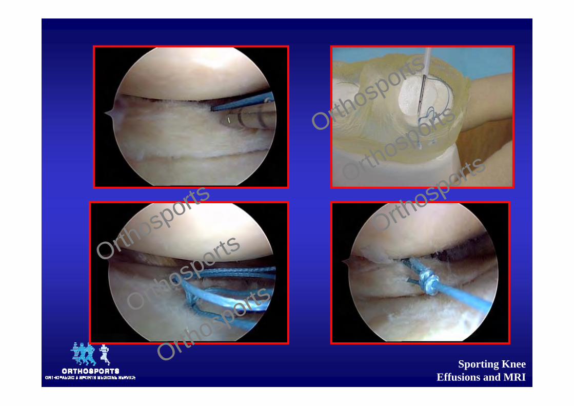
Surgical Decision Making for Meniscal Tears
• Indications for Arthroscopic Treatment:– Symptoms affecting ADLs, work, sports– Positive physical findings
• Joint line tenderness, joint effusion, limitation of motion, and provocative signs
– Failure to respond to nonsurgical treatment,– Absence of other causes of knee pain
Orthosports
O
rthosports
Orthosports
Orthosports
Orthosports
Orthosports
Sporting Knee Effusions and MRI
Surgical Decision Making for Meniscal Tears with ACL
• Most often done concurrently with ACL reconstruction– Surgical timing dictated by:
• ROM• Swelling• Quads function• Other Lig injuries• Locked knee
Orthosports
O
rthosports
Orthosports
Orthosports
Orthosports
Orthosports
Sporting Knee Effusions and MRI
Rules Of Thumb
• Ongoing pain affecting ADL’s• Meniscal pathology will do better than articular cartilage
• Younger more likely to be meniscal• Clicking more likely to be meniscal• Sudden onset of pain generally does better
• Mechanical symptoms do better
Orthosports
O
rthosports
Orthosports
Orthosports
Orthosports
Orthosports
Sporting Knee Effusions and MRI
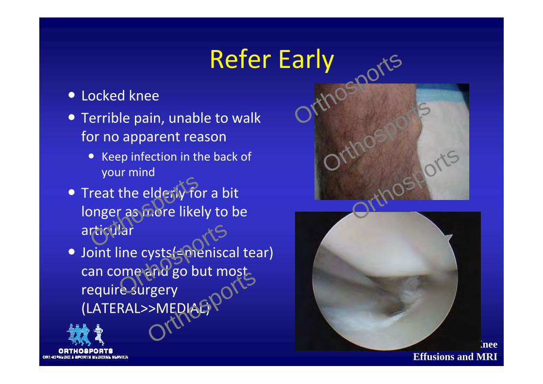
Refer Early Locked knee Terrible pain, unable to walk for no apparent reason Keep infection in the back of
your mind
Treat the elderly for a bit longer as more likely to be articular
Joint line cysts(=meniscal tear) can come and go but most require surgery (LATERAL>>MEDIAL)
Orthosports
O
rthosports
Orthosports
Orthosports
Orthosports
Orthosports
Sporting Knee Effusions and MRI
Urgent Referral
• Infection• Locked Knee• Lateral ligament
Orthosports
O
rthosports
Orthosports
Orthosports
Orthosports
Orthosports
Sporting Knee Effusions and MRI
Thank you
Orthosports
O
rthosports
Orthosports
Orthosports
Orthosports
Orthosports
Sporting Knee Effusions and MRI
Doron SherMBBS, MBiomedE, FRACS
www.kneedoctor.com.auwww.Doron.com.au
www.orthosports.com.au
160 Belmore Rd, Randwick47‐49 Burwood Rd, Concord
Orthosports
O
rthosports
Orthosports
Orthosports
Orthosports
Orthosports