serous effusions: a practical algorithmic approach to
TRANSCRIPT

Serous effusions: A practical algorithmic approach to diagnosis
Dr Ashish Chandra
MD FRCPath DipRCPath (Cytol)
Guy’s & St. Thomas’ Hosp NHSfT, London, UK

Educational objectives
• To revisit algorithms in lab handling of serous effusion samples
• To outline common diagnostic pitfalls and how to avoid them
• Algorithm for morphological evaluation and reporting terminology
• Algorithm to confirm the nature of a cellular infiltrate using ancillary tests

Serous effusions
• Abnormal accumulation of fluid in body cavities
• Presence of malignant cells implies advanced TNM Stage
• Pleural fluid
• Pericardial fluid
• Ascitic fluid
• (Peritoneal washings)
Synovial fluid and CSF not covered in this presentation

Macroscopic findings
• Volume: 75ml eliminates the influence of specimen volume on diagnostic adequacy (Rooper et al, Cancer Cytopath 2014)
• Practical implication: 75ml of fluid needed to say that a benign effusion is truly benign
• Smaller volumes acceptable for processing
Macroscopic findings
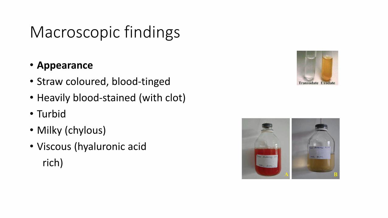
• Appearance
• Straw coloured, blood-tinged
• Heavily blood-stained (with clot)
• Turbid
• Milky (chylous)
• Viscous (hyaluronic acid
rich)

Clear/chylous fluid sample
Clear/chylous fluid sample
Cytospins
MGG & Pap
Liquid based preparations
(ThinPrep,SurePath etc)
Pap
Clot (if present)
H&E
BSCC Code of Practice for Exfoliative Cytology. Cytopathology 2009. Tiisue Pathways, RCPath www.rcpath.org

Turbid/heavily blood-stained fluid sample
Turbid/haemorrhagic fluid sample
Direct smears from sediment (MGG & Pap)
Cytospins & LBP after dilution
Clot section

Classic algorithm rare in practice
Fluid sample
Transudate Exudate
Cytology No cytology

Aliquoted at time of collection
Fluid sample
Biochemistry Cytology Microbiology
Biochemistry to ascertain whether transudate or exudate and relevant tumour markers. Cytology for malignant cells or non-neoplastic conditions. Microbiology for diagnosis of suspected infections

Common diagnostic pitfalls
• Small numbers of lesional cells and poor cellular preservation: quality may be compromised at time of collection as well as during transport & processing
• Adenocarcinoma vs mesothelial cells, reactive & neoplastic
• Cytoplasmic vacuolation including signet ring-like cells
• Single cell population
• Less frequently encountered tumours: Poorly differentiated squamous cell carcinoma, small cell carcinoma, melanoma

Terminology and diagnostic categories
• C1: Non-diagnostic
• C2: No malignant cells seen
• C3: Atypia, equivocal for malignancy (or malignancy not excluded)
• C4: Suspicious for malignancy
• C5: Malignant cells seen
• Used mainly for breast FNA samples in the UK
• Thy 1-5 in thyroid cytology
• Not a reporting requirement for serous effusions but facilitates audit
Reporting exfoliative cytology samples by UK cytotechnologists
• In the UK, cytotechnologists (Biomedical scientists) with appropriate training & qualification (Diploma of Expert Practice in non-gynae cytology) may sign out negative urine, serous effusion and respiratory tract specimens
• With the Advanced Specialist Diploma in non-gynae cytology, they may sign out abnormal exfoliative cytology results
• www.ibms.org.uk

Morphology & ancillary testing
Cytospins/LBP
(+ clot/cell block)
Non-diagnostic,
C1
Equivocal for
malignancy,
C3
Suspicious for
malignancy,
C4
No malignant
cells seen,
>75ml sample,
C2
Malignant
cells seen,
C5
Repeat sample or
clinical
follow up
Clinical follow up
Ancillary testing
Correlation with
biopsy
& clinical data
Ancillary testing to
establish primary site
and predictive markers

Ancillary testing: diagnostic & theranostic
Cytology atypical,
suspicious (diagnostic) or
malignant (theranostic)
Clot/cell block/cytospins/smears for immunochemistry,
molecular studies (paraffin curls) & cytogenetics (paraffin
sections for FISH)
Flow cytometry
Biochemical analysis (mesothelin)
As material may be limited in amount, it should be used conservatively for immunochemistry so that sufficient material remains for molecular tests if necessary

Mesothelial cells
Present singly and in small groups with cells separated by spaces (windows)Clasp-like cell junctionsPeripheral vacuolation (glycogen) and blebs (microvilli)Two-zone cytoplasmic differentiationCentral vesicular nuclei with small nucleoli

Mesothelial proliferation
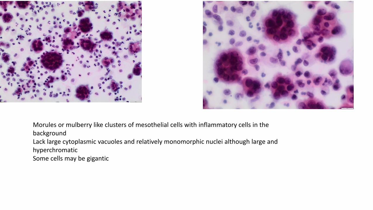
Morules or mulberry like clusters of mesothelial cells with inflammatory cells in the backgroundLack large cytoplasmic vacuoles and relatively monomorphic nuclei although large and hyperchromaticSome cells may be gigantic

MGG cytospin of the above case with identifiable mesothelial cell type, confirmed on calretiin immunostain

Ancillary testing of mesothelial proliferationsConfirm mesothelial origin
WT1, Calretinin, D2-40/Podoplanin
CK5/6, Thrombomodulin, mesothelin, HBME1
Reactive
Desmin: cytoplasmic
Neoplastic
EMA: thick membranous; p53: positive
Claudin-4: no membranous staining
GLUT-1: positive FISH: p16 deletion
IMP3, CD146, Ki67
Exclude adenocarcinoma
BerEP4, MOC31,TTF1
Particularly useful for confirming uncommon subtypes of mesothelioma such as small cell, clear cell, lymphohistiocytic , signet ring etc. Limitations of different immunostains are mentioned in the handout and also in the reference list.
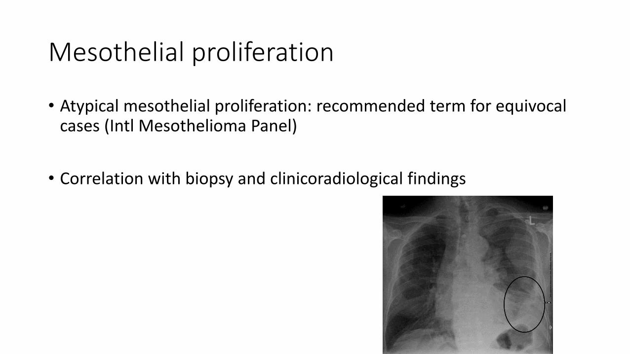
Mesothelial proliferation
• Atypical mesothelial proliferation: recommended term for equivocal cases (Intl Mesothelioma Panel)
• Correlation with biopsy and clinicoradiological findings

Epithelioid (epithelial) cells in effusions
Low power view of cytospin MGG showing a cellular infiltrate of lymphocytes, mesothelial cells and macrophages and 3 dimensional cluisters of malignant cells


MGG & Pap (x20) cohesive groups without windows or gaps between the malignant cells (unlike mesothelial cells), abundant cytoplasm with vacuolation and nuclear pleomorphism

Papillary structures in effusions are indicative of adenocarcinoma from primaries in the lung, breast, thyroid, GI tract, pancreas, kidney, ovary & uterus

Algorithm for epithelioid malignant cellsAtypical, suspicious or
malignant
epithelioid cells
Thoracic:
Breast: GATA-3
Lung: TTF1 Napsin A
Thyroid: thyroglobulin
Mesothelial: EMA, WT1
Abdominal:
Gynae: PAX8 CA125
GI: CDX2 CK20 CK7
Renal: PAX8
Bladder: GATA-3
Prostate: PSA PSMA NKX3.1
Other malignancies:
Small cell carcinoma,
melanoma, sarcoma,
histiocyte markers
Clinical data:
Thoracic /abdominal
Female/male
Check clinical data including previous cytology or other relevant samples to avoid repeating tests already performed

Theranostics
Ancillary tests
Breast & gastric cancer: Her2
Lung: Molecular tests:
Next generation sequencing or
EGFR, KRAS, BRAF
Cytogenetics: ALK, ROS1 , FGFR1
Head & neck squamous cell carcinoma: HPV ISH
Mesothelioma: p16 deletion

Other epithelioid
cells
(not site specific)
Small cell carcinoma
CD56, Chromogranin,
Synaptophysin, CAM5.2
Melanoma
HMB45, S100,
MART, Melan A
Sarcomas
Specific chromosomal
translocations

Pitfall: Cytoplasmic vacuolation
• Cytoplasmic vacuolation may be seen in adenocarcinoma but also in inflammatory cells and mesothelial cells

Cytoplasmic vacuolation in malignant cells. Large cell size, large vacuole with targetoid mucin, vacuoles with indistinct outlines or vacuoles filling up the cell indenting the nucleus which may be bulging or pushed up against the cell membrane. Epithelial marker BerEp4 confirms the epithelial nature of the vacuolated cells
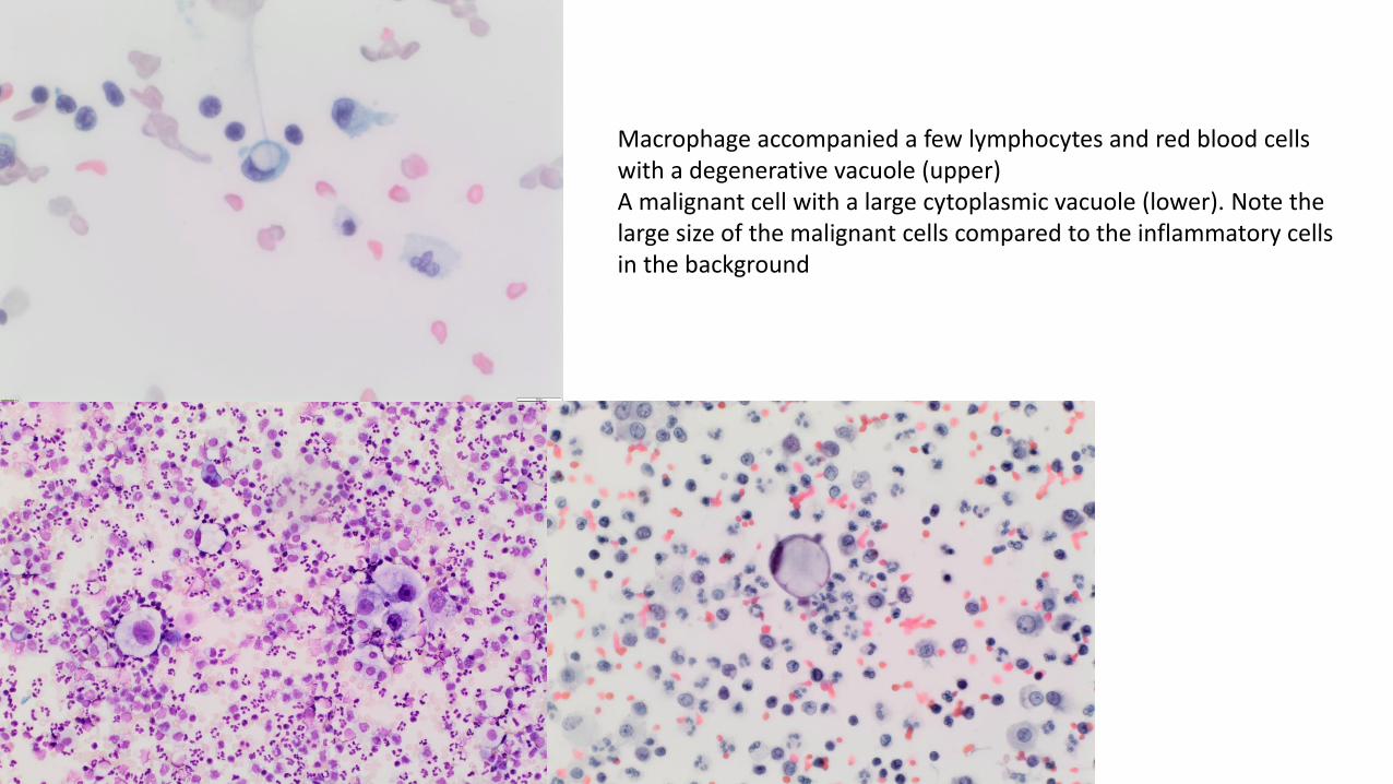
Macrophage accompanied a few lymphocytes and red blood cells with a degenerative vacuole (upper)A malignant cell with a large cytoplasmic vacuole (lower). Note the large size of the malignant cells compared to the inflammatory cells in the background

Cytoplasmic vacuolation in reactive mesothelial cellsHyaluronic acid rich mesothelial cells may show false positive staining with CEA (40%), BerEp4 (20%), LeuM1 & B72.3 (10%). False positive staining lost after pretreatment with hyaluronidase
Pitfall: Single cell population
• A mixed population of cells (mesothelial, macrophages and inflammatory cells) provide a contrast against which malignant cells stand out
• Malignant cells when present as a single cell population may be mistaken for macrophages or reactive mesothelial cells
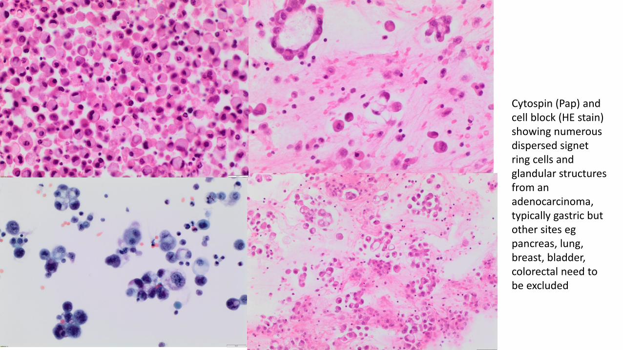
Cytospin (Pap) and cell block (HE stain) showing numerous dispersed signet ring cells and glandular structures from an adenocarcinoma, typically gastric but other sites egpancreas, lung, breast, bladder, colorectal need to be excluded

Dispersed rather than morular population of mesothelial cells may be seen in mesothelioma and may be confirmed on immunochemistry
WT1 +

Lymphocytic proliferation

Eosinophils (interspersed between lymphocytes and macrophages) indicating previous aspirations of effusion

Macrophages, multinucleated giant cells and granular debris in a rheumatoid effusion
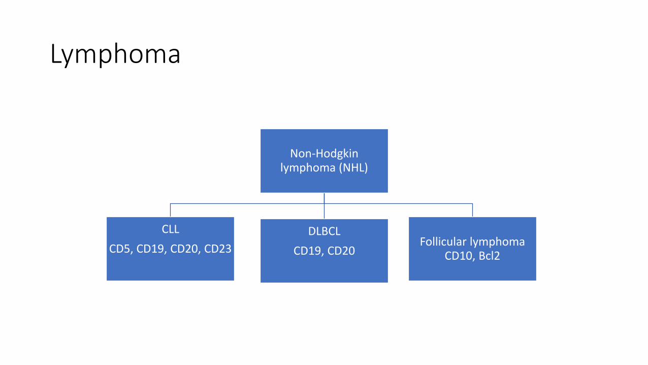
Lymphoma
Non-Hodgkin lymphoma (NHL)
CLL
CD5, CD19, CD20, CD23
DLBCL
CD19, CD20Follicular lymphoma
CD10, Bcl2
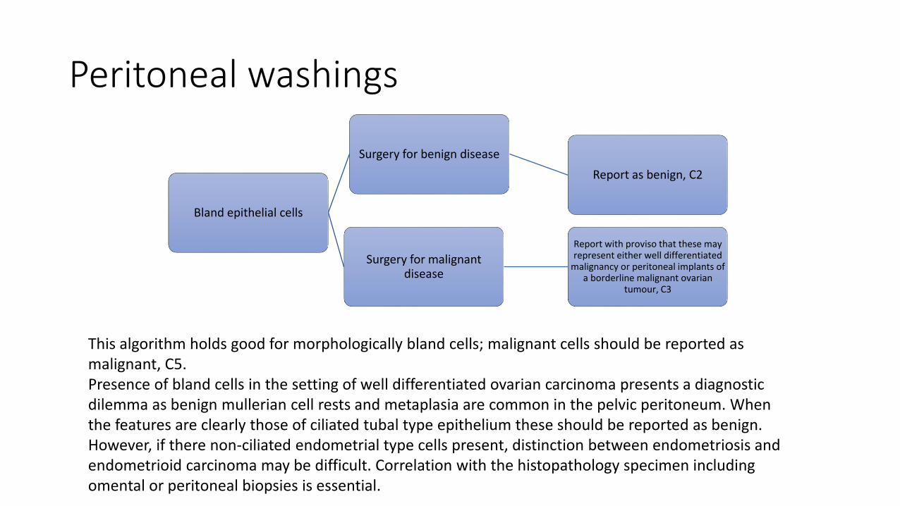
Peritoneal washings
Bland epithelial cells
Surgery for benign disease
Report as benign, C2
Surgery for malignant disease
Report with proviso that these may represent either well differentiated
malignancy or peritoneal implants of a borderline malignant ovarian
tumour, C3
This algorithm holds good for morphologically bland cells; malignant cells should be reported as malignant, C5.Presence of bland cells in the setting of well differentiated ovarian carcinoma presents a diagnostic dilemma as benign mullerian cell rests and metaplasia are common in the pelvic peritoneum. When the features are clearly those of ciliated tubal type epithelium these should be reported as benign. However, if there non-ciliated endometrial type cells present, distinction between endometriosis and endometrioid carcinoma may be difficult. Correlation with the histopathology specimen including omental or peritoneal biopsies is essential.

FIGO Ovarian carcinoma staging 2014
• Stage I: Tumour limited to one or both ovaries
• IC1: Surgical spill
• IC2: Capsule rupture before surgery or tumour on ovarian surface
• IC3: Malignant cells in ascitic fluid or peritoneal washings
• Stage IVA: Pleural effusion with malignant cells

Peritoneal washing showing artefact from the fluid used for irrigating the peritoneal cavity
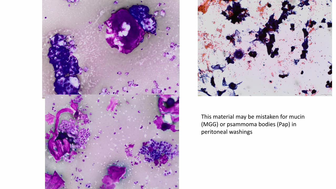
This material may be mistaken for mucin (MGG) or psammoma bodies (Pap) in peritoneal washings

Peritoneal washing containing single cells and groups of malignant cells from an adenocarcinoma

Pitfall: Tumours seen less commonly in effusions• Poorly differentiated squamous cell carcinoma
• Small cell carcinoma
• Melanoma
• Sarcoma

Fixation artefact may induce orangeophilia in mesothelial cells and being mistaken for keratinisation in squamous cell carcinoma

Small parakeratotic cells seen in mesothelioma. Their presence does not always indicate squamous cell carcinomaWT1 is helpful in distinguishing between mesothelioma and poorly differentiated squamous cell carcinoma.Intercellular bridges, keratinisation starting at the periphery of the cell.

Small cell carcinoma

Small cell carcinoma in effusions
• May show some degenerative changes and cytoplasmic vacuolation resembling adenocarcinoma, which might be TTF1. If neuroendocrine differentiation is not thought of on morphology, this important diagnosis may be missed

References
• Lisa M. Rooper, Syed Z. Ali and Matthew T. Olson, A Minimum Fluid Volume of 75 mL Is Needed to Ensure Adequacy in a Pleural Effusion: A Retrospective Analysis of 2540 Cases. Cancer (Cancer Cytopathol) 2014;122:657-65.
• Chandra A, Cross P , Denton et al. BSCC Code of Practice: Exfoliative cytology, Cytopathology 2009. 20:211-23
• Royal College of Pathologists. Tissue Pathways. www.rcpath.org
• Cook DS, Attanoos RL, Jalloh SS et al. ‘Mucin positive’ epithelioid Mesothelioma of the peritoneum: an unusual diagnostic pitfall. ‘Histopathology 2000. 37:31-36
• Shidham V and Atkinson K. Cytopathologic diagnosis of Serous effusions. 2007. 4:43-54
• Ordonez NG. Value of PAX- PAX-2, Napsin A, CAIX and Claudin-4 immunostaining in differentiation of epithelioid pleural Mesothelioma from metastatic renal cell carcinoma. Mod Pathol 2013 26(8):1132-48

References
• Davidson B, Firat P and Michael CW. Serous effusions: aetiology, diagnosis, biology and therapy (ed). Springer Verlag 2012.
• Jo VY, Cibas ES, Pinkus GS. Claudin-4 immunohistochemistry is highly effective in distinguishing adenocarcinoma from mesothelioma in effusion cytology. Cancer cytopathology 2014;122(4):299-306
• Hyun TS, Barnes M, Tabatabai ZL. The diagnostic utility D2-40, Calretinin, CK5/6, Desmin and MOC31 in the differentiation of mesothelioma from adenocarcinoma in pleural effusion cytology. Acta Cytol 2012;56:527-32
• Monaco SE, Shuai Y, Bansal M et al. The diagnostic utility of p16 FISH and GLUT-1 immunohistochemical analysis in mesothelial proliferations. Am J Clin Path 2011;135(4):619-27
• Huang C, Michael CW. Cytopathology 2014;25:112-119.