spontaneous dissection of the vertebral artery: report of three … · 2011-07-19 · there was...
TRANSCRIPT
149
Acta Neurologica Taiwanica Vol 20 No 2 June 2011
From the Department of Neurology, E-Da Hospital and I-ShouUniversity, Kaohsiung County, Taiwan.Received November 12, 2010. Revised December 31, 2010.Accepted May 5, 2011.
Correspondence to: Shih-Pin Hsu, MD. Department ofNeurology E-Da Hospital/I-Shou University, No.1, E-Da Road,Jiau-Shu Tsuen, Yan-Chau Shiang, Kaohsiung County, Taiwan.E-mail: [email protected]
Spontaneous Dissection of the Vertebral Artery:Report of Three Cases
Feng-Hsiung Chou, Chin-Sung Tung, Pei-Jung Lin, Chen-Sheng Chang, Shih-Pin Hsu
Abstract-Purpose: Vertebral artery dissection (VAD), although uncommon, is an important cause of ischemic stroke
in young adults. Without prompt diagnosis and treatment, it may cause mortality and morbidity.Spontaneous VAD related to abrupt neck position change, sudden sneezing, or severe cough and vomit-ing after fish bone chocking are rarely reported. This report describes three cases of spontaneous VADdue to seemingly harmless events.
Casp Report: The first patient developed vertebro-basilar ischemic symptoms after suddenly turning hishead to the left during an argument with a colleague. The second suffered from right lateral medullaryinfarction after sudden sneezing. The third developed left lateral medullary syndrome after she tried toextract a fish bone in her throat, which induced severe cough and vomiting. The three cases all present-ed with acute severe neck pain with posterir circulation ischemic symptoms and signs. Magnetic reso-nance imaging (MRI), magnetic resonance angiogram (MRA), and conventional angiography con-firmed the diagnosis. The first patient was treated with warfarin and recovered well. The other twocases received heparinization and then oral anticoagulant therapy, and recovered without residual neuro-logic deficits.
Conclusion: In conclusion, VAD should be among the differentials considered when encountering youngpatients presention with such clinical symptoms.
Key Words: vertebral artery dissection, magnetic resonance angiogram, magnetic resonance imaging, neckposition change, young stroke
Acta Neurol Taiwan 2011;20:149-154
INTRODUCTION
Dissection of vertebral arteries is an important butrelatively uncommon cause of stroke. In recent years,this clinical entity has drawn much attention among
clinical neurologists, thereby providing better diagnosticfacilities and more alertness on its symptomatology(1,2).Spontaneous vertebral artery dissection (VAD) is evenrarer, with an annual incidence of approximately 1-1.5per 100,000 people(3). Although it accounts for only 2%
Case Reports
of ischemic stroke in the general population, it is respon-sible for nearly 20% of stroke in young adults (<45years)(3).
Although outcome is favorable in previous studies,morbidity and mortality can be encountered(1). Possibleprecipitating factors and relevant events like trivial trau-ma or spinal manipulative therapy have been identifiedin some patients. However, there are few studies examin-ing whether common neck movements pose an indepen-dent risk for VAD(4). We present three cases of sponta-neous VAD after violent turning head, sneezing andsevere cough.
CASE REPORT
Case 1A 40-year-old healthy man presented with acute
severe right neck pain and transient consciousness lossfor a few seconds after suddenly and violently turninghis head to the left during an argument with a colleague.His past medical history was normal and there was noneck trauma or neck massage before symptom onset. Healso experienced vertigo, imbalance of gait and a tenden-cy to fall to his right side.
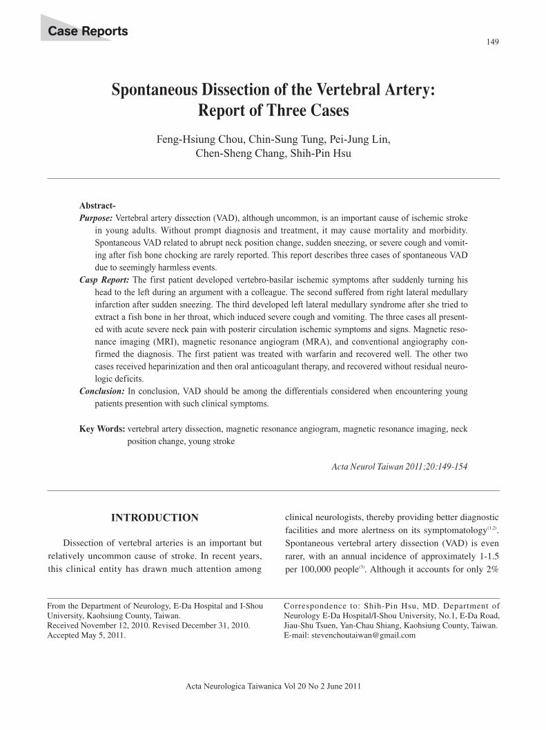
Neurologic examination (NE) revealed mild dysme-tria of right side extremities and gait imbalance.Laboratory studies were normal. Brain magnetic reso-nance imaging (MRI) done two weeks after the initialevent showed no high signal intensity on diffusion-weighted imaging (DWI) (image not shown). However,there was luminal stenosis of the right vertebral artery atthe level of the 4th cervical vertebra by magnetic reso-nance angiogram (MRA) (Fig. 1A). Catheter angiogra-phy (Fig. 1B) confirmed the right extra-cranial vertebralartery dissection (VAD).
The patient was treated with warfarin for preventionof thrombo-embolism. There were no recurrent neuro-logic symptoms on follow-up. Four months later, com-puted tomography angiography (CTA) showed totalrecovery of the right VAD (Fig. 1C).
Case 2A 36-year-old man had chronic hypertension with
adequate medical treatment. He had acute vertigo, vom-iting, and right facial numbness after a bout of sneezing.On neurological examination, right-sided Horner’s syn-drome, hiccup, ataxia, right side dysmetria and rightfacial hemi-anesthesia were found.
150
Acta Neurologica Taiwanica Vol 20 No 2 June 2011
Figure 1. (A) Luminal stenosis of the right vertebral artery from the 4th cervical vertebra by magnetic resonance angiogram (MRA)study (white arrow). (B) Conventional angiography reveals focal segmental stenosis of the right extra-cranial vertebralartery around the 4th cervical vertebra level (black arrow), and right VAD with luminal narrowing. (C) After four months,computed tomography angiography (CTA) shows total resolution of the right VAD (white arrow).
A B C

Transcranial duplex revealed focal stenotic bloodflow in right vertebral artery. Brain MRI showed highsignal intensity in the right lateral medulla on DWI (Fig.2A), and T2-weighted imaging (T2WI) revealed intra-mural hematoma in the right vertebral artery (Fig. 2B).MRA showed narrow and occlusive right vertebralartery, although the signals disappeared beyond the C2vertebrae (Fig. 2C). Conventional angiography con-firmed a steno-occlusion of the right vertebral artery(Fig. 2D).
The patient received heparin infusion, followed byoral warfarin and rehabilitation program during hospital-ization. The focal neurologic defecit was improved ondischarge and warfarin was replaced with aspirin sixmonths later.
Case 3
A 40-year-old right-handed healthy female experi-enced sudden onset vertigo and left side paresthesia aftersevere vomiting. She got fish bone stuck in her throatwhile having lunch. Left eyelid drop, blurred vision,hoarseness, and easy chocking were accompanied withacute severe left posterior neck pain.
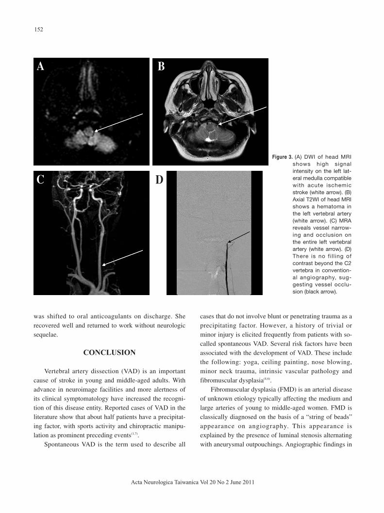
NE revealed partial Horner’s sign, left side numb-ness, dysmetria of the left extremities and ataxic gait.Laboratory studies were within normal limits. BrainMRI showed high signal intensity on the left lateralmedulla on DWI (Fig. 3A). T2WI showed intramuralhematoma of the left vertebral artery (Fig. 3B), whileMRA revealed vessel narrowing and occlusion along theentire left vertebral artery (Fig. 3C). There was no fillingof contrast beyond the C2 vertebra by conventionalangiography (Fig 3D).
Intravenous heparin infusion was administered, and
151
Acta Neurologica Taiwanica Vol 20 No 2 June 2011
A B
C D
Figure 2. (A) Diffusion-weightedimaging (DWI) of headMRI on axial viewshows high signal inten-sity of the right lateralmedulla, suggestive ofacute ischemic infarc-tion (white arrow). (B)T2 weighted imaging(T2WI) reveals ahematoma of the rightvertebral artery (whitearrow). (C) MRA showsa narrow and occlusiveright vertebral artery,but the signals disap-pear beyond the C2vertebra (white arrow).(D) Convent-ionalangiography confirms asteno-occlusive rightvertebral artery (blackarrow).
was shifted to oral anticoagulants on discharge. Sherecovered well and returned to work without neurologicsequelae.
CONCLUSION
Vertebral artery dissection (VAD) is an importantcause of stroke in young and middle-aged adults. Withadvance in neuroimage facilities and more alertness ofits clinical symptomatology have increased the recogni-tion of this disease entity. Reported cases of VAD in theliterature show that about half patients have a precipitat-ing factor, with sports activity and chiropractic manipu-lation as prominent preceding events(1,7).
Spontaneous VAD is the term used to describe all
cases that do not involve blunt or penetrating trauma as aprecipitating factor. However, a history of trivial orminor injury is elicited frequently from patients with so-called spontaneous VAD. Several risk factors have beenassociated with the development of VAD. These includethe following: yoga, ceiling painting, nose blowing,minor neck trauma, intrinsic vascular pathology andfibromuscular dysplasia(4,6).
Fibromuscular dysplasia (FMD) is an arterial diseaseof unknown etiology typically affecting the medium andlarge arteries of young to middle-aged women. FMD isclassically diagnosed on the basis of a “string of beads”appearance on angiography. This appearance isexplained by the presence of luminal stenosis alternatingwith aneurysmal outpouchings. Angiographic findings in
152
Acta Neurologica Taiwanica Vol 20 No 2 June 2011
A
C D
Figure 3. (A) DWI of head MRIshows high signalintensity on the left lat-eral medulla compatiblewith acute ischemicstroke (white arrow). (B)Axial T2WI of head MRIshows a hematoma inthe left vertebral artery(white arrow). (C) MRAreveals vessel narrow-ing and occlusion onthe entire left vertebralartery (white arrow). (D)There is no fi l l ing ofcontrast beyond the C2vertebra in convention-al angiography, sug-gesting vessel occlu-sion (black arrow).
B

our patients excluded this etiology.The role of trivial, unnoticed trauma producing dis-
section is not clear. It has been suggested that patientswith so-called “spontaneous” VAD may have had unrec-ognized trauma or sudden neck motion that has been for-gotten, overlooked, or considered insignificant by thepatient and thus not reported to the physician. In aCanadian survey(6), cervical dissections are associatedwith sudden neck movements ranging from therapeuticneck manipulation to a vigorous volleyball game,although some are during mild exertion, such as lifting apet dog or during a bout of coughing. Causative potentialtrauma, including violent coughing, neck turning duringa parade, playing basketball, dancing, swimming, andminor falls, have immediately preceded the initial symp-toms of dissection(6,13,14).
In this report, causative preceding events are suddenneck position change, acute sneezing, coughing andvomiting. These seemingly harmless movements maysuddenly increase pressure to neck vessel wall andinduced endothelial injury as well as subsequent neckpain associated with ischemic symptoms(11). Possibleexplanations for this causation remain ill defined.Authors have suggested the association of vertebralartery anomalies, tortuosity, and atherosclerosis, as wellas the duration and force of the neck movement. It isalso likely that inherent vessel wall abnormalities predis-pose to dissection upon subtle trauma(8).
Typical patients with VAD present with posteriorneck pain or headache, followed by ischemia of the ver-tebrobasilar system. Initial manifestations of VAD how-ever, are less distinct than those of carotid artery dissec-tion and usually interpreted as musculoskeletal pain(9,10).Therefore, an accurate neurologic examination anddetailed history-taking are mandatory, particularly inyoung adults, in order to search for symptoms or focalsigns of brainstem stroke.
Ischemic symptoms occur in more than 90% of VADpatients and may involve the brain stem, especially thelateral medulla (Wallenberg’s syndrome), as well as thethalamus and cerebral or cerebellar hemispheres(8,12).
Catheter angiography is the gold standard for diag-nosis of VAD. Characteristic features are vessel irregu-
larity and/or stenosis, string sign, double lumen, pseudo-aneurysm formation, or complete occlusion.Conventional angiography of our patients demonstratesome of these classic findings (See Figure). Magneticresonance techniques and CTA are replacing standardangiography in the diagnosis and follow-up of VAD inclinical practice(15,16). The resolution of MRA and CTAnow approaches that of catheter angiography and theycan show the intramural hematoma itself.
The prognosis of VAD is associated with the initialstroke severity and the extent of collateral circulation(2).The reported death rate due to dissections is < 5% andabout 75% of patients with an ischemic insult have goodrecovery(2,12). Although there is no consensus on the stan-dard treatment, anticoagulation with intravenousheparin, followed by oral warfarin, has been suggestedfor VAD patients to prevent thromboembolic complica-tions. All these three patients reported here receivedanticoagulants and no recurrent cerebrovascular eventsoccurred during follow-up.
In conclusion, VAD is an important etiology of brainstem stroke in young adults. If patients present with suchsymptomatology, detailed investigation of precipitatingevents and associated posterior circulation ischemicsymptoms and signs are mandatory. Early diagnosis andprompt treatment can reduce morbidity and mortality.
REFERENCES
1. Saeed AB, Shuaib A, Emert D, Al-Sulaiti G. Vertebral
artery dissection: warning symptoms, clinical features and
prognosis in 26 patients. Can J Neurol Sci 2000;27:292-
296.
2. Schievink WI. Spontaneous dissection of the carotid and
vertebral arteries. N Engl J Med 2001;344:898-906.
3. Dziewas R, Konrad C, Dräger B, Evers S, Besselmann M,
Ludemann P, Kuhlenbaumer G, Stogbauer F, Ringelstein
EB. Cervical artery dissection: clinical features, risk fac-
tors, therapy and outcome in 126 patients. J Neurol 2003;
250:1179-1184.
4. Rubinstein SM, Peerdeman SM, van Tulder MW, Riphagen
I, Haldeman S. A systematic review of the risk factors for
cervical artery dissection. Stroke 2005;36:1575-1580.
153
Acta Neurologica Taiwanica Vol 20 No 2 June 2011
5. Prabhakar S, Bhatia R, Khandelwal N, Lal V, Das CP.
Vertebral artery dissection due to indirect neck trauma: an
under-recognized entity. Neurol India 2001;49:384-390.
6. Norris JW, Beletsky V, Nadareishvili ZG. Sudden neck
movement and cervical artery dissection. CMAJ 2000;163:
38-39.
7. Haldeman S, Kohlbeck FJ, McGregor M. Risk factors and
precipitating neck movements causing vertebro-basilar
artery dissection after cervical trauma and spinal manipula-
tion. Spine 1999;24:785-794.
8. Caplan LR. Arterial Dissection. In: Caplan’s Stroke- A
Clinical Approach. Caplan LR (ed). Butterworth-
Heinemann; Boston 2000:295-302.
9. Silbert PL, Mokri B, Schievink WI. Headache and neck
pain in spontaneous internal carotid and vertebral artery
dissections. Neurology 1995;45:1517-1522.
10. Lanfranchi S, Di Falco M, Perini M, Zarcone D. Posterior
headache as a warning symptom of vertebral dissection: a
case report. J Headache Pain 2005;6:478-479.
11. Gutowski N, Murphy R, Beale DJ. Unilateral upper cervi-
cal posterior spinal artery syndrome following sneezing. J
Neurol Neurosurg Psychiatry 1992;55:841-843.
12. Arnold M, Bousser MG, Fahrni G, Fischer U, Georgiadis
D, Gandjour J, Benninger D, Sturzenegger M, Mattle HP,
Baumgartner RW. Vertebral artery dissection presenting
findings and predictors of outcome. Stroke 2006;37:2499-
2503.
13. Mokri B, Houser OW, Sandok BA, Piepgras DG.
Spontaneous dissections of the vertebral arteries.
Neurology 1988;38:880-885.
14. Sherman DG, Hart RG, Easton JD. Abrupt change in head
position and cerebral infarction. Stroke 1981;12:2-6.
15. Chen CJ, Tseng YC, Lee TH, Hsu HL, See LC. Multi-sec-
tion CT angiography compared with catheter angiography
in diagnosing vertebral artery dissection. AJNR 2004;5:
769-774.
16. Rizzo L, Crasto SG, Savio D, Veglia S, Davini O, Giraudo
M, Cerrato P, Lucchi RD. Dissection of cervico-cephalic
arteries: early diagnosis and follow-up with magnetic reso-
nance imaging. Emerg Radiol 2006;12:254-265.
154
Acta Neurologica Taiwanica Vol 20 No 2 June 2011