extracranial carotid and vertebral artery disease
TRANSCRIPT

ACCF/AHA Pocket Guideline
Based on the 2011 ASA/ACCF/AHA/AANN/AANS/ACR/ CNS/SAIP/SCAI/SIR/SNIS/SVM/SVS
Guideline on the Management of Patients With Extracranial Carotid and Vertebral Artery Disease
Developed in Collaboration With the American Academy of Neurology and Society of Cardiovascular Computed Tomography
January 2011


i
Guideline on the Management of Patients With Extracranial Carotid and Vertebral Artery DiseaseJanuary 2011
ACCF/AHA Writing Committee
Thomas G. Brott, MD, Co-Chair
Jonathan L. Halperin, MD, Co-Chair
Suhny Abbara, MD
J. Michael Bacharach, MD
John D. Barr, MD
Ruth L. Bush, MD, MPH
Christopher U. Cates, MD
Mark A. Creager, MD
Wesley S. Moore, MD
Peter D. Panagos, MD
Thomas S. Riles, MD
Robert H. Rosenwasser, MD

© 2011 by the American College of Cardiology Foundation and the American Heart
Association, Inc.
The following material was adapted from the 2011 ASA/ ACCF/AHA/AANN/AANS/
ACR/CNS/SAIP/SCAI/SIR/ SNIS/SVM/SVS Guideline on the Management of Patients
With Extracranial Carotid and Vertebral Artery Disease. (Executive Summary: Circulation.
2011;124:489–532; Full-Text: Circulation. 2011;124:e54–e130; Executive Summary:
Stroke. 2011;42:e420–e463; Full-Text: Stroke. 2011;42:e464–e540). This pocket guideline
is available on the World Wide Web sites of the American College of Cardiology (www.
cardiosource.org) and the American Heart Association (my.americanheart.org).
For copies of this document, please contact Elsevier Inc. Reprint Department, e-mail:
[email protected]; phone: 212-633-3813; fax: 212-633-3820.
Permissions: Multiple copies, modification, alteration, enhancement, and/or
distribution of this document are not permitted without the express permission of the
American College of Cardiology Foundation. Please contact Elsevier’s permission
department at [email protected].

Contents
1. Introduction ....................................................................................................... 3
2. Duplex Ultrasonography to Evaluate Asymptomatic Patients With Known or Suspected Carotid Stenosis ......................................... 4
3. Diagnostic Testing in Patients With Symptoms or Signs of ECVD ..... 5
4. Treatment of Hypertension .......................................................................... 6
5. Cessation of Tobacco Smoking .................................................................... 6
6. Control of Hyperlipidemia ............................................................................ 6
7. Management of Diabetes Mellitus in Patients With Atherosclerosis of the Extracranial Carotid or Vertebral Arteries ............................... 6
8. Antithrombotic Therapy in Patients With Extracranial Carotid Atherosclerotic Disease Not Undergoing Revascularization ............ 7
9. Selection of Patients for Carotid Revascularization .............................. 8
10. Periprocedural Management of Patients Undergoing CEA ................ 9
11. Management of Patients Undergoing CAS ........................................... 10

2

3
12. Management of Patients Experiencing Restenosis After CEA or CAS ........................................................................................ 10
13. Vascular Imaging in Patients With Vertebral Artery Disease .......... 11
14. Management of Atherosclerotic Risk Factors in Patients With Vertebral Artery Disease ................................................................ 11
15. Management of Patients With Occlusive Disease of the Subclavian and Brachiocephalic Arteries ............................................. 12
16. Carotid Artery Evaluation and Revascularization Before Cardiac Surgery ............................................................................. 13
17. Management of Patients With FMD of the Extracranial Carotid Arteries .......................................................................................... 13
18. Management of Patients With Cervical Artery Dissection .............. 13

4
1. Introduction
Extracranial carotid and vertebral artery disease
(ECVD) encompasses several disorders that affect
the arteries that supply the brain and is an important
cause of stroke and transient cerebral ischemic
attack. The most frequent cause is atherosclerosis,
but other causes include fibromuscular dysplasia
(FMD), cystic medial necrosis, arteritis, and
dissection. Atherosclerosis is a systemic disease,
and patients with ECVD typically face an escalated
risk of other adverse cardiovascular events,
including myocardial infarction, peripheral arterial
disease and death. To improve survival, neurological
and functional outcomes and quality of life,
preventive and therapeutic strategies must address
both cerebral and systemic risk.

5
6
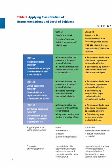
Table 1. Applying Classification of Recommendations and Level of Evidence
CLASS IIb
Benefit > Risk Additional studies with broad objectives needed; additional registry data would be helpful
Procedure/Treatment MAY BE CONSIDERED
� Recommendation’s usefulness/efficacy less well established
� Greater conflicting evidence from multiple randomized trials or meta-analyses
� Recommendation’s usefulness/efficacy less well established
� Greater conflicting evidence from single randomized trial or nonrandomized studies
� Recommendation’s usefulness/efficacy less well established
� Only diverging expert opinion, case studies, or standard of care
CLASS IIa
Benefit >> Risk Additional studies with focused objectives needed
IT IS REASONABLE to per-form procedure/administer treatment
� Recommendation in favor of treatment or procedure being useful/effective
� Some conflicting evidence from multiple randomized trials or meta-analyses
� Recommendation in favor of treatment or procedure being useful/effective
� Some conflicting evidence from single randomized trial or nonrandomized studies
� Recommendation in favor of treatment or procedure being useful/effective
� Only diverging expert opinion, case studies, or standard of care
CLASS I
Benefit >>> Risk
Procedure/Treatment SHOULD be performed/ administered
� Recommendation that procedure or treatment is useful/effective
� Sufficient evidence from multiple randomized trials or meta-analyses
� Recommendation that procedure or treatment is useful/effective
� Evidence from single randomized trial or nonrandomized studies
� Recommendation that procedure or treatment is useful/effective
� Only expert opinion, case studies, or standard of care
LEVEL A
Multiple populations evaluated*
Data derived from multiple randomized clinical trials or meta-analyses
LEVEL B
Limited populations evaluated*
Data derived from a single randomized trial or nonrandomized studies
LEVEL C
Very limited populations evaluated*
Only consensus opinion of experts, case studies, or standard of care
S I Z E O F T R E A T M E N T E F F E C T
may/might be considered
may/might be reasonable
usefulness/effectiveness is unknown/unclear/uncertain or not well established
COR III: No Benefit
is not recommended
is not indicated
should not be done
is not useful/beneficial/ effective
COR III: Harm
potentially harmful
causes harm
associated with excess morbid-ity/mortality
should not be done
should
is recommended
is indicated
is useful/effective/beneficial
Suggested phrases for writing recommendations
is reasonable
can be useful/effective/beneficial
is probably recommended or indicated
CLASS III No Benefit or CLASS III Harm Procedure/ Test Treatment
COR III: Not No Proven No benefit Helpful Benefit
COR III: Excess Cost Harmful Harm w/o Benefit to Patients or Harmful
� Recommendation that procedure or treatment is not useful/effective and may be harmful
� Sufficient evidence from multiple randomized trials or meta-analyses
� Recommendation that procedure or treatment is not useful/effective and may be harmful
� Evidence from single randomized trial or nonrandomized studies
� Recommendation that procedure or treatment is not useful/effective and may be harmful
� Only expert opinion, case studies, or standard of care
treatment/strategy A is recommended/indicated in preference to treatment B
treatment A should be chosen over treatment B
Comparative effectiveness phrases†
treatment/strategy A is probably recommended/indicated in preference to treatment B
it is reasonable to choose treatment A over treatment B

7
* Data available from clinical trials or registries about the
usefulness/efficacy in different subpopulations, such
as sex, age, history of diabetes, history of prior
myocardial infarction, history of heart failure, and prior
aspirin use. A recommendation with Level of Evidence
B or C does not imply that the recommendation is
weak. Many important clinical questions addressed in
the guidelines do not lend themselves to clinical trials.
Although randomized trials are unavailable, there may
be a very clear clinical consensus that a particular test
or therapy is useful or effective.
† For comparative effectiveness recommendations
(Class I and IIa; Level of Evidence A and B only),
studies that support the use of comparator verbs
should involve direct comparisons of the treatments or
strategies being evaluated.
CLASS IIb
Benefit > Risk Additional studies with broad objectives needed; additional registry data would be helpful
Procedure/Treatment MAY BE CONSIDERED
� Recommendation’s usefulness/efficacy less well established
� Greater conflicting evidence from multiple randomized trials or meta-analyses
� Recommendation’s usefulness/efficacy less well established
� Greater conflicting evidence from single randomized trial or nonrandomized studies
� Recommendation’s usefulness/efficacy less well established
� Only diverging expert opinion, case studies, or standard of care
CLASS IIa
Benefit >> Risk Additional studies with focused objectives needed
IT IS REASONABLE to per-form procedure/administer treatment
� Recommendation in favor of treatment or procedure being useful/effective
� Some conflicting evidence from multiple randomized trials or meta-analyses
� Recommendation in favor of treatment or procedure being useful/effective
� Some conflicting evidence from single randomized trial or nonrandomized studies
� Recommendation in favor of treatment or procedure being useful/effective
� Only diverging expert opinion, case studies, or standard of care
CLASS I
Benefit >>> Risk
Procedure/Treatment SHOULD be performed/ administered
� Recommendation that procedure or treatment is useful/effective
� Sufficient evidence from multiple randomized trials or meta-analyses
� Recommendation that procedure or treatment is useful/effective
� Evidence from single randomized trial or nonrandomized studies
� Recommendation that procedure or treatment is useful/effective
� Only expert opinion, case studies, or standard of care
LEVEL A
Multiple populations evaluated*
Data derived from multiple randomized clinical trials or meta-analyses
LEVEL B
Limited populations evaluated*
Data derived from a single randomized trial or nonrandomized studies
LEVEL C
Very limited populations evaluated*
Only consensus opinion of experts, case studies, or standard of care
S I Z E O F T R E A T M E N T E F F E C T
may/might be considered
may/might be reasonable
usefulness/effectiveness is unknown/unclear/uncertain or not well established
COR III: No Benefit
is not recommended
is not indicated
should not be done
is not useful/beneficial/ effective
COR III: Harm
potentially harmful
causes harm
associated with excess morbid-ity/mortality
should not be done
should
is recommended
is indicated
is useful/effective/beneficial
Suggested phrases for writing recommendations
is reasonable
can be useful/effective/beneficial
is probably recommended or indicated
CLASS III No Benefit or CLASS III Harm Procedure/ Test Treatment
COR III: Not No Proven No benefit Helpful Benefit
COR III: Excess Cost Harmful Harm w/o Benefit to Patients or Harmful
� Recommendation that procedure or treatment is not useful/effective and may be harmful
� Sufficient evidence from multiple randomized trials or meta-analyses
� Recommendation that procedure or treatment is not useful/effective and may be harmful
� Evidence from single randomized trial or nonrandomized studies
� Recommendation that procedure or treatment is not useful/effective and may be harmful
� Only expert opinion, case studies, or standard of care
treatment/strategy A is recommended/indicated in preference to treatment B
treatment A should be chosen over treatment B
Comparative effectiveness phrases†
treatment/strategy A is probably recommended/indicated in preference to treatment B
it is reasonable to choose treatment A over treatment B

8
2. Duplex Ultrasonography to Evaluate Asymptomatic Patients With Known or Suspected Carotid Stenosis
Class I 1. In asymptomatic patients with known or suspected
carotid stenosis, duplex ultrasonography, performed
by a qualified technologist in a certified laboratory, is
recommended as the initial test to detect
hemodynamically significant carotid stenosis. (Level
of Evidence: C)
Class IIa 1. It is reasonable to
• perform duplex ultrasonography to detect
hemodynamically significant carotid stenosis in
asymptomatic patients with carotid bruit. (Level
of Evidence: C)
• repeat duplex ultrasonography annually, by a
qualified technologist in a certified laboratory, to
assess progression or regression of disease and
response to therapy in patients with previous
atherosclerotic stenosis greater than 50%. Once
stability is established over an extended period or
candidacy for intervention has changed, longer
intervals or termination of surveillance may be
appropriate. (Level of Evidence: C)

9
Class IIb 1. Duplex ultrasonography to detect hemodynamically
significant carotid stenosis may be considered
• in asymptomatic patients with symptomatic
peripheral arterial disease, coronary artery
disease or atherosclerotic aortic aneurysm. (Level
of Evidence: C)
• to detect carotid stenosis in asymptomatic
patients without evidence of atherosclerosis who
have greater than or equal to 2 of the following:
hypertension, hyperlipidemia, tobacco smoking,
or family history of atherosclerosis before age 60
in a first degree relative or ischemic stroke. (Level
of Evidence: C)
It is unclear whether establishing a diagnosis of
ECVD would justify actions that affect clinical
outcomes. (Level of Evidence: C)

10
Class III: 1. Carotid duplex ultrasonography is not recommended
No Benefit
• for routine screening of asymptomatic patients
who have no risk factors for atherosclerosis.
(Level of Evidence: C)
• for routine evaluation of patients with
neurological or psychiatric disorders unrelated to
focal cerebral ischemia. (Level of Evidence: C)
• for patients without risk factors for atherosclerotic
carotid disease and no disease on initial vascular
testing. (Level of Evidence: C)

11
3. Diagnostic Testing in Patients With Symptoms or Signs of Extracranial Carotid Artery Disease
Class I 1. Noninvasive imaging for detection of ECVD is
recommended in the initial evaluation of patients
with transient retinal or hemispheric neurological
symptoms of possible ischemic origin. (Level of
Evidence: C)
2. Duplex ultrasonography is recommended to detect
carotid stenosis in patients who develop focal
neurological symptoms corresponding to the internal
carotid artery territory. (Level of Evidence: C)
3. In patients with acute, focal ischemic neurological
symptoms corresponding to the territory supplied by
the left or right internal carotid artery, magnetic
resonance angiography (MRA) or computed
tomography angiography (CTA) is indicated to detect
carotid stenosis when definitive sonography cannot
be obtained. (Level of Evidence: C)
4. When intracranial or ECVD is not severe enough to
account for neurological symptoms of suspected
ischemic origin, echocardiography should be
performed seeking a source of cardiogenic embolism.
(Level of Evidence: C)

12
Class IIa 1. In revascularization candidates,
• MRA or CTA can be useful when carotid duplex
ultrasonography is nondiagnostic. (Level of
Evidence: C)
• CTA, MRA or elective cerebral angiography can be
useful to search for intracranial vascular disease
when an extracranial source of ischemia is not
identified or to evaluate severity of stenosis and
identify intrathoracic or intracranial vascular
lesions not adequately assessed by
ultrasonography. (Level of Evidence: C)
• catheter-based angiography can be useful when
noninvasive imaging is not sufficient. (Level of
Evidence: C)
• MRA without contrast is reasonable to assess
extent of disease in patients with renal
insufficiency or extensive vascular calcification.
(Level of Evidence: C)
• CTA is reasonable in patients who are not
candidates for MRA because of claustrophobia,
implanted pacemakers, or other incompatible
devices. (Level of Evidence: C)
13
Class IIb 1. Duplex carotid ultrasonography might be considered
for patients with nonspecific neurological symptoms
when cerebral ischemia is a plausible cause. (Level of
Evidence: C)
2. When complete carotid arterial occlusion is
suggested by duplex ultrasonography, MRA, or CTA,
catheter-based angiography may be considered to
determine whether the arterial lumen is sufficient to
permit carotid revascularization. (Level of Evidence: C)
3. Catheter-based angiography may be reasonable in
patients with renal dysfunction to limit the amount of
radiographic contrast material required for definitive
imaging for evaluation of a single vascular territory.
(Level of Evidence: C)

14
4. Treatment of Hypertension
Class I 1. Antihypertensive treatment is recommended for
patients with hypertension and asymptomatic
atherosclerotic ECVD to maintain blood pressure
(BP) less than 140/90 mmHg. (Level of Evidence: A)
Class IIa 1. Except during the hyperacute period,
antihypertensive treatment is probably indicated in
patients with hypertension and symptomatic
atherosclerotic ECVD, but the benefit of treatment to
a specific BP has not been established in relation to
the risk of exacerbating cerebral ischemia. (Level of
Evidence: C)

15
5. Cessation of Tobacco Smoking
Class I 1. Patients with atherosclerotic ECVD who smoke
cigarettes should be advised to quit and offered
cessation interventions to reduce risk. (Level of
Evidence: B)

16
6. Control of Hyperlipidemia
Class I 1. Treatment with a statin is recommended for all
patients with atherosclerotic ECVD to lower low-
density lipoprotein cholesterol to less than 100 mg/
dL. (Level of Evidence: B)
Class IIa 1. Treatment with a statin is reasonable for all
patients with atherosclerotic ECVD who sustain
ischemic stroke to reduce low-density lipoprotein
cholesterol to a level less than or equal to 70 mg/dL.
(Level of Evidence: B)
2. If treatment with a statin does not achieve the
goal, intensifying therapy with an additional drug
from among those with evidence of improving
outcomes can be effective. (Level of Evidence: B)
3. For patients who do not tolerate statins, therapy
with bile acid sequestrants and/or niacin is
reasonable. (Level of Evidence: B)
17
7. Management of Diabetes Mellitus in Patients With Atherosclerosis of the Extracranial Carotid or Vertebral Arteries
Class IIa 1. Diet, exercise, and glucose-lowering drugs can be
useful for patients with diabetes mellitus and
atherosclerotic ECVD. The stroke prevention
benefit, however, of intensive glucose-lowering
therapy to a glycosylated hemoglobin A1c level less
than 7.0% has not been established. (Level of
Evidence: A)
2. Administration of a statin to reduce low-density
lipoprotein cholesterol less than or equal to 70 mg/
dL is reasonable in patients with diabetes mellitus
and atherosclerotic ECVD for prevention of stroke
and other ischemic events. (Level of Evidence: B)

18
8. Antithrombotic Therapy in Patients With Extracranial Carotid Atherosclerotic Disease Not Undergoing Revascularization
Class I 1. Antiplatelet therapy with aspirin, 75 to 325 mg
daily, is recommended for patients with obstructive
or nonobstructive atherosclerotic ECVD for
prevention of myocardial infarction and other
ischemic events, though benefit has not been
established for prevention of stroke in asymptomatic
patients. (Level of Evidence: A)
2. In patients with obstructive or nonobstructive
atherosclerotic ECVD who have sustained ischemic
stroke or transient ischemic stroke (TIA), antiplatelet
therapy with aspirin (75 to 325 mg daily), clopidogrel
(75 mg daily), or the combination of aspirin and
extended-release dipyridamole (25 and 200 mg twice
daily, respectively) is recommended (Level of Evidence:
B) and preferred over the combination of aspirin and
clopidogrel (Level of Evidence: B). The antiplatelet
regimen should be individualized based on risk
factors, cost, tolerance, other clinical characteristics,
and guidance from regulatory agencies.

19
3. Antiplatelet agents are recommended over oral
anticoagulation for patients with atherosclerotic
ECVD with (Level of Evidence: B) or without (Level
of Evidence: C) ischemic symptoms. (For patients
with allergy or other contraindications to aspirin, see
Class IIa recommendation #2, this section.)
Class IIa 1. In patients with atherosclerotic ECVD who have
an indication for anticoagulation, such as atrial
fibrillation or mechanical heart valve, it can be
beneficial to administer a vitamin K antagonist (such
as warfarin, dose-adjusted to achieve a target
international normalized ratio of 2.5 [range 2.0 to
3.0]) for prevention of thromboembolic ischemic
events. (Level of Evidence: C)
2. For patients with atherosclerotic ECVD in whom
aspirin is contraindicated by factors other than
active bleeding, including allergy, clopidogrel (75 mg
daily) or ticlopidine (250 mg twice daily) are
reasonable alternatives. (Level of Evidence: C)
20
Class III: 1. Full-intensity parenteral anticoagulation with
No Benefit unfractionated heparin or low-molecular-weight
heparinoids is not recommended for patients with
atherosclerotic ECVD who develop TIA or acute
ischemic stroke. (Level of Evidence: B)
2. Administration of clopidogrel in combination with
aspirin is not recommended within 3 months after
stroke or TIA. (Level of Evidence: B)

21
9. Selection of Patients for Carotid Revascularization1
Class I 1. Patients at average or low surgical risk who
experience nondisabling ischemic stroke2 or
transient cerebral ischemic symptoms, including
hemispheric events or amaurosis fugax, within 6
months (symptomatic patients) should undergo
carotid endarterectomy (CEA) if the diameter of the
lumen of the ipsilateral internal carotid artery is
reduced more than 70%3 as documented by
noninvasive imaging (Level of Evidence A) or more
than 50% as documented by catheter angiography
(Level of Evidence B) and the anticipated rate of
perioperative stroke or mortality is less than 6%
1. Recommendations for revascularization in this section assume that operators are experienced, having successfully performed the procedures in >20 cases with proper technique and a low complication rate based on independent neurological evaluation before and after each procedure.
2. Nondisabling stroke is defined by a residual deficit associated with a score ≤2 according to the Modified Rankin Scale.
3. The degree of stenosis is based on catheter-based or noninvasive vascular imaging compared with the distal arterial lumen or velocity measurements by duplex ultrasonography.

22
2. Carotid artery stenting (CAS) is indicated as an
alternative to CEA for symptomatic patients at
average or low risk of complications associated with
endovascular intervention when the diameter of the
lumen of the internal carotid artery is reduced by
more than 70% as documented by noninvasive
imaging or more than 50% as documented by
catheter angiography and the anticipated rate of
periprocedural stroke or mortality is less than 6%.
(Level of Evidence: B)
3. Selection of asymptomatic patients for carotid
revascularization should be guided by assessment of
comorbid conditions, life expectancy, and other
individual factors and should include a thorough
discussion of the risks and benefits of the procedure
with an understanding of patient preferences. (Level
of Evidence: C)

23
Class IIa 1. It is reasonable to perform CEA in asymptomatic
patients who have more than 70% stenosis of the
internal carotid artery if the risk of perioperative
stroke, myocardial infarction, and death is low.
(Level of Evidence: A)
2. It is reasonable to choose CEA over CAS when
revascularization is indicated in older patients,
particularly when arterial pathoanatomy is
unfavorable for endovascular intervention. (Level of
Evidence: B)
3. It is reasonable to choose CAS over CEA when
revascularization is indicated in patients with neck
anatomy unfavorable for arterial surgery.4 (Level of
Evidence: B)
4. When revascularization is indicated for patients
with TIA or stroke and there are no
contraindications to early revascularization,
intervention within 2 weeks of the index event is
reasonable rather than delaying surgery. (Level of
Evidence: B)
4. Conditions that produce unfavorable neck anatomy include but are not limited to arterial stenosis distal to the second cervical vertebra or proximal (intrathoracic) arterial stenosis, previous ipsilateral CEA, contralateral vocal cord paralysis, open tracheostomy, radical surgery, and irradiation.
24
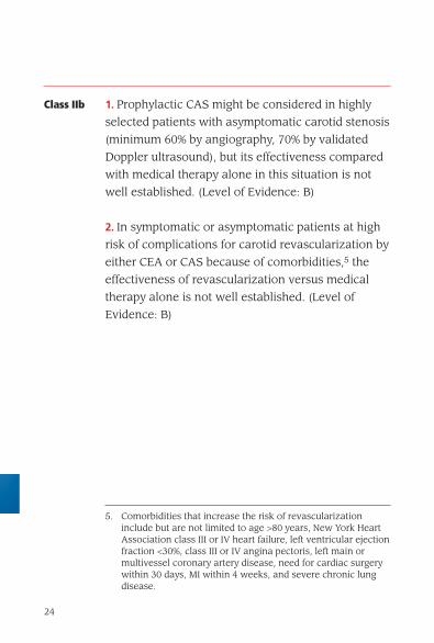
Class IIb 1. Prophylactic CAS might be considered in highly
selected patients with asymptomatic carotid stenosis
(minimum 60% by angiography, 70% by validated
Doppler ultrasound), but its effectiveness compared
with medical therapy alone in this situation is not
well established. (Level of Evidence: B)
2. In symptomatic or asymptomatic patients at high
risk of complications for carotid revascularization by
either CEA or CAS because of comorbidities,5 the
effectiveness of revascularization versus medical
therapy alone is not well established. (Level of
Evidence: B)
5. Comorbidities that increase the risk of revascularization include but are not limited to age >80 years, New York Heart Association class III or IV heart failure, left ventricular ejection fraction <30%, class III or IV angina pectoris, left main or multivessel coronary artery disease, need for cardiac surgery within 30 days, MI within 4 weeks, and severe chronic lung disease.

25
Class III: 1. Except in extraordinary circumstances, carotid
No Benefit revascularization by either CEA or CAS is not
recommended
• when atherosclerosis narrows the lumen by less
than 50%. (Level of Evidence: A)
• for patients with chronic total occlusion of the
targeted carotid artery. (Level of Evidence: C)
• for patients with severe disability6 caused by
cerebral infarction that precludes preservation of
useful function. (Level of Evidence: C)
6. In this context, severe disability refers generally to a Modified Rankin Scale of ≥3, but individual assessment is required, and intervention may be appropriate in selected patients with considerable disability when a worse outcome is projected with continued medical therapy alone.

26
10. Periprocedural Management of Patients Undergoing CEA
Class I 1. Before CEA, aspirin (81 to 325 mg daily) is
recommended. (Level of Evidence: A)
2. Beyond the first month after CEA, aspirin (75 to
325 mg daily), clopidogrel (75 mg daily), or the
combination of low-dose aspirin plus extended-
release dipyridamole (25 and 200 mg twice daily,
respectively) should be administered for long-term
prophylaxis against ischemic cardiovascular events.
(Level of Evidence: B)
3. Before and after CEA,
• administration of antihypertensive medication is
recommended as needed to control BP. (Level of
Evidence: C)
• findings on clinical neurological examination
should be documented within 24 hours. (Level of
Evidence: C)

27
Class IIa 1. After CEA,
• patch angioplasty can be beneficial for closure of
arteriotomy. (Level of Evidence: B)
• administration of statin lipid-lowering medication
is reasonable irrespective of serum lipid levels,
although the optimum agent and dose and the
efficacy for prevention of restenosis have not
been established. (Level of Evidence: B)
• noninvasive imaging of the extracranial carotid
arteries is reasonable 1 month, 6 months, and
annually. Once stability has been established over
an extended period, surveillance at longer
intervals may be appropriate. Termination of
surveillance is reasonable when the patient is no
longer a candidate for intervention. (Level of
Evidence: C)

28
11. Management of Patients Undergoing CAS
Class I 1. Before and after CAS,
• dual-antiplatelet therapy with aspirin (81 to 325 mg
daily) plus clopidogrel (75 mg daily) is recommended
(for a minimum of 30 days). For patients intolerant
of clopidogrel, ticlopidine (250 mg twice daily) may
be substituted. (Level of Evidence: C)
• administration of antihypertensive medication is
recommended to control BP. (Level of Evidence: C)
• findings on clinical neurological examination
should be documented within 24 hours. (Level of
Evidence: C)
Class IIa 1. Embolic protection device deployment during CAS
can be beneficial to reduce the risk of stroke when the
risk of vascular injury is low. (Level of Evidence: C)
2. After CAS, noninvasive imaging of the extracranial
carotid arteries is reasonable 1 month, 6 months,
and annually after revascularization to assess
patency and exclude new or contralateral lesions.
Once stability has been established over an extended
period, surveillance at extended intervals may be
appropriate and termination of surveillance is
reasonable when the patient is no longer a candidate
for intervention. (Level of Evidence: C)

29
12. Management of Patients Experiencing Restenosis After CEA or CAS
Class IIa 1. Reoperative CEA or CAS after initial
revascularization is reasonable
• in patients with symptomatic cerebral ischemia
and recurrent carotid stenosis due to intimal
hyperplasia or atherosclerosis, using the same
criteria for initial revascularization. (Level of
Evidence: C)
• when duplex ultrasound and another
confirmatory imaging method identify rapidly
progressive restenosis that indicates a threat of
complete occlusion. (Level of Evidence: C)
Class IIb 1. In asymptomatic patients who develop recurrent
carotid stenosis due to intimal hyperplasia or
atherosclerosis, reoperative CEA or CAS may be
considered using the same criteria as for initial
revascularization. (Level of Evidence: C)
Class III: 1. Reoperative CEA or CAS should not be performed
Harm in asymptomatic patients with less than 70% carotid
stenosis that has remained stable. (Level of
Evidence: C)

30
13. Vascular Imaging in Patients With Vertebral Artery Disease
Class I 1. Noninvasive imaging by CTA or MRA for detection
of vertebral artery disease
• should be part of the initial evaluation of patients
with neurological symptoms referable to the
posterior circulation and those with subclavian
steal syndrome. (Level of Evidence: C)
• should be performed in patients with
asymptomatic bilateral carotid occlusions or
unilateral carotid artery occlusion and incomplete
circle of Willis. (Level of Evidence: C)
• is recommended over ultrasound imaging for
evaluation of the vertebral arteries in patients
whose symptoms suggest posterior cerebral or
cerebellar ischemia. (Level of Evidence: C)

31
Class IIa 1. In patients with symptoms of posterior cerebral or
cerebellar ischemia
• serial noninvasive imaging of the extracranial
vertebral arteries is reasonable to assess
progression of atherosclerotic disease and
exclude the development of new lesions. (Level of
Evidence: C)
• catheter-based contrast angiography can be
useful to define vertebral artery pathoanatomy
when noninvasive imaging fails to define the
location or severity of stenosis. (Level of
Evidence: C)
2. In patients who have undergone vertebral artery
revascularization, serial noninvasive imaging of the
extracranial vertebral arteries is reasonable at
intervals similar to those for carotid
revascularization. (Level of Evidence: C)
32
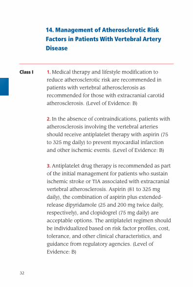
14. Management of Atherosclerotic Risk Factors in Patients With Vertebral Artery Disease
Class I 1. Medical therapy and lifestyle modification to
reduce atherosclerotic risk are recommended in
patients with vertebral atherosclerosis as
recommended for those with extracranial carotid
atherosclerosis. (Level of Evidence: B)
2. In the absence of contraindications, patients with
atherosclerosis involving the vertebral arteries
should receive antiplatelet therapy with aspirin (75
to 325 mg daily) to prevent myocardial infarction
and other ischemic events. (Level of Evidence: B)
3. Antiplatelet drug therapy is recommended as part
of the initial management for patients who sustain
ischemic stroke or TIA associated with extracranial
vertebral atherosclerosis. Aspirin (81 to 325 mg
daily), the combination of aspirin plus extended-
release dipyridamole (25 and 200 mg twice daily,
respectively), and clopidogrel (75 mg daily) are
acceptable options. The antiplatelet regimen should
be individualized based on risk factor profiles, cost,
tolerance, and other clinical characteristics, and
guidance from regulatory agencies. (Level of
Evidence: B)

33
Class IIa 1. For patients with atherosclerosis of the
extracranial vertebral arteries in whom aspirin is
contraindicated by factors other than active
bleeding, including those with allergy to aspirin,
either clopidogrel (75 mg daily) or ticlopidine (250
mg twice daily) are reasonable alternatives. (Level of
Evidence: C)
34
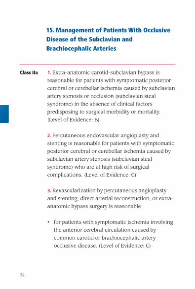
15. Management of Patients With Occlusive Disease of the Subclavian and Brachiocephalic Arteries
Class IIa 1. Extra-anatomic carotid-subclavian bypass is
reasonable for patients with symptomatic posterior
cerebral or cerebellar ischemia caused by subclavian
artery stenosis or occlusion (subclavian steal
syndrome) in the absence of clinical factors
predisposing to surgical morbidity or mortality.
(Level of Evidence: B)
2. Percutaneous endovascular angioplasty and
stenting is reasonable for patients with symptomatic
posterior cerebral or cerebellar ischemia caused by
subclavian artery stenosis (subclavian steal
syndrome) who are at high risk of surgical
complications. (Level of Evidence: C)
3. Revascularization by percutaneous angioplasty
and stenting, direct arterial reconstruction, or extra-
anatomic bypass surgery is reasonable
• for patients with symptomatic ischemia involving
the anterior cerebral circulation caused by
common carotid or brachiocephalic artery
occlusive disease. (Level of Evidence: C)

35
• for patients with symptomatic ischemia involving
upper-extremity claudication caused by
subclavian or brachiocephalic arterial occlusive
disease. (Level of Evidence: C)
2. Revascularization is reasonable for asymptomatic
patients with subclavian artery stenosis when the
ipsilateral internal mammary artery is required as a
conduit for myocardial revascularization. (Level of
Evidence: C)
Class III: 1. Asymptomatic patients with asymmetrical upper
No Benefit limb BP, periclavicular bruit, or flow reversal in a
vertebral artery caused by subclavian artery stenosis
should not undergo revascularization unless the
internal mammary artery is required for myocardial
revascularization. (Level of Evidence: C)

36
16. Carotid Artery Evaluation and Revascularization Before Cardiac Surgery
Class IIa 1. Carotid duplex ultrasound is reasonable before
elective coronary artery bypass graft surgery in
patients greater than 65 years of age and in those
with left main coronary stenosis, peripheral arterial
disease, history of cigarette smoking, history of
stroke or TIA, or carotid bruit. (Level of Evidence: C)
2. Carotid revascularization by CEA or CAS with
embolic protection before or concurrent with
myocardial revascularization surgery is reasonable
in patients with greater than 80% carotid stenosis
who have experienced ipsilateral retinal or
hemispheric cerebral ischemic symptoms within 6
months. (Level of Evidence: C)
Class IIb 1. In patients with asymptomatic carotid stenosis,
even if severe, the safety and efficacy of carotid
revascularization before or concurrent with
myocardial revascularization are not well
established. (Level of Evidence: C)

37
17. Management of Patients With FMD of the Extracranial Carotid Arteries
Class IIa 1. Annual noninvasive imaging of the carotid arteries
is reasonable initially for patients with FMD to detect
changes in disease severity, although the effect on
outcomes is unclear. Studies may be repeated less
frequently once stability is confirmed. (Level of
Evidence: C)
2. Administration of platelet-inhibitor medication
can be beneficial in patients with FMD of the carotid
arteries, but the optimum drug and dosing regimen
has not been established. (Level of Evidence: C)
3. Carotid angioplasty with or without stenting is
reasonable for patients with retinal or hemispheric
cerebral ischemic symptoms related to FMD of the
ipsilateral carotid artery, but comparative data
addressing methods of revascularization are not
available. (Level of Evidence: C)
Class III: 1. Revascularization is not recommended for
No Benefit patients with asymptomatic FMD of a carotid artery,
regardless of the severity of stenosis. (Level of
Evidence: C)

38
18. Management of Patients With Cervical Artery Dissection
Class I 1. Contrast-enhanced CTA, MRA and catheter-based
angiography are useful for diagnosis of cervical
artery dissection. (Level of Evidence: C)
Class IIa 1. Antithrombotic treatment with either an
anticoagulant (heparin, low molecular weight heparin
or warfarin*) or a platelet inhibitor (aspirin, clopidogrel
or the combination of extended-release dipyridamole
plus aspirin*) for at least 3 to 6 months is reasonable
for patients with extracranial carotid or vertebral
arterial dissection associated with ischemic stroke or
TIA. (Level of Evidence B)
* Drugs are not listed in order of preference.
Class IIb 1. CAS might be considered when ischemic neurological symptoms have not responded to antithrombotic therapy after acute carotid dissection. (Level of Evidence: C) 2. The safety and effectiveness of therapy with a-adrenergic antagonist, angiotensin inhibitor, or nondihydropyridine calcium channel antagonist to lower BP to normal and reduce arterial wall stress are not well established. (Level of Evidence: C)

39
Table 2. American Heart Association/American Stroke Association Guidelines for Antithrombotic Therapy in Patients With Ischemic Stroke of Noncardioembolic Origin (Secondary Prevention)
Guideline
Classification of Recommendation, Level of Evidence*
Antiplatelet agents recommended over oral anticoagulants
I, A
For initial treatment, aspirin (50-325 mg/d),† the combination of aspirin and extended-release dipyridamole, or clopidogrel
I, A
Combination of aspirin and extended-release dipyridamole recommended over aspirin alone
I, B
Clopidogrel may be considered instead of aspirin alone
IIb, B
For patients hypersensitive to aspirin, clopidogrel is a reasonable choice
IIa, B
Addition of aspirin to clopidogrel increases risk of hemorrhage
III, A
*Recommendation: I indicates treatment is useful and effective; IIa, conflicting evidence or divergence of opinion regarding treatment usefulness and effectiveness; IIb, usefulness/efficacy of treatment is less well established; and III, treatment is not useful or effective. Level of Evidence: A indicates data from randomized clinical trials; and B, data from a single randomized clinical trial or nonrandomized studies. †Insufficient data are available to make evidence-based recommendations about antiplatelet agents other than aspirin. Modified from Sacco RL, Adams R, Albers G, et al., Guidelines for prevention of stroke in patients with ischemic stroke or transient ischemic attack. Stroke. 2006;37:577– 617.

40
Table 3. Comparative Utility of Various Management Strategies for Patients With Carotid Stenosis in Clinical Trials
Trial, Year Patient Population Intervention Comparator Event Used to Calculate NNT ARR, % NNT*
Symptomatic CEA
NASCET (1991) Symptomatic, 70% to 99% stenosis CEA Medical therapy
Ipsilateral stroke 17.00 12
ECST (2003) Symptomatic, 70% to 99% stenosis CEA Medical therapy
Ipsilateral ischemic stroke and surgical stroke or death; ARR provided in study
18.70 27
ECST (2003) Symptomatic, 70% to 99% stenosis CEA Medical therapy
Stroke or surgical death; ARR provided in study 21.20 24
NASCET (1998) Symptomatic, 50% to 69% stenosis CEA Medical therapy
Ipsilateral stroke 6.50 77
ECST (2003) Symptomatic, 50% to 69% stenosis CEA Medical therapy
Ipsilateral ischemic stroke and surgical stroke or death; ARR provided in study
2.90 173
ECST (2003) Symptomatic, 50% to 69% stenosis CEA Medical therapy
All stroke or surgical death; ARR provided in study
5.70 88
Asymptomatic CEA
ACAS (1995) Asymptomatic CEA Medical therapy
Ipsilateral stroke and periprocedural stroke or death
6 84
ACAS (1995) Asymptomatic CEA Medical therapy
Stroke or death 0.20 1,351
ACST (2004) Asymptomatic Immediate CEA
Deferred CEA
Ipsilateral stroke in carotid artery territory 0.17 2,000
ACST (2004) Asymptomatic Immediate CEA
Deferred CEA
Stroke risks 7.20 70

41
Table 3. Comparative Utility of Various Management Strategies for Patients With Carotid Stenosis in Clinical Trials
Trial, Year Patient Population Intervention Comparator Event Used to Calculate NNT ARR, % NNT*
Symptomatic CEA
NASCET (1991) Symptomatic, 70% to 99% stenosis CEA Medical therapy
Ipsilateral stroke 17.00 12
ECST (2003) Symptomatic, 70% to 99% stenosis CEA Medical therapy
Ipsilateral ischemic stroke and surgical stroke or death; ARR provided in study
18.70 27
ECST (2003) Symptomatic, 70% to 99% stenosis CEA Medical therapy
Stroke or surgical death; ARR provided in study 21.20 24
NASCET (1998) Symptomatic, 50% to 69% stenosis CEA Medical therapy
Ipsilateral stroke 6.50 77
ECST (2003) Symptomatic, 50% to 69% stenosis CEA Medical therapy
Ipsilateral ischemic stroke and surgical stroke or death; ARR provided in study
2.90 173
ECST (2003) Symptomatic, 50% to 69% stenosis CEA Medical therapy
All stroke or surgical death; ARR provided in study
5.70 88
Asymptomatic CEA
ACAS (1995) Asymptomatic CEA Medical therapy
Ipsilateral stroke and periprocedural stroke or death
6 84
ACAS (1995) Asymptomatic CEA Medical therapy
Stroke or death 0.20 1,351
ACST (2004) Asymptomatic Immediate CEA
Deferred CEA
Ipsilateral stroke in carotid artery territory 0.17 2,000
ACST (2004) Asymptomatic Immediate CEA
Deferred CEA
Stroke risks 7.20 70

42
Trial, Year Patient Population Intervention Comparator Event Used to Calculate NNT ARR, % NNT*
Symptomatic
SPACE 2-y data (2008) Symptomatic CEA CAS All periprocedural strokes or deaths and ipsilateral ischemic strokes up to 2 y after the procedure
0.70 286
SPACE 2-y data (2008) Symptomatic CEA CAS Ipsilateral ischemic stroke within 31 d and 2 y 0.30 667
SPACE 2-y data (2008) Symptomatic CEA CAS All stroke 0.80 250
EVA-3S 4-y data (2008) Symptomatic CEA CAS Ipsilateral stroke 0 ~
EVA-3S 4-y data (2008) Symptomatic CEA CAS Composite of periprocedural stroke, death, and nonprocedural ipsilateral stroke during 4 y of
follow-up
4.90 82
EVA-3S 4-y data (2008) Symptomatic CEA CAS All strokes 5.70 71
Mixed patient populations
SAPPHIRE 1-y data (2004) Mixed population: Symptomatic, ≥50% stenosis; Asymptomatic, ≥80% stenosis
CEA CAS Stroke 1.70 58
SAPPHIRE 1-y data (2004) Mixed population: Symptomatic, ≥50% stenosis; Asymptomatic, ≥80% stenosis
CEA CAS Ipsilateral stroke 0.60 167
SAPPHIRE 1-y data (2004)†
Mixed population: Symptomatic, ≥50% stenosis; Asymptomatic, ≥80% stenosis
CEA CAS Cumulative incidence of death, stroke, or MI within 30 d after the procedure or death or
ipsilateral stroke between 31 d and 1 y
7.90 13
SAPPHIRE 3-y data (2008) Mixed population: Symptomatic, >50% stenosis; Asymptomatic, >80% stenosis
CEA CAS Composite of death, stroke, or MI within 30 d after the procedure; death or ipsilateral stroke
between 31 d and 1,080 d;1,080 d was converted to 3 y for normalization and NNT calculation
2.30 130
SAPPHIRE 3-y data (2008) Mixed population: Symptomatic, >50% stenosis; Asymptomatic, >80% stenosis
CEA CAS Stroke 0 ~
SAPPHIRE 3-y data (2008) Mixed population: Symptomatic, >50% stenosis; Asymptomatic, >80% stenosis
CEA CAS Ipsilateral stroke 1.20 250

43
Trial, Year Patient Population Intervention Comparator Event Used to Calculate NNT ARR, % NNT*
Symptomatic
SPACE 2-y data (2008) Symptomatic CEA CAS All periprocedural strokes or deaths and ipsilateral ischemic strokes up to 2 y after the procedure
0.70 286
SPACE 2-y data (2008) Symptomatic CEA CAS Ipsilateral ischemic stroke within 31 d and 2 y 0.30 667
SPACE 2-y data (2008) Symptomatic CEA CAS All stroke 0.80 250
EVA-3S 4-y data (2008) Symptomatic CEA CAS Ipsilateral stroke 0 ~
EVA-3S 4-y data (2008) Symptomatic CEA CAS Composite of periprocedural stroke, death, and nonprocedural ipsilateral stroke during 4 y of
follow-up
4.90 82
EVA-3S 4-y data (2008) Symptomatic CEA CAS All strokes 5.70 71
Mixed patient populations
SAPPHIRE 1-y data (2004) Mixed population: Symptomatic, ≥50% stenosis; Asymptomatic, ≥80% stenosis
CEA CAS Stroke 1.70 58
SAPPHIRE 1-y data (2004) Mixed population: Symptomatic, ≥50% stenosis; Asymptomatic, ≥80% stenosis
CEA CAS Ipsilateral stroke 0.60 167
SAPPHIRE 1-y data (2004)†
Mixed population: Symptomatic, ≥50% stenosis; Asymptomatic, ≥80% stenosis
CEA CAS Cumulative incidence of death, stroke, or MI within 30 d after the procedure or death or
ipsilateral stroke between 31 d and 1 y
7.90 13
SAPPHIRE 3-y data (2008) Mixed population: Symptomatic, >50% stenosis; Asymptomatic, >80% stenosis
CEA CAS Composite of death, stroke, or MI within 30 d after the procedure; death or ipsilateral stroke
between 31 d and 1,080 d;1,080 d was converted to 3 y for normalization and NNT calculation
2.30 130
SAPPHIRE 3-y data (2008) Mixed population: Symptomatic, >50% stenosis; Asymptomatic, >80% stenosis
CEA CAS Stroke 0 ~
SAPPHIRE 3-y data (2008) Mixed population: Symptomatic, >50% stenosis; Asymptomatic, >80% stenosis
CEA CAS Ipsilateral stroke 1.20 250

44
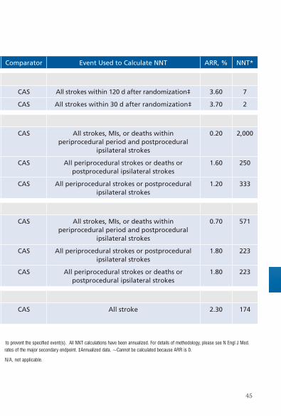
Trial, Year Patient Population Intervention Comparator Event Used to Calculate NNT ARR, % NNT*
Symptomatic
ICSS (2010) Symptomatic CEA CAS All strokes within 120 d after randomization‡ 3.60 7
ICSS (2010) Symptomatic CEA CAS All strokes within 30 d after randomization‡ 3.70 2
CREST symptomatic
CREST 4-y data (2010) Symptomatic CEA CAS All strokes, MIs, or deaths within periprocedural period and postprocedural
ipsilateral strokes
0.20 2,000
CREST 4-y data (2010) Symptomatic CEA CAS All periprocedural strokes or deaths or postprocedural ipsilateral strokes
1.60 250
CREST 4-y data (2010) Symptomatic CEA CAS All periprocedural strokes or postprocedural ipsilateral strokes
1.20 333
CREST asymptomatic
CREST 4-y data (2010) Asymptomatic CEA CAS All strokes, MIs, or deaths within periprocedural period and postprocedural
ipsilateral strokes
0.70 571
CREST 4-y data (2010) Asymptomatic CEA CAS All periprocedural strokes or postprocedural ipsilateral strokes
1.80 223
CREST 4-y data (2010) Asymptomatic CEA CAS All periprocedural strokes or deaths or postprocedural ipsilateral strokes
1.80 223
CREST mixed population
CREST 4-y data (2010) Patient population not separated in table; mixed patient population
CEA CAS All stroke 2.30 174
See 2011 full text for references and more information.
*NNT indicates number of patients needed to treat over the course of 1 year with the indicated therapy as opposed to the comparator to prevent the specified event(s). All NNT calculations have been annualized. For details of methodology, please see N Engl J Med. 2009;361:424-5. †The 1-year data from the SAPPHIRE trial included the primary endpoint; long-term data were used to calculate rates of the major secondary endpoint. ‡Annualized data. ~Cannot be calculated because ARR is 0.
ARR indicates absolute risk reduction; CAS, carotid artery stenting; CEA, carotid endarterectomy; NNT, number needed to treat; N/A, not applicable.
45
Trial, Year Patient Population Intervention Comparator Event Used to Calculate NNT ARR, % NNT*
Symptomatic
ICSS (2010) Symptomatic CEA CAS All strokes within 120 d after randomization‡ 3.60 7
ICSS (2010) Symptomatic CEA CAS All strokes within 30 d after randomization‡ 3.70 2
CREST symptomatic
CREST 4-y data (2010) Symptomatic CEA CAS All strokes, MIs, or deaths within periprocedural period and postprocedural
ipsilateral strokes
0.20 2,000
CREST 4-y data (2010) Symptomatic CEA CAS All periprocedural strokes or deaths or postprocedural ipsilateral strokes
1.60 250
CREST 4-y data (2010) Symptomatic CEA CAS All periprocedural strokes or postprocedural ipsilateral strokes
1.20 333
CREST asymptomatic
CREST 4-y data (2010) Asymptomatic CEA CAS All strokes, MIs, or deaths within periprocedural period and postprocedural
ipsilateral strokes
0.70 571
CREST 4-y data (2010) Asymptomatic CEA CAS All periprocedural strokes or postprocedural ipsilateral strokes
1.80 223
CREST 4-y data (2010) Asymptomatic CEA CAS All periprocedural strokes or deaths or postprocedural ipsilateral strokes
1.80 223
CREST mixed population
CREST 4-y data (2010) Patient population not separated in table; mixed patient population
CEA CAS All stroke 2.30 174
See 2011 full text for references and more information.
*NNT indicates number of patients needed to treat over the course of 1 year with the indicated therapy as opposed to the comparator to prevent the specified event(s). All NNT calculations have been annualized. For details of methodology, please see N Engl J Med. 2009;361:424-5. †The 1-year data from the SAPPHIRE trial included the primary endpoint; long-term data were used to calculate rates of the major secondary endpoint. ‡Annualized data. ~Cannot be calculated because ARR is 0.
ARR indicates absolute risk reduction; CAS, carotid artery stenting; CEA, carotid endarterectomy; NNT, number needed to treat; N/A, not applicable.

46

47