spinal cord and spinal nerve roots (sensory and motor) fig. 1.4.1
TRANSCRIPT
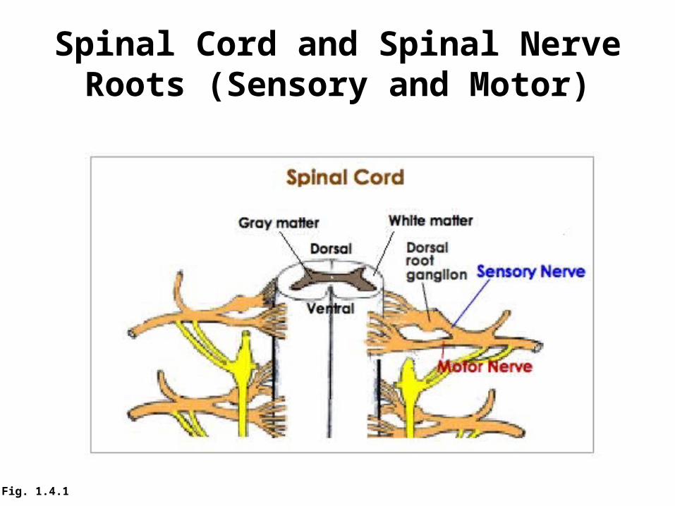
Spinal Cord and Spinal Nerve Roots (Sensory and Motor)
Fig. 1.4.1
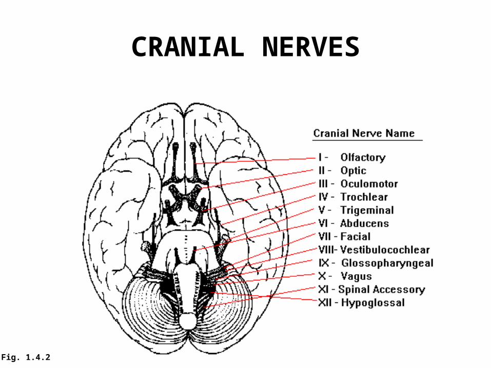
CRANIAL NERVES
Fig. 1.4.2
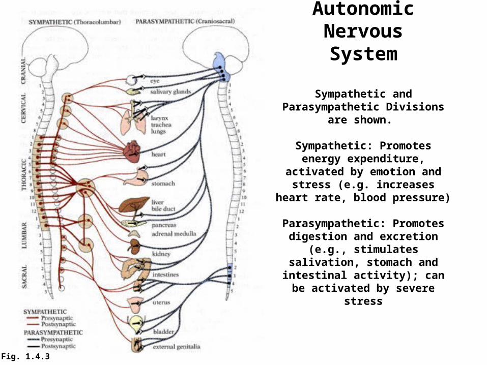
AutonomicNervousSystem
Sympathetic and Parasympathetic Divisions are shown.
Sympathetic: Promotes energy expenditure, activated by emotion
and stress (e.g. increases heart rate, blood pressure)
Parasympathetic: Promotes digestion and excretion (e.g.,
stimulates salivation, stomach and intestinal activity); can be activated by severe stress
Fig. 1.4.3
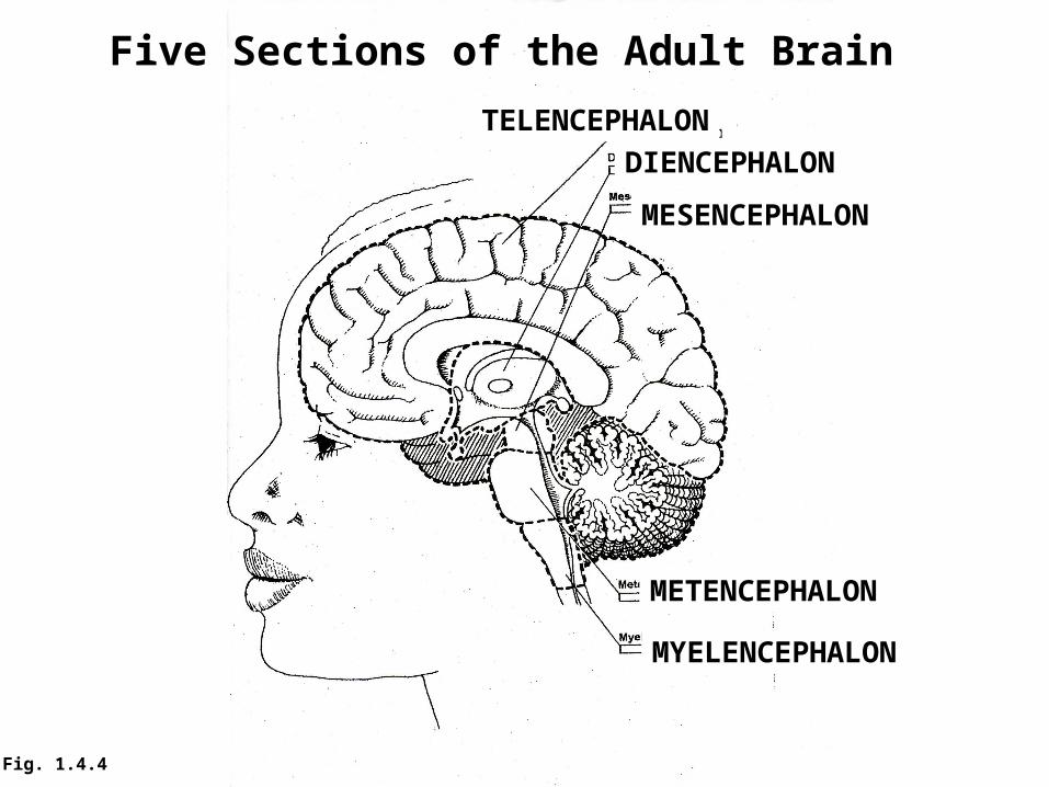
TELENCEPHALON
DIENCEPHALON
MESENCEPHALON
METENCEPHALON
MYELENCEPHALON
Five Sections of the Adult Brain
Fig. 1.4.4

TELENCEPHALON
DIENCEPHALON
MESENCEPHALON
METENCEPHALON
MYELENCEPHALONMEDULLA
Fig. 1.4.5
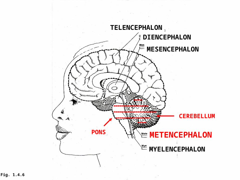
TELENCEPHALON
DIENCEPHALON
MESENCEPHALON
METENCEPHALON
MYELENCEPHALON
PONS
CEREBELLUM
Fig. 1.4.6
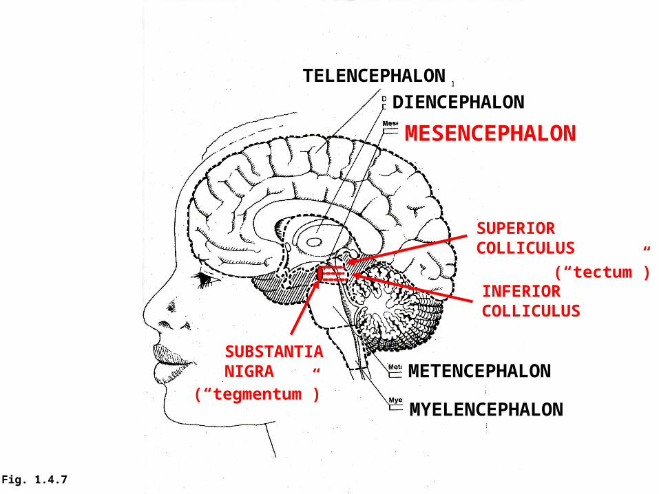
TELENCEPHALON
DIENCEPHALON
MESENCEPHALON
METENCEPHALON
MYELENCEPHALON
SUPERIOR COLLICULUS
INFERIORCOLLICULUS
SUBSTANTIANIGRA
(“tectum”)
(“tegmentum”)
Fig. 1.4.7
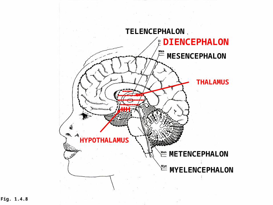
TELENCEPHALON
DIENCEPHALON
MESENCEPHALON
METENCEPHALON
MYELENCEPHALON
THALAMUS
HYPOTHALAMUS
Fig. 1.4.8
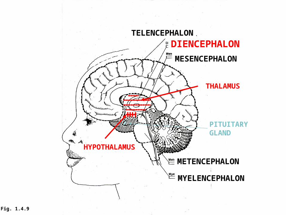
TELENCEPHALON
DIENCEPHALON
MESENCEPHALON
METENCEPHALON
MYELENCEPHALON
THALAMUS
HYPOTHALAMUS
PITUITARYGLAND
Fig. 1.4.9

TELENCEPHALONDIENCEPHALON
MESENCEPHALON
METENCEPHALON
MYELENCEPHALON
NEOCORTEX
LIMBICSYSTEMAndBASALGANGLIA(not shown)
Fig. 1.4.10
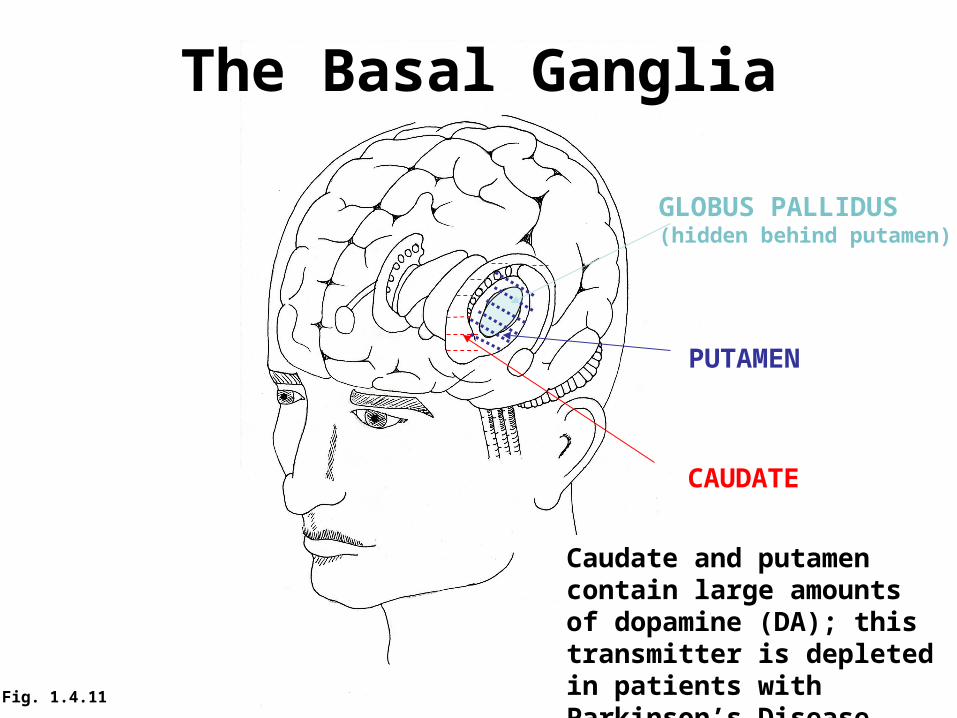
The Basal Ganglia
CAUDATE
PUTAMEN
GLOBUS PALLIDUS(hidden behind putamen)
Caudate and putamen contain large amounts of dopamine (DA); this transmitter is depleted in patients with Parkinson’s DiseaseFig. 1.4.11

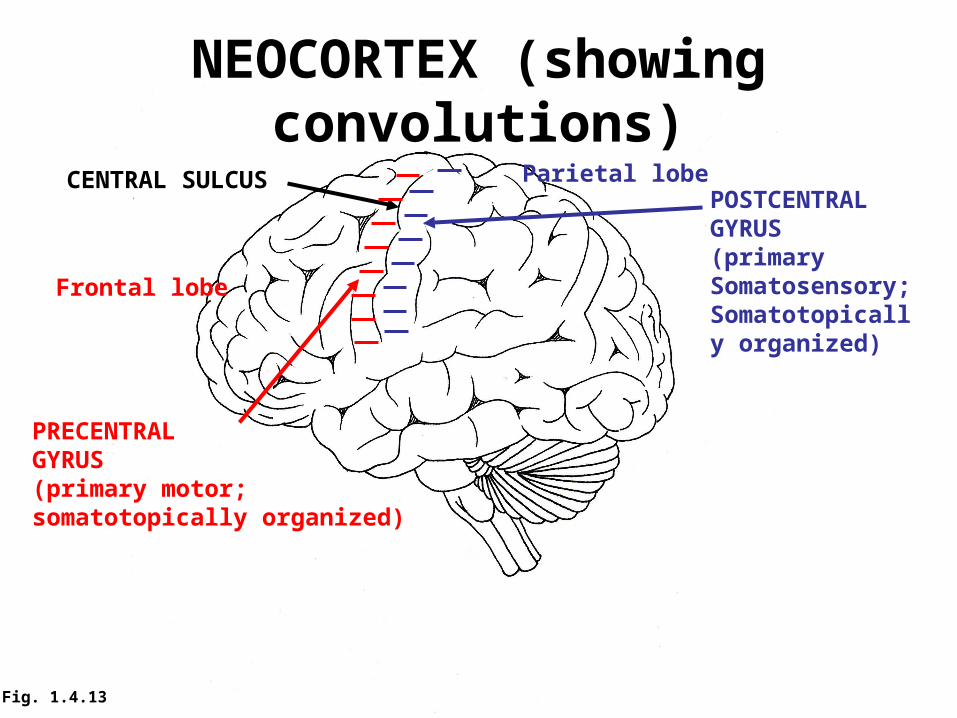
NEOCORTEX (showing convolutions)
CENTRAL SULCUS
PRECENTRALGYRUS(primary motor;somatotopically organized)
POSTCENTRALGYRUS(primary Somatosensory;Somatotopically organized)
Frontal lobe
Parietal lobe
Fig. 1.4.13

NEOCORTEX
Fig. 1.4.14
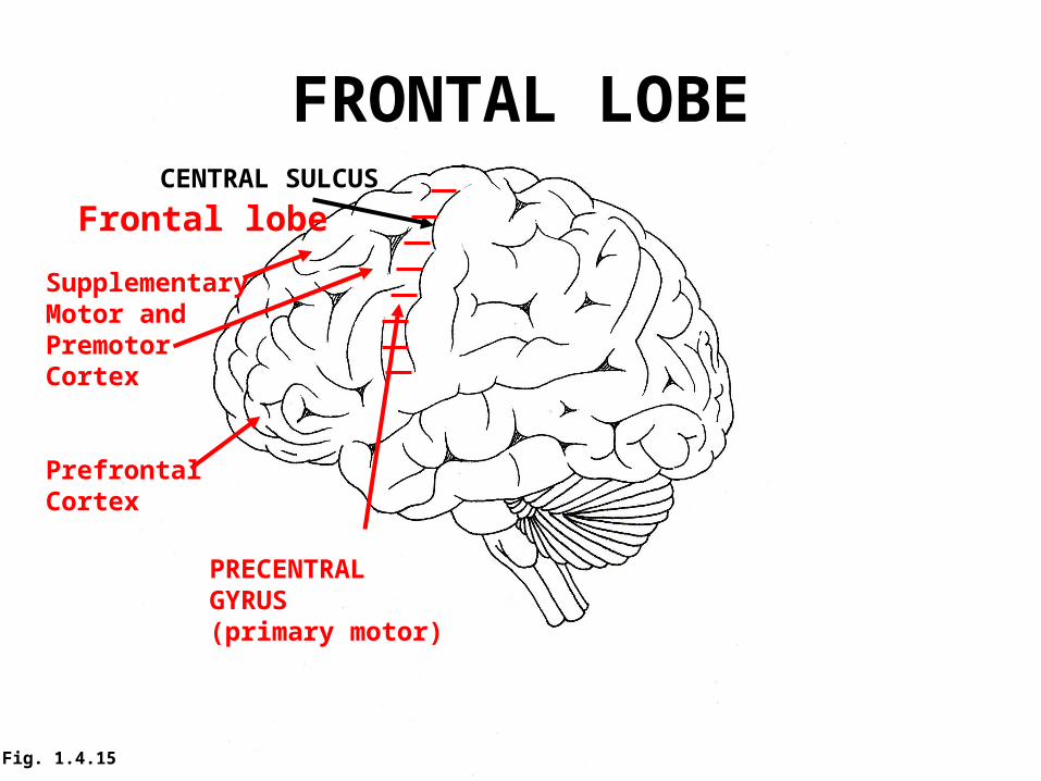
FRONTAL LOBECENTRAL SULCUS
PRECENTRALGYRUS(primary motor)
Frontal lobe
SupplementaryMotor and Premotor Cortex
PrefrontalCortex
Fig. 1.4.15
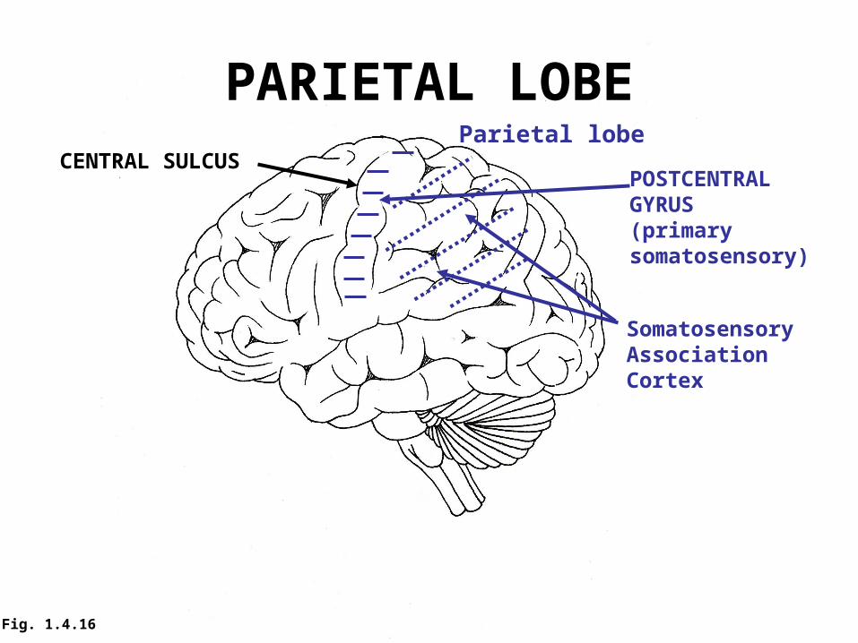
PARIETAL LOBECENTRAL SULCUS
POSTCENTRALGYRUS(primary somatosensory)
Parietal lobe
SomatosensoryAssociationCortex
Fig. 1.4.16

Occipital lobe
OCCIPITAL LOBE
PrimaryVisualCortex
VisualAssociationCortex
Fig. 1.4.17
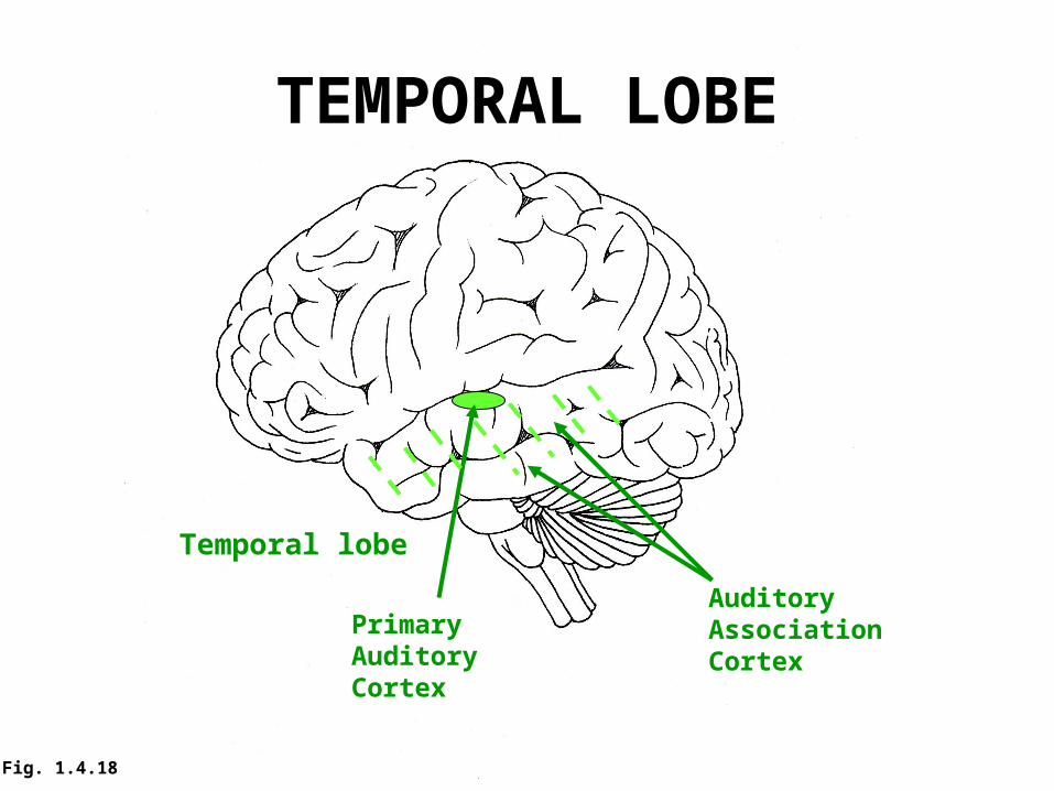
TEMPORAL LOBE
Temporal lobe
PrimaryAuditoryCortex
AuditoryAssociationCortex
Fig. 1.4.18

TELENCEPHALONDIENCEPHALON
MESENCEPHALON
METENCEPHALON
MYELENCEPHALON
NEOCORTEXCORPUSCALLOSUM(axons that connectthe two hemispheres)
Fig. 1.4.19