peripheral nerve injury alters blood–spinal cord barrier functional
TRANSCRIPT

Behavioral/Systems/Cognitive
Peripheral Nerve Injury Alters Blood–Spinal Cord BarrierFunctional and Molecular Integrity through a SelectiveInflammatory Pathway
Stefania Echeverry,1 Xiang Qun Shi,1 Serge Rivest,2 and Ji Zhang1
1The Alan Edwards Centre for Research on Pain, McGill University, Montreal, Quebec H3A 2B2, Canada, and 2Centre de Recherche du Centre Hospitalier del’Universite Laval, Quebec, Quebec G1V 4G2, Canada
Peripheral nerve lesion triggers alterations in the spinal microenvironment that contribute to the pathogenesis of neuropathic pain.While neurons and glia have been implicated in these functional changes, it remains largely underexplored whether the blood–spinalcord barrier (BSCB) is also involved. The BSCB is an important component in the CNS homeostasis, and compromised BSCB has beenassociated with different pathologies affecting the spinal cord. Here, we demonstrated that a remote injury on the peripheral nerve in ratstriggered a leakage of the BSCB, which was independent of spinal microglial activation. The increase of BSCB permeability to different sizetracers, such as Evans Blue and sodium fluorescein, was restricted to the lumbar spinal cord and prominent for at least 4 weeks afterinjury. The spinal inflammatory reaction triggered by nerve injury was a key player in modulating BSCB permeability. We identifiedMCP-1 as an endogenous trigger for the BSCB leakage. BSCB permeability can also be impaired by circulating IL-1�. In contrast,antiinflammatory cytokines TGF-�1 and IL-10 were able to shut down the openings of the BSCB following nerve injury. Peripheral nerveinjury caused a decrease in tight junction and caveolae-associated proteins. Interestingly, ZO-1 and occludin, but not caveolin-1, wererescued by TGF-�1. Furthermore, our data provide direct evidence that disrupted BSCB following nerve injury contributed to the influxof inflammatory mediators and the recruitment of spinal blood borne monocytes/macrophages, which played a major role in thedevelopment of neuropathic pain. These findings highlight the importance of inflammation in BSCB integrity and in spinal cordhomeostasis.
IntroductionAs an interface between the spinal cord and the periphery, theblood–spinal cord barrier (BSCB) constitutes a physical/bio-chemical barrier between the CNS and the systemic circulation,which serves to protect the microenvironment of the spinal cord.Similar to the blood– brain barrier (BBB), the BSCB is composedof a monolayer of nonfenestrated endothelial cells, intercon-nected by tight junctions (TJs) and surrounded by end feet ofastrocytes (Pardridge, 1999). Once thought to be static, theBSCB/BBB is actually a dynamic structure capable of rapid mod-ulation. Breakdown of the BBB/BSCB has been reported indifferent pathologies affecting the nervous system, including Alz-heimer’s disease, stroke, multiple sclerosis, and traumatic brain/
spinal cord injury; and it is mostly associated with in situ damagesin the CNS (Carvey et al., 2009).
Injury to the peripheral nervous system leading to neuropathicpain triggers multidimensional changes along the pain signalingpathway (Basbaum et al., 2009). One of the most salient observationsis the inflammatory reaction. After nerve injury, proinflammatorymediators are released from injured nerve fibers and adjacent im-mune cells (Scholz and Woolf, 2007). Within the spinal cords, re-mote injury induces substantial changes in microglia and astrocytes,which are important players in the central inflammatory response(Zhang and De Koninck, 2009). In addition, peripheral nerve injury(PNI) evoked trafficking of immune cells from circulation into thespinal cord parenchyma (Zhang et al., 2007; Cao and DeLeo, 2008;Costigan et al., 2009). These neuroimmune interactions partici-pate in the pathogenesis of chronic pain states (McMahon andMalcangio, 2009; White et al., 2009). As a key component of thespinal cord microenvironment, the BSCB could play critical roles inboth separating and conjoining the immune system and the CNS.However, the functional states of the BSCB following PNI have notbeen thoroughly explored. Many questions remain unanswered:whether a remote injury can affect the integrity of the BSCB, and ifso, what are the underlying mechanisms regulating such disturbanceand what are the consequences of a BSCB breakdown in the spinalcord homeostasis and development of pathological pain.
Two main structural features that contribute to the imperme-able characteristics of BBB/BSCB are the presence of TJs and
Received April 1, 2011; revised June 4, 2011; accepted June 8, 2011.Author contributions: S.E. and J.Z. designed research; S.E. and X.Q.S. performed research; S.R. contributed un-
published reagents/analytic tools; S.E., X.Q.S., and J.Z. analyzed data; S.E., S.R., and J.Z. wrote the paper.This work was supported by Canadian Institutes for Health Research (CIHR) Grant MOP-77624 (J.Z.), the
Fonds de la Recherche en Sante du Quebec/Quebec Pain Research Network/Pfizer partnership (J.Z.), TheLouise and Alan Edwards Foundation (J.Z.), and The Alan Edwards Center for Research on Pain/AstraZenecaAlliance (J.Z.), and by a Canadian Pain Society Trainee Grant (S.E.). We acknowledge the support from theCIHR Neuroinflammation Training Program. S.E. holds a CIHR doctoral studentship. J.Z. is a CIHR new inves-tigator, and S.R. is a Canadian Research Chair in Neuroimmunology.
Correspondence should be addressed to Dr. Ji Zhang, The Alan Edwards Centre for Research on Pain, McGillUniversity, 740, Dr. Penfield Avenue, Genome Building, Suite 3200C, Montreal, Quebec H3A 2B2, Canada. E-mail:[email protected].
DOI:10.1523/JNEUROSCI.1642-11.2011Copyright © 2011 the authors 0270-6474/11/3110819-10$15.00/0
The Journal of Neuroscience, July 27, 2011 • 31(30):10819 –10828 • 10819
fewer caveolae in CNS endothelium. TJs form a continuous wallthat limits paracellular flux of solutes and cells across the endo-thelial barriers (Huber et al., 2001). Caveolae, flask-shaped in-vaginations of the plasma membrane, participate in vesicletransport (Couet et al., 2001). How these structures are regulatedin relation to BBB/BSCB function in the context of PNI is againnot known.
In the present study, we analyzed the permeability of the BSCBfollowing a partial ligation on the sciatic nerve. Our resultsrevealed that PNI disrupted the integrity of the BSCB andinflammatory mediators are key regulators of BSCB function.Disruption of the BSCB following nerve injury causes the influxof inflammatory mediators and the recruitment of circulatingimmune cells into the spinal parenchyma, which play a criticalrole in neuroimmune interactions and the development of neu-ropathic pain.
Materials and MethodsAnimalsMale Sprague Dawley rats weighing 250 –275 g and male/female C57BL/6mice weighing �25 g (Charles River) were housed under standard 12 hlight/dark conditions and received food ad libitum. All protocols wereperformed in accordance with guidelines from the Canadian Council onAnimal Care and were approved by the McGill University and the LavalUniversity Animal Care Committees.
Generation of GFP� chimeric miceGFP � chimeric mice were generated as described previously (Zhang etal., 2007). Briefly, recipient C57BL/6 mice were exposed to irradiationusing a cobalt-60 source (Nordion) and then injected via tail vein with5 � 10 6 bone marrow cells freshly collected from GFP � transgenic mice.
Induction of peripheral nerve injury and pain behavioral testingPartial sciatic nerve ligation was performed according to the method ofSeltzer et al. (1990). Rats were anesthetized with isoflurane, and underaseptic conditions the left sciatic nerve was exposed at high-thigh level.The dorsal one-third to one-half of the nerve thickness was tightly ligatedwith 6-0 silk sutures. Sham-operated rats underwent the same surgicalprocedure, but the nerve was exposed and left intact. Survival times were3, 7, 14, 60, and 95 d after surgery. A group of naive rats was included inthe protocol as a control. Mechanical and thermal sensitivities were mon-itored using well calibrated von Frey hair (Stoelting) and Hargreavesapparatus, respectively, with the methods described previously (Har-greaves et al., 1988; Chaplan et al., 1994). Investigators were blinded tothe assignment of the groups. GFP � chimeric mice received a partialligation on the left sciatic nerve 10 weeks after irradiation and bonemarrow transplantation and were killed 14 d after surgery to collectspinal cords.
Induction of experimental autoimmune encephalomyelitisTo induce experimental autoimmune encephalomyelitis (EAE), a groupof female C57BL/6 mice were immunized with 200 �g/mouse MOG(myelin oligodendrocyte glycoprotein) peptide 35–55 (custom-synthesizedby CS Bio) emulsified in 100 �l of complete Freund’s adjuvant (CFA). Con-trol group was inoculated with equal volumes of CFA. Mice were adminis-trated intraperitoneally 200 ng of pertussis toxin dissolved in 200 �l of PBSon the day of immunization and 48 h later. Mice were monitored daily forclinical signs of EAE and scored as follows: 0, no abnormality; 1, limp tail; 2,mild hindlimb weakness; 3, severe hindlimb weakness; 4, complete hindlimbweakness; 5, quadriplegia or premoribund state; and 6, death. At 10–14 dfollowing inoculation, mice with clinical scores of 2 were used for BBB/BSCBintegrity evaluation.
Evaluation of BSCB permeabilityEvans Blue leakage. Evans Blue (EB) leakage was assessed with a wellestablished protocol adapted from Xu et al. (2001). EB dye (3% w/v insaline) [molecular weight (MW), 961 Da; Sigma-Aldrich] was injectedintravenously under anesthesia. Thirty minutes after the injection, rats
were perfused transcardially with saline and rinsed thoroughly until nomore blue dye flew out of the right atrium. The choroid plexuses andmeninges were removed. Spinal cords and brains were dissected intodifferent regions: lumbar and thoracic/cervical segments for the spinalcord, left and right cerebral cortex, and midbrain/brainstem. Each regionwas weighed for quantitative measurement of EB extravasation. EB con-tent within the parenchyma was extracted by incubating the tissue informamide for 48 h at room temperature. The diluents were filtered andsubject to spectrophotometric measurements at 620 nm. All measure-ments were within the range of detection established by the standardcurve. R2 of the standard curve was 0.9 –1.0. The dye concentrationwas calculated as the ratio of absorbance relative to the amount oftissue (n � 3–10).
Sodium fluorescein leakage. BBB and BSCB permeability was also as-sessed using a micromolecular tracer dye sodium fluorescein (NaFlu)(MW, 376; Sigma-Aldrich) according to a modified procedure by Kaya etal. (2008). Briefly, NaFlu was administered intravenously (10%; 2 ml/kg)and allowed to circulate for 30 min. Then, rats were transcardially per-fused with cold saline, for �12 min to remove intravascular NaFlu. Thechoroid plexuses and meninges were removed. Spinal cords and brainswere dissected into different regions: lumbar spinal cord, left and rightcerebral cortex, and midbrain/brainstem. Each region was weighed forquantitative measurement of NaFlu extravasation. Samples were homog-enized in 1 ml of PBS and mixed with a vortex for 2 min after the additionof equal volumes of 60% trichloroacetic acid to precipitate protein. Sam-ples were later cooled for 30 min and centrifuged at 14,000 � g for 10min. The concentration of NaFlu in supernatant was measured at exci-tation wavelength of 440 nm and emission wavelength of 525 nm using aspectrophotofluorometer. A standard curve of different amounts of Na-Flu was drawn under identical conditions of the assay for calculating dyeconcentrations in brain and spinal cord. All measurements were withinthe range of detection established by the standard curve. R2 of the stan-dard curve was 0.85– 0.9. NaFlu was expressed as micrograms per gram oftissue.
Plasma protein extravasations. Plasma protein extravasations were ad-ditionally evaluated using immunohistochemical labeling with antibod-ies against the plasma protein IgG (MW, 53 kDa) and fibronectin (MW,400 kDa). Free-floating lumbar spinal cord sections were immunola-beled with either rabbit anti-fibronectin (1:500; Millipore Bioscience Re-search Reagents) or goat anti-rat IgG (1:200; Invitrogen). Vascularmarkers CD31 and CD34 were used to delineate blood vessels.
Intrathecal infusion of minocyclineThe microglial inhibitor, minocycline hydrochloride (Sigma-Aldrich),was freshly dissolved in 0.9% sterile, isotonic saline and delivered intra-thecally at a dose of 150 �g per day for 7 d (day 0 to day 7) using anosmotic pump (Alzet) with a flow rate of 1 �l/h connected to an intra-thecal catheter (n � 5– 8 in each group). The dose for intrathecal mino-cycline was selected according to previous studies (Ledeboer et al., 2005;Lin et al., 2007).
Immune modulation of BSCB functionRecombinant proinflammatory or antiinflammatory molecules weredissolved in sterile 0.9% saline and delivered either intravenously orintrathecally into naive or nerve-injured animals. Detailed protocols arelisted in Table 1.
Permeation to circulating IL-1�BSCB permeation to circulating IL-1� was assessed using 125I-labeledIL-1� according to a previously described method (Pan et al., 2003).Briefly, 125I-IL-1� was dissolved in 0.9% saline and a bolus of 150 �l ofthe radiolabeled IL-1� (0.7 �Ci) was injected into the left ventricle. Theblood sample was collected 10 min later from the right ventricle. Theanimals were then perfused with saline and lumbar spinal cords werecollected. The 125I radioactivity in serum and in lumbar spinal cord wasmeasured separately using a gamma counter (PerkinElmer). The ratio ofradioactivity in the spinal cord to that of serum was plotted againstexposure time, the latter being the theoretical value of steady-state serumradioactivity. Ten minute exposure to 125I-IL-1� was chosen based onthe fact that, during this short period, the radioactivity in the tissue
10820 • J. Neurosci., July 27, 2011 • 31(30):10819 –10828 Echeverry et al. • Blood–Spinal Cord Barrier Integrity and Neuropathic Pain

parenchyma represents most likely intact 125I-cytokine rather than de-graded products (Pardridge, 1999). A control group was included inwhich 125I-IL-1� was dissolved in 3% EB. After measurement of theradioactivity, the spinal cord samples were incubated in formamide for48 h in the dark to extract and assess EB content in the parenchyma asdescribed above.
Microvessel isolationSpinal microvessels from naive and injured rats (day 3) were isolatedaccording to the method described by Hoehn et al. (2002). Rats weredecapitated under anesthesia, and spinal cords were removed quickly.The meninges were excised and the lumbar segments were homogenizedin microvessel isolation buffer containing protease inhibitor (Sigma-Aldrich). Dextran (26% m/v) was added to the homogenate afterward at4°C. Samples were then gently vortexed and centrifuged (5600 � g; 4°C)for 10 min, and the supernatant was discarded. Pellets were resuspendedin fresh microvessel isolation buffer and passed through a 70 �m filter(BD Biosciences). Filtered homogenates were pelleted by centrifugationat 3000 � g for 10 min. The supernatant was removed, and the pellet,which was enriched in spinal microvessels, was collected for protein ex-traction. The purity of collected microvessels was verified with immuno-
staining using two endothelial markers vonWillebrand factor (VWF) (1:1000; Abcam) andGlut-1 (1:500; Millipore).
Western blotThe pellet containing spinal microvessels wasresuspended in 500 �l of CellLytic (Sigma-Aldrich) with protease inhibitor. Thirty micro-grams of proteins extracted from the abovepreparation were loaded and separated on 10%SDS-PAGE gel. After the transfer, blots wereincubated overnight at 4°C with the followingantibodies: zonula occludens-1 (ZO-1) (1:200;Cell Signaling), occludin (1:100; Cell Signal-ing), caveolin-1 (1:250; Cell Signaling), TGF-�receptor I (TGF-�-RI) (1:200; Santa Cruz),and p-Smad2/3 (1:500; Santa Cruz). For load-ing control, blots were probed with �-actin an-tibody (1:10,000; Sigma-Aldrich). Density ofspecific bands from Western blotting wasquantified with a computer-assisted imaginganalysis system (Image Pro Plus). Three sepa-rate experiments where each treatment groupconsists of pooled microvessels from two ani-mals were included.
ImmunohistochemistryTo identify the expression of TGF-� receptorson endothelial cells, immunofluorescent stain-ing was performed on spinal microvessels ex-tracted as explained above. Microvessels werefixed with 4% PFA for 30 min and incubatedovernight at 4°C with rabbit anti-TGF-�-RIpolyclonal antibody (1:250; Santa Cruz Bio-technology) and rat anti-CD31 (for endothelialcells; 1:250; BD Biosciences), followed by a60 min incubation at room temperature influorochrome-conjugated goat secondary an-
tibody. 4�,6-Diamidino-2-phenylindole dihydrochloride (DAPI) wasalso used as a nuclear counterstain (1:10,000; Sigma-Aldrich).
The following antibodies were also used to examine the spinal micro-glial activation and immune cell trafficking following nerve injury. Free-floating sections from lumbar spinal cords were incubated overnight at4°C with rabbit anti-ionized calcium-binding adaptor molecule 1 (Iba-1)polyclonal antibody (for microglia, 1:1000; Wako Chemicals) and anti-CD3 monoclonal antibody (for T-lymphocytes, 1:200; AbD Serotec),followed by a 60 min incubation at room temperature in fluorochrome-conjugated goat secondary antibody. Anti-CD2 monoclonal antibody(1:100; AbD Serotec) was incubated for 72 h, at 4°C, and followed bytyramide signal amplification (PerkinElmer).
Image acquisitionImages were acquired using an Olympus BX51 microscope equippedwith a color digital camera (Olympus DP71) or Olympus confocal laser-scanning microscope (Fluoview 1000). GFP � cells were counted by ablind investigator in four different regions of interest [ipsilateral dorsalhorn (DHi), contralateral dorsal horn (DHc), ipsilateral ventral horn(VHi), and contralateral ventral horn (VHc)], in mice treated with sa-
Figure 1. Development of neuropathic pain following injury on sciatic nerve. Shortly after the partial ligation on the sciaticnerve, rats develop mechanical allodynia and thermal hyperalgesia in the ipsilateral hindpaws. Withdrawal threshold of bothipsilateral (A) and contralateral (B) paws to calibrated von Frey hair stimulation decreased significantly, starting from day 3 until atleast day 63 after injury. Withdrawal latency to noxious heat stimuli was also decreased at ipsilateral side (C), and to a less extentat the contralateral paw (D). N � 6/group. Values are presented as means � SEM. *p � 0.05, **p � 0.01, ***p � 0.001 frombaseline.
Table 1. Experimental protocols for immune modulation of BSCB integrity
Molecules Animals Duration Administration route Doses (source)
Rat recombinant MCP-1 Naive rats 3 d Intrathecal (osmotic pumps) 2.5 �g (PeproTech)Rat MCP-1 neutralizing antibody Nerve-injured rats 3 d (day 0 to day 3) Intrathecal (osmotic pumps) 3 �g (PeproTech)Recombinant TGF-�1 (mammalian derived) Nerve-injured rats 3 d (day 0 to day 3) Intrathecal (osmotic pumps) 2 �g (PeproTech)Recombinant IL-10 (mammalian derived) Nerve-injured rats 3 d (day 0 to day 3) Intrathecal (osmotic pumps) 3 �g (PeproTech)Recombinant TGF-�1 (mammalian derived) Naive rats 3 d Intrathecal (osmotic pumps) 2 �g (PeproTech)Recombinant TGF-�1 (mammalian derived) Nerve-injured GFP � chimeric mice 14 d (day 0 to day 14) Intrathecal, punction (every 3 d) 1 �g/injection (PeproTech)125I-recombinant human IL-1� Naive rats 10 min Intravenous 20 –5000 immune units (PerkinElmer)
Echeverry et al. • Blood–Spinal Cord Barrier Integrity and Neuropathic Pain J. Neurosci., July 27, 2011 • 31(30):10819 –10828 • 10821
line or with recombinant TGF-�1. Onlyramified GFP � cells within parenchymal graymatter were included. Colocalization of Iba-1with GFP and the distribution of GFP � cellsoutside of Glut-1-labeled vessels were ensuredwith confocal Z stacks at 0.8 �m intervals andvisualization in three-dimensional orthogonalplanes. Similar approaches were applied to thequantification of CD2 � and CD3 � cells in thedorsal horns of the spinal cords.
Statistical analysisTo determine statistical significance, unpairedt test was used for the difference betweengroups (TGF-�1/MCP-1/IL-1�/IL-10 treatedvs saline treated at each time point); paired ttest was used for the difference between ipsilat-eral and contralateral sides within the samegroup; two-way ANOVA followed by Bonfer-roni’s post tests were used for the analysis ofbehavioral data. A value of p � 0.05 was ac-cepted as statistically significant. Values arepresented as means � SEM.
ResultsDevelopment of neuropathic painfollowing peripheral nerve injuryHypersensitivity to mechanical and ther-mal stimuli are characteristic behaviorsdeveloped following nerve injury. In thisstudy, shortly after the lesion on the sciaticnerve, rats exhibited mechanical allodynia(Fig. 1A,B) and thermal hyperalgesia (Fig.1C,D), which lasted for at least 2 months,the end of the testing period.
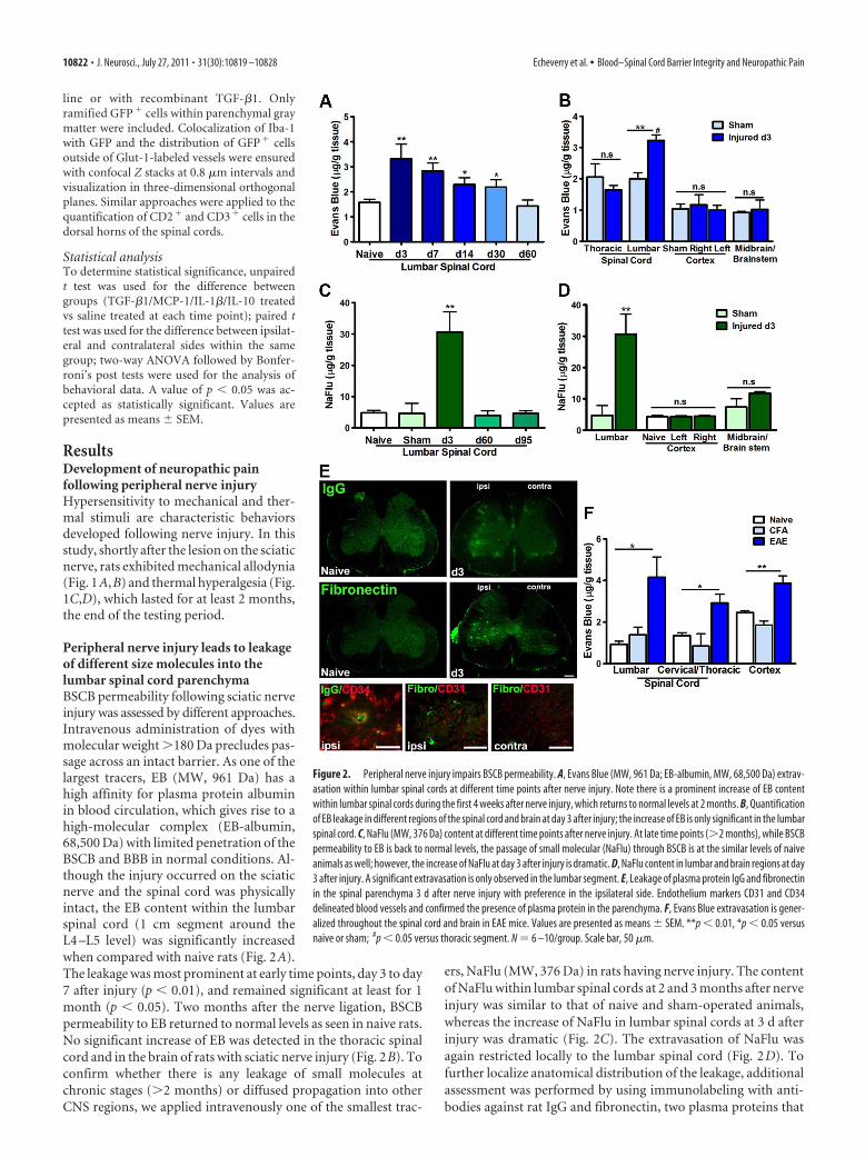
Peripheral nerve injury leads to leakageof different size molecules into thelumbar spinal cord parenchymaBSCB permeability following sciatic nerveinjury was assessed by different approaches.Intravenous administration of dyes withmolecular weight �180 Da precludes pas-sage across an intact barrier. As one of thelargest tracers, EB (MW, 961 Da) has ahigh affinity for plasma protein albuminin blood circulation, which gives rise to ahigh-molecular complex (EB-albumin,68,500 Da) with limited penetration of theBSCB and BBB in normal conditions. Al-though the injury occurred on the sciaticnerve and the spinal cord was physicallyintact, the EB content within the lumbarspinal cord (1 cm segment around theL4 –L5 level) was significantly increasedwhen compared with naive rats (Fig. 2A).The leakage was most prominent at early time points, day 3 to day7 after injury (p � 0.01), and remained significant at least for 1month (p � 0.05). Two months after the nerve ligation, BSCBpermeability to EB returned to normal levels as seen in naive rats.No significant increase of EB was detected in the thoracic spinalcord and in the brain of rats with sciatic nerve injury (Fig. 2B). Toconfirm whether there is any leakage of small molecules atchronic stages (�2 months) or diffused propagation into otherCNS regions, we applied intravenously one of the smallest trac-
ers, NaFlu (MW, 376 Da) in rats having nerve injury. The contentof NaFlu within lumbar spinal cords at 2 and 3 months after nerveinjury was similar to that of naive and sham-operated animals,whereas the increase of NaFlu in lumbar spinal cords at 3 d afterinjury was dramatic (Fig. 2C). The extravasation of NaFlu wasagain restricted locally to the lumbar spinal cord (Fig. 2D). Tofurther localize anatomical distribution of the leakage, additionalassessment was performed by using immunolabeling with anti-bodies against rat IgG and fibronectin, two plasma proteins that
Figure 2. Peripheral nerve injury impairs BSCB permeability. A, Evans Blue (MW, 961 Da; EB-albumin, MW, 68,500 Da) extrav-asation within lumbar spinal cords at different time points after nerve injury. Note there is a prominent increase of EB contentwithin lumbar spinal cords during the first 4 weeks after nerve injury, which returns to normal levels at 2 months. B, Quantificationof EB leakage in different regions of the spinal cord and brain at day 3 after injury; the increase of EB is only significant in the lumbarspinal cord. C, NaFlu (MW, 376 Da) content at different time points after nerve injury. At late time points (�2 months), while BSCBpermeability to EB is back to normal levels, the passage of small molecular (NaFlu) through BSCB is at the similar levels of naiveanimals as well; however, the increase of NaFlu at day 3 after injury is dramatic. D, NaFlu content in lumbar and brain regions at day3 after injury. A significant extravasation is only observed in the lumbar segment. E, Leakage of plasma protein IgG and fibronectinin the spinal parenchyma 3 d after nerve injury with preference in the ipsilateral side. Endothelium markers CD31 and CD34delineated blood vessels and confirmed the presence of plasma protein in the parenchyma. F, Evans Blue extravasation is gener-alized throughout the spinal cord and brain in EAE mice. Values are presented as means � SEM. **p � 0.01, *p � 0.05 versusnaive or sham; #p � 0.05 versus thoracic segment. N � 6 –10/group. Scale bar, 50 �m.
10822 • J. Neurosci., July 27, 2011 • 31(30):10819 –10828 Echeverry et al. • Blood–Spinal Cord Barrier Integrity and Neuropathic Pain

should not be found in normal spinal cord parenchyma. Asshown in Figure 2 E, while no positive IgG or fibronectin stain-ing was detected in naive spinal cords, IgG- and fibronectin-immunopositive deposits were found within the spinal cord of
rats following sciatic nerve injury. The signal (extracellular aggre-gates) was essentially located in the ipsilateral side. Extravasationof IgG and fibronectin was confirmed by costaining with thevascular markers CD34 and CD31 (Fig. 2E). To better under-stand the scope of the nerve injury-triggered BSCB disruption, weused the same protocol to compare the EB leakage in EAE mice,an animal model of multiple sclerosis where the BBB/BSCB im-pairment has been well established (Hawkins and Davis, 2005;Fabis et al., 2007). As illustrated in Figure 2F, EAE-associated EBleakage is much more pronounced and diffused in different seg-ments of the spinal cords and in the brain. In contrast, peripheralnerve injury-triggered BSCB compromise is most likely restrictedin the corresponding lumbar spinal cord.
Microglial activation is not required for BSCB disruptionTo understand whether nerve injury modulates the BSCB perme-ability directly or such effect depends on the activation of residentmicroglial cells in the spinal cord, we infused intrathecally a mi-croglial inhibitor minocycline from day 0 to day 7. As reportedpreviously (Lin et al., 2007), such a treatment significantly pre-vented the increase of Iba-1 immunoreactivity (IR) (Fig. 3A,B)and partially reduced pain development (Fig. 3C). However, mi-nocycline did not affect the extravasation of EB (Fig. 3D), whencompared with saline (intrathecally)-treated animals, which sug-gest that BSCB disruption is not dependent on microglial activa-tion in the spinal cord.
Inflammatory mediators modulate BSCB functionMCP-1 is critical in nerve injury-induced BSCB disruptionThe chemokine MCP-1 is released by damaged neurons follow-ing nerve injury (Zhang and De Koninck, 2006). To assess therole of endogenous MCP-1 in BSCB integrity, we intrathecallyinfused an antibody against MCP-1 (3 �g) in injured animals. Com-pared with injured animals infused with saline, MCP-1 antibody
Figure 3. BSCB disruption is not affected by the suppression of spinal microglial activation. A, Photomicrographs depicting Iba-1 staining in the lumbar spinal cord. Partial sciatic nerve ligationinduced a remarkable increase of Iba-1 IR at the ipsilateral side in the dorsal and ventral horns. Intrathecal minocycline (150 �g/d for 7 d) successfully reduced this Iba-1 � IR. B, Quantification ofthe area occupied by the Iba-1 � IR reveals a striking reduction of microglial activation in the minocycline-treated animals compared with saline-treated animals. C, Mechanical allodynia (left panel)and thermal hyperalgesia (right panel) were partially attenuated in minocycline-treated animals. D, Despite inhibition of microglia, EB leakage into the spinal cord remained elevated in minocycline-treated animals at day 7. Data are shown as means � SEM. *p � 0.05, **p � 0.01, ***p � 0.001, minocycline-treated versus saline-treated groups. N � 6/group. Scale bar, 250 �m.
Figure 4. Inflammatory mediators modulate BSCB integrity. A, Neutralizing endogenous MCP-1with a MCP-1 antibody (3 �g for 3 d) significantly reduced the leakage of EB in the spinal cordobserved at day 3 after injury. B, Intrathecal infusion of recombinant murine MCP-1 (2.5 �g for 3 d)produced an increase of EB content in the spinal cord in naive animals, similar to that seen in nerve-injured animals. Intrathecal catheterization provoked a slight, but nonsignificant increase of EB con-tent in the spinal cord. C, IL-1� injected intravenously caused a dose-dependent impairment of BSCBpermeability to EB. D, Intrathecal administration of antiinflammatory cytokines TGF-�1 (2 �g) andIL-10 (3 �g) for 3 d significantly reduced the EB content in the spinal cord increased by peripheralnerve injury. TGF-�1 did not affect the BSCB permeability in naive rats. N � 3– 6/group. Values arepresented as means � SEM. *p � 0.05, **p � 0.01, ***p � 0.001 versus naive rats without anytreatment; #p � 0.05 versus injured saline-treated.
Echeverry et al. • Blood–Spinal Cord Barrier Integrity and Neuropathic Pain J. Neurosci., July 27, 2011 • 31(30):10819 –10828 • 10823

significantly reduced EB extravasation inthe spinal cord at day 3 after injury (Fig. 4A)(p � 0.05 vs saline). Next, we intrathecallyinfused naive animals with recombinantMCP-1 (2.5 �g) for 3 d and then assessedpermeability of BSCB to EB. ExogenousMCP-1 evoked an increase of EB content inthe spinal parenchyma in naive animalssimilar to that seen in nerve-injured animalsat day 3 (Fig. 4B) (p � 0.01 vs naive).
Systemic IL-1� induces EB extravasationin the spinal cordLow doses of IL-1�, 10–50 immune units(IU) injected intravenously did not provokeany significant increase of EB content in thespinal cord (Fig. 3C). However, higher con-centrations of IL-1�, 1000–5000 IU diddose-dependently increase the EB perme-ation in the spinal cord, which is significantat 5000 IU (p � 0.001 vs saline) (Fig. 4C).
TGF-�1 and IL-10 block EB extravasationin the spinal cord following peripheralnerve injuryTo evaluate whether TGF-�1, as an anti-inflammatory cytokine, is effective in pre-venting nerve injury-induced BSCBdisruption, we administrated TGF-�1 (2�g) in rats having ligation on the sciaticnerve. Three day intrathecal infusion (from day 0 to day 3) ofTGF-�1 successfully prevented the EB extravasation into the spi-nal cord following PNI (p � 0.01 vs saline) (Fig. 4D). Similarblocking effects were observed using another antiinflammatorycytokine IL-10 (Fig. 4D). TGF-�1 did not alter the normal phys-iological properties of the BSCB in naive animals (Fig. 4D).
Peripheral nerve injury alters tight junction complexprotein expressionIn view of the critical roles of tight junctions in maintaining BSCBintegrity, we examined the effect of PNI on the expression of someproteins important to tight junction structure and function, includ-ing plasma protein ZO-1 and membrane protein occludin. As cave-olae complexes play an important role in transcytosis of numeroussubstrates (Tuma and Hubbard, 2003; Kirkham and Parton, 2005),including immune mediators (Middleton et al., 1997), we alsosought to determine whether the expression of caveolin-1, the majorstructural protein of caveolae (Cohen et al., 2004; Stan, 2005) wasaltered in the spinal cord by nerve injury. Western blot analysis wasperformed using microvessels isolated from lumbar spinal cord seg-ments (Fig. 5A). In comparison with naive or sham-operated rats, allthree examined proteins, ZO-1 (Fig. 5B), occludin (Fig. 5C), andcaveolin-1 (Fig. 5D), were downregulated 3 d after a partial ligationon the sciatic nerve, when maximal disruption of the BSCB wasobserved. Relative levels of targeted protein expression were deter-mined by densitometric analysis and shown in Figure 5E.
TGF-�1 prevents the downregulation of tight junctionproteins following peripheral nerve injuryTo determine how TGF-�1 prevents the BSCB dysfunction asso-ciated with nerve injury, we examined the effects of TGF-�1 onthe expression of tight junction proteins. Intrathecal infusion ofTGF-�1 was able to prevent the downregulation of tight junction
proteins ZO-1 and occludin following PNI (Fig. 6A,B,D). How-ever, TGF-�1 failed to rescue the nerve injury-associateddownregulation of caveolin-1 (Fig. 6C,D). We have reportedpreviously that, as a potent antiinflammatory cytokine, TGF-�1inhibited the neuronal MCP-1 expression and reduced the spinalcytokine level in the same nerve injury model through its pleio-tropic effects on neurons and glia (Echeverry et al., 2009). Tofurther explore the mechanism of TGF-�1 in modulating BSCBintegrity, we examined the direct effects of TGF-�1 on endothe-lial cells. TGF-�-RI was detected in isolated endothelial cells (Fig.6E). Neither nerve injury nor exogenous ligand modified signif-icantly the quantity of the receptor on microvessels (Fig. 6F).However, intrathecal infusion of TGF-�1 did activate theSmad2/3 signaling pathway in endothelial cells, evidenced by anincrease of phosphorylated Smad2/3 (Fig. 6G). Therefore, wethink that TGF-�1 regulates BSCB integrity through its directeffects on endothelial cells and its indirect effects in inhibitinglocal inflammation.
Peripheral nerve injury promotes cytokine penetration andimmune cell infiltration into the spinal cord parenchymaPermeation to circulating cytokine IL-1�125I-labeled recombinant human IL-1� was used as a tracer to testwhether the disruption of the BSCB caused by PNI could beaccompanied by a passive influx of proinflammatory cytokinesinto the CNS parenchyma. We first coadministrated 125I-IL-1�(0.7 �Ci/150 �l) with EB, to identify a minimal dose for thedetection of radioactivity and to ensure the increased entrance ofIL-1� occurred through preexisting compromised BSCB pro-voked by PNI, not due to the interference of injected IL-1� asseen in Figure 4C. As shown in Figure 7A, the EB content wassimilar in 125I-IL-1�-injected and in saline-injected rats, eithernaive or nerve-injured animals, which confirms that the dose of
Figure 5. Downregulation of tight junction proteins following peripheral nerve injury. Microvessels isolated from the spinalcord were positive for endothelium markers, VWF and Glut-1 (A). The expression of tight junction-associated protein ZO-1 (B),occludin-1 (C), and caveolin-1, a major player in the formation of caveolae (D), was significantly lower in microvessels of animalsday 3 after nerve lesion than controls (naive and sham). Quantitative assessment of protein levels is illustrated in E. Three separateexperiments in which each treatment group consists of pooled microvessels from two animals were included. Values are presentedas means � SEM. *p � 0.05, **p � 0.01. Scale bar, 20 �m.
10824 • J. Neurosci., July 27, 2011 • 31(30):10819 –10828 Echeverry et al. • Blood–Spinal Cord Barrier Integrity and Neuropathic Pain

IL-1� used in this experiment, with 10 min exposure, did notalter the BSCB permeability. However, 125I radioactivity detectedin the spinal parenchyma was significantly higher in rats 3 d afterPNI than those in naive rats (Fig. 7B).
Infiltration of GFP� bone marrow-derivedmonocytes/macrophages and CD2�/CD3� lymphocytes into the spinal cordWe previously reported a remarkable andselective infiltration of circulating mono-cytes into the spinal cord parenchyma af-ter PNI (Zhang et al., 2007). However,whether a compromised BSCB is requiredand plays a critical role in such cell recruit-ment is unknown. As shown in Figure 4D,TGF-�1 has the ability to restore BSCBfunction. Here, we injected intrathecallyrecombinant TGF-�1 in GFP� chimericmice, starting from the day of the injury(day 0) and until day 14, to evaluatewhether a compromised BSCB is neces-sary for GFP� cell infiltration in the spinalcord parenchyma. Consistent with ourprevious findings, a striking number ofGFP � cells, derived from bloodbornemonocytes/macrophages were detected inthe ipsilateral side of the L4 –L5 spinalcord in injured animals (Fig. 8A). Theserecruited GFP� cells were differentiatedinto ramified microglia and located in theparenchyma, beyond blood vessels (Fig.8A). The immune cell trafficking was al-most completely prevented in injured an-imals following recombinant TGF-�1administration (Fig. 8A). The number oframified GFP� cells in TGF-�1-injectedmice (5.42 � 0.67 in DHi and 4.91 � 0.94in VHi) was significantly lower than thatinjected with saline (29.08 � 1.17 in DHiand 30.25 � 8.92) (Fig. 8A). Although themost predominant phenotype of cells in-filtrating into the spinal cord is monocyte/macrophages, we also found few CD2�
and CD3 � lymphocytes recruited intothe CNS. This T-cell trafficking was pre-vented at the presence of TGF-�1 (Fig.8C,D).
DiscussionThe present study provides evidence that PNI can impair BSCBpermeability in a transient and restricted manner, which is me-diated by the chemokine MCP-1 released from damaged neu-rons. The compromised BSCB allows penetration of bothinflammatory molecules and immune cells into the spinal cord,participating in the central inflammatory response, a critical pro-cess for the development of neuropathic pain.
BBB/BSCB disruption has been observed in many CNS pa-thologies as a consequence of direct trauma or inflammation inthe CNS parenchyma (de Vries et al., 1997; Hawkins and Davis,2005; Banks and Erickson, 2010). Recent work has shown thatinsults in the periphery can also open the BBB/BSCB. Injection ofcomplete Freund’s adjuvant into the hindpaw increased BBB per-meability (Brooks et al., 2005). Injury on peripheral nerves al-tered BSCB function (Gordh et al., 2006; Beggs et al., 2010). Here,we describe a detailed spatial and temporal kinetics of BSCB al-teration associated with partial ligation on the sciatic nerve (Selt-zer et al., 1990). The BSCB displayed an early, but transient (up to1 month), extravasation of large plasma proteins (EB-albumin com-
Figure 6. TGF-�1 alters tight junction protein levels in spinal cord microvessels. Intrathecal administration of TGF-�1 (2.5 �gfor day 0 to day 3) successfully prevents the decrease in tight junction protein levels ZO-1 (A, D) and occludin (B, D) observed afterperipheral nerve injury. Levels of caveolae structural component caveolin-1, however, remained unchanged (C, D). The receptor forTGF-�1 (TGF-�-RI) is found in isolated spinal cord endothelial cells, confirmed by the colocalization with vascular marker CD31.DAPI staining (blue) was used for cellular identification (E). The protein levels of the TGF-�-RI remain unchanged after peripheralnerve injury, with or without TGF-�1 infusion (F ). TGF-�1 treatment induced the phosphorylation of signaling pathway proteinsSmad2/3 (pSmad2/3) in spinal cord microvessels (G). Three separate experiments in which each treatment group consists of pooledmicrovessels from two animals were included. Values are presented as means � SEM. *p � 0.05 versus d3 � saline. Scale bar, 10 �m.
Figure 7. Peripheral nerve injury induced disruption of BSCB provided access to circulatingimmune mediators into the spinal cord parenchyma. A, Intracardiac injection of 125I-IL-1� (0.7�Ci/150 �l) did not affect BSCB permeability to EB in either naive or nerve-injured rats. B,There was a significant increase in the uptake of 125I-IL-1� in the lumbar spinal cord 3 d aftersciatic nerve injury compared with control groups. Values are presented as means�SEM. *p �0.05, **p � 0.01 versus naive.
Echeverry et al. • Blood–Spinal Cord Barrier Integrity and Neuropathic Pain J. Neurosci., July 27, 2011 • 31(30):10819 –10828 • 10825

plex; MW, 68,500 Da), as well as small mol-ecules (NaFlu; MW, 376 Da) into the spinalcord parenchyma. This was restricted to thecorresponding lumbar segment, withoutany significant impact in other regions ofthe spinal cord or the brain. The BSCB com-promise was consistent, both temporallyand spatially, with the spinal inflammatoryreaction following PNI (Basbaum et al.,2009; Zhang and De Koninck, 2009). How-ever, it is in contrast with the extensive anddiffused BBB/BSCB disruption in EAE miceobserved by us and others (Bennett et al.,2010), which is accompanied by a general-ized inflammatory reaction in the diseasepathology.
Much progress has been made towardillustrating the mechanisms underlyingthe spinal inflammatory reaction in re-sponse to PNI. Among various inflamma-tory mediators released in the spinal cord,the chemokine MCP-1 stands out as animportant trigger of microglial activation(Zhang et al., 2007). Some in vitro and invivo studies have shown that MCP-1 mayalso regulate endothelial function (Stama-tovic et al., 2005; Ge et al., 2008). We dem-onstrated here that MCP-1 is necessaryfor BSCB disruption following peripheralnerve injury, because MCP-1 antibodytreatment prevented the increase of per-meability to EB, and recombinant MCP-1delivered in naive animals mimicked theeffects seen in nerve-injured rats. The ex-pression of CCR2 on brain endothelium(Dzenko et al., 2005), especially on the ab-luminal surface (Andjelkovic et al., 1999),supports the findings that parenchymallydeposited MCP-1 has the ability to openthe BSCB and to stimulate immune celltrafficking across the BSCB. Togetherwith our previous report (Zhang et al.,2007), we implicate MCP-1, released bydamaged neurons, not only as a necessarychemoattractant for microglial activation but also as an impor-tant player for regulating BSCB function. The key role ofneuronal-derived MCP-1 in altering BSCB integrity is furthersupported by the fact that activated microglia, where MCP-1 ex-pression is absent (Zhang and De Koninck, 2006), is not requiredfor opening the BSCB.
Beggs et al. (2010) have demonstrated that electrical stimula-tion of the sciatic nerve at intensity sufficient to activate C-fiberscan trigger a delayed increase in BSCB permeability. The phe-nomenon can be explained by a neurogenic inflammation fol-lowing electrical stimulation. Activation of C-fibers stimulatesthe release of substance P and CGRP (calcitonin gene-relatedpeptide) from small peptidergic neurons (Zieglgansberger et al.,2005), and subsequent release of other inflammatory mediatorsincluding cytokines (Sahbaie et al., 2009). All these molecules canbe good candidates to affect BSCB permeability. This is probablywhy there was a delay in the increase of BSCB permeability fol-lowing C-fiber stimulation, and moreover stimulation of A-fibersalone was not able to modify BSCB integrity. It should be noted
that, in addition to act as a local anesthetic, lidocaine also haspowerful antiinflammatory properties (Sinclair et al., 1993; Cra-ner et al., 2005; Gu et al., 2008). Therefore, the observed effectsusing lidocaine may not be entirely attributed to the blockade ofelectrical activities. Further investigation is needed to clarifywhether central release of MCP-1 depends on electrical firing.
Cytokines are important molecules of the immune systeminvolved in the alteration of the BBB/BSCB integrity (McColl etal., 2007), and their roles in neuropathic pain have been exten-sively studied (Ren and Torres, 2009). Elevated serum levels ofproinflammatory cytokines have been reported in neuropathicpain patients (Davies et al., 2007; Yu et al., 2009). A dose-dependent effect of systemic IL-1� on EB extravasation suggeststhat circulating inflammatory molecules also have the capabilityto modulate the BBB/BSCB permeability. Systemic IL-1�-induced alterations in BSCB can be mediated through a directinteraction with the IL-1� receptor located in the endothelium(Konsman et al., 2004) or indirectly, through IL-1�-stimulatedrelease of other chemokines having their corresponding recep-
Figure 8. Peripheral nerve injury-induced disruption of BSCB provided access to circulating immune cells into the spinal cordparenchyma. A, GFP � bone marrow-derived cells infiltrated into the spinal cords after PNI, which differentiated into Iba-1 �
microglia. Intrathecal injection of TGF-�1 prevented the entrance of circulating immune cells at 14 d after nerve injury. Evidenceon localization of infiltrated GFP � bone marrow-derived cells in the parenchyma is shown by coimmunostaining with vesselmarker Glut-1. Scale bars: left, 250 �m; middle, 10 �m; right, 20 �m. B, Quantitative analysis of GFP � cells in the spinal cords ofGFP chimeric mice after PNI and TGF-�1 treatment. C, To compare with naive animals, there was a marked infiltration of CD2 � andCD3 � lymphocytes into the ipsilateral side of spinal cords after peripheral nerve injury (white arrows). Intrathecal injection ofTGF-�1 prevented the entrance of circulating lymphocytes at 7 d after nerve injury. Scale bar, 50 �m. D, Quantitative analysis ofCD2 � and CD3 � cells in the dorsal horns of spinal cords of rats after nerve injury and TGF-�1 treatment. TGF-�1 successfullyreduced the number of cells invading the spinal parenchyma. N � 6/group. Values are expressed as means � SEM. *p � 0.05,**p � 0.01 versus controls (naive and sham); #p � 0.05, ##p � 0.01, ###p � 0.001 versus saline.
10826 • J. Neurosci., July 27, 2011 • 31(30):10819 –10828 Echeverry et al. • Blood–Spinal Cord Barrier Integrity and Neuropathic Pain
tors on blood vessels (Shaw and Greig, 1999; Ubogu et al.,2006).
Further support for such an inflammation-mediated regula-tion of BSCB/BBB function is that, while proinflammatory mol-ecules promoted the openings of BSCB, antiinflammatorycytokines, TGF-�1 or IL-10, which have been shown to be able toinhibit spinal microglial activation and to reduce the expressionof proinflammatory molecules (Moore et al., 2001; Echeverry etal., 2009; Soderquist et al., 2010), prevented the disruption ofBSCB triggered by nerve injury. These antiinflammatory cyto-kines were effective in relieving experimental neuropathic pain(Echeverry et al., 2009; Soderquist et al., 2010).
The molecular and cellular mechanisms underlying the detri-mental effects of nerve injury on BSCB permeability remain un-known. Regulation and redistribution of TJ-associated proteinshave been postulated as key players. Accompanied by a signifi-cant increase of BSCB permeability, a rapid decrease of cytoplas-mic ZO-1 and transmembrane occludin have been found inspinal microvessels 3 d after injury. In addition, the loss of TJproteins can be rescued by exogenous TGF-�1, in parallel withthe recovery of the BSCB integrity. We assume that observedeffects of TGF-�1 in modulating TJ protein expression and BBB/BSCB function might yield from its antiinflammatory effects,although they may be attributed to a direct activation of endo-thelial cells. TGF-�1 receptors are present on the surface of thesecells and the Smad2/3 signaling pathway is activated in responseto the ligand (Ronaldson et al., 2009). Another structural featurethat can contribute to the relative impermeability of BBB/BSCB iscaveolae-associated changes. Endothelial cells allow transport ofsubstances from the blood to the CNS via several routes amongwhich transcytosis is highly mediated by caveolae complexes.Caveolae functions rely on caveolin-1, a scaffolding protein thatdrives the formation of plasma membrane caveolae and anchorsthem to the actin cytoskeleton. Caveolin-1 is increased in differ-ent pathological conditions associated with alterations of BBBpermeability through enhanced transcytosis (Nag et al., 2009).However, we have detected a significant decrease of cavelolin-1protein expression in microvessels isolated from lumbar spinalcords of rats having PNI. These results might indicate a lack ofmajor involvement of the transcytosis route in PNI-triggeredBSCB compromise. Recent work, however, has also highlightedthe role of caveolin-1 in the organization of TJ protein (Nusrat etal., 2000). Interestingly, reduced expression of caveolin-1 hasbeen shown to cause TJ-associated protein disruption in a mech-anism dependent on endothelial cell exposure to MCP-1 (Song etal., 2007). Whether changes of caveolin-1 are relevant to BSCBalterations and whether such changes can affect the spatial reor-ganization of TJ in our context are not clear and warrant moreexperiments for clarification.
Loss of BSCB integrity may have manifold consequences(Banks and Erickson, 2010), through which circulating immunemediators could reach the spinal cord. Our results provided clearevidence that systemic IL-1� has access to the CNS via a disruptedBSCB. Although the active transport was not tested in this study,the passage of IL-1� most likely corresponded to a passive diffu-sion into the spinal cord parenchyma since the human IL-1� usedin this study cannot cross the BSCB through active mechanismsin the rat (Plotkin et al., 2000). Increasing evidence suggests thatproinflammatory cytokines enhance pain via central mechanisms(DeLeo and Yezierski, 2001; Kawasaki et al., 2008). We demon-strated here that, in addition to being produced in situ by acti-vated glial cells and/or damaged neurons, cytokines, such asIL-1�, can also be available by permeation from blood to the
spinal cord. The breakdown of BSCB allowed the entrance ofcirculating immune cells. PNI-induced recruitment of GFP�
cells was also significantly reduced in the spinal cord parenchymaof chimeric mice treated with TGF-�1. The ability of circulatingimmune cells to populate the CNS is further supported byT-lymphocyte infiltration observed in rats having spared nerveinjury (Costigan et al., 2009) or partial sciatic nerve injury (cur-rent study) and in mice having spinal nerve transection (Cao andDeLeo, 2008). The cross talk between the immune and nervoussystems through impaired BBB/BSCB may have significant im-pact on long-term molecular and cellular changes in pain path-ways and persistent pain behavior. Another significant clinicalconsequence of these findings that deserves to be mentioned isthat the period in which BSCB remains more permeable maydirectly influence the pharmacokinetics of drugs that are admin-istered systemically.
ReferencesAndjelkovic AV, Spencer DD, Pachter JS (1999) Visualization of chemokine
binding sites on human brain microvessels. J Cell Biol 145:403– 412.Banks WA, Erickson MA (2010) The blood-brain barrier and immune func-
tion and dysfunction. Neurobiol Dis 37:26 –32.Basbaum AI, Bautista DM, Scherrer G, Julius D (2009) Cellular and molec-
ular mechanisms of pain. Cell 139:267–284.Beggs S, Liu XJ, Kwan C, Salter MW (2010) Peripheral nerve injury and
TRPV1-expressing primary afferent C-fibers cause opening of the blood-brain barrier. Mol Pain 6:74.
Bennett J, Basivireddy J, Kollar A, Biron KE, Reickmann P, Jefferies WA,McQuaid S (2010) Blood-brain barrier disruption and enhanced vascu-lar permeability in the multiple sclerosis model EAE. J Neuroimmunol229:180 –191.
Brooks TA, Hawkins BT, Huber JD, Egleton RD, Davis TP (2005) Chronicinflammatory pain leads to increased blood-brain barrier permeabilityand tight junction protein alterations. Am J Physiol Heart Circ Physiol289:H738 –H743.
Cao L, DeLeo JA (2008) CNS-infiltrating CD4� T lymphocytes contributeto murine spinal nerve transection-induced neuropathic pain. Eur J Im-munol 38:448 – 458.
Carvey PM, Hendey B, Monahan AJ (2009) The blood-brain barrier in neu-rodegenerative disease: a rhetorical perspective. J Neurochem111:291–314.
Chaplan SR, Bach FW, Pogrel JW, Chung JM, Yaksh TL (1994) Quanti-tative assessment of tactile allodynia in the rat paw. J Neurosci Meth-ods 53:55– 63.
Cohen AW, Hnasko R, Schubert W, Lisanti MP (2004) Role of caveolae andcaveolins in health and disease. Physiol Rev 84:1341–1379.
Costigan M, Moss A, Latremoliere A, Johnston C, Verma-Gandhu M, Her-bert TA, Barrett L, Brenner GJ, Vardeh D, Woolf CJ, Fitzgerald M (2009)T-cell infiltration and signaling in the adult dorsal spinal cord is a majorcontributor to neuropathic pain-like hypersensitivity. J Neurosci29:14415–14422.
Couet J, Belanger MM, Roussel E, Drolet MC (2001) Cell biology of cave-olae and caveolin. Adv Drug Deliv Rev 49:223–235.
Craner MJ, Damarjian TG, Liu S, Hains BC, Lo AC, Black JA, Newcombe J,Cuzner ML, Waxman SG (2005) Sodium channels contribute to micro-glia/macrophage activation and function in EAE and MS. Glia49:220 –229.
Davies AL, Hayes KC, Dekaban GA (2007) Clinical correlates of elevatedserum concentrations of cytokines and autoantibodies in patients withspinal cord injury. Arch Phys Med Rehabil 88:1384 –1393.
DeLeo JA, Yezierski RP (2001) The role of neuroinflammation and neuro-immune activation in persistent pain. Pain 90:1– 6.
de Vries HE, Kuiper J, de Boer AG, Van Berkel TJ, Breimer DD (1997) Theblood-brain barrier in neuroinflammatory diseases. Pharmacol Rev49:143–155.
Dzenko KA, Song L, Ge S, Kuziel WA, Pachter JS (2005) CCR2 expressionby brain microvascular endothelial cells is critical for macrophage trans-endothelial migration in response to CCL2. Microvasc Res 70:53– 64.
Echeverry S, Shi XQ, Haw A, Liu H, Zhang ZW, Zhang J (2009) Transform-
Echeverry et al. • Blood–Spinal Cord Barrier Integrity and Neuropathic Pain J. Neurosci., July 27, 2011 • 31(30):10819 –10828 • 10827

ing growth factor-beta1 impairs neuropathic pain through pleiotropiceffects. Mol Pain 5:16.
Fabis MJ, Scott GS, Kean RB, Koprowski H, Hooper DC (2007) Loss ofblood-brain barrier integrity in the spinal cord is common to experimen-tal allergic encephalomyelitis in knockout mouse models. Proc Natl AcadSci U S A 104:5656 –5661.
Ge S, Song L, Serwanski DR, Kuziel WA, Pachter JS (2008) Transcellulartransport of CCL2 across brain microvascular endothelial cells. J Neuro-chem 104:1219 –1232.
Gordh T, Chu H, Sharma HS (2006) Spinal nerve lesion alters blood-spinalcord barrier function and activates astrocytes in the rat. Pain 124:211–221.
Gu YW, Su DS, Tian J, Wang XR (2008) Attenuating phosphorylation ofp38 MAPK in the activated microglia: a new mechanism for intrathecallidocaine reversing tactile allodynia following chronic constriction injuryin rats. Neurosci Lett 431:129 –134.
Hargreaves K, Dubner R, Brown F, Flores C, Joris J (1988) A new and sen-sitive method for measuring thermal nociception in cutaneous hyperal-gesia. Pain 32:77– 88.
Hawkins BT, Davis TP (2005) The blood-brain barrier/neurovascular unitin health and disease. Pharmacol Rev 57:173–185.
Hoehn BD, Harik SI, Hudetz AG (2002) VEGF mRNA expressed in mi-crovessels of neonatal and adult rat cerebral cortex. Brain Res Mol BrainRes 101:103–108.
Huber JD, Egleton RD, Davis TP (2001) Molecular physiology and patho-physiology of tight junctions in the blood-brain barrier. Trends Neurosci24:719 –725.
Kawasaki Y, Zhang L, Cheng JK, Ji RR (2008) Cytokine mechanisms of cen-tral sensitization: distinct and overlapping role of interleukin-1�,interleukin-6, and tumor necrosis factor-� in regulating synaptic andneuronal activity in the superficial spinal cord. J Neurosci 28:5189 –5194.
Kaya M, Gurses C, Kalayci R, Ekizoglu O, Ahishali B, Orhan N, Oku B, AricanN, Ustek D, Bilgic B, Elmas I, Kucuk M, Kemikler G (2008) Morpholog-ical and functional changes of blood-brain barrier in kindled rats withcortical dysplasia. Brain Res 1208:181–191.
Kirkham M, Parton RG (2005) Clathrin-independent endocytosis: new in-sights into caveolae and non-caveolar lipid raft carriers. Biochim BiophysActa 1745:273–286.
Konsman JP, Vigues S, Mackerlova L, Bristow A, Blomqvist A (2004) Ratbrain vascular distribution of interleukin-1 type-1 receptor immunoreac-tivity: relationship to patterns of inducible cyclooxygenase expression byperipheral inflammatory stimuli. J Comp Neurol 472:113–129.
Ledeboer A, Sloane EM, Milligan ED, Frank MG, Mahony JH, Maier SF,Watkins LR (2005) Minocycline attenuates mechanical allodynia andproinflammatory cytokine expression in rat models of pain facilitation.Pain 115:71– 83.
Lin CS, Tsaur ML, Chen CC, Wang TY, Lin CF, Lai YL, Hsu TC, Pan YY, YangCH, Cheng JK (2007) Chronic intrathecal infusion of minocycline pre-vents the development of spinal-nerve ligation-induced pain in rats. RegAnesth Pain Med 32:209 –216.
McColl BW, Rothwell NJ, Allan SM (2007) Systemic inflammatory stimuluspotentiates the acute phase and CXC chemokine responses to experimentalstroke and exacerbates brain damage via interleukin-1- and neutrophil-dependent mechanisms. J Neurosci 27:4403–4412.
McMahon SB, Malcangio M (2009) Current challenges in glia-pain biology.Neuron 64:46 –54.
Middleton J, Neil S, Wintle J, Clark-Lewis I, Moore H, Lam C, Auer M, HubE, Rot A (1997) Transcytosis and surface presentation of IL-8 by venularendothelial cells. Cell 91:385–395.
Moore KW, de Waal Malefyt R, Coffman RL, O’Garra A (2001) Interleukin-10and the interleukin-10 receptor. Annu Rev Immunol 19:683–765.
Nag S, Manias JL, Stewart DJ (2009) Expression of endothelial phosphory-lated caveolin-1 is increased in brain injury. Neuropathol Appl Neurobiol35:417– 426.
Nusrat A, Parkos CA, Verkade P, Foley CS, Liang TW, Innis-Whitehouse W,Eastburn KK, Madara JL (2000) Tight junctions are membrane mi-crodomains. J Cell Sci 113:1771–1781.
Pan W, Zhang L, Liao J, Csernus B, Kastin AJ (2003) Selective increase in
TNF alpha permeation across the blood-spinal cord barrier after SCI.J Neuroimmunol 134:111–117.
Pardridge WM (1999) Blood-brain barrier biology and methodology.J Neurovirol 5:556 –569.
Plotkin SR, Banks WA, Maness LM, Kastin AJ (2000) Differential transportof rat and human interleukin-1alpha across the blood-brain barrier andblood-testis barrier in rats. Brain Res 881:57– 61.
Ren K, Torres R (2009) Role of interleukin-1beta during pain and inflam-mation. Brain Res Rev 60:57– 64.
Ronaldson PT, Demarco KM, Sanchez-Covarrubias L, Solinsky CM, Davis TP(2009) Transforming growth factor-beta signaling alters substrate permea-bility and tight junction protein expression at the blood-brain barrier duringinflammatory pain. J Cereb Blood Flow Metab 29:1084–1098.
Sahbaie P, Shi X, Guo TZ, Qiao Y, Yeomans DC, Kingery WS, Clark JD(2009) Role of substance P signaling in enhanced nociceptive sensitiza-tion and local cytokine production after incision. Pain 145:341–349.
Scholz J, Woolf CJ (2007) The neuropathic pain triad: neurons, immunecells and glia. Nat Neurosci 10:1361–1368.
Seltzer Z, Dubner R, Shir Y (1990) A novel behavioral model of neuropathicpain disorders produced in rats by partial sciatic nerve injury. Pain43:205–218.
Shaw KT, Greig NH (1999) Chemokine receptor mRNA expression at the invitro blood-brain barrier during HIV infection. Neuroreport 10:53–56.
Sinclair R, Eriksson AS, Gretzer C, Cassuto J, Thomsen P (1993) Inhibitoryeffects of amide local anaesthetics on stimulus-induced human leukocytemetabolic activation, LTB4 release and IL-1 secretion in vitro. Acta An-aesthesiol Scand 37:159 –165.
Soderquist RG, Sloane EM, Loram LC, Harrison JA, Dengler EC, JohnsonSM, Amer LD, Young CS, Lewis MT, Poole S, Frank MG, Watkins LR,Milligan ED, Mahoney MJ (2010) Release of plasmid DNA-encodingIL-10 from PLGA microparticles facilitates long-term reversal of neuro-pathic pain following a single intrathecal administration. Pharm Res27:841– 854.
Song L, Ge S, Pachter JS (2007) Caveolin-1 regulates expression ofjunction-associated proteins in brain microvascular endothelial cells.Blood 109:1515–1523.
Stamatovic SM, Shakui P, Keep RF, Moore BB, Kunkel SL, Van Rooijen N,Andjelkovic AV (2005) Monocyte chemoattractant protein-1 regula-tion of blood-brain barrier permeability. J Cereb Blood Flow Metab25:593– 606.
Stan RV (2005) Structure of caveolae. Biochim Biophys Acta 1746:334 –348.Tuma PL, Hubbard AL (2003) Transcytosis: crossing cellular barriers.
Physiol Rev 83:871–932.Ubogu EE, Callahan MK, Tucky BH, Ransohoff RM (2006) Determinants of
CCL5-driven mononuclear cell migration across the blood-brain barrier.Implications for therapeutically modulating neuroinflammation. J Neu-roimmunol 179:132–144.
White FA, Feldman P, Miller RJ (2009) Chemokine signaling and the man-agement of neuropathic pain. Mol Interv 9:188 –195.
Xu Q, Qaum T, Adamis AP (2001) Sensitive blood-retinal barrier break-down quantitation using Evans Blue. Invest Ophthalmol Vis Sci42:789 –794.
Yu LN, Yang XS, Hua Z, Xie W (2009) Serum levels of pro-inflammatory cyto-kines in diabetic patients with peripheral neuropathic pain and the correla-tion among them (in Chinese). Zhonghua Yi Xue Za Zhi 89:469–471.
Zhang J, De Koninck Y (2006) Spatial and temporal relationship betweenmonocyte chemoattractant protein-1 expression and spinal glial activa-tion following peripheral nerve injury. J Neurochem 97:772–783.
Zhang J, De Koninck Y (2009) Central neuroglial interactions in the patho-physiology of neuropathic pain. In: Functional pain syndromes: presen-tation and pathophysiology (Mayer EA, Bushnell MC, eds), pp 319 –336.Seattle: IASP.
Zhang J, Shi XQ, Echeverry S, Mogil JS, De Koninck Y, Rivest S (2007)Expression of CCR2 in both resident and bone marrow-derived microgliaplays a critical role in neuropathic pain. J Neurosci 27:12396 –12406.
Zieglgansberger W, Berthele A, Tolle TR (2005) Understanding neuro-pathic pain. CNS Spectr 10:298 –308.
10828 • J. Neurosci., July 27, 2011 • 31(30):10819 –10828 Echeverry et al. • Blood–Spinal Cord Barrier Integrity and Neuropathic Pain