spinal cord and nerves
DESCRIPTION
Spinal Cord and Nerves. The Nervous System. Coordinates the activity of muscles, organs, senses, and actions Made up of nervous tissue Has 3 main functions: 1. Receives sensory Input 2. Integration 3. Dictates motor output. Divisions of the Nervous System. Central Nervous System (CNS) - PowerPoint PPT PresentationTRANSCRIPT
Spinal Cord and Nerves
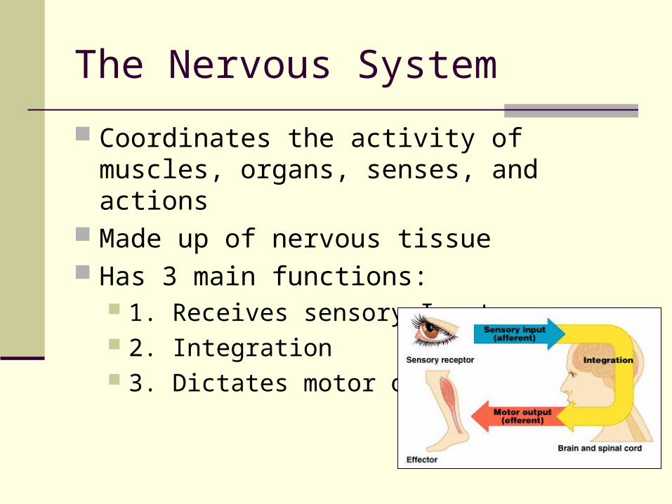
The Nervous System
Coordinates the activity of muscles, organs, senses, and actions
Made up of nervous tissue Has 3 main functions:
1. Receives sensory Input 2. Integration 3. Dictates motor output
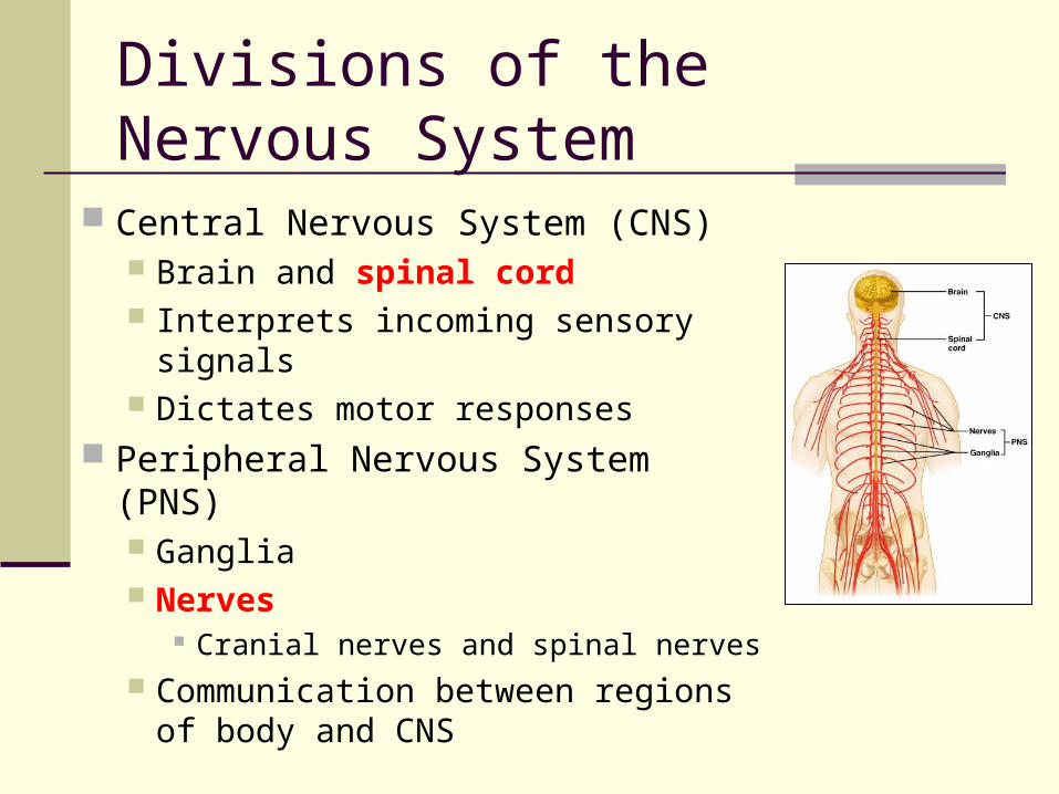
Divisions of the Nervous System
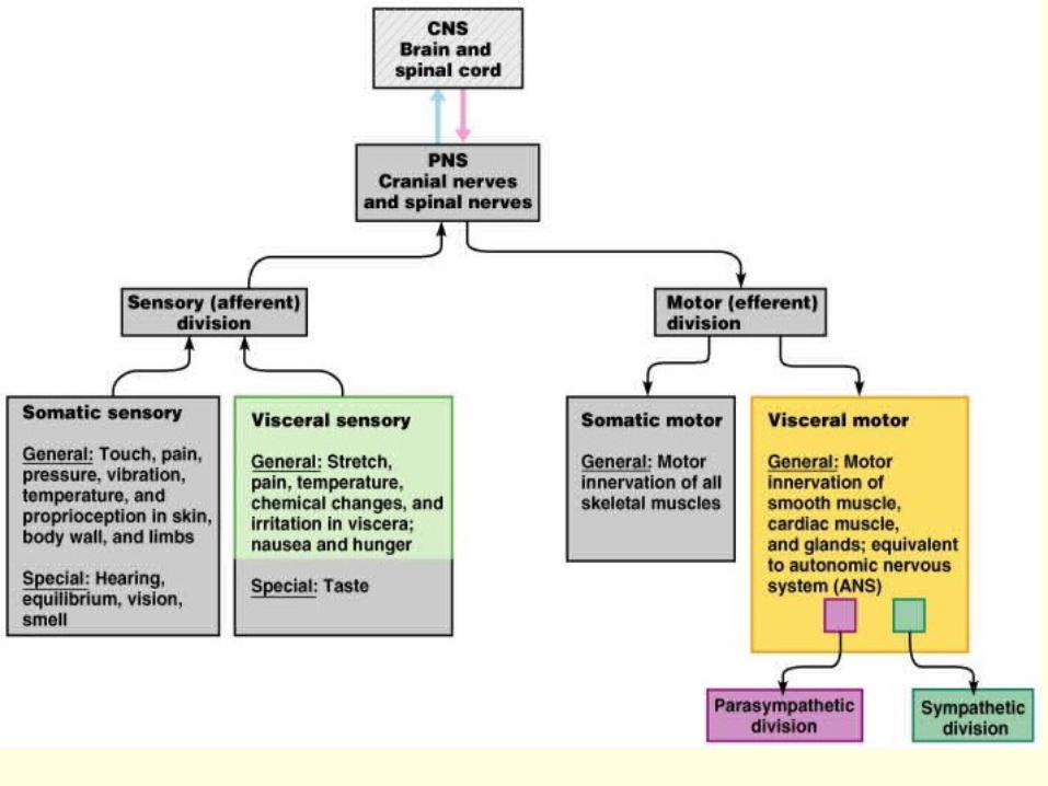
Central Nervous System (CNS) Brain and spinal cord Interprets incoming sensory signals Dictates motor responses
Peripheral Nervous System (PNS) Ganglia Nerves
Cranial nerves and spinal nerves Communication between regions of
body and CNS
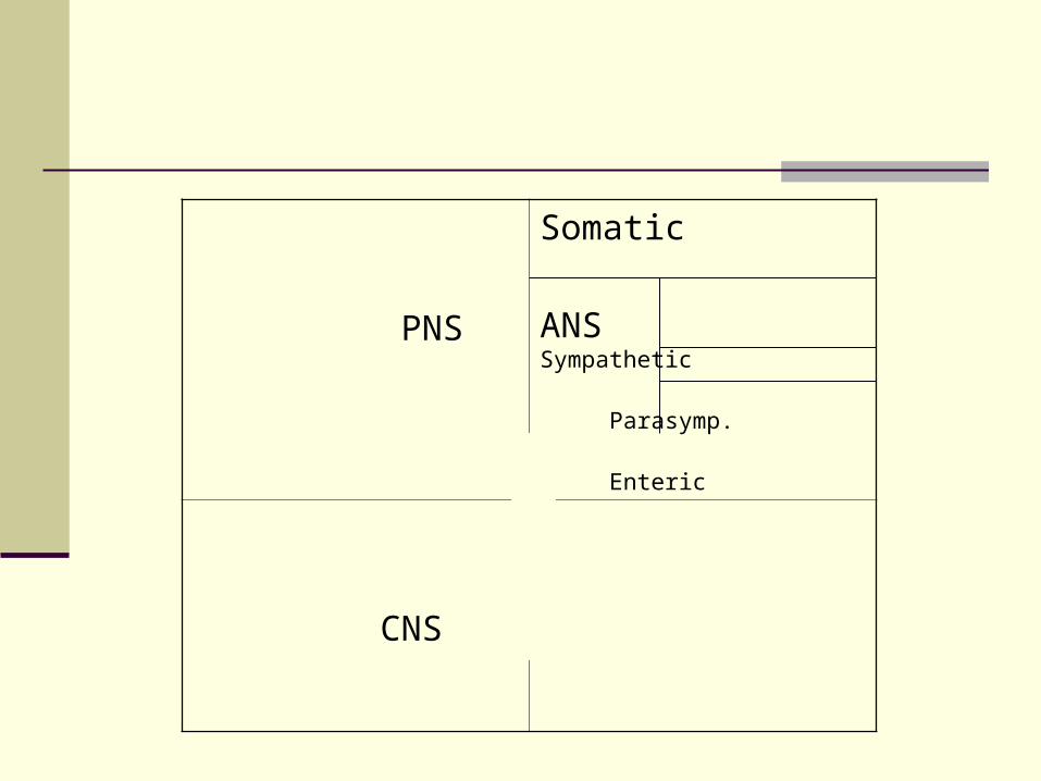
PNS
Somatic
ANS Sympathetic
Parasymp.
Enteric
CNS
PNS
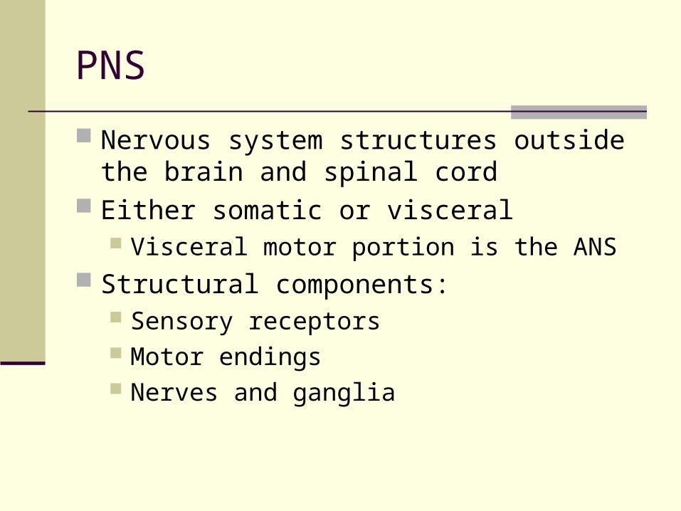
Nervous system structures outside the brain and spinal cord
Either somatic or visceral Visceral motor portion is the ANS
Structural components: Sensory receptors Motor endings Nerves and ganglia
PNS - Nervous Tissue
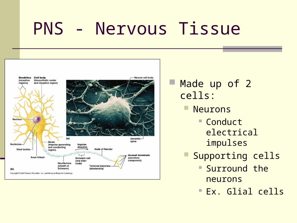
Made up of 2 cells: Neurons
Conduct electrical impulses
Supporting cells Surround the
neurons Ex. Glial cells
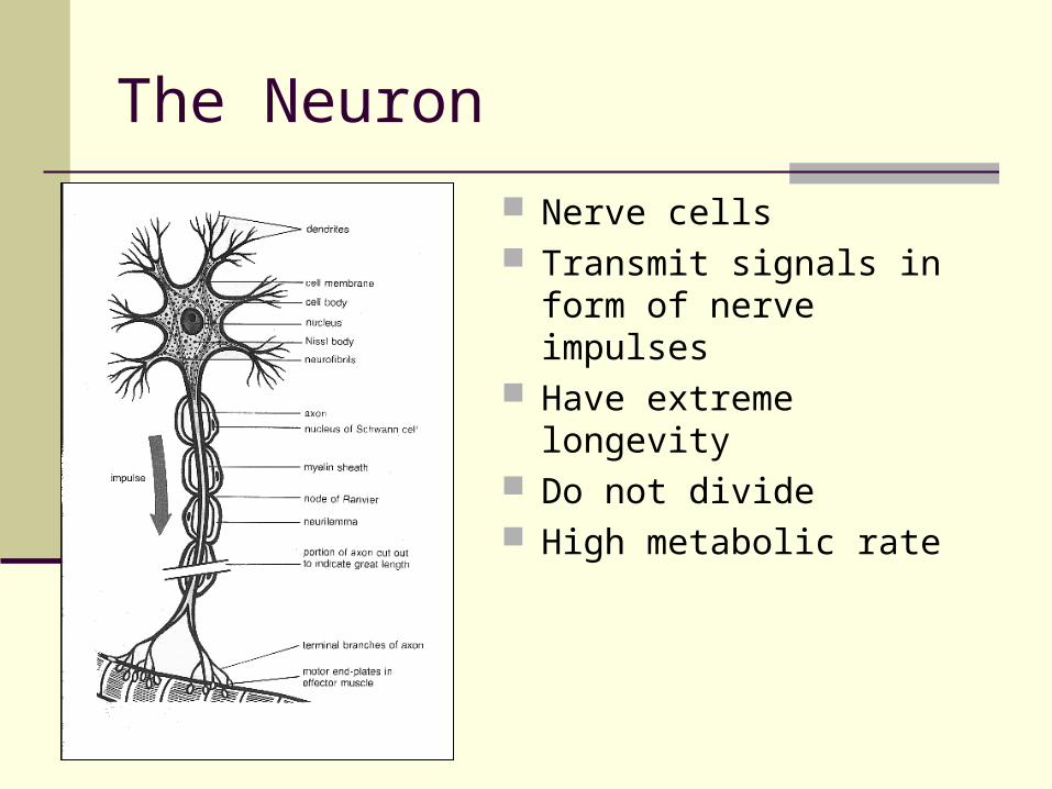
The Neuron
Nerve cells Transmit signals in form of
nerve impulses Have extreme longevity Do not divide High metabolic rate
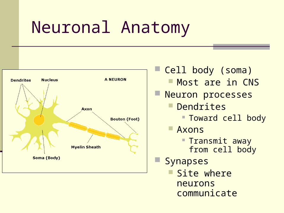
Neuronal Anatomy
Cell body (soma) Most are in CNS
Neuron processes Dendrites
Toward cell body Axons
Transmit away from cell body
Synapses Site where neurons
communicate
Neuronal Anatomy
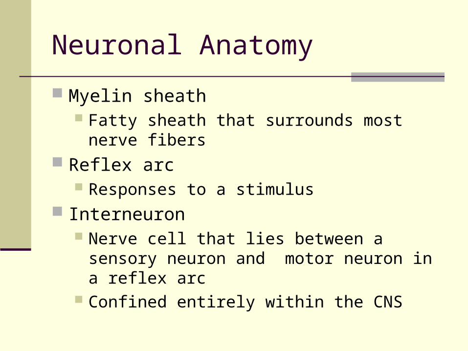
Myelin sheath Fatty sheath that surrounds most nerve fibers
Reflex arc Responses to a stimulus
Interneuron Nerve cell that lies between a sensory neuron
and motor neuron in a reflex arc Confined entirely within the CNS
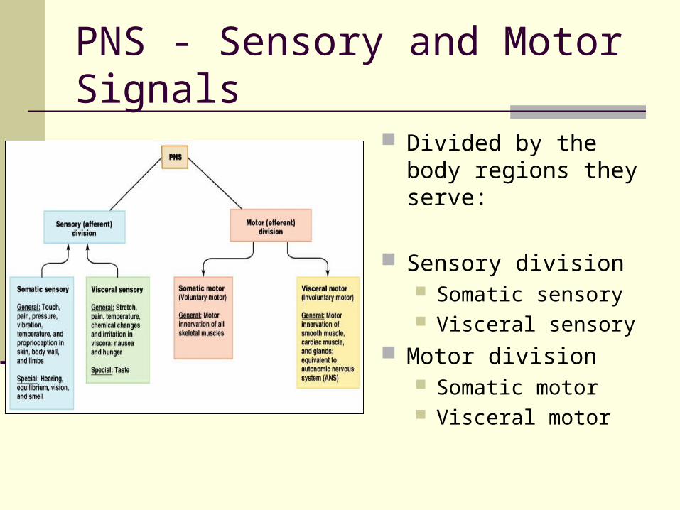
PNS - Sensory and Motor Signals
Divided by the body regions they serve:
Sensory division Somatic sensory Visceral sensory
Motor division Somatic motor Visceral motor
Types of Nerve Signals/Fibers
Sensory (afferent) Picked up by sensory receptors thru body Carried by nerve fibers of PNS into CNS
Motor (efferent) Carried away from the CNS by nerve fibers
into PNS Innervate muscles and glands Causes these organs to contract or secrete
Remember: SAME
Sensory and Motor Signals/Fibers Somatic sensory
Body senses touch, pressure, temperature, vibration of body, muscles
stretching, balance Visceral sensory
Organ senses Stretch, pain, temperature in organs (eg) nausea, hunger, cramps
Somatic motor Body movement Voluntary contraction of skeletal muscles
Visceral motor Organ movement Contraction of smooth muscle, glands = Autonomic Nervous System (involuntary)
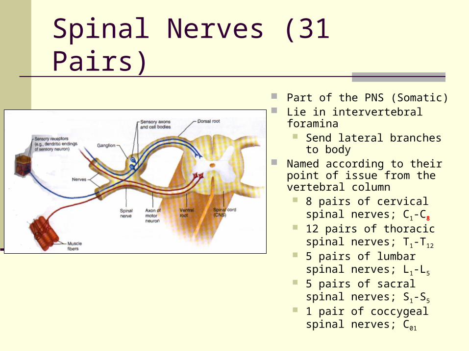
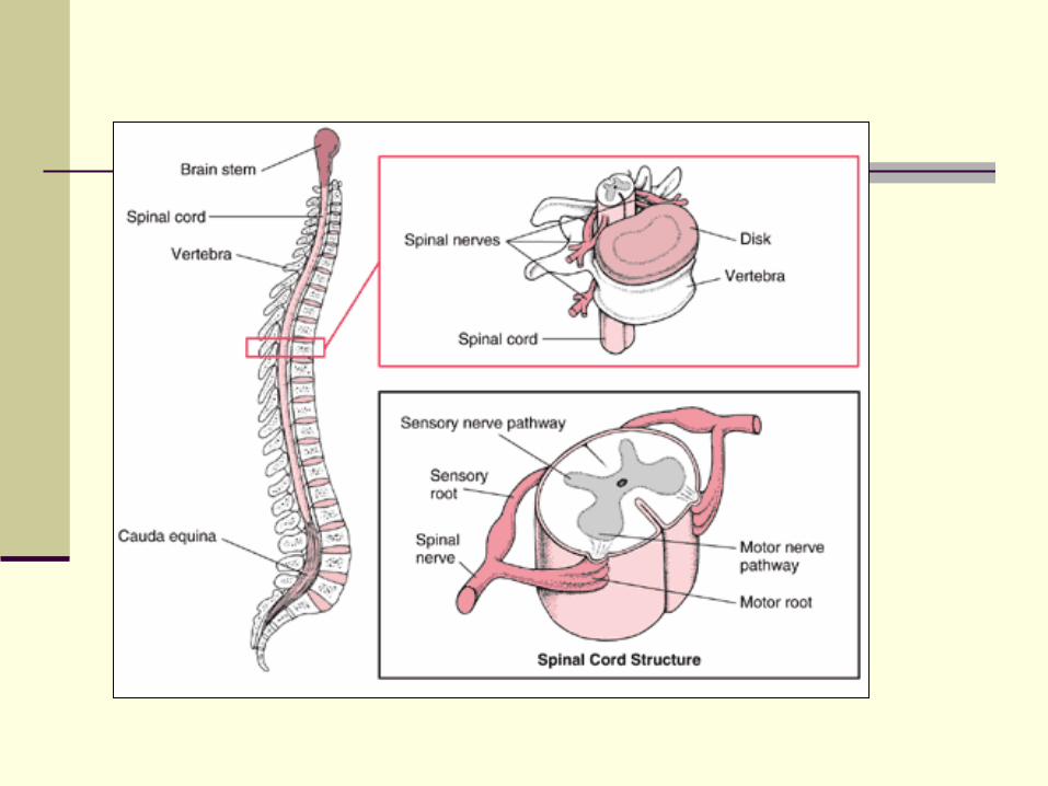
Spinal Nerves (31 Pairs)
Part of the PNS (Somatic) Lie in intervertebral foramina
Send lateral branches to body
Named according to their point of issue from the vertebral column 8 pairs of cervical spinal
nerves; C1-C8
12 pairs of thoracic spinal nerves; T1-T12
5 pairs of lumbar spinal nerves; L1-L5
5 pairs of sacral spinal nerves; S1-S5
1 pair of coccygeal spinal nerves; C01
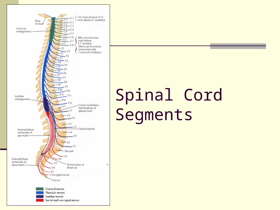
Spinal Cord Segments
Spinal Nerves
Each spinal nerve connected to spinal cord via dorsal (sensory) and ventral (motor) root
Spinal nerves branch into dorsal ramus and ventral ramus Ventral ramus
Connects to rami communicates, which then lead to sympathetic chain ganglia
Supply anterior and lateral regions of the neck, trunk, and limbs
Dorsal ramus Supply the dorsum of the neck and trunk (back)
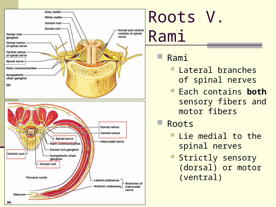
Roots V. Rami
Rami Lateral branches of
spinal nerves Each contains both
sensory fibers and motor fibers
Roots Lie medial to the spinal
nerves Strictly sensory
(dorsal) or motor (ventral)
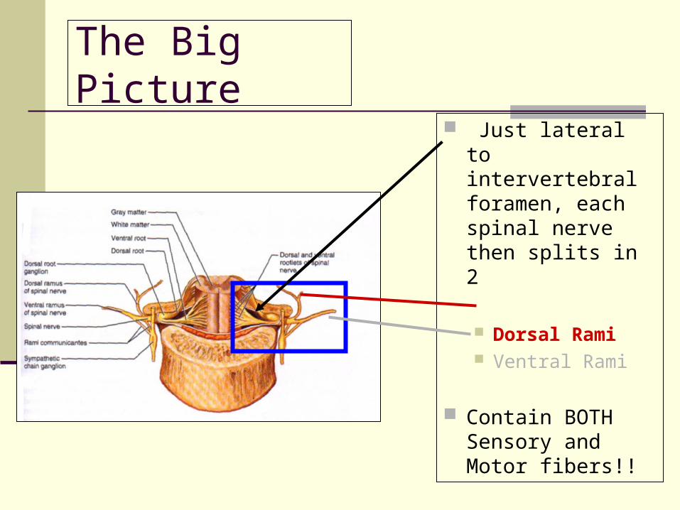
The Big Picture
Just lateral to intervertebral foramen, each spinal nerve then splits in 2
Dorsal Rami Ventral Rami
Contain BOTH Sensory and Motor fibers!!
Autonomic Nervous System
Visceral Motor Function Not easily controlled by will
Get nervous and sweat Innervate smooth muscle, cardiac muscle,
glands Regulate visceral function
Heart rate, blood pressure, digestion, urination Has 2 divisions:
Parasympathetic Sympathetic
ANS
Parasympathetic Enables body to
unwind and calm down Most active when body
at rest Routine maintenance
functions Craniosacral division
Fibers emerge from brain and sacral spinal cord
Sympathetic “fight or flight” Mobilizes the body
during extreme situations
Becomes active when extra metabolic effort needed
Thoracolumbar division
Fibers arise from thoracic and lumbar parts of spinal cord
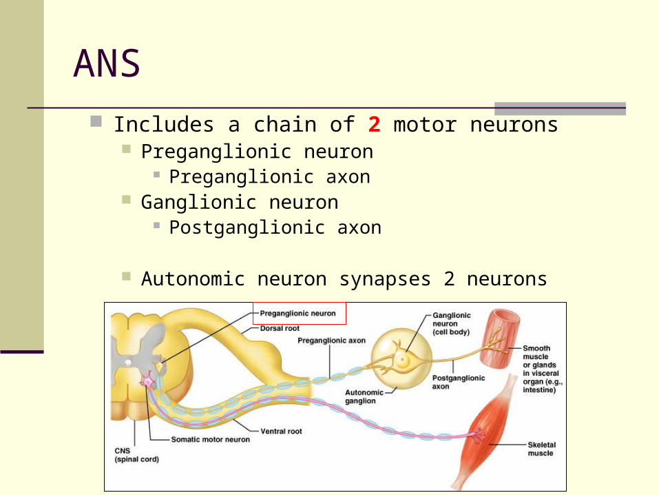
ANS Includes a chain of 2 motor neurons
Preganglionic neuron Preganglionic axon
Ganglionic neuron Postganglionic axon
Autonomic neuron synapses 2 neurons
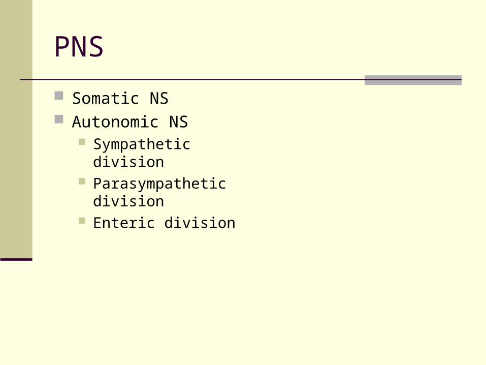
PNS
Somatic NS Autonomic NS
Sympathetic division Parasympathetic
division Enteric division
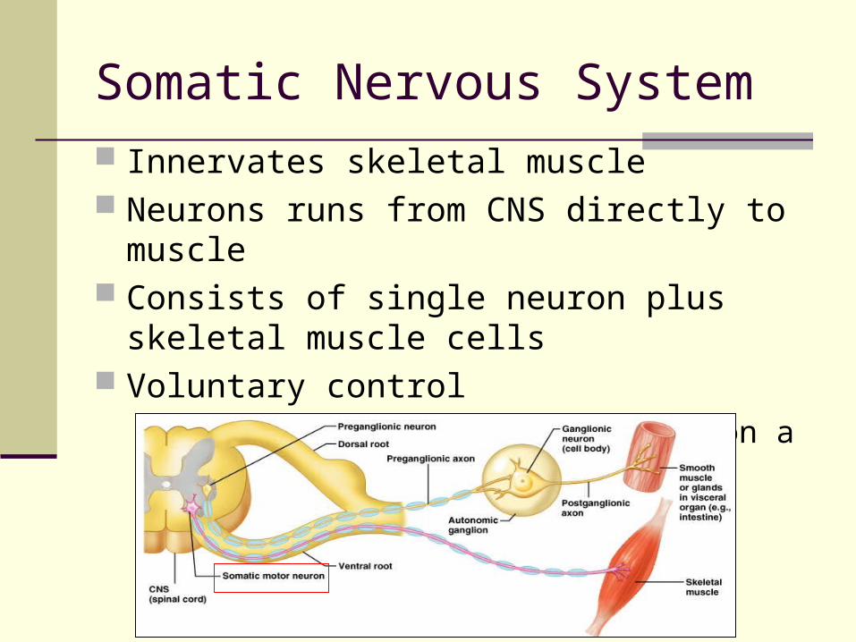
Somatic Nervous System Innervates skeletal muscle Neurons runs from CNS directly to muscle Consists of single neuron plus skeletal
muscle cells Voluntary control
Running, moving limbs, typing on a computer!
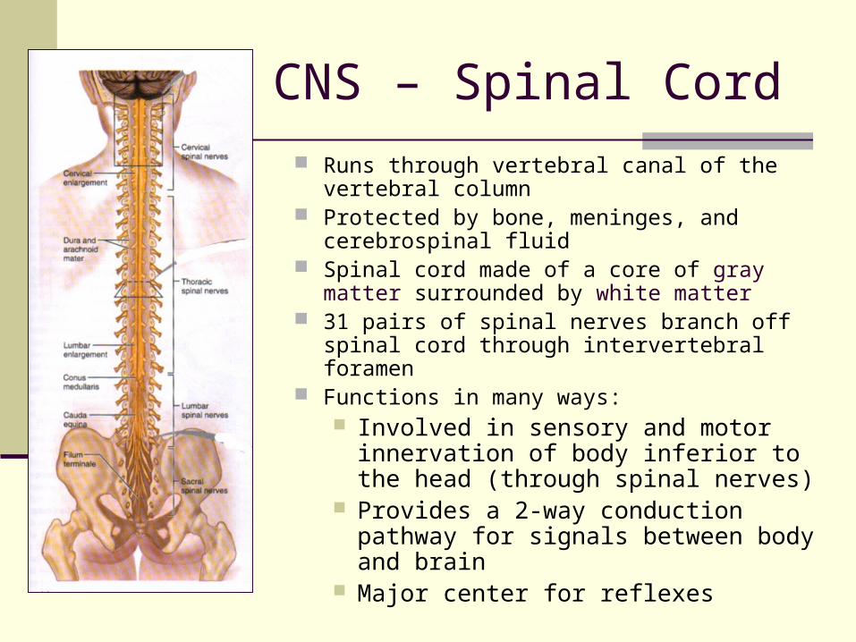
CNS – Spinal Cord
Runs through vertebral canal of the vertebral column
Protected by bone, meninges, and cerebrospinal fluid
Spinal cord made of a core of gray matter surrounded by white matter
31 pairs of spinal nerves branch off spinal cord through intervertebral foramen
Functions in many ways: Involved in sensory and motor
innervation of body inferior to the head (through spinal nerves)
Provides a 2-way conduction pathway for signals between body and brain
Major center for reflexes
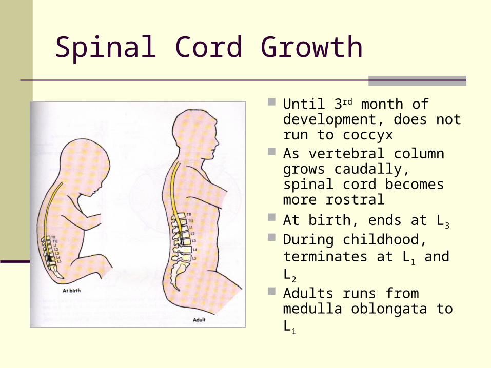
Spinal Cord Growth
Until 3rd month of development, does not run to coccyx
As vertebral column grows caudally, spinal cord becomes more rostral
At birth, ends at L3
During childhood, terminates at L1 and L2
Adults runs from medulla oblongata to L1
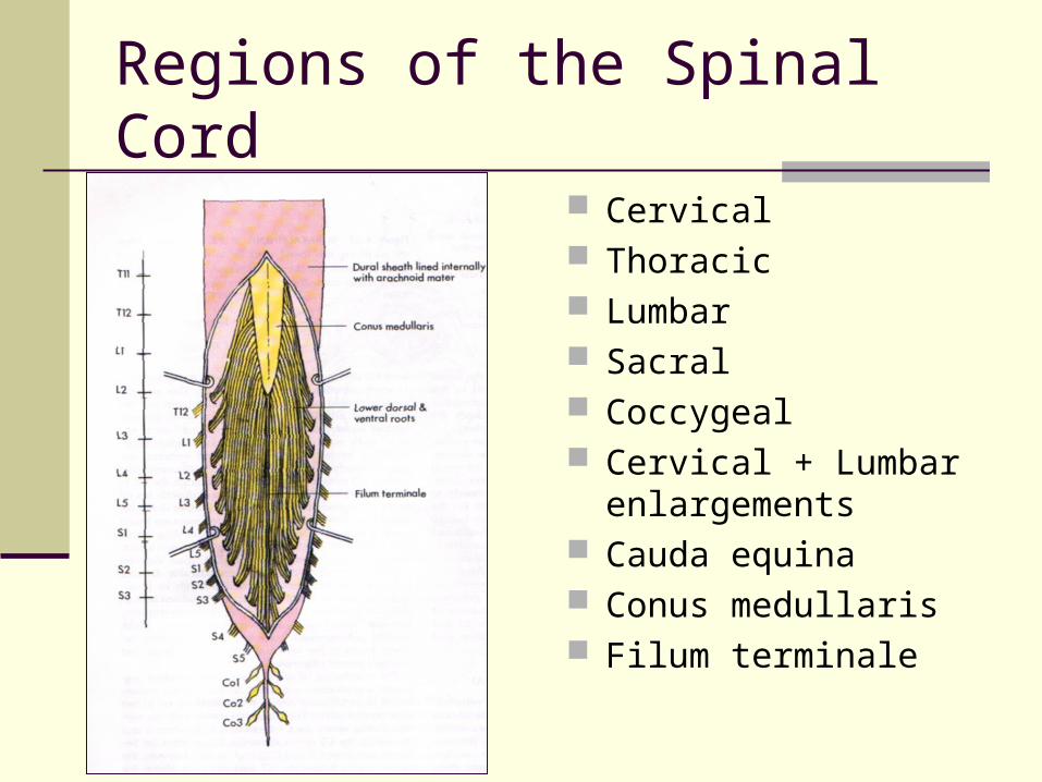
Regions of the Spinal Cord
Cervical Thoracic Lumbar Sacral Coccygeal Cervical + Lumbar
enlargements Cauda equina Conus medullaris Filum terminale
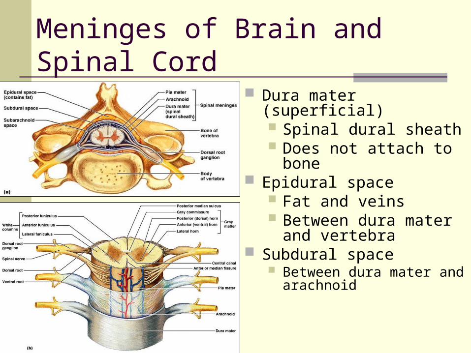
Meninges of Brain and Spinal Cord
Dura mater (superficial) Spinal dural sheath Does not attach to bone
Epidural space Fat and veins Between dura mater and
vertebra Subdural space
Between dura mater and arachnoid
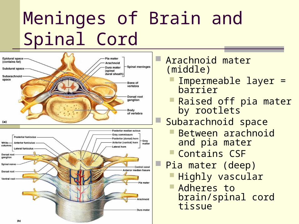
Meninges of Brain and Spinal Cord
Arachnoid mater (middle) Impermeable layer =
barrier Raised off pia mater by
rootlets Subarachnoid space
Between arachnoid and pia mater
Contains CSF Pia mater (deep)
Highly vascular Adheres to brain/spinal
cord tissue
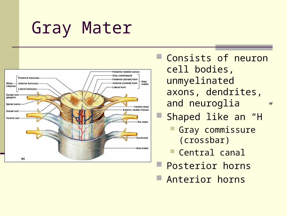
Gray Mater
Consists of neuron cell bodies, unmyelinated axons, dendrites, and neuroglia
Shaped like an “H” Gray commissure
(crossbar) Central canal
Posterior horns Anterior horns
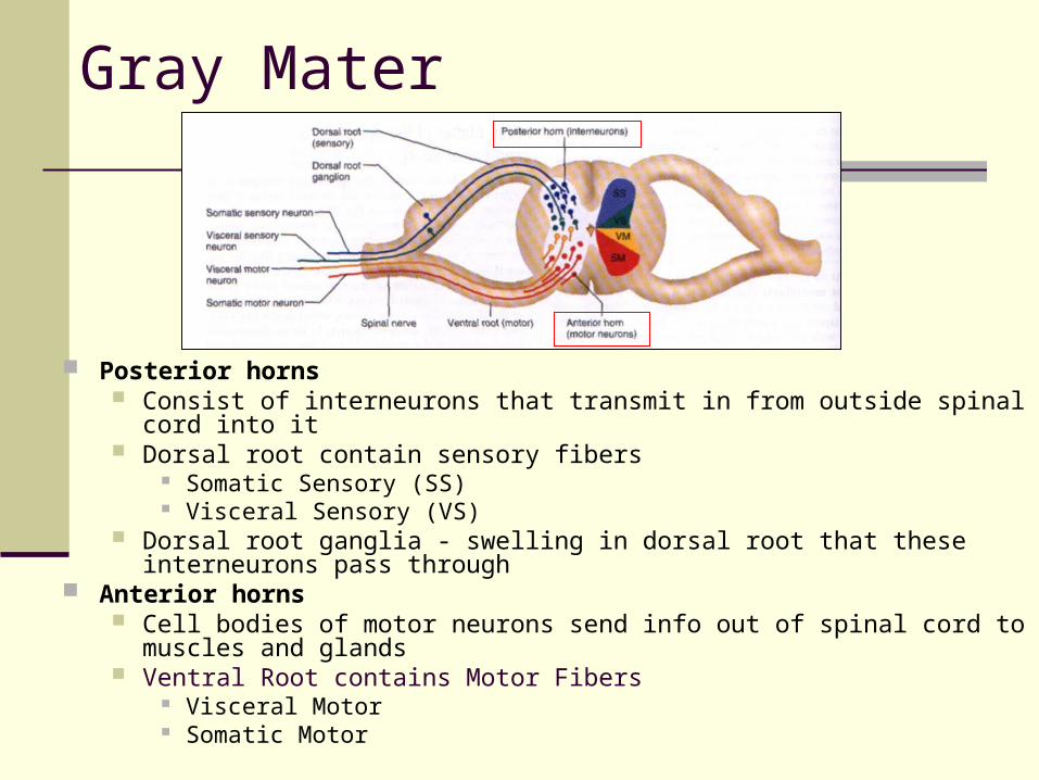
Gray Mater
Posterior horns Consist of interneurons that transmit in from outside spinal cord into it Dorsal root contain sensory fibers
Somatic Sensory (SS) Visceral Sensory (VS)
Dorsal root ganglia - swelling in dorsal root that these interneurons pass through
Anterior horns Cell bodies of motor neurons send info out of spinal cord to muscles and
glands Ventral Root contains Motor Fibers
Visceral Motor Somatic Motor
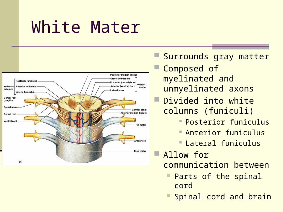
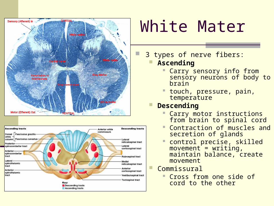
White Mater
Surrounds gray matter Composed of myelinated
and unmyelinated axons Divided into white
columns (funiculi) Posterior funiculus Anterior funiculus Lateral funiculus
Allow for communication between Parts of the spinal cord Spinal cord and brain
White Mater
3 types of nerve fibers: Ascending
Carry sensory info from sensory neurons of body to brain
touch, pressure, pain, temperature Descending
Carry motor instructions from brain to spinal cord
Contraction of muscles and secretion of glands
control precise, skilled movement = writing, maintain balance, create movement
Commissural Cross from one side of cord to the
other
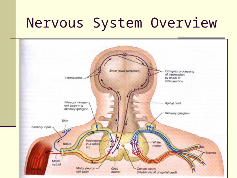
Nervous System Overview