spider phobia is associated with decreased left amygdala volume: a cross-sectional study
TRANSCRIPT

RESEARCH ARTICLE Open Access
Spider phobia is associated with decreased leftamygdala volume: a cross-sectional studyMelanie S Fisler1*, Andrea Federspiel1, Helge Horn1, Thomas Dierks1, Wolfgang Schmitt1, Roland Wiest2,Dominique J-F de Quervain3 and Leila M Soravia1
Abstract
Background: Evidence from animal and human studies imply the amygdala as the most critical structure involvedin processing of fear-relevant stimuli. In phobias, the amygdala seems to play a crucial role in the pathogenesis andmaintenance of the disorder. However, the neuropathology of specific phobias remains poorly understood. In thepresent study, we investigated whether patients with spider phobia show altered amygdala volumes as comparedto healthy control subjects.
Methods: Twenty female patients with spider phobia and twenty age-matched healthy female controls underwentmagnetic resonance imaging to investigate amygdala volumes. The amygdalae were segmented using anautomatic, model-based segmentation tool (FSL FIRST). Differences in amygdala volume were investigated bymultivariate analysis of covariance with group as between-subject factor and left and right amygdala as dependentfactors. The relation between amygdala volume and clinical features such as symptom severity, disgust sensitivity,trait anxiety and duration of illness was investigated by Spearman correlation analysis.
Results: Spider phobic patients showed significantly smaller left amygdala volume than healthy controls. Nosignificant difference in right amygdala volume was detected. Furthermore, the diminished amygdala size inpatients was related to higher symptom severity, but not to higher disgust sensitivity or trait anxiety and wasindependent of age.
Conclusions: In summary, the results reveal a relation between higher symptom severity and smaller left amygdalavolume in patients with spider phobia. This relation was independent of other potential confounders such as thedisgust sensitivity or trait anxiety. The findings suggest that greater spider phobic fear is associated with smaller leftamygdala. However, the smaller left amygdala volume may either stand for a higher vulnerability to develop aphobic disorder or emerge as a consequence of the disorder.
Keywords: Spider phobia, Amygdala, Morphology
BackgroundSpecific phobias are characterized by automatic, exaggeratedfear responses towards phobia-specific objects [1]. Conver-ging evidence implies the amygdala as the most criticalstructure involved in processing of phobia-relevant, but alsogeneral threatening stimuli [2-6]. Through its broadconnections to other brain areas, it might be involved in me-diating automatic responses to potential danger [7]. Due toits projections to the visual stream, it is further likely to
modulate sensory processing [8]. In contrast to large func-tional imaging evidence for the involvement of the amygdalain fear, there have been few reported studies on its structuralabnormality in mood and anxiety disorders [9-12]. Reducedamygdala volume (AMV) has been reported to be signifi-cantly correlated with the severity of distortion in anxiety[13,14] and panic disorder [15]. Structural brain imagingstudies in specific phobia are lacking. This raises the ques-tion whether functional differences in phobic patients ap-pear in association with structural differences. Because ofevidence of hyperactivity and structural differences in anx-iety disorders, we hypothesized that also spider phobicpatients (SP) express reduced AMV compared to healthy
* Correspondence: [email protected] of Psychiatric Neurophysiology, University Hospital ofPsychiatry, University of Bern, Bolligenstrasse 111 3000, Bern 60, SwitzerlandFull list of author information is available at the end of the article
© 2013 Fisler et al.; licensee BioMed Central Ltd. This is an Open Access article distributed under the terms of the CreativeCommons Attribution License (http://creativecommons.org/licenses/by/2.0), which permits unrestricted use, distribution, andreproduction in any medium, provided the original work is properly cited.
Fisler et al. BMC Psychiatry 2013, 13:70http://www.biomedcentral.com/1471-244X/13/70

controls. Phobic fear may be characterized by a combinationof physiological and behavioral components. In order toclarify the contribution of possible confounding factors, sev-eral aspects of phobic disorders that could be critical to thepathogenesis have to be considered. Anxiety-related person-ality traits have been suggested to represent important pre-disposing factors for anxiety-related disorders [16,17]. Otherpotential moderating variables may be the severity and dur-ation of the disease and disgust sensitivity. Therefore, the as-sociation between AMV and several clinical features hasbeen investigated in this study. To the best of our know-ledge, this is the first study investigating volumetricdifferences of the amygdala in SP compared to healthy con-trol subjects.
MethodsSubjectsTwenty female patients with a current diagnosis of spiderphobia and twenty healthy female controls matched for agewere included in the analysis. Subjects were recruited viaadvertisements. The data used for this report has beencollected in a larger project investigating the effect of corti-sol on the outcome of an exposure-based short-term grouptherapy for spider phobia. Because the study design includedan exposure task and in order to minimize motion artifacts,only patients that could keep still while facing a picture of aspider were included in the magnetic resonance paradigm.Exclusion criteria for patients were the following: any axis Iother than specific phobia for spiders, axis II disorders, themanifestation of acute or chronic medical condition, neuro-logical diseases, current drug or alcohol abuse or any contra-indication to magnetic resonance imaging (metallic objects,pregnancy) or confounding factors for structural brain stud-ies (regular medication, contraceptives, left handedness).Healthy control subjects were excluded from the study ifthey met any of the following exclusion criteria: axis I-disorders (measured with the SCL-90-R), the manifestationof acute or chronic medical condition, neurological diseases,current drug or alcohol abuse or any contraindication tomagnetic resonance imaging (metallic objects, pregnancy)or confounding factors for structural brain studies (regularmedication, contraceptives, left handedness). Informed writ-ten consent was obtained from all participants after descrip-tion of the study, which was approved by the ethicscommittee of the Canton of Bern, Switzerland (161/07) inaccordance with the principles of the Declaration of Helsinki[18]. Written informed consent was obtained from the pa-tient for publication of this report and any accompanyingimages. A copy of the written consent is available for reviewby the Editor-in-Chief of this journal.
Diagnostic instruments and questionnairesPatients: Diagnoses for specific phobia for spider wasbased on the Diagnostic and Statistical Manual of Mental
Disorders, fourth edition (DSM-IV) [1]. Specifically, weused a computer-based structured clinical interview (DIA-X) [19] which is based on the Composite InternationalDiagnostic Interview (CIDI) [20]. Patients were screenedfor possible axis II disorders, respectively personality ac-centuation using SKID-II-questionnaire [21]. The SKID-IIis an efficient, user-friendly instrument that helps to makestandardized, reliable, and accurate diagnoses of the 10DSM-IVAxis II personality disorders.Control subjects: SCL-90-R was used as short screening
to exclude Axis-I disorders (such as anxiety disorders anddepression) in the control subjects [22].All subjects filled out the German version of the
Spider Phobia Questionnaire (SPQ) [23] and a question-naire for the assessment of disgust sensitivity (FEE) [24].The SPQ is a validated self-report questionnaire widelyused for assessing spider phobic symptom severity. Itconsists of 31 items which could be answered by “true”or “false” statements. Subjects with a SPQ (range 0-31)score of less than 21 “true” statements were treated ashealthy controls. The FEE is a further development ofthe English Disgust Scale [25]. It is used to measure in-dividual differences in sensitivity to disgust, and toexamine the relationships among different kinds of dis-gust. Humans with a high disgust sensitivity show longerand more intensive disgust reactions than those withlower disgust sensitivity. Disgust sensitivity is treated asa vulnerability factor for the development and mainten-ance of disorders such as phobias. Participants had torate how disgusted they feel when confronted with sev-eral stimuli on a five-points Likert-scale (0 = ‘notdisgusting at all’ to4 = ‘very disgusting’). A total score(range: 0–148) can be calculated which captures a meas-ure for overall disgust sensitivity towards disgust-eliciting stimuli. State (both groups) and trait anxiety(only in patient group) were measured using the Germanversion of the State-Trait Anxiety Inventory (STAI) [26].The STAI consists of two 20-item scales for measuringthe anxiety intensity as an emotional state and individualdifferences in anxiety proneness as a personality trait.The STAI state reports the intensity of anxiety feelingsat the moment of assessment. Responses to the STAItrait items requests subjects to indicate how they gener-ally feel. Both scales range from 20 to 80 points. Hand-edness was assessed via the Edinburgh HandednessInventory (EHI) [27].
Data acquisitionMagnetic resonance imaging was performed on a 3 TSiemens Magnetom Trio Scanner (Erlangen, Germany)equipped with a standard radio-frequency head coil. Forstructural images, a high-resolution 3D T1-weighted im-aging protocol (modified driven equilibrium Fourier trans-form, MDEFT [28]) was used, resulting in 176 sagittal
Fisler et al. BMC Psychiatry 2013, 13:70 Page 2 of 7http://www.biomedcentral.com/1471-244X/13/70
slices of 1.0 mm thickness, 256×256 mm field of view(FOV), and a matrix size of 256×256, resulting in a voxelsize of 1x1x1mm. Further scan parameters were 7.92 msrepetition time (TR) 2.48 ms echo time (TE) and 910 msinversion time (Ti) for an optimal contrast-to-noise ratio(see [28]). Subjects were measured within their luteal phaseof their menstrual cycle, because a study in healthy femalesubjects revealed an increase in AMV during the premen-strual phase compared to the late follicular phase [29].
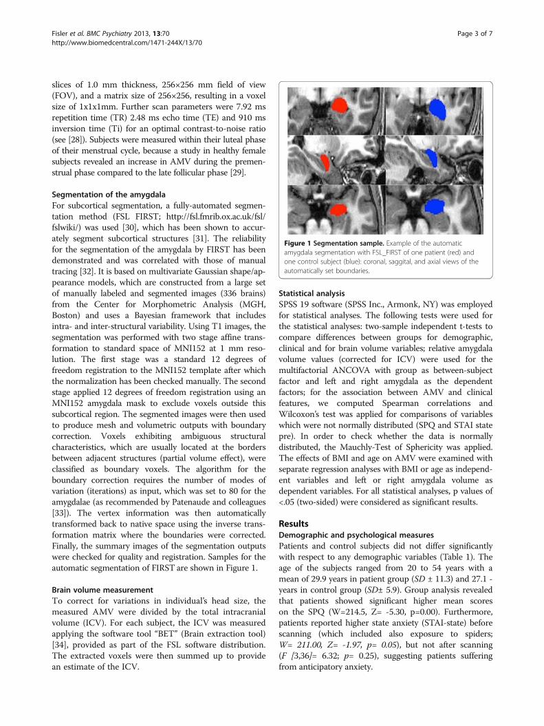
Segmentation of the amygdalaFor subcortical segmentation, a fully-automated segmen-tation method (FSL FIRST; http://fsl.fmrib.ox.ac.uk/fsl/fslwiki/) was used [30], which has been shown to accur-ately segment subcortical structures [31]. The reliabilityfor the segmentation of the amygdala by FIRST has beendemonstrated and was correlated with those of manualtracing [32]. It is based on multivariate Gaussian shape/ap-pearance models, which are constructed from a large setof manually labeled and segmented images (336 brains)from the Center for Morphometric Analysis (MGH,Boston) and uses a Bayesian framework that includesintra- and inter-structural variability. Using T1 images, thesegmentation was performed with two stage affine trans-formation to standard space of MNI152 at 1 mm reso-lution. The first stage was a standard 12 degrees offreedom registration to the MNI152 template after whichthe normalization has been checked manually. The secondstage applied 12 degrees of freedom registration using anMNI152 amygdala mask to exclude voxels outside thissubcortical region. The segmented images were then usedto produce mesh and volumetric outputs with boundarycorrection. Voxels exhibiting ambiguous structuralcharacteristics, which are usually located at the bordersbetween adjacent structures (partial volume effect), wereclassified as boundary voxels. The algorithm for theboundary correction requires the number of modes ofvariation (iterations) as input, which was set to 80 for theamygdalae (as recommended by Patenaude and colleagues[33]). The vertex information was then automaticallytransformed back to native space using the inverse trans-formation matrix where the boundaries were corrected.Finally, the summary images of the segmentation outputswere checked for quality and registration. Samples for theautomatic segmentation of FIRST are shown in Figure 1.
Brain volume measurementTo correct for variations in individual’s head size, themeasured AMV were divided by the total intracranialvolume (ICV). For each subject, the ICV was measuredapplying the software tool “BET” (Brain extraction tool)[34], provided as part of the FSL software distribution.The extracted voxels were then summed up to providean estimate of the ICV.
Statistical analysisSPSS 19 software (SPSS Inc., Armonk, NY) was employedfor statistical analyses. The following tests were used forthe statistical analyses: two-sample independent t-tests tocompare differences between groups for demographic,clinical and for brain volume variables; relative amygdalavolume values (corrected for ICV) were used for themultifactorial ANCOVA with group as between-subjectfactor and left and right amygdala as the dependentfactors; for the association between AMV and clinicalfeatures, we computed Spearman correlations andWilcoxon’s test was applied for comparisons of variableswhich were not normally distributed (SPQ and STAI statepre). In order to check whether the data is normallydistributed, the Mauchly-Test of Sphericity was applied.The effects of BMI and age on AMV were examined withseparate regression analyses with BMI or age as independ-ent variables and left or right amygdala volume asdependent variables. For all statistical analyses, p values of<.05 (two-sided) were considered as significant results.
ResultsDemographic and psychological measuresPatients and control subjects did not differ significantlywith respect to any demographic variables (Table 1). Theage of the subjects ranged from 20 to 54 years with amean of 29.9 years in patient group (SD ± 11.3) and 27.1 -years in control group (SD± 5.9). Group analysis revealedthat patients showed significant higher mean scoreson the SPQ (W=214.5, Z= -5.30, p=0.00). Furthermore,patients reported higher state anxiety (STAI-state) beforescanning (which included also exposure to spiders;W= 211.00, Z= -1.97, p= 0.05), but not after scanning(F [3,36]= 6.32; p= 0.25), suggesting patients sufferingfrom anticipatory anxiety.
Figure 1 Segmentation sample. Example of the automaticamygdala segmentation with FSL_FIRST of one patient (red) andone control subject (blue): coronal, saggital, and axial views of theautomatically set boundaries.
Fisler et al. BMC Psychiatry 2013, 13:70 Page 3 of 7http://www.biomedcentral.com/1471-244X/13/70

Amygdala volumeMultifactorial ANCOVA of AMV, adjusted for ICV,showed approximately 13% smaller AMV in patients onthe left than in controls, resulting in a significant betweengroup effect for left AMV (F [3,36]= 6.39; p= 0.02;Figure 2a). Separate regression analyses indicated that thedifference in left AMV between patients and controls werenot accounted for by differences in age (F= 1.72; df= 36,p=0.20) and BMI (F= 0.55, df= 36, p= 0.47). There was nosignificant difference of right AMV between patients andcontrols (F [3,36] = 2.28; p= 0.20; Figure 2b). Within thewhole group, SPQ scores were negatively correlated withleft AMV (r=-0.47; p=0.005; Figure 3). Separate regressionanalyses showed that smaller left amygdala occurred inde-pendently of age (r=-0.046; p=0.79) and BMI (r=-0.143;p=0.42). Correlations between left amygdala and the dis-gust score (FEE) or state anxiety (STAI trait/state) didnot reach significance, nor were there any significant
correlations between right AMV and clinical scores withinthe whole sample. Within the phobic sample, trait anxietydid not correlate with AMV. Thus, differences in AMVdid not appear to be due to age or BMI or clinical featuresof the participants.
DiscussionThe purpose of this study was to determine whether thereis evidence for AMV differences in patients with spiderphobia compared to healthy controls. As hypothesized,the findings confirm a reduction in left AMV of patients.Furthermore, the reduced left AVM is associated with theseverity of spider phobic symptoms. Our finding of abnor-mal left AMV in patients are consistent with findings thatreport diminished amygdala size in fear and anxiety-related disorders [10,35-40]. We suggest that the auto-matic manifestation of fear responses is mediated by a
Table 1 Demographic and clinical characteristics of the patients and their control group
Patients (n=20) Controls (n=20)
Characteristics Mean SD Mean SD df t p Equality ofvariance
Age (years) 29.9 11.3 27.1 5.9 38 −0.98 0.33 0.91
Age at onset 6.65 3.28 ————— ————— ————— ————— ————— ——————
Duration of illness (years) 22.14 13.01 ————— ————— ————— ————— ————— —————
Handedness scores 9.7 0.58 9.8 0.41 38 0.65 0.52 0.98
SPQ 21.55 4.29 6.1 3.83 38 −11.99 0.00 0.00
FEE 85.3 22.93 73 26.42 38 −1.47 0.15 0.17
STAI state pre / post scanning 39.85/ 34.85 10.67/ 12.9 32.5/ 30.53 4.05/ 6.61 38 −2.51/ -1.18 0.05/ 0.24 0.05/ 0.54
STAI trait 44.8 6.89 ————— ————— ————— ————— ————— —————
BMI 21.8 3.90 21.63 2.31 38 −1.18 0.87 0.61
SPQ: Spider Phobia Questionnaire; FEE: disgust sensitivity scale; BMI: body mass index.
Figure 2 Amygdala volumes of spider phobic patients and healthy controls. The central box shows the data between 25th and 75th
quartiles, with the median represented by the line. The whiskers extend from the upper and lower quartiles to a distance of 1.5 interquartilerange (IQR). Circles represent the outliers over 3 IQR below the 25th or above the 75th quartile. Relative amygdala volumes were calculated by thefollowing formula: 100x(absolute amygdala volume in mm2/intracranial volume in mm2). a: boxplot comparing relative left amygdala volume;b: boxplot comparing relative right amygdala volume.
Fisler et al. BMC Psychiatry 2013, 13:70 Page 4 of 7http://www.biomedcentral.com/1471-244X/13/70
potentiated fear network which may be associated with anamygdala deficiency.However, the differences of AMV could be also
influenced by clinical factors. The present findings showan association between left AMV and symptom severity inphobic patients in the way that the smaller the left amyg-dala, the more phobic symptoms they reported. Thisfinding seems to be attributed to spider phobia, becausewe selected patients with pure spider phobia without anycomorbidities and no other potential confounding factorhas shown to be associated with this volume reduction inspider phobic patients. It is worth remarking that findingsof smaller left AMV, as reported in this study, suggest thatabnormal functioning of this structure may underlie thesymptoms of automatic and exaggerated fear responseobserved in specific phobias, as measured with the SPQ,which reliably assesses fear of spiders [41,42].Hemispheric differences in amygdala alterations found
in this study may potentially be in line with differentroles in emotion processing for the left and right amyg-dala which has been found in several studies investigat-ing mood and anxiety disorders [39,43]. Based on thesestudies, it is assumed that the left amygdala is moreinvolved in processing sustained stimulus evaluationwhile the right one is more involved in rapid and undif-ferentiated processing of emotional stimuli [44].Some limitations of the present study need to be
mentioned. We only studied female subjects; therefore theresults cannot be generalized to men. We should furthermention the different diagnostic measurements for thecontrol and patient group. The patient group was add-itionally screened for possible personality disorders(SKID-II) and trait-anxiety (STAI-trait). Whereas the con-trol subjects were only screened for possible axisI-disorders (SCL-90-R) which implied exclusion of thestudy. Additionally, the sample size was modest and
should be extended. However, the patient group can beconsidered to be homogenous, as patients did not sufferfrom any other axis I disorders at the time of assessment.Hence, future investigations should include comparisonsof amygdala morphology in various types and degrees ofphobic disorders for a better pathophysiological distinc-tion. Longitudinal and pre-post treatment studies shouldclarify the meaning of the observed amygdala differencesover time.
ConclusionStill, the reasons for volumetric differences are so farlargely unclear. Two possible interpretations might beoffered: First, smaller left AMV might be a vulnerabilityfactor for the development of spider phobia. The secondinterpretation relates to experience or exposure-relatedstructural modifications within the amygdala. Whetherthe observed atrophy of the amygdala in mood and anx-iety disorders is progressive and already present at time ofdisease onset or develops as a result of damage secondaryto higher amygdala activity is a matter of debate [45,46].
AbbreviationsAMV: Amygdala volume; ANCOVA: Analysis of covariance; BMI: Body massindex; EHI: Edinburgh handedness inventory; FEE: Questionnaire for theassessment of disgust sensitivity; FOV: Field of view; ICV: Intracranial volume;SD: Standard deviation; SP: Spider phobic patients; SKID-II: Structured clinicalinterview for DSM-IV Axis II disorders; SPQ: Spider phobia questionnaire;STAI: State-trait anxiety inventory.
Competing interestsThe authors declare that they have no competing interests.
Authors’ contributionMF participated in the design of the study, carried out the measurements,performed the statistical analysis and drafted the manuscript. AF carried outthe sequence alignment and revised the manuscript critically. HHparticipated in the diagnostic interview and revised the manuscript critically.TD revised the manuscript critically. WS participated in the diagnosticinterviews. DQ made the conception and design of the study and revised
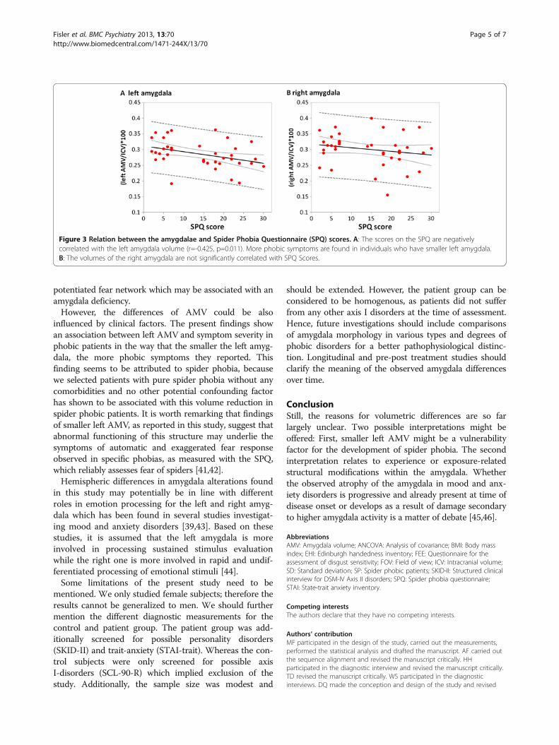
Figure 3 Relation between the amygdalae and Spider Phobia Questionnaire (SPQ) scores. A: The scores on the SPQ are negativelycorrelated with the left amygdala volume (r=-0.425, p=0.011). More phobic symptoms are found in individuals who have smaller left amygdala.B: The volumes of the right amygdala are not significantly correlated with SPQ Scores.
Fisler et al. BMC Psychiatry 2013, 13:70 Page 5 of 7http://www.biomedcentral.com/1471-244X/13/70
the manuscript critically. LS participated in the design of the study andrevised the manuscript. All authors read and approved the final manuscript.
AcknowledgementsThis work was funded by a grant from the Swiss National Science foundation(32003B_124947) and a grant from the Medical Faculty of the University ofBerne (520.10). We thank B.Sc. Joëlle Witmer, lic. phil. Yvonne Renevey, M.Sc.Céline de Buman, B.Sc. Susanne Hess, B.Sc. Basil Preisig, B.Sc. Vera Bamert, B.Sc. Esther Mahlstein, B.Sc. Isabelle Zogg and B.Sc. Veryan Thommen forresearch assistance.
Author details1Department of Psychiatric Neurophysiology, University Hospital ofPsychiatry, University of Bern, Bolligenstrasse 111 3000, Bern 60, Switzerland.2Diagnostic and Interventional Neuroradiology, Inselspital and University ofBern, OP-Ost C215, Bern 3010, Switzerland. 3Division of CognitiveNeuroscience, Faculty of Medicine & Faculty of Psychology, University ofBasel, Birmannsgasse 8 4055, Basel, Switzerland.
Received: 18 May 2012 Accepted: 20 February 2013Published: 26 February 2013
References1. American Psychiatric Association: Diagnostic and statistical manual of
mental disorders (4th ed., text rev.). Washington, DC: Author.) 2000:188–190.2. Ohman A, Mineka S: Fears, phobias, and preparedness: toward an
evolved module of fear and fear learning. Psychol Rev 2001,108(3):483–522.
3. Schienle A, Schäfer A, Walter B, Stark R, Vaitl D: Brain activation of spiderphobics towards disorder-relevant, generally disgust- and fear-inducingpictures. Neurosci Lett 2005, 388(1):1–6.
4. Straube T, Mentzel H-J, Miltner WHR: Neural mechanisms of automaticand direct processing of phobogenic stimuli in specific phobia. BiolPsychiatry 2006, 59(2):162–170.
5. Etkin A, Wager TD: Functional neuroimaging of anxiety: a meta-analysisof emotional processing in PTSD, social anxiety disorder, and specificphobia. Am J Psychiatry 2007, 164(10):1476–1488.
6. Phelps EA, LeDoux JE: Contributions of the amygdala to emotionprocessing: from animal models to human behavior. Neuron 2005,48(2):175–187.
7. Veening JG, Swanson LW, Sawchenko PE: The organization of projectionsfrom the central nucleus of the amygdala to brainstem sites involved incentral autonomic regulation: a combined retrograde transport-immunohistochemical study. Brain Res 1984, 303(2):337–357.
8. Freese JL, Amaral DG: The organization of projections from the amygdalato visual cortical areas TE and V1 in the macaque monkey. J Comp Neurol2005, 486(4):295–317.
9. Drevets W, Price J, Furey M: Brain structural and functional abnormalitiesin mood disorders: implications for neurocircuitry models of depression.Brain Struct Funct 2008, 213(1):93–118.
10. Anand A, Shekhar A: Brain imaging studies in mood and anxietydisorders. Ann N Y Acad Sci 2003, 985(1):370–388.
11. Drevets WC: Neuroimaging abnormalities in the amygdala in mooddisorders. Ann N Y Acad Sci 2003, 985(1):420–444.
12. Brambilla P, Barale F, Caverzasi E, Soares JC: Anatomical MRI findings inmood and anxiety disorders. Epidemiol Psichiatr Soc 2002, 11(2):88–99.
13. Blackmon K, Barr WB, Carlson C, Devinsky O, Dubois J, Pogash D, Quinn BT,Kuzniecky R, Halgren E, Thesen T: Structural evidence for involvement of aleft amygdala-orbitofrontal network in subclinical anxiety. Psychiatry Res2011, 194(3):296–303.
14. Spampinato MDM, Wood PDJ, De Simone MDV, Grafman PDJ: Neuralcorrelates of anxiety in healthy volunteers: a voxel-based morphometrystudy. J Neuropsychiatry Clin Neurosci 2009, 21(2):199–205.
15. Hayano F, Nakamura M, Asami T, Uehara K, Yoshida T, Roppongi T, Otsuka T,Inoue T, Hirayasu Y: Smaller amygdala is associated with anxiety inpatients with panic disorder. Psychiatry Clin Neurosci 2009, 63(3):266–276.
16. Clark LA, Watson D, Mineka S: Temperament, personality, and the moodand anxiety disorders. Journal of Abnormal Psychology J Abnorm Psychol1994, 103(1):103–116.
17. Chambers JA, Power KG, Durham RC: The relationship between traitvulnerability and anxiety and depressive diagnoses at long-term follow-up of generalized anxiety disorder. J Anxiety Disord 2004, 18(5):587–607.
18. Rickham PP: Human experimentation. Code of ethics of the worldmedical association. Declaration of helsinki. Br Med J 1964, 2(5402):177. 2.
19. Essau CA, Wittchen HU, Pfister H: DIA-X-Interview. Diagnostica 1999,45(3):163–164.
20. Rubio-Stipec M, Bravo M, Canino G: [The composite internationaldiagnostic interview (CIDI): an epidemiologic instrument suitable forusing in conjunction with different diagnostic systems in differentcultures]. Acta Psiquiatr Psicol Am Lat 1991, 37(3):191–204.
21. Fydrich T, Renneberg B, Schmitz B, Wittchen H-U: SKID-II: strukturiertesklinisches interview für DSM-IV, achse II: persönlichkeitsstörungen. Göttingen:Hogrefe; 1997.
22. Franke G, Stacker KH: Reliability and validity of the symptom checklist(SCL-90-R, derogatis, 1986) in standardized versus homogenous item-blocked sequence. Diagnostica 1995, 41(4):349–373.
23. Klorman R, Weerts TC, Hastings JE, Melamed BG, Lang PJ: Psychometricdescription of some specific-fear questionnaires. Behav Ther 1974,5(3):401–409.
24. Schienle A, Walter B, Stark R, Vaitl D: Ein fragebogen zur erfassung derekelempfindlichkeit (FEE). Z Kl Psych Psychoth 2002, 31(2):110–120.
25. Olatunji BO, Cisler JM, Deacon BJ, Connolly K, Lohr JM: The disgustpropensity and sensitivity scale-revised: psychometric properties andspecificity in relation to anxiety disorder symptoms. J Anxiety Disord 2007,21(7):918–930.
26. Spielberger CD, Gorsuch RL, Lushene RE: Manual for state-trait anxietyinventory (self-evaluation questionnaire). Palo Alto, CA: ConsultingPsychologists Press; 1970.
27. Oldfield RC: The assessment and analysis of handedness: the Edinburghinventory. Neuropsychologia 1971, 9(1):97–113.
28. Deichmann R, Schwarzbauer C, Turner R: Optimisation of the 3D MDEFTsequence for anatomical brain imaging: technical implications at 1.5 and3 T. NeuroImage 2004, 21(2):757–767.
29. Ossewaarde L, van Wingen GA, Kooijman SC, Backstrom T, Fernandez G,Hermans EJ: Changes in functioning of mesolimbic incentive processingcircuits during the premenstrual phase. Soc Cogn Affect Neur 2011,6(5):612–620.
30. Patenaude B: Bayesian statistical models of shape and appearance for subcortical brain segmentation. Oxford: University of Oxford; 2007.
31. Babalola K, Patenaude B, Aljabar P, Schnabel J, Kennedy D, Crum W, SmithS, Cootes T, Jenkinson M, Rueckert D, et al: Comparison and evaluation ofsegmentation techniques for subcortical structures in brain MRI medical imagecomputing and computer-assisted intervention – MICCAI 2008, VolumeVolume 5241. Heidelberg: Springer Berlin; 2008:409–416.
32. Morey RA, Petty CM, Xu Y, Pannu Hayes J, Wagner Ii HR, Lewis DV, LaBar KS,Styner M, McCarthy G: A comparison of automated segmentation andmanual tracing for quantifying hippocampal and amygdala volumes.NeuroImage 2009, 45(3):855–866.
33. Patenaude B, Smith SM, Kennedy DN, Jenkinson M: A Bayesian model ofshape and appearance for subcortical brain segmentation. NeuroImage2011, 56(3):907–922.
34. Smith SM: Fast robust automated brain extraction. Hum Brain Mapp 2002,17(3):143–155.
35. Milham MP, Nugent AC, Drevets WC, Dickstein DS, Leibenluft E, Ernst M,Charney D, Pine DS: Selective reduction in amygdala volume in pediatricanxiety disorders: a voxel-based morphometry investigation.Biol Psychiatry 2005, 57(9):961–966.
36. Szeszko PR, Robinson D, Alvir JMJ, Bilder RM, Lencz T, Ashtari M, Wu H,Bogerts B: Orbital frontal and amygdala volume reductions in obsessive-compulsive disorder. Arch Gen Psychiatry 1999, 56(10):913–919.
37. MacMaster FP, Mirza Y, Szeszko PR, Kmiecik LE, Easter PC, Taormina SP,Lynch M, Rose M, Moore GJ, Rosenberg DR: Amygdala and hippocampalvolumes in familial early onset major depressive disorder. Biol Psychiatry2008, 63(4):385–390.
38. Sheline YI, Barch DM, Price JL, Rundle MM, Vaishnavi SN, Snyder AZ, MintunMA, Wang S, Coalson RS, Raichle ME: The default mode network and self-referential processes in depression. Proc Natl Acad Sci USA 2009,106(6):1942–1947.
39. Pearlson GD, Barta PE, Powers RE, Menon RR, Richards SS, Aylward EH,Federman EB, Chase GA, Petty RG, Tien AY: Medial and superior temporal
Fisler et al. BMC Psychiatry 2013, 13:70 Page 6 of 7http://www.biomedcentral.com/1471-244X/13/70
gyral volumes and cerebral asymmetry in schizophrenia versus bipolardisorder. Biol Psychiatry 1997, 41(1):1–14.
40. Massana G, Serra-Grabulosa JM, Salgado-Pineda P, Gasto C, Junque C,Massana J, Mercader JM, Gomez B, Tobena A, Salamero M: Amygdalaratrophy in panic disorder patients detected by volumetric magneticresonance imaging. NeuroImage 2003, 19(1):80–90.
41. Rinck M, Bundschuh S, Engler S, Muller A, Wissmann J, Ellwart T, Becker ES:Reliability and validity of German versions of three instrumentsmeasuring fear of spiders. Diagnostica 2002, 48(3):141–149.
42. Muris P, Merckelbach H: A comparison of two spider fear questionnaires.J Behav Ther Exp Psychiatry 1996, 27(3):241–244.
43. Sheline YI, Sanghavi M, Mintun MA, Gado MH: Depression duration butnot age predicts hippocampal volume loss in medically healthy womenwith recurrent major depression. J Neurosci 1999, 19(12):5034–5043.
44. Straube T, Mentzel HJ, Miltner WH: Neural mechanisms of automatic anddirect processing of phobogenic stimuli in specific phobia. Biol Psychiatry2006, 59(2):162–170.
45. Hettema JM, Kettenmann B, Ahluwalia V, McCarthy C, Kates WR, Schmitt JE,Silberg JL, Neale MC, Kendler KS, Fatouros P: Pilot multimodal twinimaging study of generalized anxiety disorder. Depress Anxiety 2012,29(3):202–209.
46. van der Plas EA, Boes AD, Wemmie JA, Tranel D, Nopoulos P: Amygdalavolume correlates positively with fearfulness in normal healthy girls.Soc Cogn Affect Neurosci 2010, 5(4):424–431.
doi:10.1186/1471-244X-13-70Cite this article as: Fisler et al.: Spider phobia is associated withdecreased left amygdala volume: a cross-sectional study. BMC Psychiatry2013 13:70.
Submit your next manuscript to BioMed Centraland take full advantage of:
• Convenient online submission
• Thorough peer review
• No space constraints or color figure charges
• Immediate publication on acceptance
• Inclusion in PubMed, CAS, Scopus and Google Scholar
• Research which is freely available for redistribution
Submit your manuscript at www.biomedcentral.com/submit
Fisler et al. BMC Psychiatry 2013, 13:70 Page 7 of 7http://www.biomedcentral.com/1471-244X/13/70