some aspects of the phase behaviour of charged lipids
TRANSCRIPT
Biochimica etBiophysica Acta, 772 (1984) 37-50 37 Elsevier
BBA72063
SOME ASPECTS OF THE PHASE BEHAVIOUR OF CHARGED LIPIDS
HELMUT HAUSER
Laboratorium fur Biochemie, Eidgen~ssische Technische Hochschule Z~rich, CH 8092 Zhrich (Switzerland)
(Received September 15th, 1983)
Key words: Lipid swelling" Phase behavior; Charged lipid; X-ray diffraction; Electron microscopy
Based on their swelling in HzO iipids forming smectic phases may be grouped into two classes: (i) uncharged or isoelectric lipids which show no or limited swelling in H zO (the thickness of the water layer is less than or equal to 25 A); (ii) lipids with a net charge, which show continuous swelling. Up to a certain threshold concentration all the water is intercalated between the lipid bilayers: there is a single lamellar, swelling phase. Above this concentration two phases coexist: fully-hydrated unilamellar vesicles and excess water. Continu- ous swelling and the formation of unilamellar vesicles in excess water is observed with pure charged lipids and lipid mixtures. It is important to note that neutral bilayers doped with only a few percent of a positively or negatively charged lipid exhibit this behaviour. The continuous swelling is a purely electrostatic phenomenon as indicated by the reversible interconversion between continuous and limited swelling be- haviou~.
in the one-phase domain:
multilamellar
continuous swelling
excess counterion
removal of counterion
multilamellar
limited swelling
in the two-phase domain:
large unilamellar vesicles
excess counterion
removal of counterion
multilamellar structures
Introduction
The phase behaviour of phospholipids has been the subject of extensive studies in the past. The justification for this is at least two-fold: firstly, phospholipids have been shown to form integral structures of biological membranes, and the ap- preciation of their structural and functional role in
biological membranes requires a good understand- ing of their physico-chemical properties, particu- larly their phase behaviour. It is now generally accepted that the bulk of the phospholipid in biological membranes is present as a bilayer. Based upon this concept phospholipid bilayers have been widely used as model membranes and their physico-chemical properties have been studied ex-
0005-2736/84/$03.00 © 1984 Elsevier Science Publishers B.V.
38
tensively in the past. Secondly, phospholipids have been used in industry for quite some time as important ingredients of foodstuffs, emulsifiers, lubricants and cosmetics. More recently another possible industrial application of phospholipids has been considered; the use of phospholipids as drug carrier or drug delivery systems [1].
The emphasis of the past research on the phos- pholipid phase behaviour and their physico-chemi- cal properties in general, has been on neutral or isoelectric phospholipids such as phosphatidylcho- line and phosphatidylethanolamine. A great wealth of data has accumulated over the last 10 to 15 years. This is contrasted by our knowledge of charged phospholipids. Relatively little is known about the phase behaviour of pure, negatively charged phospholipids and, with a few exceptions, nothing is known about multicomponent phos- pholipid mixtures containing negatively charged phospholipids. This is a major omission since the latter phospholipid mixtures are not only more realistic membrane models but also more likely to be used on an industrial scale than pure phos- pholipids.
In this paper some common features of the phase behaviour of charged lipids are discussed which have important implications in both model membrane studies and in liposome technology.
Experimental
Materials 2,3-Dimyristoyl- and 2,3-dipalmitoyl-D-glycero-
1-phospho-L-serine (= 1,2-dimyristoyl- and 1,2-di- palmitoyl-sn-phosphatidylserine) were synthesized as described before [2]. The final product was phosphatidylserine in either the protonated form or as the NH~ salt. For conversion of the proto- nated form to the NH~- salt, sufficient NH 3 was added to the CHC13/CH3OH (60:35, v/v) solu- tion and the organic solvent was evaporated on the rotary evaporator at temperatures below 30°C [3]. Egg phosphatidylcholine, egg phosphatidic acid, the sodium salt of ox brain phosphatidylserine, wheat germ phosphatidylinositol and phosphatid- ylglycerol were purchased from Lipid Products (Surrey, U.K.). 3-Acyl-D-glycero-l-phospho-DL- glycerol (lysophosphatidylglycerol) was purchased from Sigma.
The lipids used were pure by TLC standards, applying 0.3-1 mg as a 1 cm-band on 20 x 20 cm Silica gel 60F-254 plates (Merck AG) and using two solvent systems ( C H C I 3 / C H 3 O H / N H ~, (65 : 25 : 4, v/v); CHCI3 /CH3OH/CH3COOH / H20 (50 : 30 : 8 : 4, v/v)) [3]. Unsonicated aqueous phospholipid dispersions were made by adding water or buffer to the dry phospholipid film de- posited on the glass wall of a round-bottom flask and gently shaking the flask for 5-10 min. The phospholipid film was produced by taking to dry- ness the CHC13/CH3OH solution of the phos- pholipid using the rotary evaporator and drying the film under high vacuum. Sonication of aque- ous phospholipid dispersions was carried out as described previously [4].
Methods Samples for X-ray diffraction experiments were
prepared and the X-ray diffraction studies were carried out as described previously [3]. X-ray scattering measurements were made using a Kratky slit-collimated camera.
Electron microscopy Cryofixation of the samples was carried out as
follows: a gold grid was dipped into the aqueous lipid dispersion and then placed between two low- mass copper platelets. The assembly was frozen in a propane jet [5] and the cryofixed sample was freeze-fractured in a Balzers BAF 300 at a pressure of 10-5 Pa. Contamination of the fracture-surfaces was prevented by starting the evaporation of plat inum/carbon prior to fracturing (2.5 nm P t C / 20 nm C). Replicas were rinsed with CHCI~/ CH3OH (1:1, v/v). Samples for thin-sectioning were cryofixed as described above. The sample placed between the copper platelets was fractured under liquid nitrogen and freeze-substitution was carried out as follows: after fracturing, the plate- lets were immersed in the substitution medium at - 9 5 ° C (this consisted of methanol containing 1% OsO4, 0.5% uranyl acetate and 3% glutaraldehyde); after leaving the sample for 8 h each at -95°C, -60°C and - 3 0 ° C the substituent was replaced by methanol. Low-temperature embedding and unilamellar vesicle polymerization in Lowicryl HM20 were performed at - 3 0 ° C [6]. Thin sec- tions were stained with uranyl acetate and lead citrate.
Determination of the fraction of phospholipid mole- cules present on the inner layer of the bilayer (PJ P,)
The ratio Pi/Pt (Pi = number of phospholipid molecules on the inner layer of the bilayer, Pt = total number of phospholipid molecules) was de- termined by a radiotracer method. The dry phos- pholipid film was dispersed in water containing approx. 3 nM 2ZNaCl (phospholipid concentration approx. 13 mM), There is a large excess of nega- tively charged phospholipid and under these con- ditions practically all 22Na+ will be bound to phospholipid. Phospholipid dispersions were then chromatographed on a Sepharose 4B column and phospholipid determined in each fraction as de- scribed previously [24]. During the filtration 22 Na ÷ bound to the external layer of vesicles will be released and separated from the phospholipid vesicles, the 22Na+ entrapped within the vesicle cavity (22Na+)b will be eluted with the vesicles. For each fraction eluted from the column the ratio 22Na-~/Pt=Pi/Pt was determined. This ratio is expected to be 0.5 for large unilamellar vesicles (diameter > 1000 A) and for small unilamellar vesicles with a diameter between 200 and 1000 A it will vary between 0.25 and 0.5. Multilamellar lipo- somes with concentrically arranged bilayers will give Pi/Pt ratios greater than 0.5.
Determination of the internal aqueous volume Vii of phospholipid vesicles
V i of phospholipid vesicles was determined by 35SO4 z- encapsulation. The phospholipid disper- sion was made as described above except that the water added to the dry lipid film contained 35 SO4 z-. The determination of V~ is based on the assump- tion that the concentration of 35SO4Z- in the ves- icle cavity is the same as in the external medium. To a first approximation this condition is fulfilled with negatively charged phospholipid dispersions. The external vesicle radius was calculated from V i as discussed previously [24].
Results
The swelling in H20 of various phosphatid- ylserines is shown in Fig. 1A. Phosphatidylserines, both in the uncharged, protonated form and as charged salts, have been shown to form smectic phases when dispersed in water [3,7,8]. The X-ray
39
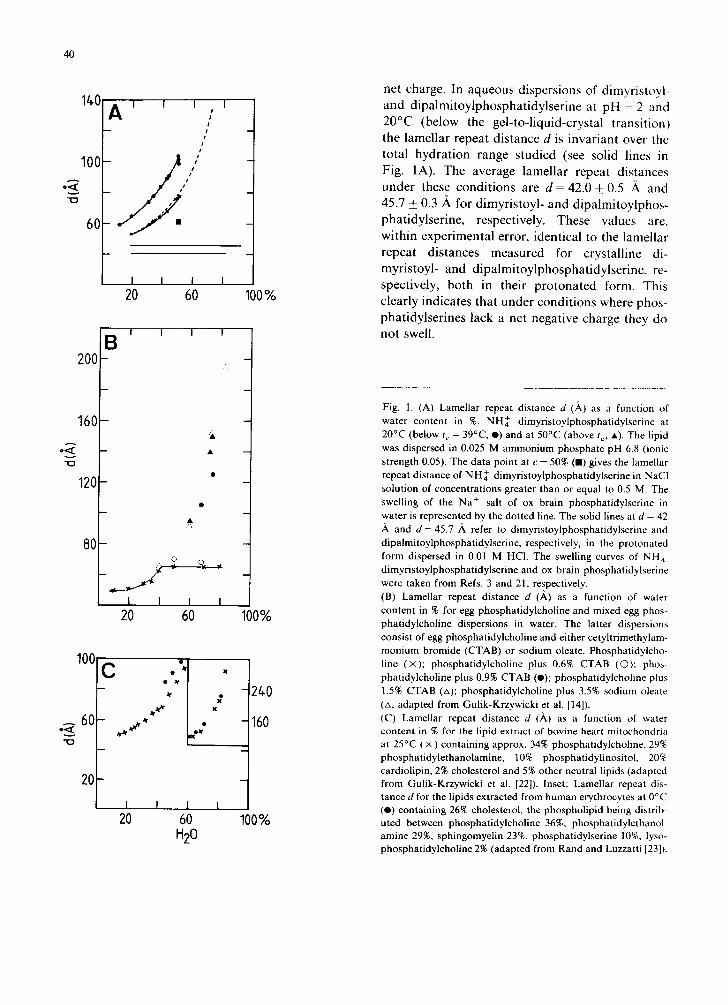
diffraction pattern of phosphatidylserine disper- sions in H20 consists at low angles of a series of equally spaced sharp lines in the ratio of 1 : (1/2) : (1/3) .. . ( l / n ) , from which the lamellar repeat distance d is derived. This distance is shown as a function of water content for various phos- phatidylserine dispersions in Fig. 1A. With NH~- dimyristoylphosphatidylserine, d increases con- tinuously with water content up to at least 50% H20. This is true for dispersions at 20°C and 50°C, where the phospholipid is below and above the gel-to-liquid-crystal transition, respectively [3]. At 20°C the wide-angle reflection at 1/4.2 .~-1 is indicative of the lipid being in the gel phase with 'hexagonal' hydrocarbon chain packing. In the gel at 20°C the lamellar repeat distance d increases from about 60 ,~ to 100 ,~ as the water content of the gel is raised from 10 to 50%. The swelling in the liquid-crystalline phase at 50°C is less over the same range of water content and is, within experi- mental error, identical to that of the Na ÷ salt of ox brain phosphatidylserine at 20°C (above the gel-to-liquid-crystal transition) (Fig. 1A). For ox brain phosphatidylserine continuous swelling could be observed up to d = 130 ,~ at about 70% H20. At higher hydration levels broadening of the low- angle diffraction lines was observed, for both phosphatidylserines, as a result of multilayer stack- ing disorders and eventually the low-angle reflec- tions were lost. Addition of sufficient NaC1 made the fully-swollen gel of NH~ dimyristoylphospha- tidylserine (at 50% water, d = 100 A) shrink. Addi- tion of 0.5 M NaC1 reduced d to 62 ,~, higher concentrations of NaCI having no further effect on the lamellar repeat distance d [9]. Qualitatively similar effects, namely a significant reduction in the lamellar repeat distance were observed when other alkali metal ions were added to aqueous dispersions of NH~ dimyristoylphosphatidyl- serine. Polyvalent cations, such as the alkaline earth metal ions and the lanthanides, produced the same effect except that the reduction in d occurred at ionic concentrations 10-50 times smaller (data not shown). The continuous swelling of the charged phosphatidylserine is contrasted by the behaviour of uncharged (isoelectric) phosphatidylserines. When dispersed in hydrochloric acid pH = 2 the carboxyl group of phosphatidylserine is fully pro- tonated [10] and hence the phospholipid has no
40
140
100;
o<
6O
200
160
o<
120
80
100
_ 60
20
A ' ' ' , ' t o
e
I I I 20 60
B I I I
~k
©
- . , , . __ . , , . . . ~J * Q. _.,
I I I I 20 60
x
Z ~rJr~ x
K
C
20 60 H20
100%
100%
24.0
160
100%
net charge. In aqueous dispersions of dimyristoyl- and dipalmitoylphosphatidylserine at pH = 2 and 20°C (below the gel-to-liquid-crystal transition) the lamellar repeat distance d is invariant over the total hydration range studied (see solid lines in Fig. 1A). The average lamellar repeat distances under these conditions are d = 42.0 + 0.5 it and 45.7 _+ 0.3 A for dimyristoyl- and dipalmitoylphos- phatidylserine, respectively. These values are, within experimental error, identical to the lamellar repeat distances measured for crystalline di- myristoyl- and dipalmitoylphosphatidylserine, re- spectively, both in their protonated form. This clearly indicates that under conditions where phos- phatidylserines lack a net negative charge they do not swell.
Fig. 1. (A) Lamellar repeat distance d (A.) as a function of water content in %. NH~- dimyristoylphosphatidylserine at 20°C (below t c = 39°C, O) and at 50°C (above to, •) . The lipid was dispersed in 0.025 M ammonium phosphate pH 6.8 (ionic strength 0.05). The data point at c = 50% (E) gives the lamellar repeat distance of N H ~ dimyristoylphosphatidylserine in NaC1 solution of concentrations greater than or equal to 0.5 M. The swelling of the Na + salt of ox brain phosphatidylserine in water is represented by the dotted line. The solid lines at d = 42
and d = 45.7 ,~ refer to dimyristoylphosphatidylserine and dipalmitoylphosphatidylserine, respectively, in the protonated form dispersed in 0.01 M HCI. The swelling curves of NH~ dimyristoylphosphatidylserine and ox brain phosphatidylserine were taken from Refs. 3 and 21, respectively. (B) Lamellar repeat distance d (,~) as a function of water content in % for egg phosphatidylcholine and mixed egg phos- phatidylcholine dispersions in water. The latter dispersions consist of egg phosphatidylcholine and either cetyltrimethylam- mon ium bromide (CTAB) or sodium oleate. Phosphatidylcho- line ( x ) ; phosphatidylcholine plus 0.6% CTAB (0) : phos- phatidylcholine plus 0.9% CTAB (o); phosphatidylcholine plus 1.5% CTAB (zx); phosphatidylcholine plus 3.5% sodium oleate (zx, adapted from Gulik-Krzywicki et al. [14]). (C) Lamellar repeat distance d (A) as a function of water content in % for the lipid extract of bovine heart mitochondria at 25°C ( × ) containing approx. 34% phosphatidylcholine, 29% phosphatidylethanolamine, 10% phosphatidylinositol, 20% cardiolipin, 2% cholesterol and 5% other neutral lipids (adapted from Gulik-Krzywicki et al. [22]). Inset: Lamellar repeat dis- tance d for the lipids extracted from human erythrocytes at 0°C (O) containing 26% cholesterol, the phospholipid being distrib- uted between phosphatidylcholine 36%, phosphatidylethanol- amine 29%, sphingomyelin 23%, phosphatidylserine 10%, lyso- phosphatidylcholine 2% (adapted from Rand and Luzzatti [23]).
That the infinite swelling of lipid bilayers bear- ing a net charge is a general phenomenon is dem- onstrated in Fig. 1B and C which were made up from data in the literature. In Fig. 1B the swelling of egg phosphatidylcholine in water (x x) is compared to the swelling of egg phosphatidylcho- line doped with small quantities of either cetyltrimethylammonium bromide (CTAB) or sodium oleate. It is seen that pure egg phosphati- dylcholine swells with increasing water content reaching an upper limit of d--- 65 ,~ at about 40% H20. Up to this limit a single, swelling, lamellar phase exists and above that a two-phase system consisting of a fully-swollen, lamellar phase with d = 65 ,~ and excess water [11,12]. A similar be- haviour of limited swelling was reported for syn- thetic phosphatidylcholines with saturated hydro- carbon chains [11] and other neutral and isoelec- tric lipids, e.g,, phosphatidylethanolamine, ethanolamine plasmalogens, sphingomyelins, monoacylglycerols, glycolipids such as cerebro- sides, mono and digalactosyl diacylglycerols [13,14]. The swelling behaviour of egg phosphati- dylcholine bilayers containing less than 0.6% cetyltrimethylammonium bromide (O O) is similar to that of pure phosphatidylcholine. How- ever, doping the phosphatidylcholine bilayer with about 1% of this detergent or 3.5% sodium oleate produced a marked change; infinite swelling as with charged phosphatidylserines is observed up to at least 80% water. It should be stressed that the infinite swelling is produced by adding only a few percent of a charged lipid to phosphatidylcholine and furthermore the effect is independent of the nature of the charge. Infinite swelling as shown in Fig. 1A and B has been reported for negatively charged phospholipids other than phosphatid- ylserine, e.g. phosphatidic acid [15,16] and phos- phatidylglycerol [17]. Furthermore, it has been shown for phospholipid mixtures containing nega- tively charged phospholipids, e.g., egg phosphatid- ylcholine containing increasing quantities of phos- phatidylglycerol [17] and egg phosphatidylcholine containing 10% phosphatidylinositol [17]; it has also been shown for lipids extracted from biologi- cal membranes which are known to contain 10% or more negatively charged lipids. The continuous swelling, up to at least 80% HzO , of the total lipids extracted from bovine heart mitochondria (×) and
41
1(3
I
5
10
I
5
- A
- B
I Fc
I O - D
I
5
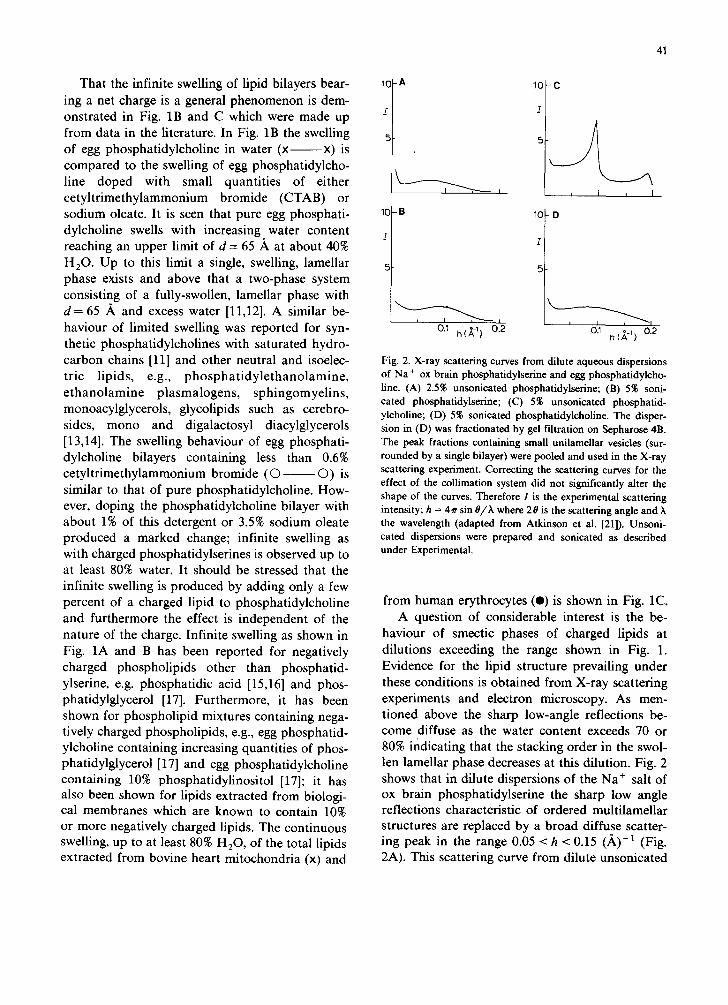
0.~ h(~-~) 0.2 %!2 Fig. 2. X-ray scattering curves from dilute aqueous dispersions of Na + ox brain phosphatidylserine and egg phosphatidylcho- line. (A) 2.5% unsonicated phosphatidylserine; (B) 5% soni- cated phosphatidylserine; (C) 5% unsonicated phosphatid- ylcholine; (D) 5% sonicated phosphatidylcholine. The disper- sion in (D) was fractionated by gel filtration on Sepharose 4B. The peak fractions containing small unilamellar vesicles (sur- rounded by a single bilayer) were pooled and used in the X-ray scattering experiment. Correcting the scattering curves for the effect of the collimation system did not significantly alter the shape of the curves. Therefore I is the experimental scattering intensity; h = 4~r sin O/h where 20 is the scattering angle and the wavelength (adapted from Atkinson et al. [21]). Unsoni- cated dispersions were prepared and sonicated as described under Experimental.
from human erythrocytes (O) is shown in Fig. 1C. A question of considerable interest is the be-
haviour of smectic phases of charged lipids at dilutions exceeding the range shown in Fig. 1. Evidence for the lipid structure prevailing under these conditions is obtained from X-ray scattering experiments and electron microscopy. As men- tioned above the sharp low-angle reflections be- come diffuse as the water content exceeds 70 or 80% indicating that the stacking order in the swol- len lamellar phase decreases at this dilution. Fig. 2 shows that in dilute dispersions of the Na ÷ salt of ox brain phosphatidylserine the sharp low angle reflections characteristic of ordered multilamellar structures are replaced by a broad diffuse scatter- ing peak in the range 0.05 < h < 0.15 (,~)-1 (Fig. 2A). This scattering curve from dilute unsonicated
42
phosphatidylserine dispersions resembles that ob- tained from sonicated Na + ox brain phosphatid- ylserine (Fig. 2B) or sonicated egg phosphatid- ylcholine (Fig. 2D) known to consist of small unilamellar vesicles. The broad diffuse scattering maxima obtained with sonicated phospholipid dis- persions are contrasted by the sharp maxima ob- tained with a diluted (5%) unsonicated egg phos- phatidylcholine dispersion in H20 (Fig. 2C). The maxima corresponding to 66 ,~ and 33 A are the first and second order diffractions from the lamel- lar repeat distance of the maximally hydrated mul- tilamellar egg phosphatidylcholine structure; the lamellar repeat distance d measured for such di- luted unsonicated phosphatidylcholine dispersions is identical to that measured at higher lipid con- centrations, i.e. at 50% lipid (cf. Fig. 1B). This clearly indicates that with phosphatidylcholine the fundamental multilamellar packing is maintained in excess water. Therefore, in excess water un- charged (isoelectric) phospholipids are markedly different from phospholipids carrying a net charge. The interpretation of the scattering curves shown in Fig. 2 is based on the computation of scattering curves for spherical, unilamellar vesicles differing in radius. A good fit with the experimental scatter- ing curves (Figs. 2A, B and D) is achieved assum- ing that the vesicle dispersions are polydisperse. Stacks of bilayers comprising more than a few bilayers can be ruled out on the basis of this computer simulation (for details, see Ref. 21). Furthermore, it can be readily shown that the broad scattering envelopes at 0.05 < h < 0.15 (A) -1 (Figs. 2A, B, D) results from the one-dimensional Fourier transform of the electron density profile of the bilayer. This strongly suggests that the funda- mental structure of charged lipids dispersed in excess water is the unilamellar vesicle.
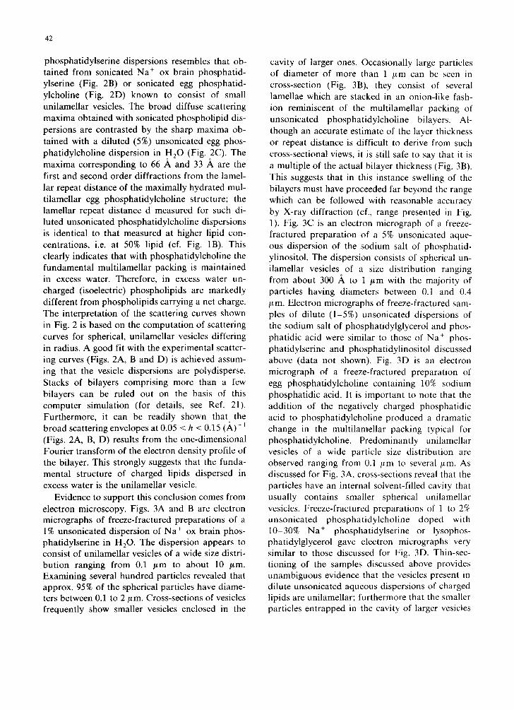
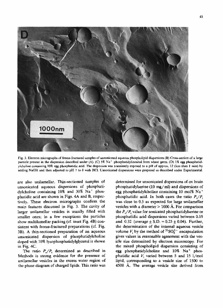
Evidence to support this conclusion comes from electron microscopy. Figs. 3A and B are electron micrographs of freeze-fractured preparations of a 1% unsonicated dispersion of Na + ox brain phos- phatidylserine in H20. The dispersion appears to consist of unilamellar vesicles of a wide size distri- bution ranging from 0.1 /Lm to about 10 ~m. Examining several hundred particles revealed that approx. 95% of the spherical particles have diame- ters between 0.1 to 2 #m. Cross-sections of vesicles frequently show smaller vesicles enclosed in the
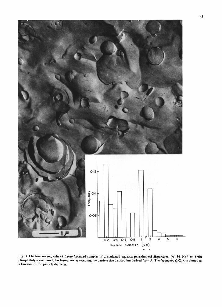
cavity of larger ones. Occasionally large particles of diameter of more than 1 /~m can be seen in cross-section (Fig. 3B), they consist of several lamellae which are stacked in an onion-like fash- ion reminiscent of the multilamellar packing of unsonicated phosphatidylcholine bilayers. Al- though an accurate estimate of the layer thickness or repeat distance is difficult to derive from such cross-sectional views, it is still safe to say that it is a multiple of the actual bilayer thickness (Fig. 3B). This suggests that in this instance swelling of the bilayers must have proceeded far beyond the range which can be followed with reasonable accuracy by X-ray diffraction (cf., range presented in Fig. 1). Fig. 3C is an electron micrograph of a freeze- fractured preparation of a 5% unsonicated aque- ous dispersion of the sodium salt of phosphatid- ylinositol. The dispersion consists of spherical un- ilamellar vesicles of a size distribution ranging from about 300 ,~ to 1 /~m with the majority of particles having diameters between 0.1 and 0.4 /~m. Electron micrographs of freeze-fractured sam- ples of dilute (1-5%) unsonicated dispersions of the sodium salt of phosphatidylglycerol and phos- phatidic acid were similar to those of Na + phos- phatidylserine and phosphatidylinositol discussed above (data not shown). Fig. 3D is an electron micrograph of a freeze-fractured preparation of egg phosphatidylcholine containing 10% sodium phosphatidic acid. It is important to note that the addition of the negatively charged phosphatidic acid to phosphatidylcholine produced a dramatic change in the multilamellar packing typical for phosphatidylcholine. Predominantly unilamellar vesicles of a wide particle size distribution are observed ranging from 0.1 /~m to several /~m. As discussed for Fig. 3A, cross-sections reveal that the particles have an internal solvent-filled cavity that usually contains smaller spherical unilamellar vesicles. Freeze-fractured preparations of 1 to 2% unsonicated phosphatidylcholine doped with 10 30% Na + phosphatidylserine or lysophos- phatidylglycerol gave electron micrographs very similar to those discussed for Fig. 3D. Thin-sec- tioning of the samples discussed above provides unambiguous evidence that the vesicles present in dilute unsonicated aqueous dispersions of charged lipids are unilamellar; furthermore that the smaller particles entrapped in the cavity of larger vesicles
43
0'15
g U.
0"05
0-2 0"4 0-6 O'B I 2 4 6 8
Part icle d i a m e t e r ( IJm)
Fig. 3. Electron micrographs of freeze-fractured samples of unsonicated aqueous phospholipid dispersions. (A) 5% Na + ox brain phosphatidylserine; inset; bar histogram representing the particle size distribution derived from A. The frequencyf~/Eif ~ is plotted as a function of the particle diameter.
44
45
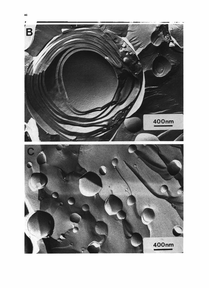
Fig. 3. Electron micrographs of freeze-fractured samples of unsonicated aqueous phospholipid dispersions (B) Cross-section of a large particle present in the dispersion described under (A). (C) 5% Na+ phosphatidylinositol from wheat germ. (D) 1% egg phosphatid-
ylcholine containing 10% egg phosphatidic acid. The dispersion was transiently exposed to a pH of approx. 12 (less than 1 min) by
adding NaOH and then adjusted to pH 7 to 8 with HCl. Unsonicated dispersions were prepared as described under Experimental.
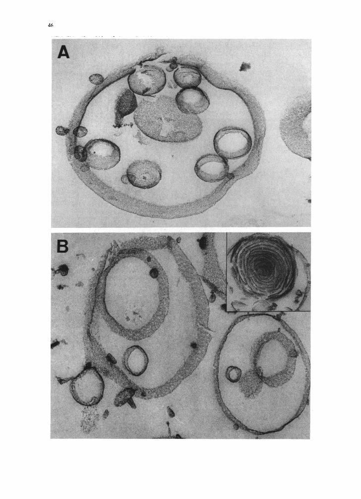
are also unilamellar. Thin-sectioned samples of unsonicated aqueous dispersions of phosphati-
dylcholine containing 10% and 30% Na+ phos- phatidic acid are shown in Figs. 4A and B, respec-
tively. These electron micrographs confirm the
main features discussed in Fig. 3. The cavity of
larger unilamellar vesicles is usually filled with
smaller ones; in a few exceptions the particles
show multilamellar packing (cf. inset Fig. 4B) con-
sistent with freeze-fractured preparations (cf. Fig.
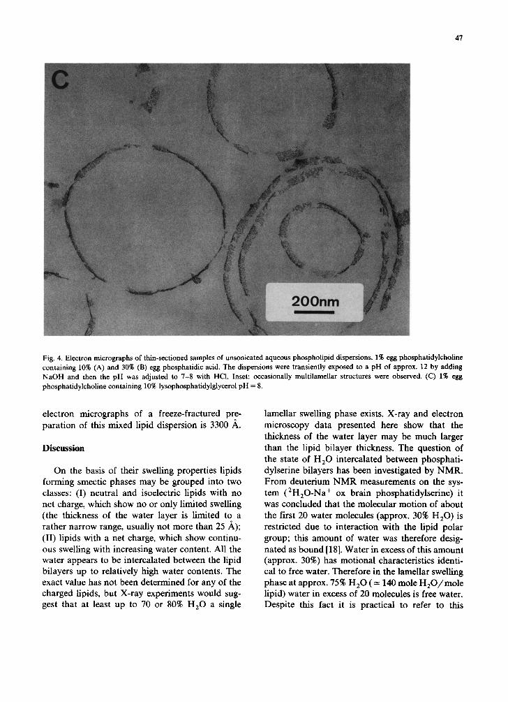
3B). A thin-sectioned preparation of an aqueous unsonicated dispersion of phosphatidylcholine
doped with 10% lysophosphatidylglycerol is shown in Fig. 4C.
The ratio Pi/P, determined as described in Methods is strong evidence for the presence of unilamellar vesicles in the excess water region of the phase diagram of charged lipids. This ratio was
determined for unsonicated dispersions of ox brain
phosphatidylserine (10 mg/ml) and dispersions of egg phosphatidylcholine containing 10 mol% Na+
phosphatidic acid. In both cases the ratio Pi/P, was close to 0.5 as expected for large unilamellar
vesicles with a diameter > 1000 A. For comparison
the Pi/P, value for sonicated phosphatidylserine or phosphatidic acid dispersions varied between 0.19 and 0.32 (average & S.D. = 0.25 f 0.04). Further,
the determination of the internal aqueous vesicle
volume Vi by the method of 3sSOi- encapsulation gives values in reasonable agreement with the ves- icle size determined by electron microscopy. For the mixed phospholipid dispersion consisting of egg phosphatidylcholine and 10% Na+ phos- phatidic acid K varied between 5 and 15 l/mol lipid, corresponding to a vesicle size of 1500 to 4500 A. The average vesicle size derived from
E
47
Fig. 4. Electron micrographs of thin-sectioned samples of unsonicated aqueous phospholipid dispersions. 1% egg phosphatidylcholine containing 10% (A) and 30% (B) egg phosphatidic acid. The dispersions were transiently exposed to a pH of approx. 12 by adding NaOH and then the pH was adjusted to 7-8 with HCI. Inset: occasionally multilamellar structures were observed. (C) 1% egg phosphatidylcholine containing 10% lysophosphatidylglycerol pH = 8.
electron micrographs of a freeze-fractured pre- paration of this mixed lipid dispersion is 3300 ~,.
Discussion
On the basis of their swelling properties lipids forming smectic phases may be grouped into two classes: (I) neutral and isoelectric lipids with no net charge, which show no or only limited swelling (the thickness of the water layer is limited to a rather narrow range, usually not more than 25 ,h,); (II) lipids with a net charge, which show continu- ous swelling with increasing water content. All the water appears to be intercalated between the lipid bilayers up to relatively high water contents. The exact value has not been determined for any of the charged lipids, but X-ray experiments would sug- gest that at least up to 70 or 80% H 2 0 a single
lamellar swelling phase exists. X-ray and electron microscopy data presented here show that the thickness of the water layer may be much larger than the lipid bilayer thickness. The question of the state of H 2 0 intercalated between phosphati- dylserine bilayers has been investigated by NMR. From deuterium N M R measurements on the sys- tem (2H20-Na+ ox brain phosphatidylserine) it was concluded that the molecular motion of about the first 20 water molecules (approx. 30% H 2 0 ) is restricted due to interaction with the lipid polar group; this amount of water was therefore desig- nated as bound [18]. Water in excess of this amount (approx. 30%) has motional characteristics identi- cal to free water. Therefore in the lameUar swelling phase at approx. 75% H 2 0 ( = 140 mole H 2 0 / m o l e lipid) water in excess of 20 molecules is free water. Despite this fact it is practical to refer to this
48
phase as a single lamellar phase as has been done throughout this paper. Although this terminology seems arbitrary, it is analogous to the one-phase system of neutral and isoelectric lipids. Character- istic of this one-phase system is a well-defined low-angle X-ray diffraction pattern.
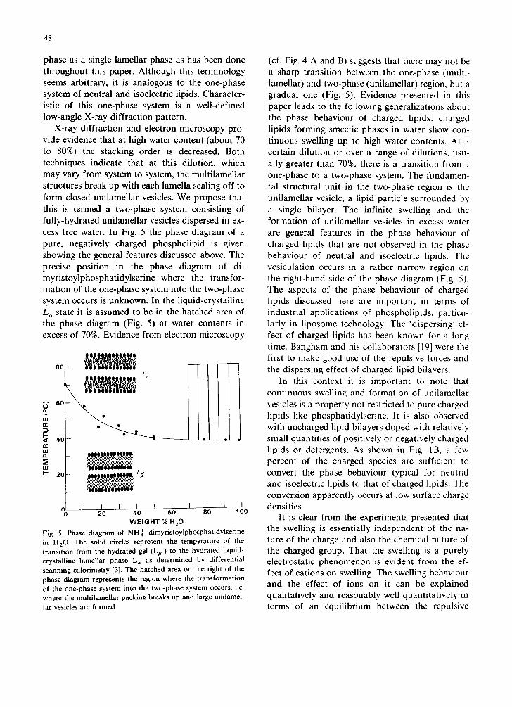
X-ray diffraction and electron microscopy pro- vide evidence that at high water content (about 70 to 80%) the stacking order is decreased. Both techniques indicate that at this dilution, which may vary from system to system, the multilamellar structures break up with each lamella sealing off to form closed unilamellar vesicles. We propose that this is termed a two-phase system consisting of fully-hydrated unilamellar vesicles dispersed in ex- cess free water. In Fig. 5 the phase diagram of a pure, negatively charged phospholipid is given showing the general features discussed above. The precise position in the phase diagram of di- myristoylphosphatidylserine where the transfor- mation of the one-phase system into the two-phase system occurs is unknown. In the liquid-crystalline L~ state it is assumed to be in the hatched area of the phase diagram (Fig. 5) at water contents in excess of 70%. Evidence from electron microscopy
k- 4 0 - " ~ - ' - ' - ~ - - = n-
uJ
,,, //ggggdgggdggg
_ gggggggggggg
I I I I I I I 0 20 40 60
WEIGHT % HzO
Fig. 5. Phase diagram of NH + dimyristoylphosphatidylserine in H20 . The solid circles represent the temperature of the transition from the hydrated gel (Lp,) to the hydrated liquid- crystalline lamellar phase L,~ as determined by differential scanning calorimetry [3]. The hatched area on the right of the phase diagram represents the region where the transformation of the one-phase system into the two-phase system occurs, i.e. where the multilamellar packing breaks up and large unilamel- lar vesicles are formed.
t I t 80 100
(cf. Fig. 4 A and B) suggests that there may not be a sharp transition between the one-phase (multi- lamellar) and two-phase (unilamellar) region, but a gradual one (Fig. 5). Evidence presented in this paper leads to the following generalizations about the phase behaviour of charged lipids: charged lipids forming smectic phases in water show con- tinuous swelling up to high water contents. At a certain dilution or over a range of dilutions, usu- ally greater than 70%, there is a transition from a one-phase to a two-phase system. The fundamen- tal structural unit in the two-phase region is the unilamellar vesicle, a lipid particle surrounded by a single bilayer. The infinite swelling and the formation of unilamellar vesicles in excess water are general features in the phase behaviour of charged lipids that are not observed in the phase behaviour of neutral and isoelectric lipids. The vesiculation occurs in a rather narrow region on the right-hand side of the phase diagram (Fig. 5). The aspects of the phase behaviour of charged lipids discussed here are important in terms of industrial applications of phospholipids, particu- larly in liposome technology. The 'dispersing' ef- fect of charged lipids has been known for a long time. Bangham and his collaborators [19] were the first to make good use of the repulsive forces and the dispersing effect of charged lipid bilayers.
In this context it is important to note that continuous swelling and formation of unilamellar vesicles is a property not restricted to pure charged lipids like phosphatidylserine. It is also observed with uncharged lipid bilayers doped with relatively small quantities of positively or negatively charged lipids or detergents. As shown in Fig. 1B, a few percent of the charged species are sufficient to convert the phase behaviour typical for neutral and isoelectric lipids to that of charged lipids. The conversion apparently occurs at low surface charge densities.
It is clear from the experiments presented that the swelling is essentially independent of the na- ture of the charge and also the chemical nature of the charged group. That the swelling is a purely electrostatic phenomenon is evident from the ef- fect of cations on swelling. The swelling behaviour and the effect of ions on it can be explained qualitatively and reasonably well quantitatively in terms of an equilibrium between the repulsive
49
forces of the double-layer potential and the attrac- tive van der Waals forces between opposing bi- layers (the quantitative treatment of this equi- librium will be the subject of a separate publica- tion). Addition of sufficient cations makes a fully swollen phosphatidylserine dispersion shrink. Ca- tions as counterions accumulate in the electrical double layer and effectively screen the negative surface charges. Water between bilayers is ex- truded and consequently d decreases. The packing of phosphatidylserine in excess NaC1 or salt in general is similar to that of phosphatidylcholine; the swelling is limited (Fig. la) and the two-phase system consists of multilamellar structures disper- sed in excess water. The addition of 0.5 M NaC1 to phosphatidylserine vesicles in excess water (two- phase system) induced aggregation and fusion of the vesicles leading, in equilibrium, to the same multilamellar structure as described above for a 50% phosphatidylserine dispersion in excess NaCI.
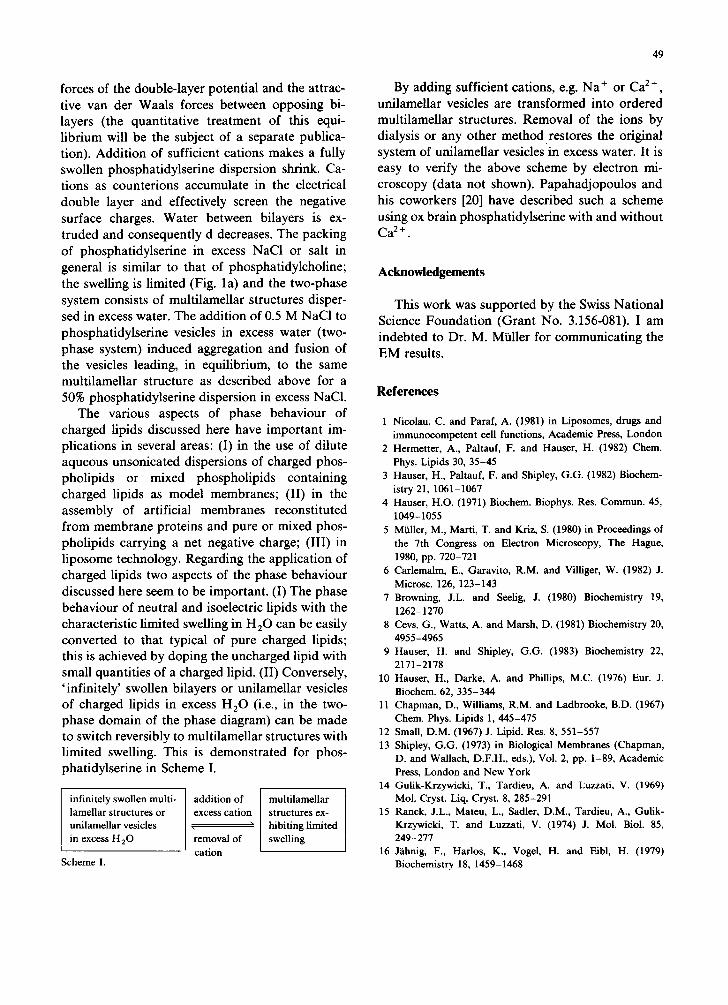
The various aspects of phase behaviour of charged lipids discussed here have important im- plications in several areas: (I) in the use of dilute aqueous unsonicated dispersions of charged phos- pholipids or mixed phospholipids containing charged lipids as model membranes; (II) in the assembly of artificial membranes reconstituted from membrane proteins and pure or mixed phos- pholipids carrying a net negative charge; (III) in liposome technology. Regarding the application of charged lipids two aspects of the phase behaviour discussed here seem to be important. (I) The phase behaviour of neutral and isoelectric lipids with the characteristic limited swelling in H20 can be easily converted to that typical of pure charged lipids; this is achieved by doping the uncharged lipid with small quantities of a charged lipid. (II) Conversely, 'infinitely' swollen bilayers or unilamellar vesicles of charged lipids in excess H20 (i.e., in the two- phase domain of the phase diagram) can be made to switch reversibly to multilamellar structures with limited swelling. This is demonstrated for phos- phatidylserine in Scheme I.
infinitely swollen multi- lamellar structures or unilamellar vesicles in excess H20
Scheme I.
addition of excess cation
removal of cation
multilamellar structures ex- hibiting limited swelling
By adding sufficient cations, e.g. Na ÷ or Ca 2+, unilamellar vesicles are transformed into ordered multilamellar structures. Removal of the ions by dialysis or any other method restores the original system of unilamellar vesiclesin excess water. It is easy to verify the above scheme by electron mi- croscopy (data not shown). Papahadjopoulos and his coworkers [20] have described such a scheme using ox brain phosphatidylserine with and without C a 2+ "
Acknowledgements
This work was supported by the Swiss National Science Foundation (Grant No. 3.156-081). I am indebted to Dr. M. Mialler for communicating the EM results.
References
1 Nicolau, C. and Paraf, A. (1981) in Liposomes, drugs and immunocompetent cell functions, Academic Press, London
2 Hermetter, A., Pahauf, F. and Hauser, H. (1982) Chem. Phys. Lipids 30, 35-45
3 Hauser, H., Paltauf, F. and Shipley, G.G. (1982) Biochem- istry 21, 1061-1067
4 Hauser, H.O. (1971) Biochem. Biophys. Res. Commun. 45, 1049-1055
5 Miiller, M., Marti, T. and Kriz, S. (1980) in Proceedings of the 7th Congress on Electron Microscopy, The Hague, 1980, pp. 720-721
6 Carlemalm, E., Garavito, R.M. and Villiger, W. (1982) J. Microsc. 126, 123-143
7 Browning, J.L. and Seelig, J. (1980) Biochemistry 19, 1262-1270
8 Cevs, G., Watts, A. and Marsh, D. (1981) Biochemistry 20, 4955-4965
9 Hauser, H. and Shipley, G.G. (1983) Biochemistry 22, 2171-2178
10 Hauser, H., Darke, A. and Phillips, M.C. (1976) Eur. J. Biochem. 62, 335-344
11 Chapman, D., Williams, R.M. and Ladbrooke, B.D. (1967) Chem. Phys. Lipids 1, 445-475
12 Small, D.M. (1967) J. Lipid. Res. 8, 551-557 13 Shipley, G.G. (1973) in Biological Membranes (Chapman,
D. and Wallach, D.F.H., eds.), Vol. 2, pp. 1-89, Academic Press, London and New York
14 Gulik-Krzywicki, T., Tardieu, A. and Luzzati, V. (1969) Mol. Cryst. Liq. Cryst. 8, 285-291
15 Ranck, J.L., Mateu, L., Sadler, D.M., Tardieu, A., Gulik- Krzywicki, T. and Luzzati, V. (1974) J. Mol. Biol. 85, 249-277
16 Jiilmig, F., Harlos, K., Vogel, H. and Eibl, H. (1979) Biochemistry 18, 1459-1468
50
17 Cowley, A.C., Fuller, N.L., Rand, R.P. and Parsegian, V.A. (1978) Biochemistry 17, 3163-3168
18 Finer, E.G. and Darke, A. (1974) Chem. Phys. Lipids 12, 1-16
19 Bang, ham, A.D. (1968) Prog. Biophys. Mol. Biol. 18, 29-95 20 Papahadjopoulos, D., Vail, W.J., Jacobson, K. and Poste,
G. (1975) Biochim. Biophys. Acta 394, 483-491
21 Atkinson, D., Hauser, H., Shipley, G.G. and Stubbs, J.M. (1974) Biochim. Biophys. Acta 339, 10-29
22 Gulik-Krzywicki, T., Rivas, E. and Luzzati, V. (1967) J. Mol. Biol. 27, 303-322
23 Rand, R.P. and Luzzati, V. (1968) Biophys, J. 8, 125-137 24 Gains, N. and Hauser, H. (1983) Biochim. Biophys. Acta
731, 31-39