solitons - federation of american scientists
TRANSCRIPT
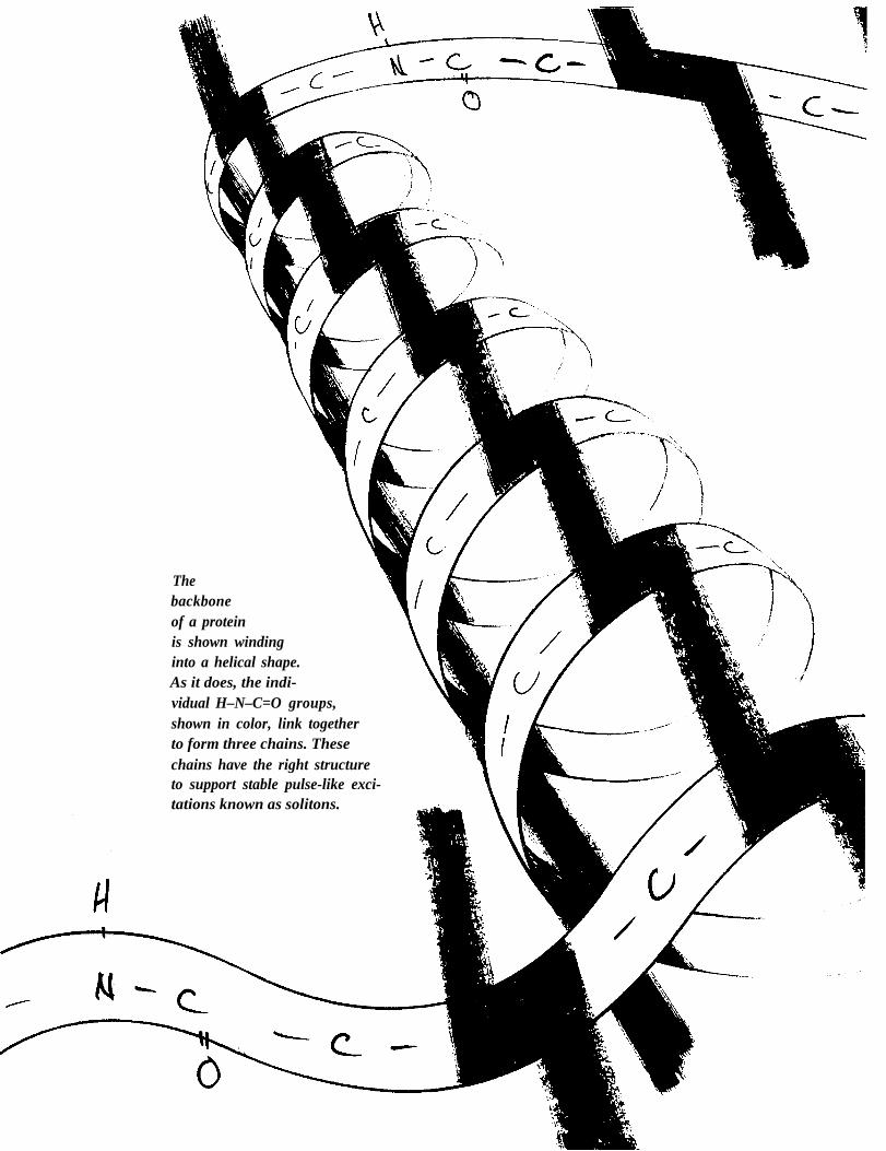
Thebackboneof a proteinis shown windinginto a helical shape.As it does, the indi-vidual H–N–C=O groups,shown in color, link togetherto form three chains. Thesechains have the right structureto support stable pulse-like exci-tations known as solitons.

Solitons●
inBiology
by Peter S. Lomdahl, Scott P. Layne, and Irving J. Bigio
I n 1973 scientists gathered at the New York Academy of Sciencesto discuss an unanswered question in bioenergetics: How is chemical energytransduced and transported in biological systems? In the same year a Sovietsolid-state physicist proposed a dynamic answer that was totally novel to theworld of biology. Exploiting the regularity in the structure of a-helical proteins,he showed that simplified models of these proteins could self-focus, or trap,energy in stable, pulse-like waves known as solitons. If self-focusing is indeed abiological reality, it may account for many aspects of protein behavior,including the efficient transport of energy. This possibility is being studied atLos Alamos through analytical, numerical, and experimental techniques.
It is widely accepted that proteins are the principal workhorses ofthe cell. They are the major organizers and manipulators of biologicalenergy and the enzymes that catalyze and maintain the life process.They are responsible for the active transport of ions into and out ofthe cell and for cellular and intracellular movement. Of course othermacromolecules, such as DNA, polysaccharides, and lipids, have anenergetic dimension. but their operation is always closely tied to thatof proteins. Therefore the discipline of bioenergetics, which is thestudy of how cells generate and transfer their energy supply, isprimarily the investigation of how proteins work. From decades ofchemical analysis and x-ray crystallography and from more recentadvances in spectroscopy, we know the composition and three-dimensional conformation of about two hundred proteins. Despitethis extensive structural knowledge. however, there is no generallyaccepted model of how proteins operate dynamically. Presently, it isfair to say that the “nuts and bolts” functioning of proteins remainsan outstanding question in bioenergetics.
The energy supply for most protein activities is provided by thehydrolysis of ATP (adenosine triphosphate). An ATP molecule bindsto a specific site on the protein. reacts with water, and under normalphysiological conditions releases 0.49 electron volt (eV) of freeenergy. This is about twenty times greater than the average energyavailable from the thermal background at 300 kelvins. The questionfor bioenergetics is what happens to this energy? How does it performuseful work? Is the energy used through a nonequilibrium process, ordoes the energy first thermalize and then work through an equilibrium
process? Molecular dynamics calculations. based on ball-and-springmodels of proteins. show that heat from a thermal bath induces avariety of motions in proteins. These equilibrium calculations showmotions ranging from localized, high-frequency vibrations of individ-ual bonds to collective, low-frequency motions of the entire protein.One may question, however. whether such equilibrium dynamicscould account for the efficient transport and use of energy over thecharacteristic lengths of proteins, which range from tens to hundredsof angstroms.
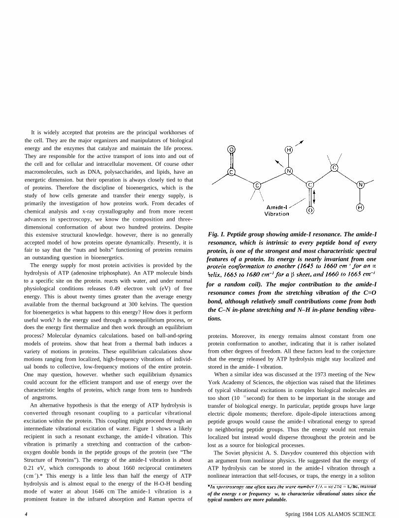
An alternative hypothesis is that the energy of ATP hydrolysis isconverted through resonant coupling to a particular vibrationalexcitation within the protein. This coupling might proceed through anintermediate vibrational excitation of water. Figure 1 shows a likelyrecipient in such a resonant exchange, the amide-I vibration. Thisvibration is primarily a stretching and contraction of the carbon-oxygen double bonds in the peptide groups of the protein (see “TheStructure of Proteins”). The energy of the amide-I vibration is about0.21 eV, which corresponds to about 1660 reciprocal centimeters(cm -1).* This energy is a little less than half the energy of ATPhydrolysis and is almost equal to the energy of the H-O-H bendingmode of water at about 1646 cm– The amide-1 vibration is aprominent feature in the infrared absorption and Raman spectra of
Fig. I. Peptide group showing amide-I resonance. The amide-Iresonance, which is intrinsic to every peptide bond of everyprotein, is one of the strongest and most characteristic spectralfeatures of a protein. Its energy is nearly invariant from one
for a random coil). The major contribution to the amide-Iresonance comes from the stretching vibration of the C=Obond, although relatively small contributions come from boththe C–N in-plane stretching and N–H in-plane bending vibra-tions.
proteins. Moreover, its energy remains almost constant from oneprotein conformation to another, indicating that it is rather isolatedfrom other degrees of freedom. All these factors lead to the conjecturethat the energy released by ATP hydrolysis might stay localized andstored in the amide- I vibration.
When a similar idea was discussed at the 1973 meeting of the NewYork Academy of Sciences, the objection was raised that the lifetimesof typical vibrational excitations in complex biological molecules aretoo short (10 12 second) for them to be important in the storage andtransfer of biological energy. In particular, peptide groups have largeelectric dipole moments; therefore. dipole-dipole interactions amongpeptide groups would cause the amide-I vibrational energy to spreadto neighboring peptide groups. Thus the energy would not remainlocalized but instead would disperse throughout the protein and belost as a source for biological processes.
The Soviet physicist A. S. Davydov countered this objection withan argument from nonlinear physics. He suggested that the energy ofATP hydrolysis can be stored in the amide-I vibration through anonlinear interaction that self-focuses, or traps, the energy in a soliton
of the energy E or frequency w, to characterize vibrational states since thetypical numbers are more palatable.
4 Spring 1984 LOS ALAMOS SCIENCE
Solitons in Biology
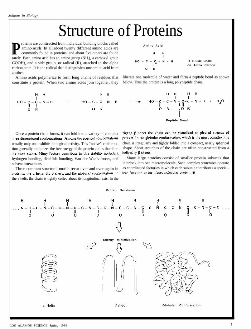
Structure of ProteinsP roteins are constructed from individual building blocks called
Amino Acidamino acids. In all about twenty different amino acids arecommonly found in proteins, and about five others are found H H
rarely. Each amino acid has an amino group (NH2), a carboxyl groupCOOH), and a side group, or radical (R), attached to the alpha HO – C – C – N – H R = Side Chain
carbon atom. It is the radical that distinguishes one amino acid fromon Alpha Carbon
O Ranother.
Amino acids polymerize to form long chains of residues that liberate one molecule of water and form a peptide bond as shownconstitute a protein. When two amino acids join together, they below. Thus the protein is a long polypeptide chain.
Peptide Bond
Once a protein chain forms, it can fold into a variety of complex
usually only one exhibits biological activity. This “native” conforma- chain is irregularly and tightly folded into a compact, nearly sphericaltion generally minimizes the free energy of the protein and is therefore shape. Short stretches of the chain are often constructed from a
hydrogen bonding, disulfide bonding, Van der Waals forces, and Many large proteins consist of smaller protein subunits thatsolvent interactions. interlock into one macromolecule, Such complex structures operate
Three common structural motifs recur over and over again in as coordinated factories in which each subunit contributes a special-
the a helix the chain is tightly coiled about its longitudinal axis. In the
Protein Backbone
Energy Minimization
o
H
GlobuIar Conformation
LOS ALAMOS SCIENCE Spring 1984 5
(see “What Is a Soliton?”). The soliton results from a nonlinearcoupling between the vibrational excitation and a deformation in theprotein structure caused by the presence of the excitation. The
excitation and the deformation balance each other. and the resultingexcitation moves through the protein uninhibited. much the wayelectrons move in the superconducting state of a metal.
Davydov worked out these ideas for one particular protein con-formation. the a helix pictured at the beginning of this article. Heintroduced a simple mathematical model to show how solitons could
travel along the three spines, or hydrogen-bonded chains, of theprotein.
Davydov first applied this idea to the problem of muscle contrac-tion. He proposed that myosin, a major contractile protein in striatedmuscle that has an a-helical tail approximately 1500 angstroms long,propagates a soliton that squeezes and pulls on the actin filamentsaround it. This action serves to slide the actin and myosin filamentstogether and thereby results in muscle contraction. In addition,Davydov and his coworkers have considered the idea that a-helicalproteins may facilitate electron transport through a soliton mecha-nism. In this case an extra electron causes a lattice distortion in theprotein that stabilizes the electron’s motion. Thus it may be reason-able to consider charge transfer across membranes. energy couplingacross membranes. and energy transport along filamentouscytoskeletal proteins in terms of a soliton mechanism. since theproteins that carry out these functions contain structural units withsignificant a-helical character.
The soliton model is one of several concepts for protein dynamicsthat should attract the careful attention of biologists. Clearly, itcannot explain every aspect of protein dynamics, but it is motivatingexciting questions and new experiments. In the following sections wewill describe Davydov’s concept in the context of the a helix andexpand it to a crystalline polymer called acetanilide, which wasobserved by G. Careri to have an anomalous spectral line near theamide-I band that might be due to a soliton. We will discussexperimental techniques for verifying the existence of solitons in a-helical proteins and acetanilide and consider the concept of self-focusing in globular proteins. (A further application of the solitonmodel is discussed in “A Possible Mechanism for GeneralAnesthesia.”)
Before discussing details of the soliton model, we will try to makethe relevant biological context more vivid to the reader by presentingthree specific examples where soliton-like dynamics may well beoperating.
Three Sites of Action at a Distance
Alpha-helical structure is quite common in proteins, and inparticular it is present where energy appears to be transported from
6
one end of a protein to the other or where two processes appear to becoupled by a protein.
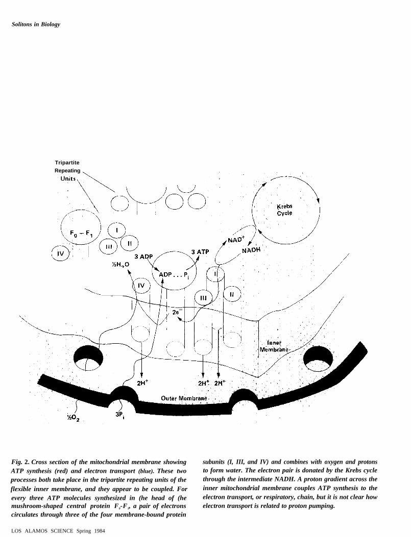
Mitochondria. Mitochondria are the energy-generating stations forliving cells. These organelles, which may have evolved from separateorganisms that were later incorporated into the cell, occupy approx-imately 20 percent of the total cellular volume. Within these or-ganelles has developed a very specialized protein unit specificallydesigned to synthesize ATP. It is called the tripartite repeating unit(Fig, 2). Numerous copies of this unit make up the flexible innermembrane of a mitochondrion. As shown in Fig. 2, each time threeATP molecules are synthesized in the head of the FO-F1 protein, a
pair of electrons (which are donated by the Krebs cycle via theintermediate NADH) circulates among the membrane-bound elec-tron-transport proteins (labeled 1, II, 111. and IV). The electronsultimately combine with oxygen and protons to produce water. Themovement of these electrons back and forth across the inner mem-brane. in turn, creates a proton gradient across the membrane thatdrives the synthesis of ATP in the head of the FO- FI protein. Atpresent. the nature of the driving mechanism is an open question.How do the cytochrome proteins in subunits 1, II. 111, and IV facilitateelectron and proton transport across the thickness (about 60angstroms) of the inner mitochondrial membrane and at the sametime couple ion transport to ATP synthesis? (Semiclassical theoriesaccount for electron tunneling between the donor and acceptor hemegroups that are attached to the cytochrome proteins and have thusexplained oxidation-reduction rates in cytochromes. These theories,however, have not connected electron tunneling to ATP synthesis. )
The dominant configuration of the cytochromes is a-helical, andthese proteins span the inner membrane. Given these facts we may
ask whether a soliton-like mechanism in these proteins may haveanything to do with the stabilization of electron transport and itsconnection to ATP synthesis.
A related question concerns the contracted configuration of amitochondrion during ATP synthesis. When a mitochondrion isinactive, its inner membrane is relaxed and spread out. but whenactive. its inner membrane abruptly contracts into a more wrinkledand twisted appearance (Fig. 3). This brings the myriad tripartiterepeating units in the inner membrane into closer apposition with oneanother. Apparently this aggregation of transmembrane proteins is aprerequisite for ATP synthesis. Whether or not this aggregationinduces a change in the conformation of the individual transmem-brane proteins is not clear. However, if a soliton-like mechanism wereoperating during ATP synthesis, it could well be affected by suchchanges in protein conformation.
Cytoskeleton. The cytoskeleton is a framework of interconnectedproteins that literally tills and bridges the inside of a cell (Fig. 4). Itprovides an internal structure on which the “bag” of the cell rests
Spring 1984 LOS ALAMOS SCIENCE
Solitons in Biology
TripartiteRepeating
Fig. 2. Cross section of the mitochondrial membrane showing subunits (I, III, and IV) and combines with oxygen and protonsATP synthesis (red) and electron transport (blue). These two to form water. The electron pair is donated by the Krebs cycleprocesses both take place in the tripartite repeating units of the through the intermediate NADH. A proton gradient across the
flexible inner membrane, and they appear to be coupled. For inner mitochondrial membrane couples ATP synthesis to the
every three ATP molecules synthesized in (he head of (he electron transport, or respiratory, chain, but it is not clear howmushroom-shaped central protein F0-F1, a pair of electrons electron transport is related to proton pumping.circulates through three of the four membrane-bound protein
LOS ALAMOS SCIENCE Spring 1984
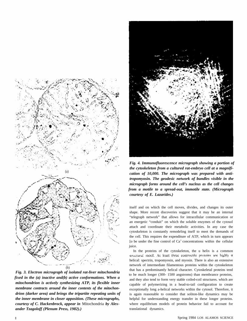
(a)
(b)Fig. 3. Electron micrograph of isolated rat-liver mitochondriafixed in the (a) inactive and(b) active conformations. When amitochondrion is actively synthesizing ATP, its flexible innermembrane contracts around the inner contents of the mitochon-drion (darker area) and brings the tripartite repeating units ofthe inner membrane in closer apposition. (These micrographs,courtesy of C. Hackenbrock, appear in Mitochondria by Alex-ander Tzagoloff (Plenum Press, 1982).)
8
Fig. 4. Immunofluorescence micrograph showing a portion ofthe cytoskeleton from a cultured rat-embryo cell at a magnifi-cation of 10,000. The micrograph was prepared with anti-tropomyosin. The geodesic network of bundles visible in themicrograph forms around the cell’s nucleus as the cell changesfrom a motile to a spread-out, immotile state. (Micrographcourtesy of E. Lazarides.)
itself and on which the cell moves, divides, and changes its outershape. More recent discoveries suggest that it may be an internal“telegraph network” that allows for intracellular communication oran energetic “conduit” on which the soluble enzymes of the cytosolattach and coordinate their metabolic activities. In any case thecytoskeleton is constantly remodeling itself to meet the demands ofthe cell. This requires the expenditure of ATP, which in turn appears[o be under the fine control of Ca2+ concentrations within the cellularjuice.
In the proteins of the cytoskeleton, the u helix is a common
helical: spectrin, tropomyosin, and myosin. There is also an extensivenetwork of intermediate filamentous proteins within the cytoskeletonthat has a predominantly helical character. Cytoskeletal proteins tendto be much longer (300- 1500 angstroms) than membrance proteins,and they also tend to form very stable coiled-coil structures. which arecapable of polymerizing in a head-to-tail configuration to createexceptionally long a-helical networks within the cytosol. Therefore, itis again reasonable to consider that soliton-like dynamics may behelpful for understanding energy transfer in these longer proteins.
where equilibrium models of protein behavior fail to account fortranslational dynamics.
Spring 1984 LOS ALAMOS SCIENCE
Solitons in Biology
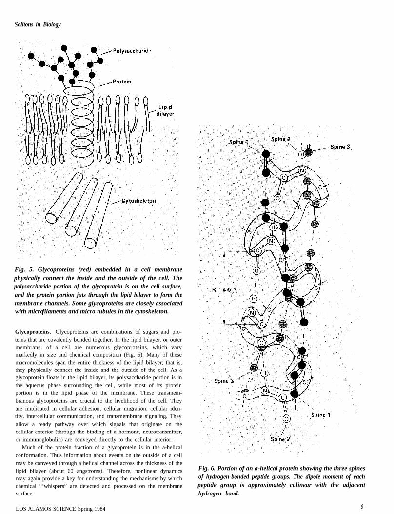
Fig. 5. Glycoproteins (red) embedded in a cell membranephysically connect the inside and the outside of the cell. Thepolysaccharide portion of the glycoprotein is on the cell surface,and the protein portion juts through the lipid bilayer to form themembrane channels. Some glycoproteins are closely associatedwith microfilaments and micro tubules in the cytoskeleton.
Glycoproteins. Glycoproteins are combinations of sugars and pro-teins that are covalently bonded together. In the lipid bilayer, or outermembrane. of a cell are numerous glycoproteins, which varymarkedly in size and chemical composition (Fig. 5). Many of thesemacromolecules span the entire thickness of the lipid bilayer; that is,they physically connect the inside and the outside of the cell. As aglycoprotein floats in the lipid bilayer, its polysaccharide portion is inthe aqueous phase surrounding the cell, while most of its proteinportion is in the lipid phase of the membrane. These transmem-branous glycoproteins are crucial to the livelihood of the cell. Theyare implicated in cellular adhesion, cellular migration. cellular iden-tity. intercellular communication, and transmembrane signaling. Theyallow a ready pathway over which signals that originate on thecellular exterior (through the binding of a hormone, neurotransmitter,or immunoglobulin) are conveyed directly to the cellular interior.
Much of the protein fraction of a glycoprotein is in the a-helical
conformation. Thus information about events on the outside of a cellmay be conveyed through a helical channel across the thickness of thelipid bilayer (about 60 angstroms). Therefore, nonlinear dynamicsmay again provide a key for understanding the mechanisms by whichchemical “’whispers” are detected and processed on the membranesurface.
LOS ALAMOS SCIENCE Spring 1984
Fig. 6. Portion of an a-helical protein showing the three spinesof hydrogen-bonded peptide groups. The dipole moment of eachpeptide group is approximately colinear with the adjacenthydrogen bond.
9
Solitons on the a Helix
Alpha-helical proteins, which are implicated in so many ways inenergy transport and energy coupling, are the context for Davydov’stheory. As the name implies, the conformation of these proteins is ahelix formed by the twisting of the protein backbone. In addition,hydrogen bonds link the peptide groups together to form three spinesthat span the length of the helix and stabilize it. (The reader might like
to make a model of the protein backbone like the one pictured in theopening figure. To form the helix. wind the backbone into a right handspiral and attach the hydrogen of the first peptide group to the oxygenof the fourth group, the hydrogen of the second peptide group to theoxygen of the fifth. and so on. Note the formation of three spines ofhydrogen-bonded peptide groups. The first spine consists of the first,fourth, seventh, tenth, etc., peptide groups. The second and thirdspines form similarly. ) The spines of an a-helical protein are notexactly linear or parallel to the axis of the helix (Fig. 6). but
are essentially in the same direction as the hydrogen bonds that definethe spine, This fact. as we will see, leads to cooperative behavior alongeach chain of hydrogen-bonded peptide groups.
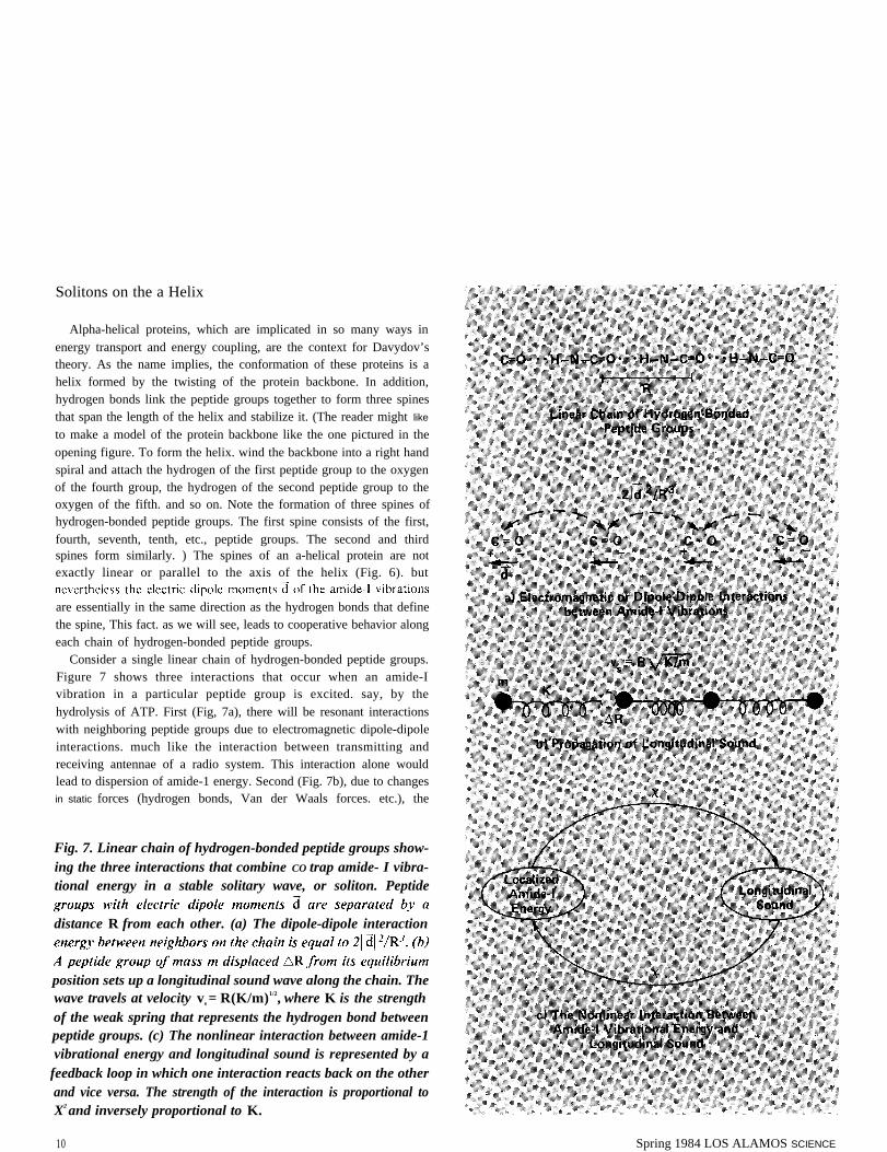
Consider a single linear chain of hydrogen-bonded peptide groups.Figure 7 shows three interactions that occur when an amide-Ivibration in a particular peptide group is excited. say, by thehydrolysis of ATP. First (Fig, 7a), there will be resonant interactionswith neighboring peptide groups due to electromagnetic dipole-dipoleinteractions. much like the interaction between transmitting andreceiving antennae of a radio system. This interaction alone wouldlead to dispersion of amide-1 energy. Second (Fig. 7b), due to changesin static forces (hydrogen bonds, Van der Waals forces. etc.), the
Fig. 7. Linear chain of hydrogen-bonded peptide groups show-ing the three interactions that combine CO trap amide- I vibra-tional energy in a stable solitary wave, or soliton. Peptide
distance R from each other. (a) The dipole-dipole interaction
position sets up a longitudinal sound wave along the chain. Thewave travels at velocity vs = R(K/m)1/2, where K is the strengthof the weak spring that represents the hydrogen bond betweenpeptide groups. (c) The nonlinear interaction between amide-1vibrational energy and longitudinal sound is represented by a
feedback loop in which one interaction reacts back on the otherand vice versa. The strength of the interaction is proportional toX2 and inversely proportional to K.
10 Spring 1984 LOS ALAMOS SCIENCE
Solitons in Biology
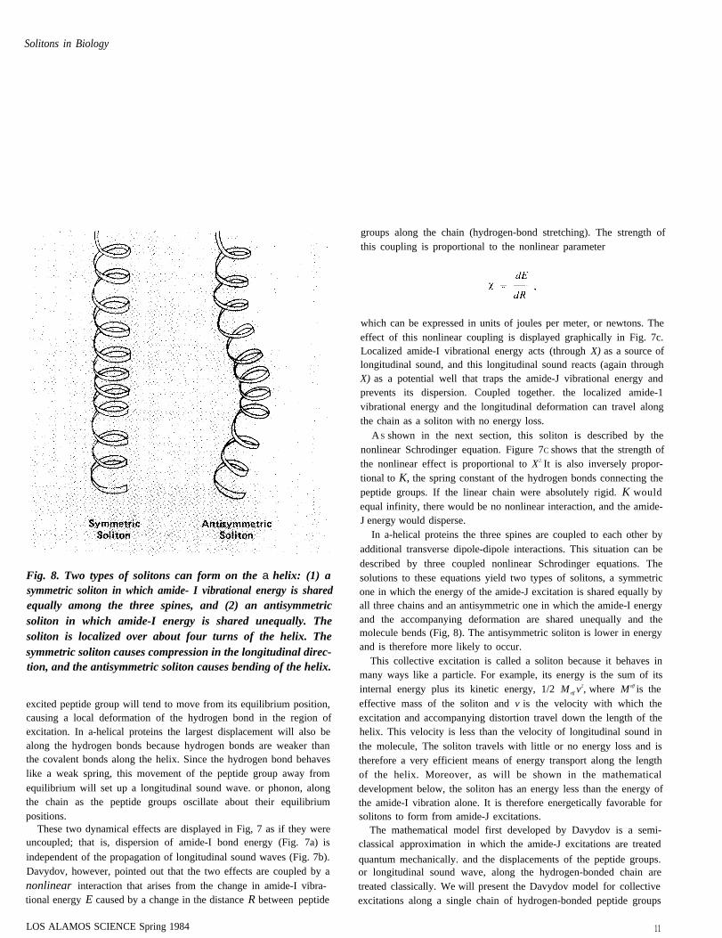
Fig. 8. Two types of solitons can form on the a helix: (1) asymmetric soliton in which amide- I vibrational energy is sharedequally among the three spines, and (2) an antisymmetricsoliton in which amide-I energy is shared unequally. Thesoliton is localized over about four turns of the helix. Thesymmetric soliton causes compression in the longitudinal direc-tion, and the antisymmetric soliton causes bending of the helix.
excited peptide group will tend to move from its equilibrium position,causing a local deformation of the hydrogen bond in the region ofexcitation. In a-helical proteins the largest displacement will also bealong the hydrogen bonds because hydrogen bonds are weaker thanthe covalent bonds along the helix. Since the hydrogen bond behaveslike a weak spring, this movement of the peptide group away fromequilibrium will set up a longitudinal sound wave. or phonon, alongthe chain as the peptide groups oscillate about their equilibriumpositions.
These two dynamical effects are displayed in Fig, 7 as if they wereuncoupled; that is, dispersion of amide-I bond energy (Fig. 7a) is
independent of the propagation of longitudinal sound waves (Fig. 7b).Davydov, however, pointed out that the two effects are coupled by anonlinear interaction that arises from the change in amide-I vibra-tional energy E caused by a change in the distance R between peptide
groups along the chain (hydrogen-bond stretching). The strength ofthis coupling is proportional to the nonlinear parameter
which can be expressed in units of joules per meter, or newtons. Theeffect of this nonlinear coupling is displayed graphically in Fig. 7c.Localized amide-I vibrational energy acts (through X) as a source oflongitudinal sound, and this longitudinal sound reacts (again throughX) as a potential well that traps the amide-J vibrational energy andprevents its dispersion. Coupled together. the localized amide-1vibrational energy and the longitudinal deformation can travel alongthe chain as a soliton with no energy loss.
A S shown in the next section, this soliton is described by thenonlinear Schrodinger equation. Figure 7C shows that the strength ofthe nonlinear effect is proportional to X2. It is also inversely propor-tional to K, the spring constant of the hydrogen bonds connecting thepeptide groups. If the linear chain were absolutely rigid. K wouldequal infinity, there would be no nonlinear interaction, and the amide-J energy would disperse.
In a-helical proteins the three spines are coupled to each other byadditional transverse dipole-dipole interactions. This situation can bedescribed by three coupled nonlinear Schrodinger equations. Thesolutions to these equations yield two types of solitons, a symmetricone in which the energy of the amide-J excitation is shared equally byall three chains and an antisymmetric one in which the amide-I energyand the accompanying deformation are shared unequally and themolecule bends (Fig, 8). The antisymmetric soliton is lower in energyand is therefore more likely to occur.
This collective excitation is called a soliton because it behaves inmany ways like a particle. For example, its energy is the sum of itsinternal energy plus its kinetic energy, 1/2 M eff v
2, where M eff is theeffective mass of the soliton and v is the velocity with which theexcitation and accompanying distortion travel down the length of thehelix. This velocity is less than the velocity of longitudinal sound inthe molecule, The soliton travels with little or no energy loss and istherefore a very efficient means of energy transport along the lengthof the helix. Moreover, as will be shown in the mathematicaldevelopment below, the soliton has an energy less than the energy ofthe amide-I vibration alone. It is therefore energetically favorable forsolitons to form from amide-J excitations.
The mathematical model first developed by Davydov is a semi-classical approximation in which the amide-J excitations are treated
quantum mechanically. and the displacements of the peptide groups.or longitudinal sound wave, along the hydrogen-bonded chain aretreated classically. We will present the Davydov model for collectiveexcitations along a single chain of hydrogen-bonded peptide groups
LOS ALAMOS SCIENCE Spring 1984 11
and show how the continuum approximation of this model leads tothe nonlinear Schrodinger equation and its well-known soliton solu-tions. (The reader unfamiliar with the formalism of quantummechanics may skip the next section without losing the main points ofthe article.)
The Davydov Model: How It Yields Soliton Solutions
The energy operator H, or Hamiltonian, for the collective excita-tion along the chain is a sum of three operators: H = H amide-1 + Hphonon
interaction , where H amide-I is the operator for the amide-I vibrationalexcitations. H phonon is the operator for the displacements of thepeptide groups, and H interaction is the operator for the interactionbetween the amide-I excitations and the displacements.
If E is the amide-f excitation energy and B+
n is an operator for
creation of this excitation on the nth peptide group, then H amide-1 isgiven by
where the summation is carried out over all N peptide groups. The
first term, E B+
nB n, defines the amide-I excitation energy, and thesecond term describes the resonance dipole interaction between
transfer of amide-I energy from peptide group n to n±1 due to thedipole-dipole interaction. The dipole-dipole interaction energy J is
The energy H phonon associated with displacing the peptide groupsaway from their equilibrium positions is given in the harmonicapproximation by
where Un is the displacement of the nth peptide group, m is the mass ofthe peptide group, and K is the spring constant, or elasticitycoefficient, of the hydrogen bonds forming the linear chain. The firstterm is kinetic energy and the second potential energy.
The Hamiltonian for the interaction between the amide-f excitationand the displacements of the peptide groups takes the form
12
where the coupling constant X, as mentioned earlier, represents thechange in amide-I energy per unit extension of an adjacent hydrogenbond.
The total Hamiltonian H = H amide-1 + Hphonon + H interaction of thesystem must satisfy the Schrodinger equation:
(4)
expressed by
(5)
and satisfies the normalization condition
(6)
amide-f vibration, that is, the ground state of the system.Substituting Eq. 5 in Eq. 4 we get, after some algebra. the following
set of differential equations:
and
Equations 7 and 8 are the main result of Davydov’s original model.They describe the time evolution of amide-I vibrational energycoupled to displacements of the hydrogen-bonded chain of peptide
amide-f energy over the individual peptide groups of the chain.In order to demonstrate that an(t), the probability amplitude of the
excitation, does behave like a soliton, we will restrict ourselves tosolutions of Eqs. 7 and 8 that vary slowly as a function of the peptidegroup number n. In this limit we can replace the functions a fl(f) and
un(t) with continuous functions a(x,t) and u(x,t), thus approximatingn with the dimensionless coordinate x. Equations 7 and 8 thenbecome
Spring 1984 LOS ALAMOS SCIENCE
Solitons in Biology
and
where
(9)
(10)
The left side of Eq. 10 is essentially, a wave equation for longitudinalsound in the system of coupled peptide groups; the sound velocity vs isgiven by vs = R(K/m) 1/2 . The right side acts as a source term forgeneration of sound.
We shall seek traveling wave solutions of Eqs. 9 and 10 in the formof excitations that propagate along the chain with a velocity v;that is,
I (11)
Inserting Eq. 11 in Eq. 10, we get
(12)
where s is the ratio of the propagation velocity to the velocity ofsound: s = v/vs < 1.
Substituting Eq. 12 into Eq. 9, we get
(13)
where K = 4x2/K( 1 – s2).Equation 13 is the nonlinear Schrodinger equation, which has
soliton solutions. For its general solution we refer the interestedreader to “Exact Theory of Two-Dimensional Self-Focusing andOne-Dimensional Self-Modulation of Waves in Nonlinear Media” byV. E. Zakharov and A. B. Shabat (Soviet Physics JETP 34( 1972):
62-69). It is sufficient for our purpose hereto look only at a stationarysolution, that is, one for whichs = 0. In this case a solution of Eq.given by
13 is
14)
The constant x0 is the position of maximum probability of amide-Iexcitation along the chain, and the pulse-shaped form given by Eq. 14falls off rapidly when one moves away from X O (see “What Is aSoliton?”). Equation 14 also satisfies the continuous equivalent of Eq.
indicating that one quantum of amide-I energy is excited on thepeptide chain.
The energy E,o) associated with the soliton is given by
Inserting the solution given by Eq. 14, we get
This is the energy of a stationary soliton. A similar but morecomplicated expression can be obtained for moving solitons.
It is instructive to see what happens in an absolutely rigid chain of
constant K in the nonlinear term of Eq. 13 is equal to zero. Equation13 is then a linear Schrodinger equation, which has solutions in theform of plane waves. This means that an excitation is uniformlydistributed along the whole chain. In other words, the amide-I energyhas dispersed and is no longer localized. It can also be seen from Eq.16 that the energy of such an extended excitation (or exciton) equalsE - 2J, which is larger by an amount X
4/3K2J than the energy E,ol ofthe spatially localized soliton excitation. It is thus more favorable forthe system to localize its energy when the nonlinear coupling betweenamide-I energy and displacement of the associated peptide group istaken into account.
LOS ALAMOS SCIENCE Spring 1984 13
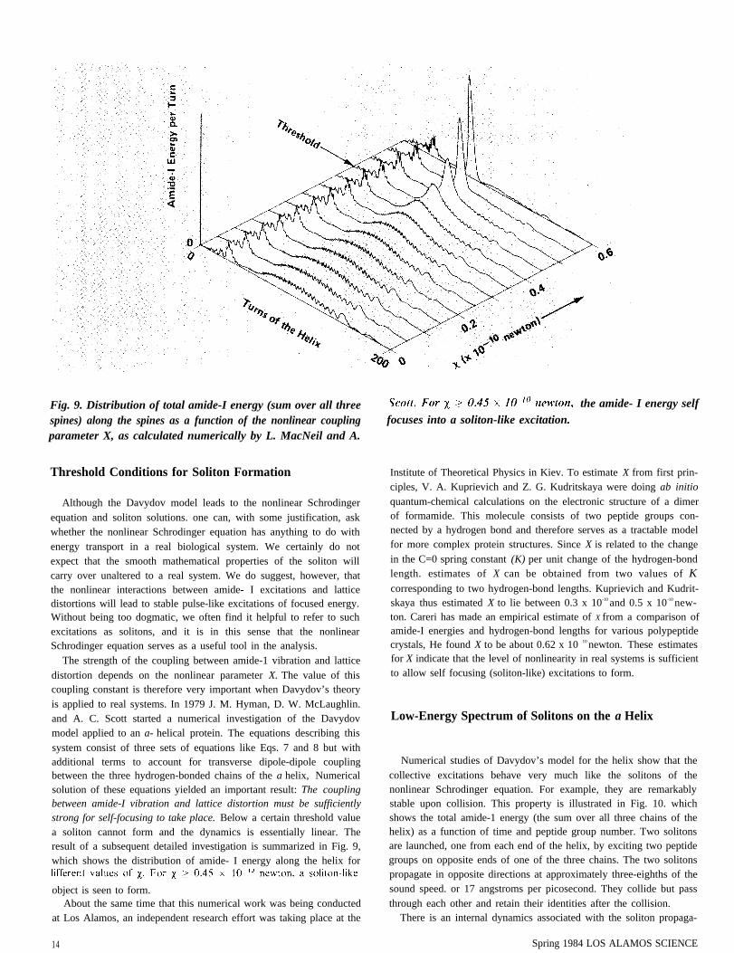
Fig. 9. Distribution of total amide-I energy (sum over all threespines) along the spines as a function of the nonlinear couplingparameter X, as calculated numerically by L. MacNeil and A.
Threshold Conditions for Soliton Formation
Although the Davydov model leads to the nonlinear Schrodingerequation and soliton solutions. one can, with some justification, askwhether the nonlinear Schrodinger equation has anything to do withenergy transport in a real biological system. We certainly do notexpect that the smooth mathematical properties of the soliton willcarry over unaltered to a real system. We do suggest, however, thatthe nonlinear interactions between amide- I excitations and latticedistortions will lead to stable pulse-like excitations of focused energy.Without being too dogmatic, we often find it helpful to refer to suchexcitations as solitons, and it is in this sense that the nonlinearSchrodinger equation serves as a useful tool in the analysis.
The strength of the coupling between amide-1 vibration and latticedistortion depends on the nonlinear parameter X. The value of thiscoupling constant is therefore very important when Davydov’s theoryis applied to real systems. In 1979 J. M. Hyman, D. W. McLaughlin.and A. C. Scott started a numerical investigation of the Davydovmodel applied to an a- helical protein. The equations describing thissystem consist of three sets of equations like Eqs. 7 and 8 but withadditional terms to account for transverse dipole-dipole couplingbetween the three hydrogen-bonded chains of the a helix, Numericalsolution of these equations yielded an important result: The couplingbetween amide-I vibration and lattice distortion must be sufficientlystrong for self-focusing to take place. Below a certain threshold valuea soliton cannot form and the dynamics is essentially linear. Theresult of a subsequent detailed investigation is summarized in Fig. 9,which shows the distribution of amide- I energy along the helix for
object is seen to form.About the same time that this numerical work was being conducted
at Los Alamos, an independent research effort was taking place at the
14
focuses into a soliton-like excitation.the amide- I energy self
Institute of Theoretical Physics in Kiev. To estimate X from first prin-ciples, V. A. Kuprievich and Z. G. Kudritskaya were doing ab initio
quantum-chemical calculations on the electronic structure of a dimerof formamide. This molecule consists of two peptide groups con-nected by a hydrogen bond and therefore serves as a tractable modelfor more complex protein structures. Since X is related to the changein the C=0 spring constant (K) per unit change of the hydrogen-bondlength. estimates of X can be obtained from two values of Kcorresponding to two hydrogen-bond lengths. Kuprievich and Kudrit-skaya thus estimated X to lie between 0.3 x 10⁻¹⁰ and 0.5 x 10⁻¹⁰ new-ton. Careri has made an empirical estimate of X from a comparison ofamide-I energies and hydrogen-bond lengths for various polypeptidecrystals, He found X to be about 0.62 x 10 10 newton. These estimatesfor X indicate that the level of nonlinearity in real systems is sufficientto allow self focusing (soliton-like) excitations to form.
Low-Energy Spectrum of Solitons on the a Helix
Numerical studies of Davydov’s model for the helix show that thecollective excitations behave very much like the solitons of thenonlinear Schrodinger equation. For example, they are remarkablystable upon collision. This property is illustrated in Fig. 10. whichshows the total amide-1 energy (the sum over all three chains of thehelix) as a function of time and peptide group number. Two solitonsare launched, one from each end of the helix, by exciting two peptidegroups on opposite ends of one of the three chains. The two solitonspropagate in opposite directions at approximately three-eighths of thesound speed. or 17 angstroms per picosecond. They collide but passthrough each other and retain their identities after the collision.
There is an internal dynamics associated with the soliton propaga-
Spring 1984 LOS ALAMOS SCIENCE
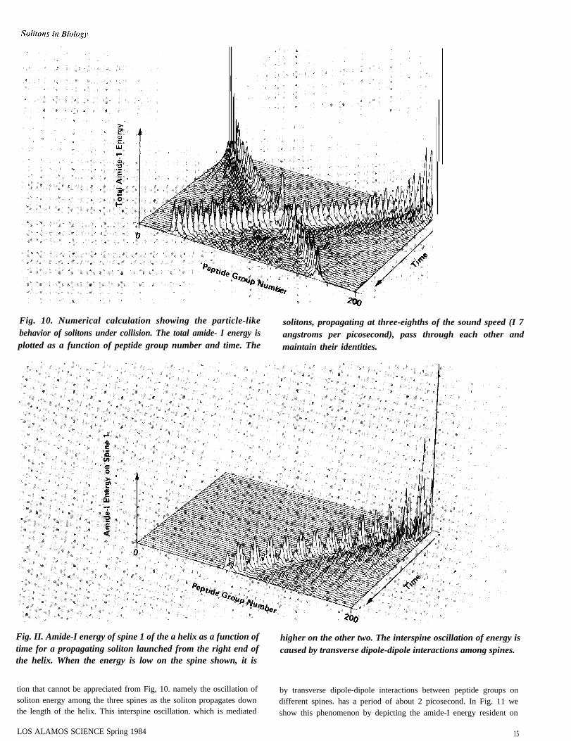
Fig. 10. Numerical calculation showing the particle-like solitons, propagating at three-eighths of the sound speed (I 7behavior of solitons under collision. The total amide- I energy is angstroms per picosecond), pass through each other andplotted as a function of peptide group number and time. The maintain their identities.
Fig. II. Amide-I energy of spine 1 of the a helix as a function of higher on the other two. The interspine oscillation of energy istime for a propagating soliton launched from the right end of caused by transverse dipole-dipole interactions among spines.the helix. When the energy is low on the spine shown, it is
tion that cannot be appreciated from Fig, 10. namely the oscillation of by transverse dipole-dipole interactions between peptide groups onsoliton energy among the three spines as the soliton propagates down different spines. has a period of about 2 picosecond. In Fig. 11 wethe length of the helix. This interspine oscillation. which is mediated show this phenomenon by depicting the amide-I energy resident on
LOS ALAMOS SCIENCE Spring 1984 15
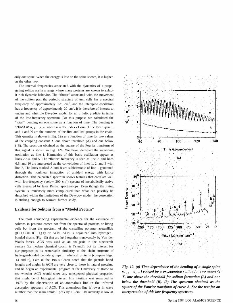
only one spine. When the energy is low on the spine shown, it is higheron the other two.
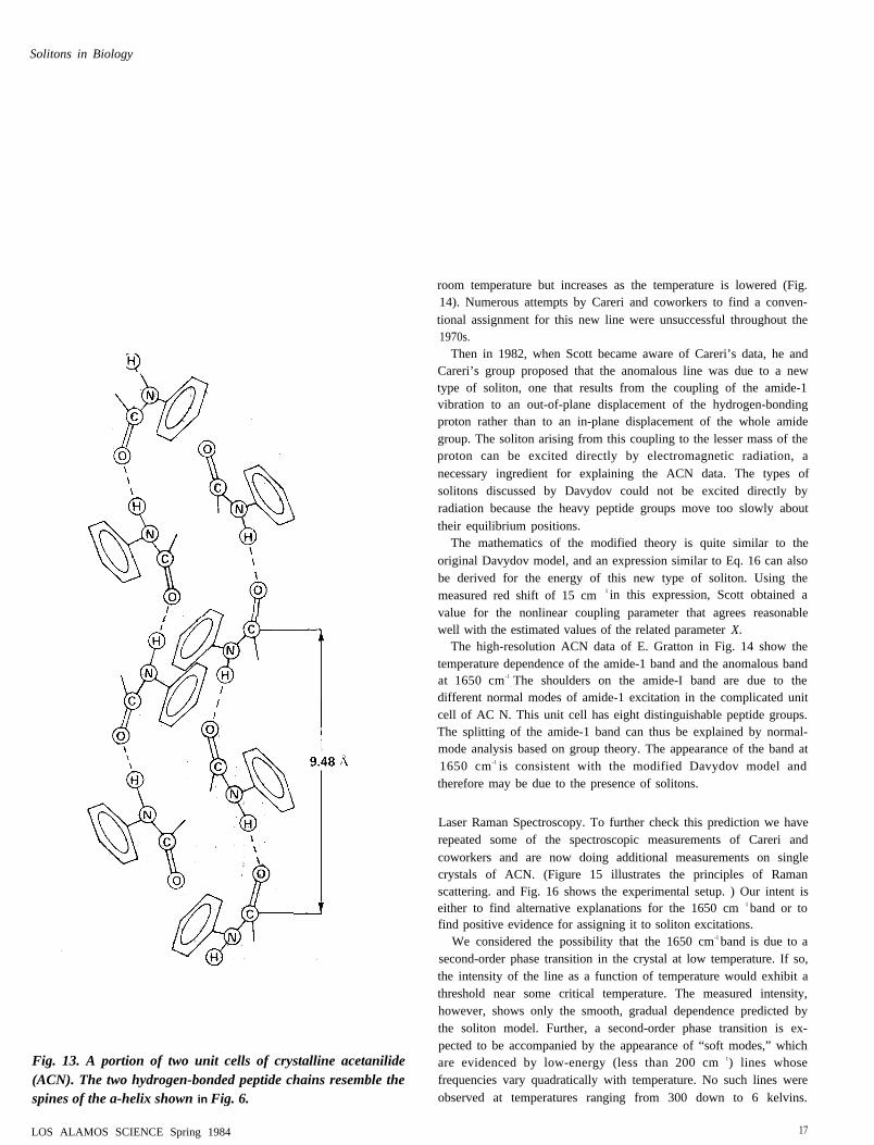
The internal frequencies associated with the dynamics of a propa-gating soliton are in a range where many proteins are known to exhib-it rich dynamic behavior. The “flutter” associated with the movementof the soliton past the periodic structure of unit cells has a spectralfrequency of approximately 125 cm-1, and the interspine oscillationhas a frequency of approximately 20 cm-1. It is therefore of interest tounderstand what the Davydov model for an u helix predicts in termsof the low-frequency spectrum. For this purpose we calculated the“total’” bending on one spine as a function of time. The bending is
and 1 and N are the numbers of the first and last groups in the chain.This quantity is shown in Fig. 12a as a function of time for two valuesof the coupling constant X. one above threshold (A) and one below( B). The spectrum obtained as the square of the Fourier transform ofthis signal is shown in Fig. 12b. We have identified the interspineoscillation as line 1. Harmonics of this basic oscillation appear aslines 2.3.4. and 5. The “flutter” frequency is seen as line 7, and lines6.8. and 10 are interpreted as the convolution of lines 1, 2, and 3 withline 7, The lines marked A and B are subharmonic of line 1 generatedthrough the nonlinear interaction of amide-I energy with latticedistortion. This calculated spectrum shows features that correlate wellwith low-frequency (below 200 cm-1) spectra of metabolically activecells measured by laser Raman spectroscopy. Even though the livingsystem is immensely more complicated than what can possibly bedescribed within the limitations of the Davydov model, the correlationis striking enough to warrant further study.
Evidence for Solitons from a “Model Protein”
The most convincing experimental evidence for the existence of
solitons in proteins comes not from the spectra of proteins or livingcells but from the spectrum of the crystalline polymer acetanilide((CH 3CONHC 6H 5) x), or ACN. ACN is organized into hydrogen-bonded chains (Fig. 13) that are held together transversely by Van derWaals forces. ACN was used as an analgesic in the nineteenthcentury (its modern chemical cousin is Tylenol), but its interest forour purposes is its remarkable similarity to the chain structure ofhydrogen-bonded peptide groups in a-helical proteins (compare Figs.13 and 6), Late in the 1960s Careri noted that the peptide bondlengths and angles in ACN are very close to those in natural proteins.and he began an experimental program at the University of Rome tosee whether ACN would show any unexpected physical propertiesthat might be of biological interest. His intuition was rewarded in1973 by the observation of an anomalous line in the infraredabsorption spectrum of ACN. This anomalous line is lower in wavenumber than the main amide-I peak by 15 cm-1. Its intensity is low at
Fig. 12. (a) Time dependence of the bending of a single spine
X, one above the threshold for soliton formation (A) and onebelow the threshold (B). (b) The spectrum obtained as thesquare of the Fourier transform of curve A. See the text for aninterpretation of this low-frequency spectrum.
16 Spring 1984 LOS ALAMOS SCIENCE
Solitons in Biology
.
Fig. 13. A portion of two unit cells of crystalline acetanilide(ACN). The two hydrogen-bonded peptide chains resemble thespines of the a-helix shown in Fig. 6.
LOS ALAMOS SCIENCE Spring 1984
room temperature but increases as the temperature is lowered (Fig.14). Numerous attempts by Careri and coworkers to find a conven-tional assignment for this new line were unsuccessful throughout the1970s.
Then in 1982, when Scott became aware of Careri’s data, he andCareri’s group proposed that the anomalous line was due to a newtype of soliton, one that results from the coupling of the amide-1vibration to an out-of-plane displacement of the hydrogen-bondingproton rather than to an in-plane displacement of the whole amidegroup. The soliton arising from this coupling to the lesser mass of theproton can be excited directly by electromagnetic radiation, anecessary ingredient for explaining the ACN data. The types ofsolitons discussed by Davydov could not be excited directly byradiation because the heavy peptide groups move too slowly abouttheir equilibrium positions.
The mathematics of the modified theory is quite similar to theoriginal Davydov model, and an expression similar to Eq. 16 can alsobe derived for the energy of this new type of soliton. Using themeasured red shift of 15 cm 1 in this expression, Scott obtained a
value for the nonlinear coupling parameter that agrees reasonablewell with the estimated values of the related parameter X.
The high-resolution ACN data of E. Gratton in Fig. 14 show thetemperature dependence of the amide-1 band and the anomalous bandat 1650 cm-1
. The shoulders on the amide-I band are due to thedifferent normal modes of amide-1 excitation in the complicated unitcell of AC N. This unit cell has eight distinguishable peptide groups.The splitting of the amide-1 band can thus be explained by normal-mode analysis based on group theory. The appearance of the band at1650 cm -1 is consistent with the modified Davydov model andtherefore may be due to the presence of solitons.
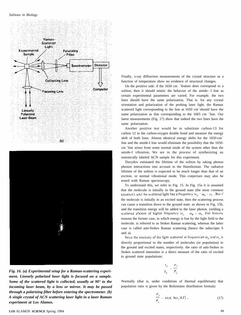
Laser Raman Spectroscopy. To further check this prediction we haverepeated some of the spectroscopic measurements of Careri andcoworkers and are now doing additional measurements on singlecrystals of ACN. (Figure 15 illustrates the principles of Ramanscattering. and Fig. 16 shows the experimental setup. ) Our intent iseither to find alternative explanations for the 1650 cm 1 band or tofind positive evidence for assigning it to soliton excitations.
We considered the possibility that the 1650 cm-1 band is due to asecond-order phase transition in the crystal at low temperature. If so,the intensity of the line as a function of temperature would exhibit athreshold near some critical temperature. The measured intensity,however, shows only the smooth, gradual dependence predicted bythe soliton model. Further, a second-order phase transition is ex-pected to be accompanied by the appearance of “soft modes,” whichare evidenced by low-energy (less than 200 cm 1) lines whosefrequencies vary quadratically with temperature. No such lines wereobserved at temperatures ranging from 300 down to 6 kelvins.
17
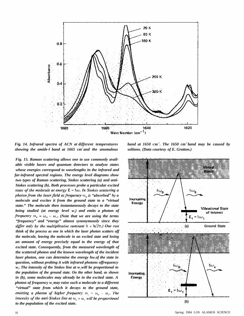
Fig. 14. Infrared spectra of ACN at different temperaturesshowing the amide-l band at 1665 cm-l and the anomalous
Fig. 15. Raman scattering allows one to use commonly avail-able visible lasers and quantum detectors to analyze stateswhose energies correspond to wavelengths in the infrared and
far-infrared spectral regions. The energy level diagrams showtwo types of Raman scattering, Stokes scattering (a) and anti-Stokes scattering (b). Both processes probe a particular excited
molecule and excites it from the ground state to a “virtualstate.” The molecule then instantaneously decays to the statebeing studied (at energy level w1) and emits a photon of
(Note that we are using the terms“frequency” and “energy” almost synonymously since they
think of the process as one in which the laser photon scatters offthe molecule, leaving the molecule in an excited state and losingan amount of energy precisely equal to the energy of thatexcited state. Consequently, from the measured wavelength ofthe scattered photon and the known wavelength of the incidentlaser photon, one can determine the energy hwl of the state inquestion, without probing it with infrared photons offrequencyw1. The intensity of the Stokes line at wI will be proportional tothe population of the ground state. On the other hand, as shownin (b), some molecules may already be in the excited state. Aphoton of frequency wo may raise such a molecule to a different“virtual” state from which it decays to the ground state,
to the population of the excited state.
18
band at 1650 cm -1. The 1650 cm-1 band may be caused bysolitons. (Data courtesy of E. Gratton.)
(a) Ground State
(b)
Spring 1984 LOS ALAMOS SCIENCE
Solitons in Biology
Fig. 16. (a) Experimental setup for a Raman-scattering experi-ment. Linearly polarized laser light is focused on a sample.Some of the scattered light is collected, usually at 90° to theincoming laser beam, by a lens or mirror. It may be passedthrough a polarizing filter before entering the spectrometer. (b)A single crystal of ACN scattering laser light in a laser Ramanexperiment at Los Alamos.
Finally, x-ray diffraction measurements of the crystal structure as afunction of temperature show no evidence of structural changes.
On the positive side. if the 1650 cm 1 feature does correspond to asoliton, then it should mimic the behavior of the amide- I line ascertain experimental parameters are varied. For example. the twolines should have the same polarization. That is. for any crystalorientation and polarization of the probing laser light. the Ramanscattered light corresponding to the line at 1650 cm-1 should have thesame polarization as that corresponding to the 1665 cm 1 line. Ourlatest measurements (Fig. 17) show that indeed the two lines have thesame polarization.
Another positive test would be to substitute carbon-13 forcarbon 12 in the carbon-oxygen double bond and measure the energyshift of both lines. Almost identical energy shifts for the 1650-cm-1
line and the amide-I line would eliminate the possibility that the 1650-cm 1 line arises from some normal mode of the system other than theamide-I vibration, We are in the process of synthesizing an
isotonically labeled ACN sample for this experiment.Davydov estimated the lifetime of the soliton by taking photon
phonon interactions into account in the Hamiltonian. The radiativelifetime of the soliton is expected to be much longer than that of anexciton. or normal vibrational mode. This conjecture may also betested with Raman spectroscopy.
To understand this, we refer to Fig. 15. In Fig. 15a it is assumedthat the molecule is initially in the ground state (the most common
the molecule is initially in an excited state, then the scattering processcan cause a transition down to the ground state. as shown in Fig. 15b,and the transition energy will be added to the laser photon. yielding a
reasons the former case, in which energy is lost by the light field to themolecule. is referred to as Stokes Raman scattering, whereas the lattercase is called anti-Stokes Raman scattering (hence the subscripts Sand a).
directly proportional to the number of molecules (or population) inthe ground and excited states, respectively. the ratio of anti-Stokes toStokes scattered intensities is a direct measure of the ratio of excitedto ground state populations:
Normally (that is, under conditions of thermal equilibrium) that
population ratio is given by the Boltzmann distribution formula:
(17)
LOS ALAMOS SCIENCE Spring 1984
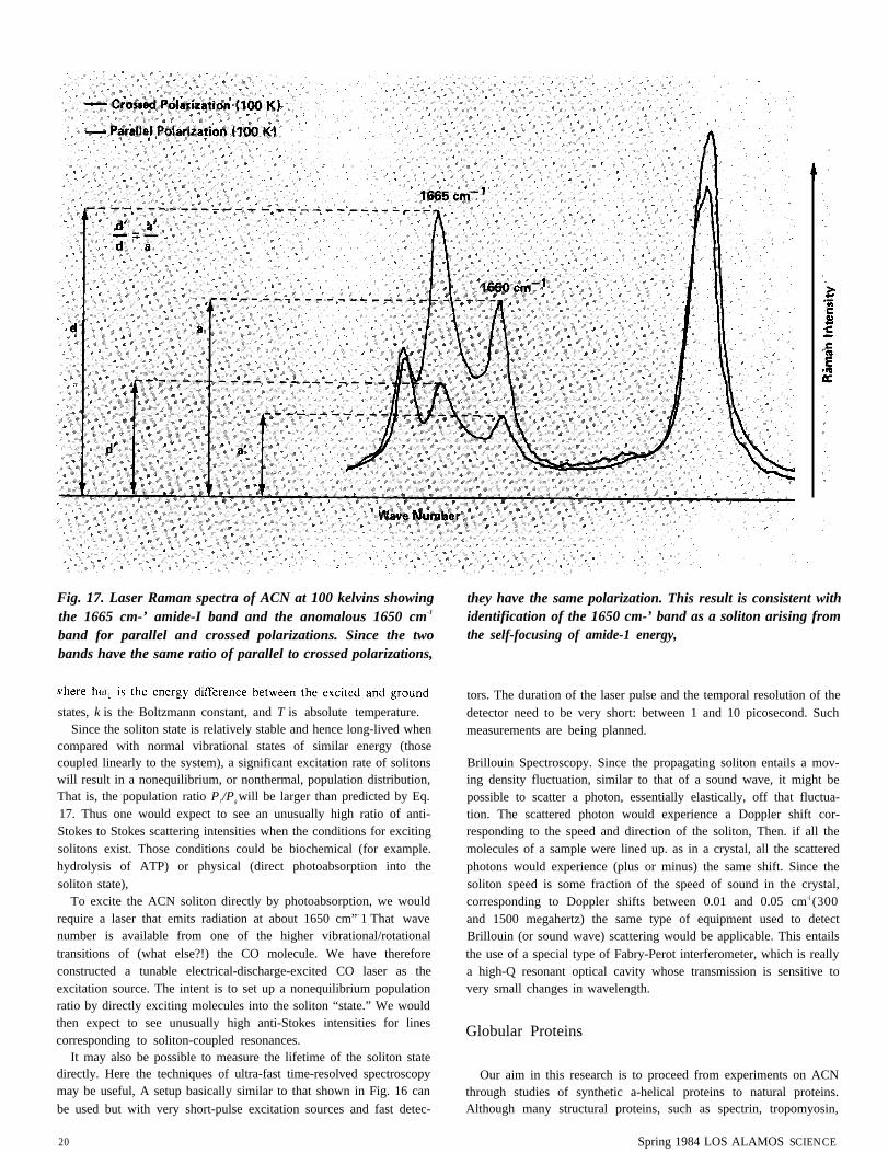
Fig. 17. Laser Raman spectra of ACN at 100 kelvins showingthe 1665 cm-’ amide-I band and the anomalous 1650 cm-1
band for parallel and crossed polarizations. Since the twobands have the same ratio of parallel to crossed polarizations,
states, k is the Boltzmann constant, and T is absolute temperature.Since the soliton state is relatively stable and hence long-lived when
compared with normal vibrational states of similar energy (thosecoupled linearly to the system), a significant excitation rate of solitonswill result in a nonequilibrium, or nonthermal, population distribution,That is, the population ratio P1/Pg will be larger than predicted by Eq.17. Thus one would expect to see an unusually high ratio of anti-Stokes to Stokes scattering intensities when the conditions for excitingsolitons exist. Those conditions could be biochemical (for example.hydrolysis of ATP) or physical (direct photoabsorption into thesoliton state),
To excite the ACN soliton directly by photoabsorption, we wouldrequire a laser that emits radiation at about 1650 cm”- 1 . That wavenumber is available from one of the higher vibrational/rotationaltransitions of (what else?!) the CO molecule. We have thereforeconstructed a tunable electrical-discharge-excited CO laser as theexcitation source. The intent is to set up a nonequilibrium populationratio by directly exciting molecules into the soliton “state.” We wouldthen expect to see unusually high anti-Stokes intensities for linescorresponding to soliton-coupled resonances.
It may also be possible to measure the lifetime of the soliton statedirectly. Here the techniques of ultra-fast time-resolved spectroscopymay be useful, A setup basically similar to that shown in Fig. 16 can
be used but with very short-pulse excitation sources and fast detec-
20
they have the same polarization. This result is consistent withidentification of the 1650 cm-’ band as a soliton arising fromthe self-focusing of amide-1 energy,
tors. The duration of the laser pulse and the temporal resolution of thedetector need to be very short: between 1 and 10 picosecond. Suchmeasurements are being planned.
Brillouin Spectroscopy. Since the propagating soliton entails a mov-ing density fluctuation, similar to that of a sound wave, it might bepossible to scatter a photon, essentially elastically, off that fluctua-tion. The scattered photon would experience a Doppler shift cor-responding to the speed and direction of the soliton, Then. if all themolecules of a sample were lined up. as in a crystal, all the scatteredphotons would experience (plus or minus) the same shift. Since thesoliton speed is some fraction of the speed of sound in the crystal,corresponding to Doppler shifts between 0.01 and 0.05 cm-[ (300and 1500 megahertz) the same type of equipment used to detectBrillouin (or sound wave) scattering would be applicable. This entailsthe use of a special type of Fabry-Perot interferometer, which is reallya high-Q resonant optical cavity whose transmission is sensitive tovery small changes in wavelength.
Globular Proteins
Our aim in this research is to proceed from experiments on ACNthrough studies of synthetic a-helical proteins to natural proteins.Although many structural proteins, such as spectrin, tropomyosin,
Spring 1984 LOS ALAMOS SCIENCE

Solitons in Biology
and myosin are almost entirely a helical, there are many other
important proteins that are globular. We have seen that the competi-tion between dispersion and focusing of amide-I energy leads—in a-helical proteins or acetanilide crystals—to the formation of a soliton-like object that can travel along the chain of hydrogen-bonded peptidegroups without changing its shape. This is essentially a manifestationof the fact that the system has perfect translational symmetry. Anatural question to ask is: “What is the result of the competition inglobular proteins?” Such proteins do not have translational in-variance among the different peptide groups, and soliton formation isnot to be expected. However, the mechanisms for dispersion andfocusing of amide-I energy are still present.
One way to generalize Davydov’s ideas to a globular protein is totake the full geometry of the molecule into account when calculatingthe dipole-dipole interactions. The size of the J term in the amide-IHamiltonian (Eq. 1) will vary from peptide group to peptide group,
and all possible dipole-dipole interactions have to be considered inorder to account for dispersion of amide-I energy. A preliminarycomputer code, based on assumptions corresponding to those leadingto Eq. 13, has been developed for arbitrary protein geometry. Thiscode provides evidence of self-focusing of amide-I vibrational energyin acetanilide (see cover) and in globular proteins. As our physicalexperiments evolve toward biologically realistic preparations, we planto make corresponding improvements in this code.
Our present understanding of energy migration in biological
systems is very much in its infancy. Our research efforts are directedtoward identifying simple but important features in this context. Thekey scientific question that we have raised in this article may bestated: Is self-trapping of amide-I energy important for transportphenomena in biological materials? Experimental and theoreticalstudies on model proteins have so far led us to expect an affirmativeanswer to this question. ■
Further Reading
A. S. Davydov and N. I. Kislukha. “Solitary Excitons in One-Dimensional Molecular Chains.” physicastatus solidi (b) 59(1973):465-470.
A. S. Davydov. “The Theory of Contraction of Proteins under Their Excitation.” Journal of TheoreticalBiology 38( 1973):559-569.
A. S. Davydov. Biology and Quantum Mechanics. Oxford: Pergamon Press Ltd., 1982.
G. Careri. “Search for Cooperative Phenomena in Hydrogen-Bonded Amide Structures.” In CooperativePhenomena, H. Haken and M. Wagner, editors (Springer-Verlag, 1973), pp. 391-394..
V. E. Zakharov and A. B. Shabat. “Exact Theory of Two-Dimensional Self-Focusing and One-Dimensional Self-Modulation of Waves in Nonlinear Media.” Soviet Physics JETP 34(1972):62-69.
J. M. Hyman, D. W. McLaughlin, and A. C. Scott. “On Davydov’s Alpha-Helix Solitons.” Physica D3D(1981):23-44.
Alwyn C. Scott. “Dynamics of Davydov Solitons.”Physical Review A 26(1982):578-595.
Ahwyn C. Scott. “The Vibrational Structure of Davydov Solitons.”Physica Scripta 25(1982):651-658.
L. MacNeil and A. C. Scott. “Launching a Davydov Soliton II: Numerical Studies.” Physica Scripta. Inpress.
P. S. Lomdahl, L. MacNeil, A. C. Scott, M. E. Stoneham, and S. J. Webb. “An Assignment to InternalSoliton Vibrations of Laser-Raman Lines from Living Cells.” Physics Letters 92 A(1982):207-210.
G. Careri, U. Buontempo, F. Carta, E. Gratton, and A. C. Scott. “Infrared Absorption in Acetanilide bySolitons.” Physical Review Letters 51(1983):304-307.
G. Careri, U. Buontempo, F. Galluzzi, A. C. Scott, E. Gratton, and E. Schyamsunder. “SpectroscopicEvidence for Davydov-Like Solitons in Acetanilide.” Los Alamos National Laboratory unclassifiedrelease LA-UR-84-483 and submitted to Physical Review B.
Peter S. Lomdahl. “Nonlinear Dynamics of Globular Proteins.” Los Alamos National Laboratoryunclassified release LA-UR-83-2252 and to be published by Plenum Press in Nonlinear Electrodynamicsin Biological Systems.
LOS ALAMOS SCIENCE Spring 1984 21
I
AUTHORS
Peter S. Lomdahl was born and grew up in Copenhagen, Denmark. In 1979hc received an M.S. in electrical engineering and in 1982 a Ph.D. inmathematical physics from the Technical University of Denmark. Hisgraduate work specialized in nonlinear wave phenomena in superconductingJosephson junctions, which is an interest he has continued after joining thelaboratory in 1982 as a postdoctoral fellow with the Center for NonlinearStudies. The main theme in most of his research has been computer studies ofnonlinear dynamics in real condensed matter materials, including conductingpolymers and proteins. He has also done work on chaos and has a stronginterest in numerical methods for partial differential equations.
Scott P. Layne was born in Chicago, Illinois. in 1954. He received his B.A. inchemistry from DePauw University in 1976 and his M.D. from Case WesternReserve University in 1980. After completing an internship at Loma LindaUniversity in 1981. he joined the Laboratory’s Center for Nonlinear Studiesas a postdoctoral fellow. Some of his interests include energy transport bybiological molecules and the molecular mechanisms of general anesthesia.
Irving J. Bigio received his B. S., M. S., and Ph.D. degrees in physics from theUniversity of Michigan in 1969. 1970, and 1974. respectively. His doctoralwork under John Ward and Peter Franken dealt with nonlinear optics, and hehas maintained a broad interest in the field of quantum electronics ever since.He came directly to Los Alamos in April 1974 as a staff member in the laserisotope separation program and has also worked in the laser fusion program.In 1976 be received a Fulbright Senior Scholar Award and spent the 1976-77academic year as a visiting professor at the Weizmann Institute of Science,Rehovot, Israel, where he taught graduate courses in laser physics andnonlinear optics and helped direct graduate student research. Since returningto Los Alamos he has resumed his research and has taught courses at theUniversity of New Mexico Graduate Center. Currently, he iS working on avariety of topics in quantum electronics and has taken an interest in theapplication of laser techniques and nonlinear optics to the solution ofbiophysics problems. He was recently appointed Deputy Leader of theDischarge Lasers Group.
22 Spring 1984 LOS ALAMOS SCIENCE
Solitons in Biology
A Possible Mechanismfor General Anesthesia
by Scott P. Layne
RELATED TOPICS
T he first general anesthesia for human surgery was administeredat the Massachusetts General Hospital in Boston in 1846. Thepatient was put to sleep by breathing diethyl ether from a glass
vesicle. and the surgeon quickly dissected a tumor located under the
jaw. After completing the operation the surgeon remarked to hisaudience, “Gentlemen, this is no humbug. ”
Since this first successful demonstration of diethyl ether. re-searchers have discovered well over twenty drugs that induce generalanesthesia. These drugs have highly diverse chemical structures andphysical properties and, as a whole, lend little insight into theirmechanism of action. In order to overcome this perplexity, H. Meyerand E. Overton (about the year 1900) originally proposed thatanesthetic potency could be related to lipid volubility. They showedthat stronger anesthetic agents were more oil-soluble than weaker
ones and used this relationship to argue that anesthetics insert into thelipid bilayer and thereby expand its volume. More recent theoriesalong this line have suggested that the expanded lipid bilayer com-presses intrinsic membrane proteins and thereby disturbs normalprotein shape and function. These theories have suggested also thatthe membrane-bound anesthetic molecules “fluidize” the lipid bilayer.This increased fluidity, in turn, alters the permeability of the mem-brane. While these popular ideas might be applicable to agents thatare both volatile and highly lipid-soluble (oil-to-gas partition coeffi-
intravenous general anesthetics that are orders of magnitude less lipid-soluble and are capable of forming hydrogen bonds. For the case ofhydrogen-bonding anesthetic agents, the simplest idea is that they actby binding directly to a particularly sensitive protein, which may ormay not be located in a lipid membrane, and inhibiting its normalfunction.
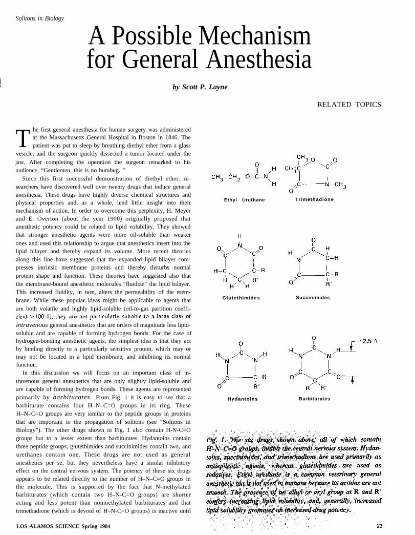
In this discussion we will focus on an important class of in-travenous general anesthetics that are only slightly lipid-soluble andare capable of forming hydrogen bonds. These agents are representedprimarily by barbiturates. From Fig. 1 it is easy to see that abarbiturate contains four H–N–C=O groups in its ring. TheseH–N–C=O groups are very similar to the peptide groups in proteinsthat are important to the propagation of solitons (see “Solitons inBiology”). The other drugs shown in Fig. 1 also contain H-N-C=Ogroups but to a lesser extent than barbiturates. Hydantoins containthree peptide groups, glutethimides and succinimides contain two, andurethanes contain one. These drugs are not used as generalanesthetics per se, but they nevertheless have a similar inhibitoryeffect on the central nervous system. The potency of these six drugsappears to be related directly to the number of H–N–C=O groups inthe molecule. This is supported by the fact that N-methylatedbarbiturates (which contain two H–N-C=O groups) are shorteracting and less potent than nonmethylated barbiturates and thattrimethadione (which is devoid of H–N-C=O groups) is inactive until
LOS ALAMOS SCIENCE Spring 1984
Ethyl Urethane Trimethadione
H
Glutethimides Succinimides
Hydantoins Barbiturates
RELATED TOPICS—
it is demethylated by hepatic enzymes. After demethylation, trimetha-dione contains two H-N-C=O groups.
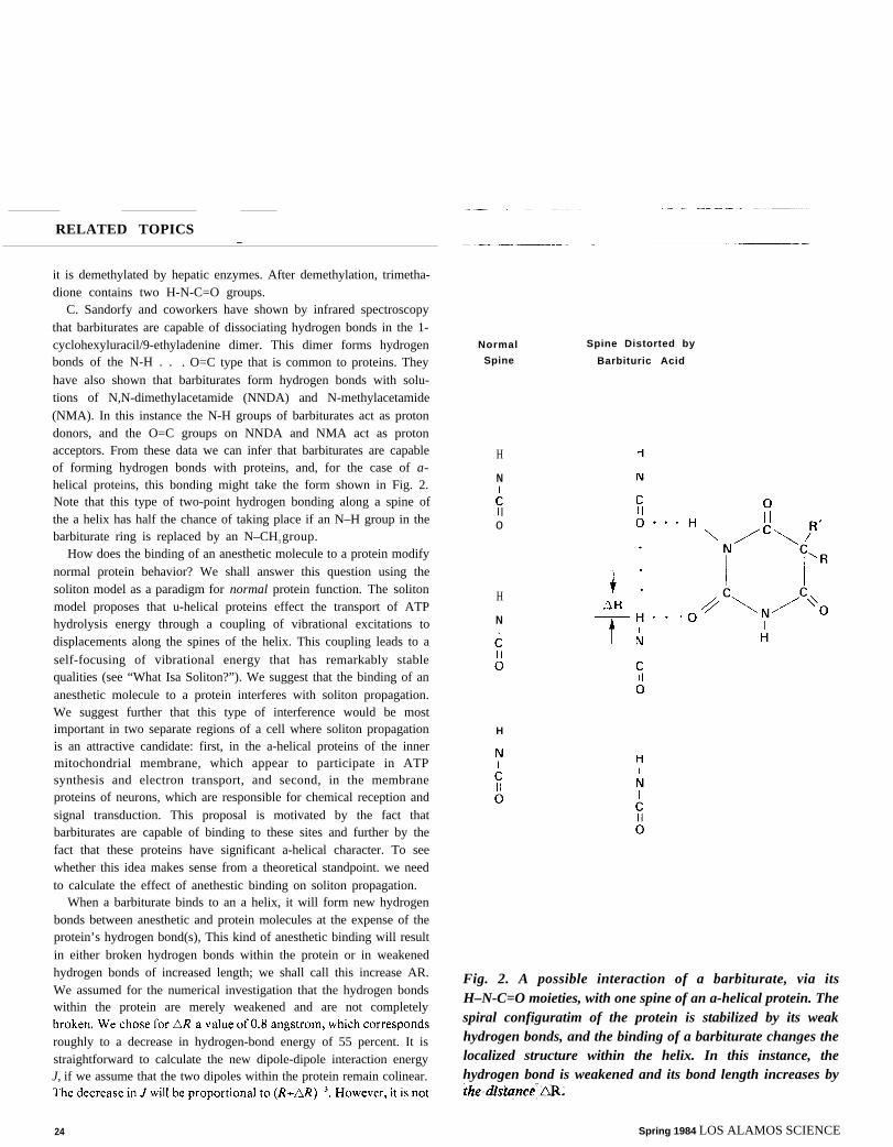
C. Sandorfy and coworkers have shown by infrared spectroscopythat barbiturates are capable of dissociating hydrogen bonds in the 1-cyclohexyluracil/9-ethyladenine dimer. This dimer forms hydrogenbonds of the N-H . . . O=C type that is common to proteins. Theyhave also shown that barbiturates form hydrogen bonds with solu-tions of N,N-dimethylacetamide (NNDA) and N-methylacetamide(NMA). In this instance the N-H groups of barbiturates act as protondonors, and the O=C groups on NNDA and NMA act as protonacceptors. From these data we can infer that barbiturates are capableof forming hydrogen bonds with proteins, and, for the case of a-helical proteins, this bonding might take the form shown in Fig. 2.Note that this type of two-point hydrogen bonding along a spine ofthe a helix has half the chance of taking place if an N–H group in thebarbiturate ring is replaced by an N–CH3 group.
How does the binding of an anesthetic molecule to a protein modifynormal protein behavior? We shall answer this question using thesoliton model as a paradigm for normal protein function. The solitonmodel proposes that u-helical proteins effect the transport of ATPhydrolysis energy through a coupling of vibrational excitations todisplacements along the spines of the helix. This coupling leads to a
self-focusing of vibrational energy that has remarkably stablequalities (see “What Isa Soliton?”). We suggest that the binding of ananesthetic molecule to a protein interferes with soliton propagation.We suggest further that this type of interference would be mostimportant in two separate regions of a cell where soliton propagationis an attractive candidate: first, in the a-helical proteins of the innermitochondrial membrane, which appear to participate in ATPsynthesis and electron transport, and second, in the membraneproteins of neurons, which are responsible for chemical reception andsignal transduction. This proposal is motivated by the fact thatbarbiturates are capable of binding to these sites and further by thefact that these proteins have significant a-helical character. To seewhether this idea makes sense from a theoretical standpoint. we needto calculate the effect of anethestic binding on soliton propagation.
When a barbiturate binds to an a helix, it will form new hydrogenbonds between anesthetic and protein molecules at the expense of theprotein’s hydrogen bond(s), This kind of anesthetic binding will resultin either broken hydrogen bonds within the protein or in weakenedhydrogen bonds of increased length; we shall call this increase AR.We assumed for the numerical investigation that the hydrogen bondswithin the protein are merely weakened and are not completely
roughly to a decrease in hydrogen-bond energy of 55 percent. It isstraightforward to calculate the new dipole-dipole interaction energyJ, if we assume that the two dipoles within the protein remain colinear.
Normal
Spine
H
N
o
H
N
H
Spine Distorted by
Barbituric Acid
Fig. 2. A possible interaction of a barbiturate, via itsH–N-C=O moieties, with one spine of an a-helical protein. Thespiral configuratim of the protein is stabilized by its weakhydrogen bonds, and the binding of a barbiturate changes thelocalized structure within the helix. In this instance, thehydrogen bond is weakened and its bond length increases by
24 Spring 1984 LOS ALAMOS SCIENCE
Solitons in Biology
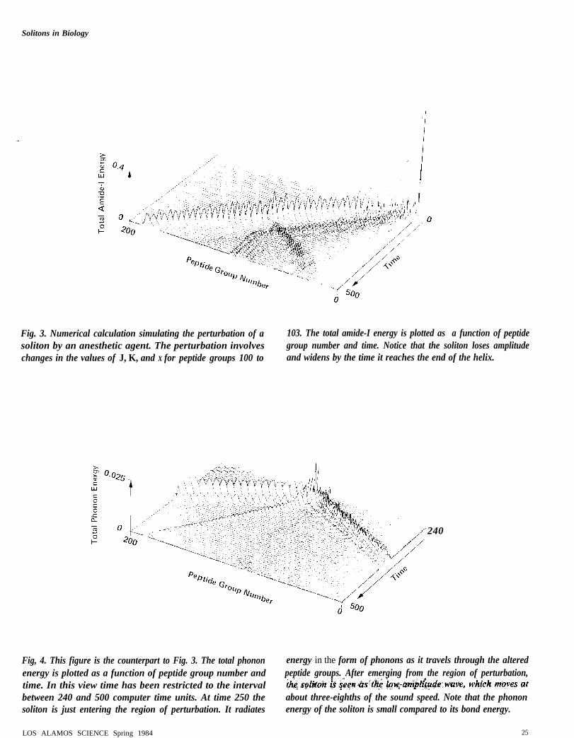
Fig. 3. Numerical calculation simulating the perturbation of a 103. The total amide-I energy is plotted as a function of peptidesoliton by an anesthetic agent. The perturbation involves group number and time. Notice that the soliton loses amplitudechanges in the values of J, K, and X for peptide groups 100 to and widens by the time it reaches the end of the helix.
240
Fig, 4. This figure is the counterpart to Fig. 3. The total phonon energy in the form of phonons as it travels through the alteredenergy is plotted as a function of peptide group number and peptide groups. After emerging from the region of perturbation,time. In this view time has been restricted to the intervalbetween 240 and 500 computer time units. At time 250 the about three-eighths of the sound speed. Note that the phononsoliton is just entering the region of perturbation. It radiates energy of the soliton is small compared to its bond energy.
LOS ALAMOS SCIENCE Spring 1984 25
RELATED TOPICS
as easy to calculate a new value for the hydrogen-bond springconstant K nor a new value for the coupling constant X in weakerhydrogen bonds. As a crude estimate we assumed that K decreasedproportionally to hydrogen-bond energy, and thus our new springconstant has the value of 0.45 K. We also assumed that X is slightlydecreased in weaker hydrogen bonds to the lower value that wascalculated by V. Kuprievich and Z. Kudritskaya. Hence, at the pointof anesthetic binding we chose X = 0.3 x 10⁻¹⁰ newton, which is justbelow threshold for soliton formation,
The results of this numerical investigation are presented in Figs. 3and 4. The decreased values of J, K, and X were restricted to peptidegroup numbers n = 100 to 103 on the three spines of the a helix. Theperturbation was restricted to this narrow region because ananesthetic molecule is expected to weaken the hydrogen bonds in onlya small region of the protein. This procedure also ensured that thesoliton was well formed before entering the perturbed region. Figure 3can be compared directly to Fig. 10 in “Solitons in Biology,” It isapparent that after 500 computer time units the soliton, whichtraveled through the perturbation, is appreciably degraded. Figure 4
reveals that energy is radiated by the soliton in the form of phonons asit travels through the perturbation. These phonons are seen to move atthe sound velocity in the a helix, which is approximately eight-thirdsthe soliton velocity. Up to this point we have neglected the fact thatthe H–N–C=O groups in the barbiturate are capable of dipole-dipole
Acknowledgment
I wish to thank Peter Lomdahl for help with the numerical code.
coupling to the H-N–C=O groups in the helix. Such a couplingshould further degrade soliton propagation. since the interactionenergy between barbiturate and a helix would be appreciable. Thedipole-dipole coupling of the anesthetic molecule to the protein willdepend on the number of H-N-C=O groups within it and on itsspatial orientation relative to the protein.
As a final consideration of this model we pose the question: Howmany proteins are inhibited during general anesthesia? Barbituratesexhibit their anesthetic activity at a concentration between 200 and1000 micromolar. At this concentration they reduce the metabolicactivity of the brain by 10 to 15 percent, as measured by oxygenutilization. Taking the average membrane protein to encompass a
volume of 20 angstroms x 20 angstroms x 40 angstroms = 1.6 x 10⁴
cubic angstroms implies that about 1 percent of typical membraneproteins are associated with an anesthetic molecule. Such a smallfigure points out that the brain is very sensitive to alterations at themolecular level. Consciousness appears to require the coordinatedeffort of almost every protein.
We have presented a simplified theoretical model for anestheticactivity, taking advantage of the fact that the a helix is an importantstructure in membrane and cytoskeletal proteins. If the Davydovsoliton finds experimental support in biology, then such a model mayhelp to explain some of the molecular mechanisms behind generalanesthesia. ■
Further Reading
R. Buchet and C. Sandorfy. “Perturbation of the Hydrogen-Bond Equilibrium in Nucleic Bases. AnInfrared Study.” Journal of Physical Chemistry 87(1983):275-280.
N. P. Franks and W. R. Lieb. “Molecular Mechanisms of General Anesthesia.” Nature300(1982):487-493.
M. Guerin, J.-M. Dumas, and C. Sandorfy. “’Vibrational Spectroscopic Studies of Molecular Associationsby Local Anesthetics.” Canadian Journal of Chemistry 58(1980):2080-2088.
Scott P. Layne. “The Modification of Davydov Solitons by the Extrinsic H–N–C=O Group.” Los AlamosNational Laboratory unclassified release LA-UR-83-2253 and to be published by Plenum Press inNonlinear Electrodynamics in Biological Systems.
Spring 1984 LOS ALAMOS SCIENCE
Solitons in Biology
What Is a Soliton?by Peter S. Lomdahl
A bout thirty years ago a remarkable discovery was madehere in Los Alamos. Enrico Fermi, John Pasta, and StanUlam were calculating the flow of energy in a one-
dimensional lattice consisting of equal masses connected by nonlinearsprings. They conjectured that energy initially put into a long-wavelength mode of the system would eventually be “thermalized,”that is, be shared among all modes of the system. This conjecture wasbased on the expectation that the nonlinearities in the system wouldtransfer energy into higher harmonic modes. Much to their surprisethe system did not thermalize but rather exhibited energy sharingamong the few lowest modes and long-time near recurrences of theinitial state.
This discovery remained largely a mystery until Norman Zabuskyand Martin Kruskal started to investigate the system again in theearly sixties. The fact that only the lowest order (long-wavelength)modes of the discrete Fermi-Pasta-Ulam lattice were “active” ledthem in a continuum approximation to the study of the nonlinearpartial differential equation
This equation (the KdV equation) had been derived in 1885 byKorteweg and de Vries to describe long-wave propagation on shallowwater. But until recently its properties were not well understood.
From a detailed numerical study Zabusky and Kruskal found thatstable pulse-like waves could exist in a system described by the KdVequation. A remarkable quality of these solitary waves was that theycould collide with each other and yet preserve their shapes and speedsafter the collision. This particle-like nature led Zabusky and Kruskalto name such waves solitons. The first success of the soliton conceptwas explaining the recurrence in the Fermi-Pasta-Ulam system. Fromnumerical solution of the KdV equation with periodic boundaryconditions (representing essentially a ring of coupled nonlinear
springs), Zabusky and Kruskal made the following observations. Aninitial profile representing a long-wavelength excitation would “breakup” into a number of solitons, which would propagate around the
system with different speeds. The solitons would collide but preservetheir individual shapes and speeds. At some instant all of the solitonswould collide at the same point, and a near recurrence of the initialprofile would occur.
This success was exciting, of course, but the soliton concept provedto have even greater impact. In fact, it stimulated very importantprogress in the analytic treatment of initial-value problems fornonlinear partial differential equations describing wave propagation.During the past fifteen years a rather complete mathematical descrip-tion of solitons has been developed. The amount of information onnonlinear wave phenomena obtained through the fruitful collabora-tion of mathematicians and physicists using this description makesthe soliton concept one of the most significant developments inmodern mathematical physics.
The nondispersive nature of the soliton solutions to the KdVequation arises not because the effects of dispersion are absent butbecause they are balanced by nonlinearities in the system. Thepresence of both phenomena can be appreciated by consideringsimplified versions of the KdV equation.
version
(2)
The most elementary wave solution of this equation is the harmonicwave
(3)
LOS ALAMOS SCIENCE Spring 1984 27
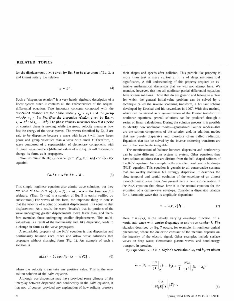
RELATED TOPICS
and k must satisfy the relation
(4)
Such a “dispersion relation” is a very handy algebraic description of alinear system since it contains all the characteristics of the originaldifferential equation, Two important concepts connected with the
of constant phase is moving, while the group velocity measures howfast the energy of the wave moves. The waves described by Eq. 2 aresaid to be dispersive because a wave with large k will have largerphase and group velocities than a wave with small k. Therefore, awave composed of a superposition of elementary components withdifferent wave numbers (different values of k in Eq. 3) will disperse, orchange its form. as it propagates.
equation
(5)
This simple nonlinear equation also admits wave solutions, but they
arbitrary. (That f(x - ut) is a solution of Eq. 5 is easily verified bysubstitution.) For waves of this form, the important thing to note isthat the velocity of a point of constant displacement u is equal to thatdisplacement. As a result, the wave “breaks”; that is, portions of thewave undergoing greater displacements move faster than, and there-fore overtake, those undergoing smaller displacements, This multi-valuedness is a result of the nonlinearity and, like dispersion, leads toa change in form as the wave propagates.
A remarkable property of the KdV equation is that dispersion andnonlinearity balance each other and allow wave solutions thatpropagate without changing form (Fig. 1), An example of such asolution is
(6)
where the velocity c can take any positive value. This is the one-soliton solution of the KdV equation.
Although our discussion may have provided some glimpse of theinterplay between dispersion and nonlinearity in the KdV equation, ithas not. of course, provided any explanation of how solitons preserve
their shapes and speeds after collision. This particle-like property ismore than just a mere curiosity; it is of deep mathematicalsignificance, A full understanding of this property requires an ex-tensive mathematical discussion that we will not attempt here. Wemention, however, that not all nonlinear partial differential equationshave soliton solutions. Those that do are generic and belong to a classfor which the general initial-value problem can be solved by atechnique called the inverse scattering transform, a brilliant schemedeveloped by Kruskal and his coworkers in 1967. With this method,which can be viewed as a generalization of the Fourier transform tononlinear equations, general solutions can be produced through aseries of linear calculations. During the solution process it is possibleto identify new nonlinear modes—generalized Fourier modes—thatare the soliton components of the solution and, in addition, modesthat are purely dispersive and therefore often called radiation.Equations that can be solved by the inverse scattering transform aresaid to be completely integrable.
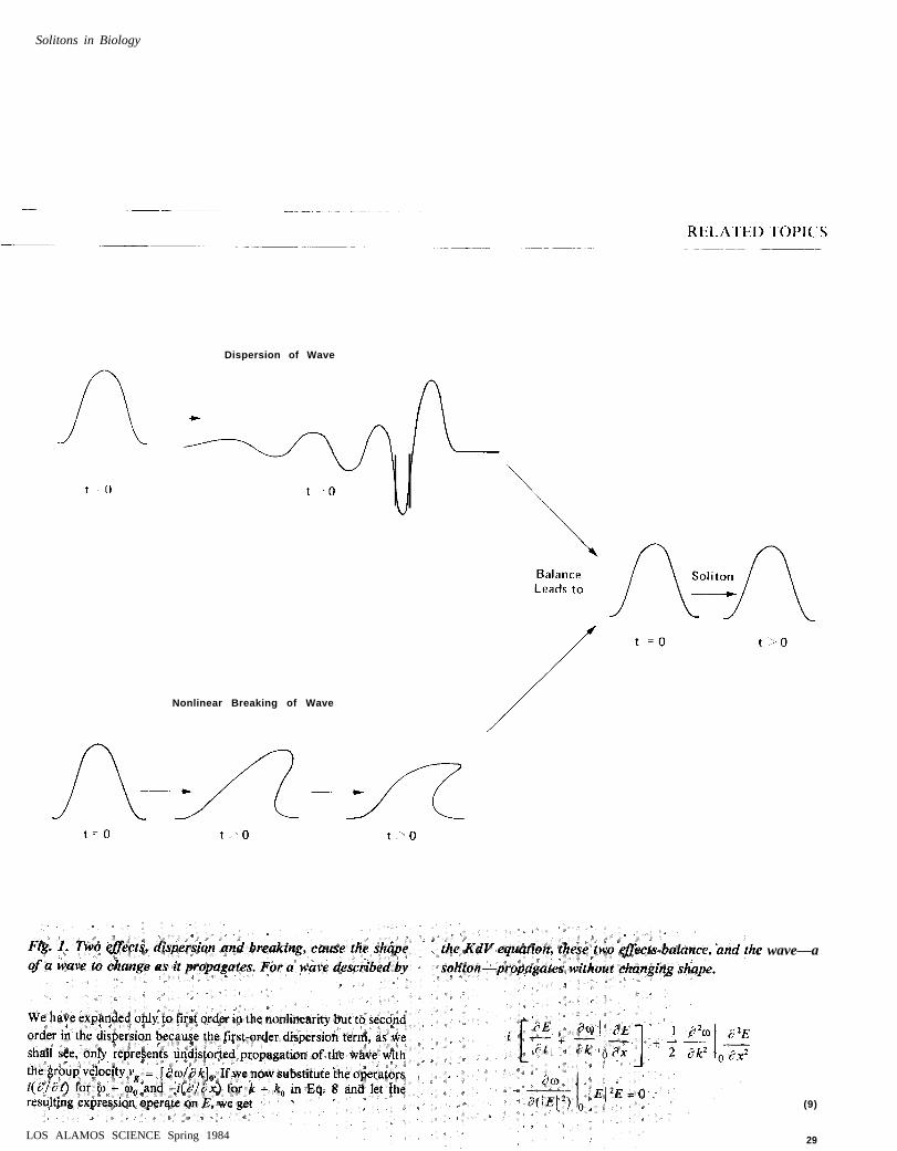
The manifestation of balance between dispersion and nonlinearitycan be quite different from system to system. Other equations thushave soliton solutions that are distinct from the bell-shaped solitons ofthe KdV equation. An example is the so-called nonlinear Schrodinger(NLS) equation. This equation is generic to all conservative systemsthat are weakly nonlinear but strongly dispersive. It describes theslow temporal and spatial evolution of the envelope of an almostmonochromatic wave train. We present here a heuristic derivation ofthe NLS equation that shows how it is the natural equation for theevolution of a carrier-wave envelope. Consider a dispersion relationfor a harmonic wave that is amplitude dependent:
(7)
Here E = E(x,t) is the slowly varying envelope function of a
situation described by Eq. 7 occurs, for example. in nonlinear opticalphenomena, where the dielectric constant of the medium depends onthe intensity of the electric signal. Other examples include surfacewaves on deep water, electrostatic plasma waves, and bond-energy
transport in proteins.
(8)
28 Spring 1984 LOS ALAMOS SCIENCE
Solitons in Biology
Dispersion of Wave
Nonlinear Breaking of Wave
wave—a
LOS ALAMOS SCIENCE Spring 1984
(9)
29
This is the nonlinear Schrodinger equation, so called because of itsresemblance to the Schrodinger equation even though its derivationoften has nothing to do with quantum mechanics. The first term ofEq. 9 represents undistorted propagation of the wave at the groupvelocity. and the second and third terms represent its linear andnonlinear distortion. respectively. This crude derivation of the NLSequation shows how it arises in systems with amplitude-dependentdispersion relations. but more formal methods are necessary if detail
It is often preferable to express Eq. 9 in a neater form. For this
to
where
(lo)
Fig. 2. Profile of a single-soliton solution of the NLS equation.
(11)
The NLS equation—like the KdV equation—is completely inte-grable and has soliton solutions. The analytic form for a single-solitonsolution is given by
amplitude, initial position, and initial phase, respectively, of thesoliton. Figure 2 shows the profile of this soliton.
Any initial excitation for the NLS equation will decompose intosolitons and/or dispersive radiation. A monochromatic wave train
tion and breaks up into separate and localized solitons. This phenom-enon is called the Benjamin-Feir instability and is well known to anysurfer on the beach who has noticed that every, say, seventh wave isthe largest. The NLS equation is in a way more universal than theKdV equation since an almost monochromatic, small-amplitudesolution of the KdV equation will evolve according to the NLSequation.
The last type of soliton we mention, which is distinctly different
30
(12)
Solitons in Biology
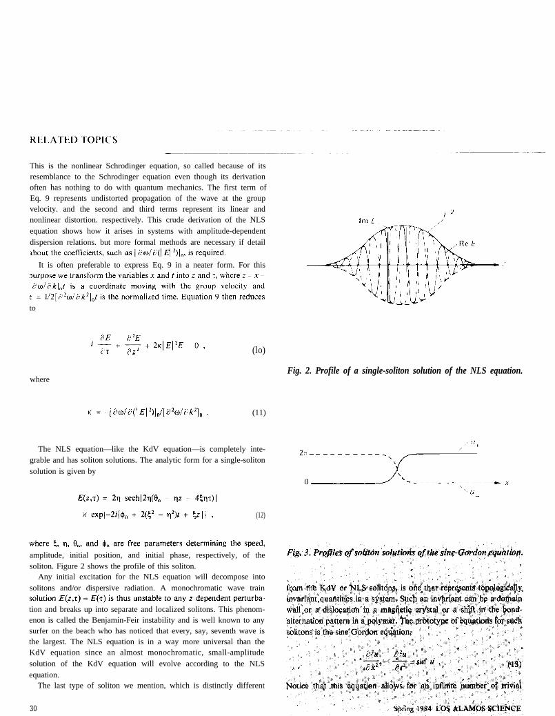
of such “degenerate ground states” also allow solutions that connecttwo neighboring ground states. Solutions of this type are often calledkinks, and for the sine-Gordon equation they are exact solitons; thatis, they collide elastically without generation of dispersive radiation.The analytic form, whose profile is shown in Fig. 3, is given by
(14)
where the solution u– is often called an antikink. The parameter
c (–1 < c < 1) determines the velocity and XO the initial position,
Other equations with degenerate ground states also have kink andantikink solutions, but they are not exact solitons like those of thesine-Gordon equation. It is interesting to note that small-amplitudesolutions of the sine-Gordon equation also can be shown to evolve
according to the NLS equation.Equations with soliton solutions are generic, and, although real
systems often contain mechanisms (impurities, dissipative forces, andmultidimensionality) that destroy exact soliton behavior, they arevery useful as a starting point for analysis. In fact, perturbationmethods—with the perturbation taking place around the soli-ton—have been developed to compute the response of the soliton toexternal forces, damping, etc. Often the result is that the parameterscharacterizing the soliton (such as velocity and amplitude) are nowtime dependent, with the time dependence governed by simpleordinary differential equations. The original equations are thereforestill very useful. Because the mechanisms that give rise to solitonequations are so prevalent, the suggestion that solitons might arise inbiology is not so surprising. The question to be asked is how well aparticular biological system satisfies the criteria underlying the soliton
Further Reading
The classic paper where the word “soliton” was introduced is “Interaction of ‘Solitons’ in a CollisionlessPlasma and the Recurrence of Initial States” by N. J. Zabusky and M. D. Kruskal in Physical ReviewLetters 15(1965):240. For many references see also “Computational Synergetics and MathematicalInnovation” by Norman J. Zabusky in Journal of Computational Physics 43(1981):95.
There are an increasing number of papers on solitons; a good review paper covering the subject to 1973 is“The Soliton: A New Concept in Applied Science” by Alwyn C. Scott, F. Y. F. Chu, and David W.McLaughlin in Proceedings of the IEEE 61( 1973): 1443.
Good accounts of the subject, together with up-to-date lists of references, can also be found in manytextbooks, including the following.
G. L. Lamb, Jr. Elements of Soliton Theory. New York: John Wiley & Sons, 1980.
Mark J. Ablowitz and Harvey Segur. Solitons and the Inverse Scattering Transform. Philadelphia:Society for Industrial and Applied Mathematics, 1981.
R. K. Dodd, J. C. Eilbeck, J. D. Gibbon, and H. C. Morris. Solitons and Nonlinear Wave Equations. NewYork: Academic Press, 1982.
LOS ALAMOS SCIENCE Spring 1984