sodium lauryl sulphate increases tiludronate paracellular transport using human epithelial caco-2...
TRANSCRIPT

ELSEVIER International Journal of Pharmaceutics 123 (1995) 71-83
international journal of pharmaceutics
Sodium lauryl sulphate increases tiludronate paracellular transport using human epithelial Caco-2 monolayers
Xavier Boulenc a,* Thierry Breul a Jean-Claude Gautier a Philippe Saudemon a, Henri Joyeux b, Claude Roques a, Yves Berger a G6rard Fabre a
a Sanofi Recherche, Department of Metabolism and Pharmacokinetics, 371 Rue du Professeur Blayac, 34184 Montpellier C~dex 4, France
b lnstitut Curie, Paris, France
Received 13 September 1994; revised 4 January 1995; accepted 23 January 1995
Abstract
The potential effect of the common pharmaceutical wetting agent, sodium lauryl sulphate (SLS), on the transport of the hydrophilic bisphosphonate, tiludronate, was investigated both by performing physico-chemical determina- tions of the SLS-tiludronate interaction and by measuring the paracellular transport of tiludronate (Boulenc et al., Biochem. Pharmacol., 46 (1993) 1591-1600) across the in vitro human intestinal epithelium model, i.e., Caco-2. S IS did not affect the contact angles determined with different liquids (glycerin, dioxane, sulphuric acid, water, mercury, heptane and decane) on tiludronate tablets. SLS influenced neither the disintegration of tiludronate tablets nor tiludronate solubility. However, both the efficiency and effectiveness of SLS, in reducing surface tension, were affected by tiludronate. Hence, the presence of 0.48 g / l tiludronate in a water solution changed the efficiency of SLS in reducing the surface tension from 1 to 0.3 g/1. Before evaluating tiludronate transport across Caco-2 monolayers, the reversible (absorption enhancement of orally administered drugs) and irreversible (cell cytotoxicity) effects of SLS on the viability of Caco-2 monolayers were investigated. Following a 1 h exposure of well-differenti- ated Caco-2 cells to SLS concentrations above 100 mg/1, mitochondrial dehydrogenase activity decreased in a concentration-dependent manner, cytosolic lactate dehydrogenase leakage occurred, mannitol transport was irre- versibly increased and a structural separation of the tight junctions was observed by electron microscopy. SLS concentrations up to 80 mg/1 did not affect mitochondrial dehydrogenase and cytosolic lactate dehydrogenase activities, while both a reversible increase in mannitol paracellular transport and tight junction opening were observed. Under these incubation conditions, tiludronate paracellular transport was increased in a concentration-de- pendent manner. These studies demonstrate that SLS increased tiludronate paracellular transport through its specific and transient effect on the permeability of the intercellular space.
Keywords: Intestinal absorption; Paracellular route; Intestinal model; Surfactant; Caco-2 monolayer
Abbreviations: TEER, trans-epithelial electrical resistance; FCS, foetal calf serum; SLS, sodium lauryl sulphate * Corresponding author. Tel. 67.10.63.25; Fax 67.10.67.67.
0378-5173/95/$09.50 © 1995 Elsevier Science B.V. All rights reserved SSDI 0378-5173(95)00041-0
72 X. Boulenc et al. / International Journal of Pharmaceutics 123 (1995) 71-83
1. Introduction
Tiludronate, or (4-chlorophenyl)thiomethylene diphosphonic acid (disodium salt), is a novel bis- phosphonate with potential use in the treatment and prevention of osteoporosis and the treatment of Paget's disease. Bisphosphonates are known to be poorly absorbed when given orally (Fogelman et al., 1986; Janner et al., 1991) and their absorp- tion varies extensively between and within indi- viduals. Among various pharmacokinetic studies performed in both animals and humans (Janner et al., 1991; Hyldstrup et al., 1993) which demon- strated their low bioavailability, recent in vitro studies performed with the Caco-2 monolayer model reported that tiludronate was slowly trans- ported across the intestinal epithelium, through the paracellular route (Boulenc et al., 1993).
The use of surfactants in formulations in- tended for administration to human subjects can lead to changes in the absorption of drugs and hence, in their pharmacological activity, although the surfactant effect on drug absorption does not readily allow generalisations to be made (Florence, 1981). These products were demon- strated to influence the disintegration and disso- lution of solid dosage forms; they can be consid- ered as absorption enhancers by increasing mem- brane permeability and/or affecting membrane integrity (Tomita et al., 1988; Van Hoogdalen et al., 1989). Moreover, it was also suggested that surfactants such as bile acids (Freel et al., 1983; Tomita et al., 1988), caprate and caprylate (Sawada et al., 1991; Anderberg et al., 1993) affected tight junction regulation. Hence, drugs for which dissolution and solubility are the rate- limiting steps for their absorption and drugs for which membrane transport is the rate-limiting step might be affected differently. Surfactants have been shown either to increase, decrease or exert no effect on the transfer of drugs across biological membranes (Attwood and Florence 1983). The influence of surfactants depends on the concentration, degree of ionization and hy- drophile-lipophile balance (HLB) of both the sur- factant and the drug, as well as drug solubility.
Sodium lauryl suphate (SLS), a hydrophilic anionic surfactant molecule, is a known wetting
agent, able to modify the surface properties of solids through the phenomenon of adsorption, and to decrease interfacial tension at solid/liquid interfaces. Moreover, like naturally occurring mi- celle formers such as bile salts and phospholipids, SLS can also form micelles and leads to the enhancement of dissolution and solubilization. These properties when added to the SLS effect on intestinal membrane permeability and in- tegrity may modify drug absorption (Muranishi, 1990).
The human adenocarcinoma cell line Caco-2 which reproducibly displays a number of the properties of differentiated intestinal cells was shown to be the most relevant in vitro system for investigating transepithelial transport processes (Artursson, 1990; Artursson and Karlsson, 1991; Dantzig and Bergin, 1990; Hilgers et al., 1990; Lundin and Artursson, 1990; Rubas et al. 1993), rapidly evaluating the permeability of a drug, defining the mechanisms of transport (Boulenc et al., 1993), and testing novel strategies for enhanc- ing drug transport (Audus et al., 1990; Wilson, 1990; Artursson, 1991).
This study was carried out in order to evaluate the qualitative and quantitative influence of SLS on the bioavailability of tiludronate, by analysing two different processes such as drug dissolution and its further absorption across the in vitro intestinal epithelium model, Caco-2.
2. Materials and methods
2.1. Chemicals
14C-labelled (spec. act. 33 mCi/mmol) and un- labelled tiludronate were obtained from Sanofi Recherche, Montpellier, France. [14C]PEG400o (Mol. Wt 4000; spec. act. 13 mCi/g), [14C]PEG400 (Mol. Wt 400; spec. act. 15.3 mCi/g) and [3H]mannitol (Mol. Wt 182; spec. act. 30 Ci/mmol) were purchased from Amersham In- ternational (Bucks, UK) and New England Nu- clear Products (Boston, USA), respectively.
SLS (98% sodium lauryl suphate) was pur- chased from Laserson and Sabetay (Etampes, France). Magnesium stearate was purchased from

X. Boulenc et al. / International Journal of Pharmaceutics 123 (1995) 71-83 73
Table 1 Tablet composition (mg)
Tablet A B
Tiludronate disodium 240.0 240.0 SLS 4.5 - Magnesium stearate 4.5 4.5 Cross-linked sodium carboxymethylcellulose 24.0 24.0 Microcrystalline monohydrated lactose 177.0 181.5
Atochem, CECA, France. Cross-linked sodium carboxymethylcellulose (Ac-Di-Sol) was pur- chased from FMC Corp. (Philadelphia, PA, USA) and microcrystallized monohydrated lactose (99.7%) from S.A. Sucre de Lait (Sains du Nord, France). Liquids used were distilled water, glyc- erin, dioxane, sulphuric acid, heptane and decane and were obtained from commercial sources.
using a microscope fitted with a goniometer eye- piece and by photographing the droplet (Adam- son, 1976, Uyama et al., 1990)
Zisman (1964) established a linear relationship between cos 0 and 1/surface tension of the wet- ting liquid for many surfaces with low surface energies. The surface tension obtained by extrap- olation to cos 0 = 1 is known as Yc the critical surface tension of the solid. The Yc value is a characteristic of a solid and may be related to the composition of the solid. The condition for wet- ting of a solid is that the surface tension of the wetting liquids must not exceed the value of Yc. Thus, tablets with low Yc are only wetted by liquids of very low surface tension. Increased 7c values improve the wetting of the tablet.
2.4. Disintegration and dissolution kinetics
2.2. Tablet composition
The tiludronate tablets studied have the com- positions listed in Table 1.
2.3. Wetting measurement
Wetting refers to the process which occurs when a solid-air interface is replaced by a solid- liquid interface. In pharmaceutical technology, wetting has usually been characterized by contact angles (Gissinger and Stamm, 1980; Buckton and Newton, 1986; Lippold and Ohm, 1986). When a drop of liquid is placed on a plane, homogeneous and solid surface, it assumes a shape which corre- sponds to the minimum free energy for the sys- tem. The contact angle formed by a drop of liquid on the horizontal surface is the angle formed by the solid surface and the tangent of the liquid surface at their intersection. The condition for minimum free energy at equilibrium is that given by Young's equation:
7S/G ---- 7S /L + YL/G COS 0
where 0 is the contact angle, determined by pho- tographing the drop placed on the surface and Ys/c, Ys/L and Ye/c denote the interfacial ten- sions at the solid/gas, liquid/gas and solid/liquid interfaces, respectively. 0 is measured directly by
The disintegration and dissolution kinetics were determined on six tablets according to the official methods of the US Pharmacopeia XXII (1990). The medium used was 0.1 M hydrochloric acid to which sodium chloride (2 g for 1000 g) was added. The final pH value was set at 1.2 and experiments were performed at 37 ° C. The con- centration of tiludronate was determined by UV spectroscopy at 261 nm on a Beckman UV spec- trophotometer.
2.5. Surface tension measurement
Measurement of the surface tension was ac- complished using the Wilhelmy plate method ac- cording to the ISO R 304 Method (1978) on a Prolabo ~ N3 tensiometer. For the purpose of comparing the performance of surfactants in re- ducing the surface tension, it was necessary to distinguish between the efficiency of the surfac- tant, i.e., the bulk phase concentration of surfac- tant required to reduce the surface tension to its minimum value whatever the surfactant concen- tration, and its effectiveness, i.e., the maximum reduction in tension that can be achieved regard- less of surfactant concentration (Rosen, 1978). Surfactant efficiency is expressed as a surfactant concentration, when surfactant effectiveness is expressed as a force per length unit.

74 X. Boulenc et al. / International Journal of Pharmaceutics 123 (1995) 71-83
2.6. Cell culture
Caco-2 cells, originating from a human col- orectal carcinoma (Fogh et al., 1977), were ob- tained from Dr A. Zweibaum (INSERM U-178, Villejuif, France). Cells were cultivated as de- scribed elsewhere (Boulenc et al., 1993). All tis- sue culture media were obtained from Eurobio Laboratories (Paris, France). Cells used in this study were between passages 90 and 120.
For transport studies Caco-2 cells were seeded onto 0.45 ~m pore collagen type I-coated inserts (Millicell-CM; pore size, 0.4 /.~m; diameter, 30 mm; Millipore, Bedford, MA) at 63 000 cells/cm 2. The monolayers used in this study were 12-20 days post-seeding or 7-16 days post-confluence.
2. 7. Integrity of the monolayers
The integrity of the monolayers was deter- mined by measurement of the potential differ- ence (TEER, transepithelial electrical resistance) and by following the transepithelial transport of a macromolecular marker, PEG4000 as previously described (Boulenc et al., 1993). The potential difference was expressed as transmembrane resis- tance (O/cm 2) after subtraction of the intrinsic resistance of the model (i.e., the resistance ob- tained over the cell-free inserts). A monolayer with low TEER was assumed to exhibit extensive leakage through imperfect occluding junctions or holes in the monolayer (Hidalgo et al., 1989; Artursson, 1990). TEER values (230 + 50 g2/cm 2) remained constant from day 12 to 20 (Hidalgo et al., 1989; Artursson, 1990).
[laC]PEG4000 (38 tzg/ml) was added to the apical side of the monolayers. The radiolabelled marker transported over Caco-2 cells was evalu- ated after 3 h at 37°C on a 0.5 ml aliquot part withdrawn from the basolateral chamber. The samples were measured in a liquid scintillation counter from Packard. Inserts without cells were used to determine the maximal transport of the marker during the same time period. The results were expressed as the percentage transported of the dose. The rate of [a4C]PEG4oo0 transported was 0.1 +_ 0.02% per h. These results are consis- tent with those previously reported by Hidalgo et
al. (1989), who showed that undamaged monolay- ers are almost impermeable to macromolecules such as PEG400o.
2.8. Preparation for transmission electron micro- scopic examination
The inserts were washed in PBS buffer (pH 7.2) and fixed in a solution containing 2% glu- taraldehyde and 0.1 M sucrose in 0.1 M cacody- late buffer (pH 7.3) for 1 h at 25 ° C. The cells were rinsed in 0.1 M Hanks buffer and fixed with 1% osmium tetroxide in 0.1 M sodium cacodylate • HC1 buffer for 1 h at 25 ° C. After dehydration, the preparation was embedded in Epon resin, sliced with a Reichert ultramicrotome, stained with uranyl acetate and lead acetate, and exam- ined on a transmission electron microscope (Jeol 100S).
2.9. Measurement of drug transport and radioac- tive scintillation counting
Drug solutions were prepared from the radio- labelled isotopes and the corresponding unla- belled compounds in Hanks buffer to give final concentrations up to 10 -3 M in the apical com- partment. All transport experiments were per- formed as described elsewhere (Boulenc et al. 1993).
2.10. Calculations
The apparent permeability coefficient (Papp) was determined as previously reported:
Papp = d Q / [ d t ×A × Co],
where d Q / d t is the transport rate (~g/s) and corresponds to the slope of the regression line determined on at least four different time points, C o denotes the initial concentration in the donor chamber (~g/ml or ~g/cm3), and A is the area of the membrane (5.7 cm a)
2.11. Cytotoxicity tests
2.11.1. Mitochondrial dehydrogenase activity The intracellular mitochondrial dehydrogenase
activity was determined by the MT'I? method.

X. Boulenc et aL / International Journal of Pharmaceutics 123 (1995) 71-83 75
Briefly, MTT is a tetrazolium salt cleaved by the mitochondrial dehydrogenase activity in living but not dead cells to a dark-blue product. Cells (ap- prox. 105 cells) were seeded in 96-well tissue culture plates and cultured for 5 days. Cells were treated with increasing SLS concentrations for 1 h. The incubation medium was removed, and cells were incubated with MTT (5 mg/ml ) for an additional 1 h. The incubation medium was mea- sured in a multiwell scanning spectrophotometer (Multiscan MCC/340, Labsystem).
2.11.2. Lactate dehydrogenase activity Lactate dehydrogenase is a cytosolic enzyme
which is recovered in the extracellular compart- ment following membrane disruption. Caco-2 monolayers were treated for 1 h with increasing concentrations of SLS and the lactate dehydroge- nase activity was determined in the extracellular compartment using the Sigma kit.
2.11.3. Mannitol transport The integrity of Caco-2 monolayers can also be
investigated by its permeability to mannitol, a compound which is transported via the paracellu- lar route. Caco-2 cell monolayers were treated for 1 h with increasing SLS concentrations ranging between 4 and 100 mg/ l . The apical compart- ment containing SLS was removed and replaced by SLS-free fresh medium. After a 24 h period of
rest, [3H]mannitol was added to the apical side of the monolayers and its rate of transport across the Caco-2 monolayer was evaluated over a 3 h period at 37 ° C, by quantification of radiolabel in the basal compartment. Inserts without cells were used to determine the maximal transport of the marker during the same period of time.
2.12. Statistical test
Values are expressed as mean _+ standard devi- ation. Three experiments were performed (n = 3), except where indicated otherwise in the figure legends. The results were analysed by one-way analysis of variance. Significance: *p < 0.1; * *p < 0.05; * * *p < 0.01.
3. Results
3.1. Physico-chemical studies
3.1.1. Influence of SLS on tablet wetting As tablet porosity plays an important role in
the variation of the contact angle of a liquid drop on the tablet, contact angle measurements were performed as a function of time with water drops. Table 2 shows the contact angles determined with different liquids (glycerin, dioxane, sulphuric acid, water, mercury, heptane and decane) on tilu-
Table 2 Contact angles and surface tension of various liquids on tiludronate tablets formulated in the absence or presence of SLS
Liquid Contact angles (°) Surface tension ( m N / m )
Tablet A Tablet B (with SLS) (without SLS)
Glycerin 70 70 63.4 Dioxane 10 10 - Sulfuric acid 45 47.5 51 50% sulfuric acid 47 - 58 Mercury 136 136 4356.5 Heptane 0 0 18.4 Decane 0 0 23.9 Water t = 0 55 50 72 t = 1 rain 45 45 t = 2 rain 40 40 t = 3 rain 35 35 t = 5 rain 30 30

76 x.. Boulenc et aL / International Journal of Pharmaceutics 123 (1995) 71-83
dronate tablets supplemented or not with SLS. The results reported in Table 2 indicate that no significant modification of the contact angle oc- curred with the different liquids on tablets sup- plemented or not with SLS. The kinetics of tablet wetting by water are not affected by the presence of SLS.
Fig. 1 illustrates the linear relationship be- tween cos 0 and 1/surface tension of the wetting liquid. The equation for this relation is as follows:
cos 0 = 88 .7 (1 /7 ) - 0.93.
The surface tension obtained by extrapolation to cos 0 = 1 is known as y~, i.e., the critical sur- face tension of the solid. The Yc value obtained for tiludronate tablets irrespective of the pres- ence of SLS is then Yc = 46 m N / m , which repre- sents the surface free energy of the tiludronate tablets.
3.1.2. Influence of SLS on tablet disintegration kinetics
The disintegration times of tiludronate tablets ranged between 6 and 7 min. SLS had no influ- ence on tiludronate tablet disintegration.
3.1.3. Influence of SLS on tiludronate solubility and dissolution kinetics
Tiludronate is soluble in water up to 160 g / l and its solubility was not modified in the pres- ence of SLS concentrations ranging between 0.1 and 10 g/1.
Sulfuric acid 1 + Water
0.5 ~ i c acid
0 ~ ~ - Glycerin
0.5
- 1 - - q I - - I I
0 0.005 0.01 0.015 0.02
1/Surface Tension (m/mN) Fig. 1. Effect of SLS on critical surface tensions of tiludronate tablets. Contact angles of different liquids were determined on tiludronate tablets formulated in the absence or presence of SLS.
c ' - o
" o
I . - -
100
80
60
40
20 -
0 4:
J m
-q~
[ I ] I
15 30 45 60
Time (Minutes) Fig. 2. Effect of SLS on tiludronate solubilisation. Solubilisa- tion of two tiludronate tablets formulated in the absence ([]) or presence (111) of SLS was investigated in a USP XXII dissolution test and the precentage of solubilisation was evalu- ated as a function of time (SD 10%).
Fig. 2 illustrates the amount of tiludronate solubilized during the dissolution test conducted with tiludronate tablets supplemented or not with SLS. These results indicated that tiludronate was solubilized more rapidly from a tablet which did not contain SLS than from a tablet supplemented with SLS. However, after a 1 h period, slightly greater amounts of tiludronate were solubilized from a tablet supplemented with SLS (96.6% vs 88.7% in the presence or absence of SLS, respec- tively; n = 6).
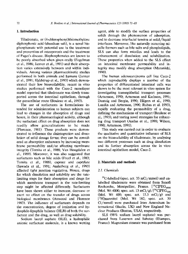
3.1.4. Influence of SLS on surface tension of tilu- dronate solutions
The influence of tiludronate on water surface tension was first investigated for concentrations up to 10 g/ l , a concentration higher than that assumed in the gastric juice. Fig. 3 illustrates the solution surface tension obtained for various con- centrations of SLS. Both the efficiency and effec- tiveness of SLS in reducing surface tension were affected by tiludronate. The presence of 0.48 g / l tiludronate in a water solution changed the effi- ciency of SLS in reducing the surface tension from 1 to 0.3 g/1. This significant change could be compared to the effect of any salt in solution with an anionic surfactant. The ranges of SLS effectiveness in reducing the surface tension were 0.01-0.3 and 0.01-1 g/1 when tests were per-
X. Boulenc et aL / International Journal of Pharmaceutics 123 (1995) 71-83 77
7O
E 60
._~ 50 oo c
~- 40
g~ 3o
20 i i 1 I 0,01 0.1 1 10 100
Sodium Lauryl Sulfate (g/I)
Fig. 3. Efficiency and effectiveness of SLS in reducing surface tension of t i ludronate solutions. Surface tension of SLS solu- tions (4.8 g / l ) was evaluated in the absence (D) or presence ( • ) of t i ludronate (SD 10%).
formed in the presence or absence of tiludronate, respectively.
3.2. Effect of SLS on tiludronate transport
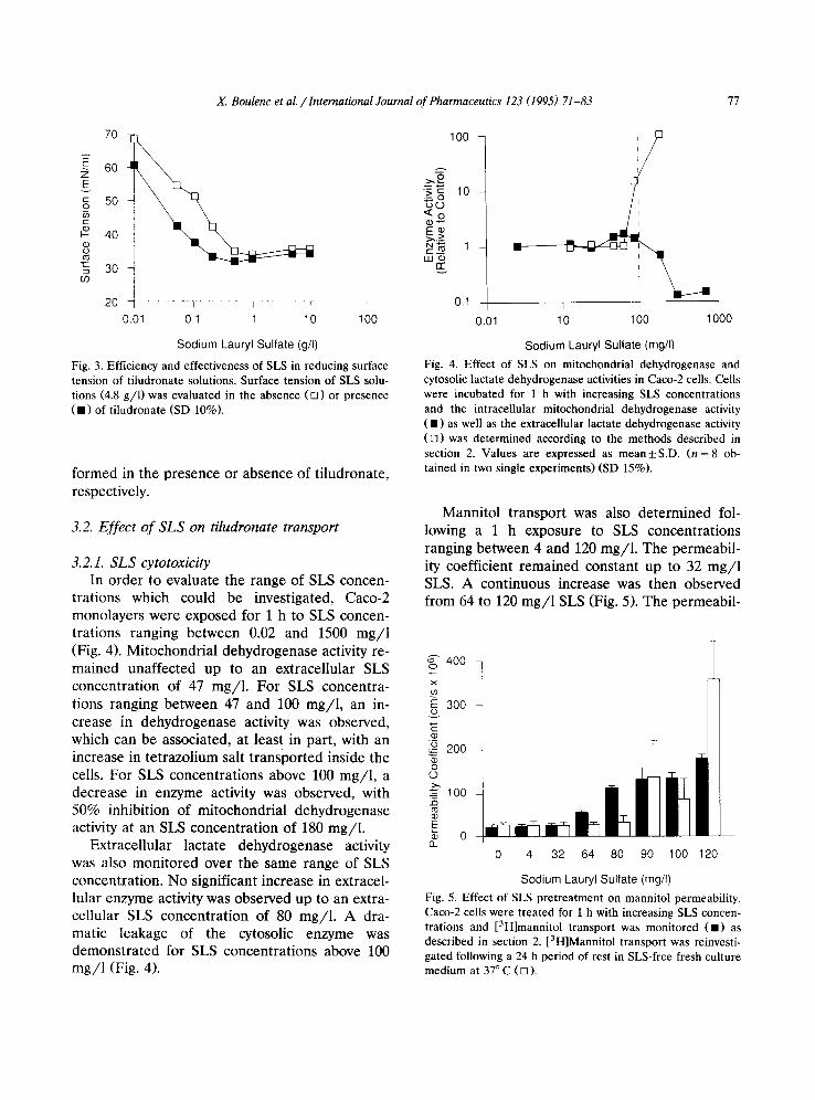
3.2.1. SLS cytotoxicity In order to evaluate the range of SLS concen-
trations which could be investigated, Caco-2 monolayers were exposed for 1 h to SLS concen- trations ranging between 0.02 and 1500 mg/1 (Fig. 4). Mitochondrial debydrogenase activity re- mained unaffected up to an extracellular SLS concentration of 47 mg/1. For SLS concentra- tions ranging between 47 and 100 mg/ l , an in- crease in dehydrogenase activity was observed, which can be associated, at least in part, with an increase in tetrazolium salt transported inside the cells. For SLS concentrations above 100 mg/1, a decrease in enzyme activity was observed, with 50% inhibition of mitochondrial dehydrogenase activity at an SLS concentration of 180 mg/1.
Extracellular lactate dehydrogenase activity was also monitored over the same range of SLS concentration. No significant increase in extracel- lular enzyme activity was observed up to an extra- cellular SLS concentration of 80 mg/1. A dra- matic leakage of the cytosolic enzyme was demonstrated for SLS concentrations above 100 mg/1 (Fig. 4).
1 O0 I
>.£ '=- ~ 10
E ¢ ~.._>
0.1 I i I
1.01 10 1 O0 1000
Sodium Lauryl Sulfate (mg/I)
Fig. 4. Effect of SLS on mitochondrial dehydrogenase and cytosolic lactate dehydrogenase activities in Caco-2 cells. Cells were incubated for 1 h with increasing SLS concentrations and the intracellular mitochondrial dehydrogenase activity ( • ) as well as the extracellular lactate dehydrogenase activity (rq) was determined according to the methods described in section 2. Values are expressed as m e a n + S . D . (n = 8 ob- tained in two single experiments) (SD 15%).
Mannitol transport was also determined fol- lowing a 1 h exposure to SLS concentrations ranging between 4 and 120 mg/1. The permeabil- ity coefficient remained constant up to 32 mg/1 SLS. A continuous increase was then observed from 64 to 120 m g / l SLS (Fig. 5). The permeabil-
g" 400 o x 03
300
5 200
8 ~ 100
~ 0 0 4 32 64 80 90 100 120
Sodium Lauryl Sulfate (mg/I)
Fig. 5. Effect of SLS pret reatment on mannitol permeability. Caco-2 cells were treated for 1 h with increasing SLS concen- trations and [3H]mannitol transport was monitored ( • ) as described in section 2. [3H]Mannitol transport was reinvesti- gated following a 24 h period of rest in SLS-free fresh culture medium at 37 ° C ([]).
78 X. Boulenc et al. / International Journal of Pharmaceutics 123 (1995) 71-83
ity coefficient of mannitol was then redetermined following a 24 h period of recovery in SLS-free fresh medium. Mannitol transport was similar to that observed in untreated cells after exposure of monolayers to SLS concentrations up to 80 mg/1. At higher SLS concentrations, an increase in mannitol transport was still observed, suggesting that monolayer integrity was not recovered fol- lowing a 24 h period of recovery, subsequent to a
1 h exposure to SLS concentrations above 80 mg/1.
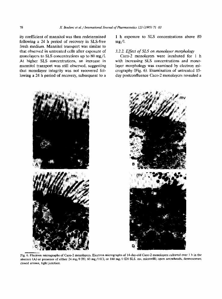
3.2.2. Effect of SLS on monolayer morphology Caco-2 monolayers were incubated for 1 h
with increasing SLS concentrations and mono- layer morphology was examined by electron mi- crography (Fig. 6). Examination of untreated 15- day postconfluence Caco-2 monolayers revealed a
: m y i
Fig. 6. Electron micrographs of Caco-2 monolayers. Electron micrographs of 14-day-old Caco-2 monolayers cultured over 1 h in the absence (A) or presence of either 24 mg/ l (B), 60 mg/ l (C), or 180 mg/ l (D) SLS. mv, microvilli; open arrowheads, desmosomes; closed arrows, tight junction.

x. Boulenc et aL / International Journal of Pharmaceutics 123 (1995) 71-83 79
well-differentiated brush border, cells being sepa- rated by desmosomes and tight junctions. No major difference was observed following a 1 h t reatment with 24 m g / l SLS, although some shortened microvilli appeared. At 60 rag/1 SLS concentration, some perturbations at the intercel- lular level were observed. A widening of intercel- lular space and of the zonula occludens was ob- served. Nevertheless, cell integrity was main- tained. When cell monolayers were treated with 180 mg/1 SLS, most of the microvilli disap- peared, nuclei were pycnotic and some extracellu- lar deposits were observed in the cytosol.
3.2.3. Ef fec t o f S L S on tiludronate transport Tiludronate transport across Caco-2 monolay-
ers, was determined following addition of the radiolabelled drug in the apical compartment , in the absence or presence of increasing SLS con- centrations, ranging between 1 and 80 mg / l . The rate of appearance of [14C]tiludronate in the basal compar tment was monitored over 2 h. The per- meability coefficient for t i ludronate transport was evaluated following addition of 1 mM tiludronate in the apical compar tment (Fig. 7). Under control conditions, i.e., in the absence of SLS, the perme- ability coefficient for t i ludronate was determined to be 4.0 × 10 -7 _+ 2.2 x 10 -7 c m / s (n = 8), based
-~ o ~ E 1.5 ~) 0 8o ~ o 1
~ o.5
~ O v
a_ 0 None 1 4 9 16 40 64 80
Sodium Lauryl Sulfate (rag/I)
Fig. 7. Effect of SLS on tiludronate transport. Radiolabeled tiludronate (1 IzM) was added in the apical compartment of Caco-2 monolayers, in the absence or presence of increasing SLS concentrations ranging between 4 and 80 mg/1. At speci- fied times and over a 1 h period, aliquot parts of culture medium were withdrawn from the basal compartment and the rate of appearance of radiolabel in the basal compartment was determined. Values are expressed as the mean_+ S.D. of four experiments performed on different days.
on experiments performed on different cell preparat ions and on 10-16-day post-confluence cells. The apparent large inter-experiment vari- ability was mostly due to the fact that t i ludronate was slowly transported by the paracellular route (Boulenc et al., 1993).
Tiludronate transport remained unaffected in the presence of SLS concentrations up to 16 mg/1. For SLS concentrations ranging between 16 and 80 mg/1, a statistically significant concen- t rat ion-dependent increase in ti ludronate trans- port was observed compared to control monolay- ers. The permeabili ty coefficient of tiludronate increased up to 13.0 x 10 -7 _ 1.0 × 10 -7 c m / s (n = 4) in the presence of 80 mg/1 SLS. Although higher SLS concentrations led to a greater in- crease in ti ludronate transport (data not shown; the tiludronate permeability coefficient = 21.0 × 10 -7 _+ 7.3 x 10 -7 c m / s (n = 3) at 200 mg/1SLS) , this phenomenon was mainly due to the cytotoxic- ity of SLS.
4. Discussion
The human intestinal epithelial cell line, Caco- 2, spontaneously differentiated, in culture, to po- lar cells possessing microvilli and enterocytic properties. Confluent monolayers form tight junc- tions between cells; thus, monolayers exhibited electrical propert ies characteristics of an intesti- nal epithelium. Epithelial cells are joined by junc- tional complexes which are comprised of three separate structures: tight junctions, intermediate junctions, and desmosomes. The intercellular spaces are sealed by tight junctions which reduce their pore radius to a few ~mgstr6m units (Madara and Dharmsathaphorn, 1985). The contribution of this paracellular pathway to the total perme- ability of the epithelial monolayer is only signifi- cant for drugs that are transported slowly across the cell membrane, e.g., hydrophilic compounds with a low molecular weight and very low parti- tion coefficients (Artursson and Karlsson, 1991). The integrity of these structures is dependent on free Ca 2+ ions (Martinez-Paolo et al., 1980).
Various molecules are able to produce a widening of the intercellular space such as surfac-

80 X. Boulenc el aL / International Journal of Pharmaceutics 123 (1995) 71-83
tants, calcium chelators, fatty acid and palmitoyl- carnitines (Muranishi, 1990). Surfactants are gen- erally incorporated into solid dosage forms, so that, when the disintegration process starts, water penetrates and forms a concentrated surfactant layer, lowering the surface tension around the drug particles or granules. Moreover, surfactant adsorption on hydrophobic drug particles below the critical micelle concentration could aid wet- ting and consequently increase their dissolution rate (Rees and Collet, 1974). Several surfactants are extensively used as pharmaceutical wetting agents. Usually, for lipophilic molecules, surfac- tants result in an enhancement of the solubilisa- tion rate. This is not the case for bisphosphonates such as tiludronate which are hydrophilic molecules. Nevertheless, a number of authors demonstrated that some surfactants exert an ef- fect on the paracellular route, explaining the ef- fect of this kind of molecule on the absorption of hydrophilic molecules (Freel et al., 1983; Sawada et al., 1991). Recently, Anderberg et al. (1993) showed that SLS induced widening of the inter- cellular space and tight junctions with Caco-2 monolayers associated with a decrease in mono- layer resistivity and an increase in the transport of paracellular marker molecules. Caco-2 mono- layers cultured under standard conditions exhib- ited a transmembrane resistance of 250 O / c m 2, i.e., consistent with that observed in the colonic mucosa, this resistance was so high that transport of hydrophilic compounds was reduced to only very low levels. Hence, even small changes in paracellular permeability were therefore readily detectable (Anderberg et al., 1992, 1993; Raeissi and Borchardt, 1993).
The intestinal absorption of various bisphos- phonates, i.e., 1-hydroxyethylidene-l , l ,bis- phosphonate (HEBP) and dichloromethylene bis- phosphonate (C12MBP), has already been de- scribed (Lamson et al., 1984; Fogelman et al., 1986; Fleich, 1993) as being low and of the order of a few percent in humans. These data are consistent with in vitro studies performed on the Caco-2 model, indicating that tiludronate, a bis- phosphonate, was transported by the paracellular pathway (Boulenc et al., 1993).
Since SLS could affect both tiludronate disso-
lution and disposition, and also membrane per- meation, these two parameters were further in- vestigated. SLS affected neither wetting nor dis- integration rates of tiludronate tablets. This could be due, at least in part, to the presence of large amounts of hydrophilic substances, such as lac- tose and disodium tiludronate itself in the tablet. Moreover, due to the considerable hydrophilicity of tiludronate, its water solubility was not af- fected by SLS. The low solubilization enhance- ment observed during the dissolution test could be mainly due to the disintegration of small parti- cles. SLS could lower the solution surface ten- sion, since a linear relationship between the dis- solution rate and the surface tension of the disso- lution medium has already been reported (Finholt and Solvang, 1968). Moreover, these experiments have been carried out in vitro, but the in vivo effect is more complex. It involves a concomitant dilution of the excipients by a complex medium, possible absorption of the surfactant and adsorp- tion of other substances onto the dissolving parti- cles. The surface tension of tiludronate solutions was reduced from approx. 60 m N / m in the ab- sence of SLS, to 30-35 m N / m in the presence of SLS. The surface tension decreased significantly, and as it reflects the concentration of the surfac- tant at the interface relative to that in the bulk liquid phase, it can be assumed that most of the SLS was adsorbed at the membrane interface after in vivo tablet disintegration.
The effect of SLS on tiludronate transport was investigated on the Caco-2 monolayer model. The range of SLS concentrations used was deter- mined on the basis of cytotoxicity. Four different approaches allowed us to demonstrate that 80 mg/1 SLS was the highest SLS concentration which could be used on this cell system. Hence, at 100 mg/1 SLS concentration, we observed a decrease in mitochondrial dehydrogenase activity, a dramatic increase in the release of cytosolic lactate dehydrogenase and a irreversible opening of tight junctions as confirmed by mannitol para- cellular transport and electron micrography of intercellular spaces.
Hence, tiludronate transport was investigated at SLS concentrations ranging between 4 and 80 mg/1, this latter concentration being considered

)L Boulenc et al. / International Journal of Pharmaceutics 123 (1995) 71-83 81
as the higher non-toxic concentration. These re- sults are similar to those described by Anderberg and Artursson (1993), who reported that a 20 min exposure of Caco-2 cells to 0.40 mM SLS (or 115 mg/1) resulted in reversible absorption enhance- men t of manni tol , 1-deamino-8-D-arginine- vasopressin and polyethylene glycol, while a 2 h exposure resulted in irreversible absorption en- hancement. Moreover, SLS improves tiludronate absorption at concentrations lower than toxic lev- els.
SLS concentrations up to 16 mg/1 did not affect t i ludronate transport. However, for SLS concentrations ranging between 16 and 80 mg/1, a significant enhancement in the permeability coefficient of ti ludronate was observed. This in- crease in ti ludronate paracellular transport was associated with an increase in mitochondrial de- hydrogenase activity, i.e., a consequence of the SLS effect on membrane permeability, and both a reversible increase in paracellular transport of mannitol and a reversible opening of the intercel- lular space including tight junctions and desmo- somes. These results show that SLS improves tiludronate absorption up to 80 mg/1 without toxic effects.
These experiments are in accordance with physico-chemical studies showing a reduction of tension from 1 to 0.3 g/1. This reduction of tension reflects interfacial adsorption of SLS at the lipidic membranes leading to reversible wounds. Hence, such membrane perturbations may lead to a paracellular route for regulation, with a widening of intercellular space via passive entrance of extracellular calcium (Anderberg and Artursson, 1993). These results would suggest that the effect of SLS on tight junctions arises from SLS adsorption at cell membranes.
The interaction of SLS with the intestinal ep- ithelium has previously been described in whole- tissue models (Muranishi, 1990). At SLS concen- trations above 2 mM (or 580 mg/1), a partial denudation of the epithelium was observed in situ, as observed in the Caco-2 model. Lower SLS concentrations, i.e., 1 mM, were reported to in- crease the permeability 2-fold in rat intestinal mucosa (Nadai et al., 1975). Sund (1975) also reported that SLS concentrations lower than 1.7
mM (or 490 mg/1) led to subtle effects on water and sodium secretion in jejunal loops of rats. Among drug solubility, dissolution rate, particle size, density, ionization, chemical stability, etc., the luminal volume played a crucial role in terms of dissolution volume. In agreement with Dress- man et al. (1985), the luminal volume was set at 250 ml, based on available information concern- ing volume, flow rates and transit time.
On the basis of this luminal volume and on in vitro studies performed on the Caco-2 epithelial model, the maximal SLS dosage which could be administered without encountering cell damage would be 30 mg according to Anderberg and Artursson (1993) and 20 mg in this study, while toxicity would occur at 125 mg (Nadai et a1.,1975) or 120 mg (Sund, 1975).
In summary, the combination of physico-chem- ical studies and of transport experiments per- formed on the Caco-2 monolayer epithelial model allowed us to demonstrate that paracellular transport of ti ludronate was maximally increased following t reatment of cells with SLS which specifically and reversibly enhances tight junction opening.
Acknowledgements
The authors wish to acknowledge Mr Max Bessoles for the quality of cell micrographs.
References
Adamson, A.W., Physical Chemistry of Surfaces, 3rd Edn, Wiley, Chichester, 1976, pp. 342-343.
Anderberg, E.K. and Artursson, P., Epithelial transport of drugs in cell culture: VIII. Effect of sodium dodecyl suphate on cell membrane and tight junction permeability in human intestinal epithelial (Caco-2) cells. J. Pharm. Sci., 82 (1993) 392-398.
Anderberg, E.K., Lindmark, T. and Artursson, P., Sodium caprate elicits dilatations in human intestinal tight junc- tions and enhances drug absorption by the paracellular route. Pharm. Res., 10 (1993) 857-864.
Anderberg, E.K., Nystrom, C. and Artursson, P., Epithelial transport of drugs in cell culture: VII. Effects of pharma- ceutical excipients and bile acids on trans-epithelial per- meability in monolayers of human intestinal epithelial (Caco-2) cells. J. Pharm. Sci., 81 (1992) 879-887.
82 X. Boulenc et al. / International Journal of Pharmaceutics 123 (1995) 71-83
Artursson, P., Cell culture as model for drug absorption across the intestinal mucosa. Crit. Rev. Ther. Drug Carrier Systems, 8 (1991) 305-330.
Artursson, P., Epithelial transport of drugs in cell culture: I. A model for studying the passive diffusion of drugs over intestinal absorptive (Caco-2) cells. J. Pharm. Sci. 79 (1990) 476-482.
Artursson, P. and Karlsson, J., Correlation between oral drug absorption in humans and apparent drug permeation coef- ficients in human intestinal epithelial (Caco-2) cells. Biochem. Biophys. Res. Commun., 175 (1991) 880-885.
Attwood, D. and Florence, A.T., Surfactant systems, their chemistry, pharmacy and biology. Biological Implications of Surfactant Presence m Formulations, Chapman and Hall, London, 1983, pp. 388-463.
Audus, K.L., Bartel, R.L., Hidalgo, I.J. and Borchardt, R.T., The use of cultured epithelial and endothelial cells for drug transport and metabolism studies. Pharm. Res., 7 (1990) 435-457.
Boulenc, X., Marti, E., Joyeux, H., Roques, C., Berger, Y. and Fabre, G., Importance of the paracellular pathway for the transport of a bisphosphonate using the human Caco-2 monolayers model. Biochem. Pharmacol., 46 (1993) 1591- 1600.
Buckton, G. and Newton, J.M., Liquid penetration as a method of assessing the wettability and surface energy of pharmaceutical powders. J. Pharm. Pharmacol., 38 (1986) 329-334.
Dantzig, A.H. and Bergin, L., Uptake of cephalosporin, cephalexin, by a dipeptide transport carrier in the human intestine cell line, Caco-2. Biochim. Biophys. Acta, 1027 (1990) 211-217.
Dressman, J.B., Amidon, G.L. and Fleisher, D., Absorption potential: estimating the fraction absorbed for orally ad- ministered compounds. J. Pharm. Sci., 74 (1985) 588-589.
Finholt, P. and Solvang, S., Dissolution kinetics of drugs in human gastric juice, the role of surface tension. J. Pharm. Sci., 57 (1968) 1322.
Fleich, H., Bisphosphonates in osteoporosis: an introduction. Osteoporosis Int., 3 (1993) $3-$5.
Florence, A.T., Surfactant interactions with biomembranes and drug absorption. Pure Appl. Chem., 53 (1981) 2057- 2068.
Fogelman, I., Smith, L., Mazess, R., Wilson, M.A. and Bevan, J.A., Absorption of oral diphosphonate in normal subjects. Clin. Endocrinol., 24 (1986) 57-62.
Fogh, J., Fogh, J.M. and Orfeo, T., Cultured human colon cell line producing tumors in nude mice. J. Natl. Acad. Sci. USA, 59 (1977) 221-226.
Freel, R.W., Hatch, M., Earnest, D.L. and Goldner, A.M., Role of tight-junctional pathways in bile salt-induced in- creases in colonic permeability. Am. J. Physiol., 245 (1983) G816-G623.
Gissinger, D. and Stamm, A., A comparative evaluation of the properties of some tablet disintegrants. Drug Dev. Ind. Pharm., 6 (1980) 511-536.
Hidalgo, I.J., Raub, T.J. and Borchardt, R.T., Characteriza-
tion of the human colon carcinoma cell line (Caco-2) as a model system for intestinal epithelial permeability. Gas- troenterology, 96 (1989) 736-749.
Hitgers, A.R., Conradi, R.A. and Burton, P.S., Caco-2 cell monolayers as a model for drug transport across the intestinal mucosa. Pharm. Res., 7 (1990) 902-910.
Hyldstrup, L., Flesh, G. and Huffe, S.A., Pharmacokinetic evaluation of pamidronate after oral administration: A study on dose proportionality, absolute bioavailability and effect of repeated administration. Calcif. Tissue Int., 53 (1993) 297-300.
Janner, M., MiJhlbauer, R.C. and Fleisch, H., Sodium EDTA enhances intestinal absorption of two bisphosphonates. Calcif. Tissue Inter. 49 (1991) 280-283.
Lamson, M.L., Fox, J.L. and Huguchi, W.I., Calcium and 1-hydroxyethylidene-l,l'-bisphosphonic acid: polynuclear complex formation in the physiological range of pH. Int. J. Pharm., 21 (1984) 143-154.
Lippold, B.C. and Ohm, A., Correlation between wettability and dissolution rate of pharmaceutical powders. Int. J. Pharm., 28 (1986) 67-74.
Lundin, S. and Artursson, P., Absorption of a vasopressin analogue, 1-deamino-8-D-arginine-vasopressin (dD-AVP), in a human intestinal epithelial cell line, Caco-2. Int. J. Pharm., 64 (1990), 181-186.
Madara, J.L. and Dharmsathaphorn, K., Occluding junctions structure-function relationship in a cultured epithelial monolayer. J. Cell. Biol., 101 (1985) 2124-2133.
Martinez-Paolo, A., Meza, I., Beaty, G. and Cereijido, M., Experimental modulation of occluding junctions in a cul- tured transporting epithelium. J. Cell. Biol., 87 (1980) 736-745.
Muranishi, S., Absorption enhancers. Crit. Rev. Drug Carrier Systems, 7 (1990) 1-33.
Nadai, T., Kuma, M., Tatamatsu, A. and Sesaki, H., Drug induced histological changes and its consequences in the permeability of the small intestine. Chem. Pharm. Bull., 23 (1975), 543-551
Raeissi, S.D. and Borchardt, R.T., Cultured human colon carcinoma cells (Caco-2) as a model to study the mecha- nism by which palmitoyl-DL-carnitine enhances intestinal permeability of drug. STP Pharm. Sci., 3 (1993) 56-62.
Rees, J.A. and Collet, J.H., The dissolution of salicylic acid in micellar solutions of polysorbate 20. J. Pharm. Pharmacol. 26, 956, (1974)
Rosen, M.J., Surfactants and Interfacial Phenomena, Wiley, New York, 1978, pp. 60-76.
Rubas, W., Jezyk, N. and Grass, G.M., Comparison of the permeability characteristics of a human colonic epithelial (Caco-2) cell line to colon of rabbit, monkey and dog intestine and human drug absorption. Pharm. Res., 10 (1993) 113-118.
Sawada, T., Ogawa, T., Tomita, M., Hayashi, M. and Awazu, S, Role of paracellular pathway in nonelectrolyte perme- ation across rat colon epithelium enhanced by sodium caprate and sodium caprylate. Pharm. Res., 8 (1991) 1365- 1371.
X. Boulenc et al. / International Journal of Pharmaceutics 123 (1995) 71-83 83
Sund, R.B., The effect of dodecylsulphate upon net sodium and water transport from tied jejunal loops in anaes- thetized rats. Acta Pharmacol. Toxicol. 37 (1975) 282-296
Tomita, M., Hagashi, M., Hrie, T., Ishizawa, T. and Awazu, S., Enhancement of colonic drug absorption by the tran- scellular permeation route. Pharm. Res., 12 (1988) 786-789.
US Pharmacopeia XXII, US Pharmacopeial Convention, Rockville, MD, 1990, pp. 1577-1578.
Uyama, Y., Inoue, H., lto, K.A., Kishida, A. and Ikada, Y.,
Comparison of different methods for contact angle mea- surement. J. Colloid Interface Sci., 139 (1990) 589-590.
Van Hoogdalen, E.J., De Boer, A.G. and Breimer, D.D., Intestinal drug absorption enhancement: an overview. Pharmacol. Ther., 44 (1989) 407-443.
Wilson, G., Cell culture techniques for the study of drug transport. Eur. J. Drug Metab., 15 (1990) 159-163.
Zisman, W.A., Relation of the equilibrium contact angle to liquid and solid constitution. Adv. Chem. 43 (1964) 1-51.