snake venom: a blessed therapeutic tool in saving life
TRANSCRIPT
Sen et al. European Journal of Biomedical and Pharmaceutical Sciences
www.ejbps.com │ Vol 8, Issue 5, 2021. │ ISO 9001:2015 Certified Journal │ 377
SNAKE VENOM: A BLESSED THERAPEUTIC TOOL IN SAVING LIFE
1Amrita Chakraborty,
1Kushal Nandi,
1Arunava Chandra Chandra,
1*Dr. Dhrubo Jyoti Sen and
2Dr. Dhananjoy Saha
1Department of Pharmaceutical Chemistry, School of Pharmacy, Techno India University, Salt Lake City, Sector‒V,
EM‒4, Kolkata‒700091, West Bengal, India. 2Deputy Director, Directorate of Technical Education, Bikash Bhavan, Salt Lake City, Kolkata‒700091, West Bengal,
India.
Article Received on 02/04/2021 Article Revised on 22/04/2021 Article Accepted on 02/05/2021
INTRODUCTION
Snake venom, synthesized and stored in venomous gland
of snakes are basically the modified parotid-salivary
gland secretion containing zootoxins. These zootoxins
get injected through tubular fangs of snakes, during bite
as a mode of its self defense. Snake venom containing
more than 20 different compounds, is mainly a
combination of several proteins (90% protein by dry
weight), polypeptides, enzymes (25 different enzymes
found in various venoms and 10 of them occurring most
commonly in venoms) show synergistic effects along
with other various substances having toxic as well as
lethal properties. A toxicological test that assess toxicity
of snake venom is called median lethal dose (LD50) that
helps in determining the toxin concentration required to
kill half the members of tested population of animals.[1-3]
SJIF Impact Factor 6.044 Research Article ejbps, 2021, Volume 8, Issue 5, 377-389.
European Journal of Biomedical AND Pharmaceutical sciences
http://www.ejbps.com
ISSN 2349-8870
Volume: 8
Issue: 5
377-389
Year: 2021
*Corresponding Author: Dr. Dhrubo Jyoti Sen
Department of Pharmaceutical Chemistry, School of Pharmacy, Techno India University, Salt Lake City, Sector‒V, EM‒4, Kolkata‒700091, West Bengal, India.
ABSTRACT Snake bite envenomation was previously considered to be a curse in life of human beings that causes painful and
traumatic death. All over the world, snake venoms were considered to be a toxin that can be the only factor to
cause a high rate of mortality. But along with time, gradually through many researches and lab-oriented studies,
snake venoms have been found to open a new horizon in drug discovery field producing innumerable life saving
drugs for cardiac diseases, nervous diseases, antimicrobial infections and diseases as well as cancer. Moreover,
there have been lot of antivenoms prepared from venomous snake species that act as life saving injection after a
snake bite. This review basically brings out the therapeutic uses of snake venoms in the field of research and
medicine.
KEYWORDS: Snake bite envenomation, Toxin, Mortality, New horizon, Drug discovery, Cardiac diseases,
Nervous diseases, Antimicrobial infections, Cancer, Antivenom.
Sen et al. European Journal of Biomedical and Pharmaceutical Sciences
www.ejbps.com │ Vol 8, Issue 5, 2021. │ ISO 9001:2015 Certified Journal │ 378
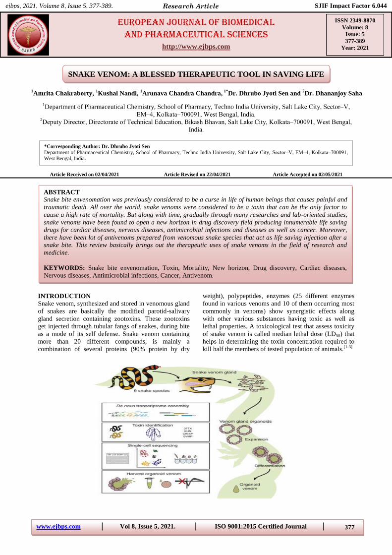
Figure-1: Snake venom organoid.
Snake venoms are complex mixtures of enzymes and
proteins of various sizes, amines, lipids, nucleosides, and
carbohydrates. Venoms also contain various metal ions
that are presumed to act as cofactors and include sodium,
calcium, potassium, magnesium, and zinc. Snake venoms
are complex mixtures of enzymes and proteins of various
sizes, amines, lipids, nucleosides, and carbohydrates.
Venoms also contain various metal ions that are
presumed to act as cofactors and include sodium,
calcium, potassium, magnesium, and zinc. Snake venoms
have been studied much more thoroughly in members of
the families Elapidae, Hydrophiidae, and Viperidae, with
considerably less knowledge regarding venoms from
Atractaspididae and Colubridae. There is a large degree
of variability in venom composition at all taxonomic
levels. In addition, within the same species, venom
components have been shown to vary considerably
among populations and across geographical areas.
Venoms act on a variety of cells and tissues with
pronounced physiological responses. Some of the actions
of venom components include the digestion of cells and
cell membranes, disruption of procoagulant and
anticoagulant activities of blood, production of oxidizing
agents, breakdown of collagen and the intercellular
matrix between cells, and the disruption of nerve tissue.
Snake toxins with defined actions include neurotoxins,
hemotoxins, cardiotoxins, cytotoxins, and myotoxins.
Snake venom components can be grouped by their
molecular weight. Low-molecular weight components
(<1500 Da) are usually considered the least
physiologically active and includes peptides, lipids,
nucleosides, carbohydrates, amines, and metal ions.
Larger venom components (mol. wt. 4500–10000 Da)
include polypeptide toxins such as post-synaptically
acting neurotoxins and myotoxins. The largest
components, the enzymes (mol. wt. 13000–150000 Da),
comprise a diverse group and produce marked
physiological effects. The percentage of enzymes in
snake venom can vary widely and can constitute as much
as 90% or more in some of the viperid venoms and as
little as 25% in some elapid venoms. There are over 30
enzymes that have been identified in snake venoms
including some that are common to all venomous snake
families. Snake venom is a heterogeneous and complex
mixture of proteins, amino acids, lipids, carbohydrates,
metal ions, and other compounds. Venom composition
varies tremendously between different species, between
different individual snakes of the same species, and even
in the same snake depending on its age and time of the
year. Though there are exceptions, the venom of the
Elapidae family is primarily neurotoxic with some
myotoxic effects. Conversely, the venom of the
Viperidae family is generally hemotoxic and myotoxic
with some (usually) minor neurotoxic effects. The study
of venom and its composition falls under the field of
toxicology and is an area of increasing interest. Multiple
pharmaceutical agents have been developed from snake
venom. Examples include tirofiban, which was derived
from the saw-scaled viper (Echis carinatus) and
angiotensin converting enzyme inhibitors from the
Brazilian viper (Bothrops jararaca). Many more
potential venom-derived agents are currently being
investigated to treat various conditions ranging from
chronic pain to multiple sclerosis. As the composition of
snake venom varies between species, so too does its
potency. Australian elapids and sea snakes are
consistently reported to have among the most potent
venom in the world. Venom is thought to have several
functions including defense, prey neutralization, and pre-
digestion. It is delivered into the prey or attackers from
the venom gland via ducts through specialized teeth or
fangs. Some snakes species, such as the vipers, have
large hollow front fangs which are very effective in
delivering large amounts of venom while others possess
poorly developed relatively ineffective grooved rear
fangs. Not all venomous snakebites result in
envenomation. Twenty-five percent or more of
snakebites are suspected to be „dry‟ bites.[4]
Types of Venoms and their effects on human body:
Depending on the nature of classification of venom,
venomous (poisonous) snakes belong to three main
categories:
1. ELAPIDAE: They mainly secrete neurotoxic
venoms that affects the nervous system, along with
high amount of cytotoxin that can cause cellular
necrosis in human. Systemic absorption of Elapidae
venom is dependent on lymphatic transport
following subcutaneous envenomation. The onset of
neurotoxic symptoms usually occurs within 4 h but
Sen et al. European Journal of Biomedical and Pharmaceutical Sciences
www.ejbps.com │ Vol 8, Issue 5, 2021. │ ISO 9001:2015 Certified Journal │ 379
can be delayed up to 10 h following a bite. The
metabolism of venom components is not well
understood. It is likely that venom components are
inactivated by enzymes within tissues where the
venom is ultimately distributed. The distribution of
venom is variable and complex and possibly reaches
different tissue sites unevenly. The biological half-
life of Elapidae venom has not been determined. It is
likely that metabolized venom fractions are
eliminated primarily by the kidneys.[5]
2.Their bite is accompanied with certain local
features like
(i) Fang marks
(ii) Burning and pain
(iii) Swelling and discoloration
(iv) Serosanguinous discharge
The systemic features of Elapidae bite exhibit two
stages
1. Pre-paralytic Stage (Vomiting, headache, giddiness,
weakness, lethargy)
2. Paralytic Stage (Convulsion, ptosis, drowsiness,
opthalmoplegia, death)
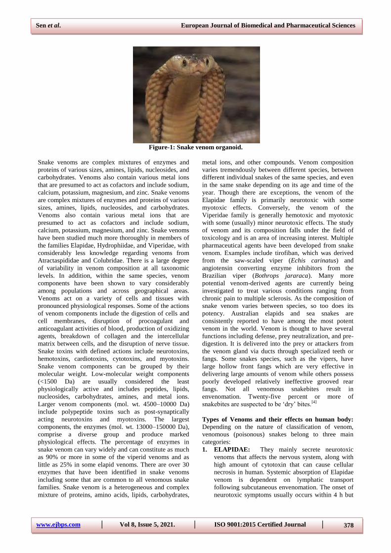
Examples of Elapidae (i) Common cobra (Naja naja)
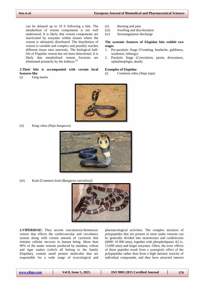
(ii) King cobra (Naja bangarus)
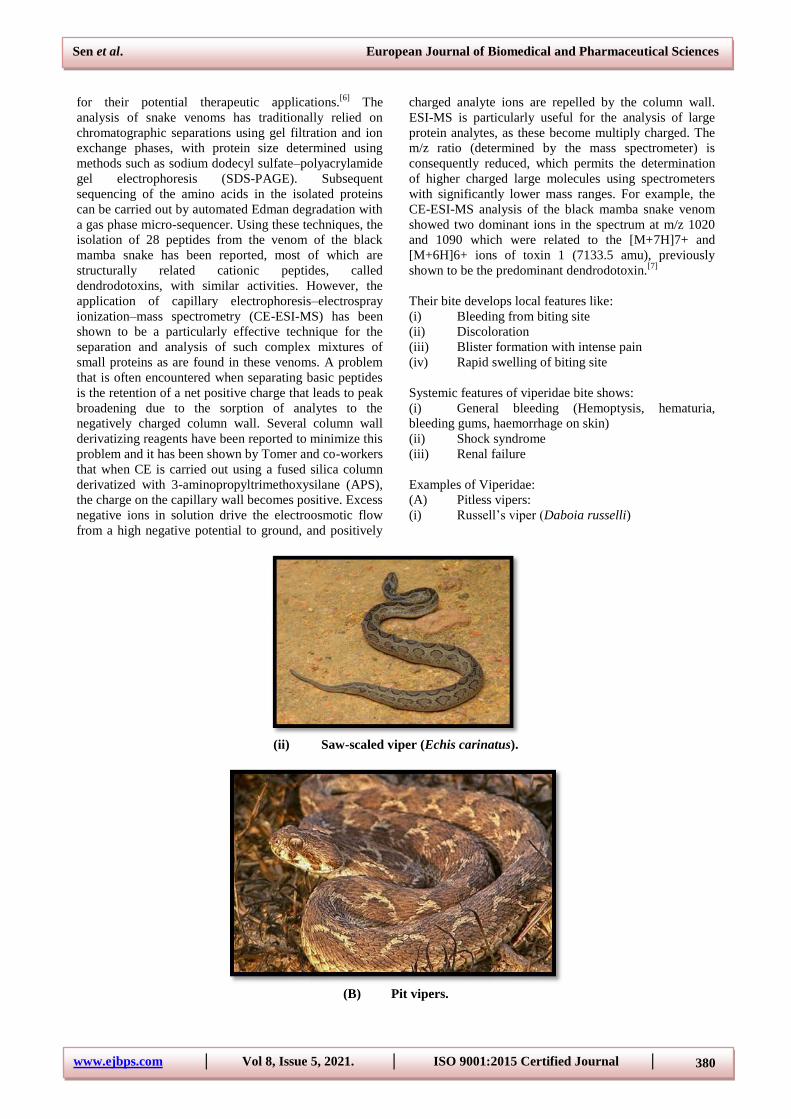
(iii) Krait [Common krait (Bangarus caeruleus)]
2.VIPERIDAE: They secrete vasculotoxic/hemotoxic
venom that effects the cardiovascular and circulatory
system along with certain amount of cytotoxin that
initiates cellular necrosis in human being. More than
90% of the snake venoms produced by mambas, cobras
and tiger snakes (which all belong to the family
Elapidae), contain small protein molecules that are
responsible for a wide range of toxicological and
pharmacological activities. The complex mixtures of
polypeptides that are present in most snake venoms can
be generally divided into neurotoxins and cardiotoxins
(6000–10 000 amu), together with phospholipases A2 (c.
13,000 amu) and larger enzymes. Often, the toxic effects
of these peptides result from a synergistic effect of the
polypeptides rather than from a high intrinsic toxicity of
individual compounds, and they have attracted interest
Sen et al. European Journal of Biomedical and Pharmaceutical Sciences
www.ejbps.com │ Vol 8, Issue 5, 2021. │ ISO 9001:2015 Certified Journal │ 380
for their potential therapeutic applications.[6]
The
analysis of snake venoms has traditionally relied on
chromatographic separations using gel filtration and ion
exchange phases, with protein size determined using
methods such as sodium dodecyl sulfate–polyacrylamide
gel electrophoresis (SDS-PAGE). Subsequent
sequencing of the amino acids in the isolated proteins
can be carried out by automated Edman degradation with
a gas phase micro-sequencer. Using these techniques, the
isolation of 28 peptides from the venom of the black
mamba snake has been reported, most of which are
structurally related cationic peptides, called
dendrodotoxins, with similar activities. However, the
application of capillary electrophoresis–electrospray
ionization–mass spectrometry (CE-ESI-MS) has been
shown to be a particularly effective technique for the
separation and analysis of such complex mixtures of
small proteins as are found in these venoms. A problem
that is often encountered when separating basic peptides
is the retention of a net positive charge that leads to peak
broadening due to the sorption of analytes to the
negatively charged column wall. Several column wall
derivatizing reagents have been reported to minimize this
problem and it has been shown by Tomer and co-workers
that when CE is carried out using a fused silica column
derivatized with 3-aminopropyltrimethoxysilane (APS),
the charge on the capillary wall becomes positive. Excess
negative ions in solution drive the electroosmotic flow
from a high negative potential to ground, and positively
charged analyte ions are repelled by the column wall.
ESI-MS is particularly useful for the analysis of large
protein analytes, as these become multiply charged. The
m/z ratio (determined by the mass spectrometer) is
consequently reduced, which permits the determination
of higher charged large molecules using spectrometers
with significantly lower mass ranges. For example, the
CE-ESI-MS analysis of the black mamba snake venom
showed two dominant ions in the spectrum at m/z 1020
and 1090 which were related to the [M+7H]7+ and
[M+6H]6+ ions of toxin 1 (7133.5 amu), previously
shown to be the predominant dendrodotoxin.[7]
Their bite develops local features like:
(i) Bleeding from biting site
(ii) Discoloration
(iii) Blister formation with intense pain
(iv) Rapid swelling of biting site
Systemic features of viperidae bite shows:
(i) General bleeding (Hemoptysis, hematuria,
bleeding gums, haemorrhage on skin)
(ii) Shock syndrome
(iii) Renal failure
Examples of Viperidae:
(A) Pitless vipers:
(i) Russell‟s viper (Daboia russelli)
(ii) Saw-scaled viper (Echis carinatus).
(B) Pit vipers.
Sen et al. European Journal of Biomedical and Pharmaceutical Sciences
www.ejbps.com │ Vol 8, Issue 5, 2021. │ ISO 9001:2015 Certified Journal │ 381
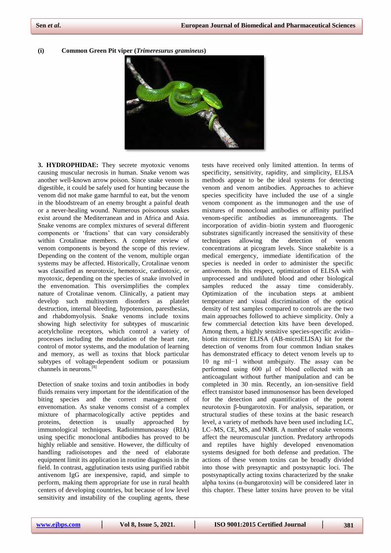
(i) Common Green Pit viper (Trimeresurus gramineus)
3. HYDROPHIDAE: They secrete myotoxic venoms
causing muscular necrosis in human. Snake venom was
another well-known arrow poison. Since snake venom is
digestible, it could be safely used for hunting because the
venom did not make game harmful to eat, but the venom
in the bloodstream of an enemy brought a painful death
or a never-healing wound. Numerous poisonous snakes
exist around the Mediterranean and in Africa and Asia.
Snake venoms are complex mixtures of several different
components or „fractions‟ that can vary considerably
within Crotalinae members. A complete review of
venom components is beyond the scope of this review.
Depending on the content of the venom, multiple organ
systems may be affected. Historically, Crotalinae venom
was classified as neurotoxic, hemotoxic, cardiotoxic, or
myotoxic, depending on the species of snake involved in
the envenomation. This oversimplifies the complex
nature of Crotalinae venom. Clinically, a patient may
develop such multisystem disorders as platelet
destruction, internal bleeding, hypotension, paresthesias,
and rhabdomyolysis. Snake venoms include toxins
showing high selectivity for subtypes of muscarinic
acetylcholine receptors, which control a variety of
processes including the modulation of the heart rate,
control of motor systems, and the modulation of learning
and memory, as well as toxins that block particular
subtypes of voltage-dependent sodium or potassium
channels in neurons.[8]
Detection of snake toxins and toxin antibodies in body
fluids remains very important for the identification of the
biting species and the correct management of
envenomation. As snake venoms consist of a complex
mixture of pharmacologically active peptides and
proteins, detection is usually approached by
immunological techniques. Radioimmunoassay (RIA)
using specific monoclonal antibodies has proved to be
highly reliable and sensitive. However, the difficulty of
handling radioisotopes and the need of elaborate
equipment limit its application in routine diagnosis in the
field. In contrast, agglutination tests using purified rabbit
antivenom IgG are inexpensive, rapid, and simple to
perform, making them appropriate for use in rural health
centers of developing countries, but because of low level
sensitivity and instability of the coupling agents, these
tests have received only limited attention. In terms of
specificity, sensitivity, rapidity, and simplicity, ELISA
methods appear to be the ideal systems for detecting
venom and venom antibodies. Approaches to achieve
species specificity have included the use of a single
venom component as the immunogen and the use of
mixtures of monoclonal antibodies or affinity purified
venom-specific antibodies as immunoreagents. The
incorporation of avidin–biotin system and fluorogenic
substrates significantly increased the sensitivity of these
techniques allowing the detection of venom
concentrations at picogram levels. Since snakebite is a
medical emergency, immediate identification of the
species is needed in order to administer the specific
antivenom. In this respect, optimization of ELISA with
unprocessed and undiluted blood and other biological
samples reduced the assay time considerably.
Optimization of the incubation steps at ambient
temperature and visual discrimination of the optical
density of test samples compared to controls are the two
main approaches followed to achieve simplicity. Only a
few commercial detection kits have been developed.
Among them, a highly sensitive species-specific avidin–
biotin microtiter ELISA (AB-microELISA) kit for the
detection of venoms from four common Indian snakes
has demonstrated efficacy to detect venom levels up to
10 ng ml−1 without ambiguity. The assay can be
performed using 600 μl of blood collected with an
anticoagulant without further manipulation and can be
completed in 30 min. Recently, an ion-sensitive field
effect transistor based immunosensor has been developed
for the detection and quantification of the potent
neurotoxin β-bungarotoxin. For analysis, separation, or
structural studies of these toxins at the basic research
level, a variety of methods have been used including LC,
LC–MS, CE, MS, and NMR. A number of snake venoms
affect the neuromuscular junction. Predatory arthropods
and reptiles have highly developed envenomation
systems designed for both defense and predation. The
actions of these venom toxins can be broadly divided
into those with presynaptic and postsynaptic loci. The
postsynaptically acting toxins characterized by the snake
alpha toxins (α-bungarotoxin) will be considered later in
this chapter. These latter toxins have proven to be vital
Sen et al. European Journal of Biomedical and Pharmaceutical Sciences
www.ejbps.com │ Vol 8, Issue 5, 2021. │ ISO 9001:2015 Certified Journal │ 382
tools in characterizing, identifying, and isolating the
nicotinic AChR.
Neurotoxins that block neuromuscular transmission by
presynaptic actions are found in the venom of many of
the same species of snakes that contain postsynaptic
toxins. The best known of these, and the first to be
described, was β-bungarotoxin, from the snake Bungarus
multicinctus. Bungarotoxins are a group of closely
related neurotoxic proteins of the three-finger toxin
superfamily found in the venom of kraits including
Bungarus multicinctus. α-Bungarotoxin inhibits the
binding of acetylcholine (ACh) to nicotinic acetylcholine
receptors; β- and γ-bungarotoxins act presynaptically
causing excessive acetylcholine release and subsequent
depletion. Both α and β forms have been characterized,
the α being similar to the long or Type II neurotoxins
from other elapid venoms.
There are four types: α-Bungarotoxin, β-Bungarotoxin
(not a three-finger toxin), γ-Bungarotoxin (Q9YGJ0) &
k-Bungarotoxin. This toxin blocks neuromuscular
transmission irreversibly by reducing ACh release. Other
neurotoxins with similar presynaptic actions are notexin
from the Australian tiger snake, Notechis scutatus,
taipoxin from the Australian taipan, Oxyuranus
scutellatus, and a myotoxin from the Asian sea snake,
Enhyodrina schistosa. β-Bungarotoxin has a triphasic
pattern of effects on ACh release. Initially, release is
decreased; this is followed by a transient increase in
release, with a subsequent progressive inhibition to
block. Actions of β-bungarotoxin have also been
demonstrated in the CNS. However, much higher
concentrations of the toxin are needed to alter transmitter
release in the brain. Its actions in the peripheral nervous
system are proposed to be specific for cholinergic
neurons; peripheral noradrenergic terminals are
unaffected by β-bungarotoxin.
The precise mechanisms underlying the various actions
of β-bungarotoxin are not yet completely understood.
Several snake venoms contain toxins with phospholipase
A2 (PLA2) activity. These include notexin, β-
bungarotoxin, and taipoxin. There is considerable
sequence homology among amino acids for β-
bungarotoxin and other snake venom PLA2 enzymes.
Snake venom PLA2 neurotoxins are thought to
hydrolyze membrane phospholipids. However, several
studies suggest that the PLA2 activity, in and of itself,
does not explain the toxin‟s effects. For example, a
potassium channel blocking action has also been reported
for β-bungarotoxin. This effect occurs independently of
the phospholipase activity and has been proposed to be
responsible for the transient increase in ACh release.
However, it is unclear how this action could be specific
for peripheral cholinergic terminals since potassium
channels would be found on other terminals as well. The
cause of the ultimate block of release of ACh by β-
bungarotoxin and other PLA2-type neurotoxins is
unknown. Suggested mechanisms include inhibition of
oxidative phosphorylation, inhibition of cytoskeletal
phosphorylation, and cytoskeleton disruption. β-
Bungarotoxin also inhibits phosphorylation of synapsin I
in rat brain synaptosomes. Synapsin I is a neuronal
phosphoprotein associated with synaptic vesicles that
have been implicated in moving vesicles from a reserve
to a releasable pool near the active zones. Ammodytoxin
A, the principal toxin in the venom of the long-nosed
viper (Vipera ammodytes ammodytes), is reported to
block neuromuscular transmission by de-energization of
the nerve terminal, resulting from mitochondrial
degeneration and subsequent impairment of coupling
between the action-potential-induced depolarization of
the nerve terminal and the evoked transmitter release.[9]
Local feature of Hydrophidae bite is accompanied with
local pain and swelling of biting site.
Systemic feature consists of:
(i) Muscle stiffness
(ii) Muscular necrosis
(iii) Myaglia
(iv) Renal failure
(v) Myoglobinuria
Examples of Hydrophidae
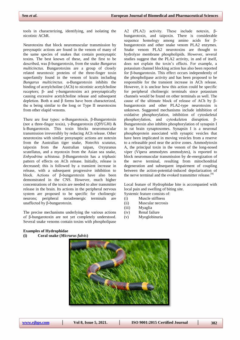
(i) Coral snake (Micrurus fulvis)
Sen et al. European Journal of Biomedical and Pharmaceutical Sciences
www.ejbps.com │ Vol 8, Issue 5, 2021. │ ISO 9001:2015 Certified Journal │ 383
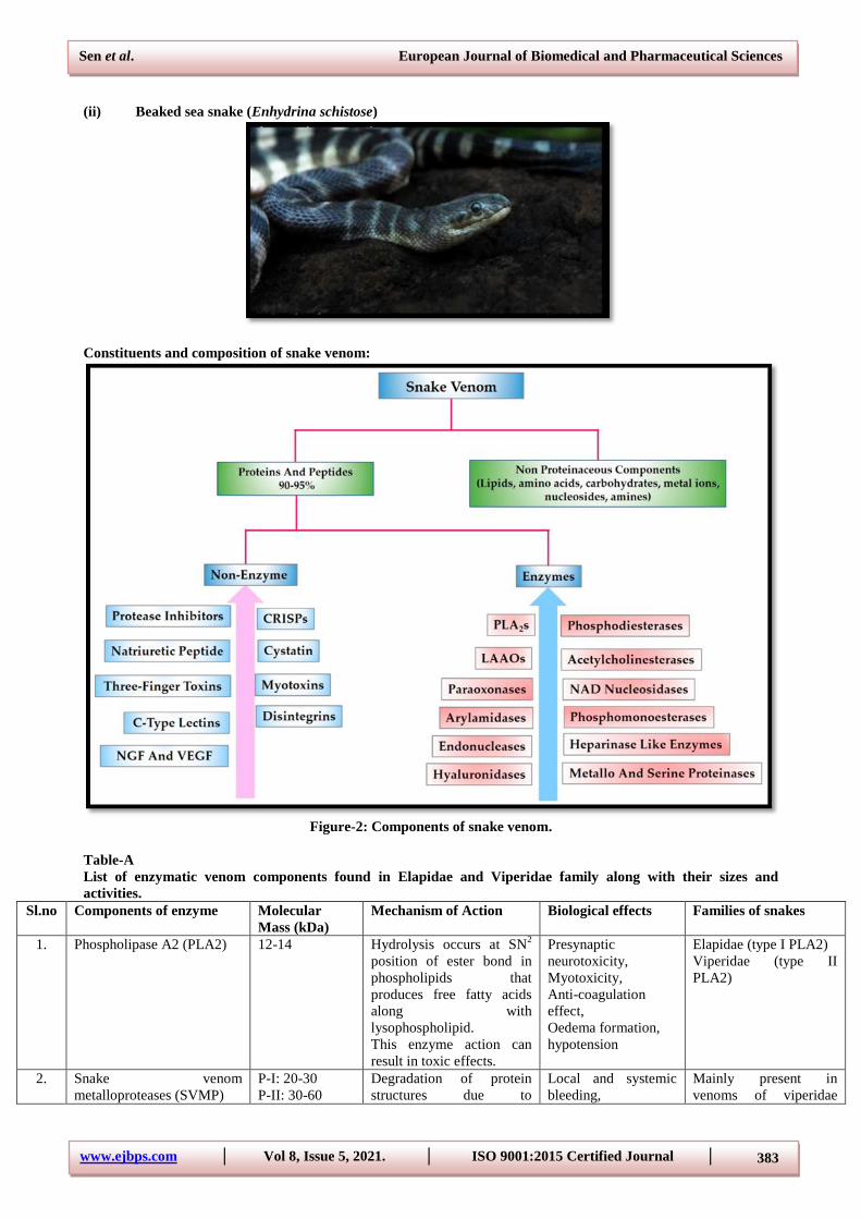
(ii) Beaked sea snake (Enhydrina schistose)
Constituents and composition of snake venom:
Figure-2: Components of snake venom.
Table-A
List of enzymatic venom components found in Elapidae and Viperidae family along with their sizes and
activities.
Sl.no Components of enzyme Molecular
Mass (kDa)
Mechanism of Action Biological effects Families of snakes
1. Phospholipase A2 (PLA2) 12-14 Hydrolysis occurs at SN2
position of ester bond in
phospholipids that
produces free fatty acids
along with
lysophospholipid.
This enzyme action can
result in toxic effects.
Presynaptic
neurotoxicity,
Myotoxicity,
Anti-coagulation
effect,
Oedema formation,
hypotension
Elapidae (type I PLA2)
Viperidae (type II
PLA2)
2. Snake venom
metalloproteases (SVMP)
P-I: 20-30
P-II: 30-60
Degradation of protein
structures due to
Local and systemic
bleeding,
Mainly present in
venoms of viperidae
Sen et al. European Journal of Biomedical and Pharmaceutical Sciences
www.ejbps.com │ Vol 8, Issue 5, 2021. │ ISO 9001:2015 Certified Journal │ 384
P-III: 60-100
[Classification
has been done
on the basis of
domain
organization]
proteolytic activities.
Haemorrhagic effects are
caused due to disintigrin-
like domain of SVMP
Haemostasis due to
anti-coagulant
property, Cytotoxic
effect causing tissue
necrosis
family
3. Serine proteases (SVSP)
Eg: Thrombin-like enzyme
26-27 In the pro-enzyme
coagulation cascade, the
hydrolysis of peptide bonds
causes pro-coagulation,
fibrinolytic/
fibrinogenolytic activities.
In some cases, SVSP
releases bradykinin due to
kallikrein – like activity.
Haemostasis,
Hypotension
Viperidae and Elapidae
venoms (Except:
Australian Elapidae
snakes)
4. L-amino acid oxidase
(LAAO)
Gel filtration
method: 110-
150
SDS/PAGE
method: 50-70
(under reducing
and non-
reducing
conditions)
Stereospecific oxidative
deamination of L-amino
acid is catalyzed that
results in producing alpha-
keto acid, NH3 and H2O2
Effect‟s platelet
aggregation, induces
cell apoptosis and
anti-microbial
activities
Both elapidae and
viperidae
5. 5‟-Nucleotidases 53-60 Phosphate monoester at 5‟-
position of DNA and RNA
is hydrolyzed
Inhibition in
aggregation of
platelets
Both elapidae and
viperidae family
6. Acetyl-cholinesterase 55-60 Acetylcholine is
hydrolyzed to choline and
acetate group
Acetylcholine
terminates neuro-
transmission
Elapidae (except:
Dendroaspis genus)
7. Hyaluronidases 33-110 Oligosaccharides N-
acetylglucosamine are
formed by hydrolysis of
hyaluronan
The structural,
rheological and
chemical properties
of extracellular matrix
are altered by this
„Spreading factor‟
Both elapidae and
viperidae
Table-B
List of non-enzymatic venom components in snakes of Elapidae and Viperidae families, their sizes and activities.
Sl.no Components of Non-
enzyme
Molecular
Mass (kDa)
Mechanism of action Biological effects Families of snakes
1. Three-finger toxins (3FTx)
Eg: alpha- neurotoxins
6-9 Interference with
neuromuscular transmission is
occurred due to inhibition in
postsynaptic nicotinic
acetylcholine receptors in
neuromuscular junction
Post synaptic effect Elapidae and very
rare in Viperidae
2. Kunitz peptides (KUN) 7 Interferes blood coagulation
and fibrinolysis by inhibiting
serine protease. Other
activities include
inflammation and ion channel
blockade
Haemostasias disruption Elapidae and
Viperidae
3. Cystine-rich secretory
proteins (CRiSP)
20-30 Cyclic nucleotide- gated
(CNG) channel blockade and
L-type calcium channel
blockade
Smooth muscle contraction
is inhibited
Common in
Viperidae
4. C-type lectins (CTL) Comprises of
two units:
Alpha (A
Blood oagulation factors or
specific platelet membrane
receptors are either activated
Anti-coagulation is
promoted along with
inhibition or promotion of
Mostly found in
Viperidae
Sen et al. European Journal of Biomedical and Pharmaceutical Sciences
www.ejbps.com │ Vol 8, Issue 5, 2021. │ ISO 9001:2015 Certified Journal │ 385
chain): 14-15
Beta (B
chain): 13-14
or inhibited by this component
in venom
platelet aggregation
5. Disintigrins (DIS) 5-10 On binding to glycoprotein
IIb/IIIa it gets expressed on
activated platelet that prevents
fibrogen interaction
Aggregation of platelet is
inhibited
Present in
Viperidae but
absent in elapidae
6. Natriuretic peptides (NP) 3.5-4 cGMP level is increased along
with subsequent cascade
signaling due to an interaction
between NPs and guanylyl
cyclase receptors.
Inhibition of angiotensin-
converting enzyme is caused
as an effect on rennin-
angiotensin by NPs
Vasodilation results in
hypotension.
Diuresis and natriuresis
promotes sodium and
water excretion
Both in Viperidae
(found abundantly)
and also in elapidae
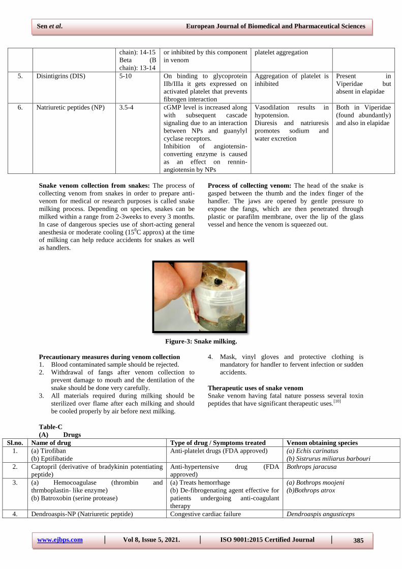
Snake venom collection from snakes: The process of
collecting venom from snakes in order to prepare anti-
venom for medical or research purposes is called snake
milking process. Depending on species, snakes can be
milked within a range from 2-3weeks to every 3 months.
In case of dangerous species use of short-acting general
anesthesia or moderate cooling (150C approx) at the time
of milking can help reduce accidents for snakes as well
as handlers.
Process of collecting venom: The head of the snake is
gasped between the thumb and the index finger of the
handler. The jaws are opened by gentle pressure to
expose the fangs, which are then penetrated through
plastic or parafilm membrane, over the lip of the glass
vessel and hence the venom is squeezed out.
Figure-3: Snake milking.
Precautionary measures during venom collection
1. Blood contaminated sample should be rejected.
2. Withdrawal of fangs after venom collection to
prevent damage to mouth and the dentilation of the
snake should be done very carefully.
3. All materials required during milking should be
sterilized over flame after each milking and should
be cooled properly by air before next milking.
4. Mask, vinyl gloves and protective clothing is
mandatory for handler to fervent infection or sudden
accidents.
Therapeutic uses of snake venom
Snake venom having fatal nature possess several toxin
peptides that have significant therapeutic uses.[10]
Table-C
(A) Drugs
Sl.no. Name of drug Type of drug / Symptoms treated Venom obtaining species
1. (a) Tirofiban
(b) Eptifibatide
Anti-platelet drugs (FDA approved) (a) Echis carinatus
(b) Sistrurus miliarus barbouri
2. Captopril (derivative of bradykinin potentiating
peptide)
Anti-hypertensive drug (FDA
approved)
Bothrops jaracusa
3. (a) Hemocoagulase (thrombin and
thrmboplastin- like enzyme)
(b) Batroxobin (serine protease)
(a) Treats hemorrhage
(b) De-fibrogenating agent effective for
patients undergoing anti-coagulant
therapy
(a) Bothrops moojeni
(b)Bothrops atrox
4. Dendroaspis-NP (Natriuretic peptide) Congestive cardiac failure Dendroaspis angusticeps
Sen et al. European Journal of Biomedical and Pharmaceutical Sciences
www.ejbps.com │ Vol 8, Issue 5, 2021. │ ISO 9001:2015 Certified Journal │ 386
[Phase II clinical study is ongoing]
5. (a) α-Cobratoxin
(b) α-Cobrotoxin
(Undergoing human trials)
Used as analgesics (a) Naja knouthia
(b) Naja nivea
6. Ancrod (Undergoing phase III clinical trials) De-fibrinogenating agent Agkistrodan rhodostoma
7. Contortrostatin/ Eristrostatin Used for treating cancer Asian sand snakes
8. Hannalgesin (oral drug) Analgesic drug Ophiophagus hannah
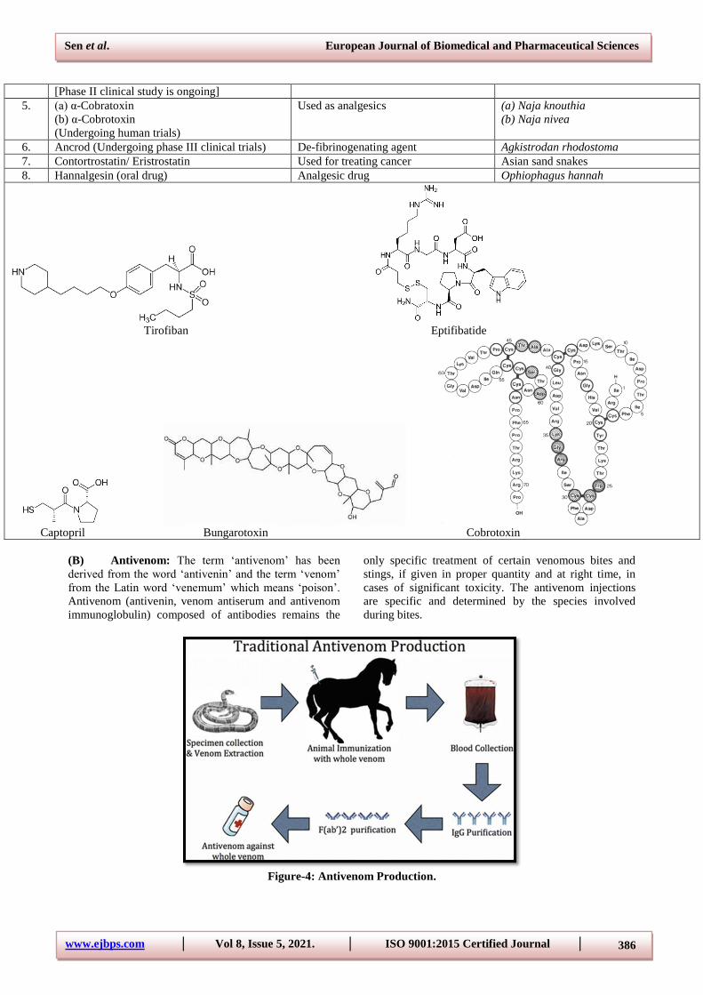
Tirofiban Eptifibatide
Captopril Bungarotoxin Cobrotoxin
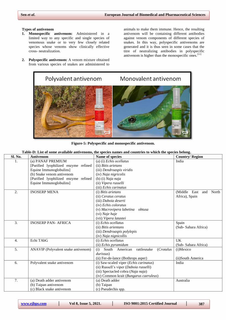
(B) Antivenom: The term „antivenom‟ has been
derived from the word „antivenin‟ and the term „venom‟
from the Latin word „venemum‟ which means „poison‟.
Antivenom (antivenin, venom antiserum and antivenom
immunoglobulin) composed of antibodies remains the
only specific treatment of certain venomous bites and
stings, if given in proper quantity and at right time, in
cases of significant toxicity. The antivenom injections
are specific and determined by the species involved
during bites.
Figure-4: Antivenom Production.
Sen et al. European Journal of Biomedical and Pharmaceutical Sciences
www.ejbps.com │ Vol 8, Issue 5, 2021. │ ISO 9001:2015 Certified Journal │ 387
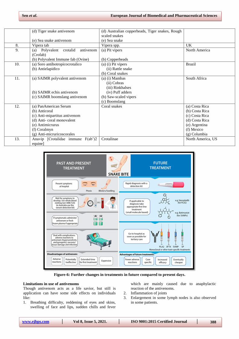
Types of antivenom
1. Monospecific antivenom: Administered in a
limited way to any specific and single species of
venomous snake or to very few closely related
species whose venoms show clinically effective
cross- neutralization.
2. Polyspecific antivenom: A venom mixture obtained
from various species of snakes are administered to
animals to make them immune. Hence, the resulting
antivenom will be containing different antibodies
against venom components of different species of
snakes. In this way, polyspecific antivenoms are
generated and it is thus seen in some cases that the
titre of neutralizing antibodies in polyspecific
antivenom is higher than the monospecific ones.[11]
Figure-5: Polyspecific and monospecific antivenom.
Table-D: List of some available antivenoms, the species names and countries to which the species belong.
Sl. No. Antivenom Name of species Country/ Region
1. (a) PANAF PREMIUM
[Purified lyophilized enzyme refined
Equine Immunoglobulins]
(b) Snake venom antivenom
[Purified lyophilized enzyme refined
Equine Immunoglobulins]
(a) (i) Echis ocellatus
(ii) Bitis arietans
(iii) Dendroaspis viridis
(iv) Naja nigricolis
(b) (i) Naja naja
(ii) Vipera russelli
(iii) Echis carinatus
India
2. INOSERP MENA (i) Bitis arietans
(ii) Ceratus ceratus
(iii) Daboia deserti
(iv) Echis coloratus
(v) Macrovipera labetina obtusa
(vi) Naje haje
(vii) Vipera latastei
(Middle East and North
Africa), Spain
3. INOSERP PAN- AFRICA (i) Echis ocellatus
(ii) Bitis arientans
(iii) Dendroaspis polylepis
(iv) Naja nignicollis
Spain
(Sub- Sahara Africa)
4. Echi TAbG (i) Echis ocellatus
(ii) Echis pyramidum
UK
(Sub- Sahara Africa)
5. ANAVIP (Polyvalent snake antivenom) (i) South American rattlesnake (Crotalus
durissus)
(ii) Fer-de-lance (Bothrops asper)
(i)Mexico
(ii)South America
6. Polyvalent snake antivenom (i) Saw-scaled viper (Echis carinatus)
(ii) Russell‟s viper (Daboia russelli)
(iii) Spectacled cobra (Naja naja)
(iv) Common krait (Bungarus caeruleus)
India
7. (a) Death adder antivenom
(b) Taipan antivenom
(c) Black snake antivenom
(a) Death adder
(b) Taipan
(c) Pseudechis spp.
Australia
Sen et al. European Journal of Biomedical and Pharmaceutical Sciences
www.ejbps.com │ Vol 8, Issue 5, 2021. │ ISO 9001:2015 Certified Journal │ 388
(d) Tiger snake antivenom
(e) Sea snake antivenom
(d) Australian copperheads, Tiger snakes, Rough
scaled snakes
(e) Sea snake
8. Vipera tab Vipera spp. UK
9. (a) Polyvalent crotalid antivenom
(Crofab)
(b) Polyvalent Immune fab (Ovine)
(a) Pit vipers
(b) Copperheads
North America
10. (a) Soro antibotropicocrotalico
(b) Antielapidico
(a) (i) Pit vipers
(ii) Rattle snake
(b) Coral snakes
Brazil
11. (a) SAIMR polyvalent antivenom
(b) SAIMR echis antivenom
(c) SAIMR boomslang antivenom
(a) (i) Mambas
(ii) Cobras
(iii) Rinkhalses
(iv) Puff adders
(b) Saw-scaled vipers
(c) Boomslang
South Africa
12. (a) PanAmerican Serum
(b) Anticoral
(c) Anti-mipartitus antivenom
(d) Anti- coral monovalent
(e) Antimicrurus
(f) Coralmyn
(g) Anti-micruricoscorales
Coral snakes (a) Costa Rica
(b) Costa Rica
(c) Costa Rica
(d) Costa Rica
(e) Argentina
(f) Mexico
(g) Columbia
13. Anavip [Crotalidae immune F(ab‟)2
equine]
Crotalinae North America, US
Figure-6: Further changes in treatments in future compared to present days.
Limitations in use of antivenoms
Though antivenom acts as a life savior, but still is
application can have some side effects on individuals
like:
1. Breathing difficulty, reddening of eyes and skins,
swelling of face and lips, sudden chills and fever
which are mainly caused due to anaphylactic
reaction of the antivenoms.
2. Inflammation of joints
3. Enlargement in some lymph nodes is also observed
in some patients.
Sen et al. European Journal of Biomedical and Pharmaceutical Sciences
www.ejbps.com │ Vol 8, Issue 5, 2021. │ ISO 9001:2015 Certified Journal │ 389
4. Due to high concentration of non-immunoglobulin
proteins several pyrogenic reactions also occur.
5. Antivenom cannot reverse the effects caused on
snake bite envenomation.
Snake venom has also been found to exhibit anti-
microbial activities like antibacterial, antiviral, antifungal
and antiparasitic properties that helps in treating several
diseases like measles, yellow fever, HIV, hepatitis C as
well as fungal species like Candida parapsilosis,
parasites of Leishmania sp. and helps in converting
gram- positive bacteria to gram-negative bacteria.
CONCLUSION
Halāhala (Sanskrit हलाहल) or kālakūṭa (Sanskrit कालकूटं,
literally: 'black mass' or 'time puzzle') is the name of a
poison (as per Hindu History) created from the sea when
Devas and Asuras churned it in order to obtain Amrita,
the nectar of immortality. Snake venoms previously have
been known to have only toxic properties that can harm
human beings causing painful and traumatic deaths. But
later on, with gradual progress in scientific research and
medical studies, snake venoms are considered to be the
most valuable therapeutic tool that acts as a “blessing” in
curing many diseases and along with the discoveries of
antivenom it has opened a new horizon in curing victims
of snake bites. Though a very small fraction of snake
venom components has been identified, but still further
more researches and studies are going on worldwide to
give it a new and broader scope in the future of drug
discovery field and to further uncover many more
therapeutic leads from snake venom.
REFERENCES
1. Mattison C. The New Encyclopedia of Snakes. New
Jersey, USA (first published in the UK): Princeton
University Press (Princeton and Oxford) first
published in Blandford, 2007; p. 117.
2. Stuart MC, Kouimtzi M, Hill SR, eds. WHO Model
Formulary, 2008. p. X.
3. Kalyan kumar, B. Antisnake venom. International
Journal on Pharmaceutical and Biomedical
Research (IJPBR), 2014; 1(3): 77 – via
documents.pub.
4. Slagboom J, Kool J, Harrison RA, Casewell
NR. Hemotoxic snake venoms: their functional
activity, impact on snakebite victima and
pharmaceutical promise. British Journal of
Haematology, 2017; 177(6): 947–959.
5. Priyanka K. Goswami, Mayuri Samant, Rashmi S
Srivastava “Snake Venom, Anti- snake venom &
Potential of snake venom”; International Journey of
Pharmacy and Pharmaceutical Sciences, 2014; 5: 4-
7.
6. Tarek Mohamed Abd El- Aziz, Antonio G Soares,
James D Stockand. “Snake venoms in Drug
Discovery: Valuable Therapeutic Tools in Life
Saving.” Toxins, 2019; 11: 564.
7. Suchaya Sanhajariya, Stephen B Duffull, Geoffrey
K Isbister. “Pharmacokinetics of snake venom.”
Toxins, 2018; 10: 73.
8. Basheruddin. Sk. “Medical uses of snake poison.”
RRJMHS, 2015.
9. Isabel Gomez-Betancur, Vedanjali Gogineni, Andra
Salazar- Ospina and Francisco Leon. “Perspective
on the Therapeutics of Anti- snake Venom.”
Molecules, 2019, 24: 3276. doi: 10.3390/molecules
24183276.
10. World Health Organization “Snakes and Snake bites
– part 2: Venoms and Antivenoms.” AFRO
Pharmaceuticals Newsletter, Number, 2008.
11. Nadim M. R. Chhipa, Dr. Dhrubo Jyoti Sen and Dr.
Bharat G. Chaudhary; Lifesaving drugs from animal
venoms: International Research Journal for
Inventions in Pharmaceutical Sciences, , 2013; 1(2):
1-58.
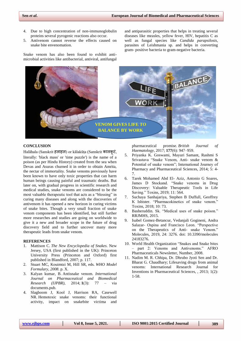
VENOM GIVES LIFE TO
BALANCE BY WORK