computational studies of snake venom toxins
TRANSCRIPT

toxins
Review
Computational Studies of Snake Venom Toxins
Paola G. Ojeda 1,2,*, David Ramírez 1,2 ID , Jans Alzate-Morales 1 ID , Julio Caballero 1,Quentin Kaas 3 and Wendy González 1,4,*
1 Center for Bioinformatics and Molecular Simulations (CBSM), Universidad de Talca, 3460000 Talca, Chile;[email protected] (D.R.); [email protected] (J.A.-M.); [email protected] (J.C.)
2 Facultad de Ciencias de la Salud, Instituto de Ciencias Biomedicas, Universidad Autonoma de Chile,3460000 Talca, Chile
3 Institute for Molecular Bioscience, The University of Queensland, Brisbane, Queensland 4072, Australia;[email protected]
4 Millennium Nucleus of Ion Channels-Associated Diseases (MiNICAD), Universidad de Talca, 3460000 Talca,Chile
* Correspondence: [email protected] (P.G.O.); [email protected] (W.G.);Tel.: +56-712-201-674 (W.G.)
Received: 6 November 2017; Accepted: 18 December 2017; Published: 22 December 2017
Abstract: Most snake venom toxins are proteins, and participate to envenomation through a diversearray of bioactivities, such as bleeding, inflammation, and pain, cytotoxic, cardiotoxic or neurotoxiceffects. The venom of a single snake species contains hundreds of toxins, and the venoms of the725 species of venomous snakes represent a large pool of potentially bioactive proteins. Despiteconsiderable discovery efforts, most of the snake venom toxins are still uncharacterized. Modernbioinformatics tools have been recently developed to mine snake venoms, helping focus experimentalresearch on the most potentially interesting toxins. Some computational techniques predict toxinmolecular targets, and the binding mode to these targets. This review gives an overview of currentknowledge on the ~2200 sequences, and more than 400 three-dimensional structures of snake toxinsdeposited in public repositories, as well as of molecular modeling studies of the interaction betweenthese toxins and their molecular targets. We also describe how modern bioinformatics have been usedto study the snake venom protein phospholipase A2, the small basic myotoxin Crotamine, and thethree-finger peptide Mambalgin.
Keywords: molecular dynamics simulations; databases; snake peptides; proteomics; molecularmodeling
1. Introduction
Snake venom is a complex mixture of proteins and peptides, and presents several medical andpharmaceutical applications [1–3]. Since the Greek antiquity, substances extracted from snake havebeen recognized for their medicinal properties, and the rod of Asclepius, a snake coiled arounda staff, is the most commonly used symbol of medicine and health. In modern times, a numberof notable molecules derived from snake toxins are used in the clinic or are in various stages ofclinical development [4]. The most famous example of snake-derived medicine is captopril (Capoten),which was developed by Bristol-Myers Squibb, and is now used as a generic medicine for treatinghypertension and congestive heart failure [5,6]. It is a small molecule inhibitor of the angiotensinconverting enzyme (ACE), and is derived from bradykinin potentiating peptides found in the venom ofthe South American snake Bothrops jararaca [5]. Another snake-derived compound potentially used forheart failure is cenderitide (CD-NP, Mayo Clinic/Capricor Therapeutics, Beverly Hills, CA, USA) [7].It is a chimera between the green mamba Dendroaspis Natriuretic Peptide DNP and the human C-type
Toxins 2018, 10, 8; doi:10.3390/toxins10010008 www.mdpi.com/journal/toxins
Toxins 2018, 10, 8 2 of 24
natriuretic peptides, and activates guanylyl cyclases. It completed Phase I/II clinical trial for chronicheart failures.
Eptifibatide (Integrilin, Millennium Pharmaceuticals/Merck, Cambridge, MA, USA), a RGD-motifcyclic heptapeptide that acts on glycoprotein IIb/IIIa integrin receptors of the blood platelets, is amedicine used in the clinic to prevent platelet aggregation and thrombus formation in acute coronarysyndromes. It was derived from a much larger protein from the pygmy rattlesnake (Sistrurus miliariusbarbouri). In the same category, anfibatide (Declotana, Lee’s Pharmaceutical, Hong Kong, China) is asnake venom-derived from platelet aggregation inhibitor peptide that antagonizes platelet glycoproteinIb receptor. It is investigated for the treatment of acute thrombotic thrombocytopenic purpura, a fatalblood clot disorder [8]. It completed phase Ib–IIa, and will undergo phase II according to Lee’s website.It also has potential for treatment of acute experimental ischaemic stroke and reperfusion injury.
A “detoxified” variant of the α-cobratoxin (RPI-78M, ReceptoPharm/Nutri Pharm, Coral Springs,FL, USA), stimulates the production of cytokines. It has application in autoimmune diseases,myasthenia gravis (MG), muscular dystrophy (MD), and amyotrophic lateral sclerosis (ALS). It recentlyreceived, from the U.S. Food and Drug Administration (FDA), an orphan drug designation for thetreatment of pediatric multiple sclerosis. Gyroxin is a serine protease from Crotalus durissus terrificus,and has applications aiming at tissue regeneration, repairing nervous system traumas and bonemarrow. Phase I clinical trial found the protein to be safe and preclinical study showed promisingresults for treating chronic venous ulcers [9].
Counterintuitively, some snake toxins can inhibit pain, and have, therefore, potential applicationas analgesics. For example, crotalphine is a 14 amino acid peptide with one disulfide bond,and has analgesic properties through TRPA1 desensitization [10]. It was reported to be developedby the Brazilian firm Biolab Farmaceutica, but the compound is not mentioned on their website.The mambalgins are three-finger toxins with antagonist activity on Acid-sensing ion channel (ASIC)channels, and are active on a range of pain models [11]. They were to be developed by Theralpha,a French startup company, which has ceased activity.
The examples mentioned above stress why snake venom peptides attract scientific and medicalinterest [3], and considerable efforts are made to mine snake venoms for interesting new compounds.The venom of a single snake species (from the roughly 725 species of venomous snakes) containshundreds of potentially pharmacologically active and useful molecules [12]. In recent years,important technological progress in transcriptomics and proteomics has resulted in a rapidly increasingknowledge of peptides and proteins in animal venoms, include those of snakes. These technologiesenable rapid discovery of the nearly complete set of toxins of a snake venom [13–15], which washitherto not achievable using previous methods based only on reversed phase-high performanceliquid chromatography (RP-HPLC) and mass spectrometry [16]. The number of toxins unraveled bymodern “omics” approaches is very large, efficient computational methods are required to mine thismassive amount of data. This review will first focus on databases that allow searching and retrievinginformation on snake toxins. Computational approaches that have been developed to help study snaketoxin activity, such as the prediction of toxin three-dimensional structures and of their interactionswith molecular targets, will be described. The successes and challenges of predicting binding affinitiesand specificities using molecular modeling will be discussed, with the future perspective to integratethe biology of venoms and prediction of toxin biological targets. Finally, we will describe how modernbioinformatics have been used to study the activity and biological targets of the snake venom proteinPLA2, the small basic myotoxin Crotamine and the three-finger peptide Mambalgin.
2. Extent of Our Knowledge on Snake Toxins
General databases, such as UniProt [17,18], NCBI Genbank/GenPept [19], and the ProteinData Bank (PDB) [20,21], play essential roles in simplifying access to information regarding proteinsequences and three-dimensional structures [22]. However, the information about peptides andproteins from snake toxins is not standardized in these resources, especially the naming of toxins
Toxins 2018, 10, 8 3 of 24
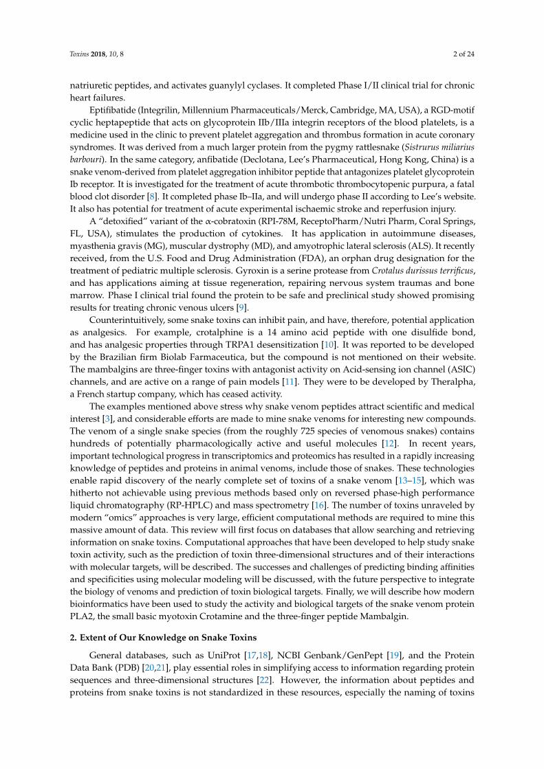
and pharmacological activities, and mining for snake venom peptides is difficult. Moreover,the database relies on depositions of information from authors to feed the data, leading to numerousduplications of entries, and a large body of work is being published in peer-reviewed articles,but never submitted to the general databases. Specialized databases, from venomous animals,are slowly emerging. Conoserver [23], Arachnoserver [24], and ISOB (Indigenous snake speciesof Bangladesh) [25] provide information on venoms from cone snail, spider, and snakes, respectively.A recently developed resource, VenomZone, is provided by the Swiss Institute of Bioinformatics(SIB), and has information about the venoms from six types of organisms, including snakes. Accessto the information is divided in taxonomy, activity, and venom protein families, making it easyto search through the website. Furthermore, all the information is linked to the venom proteininformation from the UniProtKB/Swiss-Prot database (manually annotated and reviewed) andUniProtKB/Trembl (automatically annotated). The knowledge on the activity of peptides that havebeen fully characterized helps to predict the possible function of uncharacterized peptides and proteins.In this context, specialized databases play an essential role in providing access to data, predicting thethree-dimensional structures and functions of toxins, and identifying outstanding toxins with potentialnew characteristics (Figure 1). However, there is presently no commonly established and standardway for practical annotation of toxins from the data sources mentioned above, leading sometimes to anerratic estimate for the number of toxins in the venom of one animal. Machine learning-based classifierscould help to solve this problem. ToxClassifier [26] is a machine learning tool that allows a consistentdifferentiation of toxins from non-toxin sequences, and reports the best-hit annotation, permittingassignment of a toxin into the most correct toxin protein family, providing increased curation of theseexisting databases [27].
Toxins 2017, 10, 8 3 of 23
pharmacological activities, and mining for snake venom peptides is difficult. Moreover, the database relies on depositions of information from authors to feed the data, leading to numerous duplications of entries, and a large body of work is being published in peer-reviewed articles, but never submitted to the general databases. Specialized databases, from venomous animals, are slowly emerging. Conoserver [23], Arachnoserver [24], and ISOB (Indigenous snake species of Bangladesh) [25] provide information on venoms from cone snail, spider, and snakes, respectively. A recently developed resource, VenomZone, is provided by the Swiss Institute of Bioinformatics (SIB), and has information about the venoms from six types of organisms, including snakes. Access to the information is divided in taxonomy, activity, and venom protein families, making it easy to search through the website. Furthermore, all the information is linked to the venom protein information from the UniProtKB/Swiss-Prot database (manually annotated and reviewed) and UniProtKB/Trembl (automatically annotated). The knowledge on the activity of peptides that have been fully characterized helps to predict the possible function of uncharacterized peptides and proteins. In this context, specialized databases play an essential role in providing access to data, predicting the three-dimensional structures and functions of toxins, and identifying outstanding toxins with potential new characteristics (Figure 1). However, there is presently no commonly established and standard way for practical annotation of toxins from the data sources mentioned above, leading sometimes to an erratic estimate for the number of toxins in the venom of one animal. Machine learning-based classifiers could help to solve this problem. ToxClassifier [26] is a machine learning tool that allows a consistent differentiation of toxins from non-toxin sequences, and reports the best-hit annotation, permitting assignment of a toxin into the most correct toxin protein family, providing increased curation of these existing databases [27].
Figure 1. Integration of proteomics, transcriptomics, and databases in the study of venoms. (A) Classical peptidomics analysis of toxins; (B) nucleotide discovery from transcript to precursor sequence.
2.1. Transcriptomic Analyses of Peptides and Proteins from Snake Venom Glands
Genome sequencing of venom gland is still highly expensive, and the assembly of genomic information and their analysis requires substantial bioinformatics commitment [28,29]. By contrast, sequencing cDNA libraries created from venom gland mRNA using next generation sequencing (NGS) and their assembly became mainstream in research. The transcriptomes of several snake
Figure 1. Integration of proteomics, transcriptomics, and databases in the study of venoms.(A) Classical peptidomics analysis of toxins; (B) nucleotide discovery from transcript toprecursor sequence.
2.1. Transcriptomic Analyses of Peptides and Proteins from Snake Venom Glands
Genome sequencing of venom gland is still highly expensive, and the assembly of genomicinformation and their analysis requires substantial bioinformatics commitment [28,29]. By contrast,sequencing cDNA libraries created from venom gland mRNA using next generation sequencing (NGS)
Toxins 2018, 10, 8 4 of 24
and their assembly became mainstream in research. The transcriptomes of several snake venoms havebeen reported [30–33]. NGS platforms produce near comprehensive sequence transcript informationcoding for venom peptides and proteins, complementing the traditional PCR techniques, which couldonly discover toxin transcripts related to those already discovered in other species or from proteomicstudy. The most common NGS technologies used for venom transcriptomics, i.e., 454 GS FLX Titaniumand Illumina, proceed by fragmenting the cDNA and sequencing all or part of these fragments [34].The 454 technology, which is losing momentum and will be discontinued, produces longer stretch DNAsequence (reads) than the Illumina technology (limited to read of 150 bp), but of lower quality [34,35].The fragments can be assembled back into full length or partial transcripts (the assembled fragmentsbeing called contigs) using different software, such as Trinity, Trans-Abyss or SoapdeNovo [36].
An older technology still in use today consists in using the classical Sanger sequencing ofexpressed sequence tags (ESTs), which are short DNA sequences obtained by sequencing the endsof transcript fragments. Given that most snake venom peptides are around 100 residues long,the sequencing of ESTs often yields the complete DNA sequence of a peptide. The EST approachhas been successfully applied to venom from snakes, noting that the number of retrieved sequencesare much smaller than generated by NGS, and typically only the most expressed transcripts will beidentified using Sanger/ESTs [32,37]. The ESTs can be assembled back into the original contigs usingseveral algorithms, such as CAP3 [38], Phrap [39], SeqMan [39,40], or MIRA [41].
Transcriptomic analysis generally provides insight into the peptide/protein profile of snakevenom, and can be used for discovering putative new peptides and their isoforms [15,42–45],or peptides that are lowly expressed, and consequently hard to identify by proteomic analysis [46].Finally, with advances in bioinformatics—which is no more than “the application of informationscience to biology” [47]—venom gland transcriptomic data is an excellent tool for studyingpeptides evolution [45,48], exploring antivenom and therapeutic agents [32,49], and understandingstructure–function relationships [50].
2.2. Proteomic Analyses of Peptides and Proteins from Snake Venom Glands
Snake venom proteomes are highly complex mixtures of peptides and proteins [51]. Proteomicapproaches to investigate snake venoms were recently reviewed [2,52]. These approaches generally usea combination of electrophoresis, liquid chromatography, Edman degradation sequencing, amino acidanalysis, enzymatic digestion, and mass spectrometry, among other techniques. The methods morewidely used are a combination of high performance liquid chromatography, Edman degradation,MALDI-TOF/MS of proteins, 1D or 2D PAGE and ESI/MS/MS sequencing of digested proteins [40].The first step to study the proteomes of a snake venom is the venom extraction, which is performedby “milking” the living snake. Snake milking is achieved by forcing the snake to bite into aproper container. After venom collection, the proteins are separated using high performance liquidchromatography (RP-HPLC), ultra-high performance liquid chromatography (UHPLC), and exchangechromatography [53]. Once the crude venom is fractionated, the sequences of the peptides aredetermined using a combination of mass spectrometry and Edman degradation. Prior to the MS/MSanalysis, the peptides are usually reduced, alkylated, and enzymatically digested (usually with trypsinor chemotryspin) [53]. Finally, the tertiary structure is studied using nuclear magnetic resonancespectroscopy [53,54].
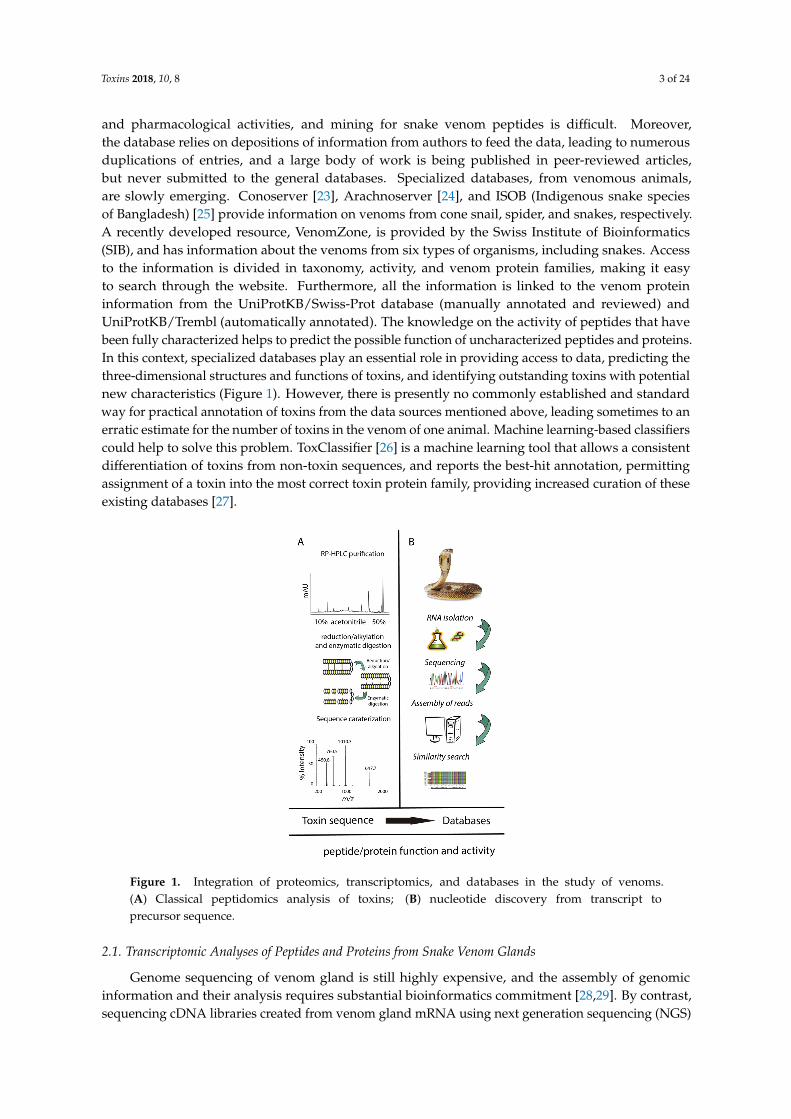
Peptides and proteins from snake venoms have a high content of cysteine residues in theirprimary sequences, and most of these cysteines form crosslinking disulfide bridges. The stabilizationunderpinned by the creation of disulfide bridges has been linked to several important features ofsnake toxins: enhancing activity, higher resistance to proteases, improving selectivity, and stabilizingsecondary structure elements [55–58]. Furthermore, the number of disulfide bridges in snake peptidesvaries (Figure 2); for instance, natriuretic peptides present one disulfide bond, sarafotoxines presenttwo disulfide bonds, and more complex toxins, such as omwarpin, have four disulfide bonds. In thewidely-studied phospholipase A2 family, the acidic phospholipase A2 subfamily, with 142 proteins
Toxins 2018, 10, 8 5 of 24
from snake venoms deposited in UniProt (search criteria: taxonomy: “Serpentes (snakes) (8570)” protein“acidic phospholipase a2” (keyword: toxin OR annotation: (type: “tissue specificity” venom)) AND reviewed: yes)share a characteristic disulfide bridge connectivity, where 14 Cys residues form seven disulfide bonds.
Toxins 2017, 10, 8 5 of 23
AND reviewed: yes) share a characteristic disulfide bridge connectivity, where 14 Cys residues form seven disulfide bonds.
Figure 2. Cys patterns of some toxins from snake venoms. Cartoon representation of a disulfide bridge (top left). The disulfide bond arrangements are shown as black lines.
3. Snake Toxin Structures and Activities
To comprehend the structure and function of snake toxins will provide a better understanding of their role in venom toxicity. Elucidation of their structures will further help us to better understand the protein–protein interactions in snake venom, as well as their target receptor/ion channels [59]. Toxins in general seem to adopt a limited number of structural scaffolds. It was initially proposed in the 1970s that the 57 snake venom toxins described at the time as being neurotoxic or cytotoxic had similar secondary structure content [60]. The first snake venom 3D structure was also solved by X-ray diffraction in the 1970s (reported on 1978) [61]. It was released in the Protein DataBank (PDB) in 1981 under the PDB ID: 1NXB (snake venom curarimimetic neurotoxins) [61]. In the past 20 years, the discovery and unraveling of snake venoms has largely paralleled the technological development in proteomics and transcriptomic sciences. Additionally, the number of 3D structures has increased, due to the amazing progress of spectroscopic techniques, such as X-ray crystallography and nuclear magnetic resonance (NMR) spectroscopy [62]. Structural genomics appeared early 2000s, and had a dramatic influence on the structural study of snake venoms [63,64]. The increased pace at which 3D structure of snake toxins are deposited in the PDB in recent years is striking, with 101 and 409 3D structures deposited before and after 2000, respectively.
The specific structure for a given toxin is important to understand the molecular events at the origin of toxin activity. Based on these experimental structures, molecular modeling has been used to understand the molecular interactions related with toxin affinity and specificity.
3.1. Classification of Snake Venom Toxins
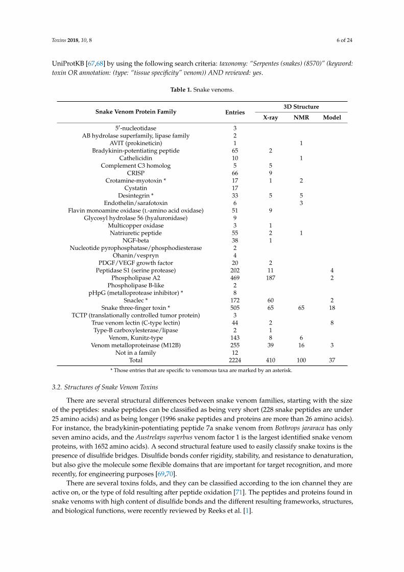
The majority of snake venom proteins, i.e., 2224 proteins and peptides, could be categorized into 30 families [65] (Table 1); whereas 12 proteins are not classified yet. To this day, 410 and 100 3D structures of snake toxins have been solved by X-ray crystallography or NMR spectroscopic techniques, respectively; and 37 3D structures have been modeled and reported in the Protein Model Portal of the PSI-Nature Structural Biology Knowledgebase [66]. This information was obtained from UniProtKB [67,68] by using the following search criteria: taxonomy: “Serpentes (snakes) (8570)” (keyword: toxin OR annotation: (type: “tissue specificity” venom)) AND reviewed: yes.
C C C C C C
C C C C C C
C C C C C CC C C C C C C C
Natriuretic peptide Sarafotoxins
Crotamine
Omwaprin Textilinin-1
C C C C C C C C C C C C C C
Acidic phospholipase A2
Figure 2. Cys patterns of some toxins from snake venoms. Cartoon representation of a disulfide bridge(top left). The disulfide bond arrangements are shown as black lines.
3. Snake Toxin Structures and Activities
To comprehend the structure and function of snake toxins will provide a better understanding oftheir role in venom toxicity. Elucidation of their structures will further help us to better understandthe protein–protein interactions in snake venom, as well as their target receptor/ion channels [59].Toxins in general seem to adopt a limited number of structural scaffolds. It was initially proposedin the 1970s that the 57 snake venom toxins described at the time as being neurotoxic or cytotoxichad similar secondary structure content [60]. The first snake venom 3D structure was also solved byX-ray diffraction in the 1970s (reported on 1978) [61]. It was released in the Protein DataBank (PDB)in 1981 under the PDB ID: 1NXB (snake venom curarimimetic neurotoxins) [61]. In the past 20 years,the discovery and unraveling of snake venoms has largely paralleled the technological developmentin proteomics and transcriptomic sciences. Additionally, the number of 3D structures has increased,due to the amazing progress of spectroscopic techniques, such as X-ray crystallography and nuclearmagnetic resonance (NMR) spectroscopy [62]. Structural genomics appeared early 2000s, and hada dramatic influence on the structural study of snake venoms [63,64]. The increased pace at which3D structure of snake toxins are deposited in the PDB in recent years is striking, with 101 and 409 3Dstructures deposited before and after 2000, respectively.
The specific structure for a given toxin is important to understand the molecular events at theorigin of toxin activity. Based on these experimental structures, molecular modeling has been used tounderstand the molecular interactions related with toxin affinity and specificity.
3.1. Classification of Snake Venom Toxins
The majority of snake venom proteins, i.e., 2224 proteins and peptides, could be categorizedinto 30 families [65] (Table 1); whereas 12 proteins are not classified yet. To this day, 410 and 1003D structures of snake toxins have been solved by X-ray crystallography or NMR spectroscopictechniques, respectively; and 37 3D structures have been modeled and reported in the Protein ModelPortal of the PSI-Nature Structural Biology Knowledgebase [66]. This information was obtained from
Toxins 2018, 10, 8 6 of 24
UniProtKB [67,68] by using the following search criteria: taxonomy: “Serpentes (snakes) (8570)” (keyword:toxin OR annotation: (type: “tissue specificity” venom)) AND reviewed: yes.
Table 1. Snake venoms.
Snake Venom Protein Family Entries3D Structure
X-ray NMR Model
5′-nucleotidase 3AB hydrolase superfamily, lipase family 2
AVIT (prokineticin) 1 1Bradykinin-potentiating peptide 65 2
Cathelicidin 10 1Complement C3 homolog 5 5
CRISP 66 9Crotamine-myotoxin * 17 1 2
Cystatin 17Desintegrin * 33 5 5
Endothelin/sarafotoxin 6 3Flavin monoamine oxidase (L-amino acid oxidase) 51 9
Glycosyl hydrolase 56 (hyaluronidase) 9Multicopper oxidase 3 1Natriuretic peptide 55 2 1
NGF-beta 38 1Nucleotide pyrophosphatase/phosphodiesterase 2
Ohanin/vespryn 4PDGF/VEGF growth factor 20 2
Peptidase S1 (serine protease) 202 11 4Phospholipase A2 469 187 2
Phospholipase B-like 2pHpG (metalloprotease inhibitor) * 8
Snaclec * 172 60 2Snake three-finger toxin * 505 65 65 18
TCTP (translationally controlled tumor protein) 3True venom lectin (C-type lectin) 44 2 8Type-B carboxylesterase/lipase 2 1
Venom, Kunitz-type 143 8 6Venom metalloproteinase (M12B) 255 39 16 3
Not in a family 12Total 2224 410 100 37
* Those entries that are specific to venomous taxa are marked by an asterisk.
3.2. Structures of Snake Venom Toxins
There are several structural differences between snake venom families, starting with the sizeof the peptides: snake peptides can be classified as being very short (228 snake peptides are under25 amino acids) and as being longer (1996 snake peptides and proteins are more than 26 amino acids).For instance, the bradykinin-potentiating peptide 7a snake venom from Bothrops jararaca has onlyseven amino acids, and the Austrelaps superbus venom factor 1 is the largest identified snake venomproteins, with 1652 amino acids). A second structural feature used to easily classify snake toxins is thepresence of disulfide bridges. Disulfide bonds confer rigidity, stability, and resistance to denaturation,but also give the molecule some flexible domains that are important for target recognition, and morerecently, for engineering purposes [69,70].
There are several toxins folds, and they can be classified according to the ion channel they areactive on, or the type of fold resulting after peptide oxidation [71]. The peptides and proteins found insnake venoms with high content of disulfide bonds and the different resulting frameworks, structures,and biological functions, were recently reviewed by Reeks et al. [1].
Toxins 2018, 10, 8 7 of 24
3.2.1. ICK Fold
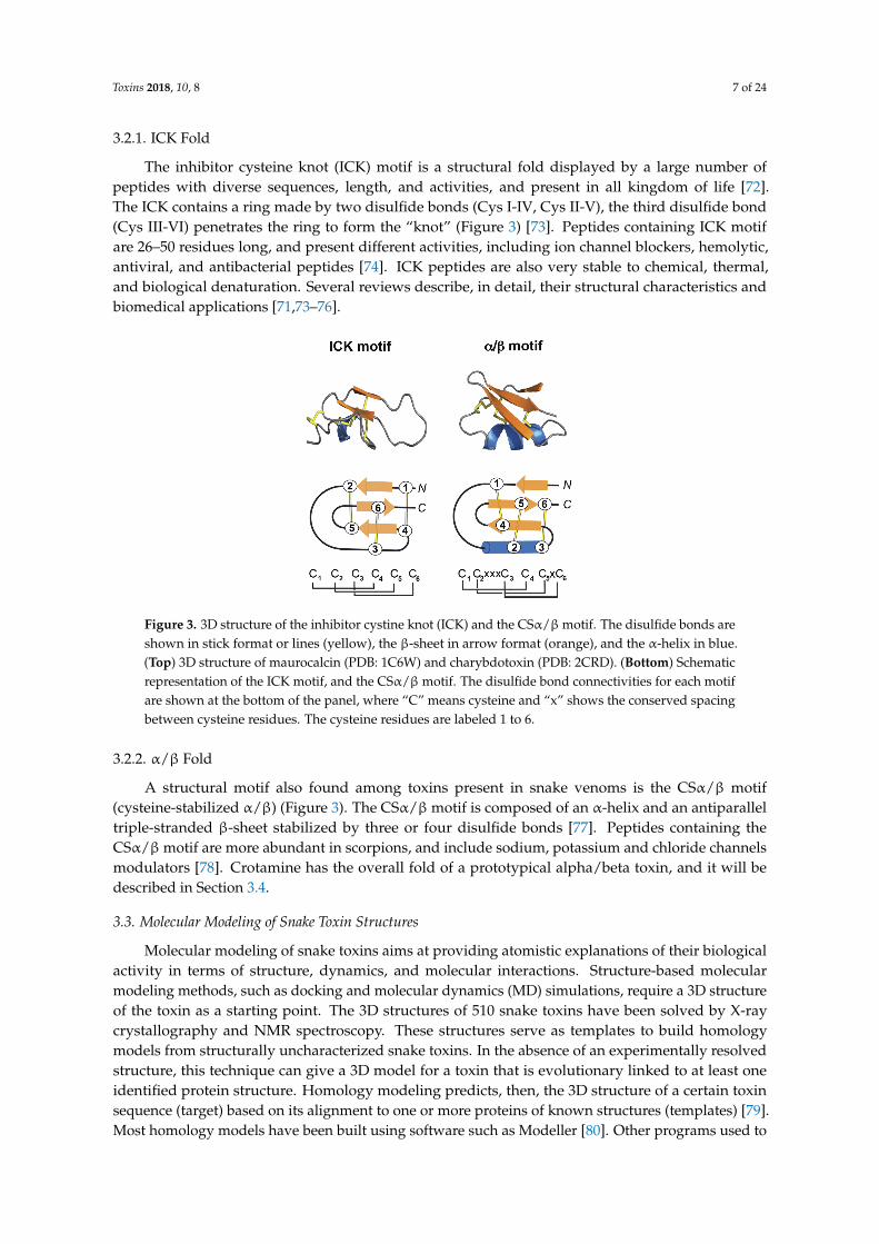
The inhibitor cysteine knot (ICK) motif is a structural fold displayed by a large number ofpeptides with diverse sequences, length, and activities, and present in all kingdom of life [72].The ICK contains a ring made by two disulfide bonds (Cys I-IV, Cys II-V), the third disulfide bond(Cys III-VI) penetrates the ring to form the “knot” (Figure 3) [73]. Peptides containing ICK motifare 26–50 residues long, and present different activities, including ion channel blockers, hemolytic,antiviral, and antibacterial peptides [74]. ICK peptides are also very stable to chemical, thermal,and biological denaturation. Several reviews describe, in detail, their structural characteristics andbiomedical applications [71,73–76].
Toxins 2017, 10, 8 7 of 23
3.2.1. ICK Fold
The inhibitor cysteine knot (ICK) motif is a structural fold displayed by a large number of peptides with diverse sequences, length, and activities, and present in all kingdom of life [72]. The ICK contains a ring made by two disulfide bonds (Cys I-IV, Cys II-V), the third disulfide bond (Cys III-VI) penetrates the ring to form the “knot” (Figure 3) [73]. Peptides containing ICK motif are 26–50 residues long, and present different activities, including ion channel blockers, hemolytic, antiviral, and antibacterial peptides [74]. ICK peptides are also very stable to chemical, thermal, and biological denaturation. Several reviews describe, in detail, their structural characteristics and biomedical applications. [71,73–76].
Figure 3. 3D structure of the inhibitor cystine knot (ICK) and the CSα/β motif. The disulfide bonds are shown in stick format or lines (yellow), the β-sheet in arrow format (orange), and the α-helix in blue. (Top) 3D structure of maurocalcin (PDB: 1C6W) and charybdotoxin (PDB: 2CRD). (Bottom) Schematic representation of the ICK motif, and the CSα/β motif. The disulfide bond connectivities for each motif are shown at the bottom of the panel, where “C” means cysteine and “x” shows the conserved spacing between cysteine residues. The cysteine residues are labeled 1 to 6.
3.2.2. α/β Fold
A structural motif also found among toxins present in snake venoms is the CSα/β motif (cysteine-stabilized α/β) (Figure 3). The CSα/β motif is composed of an α-helix and an antiparallel triple-stranded β-sheet stabilized by three or four disulfide bonds [77]. Peptides containing the CSα/β motif are more abundant in scorpions, and include sodium, potassium and chloride channels modulators [78]. Crotamine has the overall fold of a prototypical alpha/beta toxin, and it will be described in Section 3.4.
3.3. Molecular Modeling of Snake Toxin Structures
Molecular modeling of snake toxins aims at providing atomistic explanations of their biological activity in terms of structure, dynamics, and molecular interactions. Structure-based molecular modeling methods, such as docking and molecular dynamics (MD) simulations, require a 3D structure of the toxin as a starting point. The 3D structures of 510 snake toxins have been solved by X-ray crystallography and NMR spectroscopy. These structures serve as templates to build homology models from structurally uncharacterized snake toxins. In the absence of an experimentally resolved structure, this technique can give a 3D model for a toxin that is evolutionary linked to at least one identified protein structure. Homology modeling predicts, then, the 3D structure of a certain toxin sequence (target) based on its alignment to one or more proteins of known structures (templates) [79]. Most homology models have been built using software such as Modeller [80]. Other programs used
Figure 3. 3D structure of the inhibitor cystine knot (ICK) and the CSα/βmotif. The disulfide bonds areshown in stick format or lines (yellow), the β-sheet in arrow format (orange), and the α-helix in blue.(Top) 3D structure of maurocalcin (PDB: 1C6W) and charybdotoxin (PDB: 2CRD). (Bottom) Schematicrepresentation of the ICK motif, and the CSα/β motif. The disulfide bond connectivities for each motifare shown at the bottom of the panel, where “C” means cysteine and “x” shows the conserved spacingbetween cysteine residues. The cysteine residues are labeled 1 to 6.
3.2.2. α/β Fold
A structural motif also found among toxins present in snake venoms is the CSα/β motif(cysteine-stabilized α/β) (Figure 3). The CSα/βmotif is composed of an α-helix and an antiparalleltriple-stranded β-sheet stabilized by three or four disulfide bonds [77]. Peptides containing theCSα/βmotif are more abundant in scorpions, and include sodium, potassium and chloride channelsmodulators [78]. Crotamine has the overall fold of a prototypical alpha/beta toxin, and it will bedescribed in Section 3.4.
3.3. Molecular Modeling of Snake Toxin Structures
Molecular modeling of snake toxins aims at providing atomistic explanations of their biologicalactivity in terms of structure, dynamics, and molecular interactions. Structure-based molecularmodeling methods, such as docking and molecular dynamics (MD) simulations, require a 3D structureof the toxin as a starting point. The 3D structures of 510 snake toxins have been solved by X-raycrystallography and NMR spectroscopy. These structures serve as templates to build homologymodels from structurally uncharacterized snake toxins. In the absence of an experimentally resolvedstructure, this technique can give a 3D model for a toxin that is evolutionary linked to at least oneidentified protein structure. Homology modeling predicts, then, the 3D structure of a certain toxinsequence (target) based on its alignment to one or more proteins of known structures (templates) [79].Most homology models have been built using software such as Modeller [80]. Other programs used to
Toxins 2018, 10, 8 8 of 24
perform homology models are ICM [81], module Prime in Schödinger suite [82], as well as web serverssuch as SWISS-Model [83] and I-TASSER [84]. Using the force field that has been given to the atoms inthe system, it is possible to find a stable conformation or a minimum on the potential energy surfacein order to start MD. There will be more than one local minimum for a toxin. In principle there maybe a global minimum, but this will not likely be found without an extensive conformational search.The initial energy minimized structure is usually subjected to molecular dynamics to study the motionof molecules with respect to time. MD represents an option to study the structure and dynamics ofsnake toxins at atomistic resolution simultaneously. The growing significance of MD simulations forstructural prediction has been highlighted by the critical assessment of structure prediction (CASP)experiments, where MD turned out to improve the model refinement notably [85].
MD simulation [86] is based on the numerical integration of the classical Newtonian equations ofmotions for all the atoms in a system. The interactions between atoms are described by physic-basedforce fields, such as AMBER [7], CHARMM [8], Gromos [9], and OPLS [10], among others. The forcefields have been fitted to reproduce values from experiments or gas phase quantum mechanicalcalculations [87]. Short MD simulations are frequently employed to refine the conformation ofhomology models of snake toxins. MD simulations are also employed to suggest the molecularinteraction between toxins and their target, and for the rational design of novel inhibitors using aninitial pose, often resulting from docking [88,89].
3.4. Molecular Modeling of Snake Toxin—Target Complexes
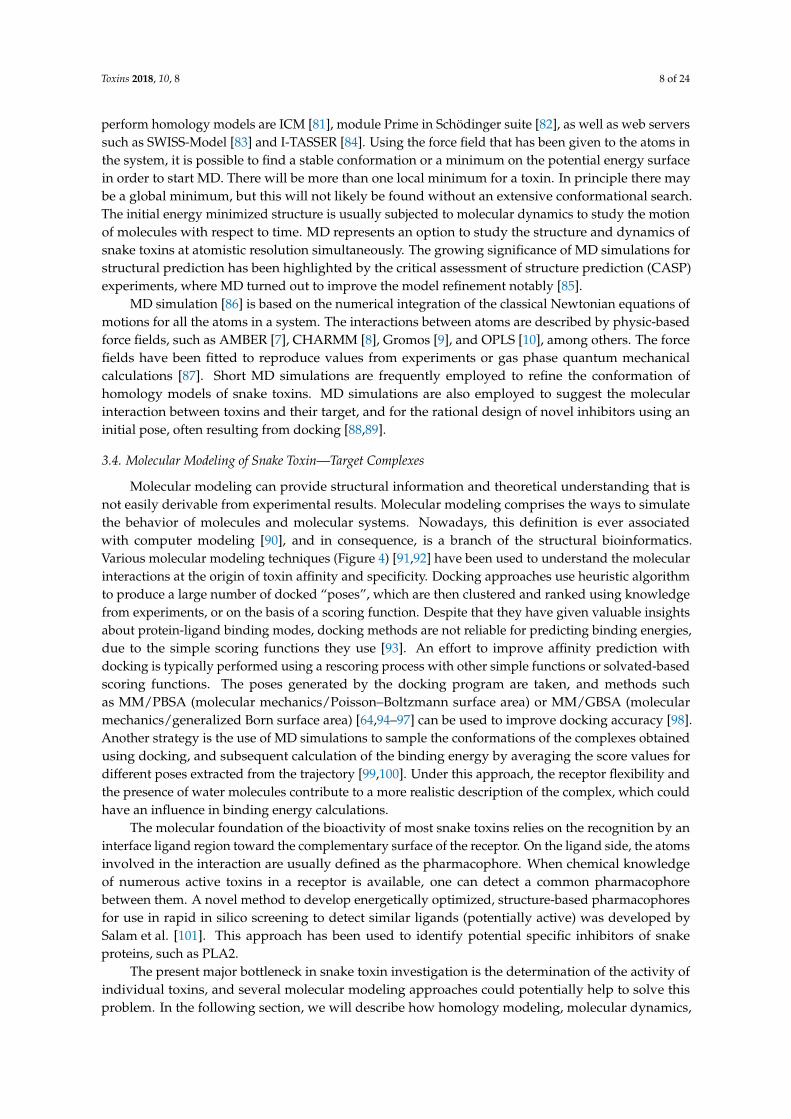
Molecular modeling can provide structural information and theoretical understanding that isnot easily derivable from experimental results. Molecular modeling comprises the ways to simulatethe behavior of molecules and molecular systems. Nowadays, this definition is ever associatedwith computer modeling [90], and in consequence, is a branch of the structural bioinformatics.Various molecular modeling techniques (Figure 4) [91,92] have been used to understand the molecularinteractions at the origin of toxin affinity and specificity. Docking approaches use heuristic algorithmto produce a large number of docked “poses”, which are then clustered and ranked using knowledgefrom experiments, or on the basis of a scoring function. Despite that they have given valuable insightsabout protein-ligand binding modes, docking methods are not reliable for predicting binding energies,due to the simple scoring functions they use [93]. An effort to improve affinity prediction withdocking is typically performed using a rescoring process with other simple functions or solvated-basedscoring functions. The poses generated by the docking program are taken, and methods suchas MM/PBSA (molecular mechanics/Poisson–Boltzmann surface area) or MM/GBSA (molecularmechanics/generalized Born surface area) [64,94–97] can be used to improve docking accuracy [98].Another strategy is the use of MD simulations to sample the conformations of the complexes obtainedusing docking, and subsequent calculation of the binding energy by averaging the score values fordifferent poses extracted from the trajectory [99,100]. Under this approach, the receptor flexibility andthe presence of water molecules contribute to a more realistic description of the complex, which couldhave an influence in binding energy calculations.
The molecular foundation of the bioactivity of most snake toxins relies on the recognition by aninterface ligand region toward the complementary surface of the receptor. On the ligand side, the atomsinvolved in the interaction are usually defined as the pharmacophore. When chemical knowledgeof numerous active toxins in a receptor is available, one can detect a common pharmacophorebetween them. A novel method to develop energetically optimized, structure-based pharmacophoresfor use in rapid in silico screening to detect similar ligands (potentially active) was developed bySalam et al. [101]. This approach has been used to identify potential specific inhibitors of snakeproteins, such as PLA2.
The present major bottleneck in snake toxin investigation is the determination of the activity ofindividual toxins, and several molecular modeling approaches could potentially help to solve thisproblem. In the following section, we will describe how homology modeling, molecular dynamics,
Toxins 2018, 10, 8 9 of 24
molecular docking, free energy, quantum chemical calculation, and e-pharmacophore approach have(Figure 4) been used to study the activity of the snake venom protein PLA2, the small basic myotoxincrotamine and the three-finger peptide mambalgin.
Toxins 2017, 10, 8 9 of 23
(Figure 4) been used to study the activity of the snake venom protein PLA2, the small basic myotoxin crotamine and the three-finger peptide mambalgin.
Figure 4. Molecular modeling is a branch of structural bioinformatics. Molecular modeling comprises several methods to simulate the behavior of molecules and molecular systems. Some of them, such as homology modeling, molecular dynamics, molecular docking, free energy, quantum chemical calculation, and e-pharmacophore approach, have been used to study the activity of snake venom proteins.
3.5. Molecular Modeling Applied to the Study of PLA2, Crotamine and Mambalgin
3.5.1. PLA2
Phospholipases A2 (PLA2; EC 3.1.1.4) are proteins present in snake venoms with a digestive role in phospholipid hydrolysis [102]. They specifically hydrolyze the sn-2 ester bond of phospholipids, releasing fatty acids from the second carbon group of glycerol, and display enhanced catalytic activity in micellar and lamellar aggregates, both in membranes and at other lipid–water interfaces [103]. When a snakebite occurs, PLA2 toxins exhibit a wide variety of pharmacological effects on the normal physiological processes of victims, such as myotoxicity, neurotoxicity, and edema-inducing activity [104,105]. Due to their toxic pathophysiological role, there is a considerable pharmacological interest towards the design and discovery of PLA2 specific inhibitors for antivenom therapies in humans.
There are several reports where computational molecular modeling methods have been used for characterizing some functional aspects of PLA2s, or the development of PLA2 inhibitors that contribute to the weakening or annihilation of snake venom toxicity. These applications use the X-ray crystallographic 3D structural information generated in the last decades, and methods such as molecular dynamics (MD) simulations and docking.
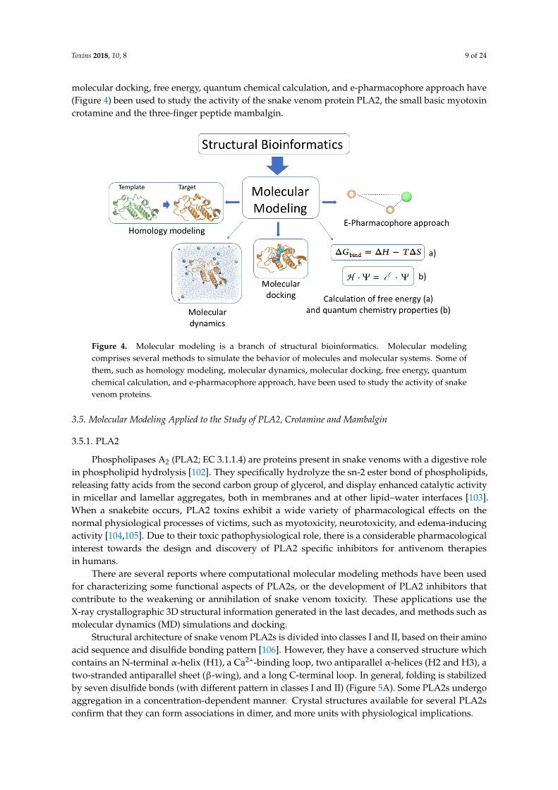
Structural architecture of snake venom PLA2s is divided into classes I and II, based on their amino acid sequence and disulfide bonding pattern [106]. However, they have a conserved structure which contains an N-terminal α-helix (H1), a Ca2+-binding loop, two antiparallel α-helices (H2 and H3), a two-stranded antiparallel sheet (β-wing), and a long C-terminal loop. In general, folding is stabilized by seven disulfide bonds (with different pattern in classes I and II) (Figure 5A). Some PLA2s undergo aggregation in a concentration-dependent manner. Crystal structures available for several PLA2s confirm that they can form associations in dimer, and more units with physiological implications.
Figure 4. Molecular modeling is a branch of structural bioinformatics. Molecular modelingcomprises several methods to simulate the behavior of molecules and molecular systems. Some ofthem, such as homology modeling, molecular dynamics, molecular docking, free energy, quantumchemical calculation, and e-pharmacophore approach, have been used to study the activity of snakevenom proteins.
3.5. Molecular Modeling Applied to the Study of PLA2, Crotamine and Mambalgin
3.5.1. PLA2
Phospholipases A2 (PLA2; EC 3.1.1.4) are proteins present in snake venoms with a digestive rolein phospholipid hydrolysis [102]. They specifically hydrolyze the sn-2 ester bond of phospholipids,releasing fatty acids from the second carbon group of glycerol, and display enhanced catalytic activityin micellar and lamellar aggregates, both in membranes and at other lipid–water interfaces [103].When a snakebite occurs, PLA2 toxins exhibit a wide variety of pharmacological effects on thenormal physiological processes of victims, such as myotoxicity, neurotoxicity, and edema-inducingactivity [104,105]. Due to their toxic pathophysiological role, there is a considerable pharmacologicalinterest towards the design and discovery of PLA2 specific inhibitors for antivenom therapiesin humans.
There are several reports where computational molecular modeling methods have been usedfor characterizing some functional aspects of PLA2s, or the development of PLA2 inhibitors thatcontribute to the weakening or annihilation of snake venom toxicity. These applications use theX-ray crystallographic 3D structural information generated in the last decades, and methods such asmolecular dynamics (MD) simulations and docking.
Structural architecture of snake venom PLA2s is divided into classes I and II, based on their aminoacid sequence and disulfide bonding pattern [106]. However, they have a conserved structure whichcontains an N-terminal α-helix (H1), a Ca2+-binding loop, two antiparallel α-helices (H2 and H3), atwo-stranded antiparallel sheet (β-wing), and a long C-terminal loop. In general, folding is stabilizedby seven disulfide bonds (with different pattern in classes I and II) (Figure 5A). Some PLA2s undergoaggregation in a concentration-dependent manner. Crystal structures available for several PLA2sconfirm that they can form associations in dimer, and more units with physiological implications.
Toxins 2018, 10, 8 10 of 24
Toxins 2017, 10, 8 10 of 23
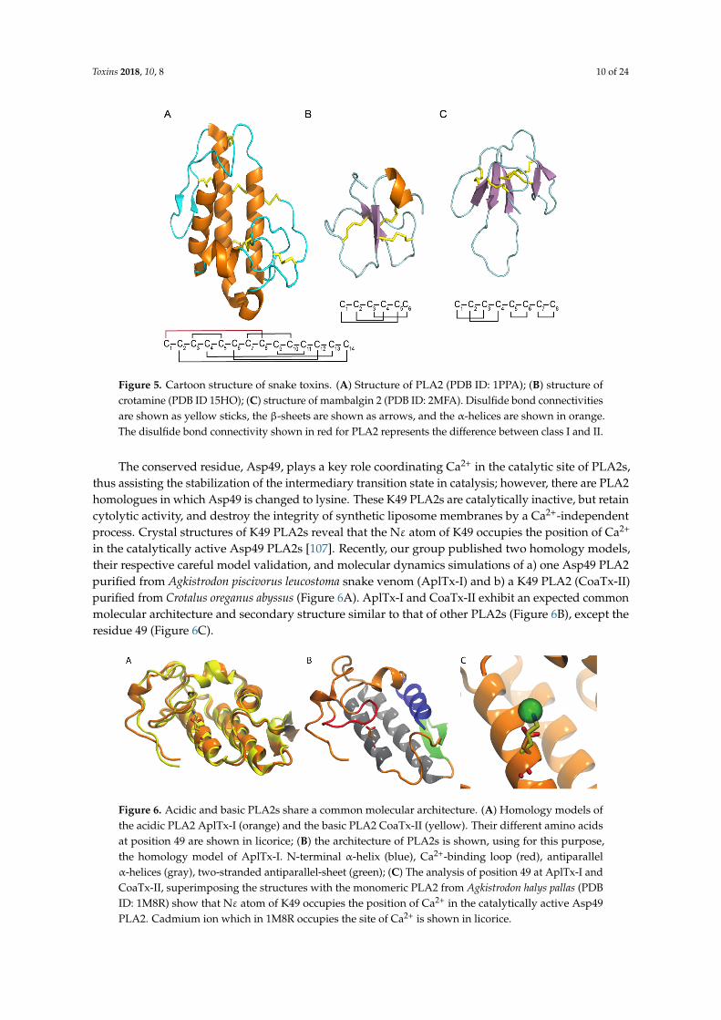
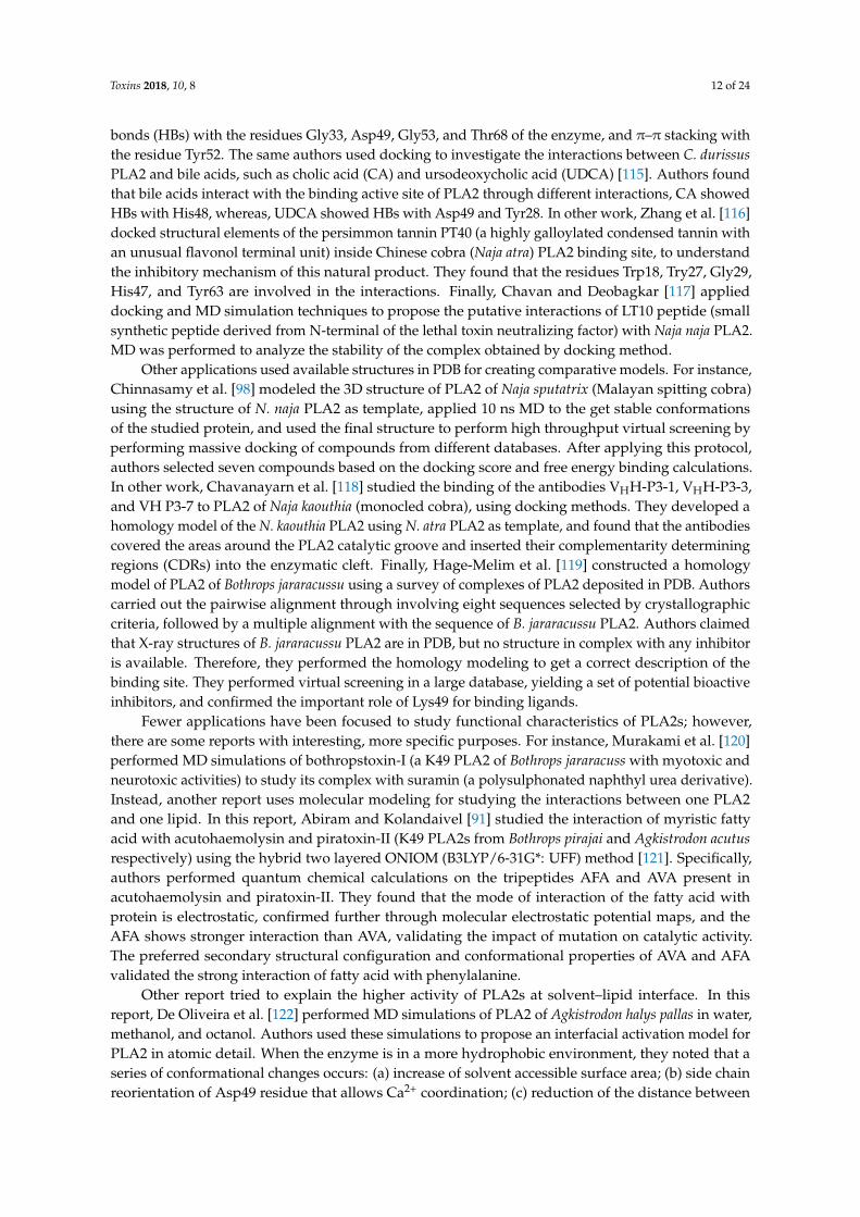
Figure 5. Cartoon structure of snake toxins. (A) Structure of PLA2 (PDB ID: 1PPA); (B) structure of crotamine (PDB ID 15HO); (C) structure of mambalgin 2 (PDB ID: 2MFA). Disulfide bond connectivities are shown as yellow sticks, the β-sheets are shown as arrows, and the α-helices are shown in orange. The disulfide bond connectivity shown in red for PLA2 represents the difference between class I and II.
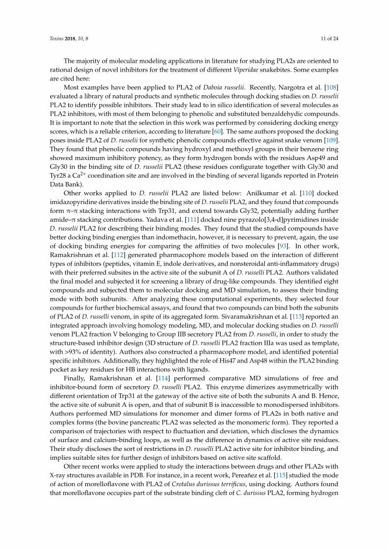
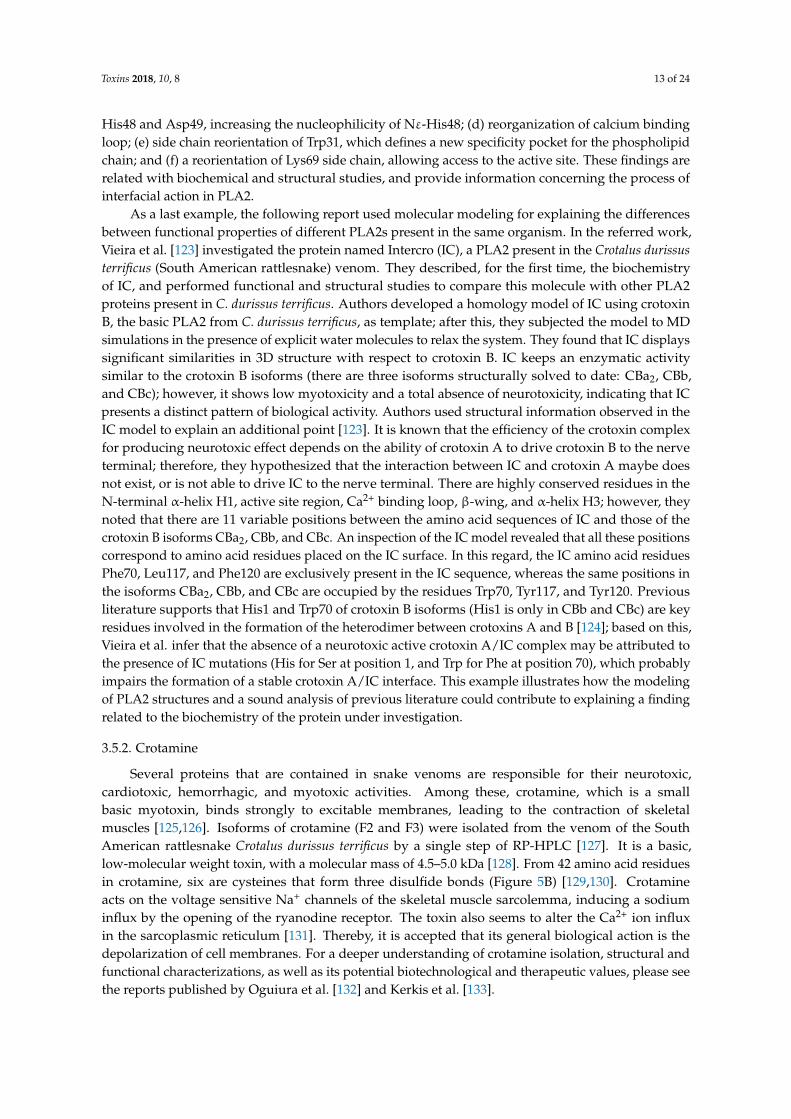
The conserved residue, Asp49, plays a key role coordinating Ca2+ in the catalytic site of PLA2s, thus assisting the stabilization of the intermediary transition state in catalysis; however, there are PLA2 homologues in which Asp49 is changed to lysine. These K49 PLA2s are catalytically inactive, but retain cytolytic activity, and destroy the integrity of synthetic liposome membranes by a Ca2+-independent process. Crystal structures of K49 PLA2s reveal that the Nε atom of K49 occupies the position of Ca2+ in the catalytically active Asp49 PLA2s [107]. Recently, our group published two homology models, their respective careful model validation, and molecular dynamics simulations of a) one Asp49 PLA2 purified from Agkistrodon piscivorus leucostoma snake venom (AplTx-I) and b) a K49 PLA2 (CoaTx-II) purified from Crotalus oreganus abyssus (Figure 6A). AplTx-I and CoaTx-II exhibit an expected common molecular architecture and secondary structure similar to that of other PLA2s (Figure 6B), except the residue 49 (Figure 6C).
Figure 6. Acidic and basic PLA2s share a common molecular architecture. (A) Homology models of the acidic PLA2 AplTx-I (orange) and the basic PLA2 CoaTx-II (yellow). Their different amino acids at position 49 are shown in licorice; (B) the architecture of PLA2s is shown, using for this purpose, the homology model of AplTx-I. N-terminal α-helix (blue), Ca2+-binding loop (red), antiparallel α-helices (gray), two-stranded antiparallel-sheet (green); (C) The analysis of position 49 at AplTx-I and CoaTx-II, superimposing the structures with the monomeric PLA2 from Agkistrodon halys pallas (PDB ID: 1M8R) show that Nε atom of K49 occupies the position of Ca2+ in the catalytically active Asp49 PLA2. Cadmium ion which in 1M8R occupies the site of Ca2+ is shown in licorice.
Figure 5. Cartoon structure of snake toxins. (A) Structure of PLA2 (PDB ID: 1PPA); (B) structure ofcrotamine (PDB ID 15HO); (C) structure of mambalgin 2 (PDB ID: 2MFA). Disulfide bond connectivitiesare shown as yellow sticks, the β-sheets are shown as arrows, and the α-helices are shown in orange.The disulfide bond connectivity shown in red for PLA2 represents the difference between class I and II.
The conserved residue, Asp49, plays a key role coordinating Ca2+ in the catalytic site of PLA2s,thus assisting the stabilization of the intermediary transition state in catalysis; however, there are PLA2homologues in which Asp49 is changed to lysine. These K49 PLA2s are catalytically inactive, but retaincytolytic activity, and destroy the integrity of synthetic liposome membranes by a Ca2+-independentprocess. Crystal structures of K49 PLA2s reveal that the Nε atom of K49 occupies the position of Ca2+
in the catalytically active Asp49 PLA2s [107]. Recently, our group published two homology models,their respective careful model validation, and molecular dynamics simulations of a) one Asp49 PLA2purified from Agkistrodon piscivorus leucostoma snake venom (AplTx-I) and b) a K49 PLA2 (CoaTx-II)purified from Crotalus oreganus abyssus (Figure 6A). AplTx-I and CoaTx-II exhibit an expected commonmolecular architecture and secondary structure similar to that of other PLA2s (Figure 6B), except theresidue 49 (Figure 6C).
Toxins 2017, 10, 8 10 of 23
Figure 5. Cartoon structure of snake toxins. (A) Structure of PLA2 (PDB ID: 1PPA); (B) structure of crotamine (PDB ID 15HO); (C) structure of mambalgin 2 (PDB ID: 2MFA). Disulfide bond connectivities are shown as yellow sticks, the β-sheets are shown as arrows, and the α-helices are shown in orange. The disulfide bond connectivity shown in red for PLA2 represents the difference between class I and II.
The conserved residue, Asp49, plays a key role coordinating Ca2+ in the catalytic site of PLA2s, thus assisting the stabilization of the intermediary transition state in catalysis; however, there are PLA2 homologues in which Asp49 is changed to lysine. These K49 PLA2s are catalytically inactive, but retain cytolytic activity, and destroy the integrity of synthetic liposome membranes by a Ca2+-independent process. Crystal structures of K49 PLA2s reveal that the Nε atom of K49 occupies the position of Ca2+ in the catalytically active Asp49 PLA2s [107]. Recently, our group published two homology models, their respective careful model validation, and molecular dynamics simulations of a) one Asp49 PLA2 purified from Agkistrodon piscivorus leucostoma snake venom (AplTx-I) and b) a K49 PLA2 (CoaTx-II) purified from Crotalus oreganus abyssus (Figure 6A). AplTx-I and CoaTx-II exhibit an expected common molecular architecture and secondary structure similar to that of other PLA2s (Figure 6B), except the residue 49 (Figure 6C).
Figure 6. Acidic and basic PLA2s share a common molecular architecture. (A) Homology models of the acidic PLA2 AplTx-I (orange) and the basic PLA2 CoaTx-II (yellow). Their different amino acids at position 49 are shown in licorice; (B) the architecture of PLA2s is shown, using for this purpose, the homology model of AplTx-I. N-terminal α-helix (blue), Ca2+-binding loop (red), antiparallel α-helices (gray), two-stranded antiparallel-sheet (green); (C) The analysis of position 49 at AplTx-I and CoaTx-II, superimposing the structures with the monomeric PLA2 from Agkistrodon halys pallas (PDB ID: 1M8R) show that Nε atom of K49 occupies the position of Ca2+ in the catalytically active Asp49 PLA2. Cadmium ion which in 1M8R occupies the site of Ca2+ is shown in licorice.
Figure 6. Acidic and basic PLA2s share a common molecular architecture. (A) Homology models ofthe acidic PLA2 AplTx-I (orange) and the basic PLA2 CoaTx-II (yellow). Their different amino acidsat position 49 are shown in licorice; (B) the architecture of PLA2s is shown, using for this purpose,the homology model of AplTx-I. N-terminal α-helix (blue), Ca2+-binding loop (red), antiparallelα-helices (gray), two-stranded antiparallel-sheet (green); (C) The analysis of position 49 at AplTx-I andCoaTx-II, superimposing the structures with the monomeric PLA2 from Agkistrodon halys pallas (PDBID: 1M8R) show that Nε atom of K49 occupies the position of Ca2+ in the catalytically active Asp49PLA2. Cadmium ion which in 1M8R occupies the site of Ca2+ is shown in licorice.
Toxins 2018, 10, 8 11 of 24
The majority of molecular modeling applications in literature for studying PLA2s are oriented torational design of novel inhibitors for the treatment of different Viperidae snakebites. Some examplesare cited here:
Most examples have been applied to PLA2 of Daboia russelii. Recently, Nargotra et al. [108]evaluated a library of natural products and synthetic molecules through docking studies on D. russeliiPLA2 to identify possible inhibitors. Their study lead to in silico identification of several molecules asPLA2 inhibitors, with most of them belonging to phenolic and substituted benzaldehydic compounds.It is important to note that the selection in this work was performed by considering docking energyscores, which is a reliable criterion, according to literature [60]. The same authors proposed the dockingposes inside PLA2 of D. russelii for synthetic phenolic compounds effective against snake venom [109].They found that phenolic compounds having hydroxyl and methoxyl groups in their benzene ringshowed maximum inhibitory potency, as they form hydrogen bonds with the residues Asp49 andGly30 in the binding site of D. russelii PLA2 (these residues configurate together with Gly30 andTyr28 a Ca2+ coordination site and are involved in the binding of several ligands reported in ProteinData Bank).
Other works applied to D. russelii PLA2 are listed below: Anilkumar et al. [110] dockedimidazopyridine derivatives inside the binding site of D. russelii PLA2, and they found that compoundsform π–π stacking interactions with Trp31, and extend towards Gly32, potentially adding furtheramide–π stacking contributions. Yadava et al. [111] docked nine pyrazolo[3,4-d]pyrimidines insideD. russelii PLA2 for describing their binding modes. They found that the studied compounds havebetter docking binding energies than indomethacin, however, it is necessary to prevent, again, the useof docking binding energies for comparing the affinities of two molecules [93]. In other work,Ramakrishnan et al. [112] generated pharmacophore models based on the interaction of differenttypes of inhibitors (peptides, vitamin E, indole derivatives, and nonsteroidal anti-inflammatory drugs)with their preferred subsites in the active site of the subunit A of D. russelli PLA2. Authors validatedthe final model and subjected it for screening a library of drug-like compounds. They identified eightcompounds and subjected them to molecular docking and MD simulation, to assess their bindingmode with both subunits. After analyzing these computational experiments, they selected fourcompounds for further biochemical assays, and found that two compounds can bind both the subunitsof PLA2 of D. russelli venom, in spite of its aggregated form. Sivaramakrishnan et al. [113] reported anintegrated approach involving homology modeling, MD, and molecular docking studies on D. russellivenom PLA2 fraction V belonging to Group IIB secretory PLA2 from D. russelli, in order to study thestructure-based inhibitor design (3D structure of D. russelli PLA2 fraction IIIa was used as template,with >93% of identity). Authors also constructed a pharmacophore model, and identified potentialspecific inhibitors. Additionally, they highlighted the role of His47 and Asp48 within the PLA2 bindingpocket as key residues for HB interactions with ligands.
Finally, Ramakrishnan et al. [114] performed comparative MD simulations of free andinhibitor-bound form of secretory D. russelli PLA2. This enzyme dimerizes asymmetrically withdifferent orientation of Trp31 at the gateway of the active site of both the subunits A and B. Hence,the active site of subunit A is open, and that of subunit B is inaccessible to monodispersed inhibitors.Authors performed MD simulations for monomer and dimer forms of PLA2s in both native andcomplex forms (the bovine pancreatic PLA2 was selected as the monomeric form). They reported acomparison of trajectories with respect to fluctuation and deviation, which discloses the dynamicsof surface and calcium-binding loops, as well as the difference in dynamics of active site residues.Their study discloses the sort of restrictions in D. russelli PLA2 active site for inhibitor binding, andimplies suitable sites for further design of inhibitors based on active site scaffold.
Other recent works were applied to study the interactions between drugs and other PLA2s withX-ray structures available in PDB. For instance, in a recent work, Pereañez et al. [115] studied the modeof action of morelloflavone with PLA2 of Crotalus durissus terrificus, using docking. Authors foundthat morelloflavone occupies part of the substrate binding cleft of C. durissus PLA2, forming hydrogen
Toxins 2018, 10, 8 12 of 24
bonds (HBs) with the residues Gly33, Asp49, Gly53, and Thr68 of the enzyme, and π–π stacking withthe residue Tyr52. The same authors used docking to investigate the interactions between C. durissusPLA2 and bile acids, such as cholic acid (CA) and ursodeoxycholic acid (UDCA) [115]. Authors foundthat bile acids interact with the binding active site of PLA2 through different interactions, CA showedHBs with His48, whereas, UDCA showed HBs with Asp49 and Tyr28. In other work, Zhang et al. [116]docked structural elements of the persimmon tannin PT40 (a highly galloylated condensed tannin withan unusual flavonol terminal unit) inside Chinese cobra (Naja atra) PLA2 binding site, to understandthe inhibitory mechanism of this natural product. They found that the residues Trp18, Try27, Gly29,His47, and Tyr63 are involved in the interactions. Finally, Chavan and Deobagkar [117] applieddocking and MD simulation techniques to propose the putative interactions of LT10 peptide (smallsynthetic peptide derived from N-terminal of the lethal toxin neutralizing factor) with Naja naja PLA2.MD was performed to analyze the stability of the complex obtained by docking method.
Other applications used available structures in PDB for creating comparative models. For instance,Chinnasamy et al. [98] modeled the 3D structure of PLA2 of Naja sputatrix (Malayan spitting cobra)using the structure of N. naja PLA2 as template, applied 10 ns MD to the get stable conformationsof the studied protein, and used the final structure to perform high throughput virtual screening byperforming massive docking of compounds from different databases. After applying this protocol,authors selected seven compounds based on the docking score and free energy binding calculations.In other work, Chavanayarn et al. [118] studied the binding of the antibodies VHH-P3-1, VHH-P3-3,and VH P3-7 to PLA2 of Naja kaouthia (monocled cobra), using docking methods. They developed ahomology model of the N. kaouthia PLA2 using N. atra PLA2 as template, and found that the antibodiescovered the areas around the PLA2 catalytic groove and inserted their complementarity determiningregions (CDRs) into the enzymatic cleft. Finally, Hage-Melim et al. [119] constructed a homologymodel of PLA2 of Bothrops jararacussu using a survey of complexes of PLA2 deposited in PDB. Authorscarried out the pairwise alignment through involving eight sequences selected by crystallographiccriteria, followed by a multiple alignment with the sequence of B. jararacussu PLA2. Authors claimedthat X-ray structures of B. jararacussu PLA2 are in PDB, but no structure in complex with any inhibitoris available. Therefore, they performed the homology modeling to get a correct description of thebinding site. They performed virtual screening in a large database, yielding a set of potential bioactiveinhibitors, and confirmed the important role of Lys49 for binding ligands.
Fewer applications have been focused to study functional characteristics of PLA2s; however,there are some reports with interesting, more specific purposes. For instance, Murakami et al. [120]performed MD simulations of bothropstoxin-I (a K49 PLA2 of Bothrops jararacuss with myotoxic andneurotoxic activities) to study its complex with suramin (a polysulphonated naphthyl urea derivative).Instead, another report uses molecular modeling for studying the interactions between one PLA2and one lipid. In this report, Abiram and Kolandaivel [91] studied the interaction of myristic fattyacid with acutohaemolysin and piratoxin-II (K49 PLA2s from Bothrops pirajai and Agkistrodon acutusrespectively) using the hybrid two layered ONIOM (B3LYP/6-31G*: UFF) method [121]. Specifically,authors performed quantum chemical calculations on the tripeptides AFA and AVA present inacutohaemolysin and piratoxin-II. They found that the mode of interaction of the fatty acid withprotein is electrostatic, confirmed further through molecular electrostatic potential maps, and theAFA shows stronger interaction than AVA, validating the impact of mutation on catalytic activity.The preferred secondary structural configuration and conformational properties of AVA and AFAvalidated the strong interaction of fatty acid with phenylalanine.
Other report tried to explain the higher activity of PLA2s at solvent–lipid interface. In thisreport, De Oliveira et al. [122] performed MD simulations of PLA2 of Agkistrodon halys pallas in water,methanol, and octanol. Authors used these simulations to propose an interfacial activation model forPLA2 in atomic detail. When the enzyme is in a more hydrophobic environment, they noted that aseries of conformational changes occurs: (a) increase of solvent accessible surface area; (b) side chainreorientation of Asp49 residue that allows Ca2+ coordination; (c) reduction of the distance between
Toxins 2018, 10, 8 13 of 24
His48 and Asp49, increasing the nucleophilicity of Nε-His48; (d) reorganization of calcium bindingloop; (e) side chain reorientation of Trp31, which defines a new specificity pocket for the phospholipidchain; and (f) a reorientation of Lys69 side chain, allowing access to the active site. These findings arerelated with biochemical and structural studies, and provide information concerning the process ofinterfacial action in PLA2.
As a last example, the following report used molecular modeling for explaining the differencesbetween functional properties of different PLA2s present in the same organism. In the referred work,Vieira et al. [123] investigated the protein named Intercro (IC), a PLA2 present in the Crotalus durissusterrificus (South American rattlesnake) venom. They described, for the first time, the biochemistryof IC, and performed functional and structural studies to compare this molecule with other PLA2proteins present in C. durissus terrificus. Authors developed a homology model of IC using crotoxinB, the basic PLA2 from C. durissus terrificus, as template; after this, they subjected the model to MDsimulations in the presence of explicit water molecules to relax the system. They found that IC displayssignificant similarities in 3D structure with respect to crotoxin B. IC keeps an enzymatic activitysimilar to the crotoxin B isoforms (there are three isoforms structurally solved to date: CBa2, CBb,and CBc); however, it shows low myotoxicity and a total absence of neurotoxicity, indicating that ICpresents a distinct pattern of biological activity. Authors used structural information observed in theIC model to explain an additional point [123]. It is known that the efficiency of the crotoxin complexfor producing neurotoxic effect depends on the ability of crotoxin A to drive crotoxin B to the nerveterminal; therefore, they hypothesized that the interaction between IC and crotoxin A maybe doesnot exist, or is not able to drive IC to the nerve terminal. There are highly conserved residues in theN-terminal α-helix H1, active site region, Ca2+ binding loop, β-wing, and α-helix H3; however, theynoted that there are 11 variable positions between the amino acid sequences of IC and those of thecrotoxin B isoforms CBa2, CBb, and CBc. An inspection of the IC model revealed that all these positionscorrespond to amino acid residues placed on the IC surface. In this regard, the IC amino acid residuesPhe70, Leu117, and Phe120 are exclusively present in the IC sequence, whereas the same positions inthe isoforms CBa2, CBb, and CBc are occupied by the residues Trp70, Tyr117, and Tyr120. Previousliterature supports that His1 and Trp70 of crotoxin B isoforms (His1 is only in CBb and CBc) are keyresidues involved in the formation of the heterodimer between crotoxins A and B [124]; based on this,Vieira et al. infer that the absence of a neurotoxic active crotoxin A/IC complex may be attributed tothe presence of IC mutations (His for Ser at position 1, and Trp for Phe at position 70), which probablyimpairs the formation of a stable crotoxin A/IC interface. This example illustrates how the modelingof PLA2 structures and a sound analysis of previous literature could contribute to explaining a findingrelated to the biochemistry of the protein under investigation.
3.5.2. Crotamine
Several proteins that are contained in snake venoms are responsible for their neurotoxic,cardiotoxic, hemorrhagic, and myotoxic activities. Among these, crotamine, which is a smallbasic myotoxin, binds strongly to excitable membranes, leading to the contraction of skeletalmuscles [125,126]. Isoforms of crotamine (F2 and F3) were isolated from the venom of the SouthAmerican rattlesnake Crotalus durissus terrificus by a single step of RP-HPLC [127]. It is a basic,low-molecular weight toxin, with a molecular mass of 4.5–5.0 kDa [128]. From 42 amino acid residuesin crotamine, six are cysteines that form three disulfide bonds (Figure 5B) [129,130]. Crotamineacts on the voltage sensitive Na+ channels of the skeletal muscle sarcolemma, inducing a sodiuminflux by the opening of the ryanodine receptor. The toxin also seems to alter the Ca2+ ion influxin the sarcoplasmic reticulum [131]. Thereby, it is accepted that its general biological action is thedepolarization of cell membranes. For a deeper understanding of crotamine isolation, structural andfunctional characterizations, as well as its potential biotechnological and therapeutic values, please seethe reports published by Oguiura et al. [132] and Kerkis et al. [133].
Toxins 2018, 10, 8 14 of 24
Crotamine was also characterized as a cell-penetrating protein (CPP) with nuclear localizationin vitro and in vivo [134]. Many biologically active compounds, including macromolecules, thatare used as various kinds of drugs, must be delivered to the interior of cell or organelles, such asmitochondria or nuclei, to achieve a therapeutic effect. CPPs are a new means for transporting ofmacromolecules through the cell membrane that became relevant in the last years. For an extensivereview on the topic, please see work published by Ruczynski et al. [135].
The use of computational methods to study crotamine is scarce, however, some computationaltechniques have been used to obtain and characterize its structure. For instance, Siqueira et al. proposeda theoretical 3D model for crotamine. They started from a homology modeling procedure, followedby intensive molecular dynamics (MD) simulations in water and complementary CD experiments.As no tridimensional structure of crotamine was available at that moment, the reported model wasthe first example for the 3D structure of this family of small basic myotoxins [136]. From this work,they proposed that key residues could be found in what they called the L1, L2, and L3 loops, whichcould serve in functions such as membrane anchoring, receptor anchoring, receptor isoform selection,and receptor inactivation. Later, Nicastro et al. reported the crotamine solution structure determined byproton NMR spectroscopy. A comparison of determined crotamine structure with human β-defensinsshowed a similar fold and a comparable net positive potential surface [137]. Moreover, the presenceof the α/β scaffold and the existence of a surface characterized by a positive electrostatic potentialseemed to justify the functional similarity with the Na+ channel affecting scorpion α-toxins. Accordingto the authors, the most significant difference between the theoretical 3D model reported by Sequeiraet al. and the NMR-derived structures from their work was the lack of a N-terminal α-helix segment.One reason for that could be the choice of the bovine β-defensin, BNBD12 [138], as a template. A new,and more refined, NMR structure determination of crotamine in aqueous solution at pH 5.8 and20 ◦C, using standard homonuclear 1H NMR spectroscopy at 900 MHz, and the automated structurecalculation software ATNOS/CANDID/DYANA was reported by Fadel et al. [139]. According to theirresults, the core of the protein is formed by an antiparallel β-sheet composed by residues 9–13 and34–38. A long, non-regular loop connects the two strands of the β-sheet. The disulfide bridges connectthe β-sheet to the N-terminal α-helix (Cys4/Cys36) and to this loop (Cys11/Cys30 and Cys18/Cys37).All nine lysines, with the sole exception of Lys35, and the two arginines, are oriented toward thesolvent, so that crotamine has extended positively charged molecular surface areas. The global foldand the cysteine-pairing pattern of crotamine were similar to the β-defensin fold [138], although thetwo proteins have low sequence homology, and displayed different biological activities. Moreover,a generally applicable new computational protocol was introduced to determine unknown disulfidebond connectivity in globular proteins.
Starting from the hypothesis that reciprocal relationships existed between antimicrobial andcytotoxic host defense peptides, Yount et al. compared in phylogeny, 3D structure, target cell specificityand mechanisms of action of the human antimicrobial peptide hBD-2 and rattlesnake venom toxincrotamine [140]. Computational molecular docking was used to compare hBD-2 versus crotamineintermolecular interactions with prototypic bacterial, fungal, or mammalian Kv channels, based on thewell-known fact that crotamine targets eukaryotic ion channels. The channel–toxin docking modelssupported direct interactions of each peptide with Kv channels. However, while crotamine localizedto occlude Kv channels in eukaryotic, but not prokaryotic cells, hBD-2 interacted with prokaryoticand eukaryotic Kv channels, but did not occlude either. The authors stated that these insights mightaccelerate development of anti-infective or therapeutic peptides that selectively target microbial orabnormal host cells.
More recently, a theoretical study, based on semi empirical, ab initio and density functionaltheory (DFT) quantum methods, was performed to investigate the structural properties of twocrotamines isolated from the venom of Crotalus durissus. Two protein models (I-a and II-a), representingcrotamine fragments (I17-C18-L19-P20-P21) and (I17-C18-I19-P20-P21) respectively, were minimized,and their chemical properties (atomic charge, orbital population, and MO energy) were calculated to
Toxins 2018, 10, 8 15 of 24
study differences in their myonecrotic activity. The authors concluded that even though there weresome variations in the chemical properties between both fragments, no rigorous relationship to theirrespective biological activities could be established [92].
3.5.3. Mambalgin-1 and -2
Mambalgin-1 and mambalgin-2 were identified as a new class of three-finger peptides fromthe venom of black mamba snake [141]. According to authors, these peptides were able to abolishpain through inhibition of acid-sensing ion channels (ASICs) expressed either in central or peripheralneurons. Mambalgins were not toxic in mice, but showed a potent analgesic effect upon central andperipheral injection that was as strong as morphine. The three-dimensional structure of mambalgin-1was modeled from five templates of three-finger snake toxins through software Modeller 9 (Version 8,Andrej Sali, San Francisco, CA, USA, 2010) [80,142]. They were composed of 57 amino acids witheight cysteine residues, and only differed by one residue at position 4. The model structure presenteda concave face commonly found in neurotoxins, and was stabilized by four disulfide bonds, with apattern identical to that observed in the crystal structure template (Cys1–Cys3, Cys2–Cys4, Cys5–Cys6,and Cys7–Cys8) (Figure 5C). Mambalgins showed a strong positive electrostatic potential, calculatedwith the adaptive Poisson–Boltzmann solver [143], that may contribute to binding to negatively chargedASIC channels. The authors concluded that their findings identified new potential therapeutic targetsfor pain, and introduced natural peptides that could block them to produce a potent analgesia [141].Later, same authors combined bioinformatic and functional approaches to uncover the molecularmechanism of channel inhibition by the mambalgin-2 pain-relieving peptide.
They first used homology modeling to obtain the structural models of rASIC1a and rASIC2athat were generated based on the structures of cASIC1a (57–90% sequence identity) using Modeller9v8 [80], to obtain the homology models of the open and desensitized forms of the trimer complexbased on the experimental structures (Protein Data Bank codes 4FZ0 and 3HGC, respectively).The three-dimensional structure of mambalgin-2 was obtained from experimental Protein Data Bank file2MFA [144]. Afterwards, molecular docking experiments were performed to model the toxin-channelinteractions. In silico rigid body docking of the toxin model onto the homology models of rat ASIC1aand ASIC2a were performed using the protein–protein docking program ZDOCK (version 2.3.2f,ZLAB, Chicopee, MA, USA, 2003) [145]. From those findings, the authors proposed a model wheremambalgin-2 traps the channel in a closed conformation by precluding the conformational changeof the palm and β-ball domains that follows proton activation. These data could help to understandinhibition by mambalgins, and provided clues for the development of new optimized blockers ofASIC channels [146]. Almost at the same time, Schroeder et al. [144] demonstrated the efficientchemical synthesis of the analgesic venom peptide mambalgin-2. To do so, they used a combination ofsolid-phase peptide synthesis and native chemical ligation. Then, using homonuclear NMR, the authorsdetermined the structure of the synthetic toxin, and moreover, they revealed an unusual three-fingertoxin fold reminiscent of functionally unrelated snake toxins. Furthermore, their functional datasuggested that the mambalgins bind near the acidic pocket of ASIC channels in a manner verysimilar to that of PcTx1, most likely by insertion of one of their protruding “fingers”. Recently,in the same spirit of abovementioned researches, Mourier et al. [147] published the first full stepwisesolid phase peptide synthesis of mambalgin-1, reported the determination of its three-dimensionalcrystal structure, and confirmed the biological activity of the synthetic toxin both in vitro and in vivo.Also, they used molecular docking experiments with ZDOCK [145] to carry out the protein–proteindocking simulations of rASIC1a to mambalgin-1 crystal structures. Considering alanine scanningdata, double mutant analysis, and X-ray structures, they generated new toxin-channel binding modepredictions by using in silico rigid body docking of toxin crystal structures onto the homology modelof rat ASIC1a channel. In that way, the functional domain of the toxin for ASIC1a inhibition wasdelineated, supporting a crucial role of loop II (more precisely, in the face containing Phe-27, Leu-32,and Leu-34 residues) in the toxin–channel interaction. Finally, the proximity of mambalgin-1 Leu32

Toxins 2018, 10, 8 16 of 24
residue with Phe350 residue in rASIC1a, suggested by double mutant cycle experiments and thelocalization of critical toxin interacting residues, were exploited to propose a structural model of thetoxin-channel complex.
Altogether, the discussed data suggest that structural study of protein–toxin interactions is veryrelevant in biotechnological and medical fields, for instance, in the search for novel drug leadsfor the treatment of diseases involving ion channels or antimicrobials, as has been pointed outrecently by Zhang et al. [148] and de Oliveira et al. [149], respectively. However, the molecularmodeling approaches, such as molecular docking, homology modeling, electronic structure methods,and MD simulations, in which the interactions between toxins, like crotamine and mambalgins, andproteins (e.g., ion channels) can be examined in atomic detail, have been scarcely used in the field.This can offer us many possibilities, from computational simulations to exploit the available structuraldata for these proteins, and other toxins from snake venoms, with the aim to find new medical orbiological applications.
3.6. Conclusions
Snake toxins have been instrumental in developing new medicines, and are actively pursuedas drug leads [4]. The venoms of snakes represent a large library of active compounds, and wehave shown here how modern computational biology and chemistry are used in many aspects oftheir initial characterization, from the discovery of genes and proteins to the determination of theirthree-dimensional structure and interaction with molecular targets. In this review, we particularlyfocused on the molecular modeling studies of PLA2, crotamine, and mambalgin, which were chosenas representative of the breadth of current computational techniques, but other important snakecompounds have been studied by molecular modeling, e.g., α-bungarotoxin and other three-fingertoxins targeting the nicotinic acetylcholine receptors [150]. Molecular modeling is an importanttechnique for suggesting a rational to structure-activity relationship results, but the ability ofcomputational approaches to predict affinity change is still challenging [151,152]. It is even morechallenging to predict the relative affinity for different molecular targets, even if some success in thisarea were made using machine learning for predicting the type of voltage-gated ion channels targetedby some venom toxins [153–156]. A major challenge of molecular modeling and bioinformatics oftoxins, such as snake toxins, is the prediction of their selectivity. Indeed, most of the toxins targetion channels that exist as multiple subtypes, the modulation of which have dramatically differentconsequences. It has been suggested that phenotypic screening approaches would have a better chanceto discover drugs with a novel mode of action, than trying to modulate a particular molecular target,an approach that is currently preferred by pharmaceutical industries [4]. In this regard, bioinformaticscould be used as a pre-screen to identify compounds that are more likely to have different activitythan the already characterized toxins. This could, for example, be done using basic physicochemicalcharacteristic predictions, sequence pattern recognition of activity at certain targets, or the predictionof structural motifs related to activity at molecular targets.
Acknowledgments: This work was supported by grants from the Fondo Nacional de Desarrollo Científico yTecnológico (FONDECYT 1140624 y 3160140).
Author Contributions: P.G.O. and W.G. conceived and designed the review. P.G.O., D.R., J.C., J.A.-M., Q.K.and W.G. wrote the manuscript. P.G.O. and W.G. prepared the review for submission. All authors revised andapproved the last version of the article.
Conflicts of Interest: The authors declare no conflict of interest.
References
1. Reeks, T.A.; Fry, B.G.; Alewood, P.F. Privileged frameworks from snake venom. Cell. Mol. Life Sci. 2015, 72,1939–1958. [CrossRef] [PubMed]
2. Georgieva, D.; Arni, R.K.; Betzel, C. Proteome analysis of snake venom toxins: Pharmacological insights.Expert Rev. Proteom. 2008, 5, 787–797. [CrossRef] [PubMed]
Toxins 2018, 10, 8 17 of 24
3. Chan, Y.S.; Cheung, R.C.; Xia, L.; Wong, J.H.; Ng, T.B.; Chan, W.Y. Snake venom toxins: Toxicity andmedicinal applications. Appl. Microbiol. Biotechnol. 2016, 100, 6165–6181. [CrossRef] [PubMed]
4. Harvey, A.L. Toxins and drug discovery. Toxicon 2014, 92, 193–200. [CrossRef] [PubMed]5. Camargo, A.C.M.; Ianzer, D.; Guerreiro, J.R.; Serrano, S.M.T. Bradykinin-potentiating peptides: Beyond
captopril. Toxicon 2012, 59, 516–523. [CrossRef] [PubMed]6. Cushman, D.W.; Cheung, H.S.; Sabo, E.F.; Ondetti, M.A. Design of potent competitive inhibitors of
angiotensin-converting enzyme—Carboxyalkanoyl and mercaptoalkanoyl amino-acids. Biochemistry 1977,16, 5484–5491. [CrossRef] [PubMed]
7. Wojta, J. Cenderitide: A multivalent designer-peptide-agonist of particulate guanylyl cyclase receptors withconsiderable therapeutic potential in cardiorenal disease states. Eur. Heart J. Cardiovasc. 2016, 2, 106–107.[CrossRef] [PubMed]
8. Zheng, L.; Mao, Y.; Li, M.; Dai, X.; Li, B.; Zheng, X.L. Therapeutic efficacy of anfibatide in a murine model ofthrombotic thrombocytopenic purpura. Blood 2015, 126, 659. [CrossRef]
9. Ferreira, R.S.; de Barros, L.C.; Abbade, L.P.F.; Barraviera, S.R.C.S.; Silvares, M.R.C.; de Pontes, L.G.;dos Santos, L.D.; Barraviera, B. Heterologous fibrin sealant derived from snake venom: From bench tobedside—An overview. J. Venom. Anim. Toxins 2017, 23, 21. [CrossRef] [PubMed]
10. Bressan, E.; Touska, F.; Vetter, I.; Kistner, K.; Kichko, T.I.; Teixeira, N.B.; Picolo, G.; Cury, Y.; Lewis, R.J.;Fischer, M.J.M.; et al. Crotalphine desensitizes trpa1 ion channels to alleviate inflammatory hyperalgesia.Pain 2016, 157, 2504–2516. [CrossRef] [PubMed]
11. Diochot, S.; Alloui, A.; Rodrigues, P.; Dauvois, M.; Friend, V.; Aissouni, Y.; Eschalier, A.; Lingueglia, E.;Baron, A. Analgesic effects of Mambalgin peptide inhibitors of acid-sensing ion channels in inflammatory andneuropathic pain. Pain 2016, 157, 552–559. [CrossRef] [PubMed]
12. Calvete, J.J.; Sanz, L.; Angulo, Y.; Lomonte, B.; Gutierrez, J.M. Venoms, venomics, antivenomics. FEBS Lett.2009, 583, 1736–1743. [CrossRef] [PubMed]
13. Calvete, J.J. Snake venomics: From the inventory of toxins to biology. Toxicon 2013, 75, 44–62. [CrossRef][PubMed]
14. Ducancel, F.; Durban, J.; Verdenaud, M. Transcriptomics and venomics: Implications for medicinal chemistry.Future Med. Chem. 2014, 6, 1629–1643. [CrossRef] [PubMed]
15. Brahma, R.K.; McCleary, R.J.; Kini, R.M.; Doley, R. Venom gland transcriptomics for identifying, cataloging,and characterizing venom proteins in snakes. Toxicon 2015, 93, 1–10. [CrossRef] [PubMed]
16. Chatrath, S.T.; Chapeaurouge, A.; Lin, Q.; Lim, T.K.; Dunstan, N.; Mirtschin, P.; Kumar, P.P.; Kini, R.M.Identification of novel proteins from the venom of a cryptic snake drysdalia coronoides by a combinedtranscriptomics and proteomics approach. J. Proteome Res. 2011, 10, 739–750. [CrossRef] [PubMed]
17. UniProt Consortium. Uniprot: A hub for protein information. Nucleic Acids Res. 2015, 43, D204–D212.18. Jain, E.; Bairoch, A.; Duvaud, S.; Phan, I.; Redaschi, N.; Suzek, B.E.; Martin, M.J.; McGarvey, P.; Gasteiger, E.
Infrastructure for the life sciences: Design and implementation of the uniprot website. BMC Bioinform. 2009,10, 136. [CrossRef] [PubMed]
19. Benson, D.A.; Clark, K.; Karsch-Mizrachi, I.; Lipman, D.J.; Ostell, J.; Sayers, E.W. Genbank. Nucleic Acids Res.2014, 42, D32–D37. [CrossRef] [PubMed]
20. Berman, H.M.; Westbrook, J.; Feng, Z.; Gilliland, G.; Bhat, T.N.; Weissig, H.; Shindyalov, I.N.; Bourne, P.E.The protein data bank. Nucleic Acids Res. 2000, 28, 235–242. [CrossRef] [PubMed]
21. Gutmanas, A.; Alhroub, Y.; Battle, G.M.; Berrisford, J.M.; Bochet, E.; Conroy, M.J.; Dana, J.M.; FernandezMontecelo, M.A.; van Ginkel, G.; Gore, S.P.; et al. Pdbe: Protein data bank in Europe. Nucleic Acids Res. 2014,42, D285–D291. [CrossRef] [PubMed]
22. Bultet, L.A.; Aguilar Rodriguez, J.; Ahrens, C.H.; Ahrne, E.L.; Ai, N.; Aimo, L.; Akalin, A.; Aleksiev, T.;Alocci, D.; Altenhoff, A.; et al. The SIB Swiss institute of bioinformatics’ resources: Focus on curateddatabases. Nucleic Acids Res. 2016, 44, D27–D37.
23. Kaas, Q.; Yu, R.; Jin, A.H.; Dutertre, S.; Craik, D.J. Conoserver: Updated content, knowledge, and discoverytools in the conopeptide database. Nucleic Acids Res. 2012, 40, D325–D330. [CrossRef] [PubMed]
24. Herzig, V.; Wood, D.L.; Newell, F.; Chaumeil, P.A.; Kaas, Q.; Binford, G.J.; Nicholson, G.M.; Gorse, D.;King, G.F. Arachnoserver 2.0, an updated online resource for spider toxin sequences and structures.Nucleic Acids Res. 2011, 39, D653–D657. [CrossRef] [PubMed]
Toxins 2018, 10, 8 18 of 24
25. Roly, Z.Y.; Hakim, M.A.; Zahan, A.S.; Hossain, M.M.; Reza, M.A. ISOB: A database of indigenous snakespecies of Bangladesh with respective known venom composition. Bioinformation 2015, 11, 107–114.[CrossRef] [PubMed]
26. Ranko Gacesa, P.L. Toxclassifier, Version 1.0. Available online: http://bioserv7.bioinfo.pbf.hr/ToxClassifier/(accessed on 20 December 2017).
27. Gacesa, R.; Barlow, D.J.; Long, P.F. Machine learning can differentiate venom toxins from other proteinshaving non-toxic physiological functions. PeerJ Comput. Sci. 2016, 2, e90. [CrossRef]
28. Castoe, T.A.; de Koning, A.P.; Hall, K.T.; Card, D.C.; Schield, D.R.; Fujita, M.K.; Ruggiero, R.P.; Degner, J.F.;Daza, J.M.; Gu, W.; et al. The Burmese python genome reveals the molecular basis for extreme adaptation insnakes. Proc. Natl. Acad. Sci. USA 2013, 110, 20645–20650. [CrossRef] [PubMed]
29. Vonk, F.J.; Casewell, N.R.; Henkel, C.V.; Heimberg, A.M.; Jansen, H.J.; McCleary, R.J.; Kerkkamp, H.M.;Vos, R.A.; Guerreiro, I.; Calvete, J.J.; et al. The king cobra genome reveals dynamic gene evolution andadaptation in the snake venom system. Proc. Natl. Acad. Sci. USA 2013, 110, 20651–20656. [CrossRef][PubMed]
30. Ching, A.T.; Rocha, M.M.; Paes Leme, A.F.; Pimenta, D.C.; de Fatima, D.F.M.; Serrano, S.M.; Ho, P.L.;Junqueira-de-Azevedo, I.L. Some aspects of the venom proteome of the Colubridae snake Philodryas olfersiirevealed from a duvernoy’s (venom) gland transcriptome. FEBS Lett. 2006, 580, 4417–4422. [CrossRef][PubMed]
31. Correa-Netto, C.; Junqueira-de-Azevedo Ide, L.; Silva, D.A.; Ho, P.L.; Leitao-de-Araujo, M.; Alves, M.L.;Sanz, L.; Foguel, D.; Zingali, R.B.; Calvete, J.J. Snake venomics and venom gland transcriptomic analysisof Brazilian coral snakes, Micrurus altirostris and M. corallinus. J. Proteom. 2011, 74, 1795–1809. [CrossRef][PubMed]
32. Suntravat, M.; Uzcategui, N.L.; Atphaisit, C.; Helmke, T.J.; Lucena, S.E.; Sanchez, E.E.; Acosta, A.R. Geneexpression profiling of the venom gland from the Venezuelan mapanare (bothrops colombiensis) usingexpressed sequence tags (ESTS). BMC Mol. Biol. 2016, 17, 7. [CrossRef]
33. Tan, K.Y.; Tan, C.H.; Chanhome, L.; Tan, N.H. Comparative venom gland transcriptomics of Naja kaouthia(monocled cobra) from Malaysia and Thailand: Elucidating geographical venom variation and insights intosequence novelty. PeerJ 2017, 5, e3142. [CrossRef] [PubMed]
34. Liu, L.; Li, Y.; Li, S.; Hu, N.; He, Y.; Pong, R.; Lin, D.; Lu, L.; Law, M. Comparison of next-generationsequencing systems. J. Biomed. Biotechnol. 2012, 2012, 251364. [CrossRef] [PubMed]
35. Goodwin, S.; McPherson, J.D.; McCombie, W.R. Coming of age: Ten years of next-generation sequencingtechnologies. Nat. Rev. Genet. 2016, 17, 333–351. [CrossRef] [PubMed]
36. Chopra, R.; Burow, G.; Farmer, A.; Mudge, J.; Simpson, C.E.; Burow, M.D. Comparisons of de novotranscriptome assemblers in diploid and polyploid species using peanut (Arachis spp.) RNA-seq data.PLoS ONE 2014, 9, e115055. [CrossRef] [PubMed]
37. Francischetti, I.M.; My-Pham, V.; Harrison, J.; Garfield, M.K.; Ribeiro, J.M. Bitis gabonica (gaboon viper)snake venom gland: Toward a catalog for the full-length transcripts (CDNA) and proteins. Gene 2004, 337,55–69. [CrossRef] [PubMed]
38. Huang, X.; Madan, A. Cap3: A DNA sequence assembly program. Genome Res. 1999, 9, 868–877. [CrossRef][PubMed]
39. Gordon, D.; Green, P. Consed: A graphical editor for next-generation sequencing. Bioinformatics 2013, 29,2936–2937. [CrossRef] [PubMed]
40. Swindell, S.R.; Plasterer, T.N. Seqman. Contig assembly. Methods Mol. Biol. 1997, 70, 75–89. [PubMed]41. Chevreux, B.; Pfisterer, T.; Drescher, B.; Driesel, A.J.; Muller, W.E.; Wetter, T.; Suhai, S. Using the miraest
assembler for reliable and automated mRNA transcript assembly and SNP detection in sequenced ESTs.Genome Res. 2004, 14, 1147–1159. [CrossRef] [PubMed]
42. Xu, N.; Zhao, H.Y.; Yin, Y.; Shen, S.S.; Shan, L.L.; Chen, C.X.; Zhang, Y.X.; Gao, J.F.; Ji, X. Combined venomics,antivenomics and venom gland transcriptome analysis of the monocoled cobra (Naja kaouthia) from China.J. Proteom. 2017, 159, 19–31. [CrossRef] [PubMed]
43. Zhang, Z.; Zhang, X.; Hu, T.; Zhou, W.; Cui, Q.; Tian, J.; Zheng, Y.; Fan, Q. Discovery of toxin-encoding genesfrom the false viper macropisthodon rudis, a rear-fanged snake, by transcriptome analysis of venom gland.Toxicon 2015, 106, 72–78. [CrossRef] [PubMed]
Toxins 2018, 10, 8 19 of 24
44. Goncalves-Machado, L.; Pla, D.; Sanz, L.; Jorge, R.J.B.; Leitao-De-Araujo, M.; Alves, M.L.M.; Alvares, D.J.;De Miranda, J.; Nowatzki, J.; de Morais-Zani, K.; et al. Combined venomics, venom gland transcriptomics,bioactivities, and antivenomics of two Bothrops jararaca populations from geographic isolated regions withinthe brazilian atlantic rainforest. J. Proteom. 2016, 135, 73–89. [CrossRef] [PubMed]
45. Junqueira-de-Azevedo, I.L.; Bastos, C.M.; Ho, P.L.; Luna, M.S.; Yamanouye, N.; Casewell, N.R. Venom-relatedtranscripts from Bothrops jararaca tissues provide novel molecular insights into the production and evolutionof snake venom. Mol. Biol. Evol. 2015, 32, 754–766. [CrossRef] [PubMed]
46. Aird, S.D.; Watanabe, Y.; Villar-Briones, A.; Roy, M.C.; Terada, K.; Mikheyev, A.S. Quantitativehigh-throughput profiling of snake venom gland transcriptomes and proteomes (Ovophis okinavensis andProtobothrops flavoviridis). BMC Genom. 2013, 14, 790. [CrossRef] [PubMed]
47. Ramsden, J. Bioinformatics: An Introduction; Springer: London, UK, 2009.48. Aird, S.D.; Aggarwal, S.; Villar-Briones, A.; Tin, M.M.; Terada, K.; Mikheyev, A.S. Snake venoms are
integrated systems, but abundant venom proteins evolve more rapidly. BMC Genom. 2015, 16, 647. [CrossRef][PubMed]
49. Leao, L.I.; Ho, P.L.; Junqueira-de-Azevedo Ide, L. Transcriptomic basis for an antiserum against Micruruscorallinus (coral snake) venom. BMC Genom. 2009, 10, 112. [CrossRef] [PubMed]
50. Sanz, L.; Escolano, J.; Ferretti, M.; Biscoglio, M.J.; Rivera, E.; Crescenti, E.J.; Angulo, Y.; Lomonte, B.;Gutierrez, J.M.; Calvete, J.J. Snake venomics of the south and central American bushmasters. Comparison ofthe toxin composition of lachesis muta gathered from proteomic versus transcriptomic analysis. J. Proteom.2008, 71, 46–60. [CrossRef] [PubMed]
51. Tasoulis, T.; Isbister, G.K. A review and database of snake venom proteomes. Toxins 2017, 9, 290. [CrossRef][PubMed]
52. Fox, J.W.; Serrano, S.M. Exploring snake venom proteomes: Multifaceted analyses for complex toxin mixtures.Proteomics 2008, 8, 909–920. [CrossRef] [PubMed]
53. Klint, J.K.; Berecki, G.; Durek, T.; Mobli, M.; Knapp, O.; King, G.F.; Adams, D.J.; Alewood, P.F.; Rash, L.D.Isolation, synthesis and characterization of omega-trtx-cc1a, a novel tarantula venom peptide that selectivelytargets l-type cav channels. Biochem. Pharmacol. 2014, 89, 276–286. [CrossRef] [PubMed]
54. Mobli, M.; King, G.F. NMR methods for determining disulfide-bond connectivities. Toxicon 2010, 56, 849–854.[CrossRef] [PubMed]
55. Craik, D.J.; Fairlie, D.P.; Liras, S.; Price, D. The future of peptide-based drugs. Chem. Biol. Drug Des. 2013, 81,136–147. [CrossRef] [PubMed]
56. Fletcher, S.; Hamilton, A.D. Targeting protein-protein interactions by rational design: Mimicry of proteinsurfaces. J. R. Soc. Interface 2006, 3, 215–233. [CrossRef] [PubMed]
57. Nguyen, L.T.; Chau, J.K.; Perry, N.A.; de Boer, L.; Zaat, S.A.; Vogel, H.J. Serum stabilities of short tryptophan-and arginine-rich antimicrobial peptide analogs. PLoS ONE 2010, 5, e12684. [CrossRef] [PubMed]
58. Brunel, F.M.; Dawson, P.E. Synthesis of constrained helical peptides by thioether ligation: Application toanalogs of gp41. Chem. Commun. 2005, 2552–2554. [CrossRef] [PubMed]
59. Doley, R.; Kini, R.M. Protein complexes in snake venom. Cell. Mol. Life Sci. 2009, 66, 2851–2871. [CrossRef][PubMed]
60. Dufton, M.J.; Hider, R.C. Snake toxin secondary structure predictions. Structure activity relationships.J. Mol. Biol. 1977, 115, 177–193. [CrossRef]
61. Tsernoglou, D.; Petsko, G.A.; Hudson, R.A. Structure and function of snake venom curarimimetic neurotoxins.Mol. Pharmacol. 1978, 14, 710–716. [PubMed]
62. Blundell, T.L.; Jhoti, H.; Abell, C. High-throughput crystallography for lead discovery in drug design. Nat.Rev. Drug Discov. 2002, 1, 45–54. [CrossRef] [PubMed]
63. Berman, H.M.; Bhat, T.N.; Bourne, P.E.; Feng, Z.; Gilliland, G.; Weissig, H.; Westbrook, J. The protein databank and the challenge of structural genomics. Nat. Struct. Biol. 2000, 7, 957–959. [CrossRef] [PubMed]
64. Ramirez, D. Computational methods applied to rational drug design. Open Med. Chem. J. 2016, 10, 7–20.[CrossRef] [PubMed]
65. Fry, B.G. From genome to “venome”: Molecular origin and evolution of the snake venom proteome inferredfrom phylogenetic analysis of toxin sequences and related body proteins. Genome Res. 2005, 15, 403–420.[CrossRef] [PubMed]
Toxins 2018, 10, 8 20 of 24
66. Haas, J.; Roth, S.; Arnold, K.; Kiefer, F.; Schmidt, T.; Bordoli, L.; Schwede, T. The protein model portal—Acomprehensive resource for protein structure and model information. Database 2013, 2013. [CrossRef][PubMed]
67. UniProt Consortium. The universal protein resource (UniProt) 2009. Nucleic Acids Res. 2009, 37, D169–D174.68. Boutet, E.; Lieberherr, D.; Tognolli, M.; Schneider, M.; Bairoch, A. Uniprotkb/Swiss-Prot. Methods Mol. Biol.
2007, 406, 89–112. [PubMed]69. Herzig, V.; King, G.F. The cystine knot is responsible for the exceptional stability of the insecticidal spider
toxin omega-hexatoxin-hv1a. Toxins 2015, 7, 4366–4380. [CrossRef] [PubMed]70. Poth, A.G.; Chan, L.Y.; Craik, D.J. Cyclotides as grafting frameworks for protein engineering and drug
design applications. Biopolymers 2013, 100, 480–491. [CrossRef] [PubMed]71. Mouhat, S.; Jouirou, B.; Mosbah, A.; De Waard, M.; Sabatier, J.M. Diversity of folds in animal toxins acting
on ion channels. Biochem. J. 2004, 378, 717–726. [CrossRef] [PubMed]72. Gelly, J.C.; Gracy, J.; Kaas, Q.; Le-Nguyen, D.; Heitz, A.; Chiche, L. The knottin website and database: A new
information system dedicated to the knottin scaffold. Nucleic Acids Res. 2004, 32, D156–D159. [CrossRef][PubMed]
73. Craik, D.J.; Daly, N.L.; Waine, C. The cystine knot motif in toxins and implications for drug design. Toxicon2001, 39, 43–60. [CrossRef]
74. Saez, N.J.; Senff, S.; Jensen, J.E.; Er, S.Y.; Herzig, V.; Rash, L.D.; King, G.F. Spider-venom peptides astherapeutics. Toxins 2010, 2, 2851–2871. [CrossRef] [PubMed]
75. King, G.F.; Hardy, M.C. Spider-venom peptides: Structure, pharmacology, and potential for control of insectpests. Annu. Rev. Entomol. 2013, 58, 475–496. [CrossRef] [PubMed]
76. King, G.F. Venoms as a platform for human drugs: Translating toxins into therapeutics. Expert Opin. Biol. Ther.2011, 11, 1469–1484. [CrossRef] [PubMed]
77. Rodriguez de la Vega, R.C.; Possani, L.D. Current views on scorpion toxins specific for k+-channels. Toxicon2004, 43, 865–875. [CrossRef] [PubMed]
78. Sunagar, K.; Undheim, E.A.; Chan, A.H.; Koludarov, I.; Munoz-Gomez, S.A.; Antunes, A.; Fry, B.G. Evolutionstings: The origin and diversification of scorpion toxin peptide scaffolds. Toxins 2013, 5, 2456–2487. [CrossRef][PubMed]
79. Saxena, A.; Singh, R.; Mishra, S. Fundamentals of homology modeling steps and comparison amongimportant bioinformatics tools: An overview. Sci. Int. 2013, 1, 237–252.
80. Sali, A.; Blundell, T.L. Comparative protein modelling by satisfaction of spatial restraints. J. Mol. Biol. 1993,234, 779–815. [CrossRef] [PubMed]
81. Abagyan, R.; Totrov, M.; Kuznetsov, D. ICM—A new method for protein modeling and design: Applicationsto docking and structure prediction from the distorted native conformation. J. Comput. Chem. 1994, 15,488–506. [CrossRef]
82. Zhu, K.; Day, T.; Warshaviak, D.; Murrett, C.; Friesner, R.; Pearlman, D. Antibody structure determinationusing a combination of homology modeling, energy-based refinement, and loop prediction. Proteins 2014, 82,1646–1655. [CrossRef] [PubMed]
83. Guex, N.; Peitsch, M.C. Swiss-model and the Swiss-Pdbviewer: An environment for comparative proteinmodeling. Electrophoresis 1997, 18, 2714–2723. [CrossRef] [PubMed]
84. Zhang, Y. I-tasser server for protein 3D structure prediction. BMC Bioinform. 2008, 9, 40. [CrossRef] [PubMed]85. Bermudez, M.; Mortier, J.; Rakers, C.; Sydow, D.; Wolber, G. More than a look into a crystal ball: Protein
structure elucidation guided by molecular dynamics simulations. Drug Discov. Today 2016, 21, 1799–1805.[CrossRef] [PubMed]
86. Durrant, J.D.; McCammon, J.A. Molecular dynamics simulations and drug discovery. BMC Biol. 2011, 9, 71.[CrossRef] [PubMed]
87. Dror, R.O.; Dirks, R.M.; Grossman, J.P.; Xu, H.; Shaw, D.E. Biomolecular simulation: A computationalmicroscope for molecular biology. Annu. Rev. Biophys. 2012, 41, 429–452. [CrossRef] [PubMed]
88. Sledz, P.; Caflisch, A. Protein structure-based drug design: From docking to molecular dynamics. Curr. Opin.Struct. Biol. 2017, 48, 93–102. [CrossRef] [PubMed]
89. Kitchen, D.B.; Decornez, H.; Furr, J.R.; Bajorath, J. Docking and scoring in virtual screening for drug discovery:Methods and applications. Nat. Rev. Drug Discov. 2004, 3, 935–949. [CrossRef] [PubMed]
90. Leach, A.R. Molecular Modelling: Principles and Applications; Prentice Hall: Upper Saddle River, NJ, USA, 2001.
Toxins 2018, 10, 8 21 of 24
91. Abiram, A.; Kolandaivel, P. Effect of piratoxin II and acutohaemolysin phospholipase (PLA2) proteins onmyristic fatty acid—An oniom and DFT study. J. Mol. Model. 2010, 16, 1853–1865. [CrossRef] [PubMed]
92. Alcântara, A.F.D.C.; Piló-Veloso, D.; Fernandes, A.J.D.N.; Dos-Santos, M.C. Theoretical investigation of thestructural properties of two crotamines isolated from the venom of Crotalus durissus. Open Nat. Prod. J. 2011,4, 16–20. [CrossRef]
93. Ramirez, D.; Caballero, J. Is it reliable to use common molecular docking methods for comparing the bindingaffinities of enantiomer pairs for their protein target? Int. J. Mol. Sci. 2016, 17, 525. [CrossRef] [PubMed]
94. Lee, M.R.; Sun, Y. Improving docking accuracy through molecular mechanics generalized born optimizationand scoring. J. Chem. Theory Comput. 2007, 3, 1106–1119. [CrossRef] [PubMed]
95. Adasme-Carreno, F.; Munoz-Gutierrez, C.; Caballero, J.; Alzate-Morales, J.H. Performance of the MM/GBSAscoring using a binding site hydrogen bond network-based frame selection: The protein kinase case.Phys. Chem. Chem. Phys. 2014, 16, 14047–14058. [CrossRef] [PubMed]
96. Mena-Ulecia, K.; Vergara-Jaque, A.; Poblete, H.; Tiznado, W.; Caballero, J. Study of the affinity between theprotein kinase pka and peptide substrates derived from kemptide using molecular dynamics simulationsand MM/GBSA. PLoS ONE 2014, 9, e109639. [CrossRef] [PubMed]
97. Rodriguez, Y.A.; Gutierrez, M.; Ramirez, D.; Alzate-Morales, J.; Bernal, C.C.; Guiza, F.M.; RomeroBohorquez, A.R. Novel N-allyl/propargyl tetrahydroquinolines: Synthesis via three-component cationicimino Diels-alder reaction, binding prediction, and evaluation as cholinesterase inhibitors. Chem. Biol.Drug Des. 2016, 88, 498–510. [CrossRef] [PubMed]
98. Chinnasamy, S.; Chinnasamy, S.; Muthusamy, K. High-affinity selective inhibitor against phospholipase a2(pla2): A computational study. J. Recept. Signal Transduct. Res. 2016, 36, 111–118. [CrossRef] [PubMed]
99. Pak, Y.; Wang, S. Application of a molecular dynamics simulation method with a generalized effectivepotential to the flexible molecular docking problems. J. Phys. Chem. B 2000, 104, 354–359. [CrossRef]
100. Caballero, J.; Alzate-Morales, J.H. Molecular dynamics of protein kinase-inhibitor complexes: A validstructural information. Curr. Pharm. Des. 2012, 18, 2946–2963. [CrossRef] [PubMed]
101. Salam, N.K.; Nuti, R.; Sherman, W. Novel method for generating structure-based pharmacophores usingenergetic analysis. J. Chem. Inf. Model. 2009, 49, 2356–2368. [CrossRef] [PubMed]
102. Six, D.A.; Dennis, E.A. The expanding superfamily of phospholipase a(2) enzymes: Classification andcharacterization. Biochim. Biophys. Acta 2000, 1488, 1–19. [CrossRef]
103. Ramirez, F.; Jain, M.K. Phospholipase a2 at the bilayer interface. Proteins 1991, 9, 229–239. [CrossRef][PubMed]
104. Kini, R.M. Excitement ahead: Structure, function and mechanism of snake venom phospholipase a2 enzymes.Toxicon 2003, 42, 827–840. [CrossRef] [PubMed]
105. Fernandes, C.A.; Borges, R.J.; Lomonte, B.; Fontes, M.R. A structure-based proposal for a comprehensivemyotoxic mechanism of phospholipase a2-like proteins from viperid snake venoms. Biochim. Biophys. Acta2014, 1844, 2265–2276. [CrossRef] [PubMed]
106. Arni, R.K.; Ward, R.J. Phospholipase a2—A structural review. Toxicon 1996, 34, 827–841. [CrossRef]107. Arni, R.K.; Ward, R.J.; Gutierrez, J.M.; Tulinsky, A. Structure of a calcium-independent phospholipase-like
myotoxic protein from Bothrops asper venom. Acta Crystallogr. D Biol. Crystallogr. 1995, 51, 311–317.[CrossRef] [PubMed]
108. Nargotra, A.; Sharma, S.; Alam, M.I.; Ahmed, Z.; Bhagat, A.; Taneja, S.C.; Qazi, G.N.; Koul, S. In silicoidentification of viper phospholipasea2 inhibitors: Validation by in vitro, in vivo studies. J. Mol. Model. 2011,17, 3063–3073. [CrossRef] [PubMed]
109. Alam, M.I.; Alam, M.A.; Alam, O.; Nargotra, A.; Taneja, S.C.; Koul, S. Molecular modeling and snake venomphospholipase a2 inhibition by phenolic compounds: Structure-activity relationship. Eur. J. Med. Chem. 2016,114, 209–219. [CrossRef] [PubMed]
110. Anilkumar, N.C.; Sundaram, M.S.; Mohan, C.D.; Rangappa, S.; Bulusu, K.C.; Fuchs, J.E.; Girish, K.S.;Bender, A.; Rangappa, K.S. A one pot synthesis of novel bioactive tri-substitute-condensed-imidazopyridinesthat targets snake venom phospholipase a2. PLoS ONE 2015, 10, e0131896. [CrossRef] [PubMed]
111. Yadava, U.; Singh, M.; Roychoudhury, M. Pyrazolo[3,4-d]pyrimidines as inhibitor of anti-coagulation andinflammation activities of phospholipase a2: Insight from molecular docking studies. J. Biol. Phys. 2013, 39,419–438. [CrossRef] [PubMed]
Toxins 2018, 10, 8 22 of 24
112. Ramakrishnan, C.; Joshi, V.; Joseph, J.M.; Vishwanath, B.S.; Velmurugan, D. Identification of novel inhibitorsof daboia Russelli phospholipase a2 using the combined pharmacophore modeling approach. Chem. Biol.Drug Des. 2014, 84, 379–392. [CrossRef] [PubMed]
113. Sivaramakrishnan, V.; Ilamathi, M.; Ghosh, K.S.; Sathish, S.; Gowda, T.V.; Vishwanath, B.S.; Rangappa, K.S.;Dhananjaya, B.L. Virtual analysis of structurally diverse synthetic analogs as inhibitors of snake venomsecretory phospholipase a2. J. Mol. Recognit. 2016, 29, 22–32. [CrossRef] [PubMed]
114. Ramakrishnan, C.; Subramanian, V.; Velmurugan, D. Molecular dynamics study of secretory phospholipasea2 of Russell’s viper and bovine pancreatic sources. J. Phys. Chem. B 2010, 114, 13463–13472. [CrossRef][PubMed]
115. Pereanez, J.A.; Patino, A.C.; Nunez, V.; Osorio, E. The biflavonoid morelloflavone inhibits the enzymatic andbiological activities of a snake venom phospholipase a2. Chem. Biol. Interact. 2014, 220, 94–101. [CrossRef][PubMed]
116. Zhang, Y.; Zhong, L.; Zhou, B.; Chen, J.Y.; Li, C.M. Interaction of characteristic structural elements ofpersimmon tannin with Chinese cobra pla2. Toxicon 2013, 74, 34–43. [CrossRef] [PubMed]
117. Chavan, S.G.; Deobagkar, D.D. In silico molecular interaction analysis of LTNF peptide-LT10 with snakevenom enzymes. Protein Pept. Lett. 2014, 21, 646–656. [CrossRef] [PubMed]
118. Chavanayarn, C.; Thanongsaksrikul, J.; Thueng-In, K.; Bangphoomi, K.; Sookrung, N.; Chaicumpa, W.Humanized-single domain antibodies (VH/VHH) that bound specifically to Naja Kaouthia phospholipase a2and neutralized the enzymatic activity. Toxins 2012, 4, 554–567. [CrossRef] [PubMed]
119. Hage-Melim, L.I.; da Silva, C.H.; Semighini, E.P.; Taft, C.A.; Sampaio, S.V. Computer-aided drug design ofnovel pla2 inhibitor candidates for treatment of snakebite. J. Biomol. Struct. Dyn. 2009, 27, 27–36. [CrossRef][PubMed]
120. Murakami, M.T.; Vicoti, M.M.; Abrego, J.R.; Lourenzoni, M.R.; Cintra, A.C.; Arruda, E.Z.; Tomaz, M.A.;Melo, P.A.; Arni, R.K. Interfacial surface charge and free accessibility to the pla2-active site-like region areessential requirements for the activity of lys49 pla2 homologues. Toxicon 2007, 49, 378–387. [CrossRef][PubMed]
121. Svensson, M.; Humbel, S.; Froese, R.D.J.; Matsubara, T.; Sieber, S.; Morokuma, K. Oniom: A multilayeredintegrated mo + mm method for geometry optimizations and single point energy predictions. A test fordiels−alder reactions and pt(p(t-bu)3)2 + h2 oxidative addition. J. Phys. Chem. 1996, 100, 19357–19363.[CrossRef]
122. De Oliveira, T.C.; de Amorim, H.L.N.; Guimarães, J.A. Interfacial activation of snake venom phospholipasesa2 (SVPLA2) probed by molecular dynamics simulations. J. Mol. Struct. THEOCHEM 2007, 818, 31–41.[CrossRef]
123. Vieira, L.F.; Magro, A.J.; Fernandes, C.A.; de Souza, B.M.; Cavalcante, W.L.; Palma, M.S.; Rosa, J.C.; Fuly, A.L.;Fontes, M.R.; Gallacci, M.; et al. Biochemical, functional, structural and phylogenetic studies on intercro, anew isoform phospholipase a2 from Crotalus durissus terrificus snake venom. Biochimie 2013, 95, 2365–2375.[CrossRef] [PubMed]
124. Faure, G.; Xu, H.; Saul, F.A. Crystal structure of crotoxin reveals key residues involved in the stability andtoxicity of this potent heterodimeric beta-neurotoxin. J. Mol. Biol. 2011, 412, 176–191. [CrossRef] [PubMed]
125. Santoro, M.L.; Sousa-e-Silva, M.C.; Goncalves, L.R.; Almeida-Santos, S.M.; Cardoso, D.F.;Laporta-Ferreira, I.L.; Saiki, M.; Peres, C.A.; Sano-Martins, I.S. Comparison of the biological activitiesin venoms from three subspecies of the south American rattlesnake (Crotalus durissus terrificus, c. Durissuscascavella and c. Durissus collilineatus). Comp. Biochem. Physiol. C Pharmacol. Toxicol. Endocrinol. 1999, 122,61–73. [PubMed]
126. Matavel, A.C.; Ferreira-Alves, D.L.; Beirao, P.S.; Cruz, J.S. Tension generation and increase involtage-activated Na+ current by crotamine. Eur. J. Pharmacol. 1998, 348, 167–173. [CrossRef]
127. Toyama, M.H.; Carneiro, E.M.; Marangoni, S.; Barbosa, R.L.; Corso, G.; Boschero, A.C. Biochemicalcharacterization of two crotamine isoforms isolated by a single step RP-HPLC from Crotalus durissus terrificus(South American rattlesnake) venom and their action on insulin secretion by pancreatic islets. Biochim.Biophys. Acta 2000, 1474, 56–60. [CrossRef]
128. Beltran, J.R.; Mascarenhas, Y.P.; Craievich, A.F.; Laure, C.J. Saxs study of the snake toxin alpha-crotamine.Eur. Biophys. J. 1990, 17, 325–329. [CrossRef] [PubMed]
129. Giglio, J.R. Analytical studies on crotamine hydrochloride. Anal. Biochem. 1975, 69, 207–221. [CrossRef]
Toxins 2018, 10, 8 23 of 24
130. Radis-Baptista, G.; Oguiura, N.; Hayashi, M.A.; Camargo, M.E.; Grego, K.F.; Oliveira, E.B.; Yamane, T.Nucleotide sequence of crotamine isoform precursors from a single South American rattlesnake(Crotalus durissus terrificus). Toxicon 1999, 37, 973–984. [CrossRef]
131. Ownby, C.L. Structure, function and biophysical aspects of the myotoxins from snake venoms. J. Toxicol.Toxin Rev. 1998, 17, 213–238. [CrossRef]
132. Oguiura, N.; Boni-Mitake, M.; Radis-Baptista, G. New view on crotamine, a small basic polypeptide myotoxinfrom South American rattlesnake venom. Toxicon 2005, 46, 363–370. [CrossRef] [PubMed]
133. Kerkis, I.; Hayashi, M.A.; Prieto da Silva, A.R.; Pereira, A.; De Sa Junior, P.L.; Zaharenko, A.J.;Radis-Baptista, G.; Kerkis, A.; Yamane, T. State of the art in the studies on crotamine, a cell penetratingpeptide from South American rattlesnake. Biomed. Res. Int. 2014, 2014. [CrossRef] [PubMed]
134. Kerkis, A.; Kerkis, I.; Radis-Baptista, G.; Oliveira, E.B.; Vianna-Morgante, A.M.; Pereira, L.V.; Yamane, T.Crotamine is a novel cell-penetrating protein from the venom of rattlesnake Crotalus durissus terrificus.FASEB J. 2004, 18, 1407–1409. [CrossRef] [PubMed]
135. Ruczynski, J.; Wierzbicki, P.M.; Kogut-Wierzbicka, M.; Mucha, P.; Siedlecka-Kroplewska, K.; Rekowski, P.Cell-penetrating peptides as a promising tool for delivery of various molecules into the cells. Folia Histochem.Cytobiol. 2014, 52, 257–269. [CrossRef] [PubMed]
136. Siqueira, A.M.; Martins, N.F.; De Lima, M.E.; Diniz, C.R.; Cartier, A.; Brown, D.; Maigret, B. A proposed 3Dstructure for crotamine based on homology building, molecular simulations and circular dichroism. J. Mol.Graph. Model. 2002, 20, 389–398. [CrossRef]
137. Nicastro, G.; Franzoni, L.; de Chiara, C.; Mancin, A.C.; Giglio, J.R.; Spisni, A. Solution structure of crotamine,a Na+ channel affecting toxin from Crotalus durissus terrificus venom. Eur. J. Biochem. 2003, 270, 1969–1979.[CrossRef] [PubMed]
138. Zimmermann, G.R.; Legault, P.; Selsted, M.E.; Pardi, A. Solution structure of bovine neutrophilbeta-defensin-12: The peptide fold of the beta-defensins is identical to that of the classical defensins.Biochemistry 1995, 34, 13663–13671. [CrossRef] [PubMed]
139. Fadel, V.; Bettendorff, P.; Herrmann, T.; de Azevedo, W.F., Jr.; Oliveira, E.B.; Yamane, T.; Wuthrich, K.Automated NMR structure determination and disulfide bond identification of the myotoxin crotamine fromCrotalus durissus terrificus. Toxicon 2005, 46, 759–767. [CrossRef] [PubMed]
140. Yount, N.Y.; Kupferwasser, D.; Spisni, A.; Dutz, S.M.; Ramjan, Z.H.; Sharma, S.; Waring, A.J.; Yeaman, M.R.Selective reciprocity in antimicrobial activity versus cytotoxicity of HBD-2 and crotamine. Proc. Natl. Acad.Sci. USA 2009, 106, 14972–14977. [CrossRef] [PubMed]
141. Diochot, S.; Baron, A.; Salinas, M.; Douguet, D.; Scarzello, S.; Dabert-Gay, A.S.; Debayle, D.; Friend, V.;Alloui, A.; Lazdunski, M.; et al. Black mamba venom peptides target acid-sensing ion channels to abolishpain. Nature 2012, 490, 552–555. [CrossRef] [PubMed]
142. Fiser, A.; Sali, A. Modeller: Generation and refinement of homology-based protein structure models.Methods Enzymol. 2003, 374, 461–491. [PubMed]
143. Baker, N.A.; Sept, D.; Joseph, S.; Holst, M.J.; McCammon, J.A. Electrostatics of nanosystems: Application tomicrotubules and the ribosome. Proc. Natl. Acad. Sci. USA 2001, 98, 10037–10041. [CrossRef] [PubMed]
144. Schroeder, C.I.; Rash, L.D.; Vila-Farres, X.; Rosengren, K.J.; Mobli, M.; King, G.F.; Alewood, P.F.; Craik, D.J.;Durek, T. Chemical synthesis, 3D structure, and asic binding site of the toxin mambalgin-2. Angew. Chem.Int. Ed. Engl. 2014, 53, 1017–1020. [CrossRef] [PubMed]
145. Chen, R.; Li, L.; Weng, Z. Zdock: An initial-stage protein-docking algorithm. Proteins 2003, 52, 80–87.[CrossRef] [PubMed]
146. Salinas, M.; Besson, T.; Delettre, Q.; Diochot, S.; Boulakirba, S.; Douguet, D.; Lingueglia, E. Binding siteand inhibitory mechanism of the mambalgin-2 pain-relieving peptide on acid-sensing ion channel 1a.J. Biol. Chem. 2014, 289, 13363–13373. [CrossRef] [PubMed]
147. Mourier, G.; Salinas, M.; Kessler, P.; Stura, E.A.; Leblanc, M.; Tepshi, L.; Besson, T.; Diochot, S.; Baron, A.;Douguet, D.; et al. Mambalgin-1 pain-relieving peptide, stepwise solid-phase synthesis, crystal structure, andfunctional domain for acid-sensing ion channel 1a inhibition. J. Biol. Chem. 2016, 291, 2616–2629. [CrossRef][PubMed]
148. Zhang, Q.; Gao, Z.; Yang, H. Computer-aided drug discovery and design targeting ion channels. Curr. Top.Med. Chem. 2016, 16, 1819–1829. [CrossRef] [PubMed]
Toxins 2018, 10, 8 24 of 24
149. de Oliveira Junior, N.G.; Franco, O.L. Snake venoms: Attractive antimicrobial proteinaceous compounds fortherapeutic purposes. Cell. Mol. Life Sci. 2013, 70, 4645–4658. [CrossRef] [PubMed]
150. Gunasekaran, D.; Sridhar, J.; Suryanarayanan, V.; Manimaran, C.; Singh, S.K. Molecular modeling andstructural analysis of NACHR variants uncovers the mechanism of resistance to snake toxins. J. Biomol.Struct. Dyn. 2017, 35, 1654–1671. [CrossRef] [PubMed]
151. Moretti, R.; Fleishman, S.J.; Agius, R.; Torchala, M.; Bates, P.A.; Kastritis, P.L.; Rodrigues, J.P.G.L.M.;Trellet, M.; Bonvin, A.M.J.J.; Cui, M.; et al. Community-wide evaluation of methods for predicting theeffect of mutations on protein-protein interactions. Proteins 2013, 81, 1980–1987. [CrossRef] [PubMed]
152. Yu, R.L.; Craik, D.J.; Kaas, Q. Blockade of neuronal alpha 7-nachr by alpha-conotoxin IMI explained bycomputational scanning and energy calculations. PLoS Comput. Biol. 2011, 7, e1002011. [CrossRef] [PubMed]
153. Wu, Y.; Zheng, Y.F.; Tang, H. Identifying the types of ion channel-targeted conotoxins by incorporating newproperties of residues into pseudo amino acid composition. Biomed. Res. Int. 2016. [CrossRef] [PubMed]
154. Ding, H.; Deng, E.Z.; Yuan, L.F.; Liu, L.; Lin, H.; Chen, W.; Chou, K.C. ICTX-type: A sequence-based predictorfor identifying the types of conotoxins in targeting ion channels. Biomed. Res. Int. 2014. [CrossRef] [PubMed]
155. Yuan, L.F.; Ding, C.; Guo, S.H.; Ding, H.; Chen, W.; Lin, H. Prediction of the types of ion channel-targetedconotoxins based on radial basis function network. Toxicol. Vitro 2013, 27, 852–856. [CrossRef] [PubMed]
156. Zhang, L.N.; Zhang, C.J.; Gao, R.; Yang, R.T.; Song, Q. Using the smote technique and hybrid features topredict the types of ion channel-targeted conotoxins. J. Theor. Biol. 2016, 403, 75–84. [CrossRef] [PubMed]
© 2017 by the authors. Licensee MDPI, Basel, Switzerland. This article is an open accessarticle distributed under the terms and conditions of the Creative Commons Attribution(CC BY) license (http://creativecommons.org/licenses/by/4.0/).