skeletal system textbook: chapter 7. functions a.support – frame on which everything else builds...
TRANSCRIPT
Skeletal System
Textbook: Chapter 7
Functions
A. Support – frame on which everything else builds
B. Protection – skull and thorax
C. Movement – connections with muscles
D. Storage – storage of Ca and other ions
E. Hematopoiesis – in red marrow
Types of Bones
1. Long Bones - humerus
2. Short Bones - carpals
3. Flat Bones - frontal
4. Irregular Bones - vertebrae
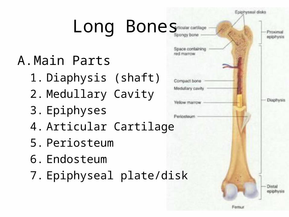
Long Bones
A. Main Parts1. Diaphysis (shaft)2. Medullary Cavity3. Epiphyses4. Articular Cartilage5. Periosteum6. Endosteum7. Epiphyseal plate/disk

A. Structure of Bone
The skeletal system is composed of two major types of tissue – bone and cartilage
1.Bone cells = osteocytes2.Cartilage cells = chondrocytes
B. Types of bone
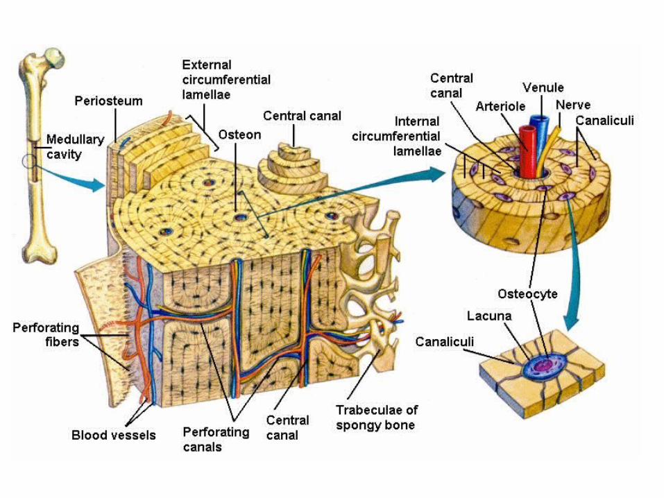
1. Compact Bonea. Covered by periosteumb. Appears solidc. Composed of osteons
1. Lamella - rings2. Lacunae – b/w lamella, contain osteocytes3. Calaliculi – canals that reach osteocytes4. Central canal – blood vessel
B. Types of bone
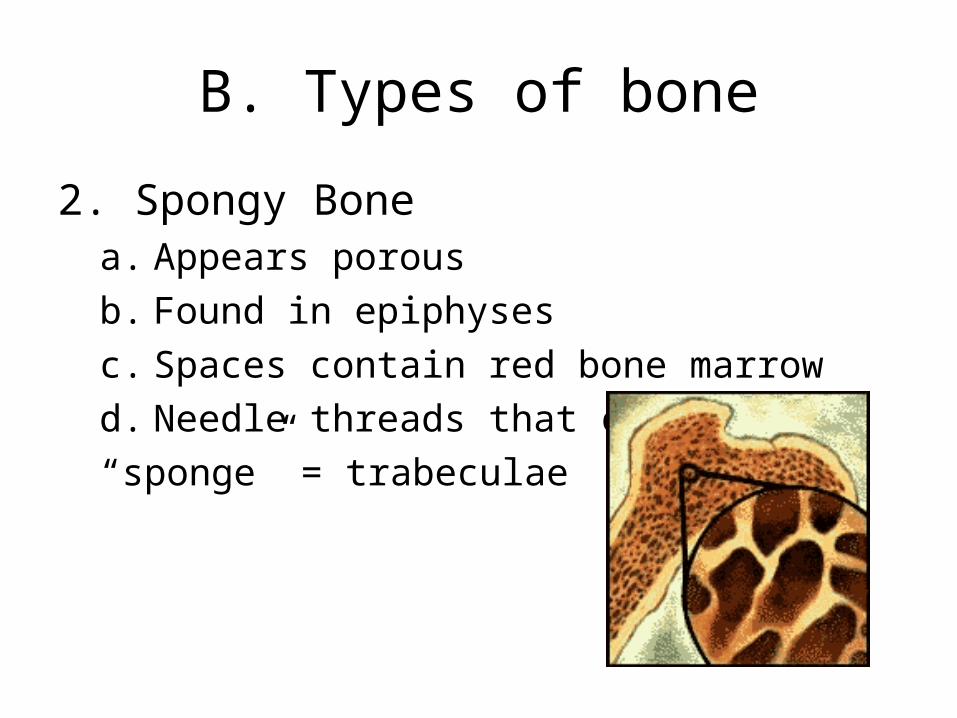
2. Spongy Bonea. Appears porousb. Found in epiphysesc. Spaces contain red bone marrowd. Needle threads that create “sponge” = trabeculae
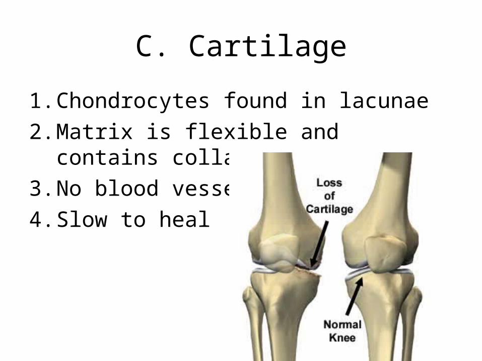
C. Cartilage
1. Chondrocytes found in lacunae2. Matrix is flexible and contains collagen3. No blood vessels4. Slow to heal
Bone Formation and Growth
A. New bone cells = osteoblasts; Resorbing cells = osteoclasts
B. Ability of bone to grow and change is dependent on the osteoblasts and osteoclasts
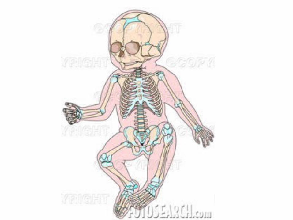
C. Bone Growth = ossification1. Endochondral Ossification –formed from cartilage2. Intramembranous Ossification3. Hematopoietic Ossification
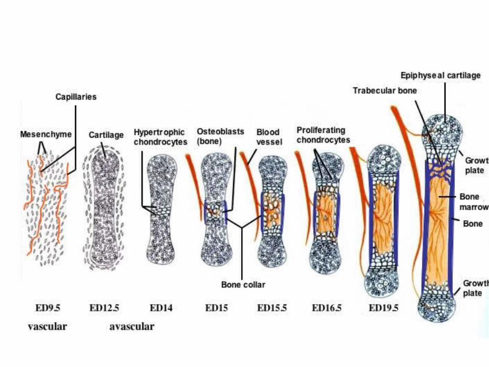
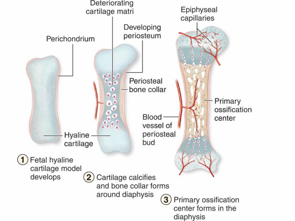
Endochondral Ossification
1. Bone formation begins from cartilage2. Blood vessel invades at diaphysis3. Osteoblasts/clasts result – center of
ossification appears in diaphysis4. Centers of ossification appear in epiphyses5. Ephyseal Plate appears6. Bone grows
Endochondral Ossification
• Bone continues to grow while the cartilaginous cells of the epiphyseal plate are active –once ossification center meet growth stops
• Osteoblasts deposit bone material under periosteum – bone grows
• Osteoclasts erode medullary cavity

Skeleton Organization
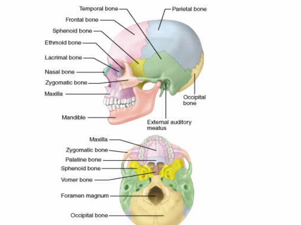
1. Axial Skeletona. Skull
i. 8 Cranial Bones1. Frontal2. Parietal (2)3. Occipital 4. Temporal (2)5. Sphenoid6. Ethmoid
ii. 14 Facial Bones1. maxilla (2)2. zygomatic (2)3. palatine (2)4. Inferior nasal concha (2)5. Mandible 6. Lacrimal (2)7. Nasal (2)8. Vomer

Skeletal Organization
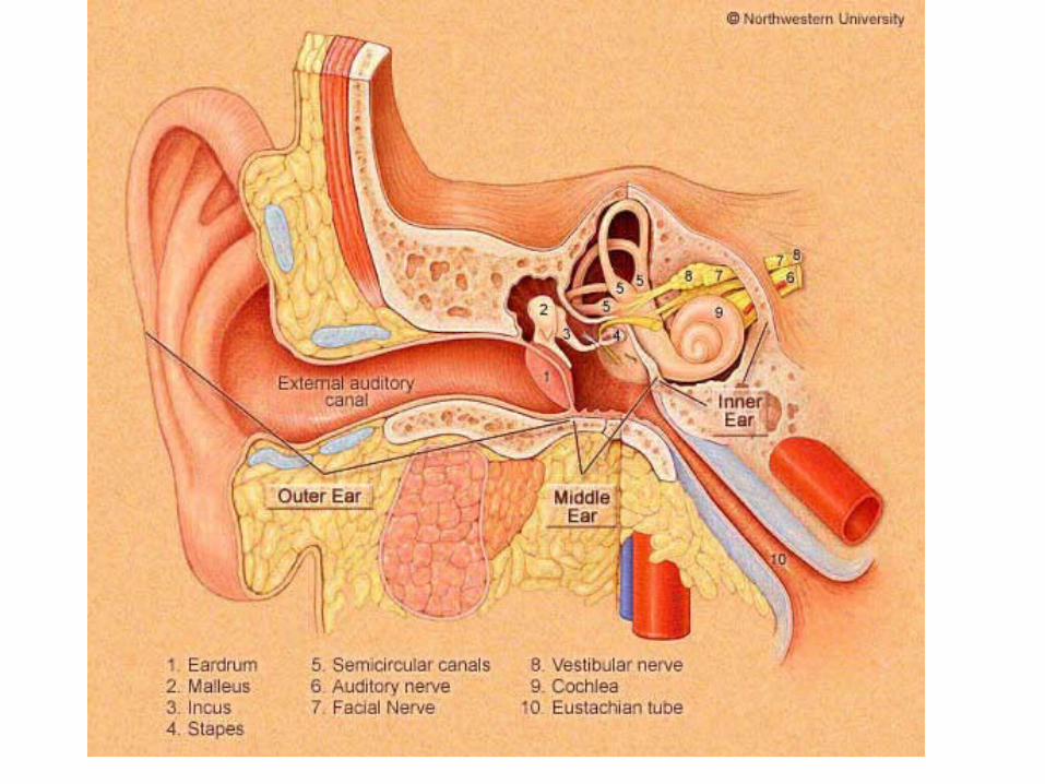
b. Middle ear bones1. malleus (2)2. incus (2)3. stapes (2)
Skeletal Organization
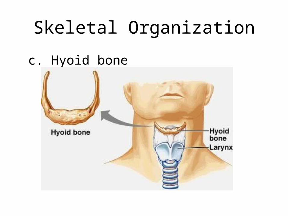
c. Hyoid bone

Skeletal Organizationd. Vertebral Column
1. Cervical Vertebrae (7)-atlas supports head
2. Thoracic Vertebrae (12)-larger than cervical-facets on sides articulate with ribs
3. Lumbar Vertebrae (5)-large and strong
4. Sacrum -five fused vertebrae
5. Coccyx-four fused vertebrae-shock absorber

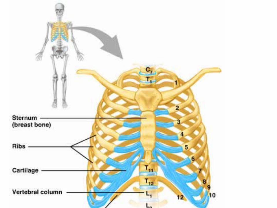
Skeletal Organization
e. Thoracic Cage1. Ribs
a. 7 True Ribsb. 5 False Ribsc. 2 Floating Ribs
2. Sternum-consists of manubrium, body, and xiphoid process-articulates with clavicles
Skeleton Organization
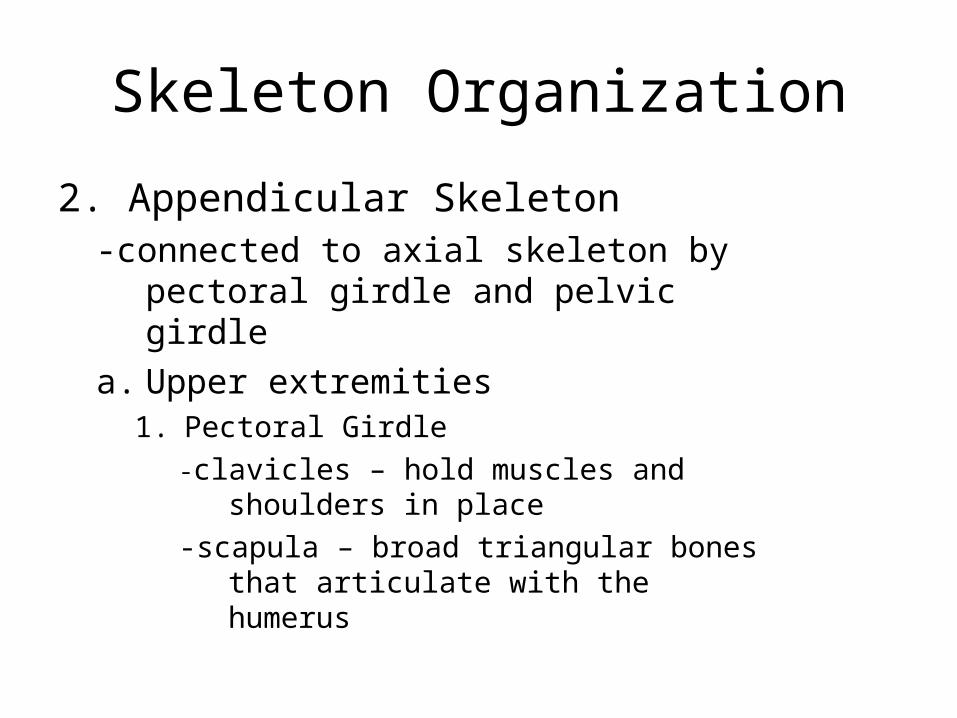
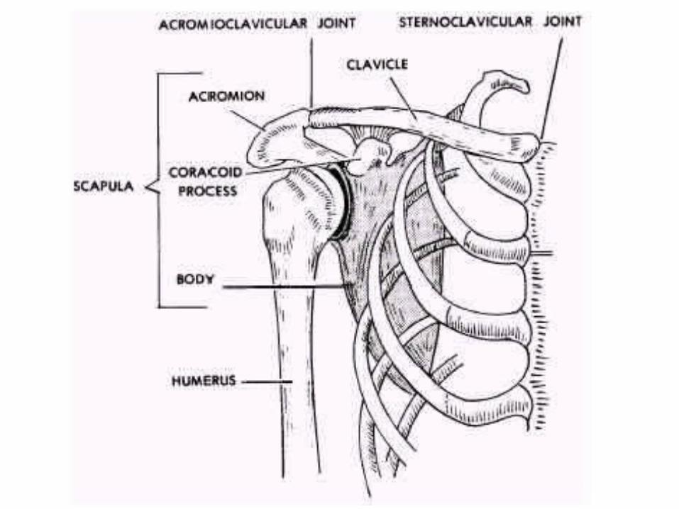
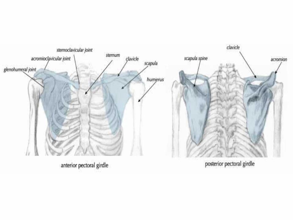
2. Appendicular Skeleton-connected to axial skeleton by pectoral
girdle and pelvic girdlea. Upper extremities
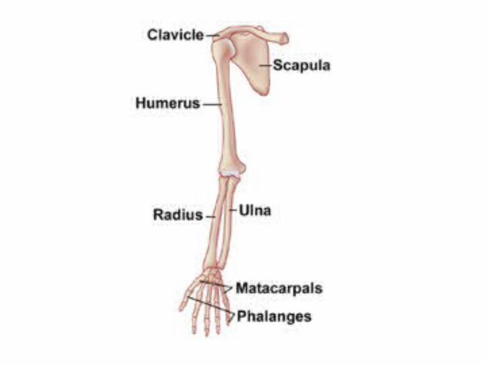
1. Pectoral Girdle-clavicles – hold muscles and shoulders in
place-scapula – broad triangular bones that
articulate with the humerus
Skeletal Organization
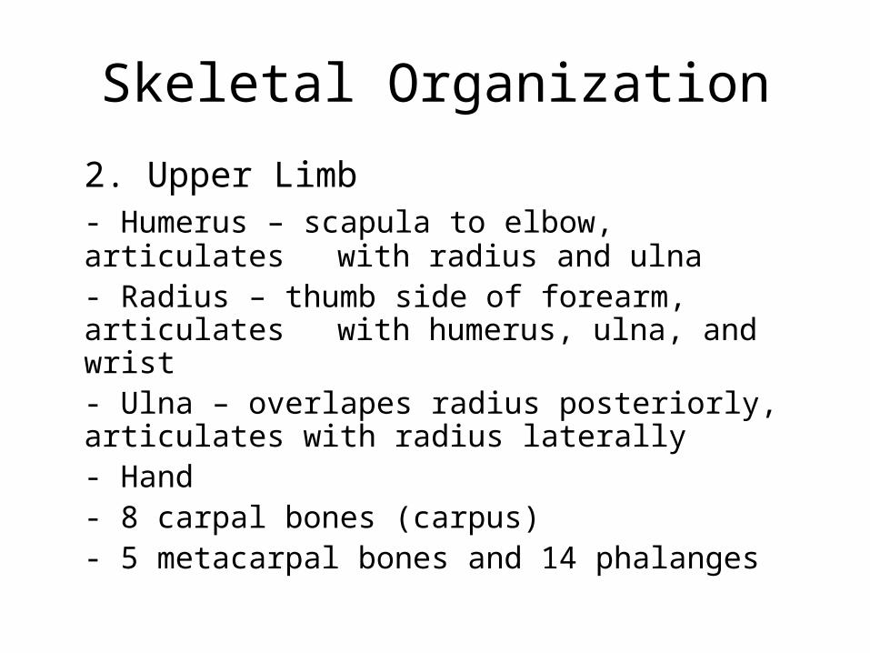
2. Upper Limb- Humerus – scapula to elbow, articulates with radius and ulna- Radius – thumb side of forearm,
articulates with humerus, ulna, and wrist- Ulna – overlapes radius posteriorly, articulates with radius laterally - Hand
- 8 carpal bones (carpus)- 5 metacarpal bones and 14
phalanges
Skeletal Organization
b. Lower extremities1. Pelvic Girdle
- sacrum, coccyx, and pelvic girdle = pelvis- coxal bones
-ilium – joins sacrum-ischium – lowest part,
supports weight when sitting-pubis – anterior portion, fused anteriorly

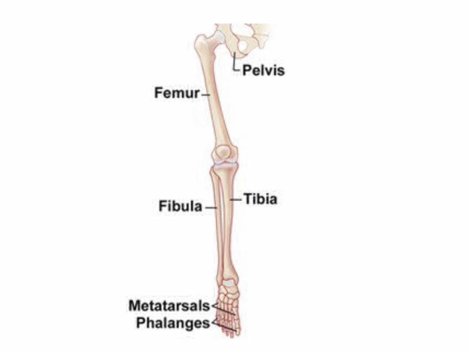
Skeletal Organization2. Lower Limb
- Femur – hip to knee, articulates with patella- Tibia – medial side of leg, articulates
with talus- Fibula – lateral side of tibia, articulates with ankle - Foot
-tarsus- 5 metatarsals-14 phalanges