sindrome di rett - morecomunicazione.it · joussef hayek u.o.c. neuropsichiatria infantile, azienda...
TRANSCRIPT

Joussef Hayek
U.O.C. Neuropsichiatria Infantile, Azienda Ospedaliera Universitaria Senese (AOUS), Siena
12 Maggio 2018
Sindrome di Rett

“Definition and diagnosis”
Rett syndrome (RTT) (OMIM #312750) is an X-linked neurodevelopmental disease that affects predominantly females (incidence 1 in 10,000 female births). Described for the first time by Dr. Rett (1966), but not widely recognized until 1983 Despite the association of RTT with mutations in the MECP2 gene (Amir et al.,1999), it remains a clinical diagnosis. Clinical diagnostic criteria guidelines (Neul et al., 2010): - history of a period of regression followed by recovery or stabilization - regression of purposeful hand use - regression of spoken language - development of gait abnormalities - development of hand stereotypies Supportive criteria (breathing abnormalities when awake, bruxism, sleep disturbances, abnormal muscle tone, vasomotor disturbances of the extremities, scoliosis/kyphosis, growth retardation, small cold hands and feet, unprovoked laughing/screaming, diminished pain response, intense eye gaze)

MeCP2 is a 4 exons gene encoding a 486 AA, 53 kDa protein
Protein Domains MBD: Methyl-binding domain TRD: Transcription repression domain C-Term: COOH-terminal domain
Over 240 mutations known (de novo in 99% of cases)
“RTT is mainly caused (∼90–95% of cases) by loss-of-function mutations in the X-
linked methyl-CpG-binding protein 2 gene (MECP2). Although mutations in other
genes are more rarely found CDKL5 mutations in the infantile seizure in the infantile
seizure onset variant and FOXG1 mutations in the congenital variant”.
Mutazioni MECP2 e sindrome di Rett

Rett syndrome: clinical features and evolution
Chahrour M & Zoghbi HY, Neuron 2007;56:422
Final picture
Severe mental retardation and motor impairments, including ataxia, apraxia, and tremors.
Seizures, respiratory dysfunction (hyperventilation, apnea), GI (gastroesophageal reflux, air swallowing with abdominal distention, chronic constipation) and orthopedic problems are also common.

Clinical features MECP2-mutated RTT
patients
CDKL5-mutated RTT
patients
Neurological regression history 100 % 100 %
Somatic growth defiency 31.2 % 50 %
Microcephaly 100 % 100 %
Loss of spontaneous ambulation 43.7 % 87.5 %
Loss of purposeful hand use 87.5 % 87 %
Scoliosis 37.5 %
Lack of verbal language 93.7 % 100 %
Impaired nonverbal language 6.2 %
Respiratory dysfunction 43.7 % 12.5 %
Autonomic nervous system signs 62.5 %
Stereotypies 100 % 100 %
Epilepsy 43.7 % 100 %
Clinical features in Rett syndrome
Leoncini S. et al. 2015

Sindrome di Rett:
patologia multisistemica
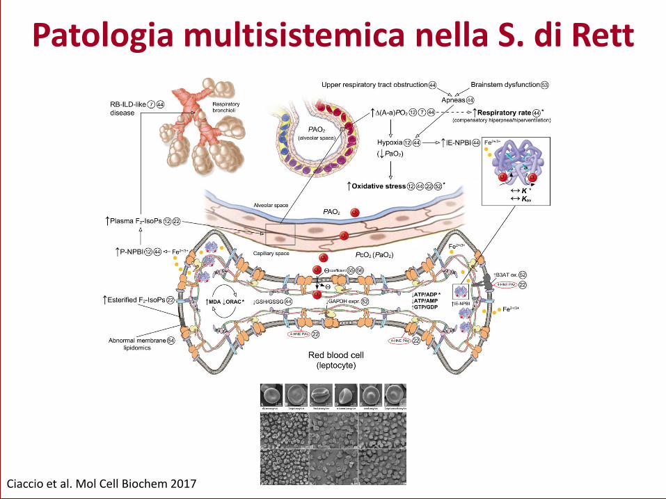
Ciaccio et al. Mol Cell Biochem 2017
Patologia multisistemica nella S. di Rett

“Cardiac dysautonomia in Rett syndrome” (J Child Neurol. 2001, Arch Dis Child. 2006 )
“Increased leptin levels in RTT” (Blardi et al. J Pediatr 2007)
“Subclinical myocardial dysfunction in Rett syndrome” (De Felice et al. European Heart Journal - Cardiovascular
Imaging 2012)
“Microvascular abnormalities in Rett syndrome” (Bianciardi et al. Clin Hemorheol Microcirc. 2013)
Clinical evidences (2001-2013) in Rett syndrome

Clinical evidences (2001-2013) in Rett syndrome
Pulmonary abnormalities in RTT patients (De Felice et al., Chest 2010)
Abnormalities in RBCs shape (Prevalence of leptocytes ~ 51.0 %) (Ciccoli L. et al. 2012)
Bronchiolitis-associated interstitial lung disease-like in MECP2-RTT patients
Healthy subject MECP2-mutated RTT patient

Inflammation in Rett syndrome
Presence of subclinical inflammatory status (Cortelazzo A. et al., Mediators Inflamm. 2014)
Inflammatory lung disease and pulmonary gas exchange abnormality in Rett syndrome (De Felice C. et
al., Mediators Inflamm. 2014)
Cytokine dysregulation in MECP2- and CDKL5-RTT (Leoncini S. et al., Oxid Med Cell Longev. 2015)
«Low pattern» abnormality «High pattern» abnormality Simple V/Q mismatch «Mixed pattern» abnormality
( 34.8 %) (39.8 %) (5.9 %) (19.6 %)
* P < 0.0001

Presence of antineuronal (brain proteins) antibodies in CSF from RTT patients (Hayek et al. 1996)
Absence of antineuronal and antiganglioside antibodies in serum from RTT patients (Fiumara et
al. 1999)
Absence of antinuclear, antistriated muscle and antismooth muscle antibodies (Fiumara et al.
1999)
Presence of brain-directed autoantibodies (AAB) in serum from RTT patients (Klushnik et al. 2001)
Increased serum levels of autoantibodies (AAB) to nerve growth factor (NGF) in RTT patients (Gratchev et al. 2001)
High levels of MeCP2 may contribute to the repression of MHC I expression in mature
neuronal cells (Miralvès et al. 2007)
Presence of folate receptor (FR) autoantibodies in serum from RTT patients (24% of
population) (Ramaekers et al. 2007)
Reduced frequency of HLA-B39 haplotype is present in MECP2-RTT (Ariani et al. 2012)
Absence of antithyroglobulin autoantibodies, antithyroid peroxidase, or anti-TSHr in RTT (Stagi et al. 2015)
An association between MECP2 gene polymorphisms and autoimmune disease, such as
systemic lupus erythematosus (SLE) (Sawalha et al. 2008, Webb et al. 2009) and primary Sj¨ogren’s
syndrome (Cobb et al. 2010), has been reported.
Association of an activity-enhancing variant of IRAK1 and an MECP2-IRAK1 haplotype with
increased susceptibility to rheumatoid arthritis (RA) (Han et al. 2013)
MeCP2 & Autoimmunity (Lit-Up)

Elevated autoantibodies anti-CSF114(Glc) and total IgM titers in Rett syndrome
«Immune Dysfunction in Rett Syndrome Patients Revealed by High Levels of Serum Anti-N(Glc) IgM Antibody Fraction»
Papini AM et al. 2014

Inflammation
Autoimmunity
Immunity
Rett
syndrome

Perché fare ricerca sulla sindrome di Rett?
Modelli sperimentali animali

Modelli Mepc2-308 femmine

Cortelazzo et al.

Proteomic analysis of the Rett syndrome experimental model mecp2Q63X mutant zebrafish (Cortelazzo A et al. J Proteomics 2017)
Modelli «zebrafish»

Evidenze cliniche e
possibili target molecolari

Relevant clinical findings observed in the patients:
- elevated blood lactate levels - elevate blood pyruvate levels - mitochondrial dysfunction - low plasma carnitine levels - increased CSF β-endorphin levels - low CSF levels of folate - increased ESR - alterated cholesterol metabolism
Clinical trials addressed on 1) general cellular processes, 2) growth factors, 3) neurotrasmitter modulators.
The most feasible strategy for treating RTT is to target events downstream to the primary gene abnormality and MECP2 deficit.
Multiple global and cell- and pathway-specific targets have been identified.

Sindrome di Rett
ed Autismo

Principali caratteristiche: sindrome di Rett vs. Autismo

Principali caratteristiche: sindrome di Rett vs. Autismo

N° di Pubblicazioni /anno 1993-2013: ASDs vs. Rett
1 = Autismo
2 = s. di Rett
ASDs: 1 p./4,5 hh
s. Rett: 1 p/1,9 gg
Fonte: PubMed

Disturbi gastrointestinali
Sindrome di Rett a. Stipsi+++
(Dismotilità intestinale) b. Stato infiammatorio
c. Stress ossidativo Disregolazione citochinica
d. Alterazione immunità/autoimmunità
e. Aumento «gluten sensitivity»
Spettro autistico a. Ipotizzato ruolo attivo microbiota
intestinale nella patofisiologia «microbiome-gut-brain-axis»
b. Disregolazione citochinica
Principali referenze Motil KJ, et al. Gastrointestinal and nutritional problems occur frequently throughout life in girls and women with Rett syndrome. J Pediatr Gastroenterol Nutr. 2012;55(3):292–8. Leonard H, et al. Assessment and management of nutrition and growth in Rett syndrome. J Pediatr Gastroenterol Nutr. 2013;57(4):451–60. Leoncini S, et al. Cytokine dysregulation in MECP2- and CDKL5-related Rett syndrome: relationships with aberrant redox homeostasis, inflammation, and omega-3 PUFAs. Oxidative Med Cell Longev. 2015;2015:421624. Cortelazzo A, et al. Subclinical inflammatory status in Rett syndrome. Mediat Inflamm. 2014;2014:480980. De Felice C, et al.Rett syndrome: An autoimmune disease? Autoimmun Rev. 2016 Apr;15(4):411-6.
Principali referenze McElhanon BO, et al. Gastrointestinal symptoms in autism spectrum disorder: a meta-analysis. Pediatrics. 2014;133(5):872–83.
Hsiao EY. Gastrointestinal issues in autism spectrum disorder. Harv Rev Psychiatry. 2014;22(2):104–11.
Furuta GT, et al. Management of constipation in children and adolescents with autism spectrum disorders. Pediatrics. 2012;130 Suppl 2:S98–105.
Gorrindo P, et al. Gastrointestinal dysfunction in autism: parental report, clinical evaluation, and associated factors. Autism Res. 2012;5(2):101–8.
Mayer EA, et al. Altered brain-gut axis in autism: comorbidity or causative mechanisms? BioEssays. 2014;36(10):933–9.

Perché studiare il microbiota intestinale nella sindrome di Rett?

Fattori che influenzano l’attività intestinale Produzione di «SCFAs»

Bifidobacterium
Lactobacillus
Enterococcus
Anaerostipes
Clostridium XIVa
Clostridium XIVb
Lachnospiraceae
Erysipelotrichaceae
Acetate
Lactate
Propionate
Butyrate
Constipation
Mucin layer
reduction Pro-inflammatory
commensals translocation
Escherichia/Shigella
Candida spp.
up up
Cross-feeding mechanisms
Intestinal inflammation
Alteration of RTT GI physiology
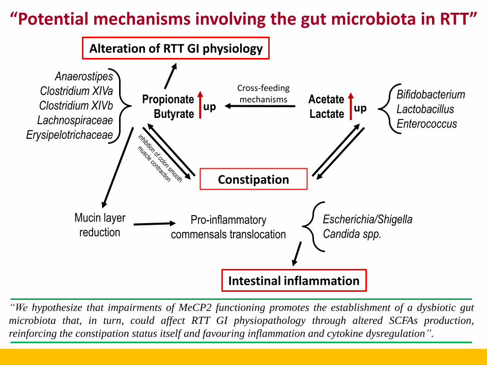
“Potential mechanisms involving the gut microbiota in RTT”
“We hypothesize that impairments of MeCP2 functioning promotes the establishment of a dysbiotic gut
microbiota that, in turn, could affect RTT GI physiopathology through altered SCFAs production,
reinforcing the constipation status itself and favouring inflammation and cytokine dysregulation”.

Altered bacterial gut microbiota in RTT
RTT is characterized by a dysbiotic
gut microbiota showing an overall
reduction of the microbial richness
and diversity as well as an altered
composition of the microbial
community structure.
Alterations of the gut microbiota do
not depend on the constipation
status of RTT subjects.
Strati F et al. Microbiome 2016, 4:41

Bifidobacteriumis the hallmark of intestinal dysbiosis in RTT
The increase in the relative abundance of Bifidobacterium,
different Clostridia and Escherichia/Shigella drives the
dysbiotic state associated with RTT. Bifidobacterium was the
most abundant taxa identified as confirmed by qPCR analysis.
Strati F et al. Microbiome 2016, 4:41

High levels of SCFAs in RTT subjects
The altered production of short chain fatty acids associated with this microbiota might reinforce
the constipation status of RTT subjects and contribute to RTT gastrointestinal physiopathology.
Strati F et al. Microbiome 2016, 4:41

Altered gut mycobiota in RTT
As observed for the bacterial
microbiota, RTT harbour an
altered gut mycobiota in which
predominates the genus Candida.
Strati F et al. Microbiome 2016, 4:41

Microbiota intestinale nella S. di Rett
Disbiosi a carico della componente batterica e fungina
Patofisiologia gastrointestinale
(alterazione infiammatoria?)
(alterazione immunitaria?)
Alterata produzione di SCFAs (short chain fatty acids)

Team di ricerca
Claudio De Felice Joussef Hayek
Cinzia Signorini Silvia Leoncini Alessio Cortelazzo
Ringraziamenti Francesco Strati - Davide Albanese – Lisa Rizzetto Computational Biology Research Unit, Research and Innovation Centre, Fondazione Edmund Mach, Via E. Mach 1, San Michele all’ Adige, Italy. Francesco Strati - Olivier Jousson Centre for Integrative Biology, University of Trento, Via Sommarive 9, Trento, Italy. Massimo Pindo Department of Genomics and Biology of Fruit Crop, Research and Innovation Centre, Fondazione Edmund Mach, Via E. Mach 1, San Michele all’ Adige, Italy. Duccio Cavalieri Department of Biology, University of Florence, Via Madonna del Piano 6, Sesto Fiorentino, Florence, Italy. Antonio Calabrò – Daniela Renzi Department of Experimental and Clinical Biomedical Sciences, Gastroenterology Unit, University of Florence, Viale Morgagni 40, Florence, Italy. Carlotta De Filippo Institute of Agriculture Biology and Biotechnology, CNR, Via Moruzzi 1, Pisa, Italy

Dedicato alle bimbe dagli occhi belli

Grazie per l’Attenzione!