signs and symptoms in cardiovascular problems
TRANSCRIPT

Signs and symptoms in cardiovascular problems
Anna Własienko, MD
Department of Paediatrics with Observation Ward
Warsaw Medical University
Head of Department: Ernest Kuchar
English Division

Agenda
1. Examination of the heart
• Heart sounds
• Heart murmurs (systolic, diastolic)
2. Cyanosis
3. Clubbing
4. Heart failure- signs & symptoms
5. Practical approach to examination of cardiac system

History taking in cardiac problems
• Prenatal history➢ previos prenatal US/scans,➢ prenatal echocariography
• Neonatal period- echocardiography
• Eating problems (breastfeeding?)- neonatal pediod, infancy
• Excesive sweating of the child
• Physical activity- compare to peers, NYHA scale
• Fainting? / Syncope?- anytime? In what conditions?
• Condition of teeth !
• Family history of ➢ congenital heart defects, ➢ cardiac arrest, ➢ sudden deaths➢ arrythmia?

History taking in cardiac problems• Prenatal history
➢ previos prenatal US/scans,➢ prenatal echocariography
• Neonatal period- echocardiography
• Eating problems (breastfeeding?)- neonatal pediod, infancy
• Excesive sweating of the child
• Physical activity- compare to peers
• Fainting? / Syncope?- anytime? In what conditions?
• Condition of teeth !
• Family history of ➢ congenital heart defects, ➢ cardiac arrest, ➢ sudden deaths➢ arrythmia?

Infective endocarditis

History taking in cardiac problems
• Prenatal history➢ previos prenatal US/scans,➢ prenatal echocariography
• Neonatal period- echocardiography
• Eating problems (breastfeeding?)- neonatal pediod, infancy
• Excesive sweating of the child
• Physical activity- compare to peers, NYHA scale
• Fainting? / Syncope?- anytime? In what conditions?
• Condition of teeth !
• Family history of ➢ congenital heart defects, ➢ cardiac arrest, ➢ sudden deaths➢ arrythmia?

EXAMINATION OF THE HEART SOUNDS & MURMURS

THE CARDIAC CYCLE
SYSTOLE DIASTOLE


WHERE DO WE AUSCULTATE HEART SOUNDS?

Bates’ Pocket Guide to Physical Examination and History Taking


NORMAL HEART SOUNDS=WHAT DO I HEAR?

Normal heart sound
https://www.youtube.com/watch?v=FtXNnmifbhE

ABNORMAL HEART SOUNDS & MURMURS
1. Extra heart sounds (S3, S4)
2. Spliting of S1 & S2
3. Alteration in intensity
4. Additional heart sounds (extrasystoly)
5. Murmurs

NORMAL HEART SOUNDS=WHAT DO I HEAR?

A third heart sound (S3)
• is heard roughly 0.1 seconds after the second heart sound.
• Due to rapid ventricular filling causing the chordae tendineae to pull to their full length &
• „twang like a guitar string”
• can be normal in children, teenagers and young healthy adults (15-40 years) -because the heart functions so well that the ventricles easily allow rapid filling
• In older patients it can indicated heart failure, as the ventricles and chordae are stiff and weak so they reach their limit much faster than normal.

S3 słuchamy
https://www.youtube.com/watch?v=_i2D1KZkN1w

NORMAL HEART SOUNDS=WHAT DO I HEAR?

A fourth heart sound (S4)
• is heard directly before S1.
• is always abnormal
• relatively rare to hear
• indicates a stiff or hypertrophic ventricle and is caused by turbulent flow from an atria contracting against a non-compliant ventricle

S4- słuchamy
https://www.youtube.com/watch?v=KcMF8rJDTIk

NORMAL HEART SOUNDS=WHAT DO I HEAR?

Split of S1
• You will listen to S1 split when MV and TV do not closeexactly at the same time
• M1 usually closes first (M1), then the TV (T1)
• S1 split is usually normal finding
• May be heard in RBBB

Split S1-słuchamy
https://www.youtube.com/watch?v=kvQ2IU3ILRo

NORMAL HEART SOUNDS=WHAT DO I HEAR?
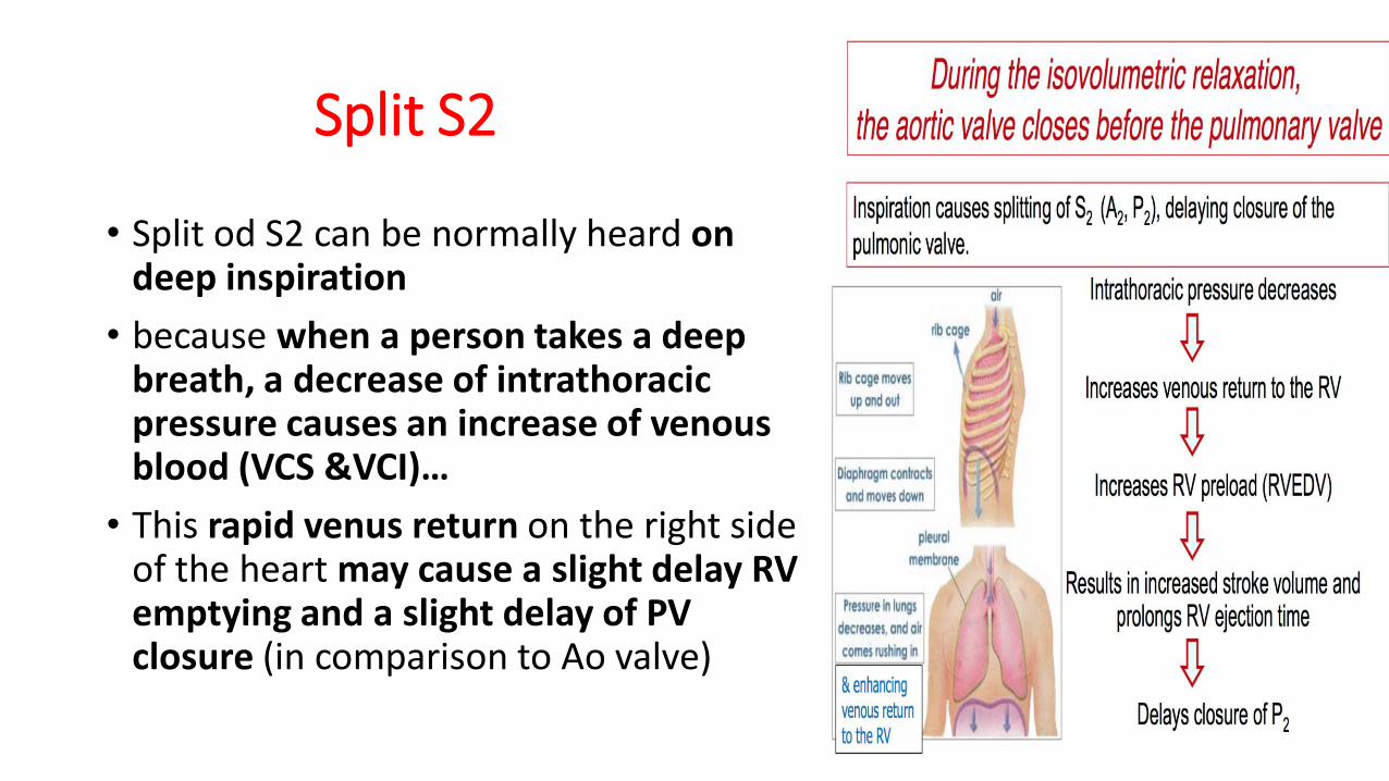
Split S2
• Split od S2 can be normally heard on deep inspiration
• because when a person takes a deepbreath, a decrease of intrathoracicpressure causes an increase of venousblood (VCS &VCI)…
• This rapid venus return on the right sideof the heart may cause a slight delay RV emptying and a slight delay of PV closure (in comparison to Ao valve)

Split S2 – słuchamy
https://www.youtube.com/watch?v=98HM1fr3cq4

HEART MURMURS


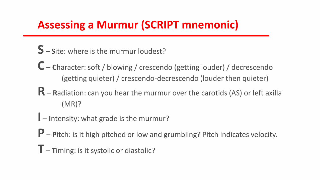
Assessing a Murmur (SCRIPT mnemonic)
S – Site: where is the murmur loudest?
C – Character: soft / blowing / crescendo (getting louder) / decrescendo
(getting quieter) / crescendo-decrescendo (louder then quieter)
R – Radiation: can you hear the murmur over the carotids (AS) or left axilla
(MR)?
I – Intensity: what grade is the murmur?
P – Pitch: is it high pitched or low and grumbling? Pitch indicates velocity.
T – Timing: is it systolic or diastolic?

MURMURSSYSTOLIC DIASTOLIC

MURMURS
• Holo- systolic= Pansystolic
• Early- systolic
• Mid- systolic
• End- systolic

MURMURS
• End-diastolic
• Early- diastolic
• Holo- diastolic
• Mid- diastolic


Aortic stenosis (AoS)
• In a child with AoS,because the AoV is verynarrow, the pressure in LV is much higher than normal and the heart must work harder to pump blood out into the body arteries
• Over time this CHD can cause hypertrophy and damage to the overworked heart muscle-should be treated (baloon or surgery)
• On examination- systolic ejection murmur dueto turbulent flow through the AoV

Aortic stenosis- słuchamy
https://www.youtube.com/watch?v=pgDWz1JybzE&t=15s


Ao regurgitation- słuchamy
https://www.youtube.com/watch?v=uZysrKXHJMM

Mitral stenosis- słuchamy
https://www.youtube.com/watch?v=5oCPtZo4pUY

→ MV cannot closecompletely causingleakge of the bloodduring systole of the heart cycle

Mitral- Tricuspid Valve RegurgitationHolosystolic Murmurhttps://www.youtube.com/watch?v=MzORJbyHTT0

Continuous machinery murmur- PDA(Gibson’s murmur)
• In PDA, abnormal blood flow occurs between Aoand pumonary trunk
• Typical machinerysystolic-diastolicmurmur

Continuous murmur- PDA
https://www.youtube.com/watch?v=LduIjbtF7kA

Heart murmurs- summary

Murmur Grade- Levine’s grading
1. Difficult to hear
2. Quiet
3. Easy to hear with sthetoscope, but no palpable thrill
4. Easy to hear with a palpable thrill
5. Can hear with stethoscope barely touching chest
6. Can hear with stethoscope off the chest
→ Grading a murmur is quite subjective but is helpful is assessing the severity of the defect and will make you sound clever.
→ If in doubt it is probably grade 2 or 3.
Assessing a Murmur (SCRIPT mnemonic)
S – Site: where is the murmur loudest?
C – Character: soft / blowing / crescendo (getting louder) / decrescendo
(getting quieter) / crescendo-decrescendo (louder then quieter)
R – Radiation: can you hear the murmur over the carotids (AS) or left axilla
(MR)?
I – Intensity: what grade is the murmur?
P – Pitch: is it high pitched or low and grumbling? Pitch indicates velocity.
T – Timing: is it systolic or diastolic?

Special manoeuvres
• can be used to emphasise certain murmurs:
• Patient on their left side → mitral stenosis
• Patient sat up, learning forward and holding exhalation → aortic regurgitation


But….
… not every murmur is a pathology

Innocent murmurs
• In healthy children• Caused by turbulent blood flow through anatomically healthy heart• Asymptomatic• Always systolic• Postural- volume varies with siting/ standing• Intensifies with increased cardiac output (eg.fever, emotions, excercise)• Short duration• Soft and quite in quality < 3 grade• No radiation• Otharwise normal physical examination- no palpble thrill, no SOB, no FTT,
normal BP, HR, SaO2

Innocent murmur- słuchamy
https://www.youtube.com/watch?v=uFyWHPfrRak


CYANOSIS

CYANOSIS
• the bluish or purplish discoloration of the skin or/ and mucous membranes due to low oxygen saturation of the tissues near the skin surface
• Is a result of deoxygenated haemoglibin or abnormal haemoglobin in the blood
• Is apparent when there is ≥ 5 g/dl of reduced haemoglobin
• Anemic patients may not become cyanotic even in the presence of markedarterial desaturation
• In the light-skinned patients cynanosis is usualy noted with arterial SaO2 < 85%, whereas:
• In the dark-skinned patients, the Sa02 may be lower

CYANOSIS
CENTRALis due to a circulatory or ventilatory problem that leads to
poor blood oxygenation in the lungs
• lips
• tongue
PERIFERALIs due to an inadequate or obstructed circulation
• only the extremities or fingers


Causes of central cyanosis
1. Cardiovascular diseases:
• Congenital heart disease with R-L shunt
• Heart failure
• Valvular heart disease
• Myocardial infarction

Causes of central cyanosis
2. Respiratory system:
• Severe pneumonia• Bronchiolitis• Bronchospasm• Pulmonary hypertension• Pulmonary embolism• Hypoventilation• Chronic obstructive pulmonary disease• Cystic fibrosis

Causes of central cyanosis
3. Central nervous system (impairing normal ventilation):
• Intracranial hemorrhage
• Drug overdose (e.g. heroin) → apnea or/ and airway obstruction
• Tonic–clonic seizure (e.g. grand mal seizure)

Causes of central cyanosis
4. Blood :
• Congenital cyanosis (HbM Boston) arises from a mutation in the α-codon which results in a change of primary sequence
• Methemoglobinemia- patient appears cyanosedeven in the presence of a normal arterial oxygenlevel due to conversion of iron in hemoglobin from the ferrous [Fe2+] to the ferric [Fe3+] → aquired (drugs, chemicals & toxins eg. anilinedyes, chlorates, and bromates )
• Polycythaemia

Causes of central cyanosis
5. Others:
• High altitude, cyanosis may develop in ascents to altitudes >2400 m.
• Hypothermia
• Severe obstructive sleep apnea (apnea)

Peripheral cyanosis
may be due to the following causes:
• All common causes of central cyanosis
• Reduced cardiac output (e.g. heart failure or hypovolaemia)
• Cold exposure
• Chronic obstructive pulmonary disease (COPD)
• Arterial obstruction (e.g. Raynaud phenomenon)
• Venous obstruction (e.g. deep vein thrombosis)

Is it cyanosis?

Is it cyanosis?
Argyria or argyrosis is a condition caused by excessive exposure to chemical compounds of the element silver or to silver dust
• skin turns purple or purple-grey
• Generalised (with mucus membranes, eyes) or local
• Argyria worsens and builds up as exposure to silver continues, and does not resolve once exposure stops

CLUBBING

NAIL CLUBBING

NAIL CLUBBING
• is a deformity of the fingers and/or toes nails associated with a number of diseases, mostly of the heart and lungs
• occurs when the tips of the fingers enlarge and the nails curve around the fingertips,
• usually over the course of years
• is often the result of low oxygen in the blood and could be a sign of various types of heart and/ or lung disease
• Patomechanism- unknown

NAIL CLUBBING< 180 °


NAIL CLUBBING- TEST (Schamroth sign)

NAIL CLUBBING- TEST

NAIL CLUBBING


NAIL CLUBBING↔ DRUMSTICS

NAIL CLUBBING

NAIL CLUBBING

NAIL CLUBBING

NAIL CLUBBING
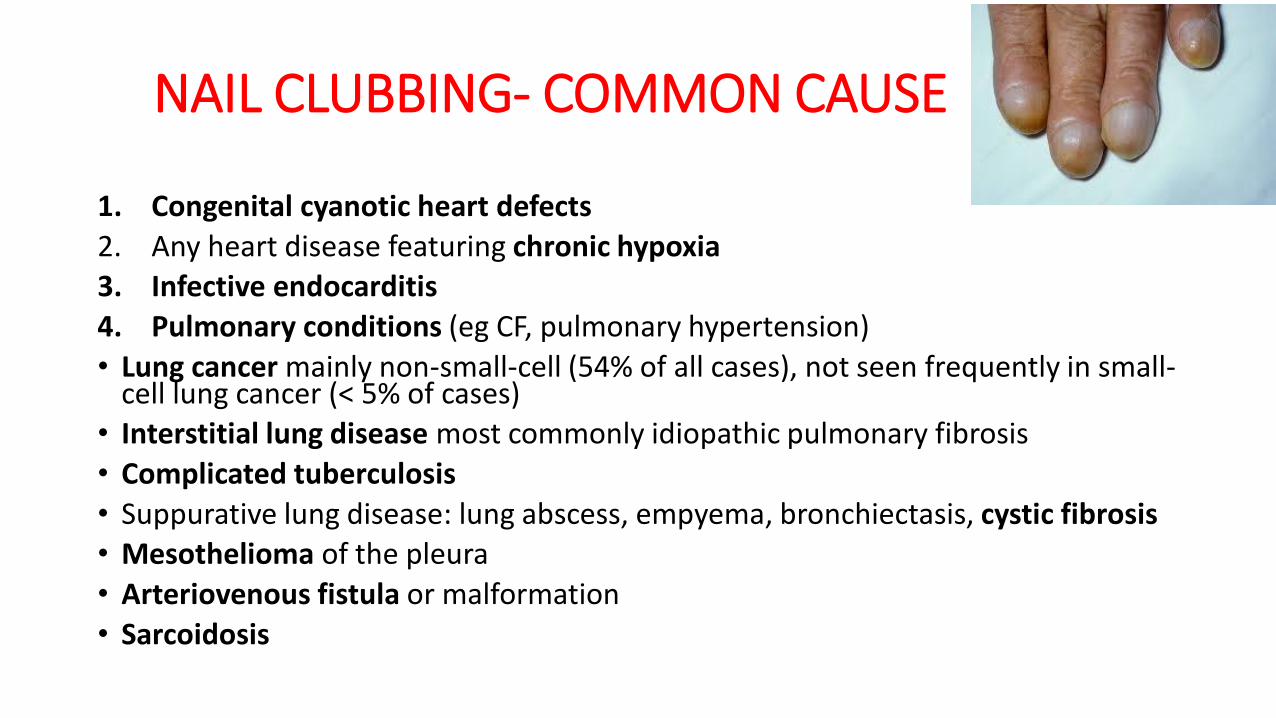
NAIL CLUBBING- COMMON CAUSE
1. Congenital cyanotic heart defects
2. Any heart disease featuring chronic hypoxia
3. Infective endocarditis
4. Pulmonary conditions (eg CF, pulmonary hypertension)
• Lung cancer mainly non-small-cell (54% of all cases), not seen frequently in small-cell lung cancer (< 5% of cases)
• Interstitial lung disease most commonly idiopathic pulmonary fibrosis
• Complicated tuberculosis
• Suppurative lung disease: lung abscess, empyema, bronchiectasis, cystic fibrosis• Mesothelioma of the pleura
• Arteriovenous fistula or malformation
• Sarcoidosis

NAIL CLUBBING- COMMON CAUSE
5. Hereditary
6. GI disease (Crohn’s disease, ulcerative cllitis, cirrhosis, especially in primary billiary cirrhosis)
7. Idiopathic

HEART FAILURE

HEART FAILURE IN ADULTS

Heart failure in children
• May be manifested by symptoms of poor tissue perfussion alone(eg. fatigue, poor excercise tolerance, cinfusion) or
• by symptoms of congestion of circularion (e. SOB, pleural effusion, pulmonary or peripheral oedema, hepatomegaly) without evokingcompensatory mechanisms
• Underlying pathophysiology mechanisms leading to HF include
➢ increased afterload (preassure work) eg. valves stenosis
➢Increased preload (volume work) eg. shunts
➢Myocardial abnormalitries (eg. Cadiomiopathies)
➢Tachyarhhythmias

HEART FAILURE IN CHILDREN
Tom Lissauer, Avroy Fanaroff, Neonatology at a Glance


Common causes of HF in children

Practical approach to physical examination of circulation• Airway
• Breathing• Respiratory Rate• Tidal Volume• Work of extra muscles• Oxygenation
• Circulaton• HR• BP• PULSE (PRESENT? AMPLITUDE?)• PERFUSSION (CRT, SKIN- COLUOR- CYANOSIS?, WARM? / COLD?, SWEATY?)• PRELOAD (JUGULAR VEINS, HEPATOMEGALY, CRACLES IN LUNGS)• DIURESIS (RENAL PERFUSSION)

Checking the pulse in children


Checking the pulse in neonates

Thank you for yourattention!
