signal transduction pathways ii: enzyme-linked receptors nuclear receptors stochasticity in...
TRANSCRIPT

Signal transduction pathways II:
Enzyme-linked receptors
Nuclear receptors
Stochasticity in signalling.

Enzyme-linked receptors
signalling through growth factors, hormones, cytokines acting locally at very low concentrations (10-9 – 10-11M) response is usually slow (min-hours) and requires many intracellular steps, leading to
changes in gene expression1. receptor tyrosin kinases (RTKs)2. tyrosin kinases associated receptors3. receptor serin/threonin kinases4. histidin kinase associated receptors5. receptor guanylyl cyclases

1. Receptor tyrosin kinases (RTKs)
Ligands - EGF, PDGF, HGF, NGF, insulin, ephrins...Signalling through monomeric GTPases or through lipid modifications by PI3K
Figure 15-52 Molecular Biology of the Cell (© Garland Science 2008)

Table 15-4 Molecular Biology of the Cell (© Garland Science 2008)

Figure 15-16c Molecular Biology of the Cell (© Garland Science 2008)
- ligand is a dimer (PDGF)- ligand is monomer but binds to TM heparan sulphate proteoglycans to form multimers (FGF)- clustering of TM ligand in a neighbouring cell (ephrins)- receptor is a multimer to start with (insulin)- ligand is monomer but binds two receptors simultaneously (EGF)
1. Clusterring of receptors

Figure 15-53a Molecular Biology of the Cell (© Garland Science 2008)
2. induced proximity trans/auto phosphorylation

Figure 15-53b Molecular Biology of the Cell (© Garland Science 2008)
kinase-deaddimerization OK
How would you create a DN form of enzyme associated receptor?

3. transient assembly of intracellular signalling complexes
• docking via SH2 domains, PTB domains, PH domains and others• some are kinases (Src, PI3K)• some are adaptor proteins (Grb) that connect to a protein with enzymatic a.• some are inhibitors (e.g. c-Cbl, monoUbiq. and endocytosis)

SH2 domain, PTB domain – recognize phosporylated tyrosines
SH3 domain – recognizes proline rich sequences
PH domain – recognizes modified lipids
SH2 domain

4. spreading of the intracellular signalling events through the
Ras superfamily of monomeric GTPases
Only Ras and Rho signal through cell surface receptors
Often tethered to membrane via lipid anchor

Figure 15-19 Molecular Biology of the Cell (© Garland Science 2008)
Ras: hyperactive forms such as RasV12
• dominant, constitutively active
• resistant to inactivation by GAP
(no GTP hydrolysis)
• Ras required for cell proliferation;
mutant in 30% of human cancers
RTKs activate a Ras-GEF or inhibit a Ras-GAP
GEF – guanine exchange factorGAP – GTPase activating proteins

Figure 15-56 Molecular Biology of the Cell (© Garland Science 2008)
Signalling through Ras:EGFR signalling
lesson from the Drosophila eye
760 ommatidia: 8 photoreceptor cells (R1-8) and 11 support cells, pigment cells, and a
cornea

Dominant allele of EGFR, constitutively active
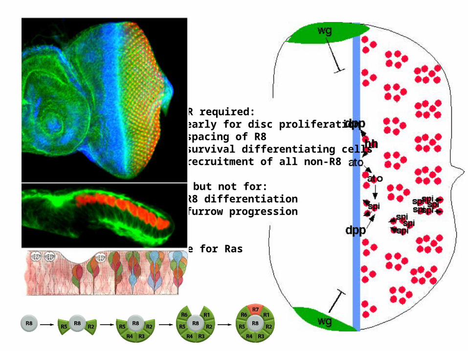
EGFR required:• early for disc proliferation• spacing of R8• survival differentiating cells• recruitment of all non-R8
... but not for:• R8 differentiation• furrow progression
Same for Ras
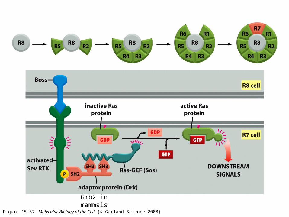
Figure 15-57 Molecular Biology of the Cell (© Garland Science 2008)
Grb2 in mammals
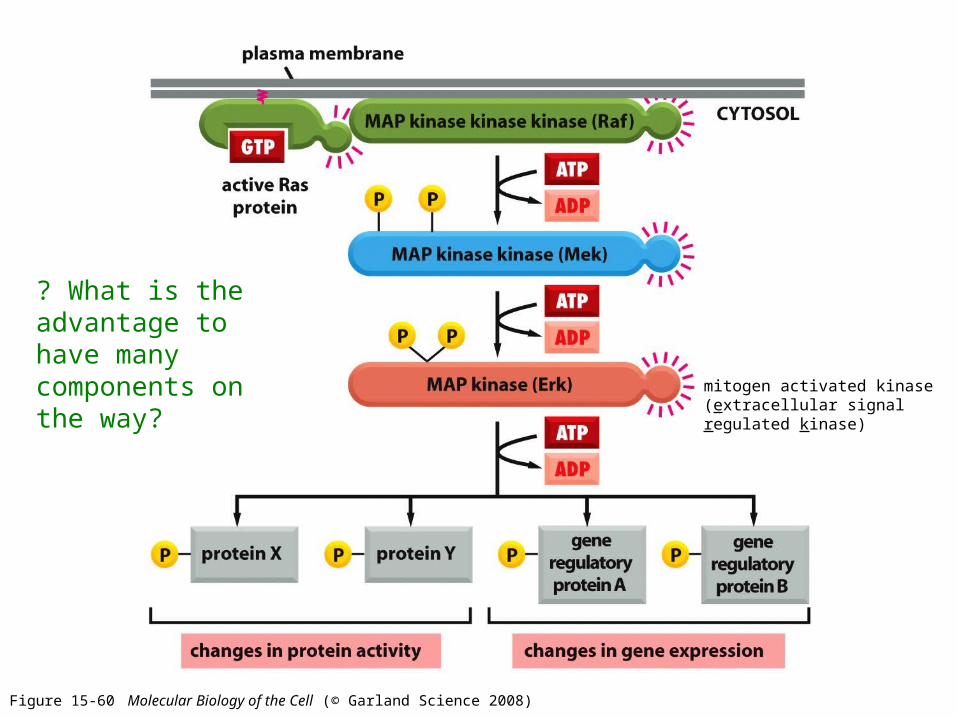
Figure 15-60 Molecular Biology of the Cell (© Garland Science 2008)
mitogen activated kinase(extracellular signal regulated kinase)
? What is the advantage to have many components on the way?

MAP kinase modules
- complexes of three protein kinases interconnected in series, often bound to a scaffold- after activation the MAPK-MAPKK interactions may be weakened, enabling MAPK to travel to the nucleus

Stress modules
MAPKKK
MAPKK
MAPK
Mitogenesis module
MAP kinase modules
Binding to a scaffold reduces crosstalk BUT also avoids difussion and spreading of signal

Signal amplification (of undocked kinases)
input signal
MEKK1
MKK4
output signal
MKK4MKK4MKK4
MKK4MKK4
MKK4MKK4
JNKJNK
JNKJNK
JNKJNK
JNK
modules avoid spreading

Downstream of MAPK – the effectors
- mostly to promote proliferation and prevent apoptosis- substrates of MAPK: transcription factors, further kinases (MAPKAP kinases), also negative feedback loops through phosphorylation and activation of MAPK phosphatases or ras-GAPs
MAPKAP kinases:- MAP kinase activated protein kinases- activation of proliferation and survival programs through phosphorylation of transcription factors or other regulatory proteins- p90 RSK = ribosomal S6 kinase MSK = mitogen and stress activated kinase MNK = MAPK interacting kinase
Transctiption factors:- Ets family (Elk-1, TCF... after phosphorylation often bind another TF like SRF)- AP1(homo- or hetero-dimers of c-fos, c-jun, ATF2 and MAF through leucine zipper)- MEF2, MAX, NFAT.....
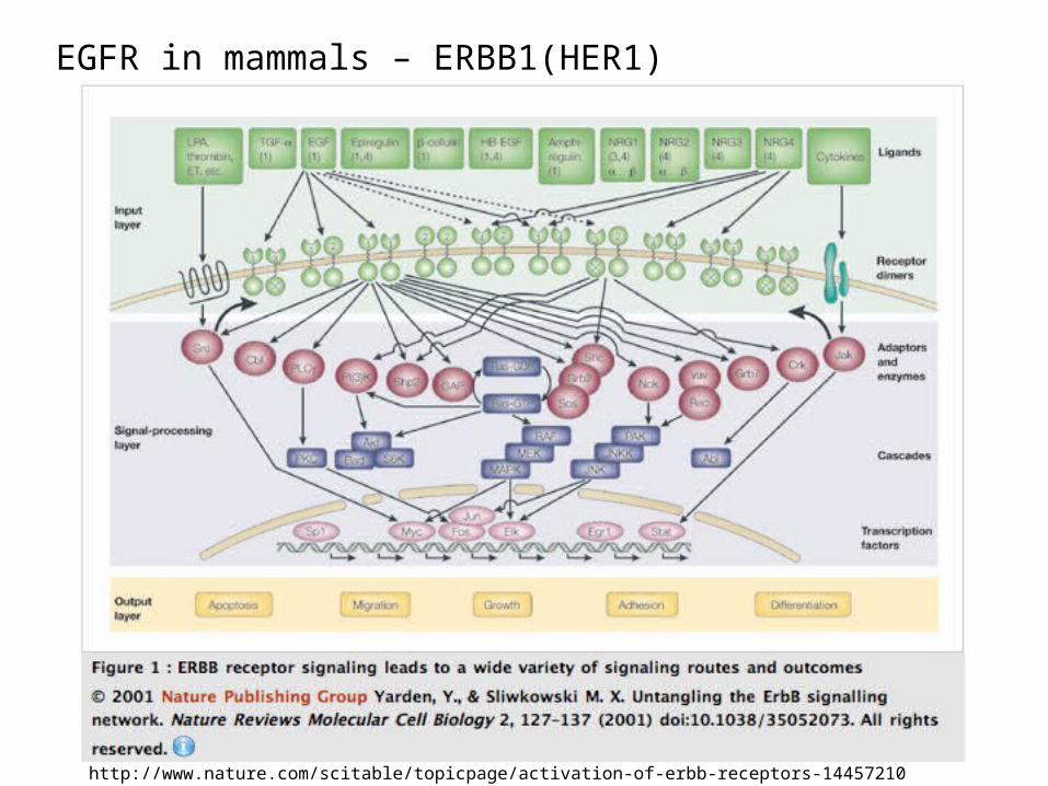
EGFR in mammals – ERBB1(HER1)
http://www.nature.com/scitable/topicpage/activation-of-erbb-receptors-14457210

ERBB family of tyrosin kinases
4 receptors ERBB1-4 (HER1-4)
different numbers of phosphorylation sites on their C-termini
cellular outcome depends on the dimerization partners and ligands
exists as homo or heterodimers
Ligands : growth factors (EGF family, NGF,TGF-a)


Positive and negative feedback loops
Tyrosine specific phosphatases and GAPs inactivate Ras.
Neuronal precursor cell:EGF activation – peaks in 5 minutes, cells later dividesNGF activation – lasts many hours, cell stops dividing and differentiates into neurons
Duration of response can influence the final outcome

- regulate actin and microtubule cytoskeleton- also regulate cell cycle progression, transcription, membrane transport- used by ephrin receptors but also by integrins, GF receptors, GPCRs…
(Ras protein too)- activated by rho-GEFs (60 in human), inactivated by rho-GAPs (70 in human)- inactive rho bound in cytosol to guanine nucleotide dissociating inhibitor (GDI), preventing it to bind to GEF in the membrane
RhoA, Rac, cdc48...
4. Signalling through the Rho family of monomeric GTPases

Growth cone extension in axons through the guidance molecules ephrinsThe most abundant class of protein tyrosine kinases.
http://www.youtube.com/watch?v=_1zGQHrvoRo&feature=results_video&playnext=1&list=PLD424D0006F154019

Enzyme-linked receptors
1. receptor tyrosin kinases (RTKs)2. tyrosin kinases associated receptors3. receptor serine/threonine kinases4. histidin kinase associated receptors5. receptor guanylyl cyclases

Similar as receptor tyrosine kinases but the kinase is non-covalently bound to the receptor
Tyrosin kinases associated receptors
Receptors for antigens on leucocytes, for cytokines, hormones, integrin receptors...
kinase kinase kinase kinase
ligand

Tyrosin kinases associated with receptors
Src family: Src,Yes, Fgr, Fyn, Lck, Lyn, Hck, and Blk
- cytoplasmic localization through lipid anchor and through association with receptors- SH2 and SH3 domains- can also be activated by some RTKs and GPCR
FAK (focal adhesion kinase)
JAK (Janus kinase)

Src- first tyrosine kinase discovered, from the chicken Rous sarcoma virus (Peyton
Rous, Nobel prize in 1966) = first transmissable agent inducing cancer- oncoproteins are gain of function cellular proteins (Bishop and Varmus ,1989)- v-src, c-src
(receptor orother proteins)

FAK (focal adhesion kinase)
- associated with integrin receptors in focal adhesion points (places of cell contact with extracellular matrix) - recruit src, mutual phosphorylation and activation of cell survival and proliferation programme

JAK
- family of cytokine receptors, receptors for growth hormone and prolactine- stable association with JAK (Janus kinase, JAK1, JAK2, JAK3 and Tyk)- activated JAKs recruits STATs (signal transducers and activators of transcription), in the cytoplasm and phosphorylates them- phosphorylated STATs migrate to the nucleus and activate transcription
JAK-STAT pathway
Enzyme-linked receptors
1. receptor tyrosin kinases (RTKs)2. tyrosin kinases associated receptors3. receptor serine/threonine kinases4. histidin kinase associated receptors5. receptor guanylyl cyclases

TGF-beta and BMP signalling
transforming growth factor beta and bone morphogenetic protein

- SMADs continuously shuttle between cytoplasm and nucleus during signalling (dephosphorylated in nucleus, rephosphorylated in cytoplasm)- secreted inhibitory proteins regulate the morphogen gradient: noggin and chordin (for BMP), follistatin (for TGFbeta)- inhibitory SMADs (SMAD6 and SMAD7)
- compete for activating SMADs - recruits ubiquitin ligase Smurf to the receptor, promoting its internalization
and degradation- recruits phosphatases to inactivate the receptor


Enzyme-linked receptors
1. receptor tyrosin kinases (RTKs)2. tyrosin kinases associated receptors3. receptor serine/threonine kinases4. histidin kinase associated receptors5. receptor guanylyl cyclases

- bacteria, yeast and plants, not in animals- bacterial chemotaxis:
Histidin kinase associated receptors
Default movement anticlockwise (straight) with occasional clockwise tumbling (stop).

Chemotaxis receptors to various atractants and repelents
- repellent activates receptor (tumbling), atractant inactivates it (going forward)
- histidin kinase CheA (adaptor CheW)- regulator protein CheY binds motor and makes it rotate clockwise- intrinsic phosphatase activity of CheY (accelerated by CheZ)

Adaptation through receptor methylation and demethylation:
Attractant:(1) decreases the ability of the receptor to activate CheA (2) slowly (over minutes) alters the receptor so that it can be methylated by a methyl transferase, which returns the receptor’s ability to activate CheA to its original level (tumbling).
Thus, the unmethylated receptor without a bound atractant has the same activityas the methylated receptor with a bound atractant, and the tumbling frequency ofthe bacterium is therefore the same in both cases. Bacteria adapts to certain concentration of atractant.
Bacteria senses the difference in the concentrations of repellents and attractants,not the exact concentration!

Enzyme-linked receptors
1. receptor tyrosin kinases (RTKs)2. tyrosin kinases associated receptors3. receptor serine/threonine kinases4. histidin kinase associated receptors5. receptor guanylyl cyclases
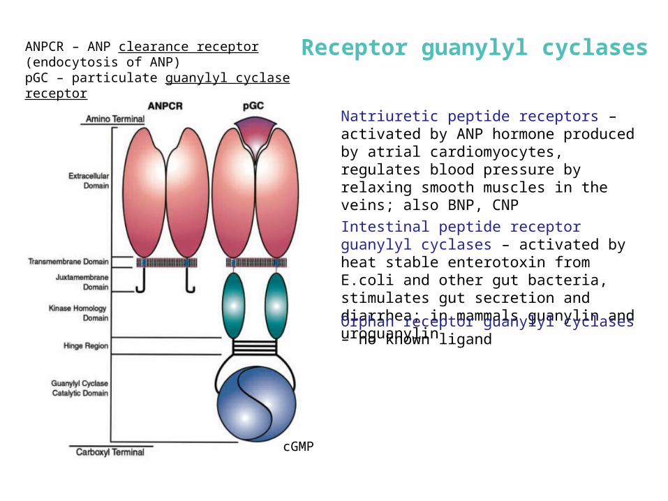
ANPCR – ANP clearance receptor (endocytosis of ANP)pGC – particulate guanylyl cyclase receptor
Natriuretic peptide receptors – activated by ANP hormone produced by atrial cardiomyocytes, regulates blood pressure by relaxing smooth muscles in the veins; also BNP, CNP
cGMP
Intestinal peptide receptor guanylyl cyclases – activated by heat stable enterotoxin from E.coli and other gut bacteria, stimulates gut secretion and diarrhea; in mammals guanylin and uroguanylin
Orphan receptor guanylyl cyclases – no known ligand
Receptor guanylyl cyclases

NUCLEAR RECEPTORS

Figure 15-13 Molecular Biology of the Cell (© Garland Science 2008)
NUCLEAR RECEPTORS
Ligands: lipophylic molecules• Steroid hormones (from cholesterol: sex hormones, vitaminD, cortisol, ecdysone in insects) • Thyroid hormones (from tyrosine)• Retinoids (from vitaminA)

activation
repression

Video tutorial: http://www.nursa.org/flash/gene/nuclearreceptor/start.html
III.
Pardee et al.: A Handbook of Transcription Factors, Subcellular Biochemistry 52, 2011
mineralokortikoid r.
glucocorticoid r.estrogen r.
androgen r.
progesteron r.
vitamin D
retinoic acid
ultraspiracle
peroxisome proliferatorecdysone
hepatocyte nuclear f.
Types of nuclear receptors

Types of nuclear receptors
Sever and Glass: Cold Spring Harb Perspect Biol 2013;5:a016709

inactive Met (juvenile hormone receptor)
homodimer
...and formation of active transcription factor with
Taiman
JH binding to Met promotes dissociation of the inactive complex...
MetJH
Tai
JH
Met
Met
Receptor for juvenile hormone (Met)new class of nuclear receptors
Marek Jindra: Proc Natl Acad Sci U S A. 2011 Dec 27;108(52):21128-33, 2011

Pardee et al.: A Handbook of Transcription Factors, Subcellular Biochemistry 52, 2011
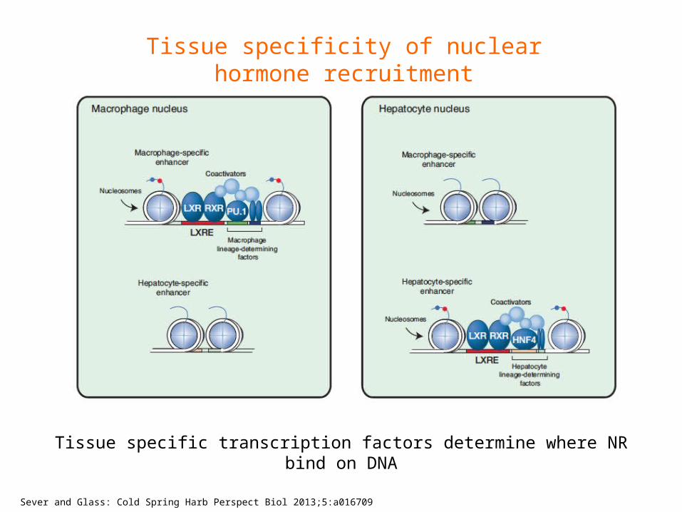
Tissue specificity of nuclear hormone recruitment
Tissue specific transcription factors determine where NR bind on DNA
Sever and Glass: Cold Spring Harb Perspect Biol 2013;5:a016709

In vivo ligand sensors

STOCHASTICITY IN CELL SIGNALLING
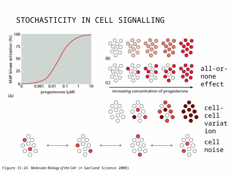
STOCHASTICITY IN CELL SIGNALLING
Figure 15-24 Molecular Biology of the Cell (© Garland Science 2008)
all-or-none effect
cell-cellvariation
cell noise

Sprinzak and Elowitz, Nature, 2010
Reporter assay with a GFP reporter
Stochastic response

ERK2 gene
ERK2-YFP fusion in genomic locus
YFP
Cohen-Saidon C, Alon U et al.: Molecular Cell (2009)
Response to EGF

Cohen-Saidon C, Alon U et al.: Molecular Cell (2009)
Different basal levels……fold response after EGF stimulation similar between cells
‘Amount of ERK2 entering the nucleus is proportional to the basal level of nuclear ERK2 in each cell, suggesting a fold-change response mechanism.’

EstrogensignalingWeber’s law (1834)
Human perception of light and sound is not based on absolute levels but on the magnitude of stimulus relative to the background levels.

Why fold change rather than absolute level of detection?
How is the fold change detected by the cell?
It might be easier for the cell to generate reliable fold changes than reliable absolute changes.
Negative feed-forward loops.
It might serve as an adjustable noise filter. Absolute variations present when background level of stimulus is high tends to be higher than when background level is low.

Consequence for regulated gene Z: Pulse of activity
Incoherent feed-forward loop as a fold-change detector
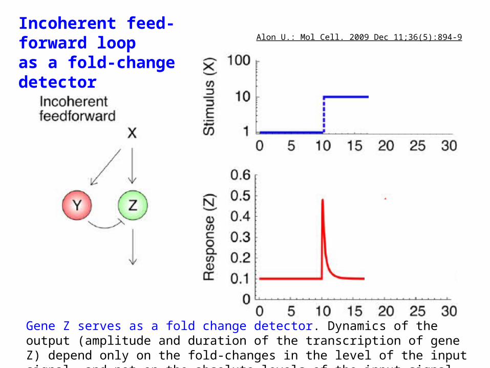
Incoherent feed-forward loop as a fold-change detector
Gene Z serves as a fold change detector. Dynamics of the output (amplitude and duration of the transcription of gene Z) depend only on the fold-changes in the level of the input signal, and not on the absolute levels of the input signal.
Alon U.: Mol Cell. 2009 Dec 11;36(5):894-9
