severe cryptogenic multifocal ulcerous stenosing enteritis
TRANSCRIPT

Introduction
Cryptogenic multifocal ulcerous stenosing enteritis(CMUSE) is a rare condition characterised by chronic orrelapsing moderate ileous episodes resulting from multiplesmall intestinal strictures, multiple shallow ulcers of thesmall bowel and beneficial therapeutical effect of gluco-corticosteroids (1, 2). Japanese gastroenterologists call thissyndrome chronic non-specific multiple ulcers of the smallintestine (CNSU) (3). The aetiology of CMUSE has notbeen clarified yet and pathogenesis is still poorly under-stood. Some authors even doubt the real existence of thisentity.
Patients with CMUSE are often referred for surgery be-cause of symptomatic small intestinal strictures and severalresections of the small bowel had to be performed.
We report three cases of CMUSE diagnosed within thepast 10 years at a single tertiary centre, where more thaneight thousand GI endoscopies are performed per year.Review of available relevant literature is provided.
Case reports
Case 1
A 35-year-old woman was admitted to our departmentbecause of chronic diarrhoea, colicky abdominal pain, weight
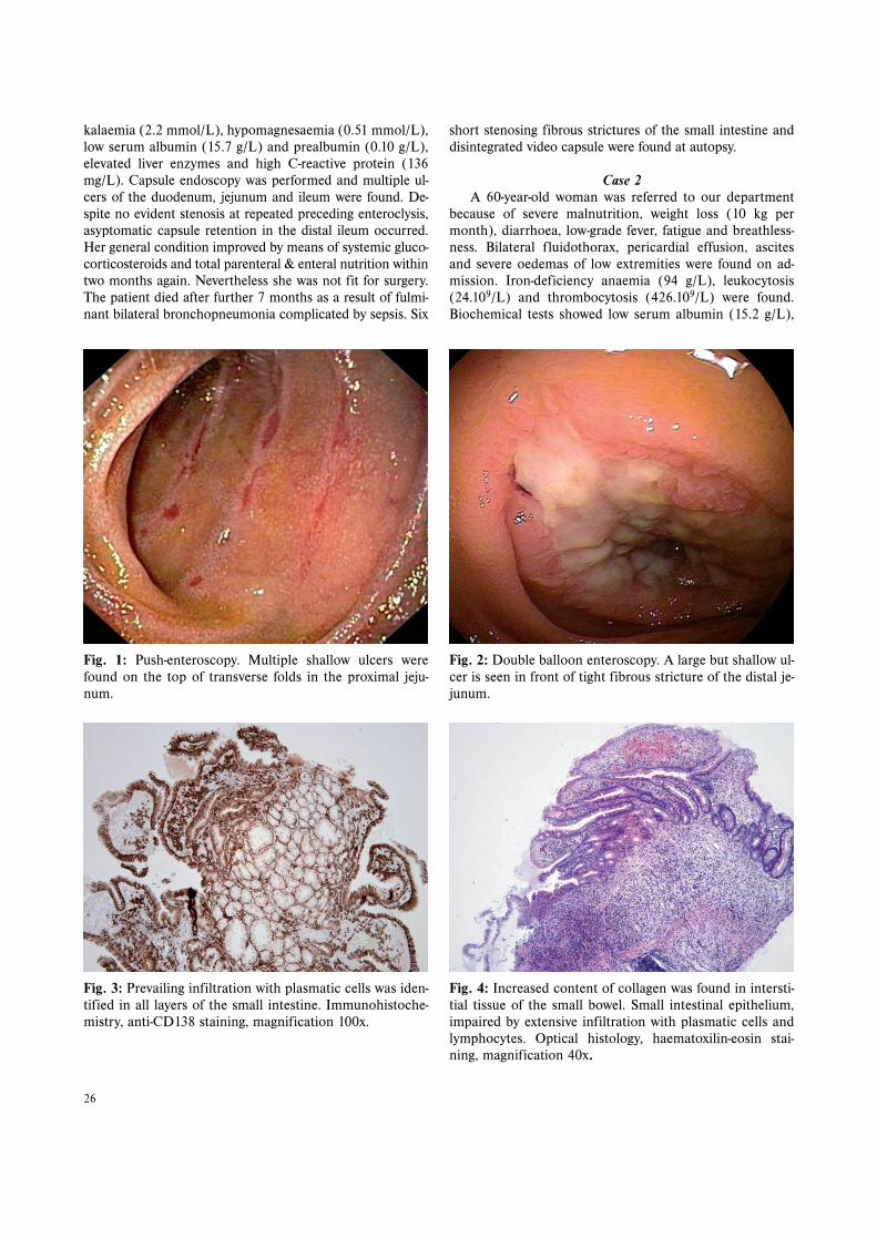
loss (16 kg during previous five years) and repeated mode-rate ileous episodes. She was cachectic (body-mass index12.8 kg/m2). Laboratory tests showed iron-deficiency anae-mia (haemoglobin 96 g/L), thrombocytosis (459.109 g/L)and proteino-energy malnutrition (serum prealbumin 0.17g/L). Abdominal ultrasound revealed strenuous small in-testinal peristalsis and a large volume of fluid in the intesti-nal loops. Enteroclysis provided no further information.Gastroscopy and colonoscopy were normal. Push-entero-scopy found multiple shallow ulcers of the distal duodenumand proximal jejunum (Fig. 1) and increased stagnant fluidcontent in the proximal part of the small bowel. A non-spe-cific inflammatory process was detected at histology andelectron microscopy of small intestinal biopsy specimens.Coeliac disease, lymphoma, Crohn’s disease and vasculitiswere excluded. No infective agent was found. Total paren-teral nutrition and glucocorticosteroids improved her gene-ral condition including nutritional status (serum albumin35.8 g/L, prealbumin 0.29 g/L, body-mass index 13.4 kg/m2).The patient was discharged on enteral nutrition, gluco-corticosteroids (prednisone was gradually replaced by bu-desonide) and 5-aminosalicylates. She was followed-up atregular controls. Nine months later the patient had to beadmitted because of a worsened general condition (fre-quent watery stools, fatigue, oedemas of lower extremities,fluidothorax and ascites). Laboratory tests revealed hypo-
25
CASE REPORT
SEVERE CRYPTOGENIC MULTIFOCAL ULCEROUS STENOSING ENTERITIS.A REPORT OF THREE CASES AND REVIEW OF THE LITERATURE
Darina Kohoutová1, Jan Bureš1, Věra Tyčová2, Jolana Bártová1, Ilja Tachecí1, Stanislav Rejchrt1, Zdeněk Vacek3,
Rudolf Repák1, Marcela Kopáčová1
Charles University in Prague, Faculty of Medicine and University Hospital Hradec Králové, Czech Republic: 2nd Departmentof Internal Medicine, Division of Gastroenterology1, The Fingerland Department of Pathology2, Department of Radiology3
Summary: Cryptogenic multifocal ulcerous stenosing enteritis (CMUSE) is a rare condition characterised by chronic orrelapsing moderate ileous episodes resulting from multiple small intestinal strictures, multiple shallow ulcers of the smallbowel and favourable therapeutical effect of glucocorticosteroids. The aim of this paper was to evaluate three cases ofCMUSE diagnosed within 10 years at a tertiary gastroenterology centre. Three females (35, 50, 60 years) were presentedwith colicky pain, repeated moderate ileous episodes and weight loss. Multiple fibrous strictures and ulcers of the smallbowel were found. All three patients responded to glucocorticosteroid treatment. Tandem tight jejunal stenoses were di-lated endoscopically by means of double balloon enteroscopy. In conclusion, CMUSE should always be considered whenchronic moderate ileous episodes and multiple small intestinal strictures and ulcers of uncertain aetiology are found.Double balloon enteroscopy enables precise diagnostic work, possible endoscopic treatment of stenoses, may obviate theneed for surgery and prevent excessive small bowel resections.
Key words: Cryptogenic multifocal ulcerous stenosing enteritis; Moderate ileous episodes; Small intestinal ulcers;Glucocorticosteroids
ACTA MEDICA (Hradec Králové) 2010;53(1):25–29
kalaemia (2.2 mmol/L), hypomagnesaemia (0.51 mmol/L),low serum albumin (15.7 g/L) and prealbumin (0.10 g/L),elevated liver enzymes and high C-reactive protein (136mg/L). Capsule endoscopy was performed and multiple ul-cers of the duodenum, jejunum and ileum were found. De-spite no evident stenosis at repeated preceding enteroclysis,asyptomatic capsule retention in the distal ileum occurred.Her general condition improved by means of systemic gluco-corticosteroids and total parenteral & enteral nutrition withintwo months again. Nevertheless she was not fit for surgery.The patient died after further 7 months as a result of fulmi-nant bilateral bronchopneumonia complicated by sepsis. Six
short stenosing fibrous strictures of the small intestine anddisintegrated video capsule were found at autopsy.
Case 2
A 60-year-old woman was referred to our departmentbecause of severe malnutrition, weight loss (10 kg permonth), diarrhoea, low-grade fever, fatigue and breathless-ness. Bilateral fluidothorax, pericardial effusion, ascitesand severe oedemas of low extremities were found on ad-mission. Iron-deficiency anaemia (94 g/L), leukocytosis(24.109/L) and thrombocytosis (426.109/L) were found.Biochemical tests showed low serum albumin (15.2 g/L),
26
Fig. 1: Push-enteroscopy. Multiple shallow ulcers werefound on the top of transverse folds in the proximal jeju-num.
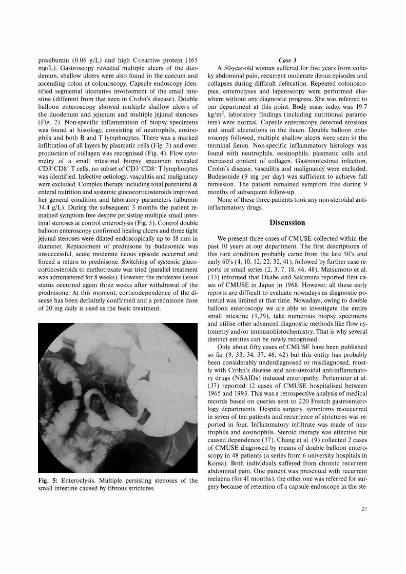
Fig. 2: Double balloon enteroscopy. A large but shallow ul-cer is seen in front of tight fibrous stricture of the distal je-junum.
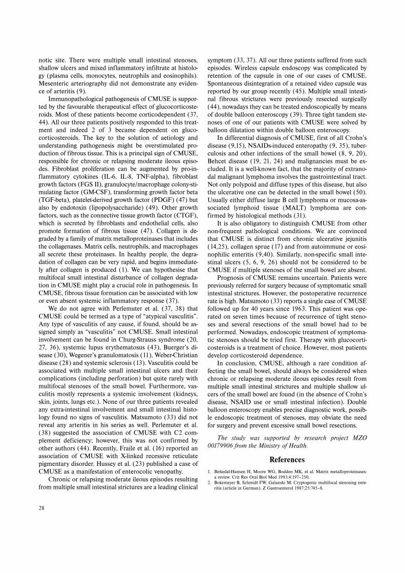
Fig. 3: Prevailing infiltration with plasmatic cells was iden-tified in all layers of the small intestine. Immunohistoche-mistry, anti-CD138 staining, magnification 100x.
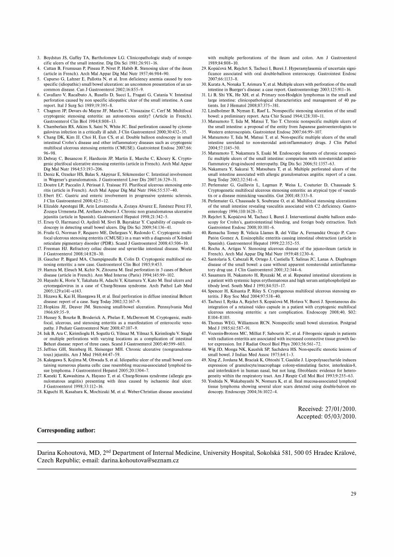
Fig. 4: Increased content of collagen was found in intersti-tial tissue of the small bowel. Small intestinal epithelium,impaired by extensive infiltration with plasmatic cells andlymphocytes. Optical histology, haematoxilin-eosin stai-ning, magnification 40x.
27
prealbumin (0.06 g/L) and high C-reactive protein (165mg/L). Gastroscopy revealed multiple ulcers of the duo-denum, shallow ulcers were also found in the caecum andascending colon at colonoscopy. Capsule endoscopy iden-tified segmental ulcerative involvement of the small inte-stine (different from that seen in Crohn’s disease). Doubleballoon enteroscopy showed multiple shallow ulcers ofthe duodenum and jejunum and multiple jejunal stenoses(Fig. 2). Non-specific inflammation of biopsy specimenswas found at histology, consisting of neutrophils, eosino-phils and both B and T lymphocytes. There was a markedinfiltration of all layers by plasmatic cells (Fig. 3) and over-production of collagen was recognised (Fig. 4). Flow cyto-metry of a small intestinal biopsy specimen revealedCD3+CD8+ T cells, no subset of CD3+CD8– T lymphocyteswas identified. Infective aetiology, vasculitis and malignancywere excluded. Complex therapy including total parenteral &enteral nutrition and systemic glucocorticosteroids improvedher general condition and laboratory parameters (albumin34.4 g/L). During the subsequent 3 months the patient re-mained symptom free despite persisting multiple small intes-tinal stenoses at control enteroclysis (Fig. 5). Control doubleballoon enteroscopy confirmed healing ulcers and three tightjejunal stenoses were dilated endoscopically up to 18 mm indiameter. Replacement of prednisone by budesonide wasunsuccessful, acute moderate ileous episode occurred andforced a return to prednisone. Switching of systemic gluco-corticosteroids to methotrexate was tried (parallel treatmentwas administered for 8 weeks). However, the moderate ileousstatus occurred again three weeks after withdrawal of theprednisone. At this moment, corticodependence of the di-sease has been definitely confirmed and a prednisone doseof 20 mg daily is used as the basic treatment.
Case 3
A 50-year-old woman suffered for five years from colic-ky abdominal pain, recurrent moderate ileous episodes andcollapses during difficult defecation. Repeated colonosco-pies, enteroclyses and laparoscopy were performed else-where without any diagnostic progress. She was referred toour department at this point. Body mass index was 19.7kg/m2, laboratory findings (including nutritional parame-ters) were normal. Capsule enteroscopy detected erosionsand small ulcerations in the ileum. Double balloon ente-roscopy followed, multiple shallow ulcers were seen in theterminal ileum. Non-specific inflammatory histology wasfound with neutrophils, eosinophils, plasmatic cells andincreased content of collagen. Gastrointestinal infection,Crohn’s disease, vasculitis and malignancy were excluded.Budesonide (9 mg per day) was sufficient to achieve fullremission. The patient remained symptom free during 9months of subsequent follow-up.
None of these three patients took any non-steroidal anti-inflammatory drugs.
Discussion
We present three cases of CMUSE collected within thepast 10 years at our department. The first descriptions ofthis rare condition probably came from the late 50’s andearly 60’s (4, 10, 12, 22, 32, 41), followed by further case re-ports or small series (2, 3, 7, 18, 46, 48). Matsumoto et al.(33) informed that Okabe and Sakimura reported first ca-ses of CMUSE in Japan in 1968. However, all these earlyreports are difficult to evaluate nowadays as diagnostic po-tential was limited at that time. Nowadays, owing to doubleballoon enteroscopy we are able to investigate the entiresmall intestine (9,29), take numerous biopsy specimensand utilise other advanced diagnostic methods like flow cy-tometry and/or immunohistochemistry. That is why severaldistinct entities can be newly recognised.
Only about fifty cases of CMUSE have been publishedso far (9, 33, 34, 37, 46, 42) but this entity has probablybeen considerably underdiagnosed or misdiagnosed, most-ly with Crohn’s disease and non-steroidal anti-inflammato-ry drugs (NSAIDs) induced enteropathy. Perlemuter et al.(37) reported 12 cases of CMUSE hospitalised between1965 and 1993. This was a retrospective analysis of medicalrecords based on queries sent to 220 French gastroentero-logy departments. Despite surgery, symptoms re-occurredin seven of ten patients and recurrence of strictures was re-ported in four. Inflammatory infiltrate was made of neu-trophils and eosinophils. Steroid therapy was effective butcaused dependence (37). Chang et al. (9) collected 2 casesof CMUSE diagnosed by means of double balloon entero-scopy in 48 patients (a series from 6 university hospitals inKorea). Both individuals suffered from chronic recurrentabdominal pain. One patient was presented with recurrentmelaena (for 41 months), the other one was referred for sur-gery because of retention of a capsule endoscope in the ste-
Fig. 5: Enteroclysis. Multiple persisting stenoses of thesmall intestine caused by fibrous strictures.
notic site. There were multiple small intestinal stenoses,shallow ulcers and mixed inflammatory infiltrate at histolo-gy (plasma cells, monocytes, neutrophils and eosinophils).Mesenteric arteriography did not demonstrate any eviden-ce of arteritis (9).
Immunopathological pathogenesis of CMUSE is suppor-ted by the favourable therapeutical effect of glucocorticoste-roids. Most of these patients become corticodependent (37,44). All our three patients positively responded to this treat-ment and indeed 2 of 3 became dependent on gluco-corticosteroids. The key to the solution of aetiology andunderstanding pathogenesis might be overstimulated pro-duction of fibrous tissue. This is a principal sign of CMUSE,responsible for chronic or relapsing moderate ileous episo-des. Fibroblast proliferation can be augmented by pro-in-flammatory cytokines (IL-6, IL-8, TNF-alpha), fibroblastgrowth factors (FGS II), granulocyte/macrophage colony-sti-mulating factor (GM-CSF), transforming growth factor beta(TGF-beta), platelet-derived growth factor (PDGF) (47) butalso by endotoxin (lipopolysaccharide) (49). Other growthfactors, such as the connective tissue growth factor (CTGF),which is secreted by fibroblasts and endothelial cells, alsopromote formation of fibrous tissue (47). Collagen is de-graded by a family of matrix metalloproteinases that includesthe collagenases. Matrix cells, neutrophils, and macrophagesall secrete these proteinases. In healthy people, the degra-dation of collagen can be very rapid, and begins immediate-ly after collagen is produced (1). We can hypothesise thatmultifocal small intestinal disturbance of collagen degrada-tion in CMUSE might play a crucial role in pathogenesis. InCMUSE, fibrous tissue formation can be associated with lowor even absent systemic inflammatory response (37).
We do not agree with Perlemuter et al. (37, 38) thatCMUSE could be termed as a type of “atypical vasculitis”.Any type of vasculitis of any cause, if found, should be as-signed simply as “vasculitis” not CMUSE. Small intestinalinvolvement can be found in Churg-Strauss syndrome (20,27, 36), systemic lupus erythematosus (43), Buerger’s di-sease (30), Wegener’s granulomatosis (11), Weber-Christiandisease (28) and systemic sclerosis (13). Vasculitis could beassociated with multiple small intestinal ulcers and theircomplications (including perforation) but quite rarely withmultifocal stenoses of the small bowel. Furthermore, vas-culitis mostly represents a systemic involvement (kidneys,skin, joints, lungs etc.). None of our three patients revealedany extra-intestinal involvement and small intestinal histo-logy found no signs of vasculitis. Matsumoto (33) did notreveal any arteritis in his series as well. Perlemuter et al.(38) suggested the association of CMUSE with C2 com-plement deficiency; however, this was not confirmed byother authors (44). Recently, Fraile et al. (16) reported anassociation of CMUSE with X-linked recessive reticulatepigmentary disorder. Hussey et al. (23) published a case ofCMUSE as a manifestation of enterocolic venopathy.
Chronic or relapsing moderate ileous episodes resultingfrom multiple small intestinal strictures are a leading clinical
symptom (33, 37). All our three patients suffered from suchepisodes. Wireless capsule endoscopy was complicated byretention of the capsule in one of our cases of CMUSE.Spontaneous disintegration of a retained video capsule wasreported by our group recently (45). Multiple small intesti-nal fibrous strictures were previously resected surgically(44), nowadays they can be treated endoscopically by meansof double balloon enteroscopy (39). Three tight tandem ste-noses of one of our patients with CMUSE were solved byballoon dilatation within double balloon enteroscopy.
In differential diagnosis of CMUSE, first of all Crohn’sdisease (9,15), NSAIDs-induced enteropathy (9, 35), tuber-culosis and other infections of the small bowel (8, 9, 20),Behcet disease (19, 21, 24) and malignancies must be ex-cluded. It is a well-known fact, that the majority of extrano-dal malignant lymphoma involves the gastrointestinal tract.Not only polypoid and diffuse types of this disease, but alsothe ulcerative one can be detected in the small bowel (50).Usually either diffuse large B cell lymphoma or mucosa-as-sociated lymphoid tissue (MALT) lymphoma are con-firmed by histological methods (31).
It is also obligatory to distinguish CMUSE from othernon-frequent pathological conditions. We are convincedthat CMUSE is distinct from chronic ulcerative jejunitis(14,25), collagen sprue (17) and from autoimmune or eosi-nophilic enteritis (9,40). Similarly, non-specific small inte-stinal ulcers (5, 6, 9, 26) should not be considered to beCMUSE if multiple stenoses of the small bowel are absent.
Prognosis of CMUSE remains uncertain. Patients werepreviously referred for surgery because of symptomatic smallintestinal strictures. However, the postoperative recurrencerate is high. Matsumoto (33) reports a single case of CMUSEfollowed up for 40 years since 1963. This patient was ope-rated on seven times because of recurrence of tight steno-ses and several resections of the small bowel had to beperformed. Nowadays, endoscopic treatment of symptoma-tic stenoses should be tried first. Therapy with glucocorti-costeroids is a treatment of choice. However, most patientsdevelop corticosteroid dependence.
In conclusion, CMUSE, although a rare condition af-fecting the small bowel, should always be considered whenchronic or relapsing moderate ileous episodes result frommultiple small intestinal strictures and multiple shallow ul-cers of the small bowel are found (in the absence of Crohn’sdisease, NSAID use or small intestinal infection). Doubleballoon enteroscopy enables precise diagnostic work, possib-le endoscopic treatment of stenoses, may obviate the needfor surgery and prevent excessive small bowel resections.
The study was supported by research project MZO00179906 from the Ministry of Health.
References1. Birkedal-Hansen H, Moore WG, Bodden MK, et al. Matrix metalloproteinases:
a review. Crit Rev Oral Biol Med 1993;4:197–250.2. Bokemeyer B, Schmidt FW, Galanski M. Cryptogenic multifocal stenosing ente-
ritis (article in German). Z Gastroenterol 1987;25:745–8.
28
29
3. Boydstun JS, Gaffey TA, Bartholomew LG. Clinicopathologic study of nonspe-cific ulcers of the small intestine. Dig Dis Sci 1981;26:911–16.
4. Cattan B, Frumusan P, Pineau P, Nivet P, Habib R. Stenosing ulcer of the ileum(article in French). Arch Mal Appar Dig Mal Nutr 1957;46:984–90.
5. Capurso G, Lahner E, Pallotta N, et al. Iron deficiency anemia caused by non-specific (idiopathic) small bowel ulceration: an uncommon presentation of an un-common disease. Can J Gastroenterol 2002;16:855–9.
6. Cavallaro V, Racalbuto A, Rusello D, Succi L, Fragati G, Catania V. Intestinalperforation caused by non specific idiopathic ulcer of the small intestine. A casereport. Ital J Surg Sci 1989;19:395–8.
7. Chagnon JP, Devars du Mayne JF, Marche C, Vissuzaine C, Cerf M. Multifocalcryptogenic stenosing enteritis: an autonomous entity? (Article in French).Gastroenterol Clin Biol 1984;8:808–13.
8. Chamberlain RS, Atkins S, Saini N, White JC. Ileal perforation caused by cytome-galovirus infection in a critically ill adult. J Clin Gastroenterol 2000;30:432–35.
9. Chang DK, Kim JJ, Choi H, Eun CS, et al. Double balloon endoscopy in smallintestinal Crohn’s disease and other inflammatory diseases such as cryptogenicmultifocal ulcerous stenosing enteritis (CMUSE). Gastrointest Endosc 2007;66:96–98.
10. Debray C, Besancon F, Harduoin JP, Martin E, Marche C, Khoury K. Crypto-genic plurifocal ulcerative stenosing enteritis (article in French). Arch Mal ApparDig Mal Nutr 1964;53:193–206.
11. Deniz K, Ozseker HS, Balas S, Akpýnar E, Sökmensüer C. Intestinal involvementin Wegener’s granulomatosis. J Gastroenterol Liver Dis 2007;16:329–31.
12. Doutre LP, Paccalin J, Périssat J, Traissac FJ. Plurifocal ulcerous stenosing ente-ritis (article in French). Arch Mal Appar Dig Mal Nutr 1966;55:537–40.
13. Ebert EC. Gastric and enteric involvement in progressive systemic sclerosis.J Clin Gastroenterol 2008;42:5–12.
14. Elizalde Apestegui IR, Arín Letamendía A, Zozaya Alvarez E, Jiménez Pérez FJ,Zozaya Urmeneta JM, Arellano Aburto J. Chronic non granulomatous ulcerativejejunitis (article in Spanish). Gastroenterol Hepatol 1998;21:342–5.
15. Ersoy O, Harmanci O, Aydinli M, Sivri B, Bayraktar Y. Capability of capsule en-doscopy in detecting small bowel ulcers. Dig Dis Sci 2009;54:136–41.
16. Fraile G, Norman F, Requero ME, Defargues V, Redondo C. Cryptogenic multi-focal ulcerous stenosing enteritis (CMUSE) in a man with a diagnosis of X-linkedreticulate pigmentary disorder (PDR). Scand J Gastroenterol 2008;43:506–10.
17. Freeman HJ. Refractory celiac disease and sprue-like intestinal disease. WorldJ Gastroenterol 2008;14:828–30.
18. Gaucher P, Bigard MA, Champigneulle B, Colin D. Cryptogenic multifocal ste-nosing enteritis: a new case. Gastroenterol Clin Biol 1985;9:453.
19. Hamza M, Eleuch M, Kchir N, Zitouma M. Ileal perforation in 3 cases of Behcetdisease (article in French). Ann Med Interne (Paris) 1994;145:99–102.
20. Hayashi K, Horie Y, Takahata H, Adachi Y, Kitamura Y, Kato M. Ileal ulcers andcytomegalovirus in a case of Churg-Strauss syndrome. Arch Pathol Lab Med2005;129:e141–e143.
21. Hizawa K, Kai H, Hasegawa H, et al. Ileal perforation in diffuse intestinal Behcetdisease: report of a case. Surg Today 2002;32:167–9.
22. Hopkins JE, Deaver JM. Stenosing small-bowel ulceration. Pennsylvania Med1966;69:35–9.
23. Hussey S, Bourke B, Broderick A, Phelan E, McDermott M. Cryptogenic, multi-focal, ulcerous, and stenosing enteritis as a manifestation of enterocolic veno-pathy. J Pediatr Gastroenterol Nutr 2008;47:107–9.
24. Isik B, Ara C, Kirimlioglu H, Sogutlu G, Yilmaz M, Yilmaz S, Kirimlioglu V. Singleor multiple perforations with varying locations as a complication of intestinalBehcet disease: report of three cases. Scand J Gastroenterol 2005;40:599–603.
25. Jeffries GH, Steinberg H, Sleisenger MH. Chronic ulcerative (nongranuloma-tous) jejunitis. Am J Med 1968;44:47–59.
26. Kakegawa S, Kojima M, Ohwada S, et al. Idiopathic ulcer of the small bowel con-taining numerous plasma cells: case resembling mucosa-associated lymphoid tis-sue lymphoma. J Gastroenterol Hepatol 2005;20:1304–7.
27. Kaneki T, Kawashima A, Hayano T, et al. Churg-Strauss syndrome (allergic gra-nulomatous angiitis) presenting with ileus caused by ischaemic ileal ulcer.J Gastroenterol 1998;33:112–16.
28. Kiguchi H, Kasahara K, Mochizuki M, et al. Weber-Christian disease associated
with multiple perfeorations of the ileum and colon. Am J Gastroenterol1989;84:808–10.
29. Kopáčová M, Rejchrt S, Tachecí I, Bureš J. Hyperamylasemia of uncertain signi-ficance associated with oral double-balloon enteroscopy. Gastrointest Endosc2007;66:1133–8.
30. Kurata A, Nonaka T, Arimura Y, et al. Multiple ulcers with perforation of the smallintestine in Buerger’s disease: a case report. Gastroenterology 2003;125:911–16.
31. Li B, Shi YK, He XH, et al. Primary non-Hodgkin lymphomas in the small andlarge intestine: clinicopathological characteristics and management of 40 pa-tients. Int J Hematol 2008;87:375–381.
32. Lindholmer B, Nyman E, Raef L. Nonspecific stenosing ulceration of the smallbowel: a preliminary report. Acta Chir Scand 1964;128:310–11.
33. Matsumoto T, Iida M, Matsui T, Yao T. Chronic nonspecific multiple ulcers ofthe small intestine: a proposal of the entity from Japanese gastroenterologists toWestern enteroscopists. Gastrointest Endosc 2007;66:99–107.
34. Matsumoto T, Iida M, Matsui T, et al. Non-specific multiple ulcers of the smallintestine unrelated to non-steroidal anti-inflammatory drugs. J Clin Pathol2004;57:1145–50.
35. Matsumoto T, Nakamura S, Esaki M. Endoscopic features of chronic nonspeci-fic multiple ulcers of the small intestine: comparison with non-steroidal anti-in-flammatory drug-induced enteropathy. Dig Dis Sci 2006;51:1357–63.
36. Nakamura Y, Sakurai Y, Matsubara T, et al. Multiple perforated ulcers of thesmall intestine associated with allergic granulomatous angiitis: report of a case.Surg Today 2002;32:541–6.
37. Perlemuter G, Guillevin L, Legman P, Weiss L, Couturier D, Chaussade S.Cryptogenetic multifocal ulcerous stenosing enteritis: an atypical type of vasculi-tis or a disease mimicking vasculitis. Gut 2001;48:333–8.
38. Perlemuter G, Chaussade S, Soubrane O, et al. Multifocal stenosing ulcerationsof the small intestine revealing vasculitis associated with C2 deficiency. Gastro-enterology 1996;110:1628–32.
39. Rejchrt S, Kopáčová M, Tachecí I, Bureš J. Interventional double balloon endo-scopy for Crohn’s, gastrointestinal bleeding, and foreign body extraction. TechGastrointest Endosc 2008;10:101–6.
40. Remacha Tomey B, Velicia Llames B, del Villar A, Fernandéz Orcajo P, Caro-Patón Gomez A. Eosinophilic enteritis causing intestinal obstruction (article inSpanish). Gastroenterol Hepatol 1999;22:352–55.
41. Rocha A, Artigas V. Stenosing ulcerous disease of the jejuno-ileum (article inFrench). Arch Mal Appar Dig Mal Nutr 1959;48:1230–6.
42. Santolaria S, Cabezali R, Ortego J, Castiella T, Salinas JC, Lanas A. Diaphragmdisease of the small bowel: a case without apparent nonsteroidal antiinflamma-tory drug use. J Clin Gastroenterol 2001;32:344–6.
43. Sasamura H, Nakamoto H, Ryuzaki M, et al. Repeated intestinal ulcerations ina patient with systemic lupus erythematosus and high serum antiphospholipid an-tibody level. South Med J 1991;84:515–17.
44. Spencer H, Kitsanta P, Riley S. Cryptogenous multifocal ulcerous stenosing en-teritis. J Roy Soc Med 2004;97:538–40.
45. Tachecí I, Ryška A, Rejchrt S, Kopáčová M, Hořava V, Bureš J. Spontaneous dis-integration of a retained video capsule in a patient with cryptogenic multifocalulcerous stenosing enteritis: a rare complication. Endoscopy 2008;40, S02:E104–E105.
46. Thomas WEG, Williamson RCN. Nonspecific small bowel ulceration. PostgradMed J 1985;61:587–91.
47. Vozenin-Brotons MC, Milliat F, Sabourin JC, et al. Fibrogenic signals in patientswith radiation enteritis are associated with increased connective tissue growth fac-tor expression. Int J Radiat Oncol Biol Phys 2003;56:561–72.
48. Wig JD, Monga NK, Kaushik SP, Sachdeva HS. Non-specific stenotic lesions ofsmall bowel. J Indian Med Assoc 1975;64:1–3.
49. Xing Z, Jordana M, Braciak K, Ohtoshi T, Gauldie J. Lipopolysaccharide inducesexpression of granulocyte/macrophage colony-stimulating factor, interleukin-8,and interleukin-6 in human nasal, but not lung, fibroblasts: evidence for hetero-geneity within the respiratory tract. Am J Respir Cell Mol Biol 1993;9:255–63.
50. Yoshida N, Wakabayashi N, Nomura K, et al. Ileal mucosa-associated lymphoidtissue lymphoma showing several ulcer scars detected using double-baloon en-doscopy. Endoscopy 2004;36:1022–4.
Corresponding author:
Darina Kohoutová, MD, 2nd Department of Internal Medicine, University Hospital, Sokolská 581, 500 05 Hradec Králové,Czech Republic; e-mail: [email protected]
Received: 27/01/2010.Accepted: 05/03/2010.