setdb1: a novel kap-1-associated histone h3, lysine 9...
TRANSCRIPT
SETDB1: a novel KAP-1-associatedhistone H3, lysine 9-specificmethyltransferase that contributes toHP1-mediated silencing of euchromaticgenes by KRAB zinc-finger proteinsDavid C. Schultz,1,2 Kasirajan Ayyanathan, Dmitri Negorev, Gerd G. Maul, and Frank J. Rauscher III2
The Wistar Institute, Philadelphia, Pennsylvania 19104, USA
Posttranslational modification of histones has emerged as a key regulatory signal in eukaryotic geneexpression. Recent genetic and biochemical studies link H3-lysine 9 (H3-K9) methylation to HP1-mediatedheterochromatin formation and gene silencing. However, the mechanisms that target and coordinate theseactivities to specific genes is poorly understood. Here we report that the KAP-1 corepressor for the KRAB-ZFPsuperfamily of transcriptional silencers binds to SETDB1, a novel SET domain protein with histoneH3-K9-specific methyltransferase activity. Although acetylation and phosphorylation of the H3 N-terminal tailprofoundly affect the efficiency of H3-K9 methylation by SETDB1, we found that methylation of H3-K4 doesnot affect SETDB1-mediated methylation of H3-K9. In vitro methylation of the N-terminal tail of histone H3by SETDB1 is sufficient to enhance the binding of HP1 proteins, which requires both an intact chromodomainand chromoshadow domain. Indirect immunofluoresence staining of interphase nuclei localized SETDB1predominantly in euchromatic regions that overlap with HP1 staining in nonpericentromeric regions ofchromatin. Moreover, KAP-1, SETDB1, H3-MeK9, and HP1 are enriched at promoter sequences of aeuchromatic gene silenced by the KRAB–KAP-1 repression system. Thus, KAP-1 is a molecular scaffold that istargeted by KRAB-ZFPs to specific loci and coordinates both histone methylation and the deposition of HP1proteins to silence gene expression.
[Key Words: Histone methylation; SET domain; chromatin; KRAB domain]
Received December 27, 2001; revised version accepted February 21, 2002.
Macromolecular protein complexes containing enzymat-ic activities that modify the N-terminal tails of the corehistones have emerged as key regulators of gene expres-sion in eukaryotes. The constellation of histone modifi-cations, including acetylation, phosphorylation, ubiqui-nation, and methylation, create both synergistic and an-tagonistic signals that correlate with the transcriptionalactivity of a gene. This emerging histone code is hypoth-esized to create an architecture in chromatin that is rec-ognized by nonhistone chromosomal proteins, whichthen effect the dynamic transition between transcrip-tionally active versus transcriptionally silent chromatindomains (Jenuwein and Allis 2001). Moreover, the com-
binatorial nature of these histone modifications and thechromatin-associated proteins that recognize these sig-nals may represent an epigenetic marking system re-sponsible for setting and maintaining heritable programsof gene expression during cellular differentiation and or-ganism development.
The role of histone acetylation and phosphorylation inregulation of transcription has been extensively charac-terized (Cheung et al. 2000; Strahl and Allis 2000; Berger2001). Although it is well established that arginine andlysine methylation of histones occurs in vivo, the func-tion of these specific modifications remains to be fullydescribed (Strahl et al. 1999). Similar to the discovery ofhistone acetyltransferases (HATs) and deacetylases(HDACs), the role of histone methylation in the regula-tion of chromatin structure and gene expression has beengreatly facilitated by the identification of the responsibleenzymes. The discovery that the nuclear receptor co-activator associated protein CARM1 is an H3-specificarginine methyltransferase and that the mammalian
1Present address: Department of Pharmacology, Case Western ReserveUniversity, 10900 Euclid Avenue, Cleveland, OH 44106-4965, USA.2Corresponding authors.E-MAIL [email protected]; FAX (216) 368-3395.E-MAIL [email protected]; FAX (215) 898-3929.Article and publication are at http://www.genesdev.org/cgi/doi/10.1101/gad.973302.
GENES & DEVELOPMENT 16:919–932 © 2002 by Cold Spring Harbor Laboratory Press ISSN 0890-9369/02 $5.00; www.genesdev.org 919
Cold Spring Harbor Laboratory Press on August 29, 2018 - Published by genesdev.cshlp.orgDownloaded from

homologs of the Drosophila melanogaster heterochro-matin protein Su(var)3-9 are H3-specific lysine methyl-transferases significantly supported the involvementof histone methylation in gene regulation (Chen et al.1999; Rea et al. 2000). In the later case, methylationwas highly selective for Lys 9, with the methyltrans-ferase function mapping to the evolutionarily con-served SET domain homology (Rea et al. 2000). TheH3-K9 methylation (H3-MeK9) mark establishes ahigh-affinity binding site for the recruitment of theHP1 family of heterochromatin proteins through itschromodomain (Jacobs et al. 2001; Jacobs and Khorasan-izadeh 2002). Furthermore, genetic experiments link thelocalization of Swi6 and HP1 at condensed chromatinsequences to the histone methyltransferase activity ofClr4 and SUVAR39H1, respectively (Bannister et al.2001; Lachner et al. 2001; Nakayama et al. 2001). Thus,substantial genetic, biochemical, and cytological evi-dence links the selective methylation of histone H3-K9and the deposition of HP1 proteins to chromatin se-quences whose transcription is epigenetically silenced.Additional studies have shown that the H3-MeK9 epit-ope globally distinguishes transcriptionally silent fromactive chromatin domains in vivo, and methylation ofhistone H3 Lys 9 represents an early molecular mark onthe X chromosome during X inactivation (Heard et al.2001; Litt et al. 2001; Noma et al. 2001; Boggs et al.2002). However, the mechanisms that target H3-K9methylation and coordinate HP1 deposition to specificcis regulatory sequences in vivo remain to be fully de-fined.
A prime candidate for a molecule that could coordi-nate these activities is the KAP-1 corepressor. Thisprotein serves as a universal, obligatory corepressorfor the >220 KRAB domain zinc-finger proteins (ZFPs)that are encoded by the human genome (Friedmanet al. 1996). Each KRAB-ZFP contains an N-terminal75-amino-acid KRAB box that binds directly to KAP-1,and a C-terminal array of C2H2 zinc fingers, whichmediate sequence-specific DNA binding (Mark et al.1999). The tripartite RBCC region of KAP-1 functionsas an integrated structural unit that is necessary andsufficient for oligomerization and KRAB binding (Fig.1A; Peng et al. 2000a,b). The PHD finger and bromo-domain of KAP-1 form a cooperative transcriptional re-pression unit that recruits the NuRD HDAC complex(Capili et al. 2001; Schultz et al. 2001). A separate repres-sion domain in KAP-1 containing a core PxVxL motifbinds directly to the chromoshadow domain of the HP1protein family (Ryan et al. 1999; Lechner et al. 2000).From these data we postulate that KAP-1 functions as ascaffold that, in turn, coordinates the activities of largemacromolecular complexes that modify chromatinstructure to silence gene expression. Because the HP1chromodomain can bind to the methylated Lys 9 of his-tone H3, we reasoned that KAP-1 repression complexesin vivo might contain histone H3-K9 methyltransferaseactivity, which would function to coordinate HP1-medi-ated repression of transcription at KRAB-ZFP targetgenes.
Results
KAP-1 associates with a novel SET-domainprotein, SETDB1
To identify effectors of KAP-1-directed transcriptionalrepression, we used the PHD finger and bromodomain ofKAP-1 as bait in a two-hybrid screen. We previously re-ported the identification of a novel isoform of the Mi-2�subunit of the NuRD histone deacetylase complex thatbound to this bipartite repression domain (Schultz et al.2001). Here we report that KAP-1 specifically associatedwith two independent overlapping amino acid sequences(KIP21 and KIP41) that are encoded by the putative his-tone methyltransferase (HMTase) gene SETDB1 (Fig. 1B;Harte et al. 1999). As illustrated in Figure 1C, mutationsdeleterious to the repression activity of the KAP-1 PHDfinger and bromodomain significantly impaired the as-sociation between KAP-1 and either SETDB1 or Mi-2�(Schultz et al. 2001). Note that some of the mutationsdifferentially affect Mi-2� and SETDB1 binding, whichraises the possibility that Mi-2� and SETDB1 may bindto different surfaces of the KAP-1 protein. To confirmthe association between KAP-1 and SETDB1 in vivo, wegenerated a full-length Flag-epitope-tagged expressionvector and immunopurified SETDB1 from transfectedHEK293 cells. The spectrum of polypeptides was sub-jected to MS/MS peptide analyses, which definitivelyidentified KAP-1 as a nonstoichiometric, associatedpolypeptide (Fig. 1D,E). Eleven KAP-1 polypeptides wereidentified spanning a significant portion of the KAP-1open reading frame (amino acids 239–790). From thesedata we conclude that the KAP-1 corepressor interactswith the SETDB1 protein in vivo.
SETDB1 is a novel histone H3-specificmethyltransferase
The primary amino acid sequence of SETDB1 revealedseveral interesting signature motifs including a CpG-DNA methyl binding domain of the MeCP2 family, andhomology to the SET (SuVar3-9, Enhancer of Zeste, Tri-thorax) domain (Fig. 2A). Interestingly, the SET-domainhomology of SETDB1 is interrupted by a 347-amino-acidinsertion to create a bifurcated domain (Harte et al.1999). This unique insertion is evolutionarily conservedfrom the human protein to lower eukaryotes, includingCaenorhabditis elegans and D. melanogaster, suggestingthat the SET domain may possess functionally separabledomains. It has been previously shown that the SET-domain homology of SUV39H1 and the two adjacent cys-teine-rich regions (pre-SET and post-SET) possess intrin-sic histone methyltransferase (HMTase) activity that isdependent on the integrity of all three domains (Rea etal. 2000). To test whether SETDB1 possessed intrinsicHMTase activity, we expressed and purified two differ-ent recombinant GST–SETDB1 fusion proteins that en-code the entire putative catalytic domain (amino acids585–1291 and 661–1291, respectively). Unlike recombi-nant PRMT1, SUV39H1 and G9a proteins, the recombi-
Schultz et al.
920 GENES & DEVELOPMENT
Cold Spring Harbor Laboratory Press on August 29, 2018 - Published by genesdev.cshlp.orgDownloaded from

nant SETDB1 proteins failed to show any appreciablemethylation of core histones (Fig. 2B). However, SETDB1that was immunopurified from transiently transfected
HEK293 cells (Fig. 1D) showed a robust histone H3-spe-cific methyltransferase activity for core histones andmononucleosome substrates (Fig. 2C). Identical enzy-
Figure 1. The KAP-1 corepressor interacts with the putative histone methyltransferase SETDB1. (A) Schematic illustration of theKAP-1 corepressor. The oligomerization and KRAB-binding domain map to the RBCC region of KAP-1. The chromoshadow domainof the HP1 family of chromosomal proteins directly binds to a PxVxL motif in KAP-1. The PHD finger and bromodomain of KAP-1form a cooperative repression domain that interacts with Mi-2� and SETDB1. (B) The KAP-1 PHD finger and bromodomain interactwith Mi-2� (KIP54) and SETDB1 (KIP21 and KIP 41). (C) Mutations in the PHD finger and bromodomain that impair transcriptionalrepression by KAP-1 impair the association with SETDB1 (KIP21). (D) Coomassie blue staining of anti-Flag immunopurified SETDB1from transfected HEK293 cells. MS/MS peptide identification, definitively identified 11 overlapping peptides of KAP-1, illustrated atthe right in single-letter amino acid abbreviations. (E) Anti-KAP-1 Western blot of Flag immunoprecipitates from HEK293-transfectednuclear extracts.
KRAB-ZFP targeting of histone methylation
GENES & DEVELOPMENT 921
Cold Spring Harbor Laboratory Press on August 29, 2018 - Published by genesdev.cshlp.orgDownloaded from

Figure 2. SETDB1 is a histone H3-specific methyltransferase. (A) Schematic illustration of the SETDB1 protein. The position of thepre-SET, SET, and post-SET (C) homologies at the C terminus are indicated. The 347-amino-acid insertion in the SET domain isindicated by the gray box. (MBD) A CpG DNA methyl-binding domain. The minimal KAP-1 interaction domain (KID) is defined byamino acids of SETDB1 present in two-hybrid clone KIP21. The region of SETDB1 (amino acids 1–377) used to raise a polyclonalantibody is illustrated. (B) Schematic illustration (top) of recombinant GST-PRMT1, GST-SUV39H1, GST-G9a, and GST-SETDB1histone methyltransferase proteins and enzymatic activities (bottom) of affinity-purified proteins expressed in E. coli. Coomassie stainillustrates the affinity-purified GST–proteins. Autoradiograph shows [3H]methyl-labeled histone H3 and H4 from an in vitro HMTaseassay with each respective methyltransferase. (Bottom panel) A Coomassie stain representing equal amounts of core histone octamersper each reaction, whose identities are labeled respectively. (C) Peptide eluate of anti-Flag immunopurified SETDB1 from transientlytransfected HEK293 cells (Fig. 1D) revealed histone H3-specific methyltransferase activity in an in vitro HMTase assay with either corehistones or chicken erythrocyte mononucleosomes as substrates. Coomassie blue stain shows the loading of histones. Autoradiographshows corresponding [3H]methyl-labeled products. (D) Strategy to affinity-purify enzymatically active SETDB1 expressed in Sf9baculovirus-infected cells. (E) Coomassie stain illustrates Ni2+-NTA and �-Flag M2 affinity-purified SETDB1 from baculovirus-in-fected Sf9 cells. Autoradiograph illustrates [3H]methyl-labeled histone H3. Western blot confirms identity of SETDB1 during thepurification. (Bottom panel) A Coomassie stain representing equal amounts of core histone octamers per each reaction.
Schultz et al.
922 GENES & DEVELOPMENT
Cold Spring Harbor Laboratory Press on August 29, 2018 - Published by genesdev.cshlp.orgDownloaded from
matic activity was observed for a SETDB1 protein ex-pressed and affinity-purified to near homogeneity frombaculovirus-infected Sf9 cell extracts (Fig. 2E). Therefore,we postulate that SETDB1 may require posttranslationalmodification or a small molecular weight cellular cofac-tor(s) to function as a histone methylase.
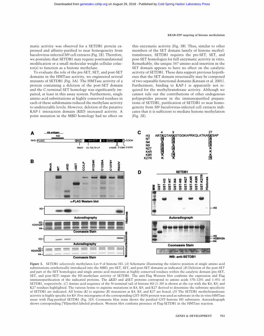
To evaluate the role of the pre-SET, SET, and post-SETdomains in the HMTase activity, we engineered severalmutants of SETDB1 (Fig. 3A). The HMTase activity of aprotein containing a deletion of the post-SET domainand the C-terminal SET homology was significantly im-paired, at least in this assay system. Furthermore, singleamino acid substitutions at highly conserved residues ineach of these subdomains reduced the methylase activityto undetectable levels. However, deletion of the putativeKAP-1 interaction domain (KID) increased activity. Apoint mutation in the MBD homology had no effect on
this enzymatic activity (Fig. 3B). Thus, similar to othermembers of the SET domain family of histone methyl-transferases, SETDB1 requires the pre-SET, SET, andpost-SET homologies for full enzymatic activity in vitro.Remarkably, the unique 347-amino-acid insertion in theSET domain appears to have no effect on the catalyticactivity of SETDB1. These data support previous hypoth-eses that the SET domain structurally may be composedof two separable functional domains (Katsani et al. 2001).Furthermore, binding to KAP-1 is apparently not re-quired for the methyltransferase activity. Although wecannot rule out the contributions of other endogenouspolypeptides present in the immunopurified prepara-tions of SETDB1, purification of SETDB1 to near homo-geneity from Sf9 baculovirus-infected cell extracts indi-cates that it is sufficient to mediate histone methylation(Fig. 2E).
Figure 3. SETDB1 selectively methylates Lys 9 of histone H3. (A) Schematic illustrating the relative position of single amino acidsubstitutions synthetically introduced into the MBD, pre-SET, SET, and post-SET domains as indicated. (B) Deletion of the post-SETand part of the SET homologies and single amino acid mutations at highly conserved residues within the catalytic domain (pre-SET,SET, and post-SET) impair the H3-methylase activity of SETDB1. The anti-Flag Western blot confirms the expression and Flagimmunopurification of the indicated proteins. The �KID and �SET proteins correspond to amino acids 570–1291 and 1–951 ofSETDB1, respectively. (C) Amino acid sequence of the N-terminal tail of histone H3 (1–30) is shown at the top with the K4, K9, andK27 residues highlighted. The various lysine to arginine mutations in K4, K9, and K27 derived to determine the substrate specificityof SETDB1 are indicated. All lysine (K) to arginine (R) mutations at K4, K9, and K27 are boxed. (D) The SETDB1 methyltransferaseactivity is highly specific for K9. Five micrograms of the corresponding GST–H3N protein was used as substrate in the in vitro HMTaseassay with Flag-purified SETDB1 (Fig. 1D). Coomassie blue stain shows the purified GST–histone H3 substrates. Autoradiographshows corresponding [3H]methyl-labeled products. Western blot confirms presence of Flag-SETDB1 in the HMTase reaction.
KRAB-ZFP targeting of histone methylation
GENES & DEVELOPMENT 923
Cold Spring Harbor Laboratory Press on August 29, 2018 - Published by genesdev.cshlp.orgDownloaded from

SETDB1 is a highly selective H3-K9 methyltransferase
To define the site specificity of H3 methylation bySETDB1, we used a series of purified, recombinant GST-histone tail proteins with several lysine-to-arginine sub-stitutions as substrates (Fig. 3C; Tachibana et al. 2001).We found that H3 methylation by SETDB1 is highly se-lective for Lys 9 (Fig. 3D). A substrate (NT) in which K4,K9, and K27 were each mutated to arginine failed to bemethylated. Substrates with double lysine-to-argininemutations (N4, N9, N27) revealed methylation of a sub-strate with only K9 (N9) preserved. A substrate with asingle arginine substitution at K9 (K9R) confirmed thespecificity of SETDB1 for K9. These data confirm thatadditional posttranslational modifications (i.e., acetyla-tion, phosphorylation, methylation) of the substrate arenot required for H3-K9 methylation by SETDB1. More-over, when K9 was mutated to arginine (K9R), SETDB1did not change its specificity to K27, despite the fact thatthis residue lies in a strikingly similar amino acid se-quence (TKxxARKS) as K9.
The histone code and H3-K9 methylation by SETDB1
We next determined whether preexisting modificationsto histone tails could influence the ability of SETDB1 tomethylate its substrate. The core histones isolated fromcalf thymus were pretreated with catalytic amounts of ahomogeneously pure preparation of the histone deacety-lase complex NuRD (data not shown). We then evaluatedthe efficiency of SETDB1 to methylate this deacetylatedcore histone octamer substrate. As illustrated in Figure4A, pretreatment of the histone substrate with the puri-fied NuRD complex resulted in an approximately two-
fold enhancement in methylation, which was in agree-ment with a global decrease in H3 acetylation, suggest-ing that the site of methylation is either naturallyacetylated, or that global acetylation of H3 interfereswith the enzyme’s recognition of the substrate.
To define which posttranslational modifications (i.e.,acetylation, methylation, and phosphorylation) of his-tone H3 affect the SETDB1 methylase activity, we testedthe activity of SETDB1 against a panel of peptide sub-strates possessing either an individual or a combinationof modifications (Fig. 4B). Flag-purified SETDB1 robustlymethylated the unmodified H3 substrate, but not an H4peptide. Interestingly, a peptide substrate methylated atK4 had no apparent effect on this activity. As expected,any modification (methylation or acetylation) of H3-K9inhibited SETDB1-mediated methylation. Furthermore,phosphorylation of S10 or acetylation of K14 also dra-matically inhibited the methylation of the substrate.These observations are similar to that previously ob-served for the related K9-specific histone H3 methyl-transferase SUV39H1, indicating that these proteinslikely recognize the substrate in a similar fashion andpossess a similar catalytic mechanism. We thereforeconclude that in vivo the ability of SETDB1 to methylatehistones within a target locus will likely require coordi-nation with deacetylase complexes and putative histonephosphatases.
H3-K9 methylation by SETDB1 enhances HP1 binding
Because HP1 proteins bind methylated K9 histone pep-tides, we tested whether SETDB1 could stimulate HP1binding to the N-terminal tails of histone H3. As illus-trated in Figure 5, methylation of the GST-H3 and GST-
Figure 4. Dissecting the histone code and methylation by SETDB1. (A) Pretreatment of a core histone substrate with a homogenouslypure histone deacetylase complex, NuRD, enhanced methylation of histone H3 in vitro, concomitantly with deacetylation of histoneH3 (anti-AcH3 Western blot). Coomassie blue stain shows equal loading of histone proteins. Autoradiograph shows corresponding[3H]methyl-labeled products. Anti-SETDB1 and anti-HDAC2 Western blots show the presence of SETDB1 and HDACs in the corre-sponding HMTase reactions. (B) Effect of histone modifications on the enzymatic activity of SETDB1. One microgram of unmodifiedor acetylated (K9-Ac, K14-Ac, K9,K14-Ac), phosphorylated (S10-phos), or methylated (K4-diMe, K9-diMe) peptides corresponding to theN-terminal tail of histone H3 and H4 were used as substrates in the in vitro methylation assay with Flag-purified SETDB1. Methyl-ation was quantified via a filter binding assay and represented as raw counts per minute (C.P.M.) incorporated.
Schultz et al.
924 GENES & DEVELOPMENT
Cold Spring Harbor Laboratory Press on August 29, 2018 - Published by genesdev.cshlp.orgDownloaded from

N9 substrates by SETDB1 significantly enhanced the ef-ficiency of HP1� binding to the N-terminal tail of his-tone H3. This binding activity was abolished by amutation in the chromodomain (V21M) of HP1�. Fur-thermore, a mutation in the chromoshadow domain(I165K) that affects the dimerization of HP1� signifi-cantly impaired the HP1:histone interaction (Lechner etal. 2000). From this series of data we conclude thatSETDB1 is a highly selective histone H3-K9 methylasefully capable of stimulating the binding of HP1 proteinsto histone H3.
Endogenous SETDB1 is a euchromaticH3-specific methyltransferase
To confirm that endogenous SETDB1 possessed methyl-transferase activity we produced a polyclonal antibodythat specifically recognizes the protein (Fig. 2A). Westernblot analysis of phosphocellulose-fractionated solubleHeLa nuclear extract revealed that SETDB1 primarilyelutes in the 0.1 M and 0.3 M KCl elutions, whereasSUV39H1 is present in the 0.5 M and 1.0 M KCl elutions.The fractions containing either protein showed robustH3 methylase activity (Fig. 6A). Antibodies againstSETDB1 efficiently immunodepleted nearly all the his-tone H3 methylase activity from the 0.1 M P11 extract
without affecting the H4 activity (Fig. 6B). Moreover, thepellet of the SETDB1 immunoprecipitate retained astrong histone H3 activity that is comparable to that ofFlag-purified SETDB1 (Fig. 1D). These observationsstrongly suggest that endogenous SETDB1 represents anabundant histone H3 methyltransferase. Indirect immu-nofluoresence staining of asynchronous populations ofNIH/3T3 cells revealed that SETDB1 is localized pre-dominantly in euchromatic regions of interphase nucleiand excluded from nucleoli and islands of condensedchromatin, as determined by Hoechst stain and immu-nostaining with a monoclonal antibody to HP1� (Fig.6C). However, there is significant overlap betweenSETDB1 and HP1� in euchromatic regions of thenucleus. Therefore, we propose that SETDB1 functionsindependently of SUV39H1/H2 and is one cellularHMTase responsible for global euchromatic H3-K9methylation maintained in the Suv39h double knockoutmouse (Peters et al. 2001).
SETDB1 enhances H3-K9 methylation and HP1 at anendogenous locus stably silenced by the KRAB–KAP-1repression system
The above results suggest that SETDB1 functions tomethylate histone H3-K9 in euchromatic territories ofthe nucleus to facilitate HP1 deposition. Furthermore,our biochemical data suggest that the KRAB-ZFP–KAP-1repression complex could target H3-K9 methylation ofendogenous gene promoters by SETDB1 facilitating thedeposition of HP1 proteins to silence gene expression. Totest this hypothesis, chromatin immunoprecipitation(ChIP) experiments were done with a cell line that con-tains a stably integrated, euchromatic luciferase trans-gene that is subject to KRAB-mediated repression (Fig.7A). This two-plasmid system is based on a hormone-regulatable DNA-binding KRAB domain fusion (Ayya-nathan et al. 2000) and a TK-luciferase reporter transgeneas its target. The fusion protein is fully capable of form-ing a ternary complex with KAP-1 and HP1 (data notshown). A comprehensive analysis of clonal NIH3T3 celllines that contain these two plasmids has shown thefollowing: (1) The KRAB domain is a strong repressor ofan integrated target gene; (2) repression is relativelyshort-range, and accompanied by localized chromatincompaction in the promoter region and spatial recruit-ment to subnuclear territories enriched in condensedchromatin; (3) the KAP-1 corepressor and HP1�/� pro-teins are physically associated with the repressed gene ina highly localized manner as judged by ChIP assays.Most remarkable, a large fraction of cells from a hor-mone-treated population can be isolated that maintain asilent expression state of the transgene for >40 popula-tion doublings in the absence of hormone, suggestingthat the silenced state is mitotically heritable.
This system provides a model for HP1-dependent si-lencing and variegation of a euchromatic gene expressionin a mammalian cell line (K. Ayyanathan and F.J.Rauscher III, in prep.), and we have used it as describedbelow to evaluate the role of SETDB1 and histone H3
Figure 5. SETDB1 methylation of histone H3-K9 enhancesHP1 binding. In vitro GST-binding assay between HP1� andGST–H3N. GST–H3N substrates were premethylated withFlag-purified SETDB1 (Fig. 1D) and 15 µM S-adenosyl-L-methi-onine (Sigma). 35S-L-methinonine labeled in vitro translatedHP1� proteins were incubated with the methylated GST–H3Nproteins. HP1-histone complexes were eluted by denaturationand resolved on 10% SDS-PAGE gels, and bound HP1� wasvisualized by fluorography. Coomassie blue stain shows the pu-rified, methylated GST–histone H3 substrates. Schematic dia-gram of HP1� to the right illustrates the domain organization(CD, chromodomain; CSD, chromoshadow domain) of this pro-tein family and the relative position of the V21M and I165Kmutations (Lechner et al. 2000).
KRAB-ZFP targeting of histone methylation
GENES & DEVELOPMENT 925
Cold Spring Harbor Laboratory Press on August 29, 2018 - Published by genesdev.cshlp.orgDownloaded from

MeK9 in the stable silencing of the luciferase transgene.We used clonal cell lines that showed either robust ex-pression of luciferase (cl-49) or nearly complete silencingof the luciferase transgene (cl-74; Fig. 7B). In cells con-taining a silenced luciferase transgene (cl-74), the ChIPexperiments indicated that both KAP-1 and SETDB1readily cross-linked to the luciferase transgene and weresignificantly colocalized around the TK promoter regionof the integrated reporter. In contrast, KAP-1 andSETDB1 were undetectable by ChIP analysis at the TKpromoter region in cells (cl-49) showing significant lu-ciferase activity (Fig. 7C). Furthermore, we observedlittle binding of these proteins to the promoter region ofthe linked Zeocin resistance locus, which is nearly 3.0kb downstream of the TK promoter, or at the unlinkedNeomycin resistance gene present in the same cells (Fig.7C). Clonal populations of cells containing only the in-
tegrated luciferase transgene failed to show any localiza-tion of KAP-1 or SETDB1 to the TK promoter region,suggesting that a DNA-bound KRAB repression modulewas required for KAP-1 and SETDB1 recruitment (datanot shown). We next determined whether the localiza-tion of SETBD1 to the TK promoter region enhancedH3-K9 methylation and the recruitment of HP1. As il-lustrated in Figure 7D, a comparison between these twocell lines has revealed that HP1� and its chromatin li-gand, H3-MeK9, are enriched in chromatin containingthe TK promoter sequences in cells with a hormone-induced, stably silenced transgene. These data suggestthat the KAP-1 corepressor functions as a molecular plat-form that coordinates the sequential recruitment of his-tone methyltransferases and the deposition of HP1 at aeuchromatic locus to stably silence gene expression thatis mitotically heritable.
Figure 6. Endogenous SETDB1 represents a major histone H3-specific methyltransferase. (A) Biochemical fractionation of H3-specificmethlytransferases from HeLa nuclear extract. HeLa nuclear extract was fractionated by phosphocellulose (P11) chromatography aspreviously described (Bochar et al. 2000). HMTase activity was monitored by the in vitro methylation assay. Elution of SETDB1 andSUV39H1 from the P11 column was monitored by Western blot analysis. (B) Supernatants of SETDB1 immunodepleted nuclear extractwere devoid of H3 HMTase activity. We incubated 150 µg of the 0.1 M P11 fractionated nuclear extract with protein A-agarose andeither affinity-purified anti-GST or anti-SETDB1 IgG. Supernatants and pellets from these immunoprecipitates were assayed forHMTase activity. Coomassie blue stain shows equal amounts of core histone substrate in each reaction. Autoradiograph showscorresponding [3H]methyl-labeled products. Anti-SETDB1 Western blot shows efficient immunodepletion of SETDB1 from the 0.1 MP11 extract. (C) Indirect immunofluoresence of interphase nuclei of NIH/3T3 cells. Affinity-purified polyclonal SETDB1-specific IgGglobally stained euchromatic nuclear territories of interphase nuclei (FITC) with little overlap in A-T-rich condensed chromatindomains visualized by Hoechst stain and monoclonal HP1� IgG (Texas Red).
Schultz et al.
926 GENES & DEVELOPMENT
Cold Spring Harbor Laboratory Press on August 29, 2018 - Published by genesdev.cshlp.orgDownloaded from

Discussion
The role of histone modifications in the regulation ofchromatin structure and subsequent regulation of eu-karyotic gene expression is rapidly being defined (Strahland Allis 2000; Jenuwein and Allis 2001). The hypothesisthat is emerging from these studies is that the combina-tion of modifications on a histone or perhaps other his-tones within the same nucleosome creates an indexingsystem, which formats chromatin to properly expressthe genetic information of the genome, and facilitatesthe establishment of specialized nuclear structures.
However, key questions in understanding all of thesehistone modifications include the following: (1) How arethey targeted to specific gene regulatory elements? (2)What non-histone chromosomal proteins interpret thishistone code? (3) How is recognition of this code trans-lated into a change in gene activity?
Here we report that the KAP-1 corepressor of theKRAB-ZFP family of sequence-specific transcriptionalrepressors associates with SETDB1, a histone H3 Lys 9methyltransferase, and targets it to gene promoters tran-scriptionally silenced by a model KRAB repressor pro-tein. Mutation of conserved amino acid residues in thecatalytic domain, including the pre-SET, SET, and post-
Figure 7. The KRAB–KAP-1 repression system targets SETDB1 and enhances H3-K9 methylation and HP1 recruitment to promotersof transcriptionally silenced genes. (A) Schematic representation of a two-plasmid system used to create a stably integrated luciferasetransgene in NIH/3T3 cells that is regulated by a heterologous KRAB repressor protein. Numbered arrow sets represent the relativeposition of PCR primers used for PCR amplification of DNA retained by ChIP. (B) Two single-cell subclones containing the heter-ologous KRAB–PAX3–HBD transcriptional repressor and the integrated luciferase transgene, which is either expressed (cl-49) or stablysilenced (cl-74) following hormone treatment. Luciferase activities were measured in subconfluent populations of cells and reportedas relative light units per milligram of protein. (C) ChIP experiments showing the colocalization of KAP-1 and SETDB1 at the TKpromoter region of the luciferase transgene in the cells where transcription of the luciferase gene has been stably silenced (cl-74).Formaldehyde cross-linked chromatin from cl-49 and cl-74 cells was immunoprecipitated with either affinity-purified KAP-1 orSETDB1 IgG. An equal amount of promoter sequence in cl-49 and cl-74 nucleosomal preparations was determined by PCR from 1%of the input chromatin. PCR-amplified DNA fragments are illustrated in A. cl-2 represents a negative control cell line. (D) ChIPs ofcross-linked chromatin with KAP-1, SETDB1, HP1�, and MeK9 antiserum as in C. Bold numbers below each lane represent quanti-tation of amplified DNA, expressed as percentage of signal intensity for the amplified input DNA.
KRAB-ZFP targeting of histone methylation
GENES & DEVELOPMENT 927
Cold Spring Harbor Laboratory Press on August 29, 2018 - Published by genesdev.cshlp.orgDownloaded from

SET domains, significantly impairs the enzymatic activ-ity of SETDB1. Although acetylation and phosphoryla-tion of the H3 N-terminal tail profoundly affect the ef-ficiency of H3-K9 methylation by SETDB1, we foundthat a substrate methylated at H3-K4 does not affectmethylation of H3-K9 by SETDB1. In vitro methylationof the N-terminal tails of histone H3 by SETDB1 is suf-ficient to enhance the binding of HP1 proteins. Surpris-ingly, SETDB1 localizes exclusively in euchromatic ter-ritories of interphase nuclei and overlaps with HP1 stain-ing in nonpericentromeric regions of chromatin.Furthermore, we show that SETDB1 can be targeted to astably silenced euchromatic gene via the KAP-1 core-pressor. Moreover, recruitment of SETDB1 enhances H3-K9 and HP1 localization to cis-regulatory sequences of agene silenced by the KRAB–KAP-1 repression system.We propose that KRAB-ZFPs bind to its cognate recog-nition sequence and then recruits KAP-1 to form a scaf-fold that coordinates the assembly histone deacetylases,histone methylases, and the deposition of HP1 proteinsto silence gene expression by forming a facultative het-erochromatin environment.
SETDB1 mediates K9 methylation
The role of histone methylation in the emerging histonecode has been revolutionized by the discovery that pro-teins with the highly conserved SET domain function aslysine-specific histone methyltransferases (Rea et al.2000). Analysis of the primary amino acid sequence ofSETDB1 revealed the presence of amino acid moduleshighly homologous to the pre-SET, SET, and post-SETdomains of the SUV39H1 protein family (data notshown). Interestingly, the SET-domain homology ofSETDB1 is interrupted by a 347-amino-acid insertionthat is evolutionarily conserved in homologous proteinsin lower eukaryotes. Although a recombinant SETDB1composed of the minimal catalytic domain (pre-SET,SET, and post-SET domains) was not active in our meth-yltransferase assay, SETDB1 purified from eukaryoticcell extracts showed robust H3-specific HMTase activityusing calf thymus core histones, chicken mononucleo-somes, or H3 N-terminal peptides as substrates. It is notclear why the recombinant SETDB1 proteins are not ac-tive when compared with other histone methyltransfer-ases that are active as recombinant proteins (SUV39H1,G9a, PRMT1). Although we cannot rule out a small-mo-lecular-weight cofactor such as NAD or FAD, we favor ahypothesis that SETDB1 must require posttranslationalmodification(s) for activity over a cellular protein cofac-tor, because SETDB1 expressed in baculovirus-infectedSf9 cells and affinity-purified to near homogeneity wassufficient to show identical enzymatic activity as endog-enous sources of SETDB1. During our affinity purifica-tion of enzymatically active SETDB1, we consistentlyobserved two forms. Moreover, we noticed a tight corre-lation between the slower-migrating protein species bySDS-PAGE/Western blotting and the enzymatic activityof SETDB1 (Fig. 3B). It is possible that the faster-migrat-ing species is a degradation product. However, peptide-
mapping experiments revealed changes in the peptideprofiles between the two species that are consistent withsmall-molecular-weight modifications. In this regard, wefound that SETDB1 is constitutively phosphorylatedin asynchronous populations of cells, a finding consis-tent with other members of the SUV39H1 family (D.C.Schultz and F.J. Rauscher III, unpubl.). Nonetheless, theexact types and extent of posttranslational modificationof SETDB1 will need to be determined to fully assess therole they have in its enzymatic activity.
Mechanism of K9 methylation
Although methylation of histones has long been estab-lished, only recently have the functional consequencesof site-specific methylation in modulation of chromatinstructure and gene transcription begun to be elucidated.The recent discovery that the H3-MeK9 mark estab-lishes a high-affinity ligand for binding the HP1 family ofheterochromatin proteins and targeting its proper sub-cellular location has provided one of the first links be-tween a histone modification and a repressive chromatinenvironment for gene expression (Bannister et al. 2001;Jacobs et al. 2001; Lachner et al. 2001; Nakayama et al.2001; Jacobs and Khorasanizadeh 2002). Here we showthat de novo methylation of recombinant histone H3N-terminal tails by SETDB1 is sufficient to enhance theaffinity of HP1 binding. Moreover, the recruitment ofSETDB1 to a chromatinized locus targeted for repressionby the KRAB–KAP-1 repression system coincides withsignificant enrichment of H3-K9 methylation and HP1deposition. In both the in vitro and in vivo experiments,enhanced HP1 binding was completely dependent onH3-K9 methylation. As expected, HP1 binding to themethylated histone tail in vitro required a functionalchromodomain. Furthermore, we found that optimalHP1 binding also required a functional chromoshadowdomain, as a mutation that disrupts the dimerization ofthe HP1 protein significantly impaired this association.Thus, HP1 binding to methylated H3-K9 is not exclu-sively chromodomain-dependent. The later observationimplies a potential role for any of the chromoshadowdomain binding partners (i.e., KAP-1, CAF150, SP100) instabilizing of the H3-MeK9–HP1 interaction. Addition ofrecombinant KAP-1 did not stimulate or enhance HP1binding to the methylated H3 tail in an in vitro solution-binding assay (D.C. Schultz, unpubl.). One importantquestion that remains to be answered about the HP1–H3-MeK9 interaction is whether the chromodomains ofa single HP1 molecule bind to the methylated H3-K9tails of a single nucleosome or two adjacent nucleo-somes. Because KAP-1 exists as a homotrimer in solu-tion, it is possible that KAP-1 may increase the stoichi-ometry of HP1–H3-MeK9 interactions at a targeted locusby functioning as a nucleosome cross-linking agent.
A possible interplay between H3-K4 and H3-K9methylation
A more global analysis of H3-K9 methylation at specificloci in Schizosaccharomyces pombe and higher eukary-
Schultz et al.
928 GENES & DEVELOPMENT
Cold Spring Harbor Laboratory Press on August 29, 2018 - Published by genesdev.cshlp.orgDownloaded from

otes found this chromatin mark to be preferentially as-sociated with transcriptionally inactive chromatin. Incontrast, histone H3 Lys 4 methylation has been shownto correlate with histone H3 acetylation and chromatinregions permissive to gene transcription. Moreover,these data indicate that H3-K4 and H3-K9 methylationare mutually exclusive at these two specific loci (Heardet al. 2001; Litt et al. 2001; Noma et al. 2001; Boggs et al.2002). Furthermore, H3-K4 methylation has been exclu-sively associated with transcriptionally active macronu-clei, but not inactive micronuclei, of Tetrahymena(Strahl et al. 1999). Interestingly, we found that a sub-strate methylated at H3-K4 had little effect on the abilityof SETDB1 to methylate H3-K9. This observation ap-pears to be counterintuitive with the mutual exclusivehypothesis for these two modifications. Furthermore,this observation is in stark contrast to findings forSUV39H1, whose enzymatic activity for H3-K9 is sig-nificantly reduced by a methylated H3-K4 substrate(Nishioka et al. 2002). One explanation for such differ-ences might be explained by a histone code hypothesis.In this particular case the pattern of histone modifica-tions on the same histone appear to independently regu-late the enzymatic activity of two different enzymes thatmodify the same residue. In this regard, SETDB1 may beinstrumental in maintaining H3-K9 methylation atboundary elements and, thus, prevent the spreading oftranscriptionally active chromatin domains from juxta-posed regions enriched in H3-K4 methylation. Alterna-tively, SETDB1 methylation of H3-K9 may function inconcert with H3-K4 to repress transcription in highlyspecialized chromatin territories or structures. Consis-tent with this hypothesis, H3-K4 methylation by theSaccharomyces cerevisae protein Set1, the predominantH3-K4 methyltransferase, is required for repression ofRNA polymerase II transcription within rDNA, telo-meres, and the silent mating-type locus (Nislow et al.1997; Briggs et al. 2001; Miller et al. 2001; Roguev et al.2001). Characterization of the H3-K4 methylation statusat our engineered transgene and further identification ofendogenous loci regulated by SETDB1 will be required todefine more accurately the role SETDB1 has in regula-tion of chromatin structure containing both H3-K4 andH3-K9 methylation.
Histone methylation and DNA methylation
Interestingly, SETDB1 is a unique member of a subclassof SET proteins that possess a canonical CpG DNAmethyl binding domain (MBD), which other proteins useto bind methylated DNA (Wade and Wolffe 2001). Onemodel would predict that SETDB1 binds directly to hy-poacetylated regions (imprinting centers, inactive Xchromosome, transposons, etc.) of methylated DNA andsubsequently methylates the corresponding histones ofsurrounding nucleosomes. Alternatively, it may be thatH3-K9 methylation by SETDB1 plays a role in gene-spe-cific targeting of DNA methylation, based on a recentgenetic study in Neurospora crassa that has linked main-tenance of genomic DNA methylation to histone H3-K9
methylation (Tamaru and Selker 2001). Preliminary re-sults suggest that modifying agents of DNA methylationcan alter the silenced state of the integrated transgene (K.Ayyanathan and F.J. Rauscher III, unpubl.). The KRAB–KAP-1 repression system may therefore provide a usefultool to dissect the interplay between histone methyl-ation and DNA methylation in establishing epigeneticstates of gene silencing.
Role of KRAB-ZFPs in establishingfacultative heterochromatin
These observations strongly suggest a role for the KRAB-ZFP superfamily of transcriptional repressors in se-quence-specific establishment of gene silencing. KAP-1appears to have the capacity to coordinate the biochemi-cal activities required to induce and maintain the assem-bly of higher-order chromatin structure. We propose thatKRAB-ZFPs selectively bind to cognate cis-regulatory el-ements and recruit the KAP-1 corepressor to the targetedlocus. Because KAP-1 is obligatory for KRAB-mediatedrepression, it appears that the effector molecules of si-lencing are likely from the network of proteins that in-teract with KAP-1. Through our studies, we defineKAP-1 as a molecular scaffold that coordinates at leastfour activities necessary for gene specific silencing: (1)targeting to specific promoters (via the >220 KRAB zinc-finger proteins in the human genome); (2) histonedeacetylation via the NuRD/HDAC complex; (3) H3-K9methylation via SETDB1; and (4) deposition of HP1 pro-tein, which collectively facilitate the nucleation of fac-ultative heterochromatin to silence gene expression. Todate, Rb is the only other corepressor protein that seemsto coordinate similar activities in the repression of E2Ftarget genes (Nielsen et al. 2001). Thus, the KRAB–KAP-1 repression system is one of the best-characterizedmammalian systems for gene-specific silencing of eu-chromatic genes by targeting HP1 proteins. Moreover,the abundance of the KRAB domain zinc-finger proteinsin the human proteome and the potentially diverse arrayof DNA sequences they recognize potentially make thisfamily of gene-specific silencers a master regulator inestablishing epigenetic programs of gene silencing dur-ing cellular differentiation and organism development.
Materials and methods
Plasmids
Full-length human SETDB1 (KIAA0067) was obtained from theKazasu DNA Research Institute. Coding sequences for SETDB1were subcloned NotI/BamHI into pCMV2 (Sigma) to create theCMV-driven Flag-tagged SETDB1 mammalian expression vec-tor. The �KID (amino acids 570–1291) expression construct wascreated by subcloning a HindIII/BamHI fragment into pCMV2.The �SET (amino acids 1–951) expression construct was createdby subcloning a NotI/BglII fragment into pCMV2. Amino acidsubstitutions in SETDB1 (R643V, CC 729, 731 LP, H1224K,C1226A, C1279Y) were created using Quick Change PCR mu-tagenesis strategies (Stratagene). For protein expression in Esch-erichia coli, a 2.2-kb BamHI fragment encoding amino acids
KRAB-ZFP targeting of histone methylation
GENES & DEVELOPMENT 929
Cold Spring Harbor Laboratory Press on August 29, 2018 - Published by genesdev.cshlp.orgDownloaded from

661–1291 of human SETDB1 was subcloned into pGEX-5X-1.Similarly, a 2.6-kb XhoI/SalI fragment encoding amino acids585–1291 was subcloned into pGEX-5X-1. For antigen produc-tion, a 1.4-kb XhoI fragment encoding amino acids 1–377 frompACT-KIP41 was subcloned into pGEX-4T-1. Previously de-scribed PHD finger and bromodomain mutations in KAP-1 weresubcloned into the SmaI site of pBTM116 (Capili et al. 2001;Schultz et al. 2001). The GST–histone H3 and GST–G9a bacte-rial expression plasmids were previously described (Tachibanaet al. 2001). Bacterial protein expression and in vitro GST-bind-ing assays were performed as described previously (Ryan et al.1999; Lechner et al. 2000). Appropriate reading frame fusionsand integrity of flanking sequences for all constructs created byPCR were confirmed by DNA sequence analysis of both strands.
Baculovirus SETDB1
The full-length coding sequence for SETDB1 was subcloned viaEcoRI/BamHI into pAcHLTa (Pharmingen) to create a tandem6His/Flag-tagged SETDB1 baculovirus transfer vector. Recom-binant baculovirus-infected Sf9 cells (mock or SETDB1) wereharvested 48 h postinfection by lysing in RIPA buffer (50 mMTris at pH 7.9, 150 mM NaCl, 0.5% deoxycholate, 0.1% SDS, 1mM PMSF, 10 µg/mL pepstatin, 10 µg/mL aprotonin, 10 µg/mLleupeptin, 1 mM Benzamidine, 50 mM NaF, 10 mM NaOV4).Whole-cell extracts were clarified at 100,000g, and supernatantswere incubated in batch with Ni2+-NTA agarose (QIAGEN),washed with 20 column volumes of RIPA buffer, followed by 10column volumes of BC100 (20 mM Tris-HCl at pH 8.0, 100 mMNaCl, 0.2 mM EDTA, 10% glycerol, 0.2 mM PMSF, 0.2%Tween 20). Bound proteins were step-eluted with BC100 supple-mented with 300 mM imidazole. Ni2+-NTA eluates were im-mediately incubated with anti-Flag M2-conjugated agarose(Sigma) at 4°C for 2 h, washed with 20 column volumes ofBC1000 (1 M NaCl), followed by 10 column volumes of BC100.Bound proteins were eluted twice in batch with 2 column vol-umes of BC100 supplemented with 400 µg/mL Flag M2 peptideat 4°C for 1 h each.
Yeast two-hybrid system
The yeast two-hybrid system as modified by Stan Hollenbergwas used for all yeast experiments. A human oligo(dT)-primedB-cell cDNA library was screened as described previously(Jensen et al. 1998).
Immunoprecipitation
HEK293 cells were transiently transfected with lipofectamine,and nuclear extracts were prepared 36–48 h posttransfection, aspreviously described (Ryan et al. 1999). Then 5–10 mg of nuclearextract adjusted to 100 mM NaCl was incubated with 100 µg ofanti-Flag M2 (Sigma) at 4°C for 2–4 h. Immune complexes werewashed three times with BC500 (20 mM Tris-HCl at pH 8.0, 500mM NaCl, 0.2 mM EDTA, 10% glycerol, 0.2 mM PMSF, 0.2%Tween 20), once with BC100 (20 mM Tris-HCl at pH 8.0, 100mM NaCl, 0.2 mM EDTA, 10% glycerol, 0.2 mM PMSF, 0.2%Tween 20), and eluted with 400 µg/mL Flag M2 peptide. Elutedproteins were resolved on a 4%–12% NuPAGE gel in MOPSrunning buffer (Invitrogen). Proteins were visualized by silverstaining or Western blotting to PVDF as previously described(Ryan et al. 1999). For endogenous SETDB1 immunoprecipita-tion studies, 100 µg of a DEAE-bound, 0.1 M phosphocelluloseelution of HeLa nuclear extract was incubated with 5 µg ofaffinity-purified SETDB1 antibody and 5 µL of protein G-Seph-arose (Pharmacia) at 4°C for 2 h. Bound immune complexes
were washed three times with BC100 and twice with HMTasebuffer prior to assaying for HMTase activity. MS/MS peptideidentification was performed at The Wistar Institute ProteinMicrochemistry Facility, using microcapillary reverse phaseHPLC nanospray tandem mass spectrometry on a Finnigan LCQquadrupole ion trap mass spectrometer.
In vitro histone methyltransferase reactions
In a 40-µL reaction volume, enzyme, 5 µg of core histones(Roche Biochemicals), 2 µg of chicken erythrocyte mononucleo-somes, or 5 µg of GST–H3N, and 500 nCi of S-adenosyl-[3H-methyl]-L-methionine (3H-AdoMet; 72 Ci/mmole; NEN LifeScience Products) were incubated at 37°C for 1 h in 50 mM Tris(pH 8.5), 20 mM KCl, 10 mM MgCl2, 10 mM �-mercaptoetha-nol, and 250 mM sucrose. Reactions were terminated by theaddition of 5× SDS-buffer. Histones were resolved on 4%–12%NuPage gels in MES running buffer and visualized by Coo-massie blue R250 stain. [3H]Methyl labeling was detected byfluorography in 22% PPO solution. Dried gels were exposed toKodak MRX film. Western Blotting was done as previously de-scribed (Ryan et al. 1999).
Immunofluoresence
NIH3T3 cells were grown on glass coverslips in DMEM me-dium containing 10% calf serum and immunostained as previ-ously described (Maul et al. 1998). The murine SETDB1 proteinwas visualized by indirect immunofluoresence with an antigen-purified rabbit polyclonal antibody diluted 1:400. DNA wascounterstained with Hoechst 33258 (Sigma), and coverslipswere mounted with Fluoromount G (Fisher Scientific). Cellswere visualized with an inverted light microscope (Leica).
Chromatin immunoprecipitation
The chromatin immunoprecipitation (ChIP) experiments weredone essentially as previously described with some modifica-tions (Orlando et al. 1997). Cells were cross-linked with 1%formaldehyde at 37°C for 20 min. The cross-linking reactionwas quenched by washing the cells several times with cold TBS(50 mM Tris at pH 8.0, 200 mM NaCl). Cells were scraped intocold TBS supplemented with 5 mM butyric acid. Chromatinwas enriched for by washing the cells once in 20 mM Tris HCl(pH 8.0), 0.25% Triton X-100, 200 mM NaCl, 10 mM EDTA, 0.5mM EGTA, 1 µg/mL aprotonin, leupetitn, pepstatin, 1 mMBenzamidine, 50 mM NaF, 10 mM NaOV3, and 5 mM butyricacid. The cells were centrifuged, resuspended in IP buffer (20mM NaCl, 0.05% DOC, 0.5% Triton X-100, 0.5% NP-40, 200mM NaCl, 5 mM butyrate, and protease inhibitors), and soni-cated to an average fragment size of 300–500 bp. Solubilizedchromatin was clarified by centrifugation at 12,000g, and thesupernatant was preincubated for 2 h with protein A agarosebeads blocked with salmon sperm DNA and BSA. Preclearedchromatin was incubated with 5–10 µg of anti-KAP-1 (Schultzet al. 2001), anti-SETDB1, anti-H3 MeK9 (Upstate Biotechnol-ogy), and anti-HP1� (D.C. Schultz, unpubl.) at 4°C for 12–16 h.Immune complexes were bound to protein A agarose beads at4°C for an additional 2–3 h. The beads were washed four timeswith IP buffer, two times with high salt buffer (IP buffer with 0.4M NaCl), once with LiCl buffer (10 mM Tris at pH 8.0, 250 mMLiCl, 0.5% NP40, 1% Triton X-100, 0.1% DOC, 5 mM EDTA,and protease inhibitors), and two times with TE. The DNA–protein complexes were eluted from the protein A beads with 50mM Tris (pH 8.0), 200 mM NaCl, 5 mM EDTA, and 1% SDS atroom temperature for 1 h. The supernatant was transferred to a
Schultz et al.
930 GENES & DEVELOPMENT
Cold Spring Harbor Laboratory Press on August 29, 2018 - Published by genesdev.cshlp.orgDownloaded from

fresh tube and cross-links were reversed at 65°C for 6–12 h.Samples were treated with 30 µg of Proteinase K (Roche Bio-chemicals) at 55°C for 2 h, extracted once with phenol, and theDNA was precipitated with 2.5 volumes of ethanol plus 20 µg ofglycogen as carrier. Precipitated DNA was pelleted, washedonce with 70% ethanol, dried, and resuspended in 25 µL ofwater. The DNA was analyzed by PCR using specific primerpairs to promoter sequences of the integrated plasmids.
Acknowledgments
We thank Brian D. Strahl and C. David Allis for anti-H3 MeK9IgG, chicken mononucleosomes, and histone H3 MeK4, MeK9peptides. We thank Yoichi Shinkai for GST–H3N and mutantplasmids. We thank David Speicher and The Wistar InstituteCancer Center Core Protein Microchemistry Facility for theMS/MS analysis. We thank William Wunner and the WistarInstitute Cancer Center Core Protein Expression Facility forproduction of baculovirus-expressed SETDB1. D.C.S. was sup-ported by DAMD 17-98-1-8269 and The Wistar Institute CancerTraining Grant CA 09171. G.M. is supported by AI41136. F.J.R.is supported in part by National Institutes of Health grants CA92088, Core grant CA 10815, GM 54220, DAMD 17-96-1-6141,ACS NP-954, the Irving A. Hansen Memorial Foundation, andThe Susan G. Komen Breast Cancer Foundation. Early parts ofthis work were supported by the Pew Scholars Program in theBiomedical Sciences (F.J.R.).
The publication costs of this article were defrayed in part bypayment of page charges. This article must therefore be herebymarked “advertisement” in accordance with 18 USC section1734 solely to indicate this fact.
References
Ayyanathan, K., Fredericks, W.J., Berking, C., Herlyn, M., Bal-akrishnan, C., Gunther, E., and Rauscher III, F.J. 2000. Hor-mone-dependent tumor regression in vivo by an inducibletranscriptional repressor directed at the PAX3-FKHR onco-gene. Cancer Res. 60: 5803–5814.
Bannister, A.J., Zegerman, P., Partridge, J.F., Miska, E.A., Thom-as, J.O., Allshire, R.C., and Kouzarides, T. 2001. Selectiverecognition of methylated lysine 9 on histone H3 by the HP1chromo domain. Nature 410: 120–124.
Berger, S.L. 2001. An embarrassment of niches: The many co-valent modifications of histones in transcriptional regula-tion. Oncogene 20: 3007–3013.
Bochar, D.A., Savard, J., Wang, W., Lafleur, D.W., Moore, P.,Cote, J., and Shiekhattar, R. 2000. A family of chromatinremodeling factors related to Williams syndrome transcrip-tion factor. Proc. Natl. Acad. Sci. 97: 1038–1043.
Boggs, B.A., Cheung, P., Heard, E., Spector, D.L., Chinault, A.C.,and Allis, C.D. 2002. Differentially methylated forms of his-tone H3 show unique association patterns with inactive hu-man X chromosomes. Nat. Genet. 30: 73–76.
Briggs, S.D., Bryk, M., Strahl, B.D., Cheung, W.L., Davie, J.K.,Dent, S.Y., Winston, F., and Allis, C.D. 2001. Histone H3lysine 4 methylation is mediated by Set1 and required forcell growth and rDNA silencing in Saccharomyces cerevi-siae. Genes & Dev. 15: 3286–3295.
Capili, A.D., Schultz, D.C., Rauscher III, F.J., and Borden, K.L.2001. Solution structure of the PHD domain from the KAP-1corepressor: Structural determinants for PHD, RING andLIM zinc-binding domains. EMBO J. 20: 165–177.
Chen, D., Ma, H., Hong, H., Koh, S.S., Huang, S.M., Schurter,B.T., Aswad, B.W., and Stallcup, M.R. 1999. Regulation of
transcription by a protein methyltransferase. Science284: 2174–2177.
Cheung, P., Allis, C.D., and Sassone-Corsi, P. 2000. Signaling tochromatin through histone modifications. Cell 103:263–271.
Friedman, J.R., Fredericks, W.J., Jensen, D.E., Speicher, D.W.,Huang, X.P., Neilson, E.G., and Rauscher III, F.J. 1996. KAP-1, a novel corepressor for the highly conserved KRAB repres-sion domain. Genes & Dev. 10: 2067–2078.
Harte, P.J., Wu, W., Carrasquillo, M.M., and Matera, A.G. 1999.Assignment of a novel bifurcated SET domain gene,SETDB1, to human chromosome band 1q21 by in situ hy-bridization and radiation hybrids. Cytogenet. Cell Genet.84: 83–86.
Heard, E., Rougeulle, C., Arnaud, D., Avner, P., Allis, C.D., andSpector, D.L. 2001. Methylation of histone H3 at Lys-9 is anearly mark on the X chromosome during X inactivation. Cell107: 727–738.
Jacobs, S.A. and Khorasanizadeh, S. 2002. Structure of HP1chromodomain bound to a lysine 9-methylated histone H3tail. Science 295: 2080–2083.
Jacobs, S.A., Taverna, S.D., Zhang, Y., Briggs, S.D., Li, J., Eis-senberg, J.C., Allis, C.D., and Khorasanizadeh, S. 2001.Specificity of the HP1 chromo domain for the methylatedN-terminus of histone H3. EMBO J. 20: 5232–5241.
Jensen, D.E., Proctor, M., Marquis, S.T., Gardner, H.P., Ha, S.I.,Chodosh, L.A., Ishov, A.M., Tommerup, N., Vissing, H.,Sekido, Y., et al. 1998. BAP1: A novel ubiquitin hydrolasewhich binds to the BRCA1 RING finger and enhancesBRCA1-mediated cell growth suppression. Oncogene16: 1097–1112.
Jenuwein, T. and Allis, C.D. 2001. Translating the histone code.Science 293: 1074–1080.
Katsani, K.R., Arredondo, J.J., Kal, A.J., and Verrijzer, C.P. 2001.A homeotic mutation in the trithorax SET domain impedeshistone binding. Genes & Dev. 15: 2197–2202.
Lachner, M., O’Carroll, D., Rea, S., Mechtler, K., and Jenuwein,T. 2001. Methylation of histone H3 lysine 9 creates a bindingsite for HP1 proteins. Nature 410: 116–120.
Lechner, M.S., Begg, G.E., Speicher, D.W., and Rauscher III, F.J.2000. Molecular determinants for targeting heterochromatinprotein 1-mediated gene silencing: Direct chromoshadowdomain-KAP-1 corepressor interaction is essential. Mol.Cell. Biol. 20: 6449–6465.
Litt, M.D., Simpson, M., Gaszner, M., Allis, C.D., and Felsen-feld, G. 2001. Correlation between histone lysine methyl-ation and developmental changes at the chicken �-globinlocus. Science 293: 2453–2455.
Mark, C., Abrink, M., and Hellman, L. 1999. Comparativeanalysis of KRAB zinc finger proteins in rodents and man:Evidence for several evolutionarily distinct subfamilies ofKRAB zinc finger genes. DNA Cell Biol. 18: 381–396.
Maul, G.G., Jensen, D.E., Ishov, A.M., Herlyn, M., and RauscherIII, F.J. 1998. Nuclear redistribution of BRCA1 during viralinfection. Cell Growth Differ. 9: 743–755.
Miller, T., Krogan, N.J., Dover, J., Erdjument-Bromage, H.,Tempst, P., Johnston, M., Greenblatt, J.F., and Shilatifard, A.2001. COMPASS: A complex of proteins associated with atrithorax-related SET domain protein. Proc. Natl. Acad. Sci.98: 12902–12907.
Nakayama, J., Rice, J.C., Strahl, B.D., Allis, C.D., and Grewal,S.I. 2001. Role of histone H3 lysine 9 methylation in epige-netic control of heterochromatin assembly. Science 292:110–113.
Nielsen, S.J., Schneider, R., Bauer, U.M., Bannister, A.J., Morri-son, A., O’Carroll, D., Firestein, R., Cleary, M., Jenuwein, T.,
KRAB-ZFP targeting of histone methylation
GENES & DEVELOPMENT 931
Cold Spring Harbor Laboratory Press on August 29, 2018 - Published by genesdev.cshlp.orgDownloaded from

Herrera, R.E., et al. 2001. Rb targets histone H3 methylationand HP1 to promoters. Nature 412: 561–565.
Nishioka, K., Chuikov, S., Sarma, K., Erdjument-Bromage, H.,Allis, C.D., Tempst, P., and Reinberg, D. 2002. Set9, a novelhistone H3 methyltransferase that facilitates transcriptionby precluding histone tail modifications required for hetero-chromatin formation. Genes & Dev. 16: 479–489.
Nislow, C., Ray, E., and Pillus, L. 1997. SET1, a yeast memberof the trithorax family, functions in transcriptional silencingand diverse cellular processes. Mol. Biol. Cell 8: 2421–2436.
Noma, K., Allis, C.D., and Grewal, S.I. 2001. Transitions indistinct histone H3 methylation patterns at the heterochro-matin domain boundaries. Science 293: 1150–1155.
Orlando, V., Strutt, H., and Paro, R. 1997. Analysis of chromatinstructure by in vivo formaldehyde cross-linking. Methods11: 205–214.
Peng, H., Begg, G.E., Harper, S.L., Friedman, J.R., Speicher,D.W., and Rauscher III, F.J. 2000a. Biochemical analysis ofthe Kruppel-associated box (KRAB) transcriptional repres-sion domain. J. Biol. Chem. 275: 18000–18010.
Peng, H., Begg, G.E., Schultz, D.C., Friedman, J.R., Jensen, D.E.,Speicher, D.W., and Rauscher III, F.J. 2000b. Reconstitutionof the KRAB-KAP-1 repressor complex: A model system fordefining the molecular anatomy of RING-B box–coiled-coildomain-mediated protein–protein interactions. J. Mol. Biol.295: 1139–1162.
Peters, A.H., O’Carroll, D., Scherthan, H., Mechtler, K., Sauer,S., Schofer, C., Weipoltshammer, K., Pagani, M., Lachner,M., Kohlmaier, A., et al. 2001. Loss of the suv39h histonemethyltransferases impairs mammalian heterochromatinand genome stability. Cell 107: 323–337.
Rea, S., Eisenhaber, F., O’Carroll, D., Strahl, B.D., Sun, Z.W.,Schmid, M., Opravil, S., Mechtler, K., Ponting, C.P., Allis,C.D., et al. 2000. Regulation of chromatin structure by site-specific histone H3 methyltransferases. Nature 406:593–599.
Roguev, A., Schaft, D., Shevchenko, A., Pijnappel, W.W., Wilm,M., Aasland, R., and Stewart, A.F. 2001. The Saccharomycescerevisiae Set1 complex includes an Ash2 homologue andmethylates histone 3 lysine 4. EMBO J. 20: 7137–7148.
Ryan, R.F., Schultz, D.C., Ayyanathan, K., Singh, P.B., Fried-man, J.R., Fredericks, W.J., and Rauscher III, F.J. 1999. KAP-1corepressor protein interacts and colocalizes with hetero-chromatic and euchromatic HP1 proteins: A potential rolefor Kruppel-associated box-zinc finger proteins in hetero-chromatin-mediated gene silencing. Mol. Cell. Biol.19: 4366–4378.
Schultz, D.C., Friedman, J.R., and Rauscher III, F.J. 2001. Tar-geting histone deacetylase complexes via KRAB-zinc fingerproteins: The PHD and bromodomains of KAP-1 form a co-operative unit that recruits a novel isoform of the Mi-2�
subunit of NuRD. Genes & Dev. 15: 428–443.Strahl, B.D. and Allis, C.D. 2000. The language of covalent his-
tone modifications. Nature 403: 41–45.Strahl, B.D., Ohba, R., Cook, R.G., and Allis, C.D. 1999. Meth-
ylation of histone H3 at lysine 4 is highly conserved andcorrelates with transcriptionally active nuclei in Tetrahy-mena. Proc. Natl. Acad. Sci. 96: 14967–14972.
Tachibana, M., Sugimoto, K., Fukushima, T., and Shinkai, Y.2001. Set domain-containing protein, G9a, is a novel lysine-preferring mammalian histone methyltransferase with hy-peractivity and specific selectivity to lysines 9 and 27 ofhistone H3. J. Biol. Chem. 276: 25309–25317.
Tamaru, H. and Selker, E.U. 2001. A histone H3 methyltrans-ferase controls DNA methylation in Neurospora crassa. Na-ture 414: 277–283.
Wade, P.A. and Wolffe, A.P. 2001. ReCoGnizing methylatedDNA. Nat. Struct. Biol. 8: 575–577.
Schultz et al.
932 GENES & DEVELOPMENT
Cold Spring Harbor Laboratory Press on August 29, 2018 - Published by genesdev.cshlp.orgDownloaded from

10.1101/gad.973302Access the most recent version at doi: 16:2002, Genes Dev.
David C. Schultz, Kasirajan Ayyanathan, Dmitri Negorev, et al. genes by KRAB zinc-finger proteinsmethyltransferase that contributes to HP1-mediated silencing of euchromatic SETDB1: a novel KAP-1-associated histone H3, lysine 9-specific
References
http://genesdev.cshlp.org/content/16/8/919.full.html#ref-list-1
This article cites 41 articles, 24 of which can be accessed free at:
License
ServiceEmail Alerting
click here.of the article or
Receive free email alerts when new articles cite this article - sign up in the box at the top right corner
Cold Spring Harbor Laboratory Press
Cold Spring Harbor Laboratory Press on August 29, 2018 - Published by genesdev.cshlp.orgDownloaded from