serum response factor is required for cortical axon growth
TRANSCRIPT

Washington University School of MedicineDigital Commons@Becker
Open Access Publications
2011
Serum response factor Is required for cortical axongrowth but Is dispensable for neurogenesis andneocortical laminationPaul P.Y. LuWashington University School of Medicine in St. Louis
Narendrakumar RamananWashington University School of Medicine in St. Louis
Follow this and additional works at: https://digitalcommons.wustl.edu/open_access_pubs
Part of the Medicine and Health Sciences Commons
This Open Access Publication is brought to you for free and open access by Digital Commons@Becker. It has been accepted for inclusion in OpenAccess Publications by an authorized administrator of Digital Commons@Becker. For more information, please contact [email protected].
Recommended CitationLu, Paul P.Y. and Ramanan, Narendrakumar, ,"Serum response factor Is required for cortical axon growth but Is dispensable forneurogenesis and neocortical lamination." Journal of Neuroscience.,. 16651-16664. (2011).https://digitalcommons.wustl.edu/open_access_pubs/694

Development/Plasticity/Repair
Serum Response Factor Is Required for Cortical AxonGrowth But Is Dispensable for Neurogenesis and NeocorticalLamination
Paul P. Y. Lu and Narendrakumar RamananDepartment of Anatomy and Neurobiology, Washington University School of Medicine, St. Louis, Missouri 63110
Previous studies have shown that neuron-specific deletion of serum response factor (SRF) results in deficits in tangential cell migration,guidance-dependent circuit assembly, activity-dependent gene expression, and synaptic plasticity in the hippocampus. Furthermore,SRF deletion in mouse embryonic stem cells causes cell death in vitro. However, the requirement of SRF for early neuronal developmentincluding neural stem cell homeostasis, neurogenesis, and axonal innervations remains unknown. Here, we report that SRF is critical fordevelopment of major axonal tracts in the forebrain. Conditional mutant mice lacking SRF in neural progenitor cells (Srf-Nestin-cKO)exhibit striking deficits in cortical axonal projections including corticostriatal, corticospinal, and corticothalamic tracts, and they showa variable loss of the corpus callosum. Neurogenesis and interneuron specification occur normally in the absence of SRF and the deficitsin axonal projections were not due to a decrease or loss in cell numbers. Radial migration of neurons and neocortical lamination were alsonot affected. No aberrant cell death was observed during development, whereas there was an increase in the number of proliferative cellsin the ventricular zone from embryonic day 14 to day 18. Similar axonal tract deficits were also observed in mutant mice lacking SRF in thedeveloping excitatory neurons of neocortex and hippocampus (Srf-NEX-cKO). Together, these findings suggest distinct roles for SRFduring neuronal development; SRF is specifically required in a cell-autonomous manner for axonal tract development but is dispensablefor cell survival, neurogenesis, neocortical lamination, and neuronal differentiation.
IntroductionNeuronal development in the CNS is an intricately coordinatedprocess that involves proliferation and maintenance of neuralprecursor cells (NPCs), neurogenesis, growth and extension ofaxons and dendrites, and structural organization within specificbrain regions. At the molecular level, these processes are regu-lated by several extracellular cues through activation of specifictranscriptional programs (Goldberg et al., 2002; Zhou andSnider, 2006). Serum response factor (SRF) is a stimulus-dependent transcription factor belonging to the Mcm1-Agamous-Deficiens-SRF-domain family of transcriptionalregulators. Thus far, the roles of SRF in CNS development remainpoorly understood because of early embryonic lethality of SRF-null mice (Arsenian et al., 1998). Recent studies using conditionalSRF mutant mice have begun to elucidate the importance of SRFin CNS development and adult function. Perinatal neuron-
specific deletion of SRF results in several developmental abnor-malities, including defects in tangential neuronal migrationalong the rostral migratory stream, deficits in axon guidancewithin the hippocampal mossy fiber circuitry, hippocampal lam-ination and dendritic complexity of hippocampal pyramidal neu-rons, and ultimately resulting in lethality by 3 weeks of age(Alberti et al., 2005; Knoll et al., 2006; Stritt and Knoll, 2010).
In contrast, mice carrying postnatal forebrain-specific dele-tion of SRF are viable and fertile, and do not exhibit any of theabove developmental abnormalities (Ramanan et al., 2005; Etkinet al., 2006). Instead, these mice exhibit specific deficits inactivity-dependent expression of several immediate early genes(IEG), including c-Fos, Egr-1, and Arc, in the hippocampus andneocortex (Ramanan et al., 2005). SRF ablation does not affectbasal synaptic transmission but disrupts both early and latephases of LTP and LTD in hippocampus and in cultured cerebel-lar Purkinje neurons (Ramanan et al., 2005; Etkin et al., 2006;Smith-Hicks et al., 2010). Interestingly, SRF loss does not affectneuronal cell survival and maintenance (Ramanan et al., 2005).Defects in activity-dependent transcription and synaptic plastic-ity are the likely underlying causes of learning and memory def-icits observed in these mice (Etkin et al., 2006; Johnson et al.,2011).
Despite these advances, the role of SRF in neural progenitorcell homeostasis, neurogenesis, and neuronal maturation duringearly brain development remains unknown. In this study, weconditionally deleted SRF in NPCs using a nestin-cre transgenicline to investigate earlier developmental roles of SRF. Srf-Nestin-
Received June 15, 2011; revised Sept. 6, 2011; accepted Sept. 26, 2011.Author contributions: N.R. designed research; P.P.L. and N.R. performed research; P.P.L. and N.R. analyzed data;
P.P.L. and N.R. wrote the paper.This work was supported by Andrew B. and Virginia C. Craig Faculty Fellowship (N.R.) and a Whitehall Foundation
Award (N.R.). We thank Anna Oldenborg and Deanna Young for expert technical assistance, Dr. A. Burkhalter withhelp with DiI labeling, and B. Hall (Tulane University) for the NEX-Cre mice. We thank members of the Ramananlaboratory for comments on this manuscript and useful discussions.
Correspondence should be addressed to Dr. Narendrakumar Ramanan, Department of Anatomy and Neurobiol-ogy, Washington University School of Medicine, 660 South Euclid Avenue, Campus Box 8108, St. Louis, MO 63110.E-mail: [email protected].
DOI:10.1523/JNEUROSCI.3015-11.2011Copyright © 2011 the authors 0270-6474/11/3116651-14$15.00/0
The Journal of Neuroscience, November 16, 2011 • 31(46):16651–16664 • 16651

cKO mutants exhibited neonatal lethality along with several ab-normalities in brain architecture. Closer analysis revealed thatloss of SRF affected the development of major CNS axonal fibertracts. However, neurogenesis, neuronal subtype specification,and neuronal survival were unaffected. Similarly, Srf-NEX-cKOmutant mice, lacking SRF only in postmitotic glutamatergic neu-rons in the neocortex and hippocampus, also exhibited defects inaxonal projections suggesting a cell-autonomous role of SRF inaxon growth in vivo. Contrary to previous findings, neocorticallamination occurs normally in both these lines of mutant mice.Last, examination of NPCs revealed an accumulation of precur-sors in Srf-Nestin-cKO mutants suggesting that SRF plays animportant role in NPC homeostasis. Thus, our study reveals acritical role for SRF in NPC maintenance and axon outgrowthduring CNS development.
Materials and MethodsAnimals. Srf f/f mice (control) were maintained as a homozygous colonyas previously described (Ramanan et al., 2005). The Srf f/f were crossed tothe nestin-Cre transgenic mouse strain (Tronche et al., 1999) (The Jack-son Laboratory, Stock # 003771) to generate Srf f/�; Nestin-Cre doubleheterozygous mice. The double heterozygous mice were bred to Srf f/f
mice to obtain Srf f/f; Nestin-Cre (Srf-Nestin-cKO) mutant mice in theexpected Mendelian ratio. Similarly, Srf f/f mice were bred to the NEX-cre transgenic mice to generate Srf f/f; NEX-Cre (Srf-NEX-cKO) mice (Goe-bbels et al., 2006). The Srf-NEX-cKO mice were viable and were bred toSrf f/f mice to propagate the colony. For experiments that required em-bryos of various developmental stages, we set up timed pregnancies withthe day following detection of a vaginal plug being identified as embry-onic day 0.5 (E0.5). All experiments were approved by the Animals Stud-ies Committee, Division of Comparative Medicine, WashingtonUniversity School of Medicine, St. Louis, MO.
Immunohistochemistry. Immunohistochemistry was performed aspreviously described (Ramanan et al., 2005). Briefly, postnatal day 0.5(P0.5) and older animals were fixed by transcardial perfusion. The brainswere cryopreserved in 30% sucrose, frozen, and stored at �80°C untiluse. For staining, 12–16 �m cryosections were made and incubated inblocking/permeabilization solution containing 3% normal goat serumand 0.3% Triton-X in PBS. Embryos until E18.5 were drop-fixed in 4%PFA followed by cryopreservation in 30% sucrose. The following pri-mary antibodies were used: NeuN (1:1000, Millipore Bioscience Re-search Reagents), SRF (1:1500, Santa Cruz Biotechnology), 2H3/Neurofilament (1:1000, DSHB), activated-Caspase 3 (1:1500, Millipore),Tbr2/EOMES (1:50, Santa Cruz Biotechnology), Tbr1 (1:250, Santa CruzBiotechnology), Cux1 (1:5000, Santa Cruz Biotechnology), 40E-C/Vimentin (1:50, DSHB), somatostatin (SST; 1:600, Santa Cruz Biotech-nology), calbindin (1:2000, Sigma-Aldrich), parvalbumin (1:1000, Sigma-Aldrich), Gad-6 (1:500, DHSB), p-histone H3 (1:500, Sigma-Aldrich),Ki67 (1:500, BD Biosciences), and Sox2 (1:100, Santa Cruz Biotechnol-ogy). The following secondary antibodies were used: anti-goat Cy3 (1:300, Jackson ImmunoResearch), anti-mouse Alexa Fluor 594 and AlexaFluor 488 (1:500, Invitrogen), and anti-rabbit Alexa Fluor 488 and AlexaFluor 594 (1:500, Invitrogen).
TUNEL. Embryonic and neonatal brains were perfused with 4% PFAand cryoprotected in 30% sucrose. Samples were sectioned at 12–16 �m.Before staining, sections were permeabilized with 0.1 M citrate buffer, pH6.0, at 80°C for 30 min. Slides were rinsed with PBS and immersed in 0.1M Tris-HCl containing 3% BSA and 20% bovine serum for 30 min atroom temperature. Finally, 50 –100 �l of terminal deoxynucleotidyltransferase dUTP nick end labeling (TUNEL) reaction mixture (Roche)was added per slide and incubated at 37°C in a humidified chamber indark to complete the staining.
In situ hybridization. In situ hybridization was performed as previouslydescribed (Ramanan et al., 2005). The cDNA clones of Cux2, Klf6, Lhx5,Lmo4, Nfix, Nr4a2, and Sox5 for riboprobes were generously provided byPaul Gray, Washington University School of Medicine, St. Louis, MO.Both sense and antisense riboprobes were synthesized and hybridized
and sense strand probes did not produce any signal above that of thebackground.
Cell counts. High-magnification (10� or 20�) images of 10 noncon-secutive bregma axis-matched sections were taken using a Nikon 80iepifluorescence microscope. A universal threshold determined by signalto background ratio was applied to all images from control and knock-out samples. Positive cells based on their nuclear staining were countedusing Analyze Particle function with constraints on the particle size inpixels (300 –2000 pixels) and circularity of the particle (0.4 –1.0) in Im-ageJ software. Total number of counts per area in pixel square was com-puted and converted to counts per square micrometers based on themagnification of the image.
DiI labeling. Tiny crystals of 1,1-dioctadecyl-3,3,3�,3�-tetramethyl-indocarbocyanine perchlorate (DiI) similar in size were placed on thesurface of the motor and the visual cortices (ipsilateral hemisphere) ofneonatal Srf-Nestin-cKO, Srf-NEX-cKO, and control littermate brains,using an insect needle pin. Control and knock-out littermate brain pairswere positioned next to each other to ensure crystal placements were ascomparable as possible. Samples were incubated in 37°C for 2– 4 weeksand then sectioned coronally, sagittally, or horizontally at 100 �m thick-ness using a vibratome. Sections were collected as floating sections andmounted serially on glass microscope slides using Vectashield mountingmedium with DAPI (Vector Laboratories). For DiI staining of thalamo-cortical axons in sections, glass beads (250 �m, acid-washed, Supelco)were coated with DiI (2 mg of DiI in 100 �l of methylene chloride to coat300 mg of glass beads). A single DiI-coated bead was placed in the ventralthalamus of 100 �m paraformaldehyde-fixed coronal section and incu-bated for 3 weeks at 37°C.
Quantification of axonal projections. DiI-labeled corticostriatal projec-tions in 2–3 slices were measured for projection length using ImageJ totrack and record the absolute length in pixels and then converted tomicrometers. For measuring the target innervation of corticothalamicprojection, DiI-labeled thalamic area was measured using ImageJ insquare pixels and then converted to square micrometers. Mean of pro-jection length or target innervation area of comparable sections fromthree pairs of control and mutant animals was calculated to quantify forthe difference between control and mutants.
Nissl staining. Fresh frozen brains were sectioned at 20 �m andmounted on gelatin-coated glass slides. After overnight drying, slideswere immersed in 0.5% cresyl violet in water for 10 min; rinsed in H2O;dehydrated serially in 50%, 75%, 95%, and 100% ethanol (2 min each),followed by two rinses in xylenes (3 min each); and then coverglassmounted with permount histology mounting medium.
Statistical analyses. The mean, SD and SEM for cell counts were calcu-lated from images, which were sampled serially to encompass a structuralregion. Pups of either sex (n � 3–5) from at least two different litters wereused in all experiments. Statistical significance between control and mu-tant pair was determined by Student’s t test.
ResultsAblation of SRF in Srf-Nestin-cKO mutant brainTo determine the role of SRF in neurogenesis and CNS develop-ment, we deleted SRF using a nestin-Cre transgenic mouse line,in which Cre-mediated recombination has been shown to occur�E9.5 to E11.5 (Tronche et al., 1999). Srf-Nestin-cKO animalswere born in the expected Mendelian ratio, but the mutant micedid not survive beyond P1 due to unknown reasons. The Srf-Nestin-cKO mice were physically indistinguishable from controllittermates (Srf f/f) but exhibited neonatal hemorrhage starting�12–16 h after birth (Fig. 1A). In the Srf-Nestin-cKO mutantmice, SRF deletion begins �E12.5 and is complete by E14.5 in thebrain and spinal cord as determined by immunohistochemistry(data not shown). SRF loss was also confirmed by immunoblot-ting using whole brain lysates from neonatal control and Srf-Nestin-cKO mice (Fig. 1B). A closer examination of spatialdeletion of SRF at P0.5 in Srf-Nestin-cKO brains by immuno-staining using anti-SRF antibody demonstrates loss of SRF in all
16652 • J. Neurosci., November 16, 2011 • 31(46):16651–16664 Lu and Ramanan • SRF Is Critical for Axon Growth and Innervation
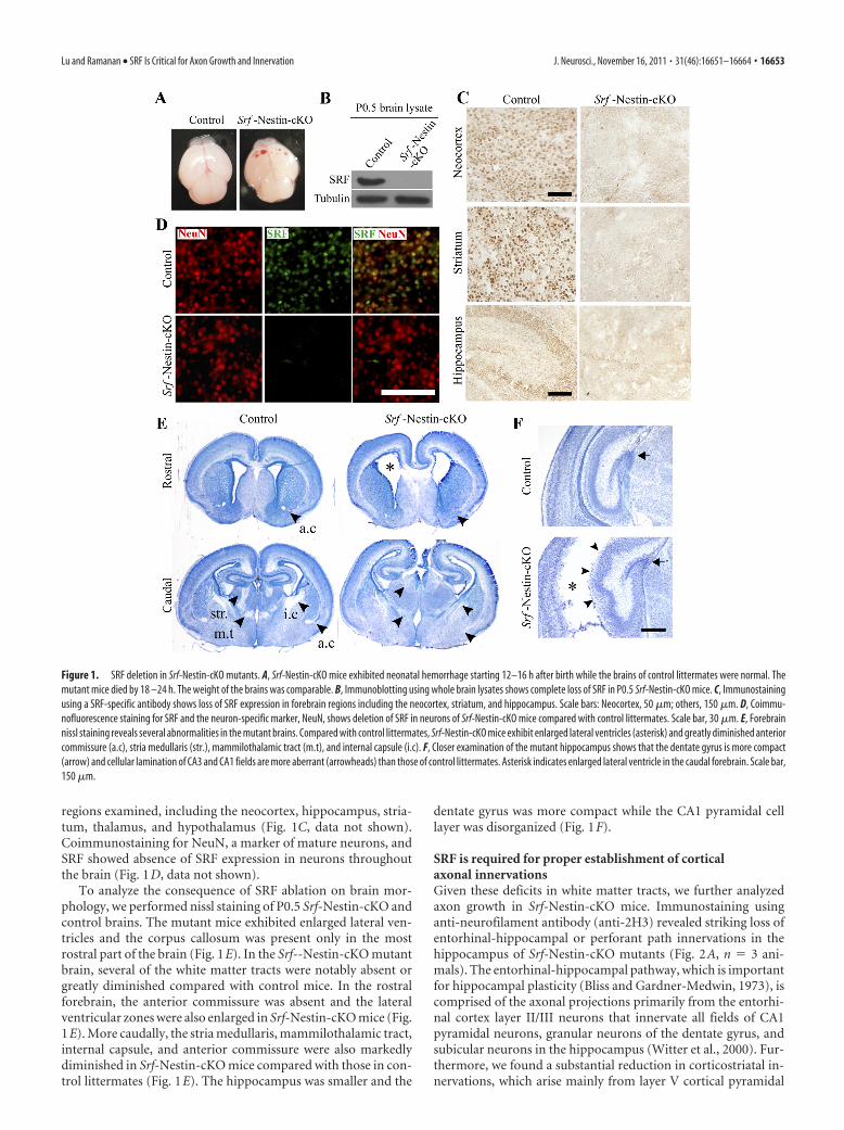
regions examined, including the neocortex, hippocampus, stria-tum, thalamus, and hypothalamus (Fig. 1C, data not shown).Coimmunostaining for NeuN, a marker of mature neurons, andSRF showed absence of SRF expression in neurons throughoutthe brain (Fig. 1D, data not shown).
To analyze the consequence of SRF ablation on brain mor-phology, we performed nissl staining of P0.5 Srf-Nestin-cKO andcontrol brains. The mutant mice exhibited enlarged lateral ven-tricles and the corpus callosum was present only in the mostrostral part of the brain (Fig. 1E). In the Srf--Nestin-cKO mutantbrain, several of the white matter tracts were notably absent orgreatly diminished compared with control mice. In the rostralforebrain, the anterior commissure was absent and the lateralventricular zones were also enlarged in Srf-Nestin-cKO mice (Fig.1E). More caudally, the stria medullaris, mammilothalamic tract,internal capsule, and anterior commissure were also markedlydiminished in Srf-Nestin-cKO mice compared with those in con-trol littermates (Fig. 1E). The hippocampus was smaller and the
dentate gyrus was more compact while the CA1 pyramidal celllayer was disorganized (Fig. 1F).
SRF is required for proper establishment of corticalaxonal innervationsGiven these deficits in white matter tracts, we further analyzedaxon growth in Srf-Nestin-cKO mice. Immunostaining usinganti-neurofilament antibody (anti-2H3) revealed striking loss ofentorhinal-hippocampal or perforant path innervations in thehippocampus of Srf-Nestin-cKO mutants (Fig. 2A, n � 3 ani-mals). The entorhinal-hippocampal pathway, which is importantfor hippocampal plasticity (Bliss and Gardner-Medwin, 1973), iscomprised of the axonal projections primarily from the entorhi-nal cortex layer II/III neurons that innervate all fields of CA1pyramidal neurons, granular neurons of the dentate gyrus, andsubicular neurons in the hippocampus (Witter et al., 2000). Fur-thermore, we found a substantial reduction in corticostriatal in-nervations, which arise mainly from layer V cortical pyramidal
Figure 1. SRF deletion in Srf-Nestin-cKO mutants. A, Srf-Nestin-cKO mice exhibited neonatal hemorrhage starting 12–16 h after birth while the brains of control littermates were normal. Themutant mice died by 18 –24 h. The weight of the brains was comparable. B, Immunoblotting using whole brain lysates shows complete loss of SRF in P0.5 Srf-Nestin-cKO mice. C, Immunostainingusing a SRF-specific antibody shows loss of SRF expression in forebrain regions including the neocortex, striatum, and hippocampus. Scale bars: Neocortex, 50 �m; others, 150 �m. D, Coimmu-nofluorescence staining for SRF and the neuron-specific marker, NeuN, shows deletion of SRF in neurons of Srf-Nestin-cKO mice compared with control littermates. Scale bar, 30 �m. E, Forebrainnissl staining reveals several abnormalities in the mutant brains. Compared with control littermates, Srf-Nestin-cKO mice exhibit enlarged lateral ventricles (asterisk) and greatly diminished anteriorcommissure (a.c), stria medullaris (str.), mammilothalamic tract (m.t), and internal capsule (i.c). F, Closer examination of the mutant hippocampus shows that the dentate gyrus is more compact(arrow) and cellular lamination of CA3 and CA1 fields are more aberrant (arrowheads) than those of control littermates. Asterisk indicates enlarged lateral ventricle in the caudal forebrain. Scale bar,150 �m.
Lu and Ramanan • SRF Is Critical for Axon Growth and Innervation J. Neurosci., November 16, 2011 • 31(46):16651–16664 • 16653

neurons in the Srf-Nestin-cKO mutants compared with controllittermates (Fig. 2B, n � 3 animals). As observed with nissl stain-ing, anti-2H3 staining also revealed a lack of or deficits in anteriorcommissure, fasciculus retroflexis, and internal capsule in Srf-Nestin-cKO mice compared with control mice (Fig. 2C, n � 3mice). We also performed anti-�-III tubulin (anti-Tuj1) immu-nostaining of sagittal brain sections to visualize the corticospinaltract projections of pyramidal neurons in layer V of the motorcortex. We observed significantly less abundant corticospinalprojections through the internal capsule and cerebral peduncle inthe Srf-Nestin-cKO brains as compared with control littermates(data not shown).
In addition to immunostaining, we used DiI labeling to visu-alize axonal projections in vivo. DiI crystals were placed on thesurface of the brain corresponding to the motor and visual corti-ces of one hemisphere (Fig. 3A). To ensure the comparisons be-tween control and mutant brain sections are made accurately, wecross-examined all coronal serial sections from the anterior to the
posterior forebrain. As observed for anti-neurofilament immu-nostaining (anti-2H3), DiI labeling showed significant deficits inthe corticostriatal projections in Srf-Nestin-cKO brains. In theanterior forebrain region of control mice, the projections fromthe cortical neurons clearly innervated the striatum. However, inSrf-Nestin-cKO brains these projections mostly terminate in thelateral corpus callosum and fail to reach their targets in the stria-tum (Fig. 3B,D; projection length: control, 460 � 13 �m; Srf-Nestin-cKO, 248 � 55 �m; n � 2 mice; p � 0.05). In the posteriorforebrain of control mice, we observed robust corticothalamicprojections innervating the thalamus. Strikingly, these cortico-thalamic innervations were absent in Srf-Nestin-cKO brain (Fig.3C,D; projection area: control, 20,875 � 1127 �m2; Srf-Nestin-cKO, 1619 � 2894 �m2; p � 0.01). Furthermore, we also observedlack of innervations to the dorsal hippocampal commissure in theSrf-Nestin-cKO mice (Fig. 3C). DiI labeling further confirmedthe deficits in anterior commissure and corpus callosum ob-served using anti-2H3 immunostaining (Fig.3 E,F). We then
Figure 2. Srf-Nestin-cKO mutant mice exhibit axonal growth deficits. A, Neurofilament immunostaining using anti-2H3 antibody reveals absence of entorhinal-hippocampal innervation in thehippocampus of Srf-Nestin-cKO mice. Inset shows the magnified view of the region indicated by the arrow. B, Examination of neurofilament expression in striatum shows less abundantcorticostriatal projections in Srf-Nestin-cKO brains than in control littermates, n � 3 mice. Right, Magnified views of the boxed regions in B, showing sparse axonal projections in the Srf-Nestin-cKOmice compared with control littermates. C, Anti-2H3 neurofilament staining shows absence or highly reduced anterior commissure (arrows), fasciculus retroflexis (arrows), and internal capsule.Enlarged view of the boxed region is shown for each fiber tract. Scale bars: A and C, large panels, and B, left, 500 �m; A, insets, and B, right, 100 �m. DG, Dentate gyrus; Ctx, neocortex; Str, striatum;Hip, hippocampus.
16654 • J. Neurosci., November 16, 2011 • 31(46):16651–16664 Lu and Ramanan • SRF Is Critical for Axon Growth and Innervation
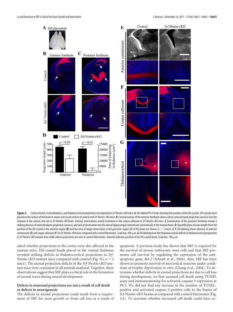
asked whether projections to the cortex were also affected in themutant mice. DiI-coated beads placed in the ventral thalamusrevealed striking deficits in thalamocortical projections in Srf-Nestin-cKO mutant mice compared with control (Fig. 3G, n � 2mice). The axonal projection deficits in the Srf-Nestin-cKO mu-tant mice were consistent in all animals analyzed. Together, theseobservations suggest that SRF plays a critical role in the formationof axonal tracts during neural development.
Defects in neuronal projections are not a result of cell deathor defects in neurogenesisThe deficits in axonal projections could result from a require-ment of SRF for axon growth or from cell loss as a result of
apoptosis. A previous study has shown that SRF is required forthe survival of mouse embryonic stem cells and that SRF pro-motes cell survival by regulating the expression of the anti-apoptotic gene, Bcl-2 (Schratt et al., 2004). Also, SRF has beenshown to promote survival of neocortical neurons under condi-tions of trophic deprivation in vitro (Chang et al., 2004). To de-termine whether deficits in axonal projections are due to cell lossduring development, we first assessed cell death using TUNELassay and immunostaining for activated-caspase 3 expression atP0.5. We did not find any increase in the number of TUNEL-positive and activated caspase-3-positive cells in the brains ofSrf-Nestin-cKO brains as compared with control littermates (Fig.4A). To ascertain whether increased cell death could have oc-
Figure 3. Corticostriatal, corticothalamic, and thalamocortical projections are impaired in Srf-Nestin-cKO mice. A, DiI-labeled P0.5 brain showing the position of the DiI crystals. DiI crystals wereplaced on the surface of the brain in motor and visual cortices of control and Srf-Nestin-cKO mice. B, Coronal section of the anterior forebrain shows robust corticostriatal projection (arrows) into thestriatum in the control, but not in Srf-Nestin-cKO brain. Instead, innervations mostly terminate in the corpus callosum in Srf-Nestin-cKO mice. C, Examination of the posterior forebrain reveals astriking absence of corticothalamic projection (arrows) and lack of innervation into the dorsal hippocampal commissure (arrowhead) in the mutant brain. D, Quantification of axon length from theposition of the DiI crystal in the anterior region (B) and the area of target innervation in the posterior region (C) of the brain are shown (n � 3 mice). E, F, DiI labeling shows absence of anteriorcommissure (E) and corpus callosum (F ) in Srf-Nestin-cKO mice compared with control littermates. Scale bar, 200 �m. G, DiI labeling from the thalamus reveals deficits in thalamocortical projectionsin Srf-Nestin-cKO mutant mice while robust projections are seen in control littermates. Asterisk indicates position of the DiI-coated bead. Scale bar, 100 �m.
Lu and Ramanan • SRF Is Critical for Axon Growth and Innervation J. Neurosci., November 16, 2011 • 31(46):16651–16664 • 16655

curred earlier during brain development, we analyzed the brainsof control and Srf-Nestin-cKO mice at E14.5, E16.5, and E18.5.We did not observe any difference in cell death at any of thesestages between Srf-Nestin-cKO and control littermates, suggest-ing that SRF-deletion does not cause apoptotic cell death duringbrain development (Fig. 4B–D).
Although we did not see any increased cell death in the brainsof Srf-Nestin-cKO mice during development, it is possible thatthe deficits in axonal tract formation observed in these mutantmice could be due to deficits in the total number of neuronsgenerated. To investigate the effect of SRF loss on neurogenesis,we determined the number of neurons in control and Srf-Nestin-cKO brains by immunostaining for NeuN, a marker for matureneuronal cell nuclei. We found that the total number of NeuN-positive cells was similar in both Srf-Nestin-cKO and controllittermates (Fig. 5A–D; neocortex: control 100 � 4.5% andknock-out 108.95 � 6.2%; striatum: control 100 � 3.5% andknock-out 105.2 � 4.4%; thalamus: control 100 � 6.0%and knock-out 106.1 � 2.7%; hippocampus: control 100 � 5.9%and knock-out 103.5 � 8.0%; and dentate gyrus: control 100 �7.6% and knock-out 92.8 � 3.1%. Data shown are mean � SEMas a percentage of mean; n � 3 mice). Although the dentate gyrusin Srf-Nestin-cKO mice appeared smaller, it had a higher celldensity than that of control animals and there were no apprecia-ble differences in total neuronal numbers (Fig. 5C, data notshown). We also analyzed the number of intermediate neuronalprecursors (INPs), as identified by Tbr2 (or Eomes) expression,and found no statistically significant difference in the totalnumber of INPs within the neocortex of Srf-Nestin-cKO miceand control littermates (Fig. 5E; control 100 � 6.5% andknock-out 96 � 5.8%; p � 0.001; n � 3 mice). Together, these
results demonstrate that SRF is not required for cell survivaland neurogenesis and that the deficits in axonal projectionsobserved in Srf-Nestin-cKO brains reflect a specific require-ment for SRF for axon growth.
SRF is not required for projection neuron subtypespecification and cortical laminationWe next asked whether SRF was required for neuronal subtypespecification. The excitatory projection neurons reside in definedlayers of the neocortex and make intracortical, subcortical, orsubcerebral connections (Molyneaux et al., 2007). A number oftranscription factors, including Fezf2 and Ctip2, are critical forspecification of cortical projection neuron subtypes and their lossresults in absence of subcerebral and subcortical projections (Ar-lotta et al., 2005; Chen et al., 2005; Molyneaux et al., 2005; Chenet al., 2008). Therefore, it is possible that the lack of corticalprojections observed in Srf-Nestin-cKO mutants could be attrib-uted to a switch in projection neuron identity, a result that couldaffect neocortical lamination. To study this, we probed for ex-pression of Tbr1, a deep-layer neuronal marker, and Cux1, whichis specifically expressed in neocortical layers 2– 4 (Hevner et al.,2001; Ferrere et al., 2006). Immunostaining of P0.5 brains re-vealed no differences in the layer-specific expression patterns ofboth Tbr1 and Cux1 in Srf-Nestin-cKO mice and control litter-mates (Fig. 6A,B). Additionally, we also probed for expression ofother neocortical layer-specific transcription factors by in situhybridization (Gray et al., 2004). Expression patterns of severaltranscription factors, including Cux2, Klf6, Lhx5, Lmo4, Nfix,Nr4a2, and Sox5, which specify the identity and position of pro-jection neuron subtypes, were similar between Srf-Nestin-cKOmice and control littermates (Fig. 6C, data not shown). These
Figure 4. Loss of SRF does not cause apoptotic cell death during brain development. A, TUNEL cell death assay and immunostaining for cleaved activated-caspase3 (act-Casp3) show no detectableelevation of apoptotic cell death in vivo in the absence of SRF at birth. Scale bar, 100 �m. B–D, Both TUNEL assay and immunostaining against activated-Caspase3 at E18.5, E16.5, and E14.5 indicateno significant elevation in the number of apoptotic cells in Srf-Nestin-cKO neocortex. CP, Cortical plate; IZ, intermediate zone; SVZ, subventricular zone; VZ, ventricular zone. Scale bar, 100 �m.
16656 • J. Neurosci., November 16, 2011 • 31(46):16651–16664 Lu and Ramanan • SRF Is Critical for Axon Growth and Innervation

results indicate that there are no changes in the establishment oflayer-specific neuronal subtypes and neocortical lamination atP0.5 in the brains of Srf-Nestin-cKO mice.
The proper lamination of the neocortex in Srf-Nestin-cKOmice suggested that radial migration of neurons is not affected inthe absence of SRF. We found that SRF is also deleted in radialglial cells in Srf-Nestin-cKO mice (data not shown). We immu-nostained P0.5 brains using anti-vimentin antibody, which labelsradial glial processes, and found no gross alteration in the abun-dance of radial glial processes or the integrity of projectionswithin multiple regions examined including the ventricular zone,neocortex, hippocampus, and corpus callosum (Fig. 6D,E, data
not shown). Together, these findings suggest that loss of SRF doesnot affect radial glial projections and thereby, the radial migra-tion of neurons and lamination of neocortex.
SRF is not required for interneuron subtype specificationGiven the role of SRF in mediating differentiation and develop-ment of several cell types in other tissues, we next sought todetermine whether SRF is required for specification of interneu-rons. Interneurons, which show astonishing differences in theirelectrophysiological, morphological, and molecular properties,are primarily derived from the medial and caudal ganglionic em-inences during brain development and then migrate to populate
Figure 5. Loss of SRF does not affect neurogenesis. A, Srf-Nestin-cKO mutants exhibit no significant changes in the number of neurons generated, as indicated by NeuN immunostaining, in theneocortex. ctx, Neocortex; c.c, corpus callosum. A’, Magnified view of boxed regions shown in A. B, C, Immunostaining for NeuN in the striatum and thalamus (B) and hippocampus (C) of control andSrf-Nestin-cKO mice. Although the dentate gyrus is more compact in Srf-Nestin-cKO mice, there is no significant difference in the number of cells compared with control mice. Scale bars: A–C, 100�m. D, Quantification of total number of NeuN-positive cells in neocortex, striatum, thalamus, and hippocampus. Error bars represent SEM. Student’s t test analysis between control andSrf-Nestin-cKO animals shows no statistically significant differences. E, Immunostaining for Tbr2 (or Eomes), a marker for intermediate neuronal progenitors, showed no difference in total numberof committed neuronal precursors in Srf-Nestin-cKO mice and control littermates. Scale bar, 50 �m.
Lu and Ramanan • SRF Is Critical for Axon Growth and Innervation J. Neurosci., November 16, 2011 • 31(46):16651–16664 • 16657

the neocortex as the brain matures (Marín and Rubenstein, 2003;Wonders and Anderson, 2006). Since Srf-Nestin-cKO mutantsdo not survive beyond P1, we restricted our analysis to interneu-ron populations in the striatum. We used immunostaining foranti-Gad-6 to identify the expression of GAD, an enzyme thatsynthesizes GABA neurotransmitters in all interneurons (Fig.7A). We found no significant difference in the overall numbers ofinterneurons between control and Srf-Nestin-cKO mice (Fig.7A,C; control 100 � 6.8% and knock-out 98.8 � 10.8%). Next,we analyzed for different interneuron subtypes, including SST-positive, parvalbumin-positive, and calbindin-positive cells. Weobserved no difference in their numbers between Srf-Nestin-cKOmutants and control littermates (Fig. 7B,C; SST� cells: control100 � 7.3% and knock-out 92.6 � 7.5%; parvalbumin� cells:control 100 � 6.4% and knock-out 117.8 � 11.2%; calbindin:control 100 � 7.8% and knock-out 116.0 � 10.1%). Together,these findings demonstrate that, in addition to projection neuronsubtype specification, the establishment of interneuron subtypes
occurs normally in the absence of SRF during neuronal lineagecommitment.
Neural precursor cell population increases inSrf-Nestin-cKO mutant miceQuiescent cells that rest in the G0 state can be induced by extra-cellular stimuli to express immediate early gene (IEG) programs(Herschman, 1991). A number of transcription factor-encodingIEGs, such as cFos, c-Myc, Egr-1, and JunB, and are then respon-sible for activating gene cascades that enable cell progression tothe G1 state (Greenberg and Ziff, 1984; Lau and Nathans, 1985).SRF-mediated transcription was demonstrated to be necessaryfor inducing IEG expression in embryonic stem cells and in neu-rons (Norman et al., 1988; Schratt et al., 2001; Ramanan et al.,2005). Sequestration of functional SRF was also shown to impederat embryonic fibroblast and myoblast proliferation, but not self-renewal of embryonic stem cells (Gauthier-Rouviere et al., 1991;Soulez et al., 1996). We therefore assessed whether SRF deletion
Figure 6. SRF is dispensable for neocortical lamination and projection neuron subtype specification. A, Immunostaining for Tbr1 expression, a marker of deep layer neurons in the neocortex,shows normal layer VI lamination in Srf-Nestin-cKO and control brains. Shown here is the retrosplenial cortex. B, Immunofluorescence staining for expression of Tbr1 and Cux 1 (a marker forsuperficial layer neurons) in the neocortex shows that both layer 2/3 and layer VI neurons are specified and positioned normally in knock-out mice. c.c., Corpus callosum. C, In situ hybridization oflayer-specific transcription factors, including Lmo4, Cux2, and Lhx5, shows that neocortical lamination and the specification of those subtypes are normal in the absence of SRF. Arrows point to Cux2-or Lhx5-expressing upper layer neurons. Scale bars: A, C, 200 �m; B, 100 �m. D, Immunostaining using anti-vimentin antibody, expressed in radial glial processes, show that the structural integrityof radial glia is normal in Srf-Nestin-cKO brain. E, Magnified images of boxed regions in D show normal appearance of radial glial processes in Srf-Nestin-cKO mice compared with control mice. Scalebar, 20 �m.
16658 • J. Neurosci., November 16, 2011 • 31(46):16651–16664 Lu and Ramanan • SRF Is Critical for Axon Growth and Innervation

affects NPC growth and maintenance in vivo. Proliferating pro-genitor cells are identified by the expression of phospho-histoneH3, which is a modification event that occurs specifically duringcell division events of both mitosis and meiosis (Hans and Dim-itrov, 2001). Surprisingly, we observed an increase in phospho-histone H3-positive cells in the proliferative subventricular zone(SVZ) of Srf-Nestin-cKO brains at both E14.5 and E18.5 (Fig.8A–D). Quantitative analyses of the number of phospho-histoneH3-positive cells per area of the parameter of the SVZ revealed a20% and 80% increase in mutants at E14.5 and E18.5, respec-tively (Fig. 8E; E14.5 p-histone H3 normalized: control 100 �7.3% and knock-out 120.7 � 7.8%; and E18.5 p-histone H3 nor-malized: control 100 � 3.0% and knock-out 180.6 � 4.2%). Weconfirmed the increase in proliferative progenitor cell numbersusing two additional markers: Ki-67, which is expressed by cellsin the cell-cycle phases G1, S, and G2 and in mitosis; and Sox2, atranscription factor expressed in NPC. At E14.5, immunohisto-chemistry staining of Ki-67 in Srf-Nestin-cKO brains showed anincrease in NPC numbers per square micrometer in the SVZ andthe neocortex compared with control (Fig. 8E,F; Ki-67 normal-ized: control 100 � 18.0% and knock-out 140 � 6.3%). Similarly,neocortex and SVZ in Srf-Nestin-cKO showed markedly in-creased numbers of Sox2-expressing cells compared with those incontrol littermates (Fig. 8F). These observations suggest that lossof SRF affects NPC homeostasis during development withoutaffecting NPC survival.
Conditional deletion of SRF in developing forebrain neuronsOur analyses of the Srf-Nestin-cKO mice suggested that SRFplays a specific role in the development of axonal tracts without
affecting neurogenesis, neuronal survival, and neuronal subtypespecification. However, since SRF deletion occurs in all neuralprecursor cells before cellular differentiation occurs, it is possiblethat axon growth defects in Srf-Nestin-cKO mice could be due toa non-cell autonomous requirement of SRF for axon growth. Toascertain whether SRF is required cell autonomously for axongrowth, we generated a neuron-specific deletion of SRF using theNEX-Cre transgenic mouse. In the NEX-Cre mouse, cre expres-sion is controlled by the onset of expression of the NEX gene (alsoknown as Math2 or NeuroD6), an early neuronal basic helix-loop-helix gene expressed specifically in differentiating neurons(Schwab et al., 1998; Goebbels et al., 2006). Cre recombinase-mediated excision in the NEX-Cre mouse has been shown to takeplace starting at �E11.5 and is restricted only to the glutamater-gic neurons in the neocortex and hippocampus, whereas cre ex-pression is not observed in the interneurons and glial cells(Brockschnieder et al., 2004; Goebbels et al., 2006; Kashani et al.,2006). The Srf-NEX-cKO mice were born in the expected Men-delian ratio but unlike the Srf-Nestin-cKO mice, these mice didnot exhibit neonatal lethality and grew to adulthood. We firstconfirmed deletion at P0.5 by immunostaining and found thatSRF deletion was restricted to the neocortex and hippocampusbut not in the striatum and other regions of the brain, consistentwith previous findings (Fig. 9A—D, data not shown).
We next asked whether lamination occurs normally in Srf-NEX-cKO mice. We performed immunostaining for laminar-specific markers, Cux1 and Tbr1, on P21 brains. We did not findany deficits in neocortical lamination in Srf-NEX-cKO mice ascompared with control littermates (Fig. 9E). At birth, the lack ofdefects in lamination in Srf-NEX-cKO mice was similar to that
Figure 7. SRF is not required for interneuron subtype specification. A, Immunostaining for Gad-6, which labels all GABAergic interneurons, showed normal expression of Gad-6 in Srf-Nestin-cKOand control mice. Inset shows enlarged image of a single stained neuron. B, Immunohistochemistry staining showing expression of SST, parvalbumin, and calbindin, which label unique subtypes ofinterneurons, suggests no apparent change in the population of interneurons in brains of Srf-Nestin-cKO mice. The striatal region from control and mutant is shown as magnified images. Inset showsenlarged image of a single stained neuron. C, Quantification of cell numbers for different interneurons subtypes for B. Student’s t test showed no statistically significant difference in the number ofeach interneuron subtype between paired control and knock-out animals. Error bars represent SEM.
Lu and Ramanan • SRF Is Critical for Axon Growth and Innervation J. Neurosci., November 16, 2011 • 31(46):16651–16664 • 16659

observed for Srf-Nestin-cKO mutantmice (data not shown). Together, thesefindings suggest neocortical lamination isproperly established in the absence ofSRF.
SRF mediates cortical neuron targetinnervation cell autonomouslyWe next analyzed axonal projections inSrf-NEX-cKO mice at P0.5 using DiI la-beling. Two weeks following DiI labeling,sagittal sections of Srf-NEX-cKO mousebrains revealed greatly diminished corti-cospinal projections, and cortical motorneuron axons passing through the inter-nal capsule were less abundant with veryfew projections reaching the cerebral pe-duncle (Fig. 10A,A’,A”; n � 3 mice).Comparison of parallel serial sectionsfrom the lateral side to the medial regionof the forebrain between control and Srf-NEX-cKO mice showed a severe reduc-tion in corticospinal projections in themutant neocortex (Fig. 10B; n � 3 mice).
We then examined horizontal sectionsof control and Srf-NEX-cKO brains after6 weeks of DiI labeling. On the ventral sideof the brain, we observed that the intra-cortical and corticostriatal connections inSrf-NEX-cKO mice were less abundantand shorter than those observed in con-trol littermates. In particular, a region ofthe thalamus is clearly innervated in thecontrol brain; however, this innervationis less prominent in the Srf-NEX-cKObrain (Fig. 10C; n � 3 mice). In the me-dial region of the brains, similar to thatobserved in 2-week-old sagittal sections, itwas evident that corticospinal innervationsthrough the striatum to the cerebral pedun-cle were greatly reduced overall in Srf-NEX-cKO mutants compared with controllittermates (Fig. 10D).
We further examined serial coronal sec-tions of control and Srf-NEX-cKO brains at4 weeks after DiI labeling. In rostral sections,DiI tracing showed that callosal innervations that form the corpuscallosum are diminished and shorter in the Srf-NEX-cKO brains(Fig. 10E). We found less abundant corticostriatal projections in theSrf-NEX-cKO brain (Fig. 10E,F; projection area: control, 205,737 �496 �m2; Srf-NEX-cKO, 12,148 � 427 �m2, n � 3 mice), an obser-vation that is consistent with that made in brains of Srf-Nestin-cKOmice using anti-2H3 immunostaining. Toward the caudal end of theforebrain, retrograde DiI labeling revealed that the corticothalamicconnections, which are important relays of sensory information be-tween the visual cortex and the lateral geniculate nucleus of the thal-amus, were also less robustly established in the Srf-NEX-cKO micecompared with control mice (Fig. 10E,F; projection area: control,41,690 � 1643 �m2; Srf-NEX-cKO, 21,229 � 1207 �m2, n � 2mice). These observations were consistent in all the mutant miceanalyzed. These neuroanatomical tracing experiments demonstratethat SRF plays a critical cell-autonomous role in regulating axonalgrowth and establishment of axonal projections in vivo.
DiscussionNeuronal development in the CNS involves several critical stagesincluding neurogenesis and maturation of neurons, growth andextension of axons, and structural organization within the brain.Our current understanding of the role of SRF in neuronal devel-opment comes mainly from studies using mice carrying condi-tional neuron-specific deletion of SRF in late gestation or inpostnatal brain. However, the requirement of SRF for early stagesof neuronal development remains unknown. In the presentstudy, we show that conditional deletion of SRF in neural precur-sor cells (Srf-Nestin-cKO) results in severe deficits in the devel-opment of major axonal projections in the forebrain, includingcorticospinal, corticothalamic, corticostriatal, and thalamocorti-cal tracts along with a variable loss of the corpus callosum. Axonaldeficits were seen as early as E14.5 in the Srf-Nestin-cKO miceand there was little or no cell death during development. Inter-estingly, there was a significant increase in the number of prolif-
Figure 8. Loss of SRF results in an increase in the number of neural precursor cells. A, Proliferating NPCs are identified in thecontrol and Srf-Nestin-cKO forebrains using anti-p-histone H3 antibody at E14.5. B, Magnification of the boxed regions in A. C,Comparison of NPC populations at E18.5. D, Magnified view of the boxed regions in C. E, Cell count analyses of the number ofp-histone-H3- and Ki67-positive cells at E14.5 indicate a statistically significant increase in the number of proliferating cells in theventricular zone of Srf-Nestin-cKO mice. The difference in relative numbers of NPCs between the control and mutant brain is morepronounced at E18.5. F, Immunostaining for two additional proteins, Ki67, a marker of cells in the active phase of cell cycle, andSox2, a marker of neural precursor cells, was used to visualize proliferating cells in the neocortex at E14.5. Mutant brains not onlydisplay more Ki67-positive cells but also show a broadened layer of Sox2-positive cells. Scale bars, 50 �m.
16660 • J. Neurosci., November 16, 2011 • 31(46):16651–16664 Lu and Ramanan • SRF Is Critical for Axon Growth and Innervation

erating cells in the ventricular zone in Srf-Nestin-cKO mice.Conditional deletion of SRF in embryonic forebrain neurons(Srf-NEX-cKO) also resulted in severe deficits in major axonalprojections. Neurogenesis, radial neuronal migration in the neo-cortex, neocortical lamination, and neuronal subtype specifica-tion were unaffected by SRF loss. Together, these findings suggestthat SRF is required in a cell-autonomous manner for axongrowth and extension. Similar to that observed in mice with pre-natal and postnatal deletion of SRF, SRF is dispensable for neu-ronal survival. Our study identifies a specific role for SRF inpromoting axon growth during neuronal development withoutaffecting neurogenesis and neuronal differentiation.
Previous studies have shown that neuron-specific SRF dele-tion during late gestation in the brain causes deficits in terminaltargeting of mossy fiber axons in the hippocampus, while SRFloss in developing sensory neurons in the peripheral nervoussystem affects NGF-dependent terminal arborization and targetinnervation (Knoll et al., 2006; Wickramasinghe et al., 2008).However, proximal axon growth in the peripheral nervous sys-tem is not affected while the role of SRF in axon growth in theCNS remains unknown. We found that deleting SRF in neuralprecursor cells results in severe deficits in axon growth and tar-geting of cortical axon projections. Observations made in cul-tured hippocampal neurons have shown that SRF is required forcontact-mediated axon repulsion (Knoll et al., 2006). We did notobserve any mistargeted axonal tracts in the brains of Srf-Nestin-
cKO mice, suggesting that the lack of target innervation seen inthe SRF mutant mice is primarily due to defects in axon growthand not due to defects in axon guidance. We found similar axonalgrowth defects when SRF was deleted in developing postmitoticneurons in neocortex and hippocampus in Srf-NEX-cKO mutantmice. Unlike Srf-Nestin-cKO mice, the Srf-NEX-cKO mice sur-vived to adulthood, and in preliminary observations, we foundthat adult Srf-NEX-cKO mice exhibited clasping of limbs in adystonic manner when subjected to the tail suspension test,which is suggestive of motor dysfunction (Carter et al., 1999;Yamamoto et al., 2000). Furthermore, consistent with previousobservations, we also found that SRF-deficient neurons exhibithighly attenuated axon growth in culture (Knoll et al., 2006) (C.Li and N. Ramanan, unpublished observations). The similaritiesin deficits in axon growth in the brains of Srf-Nestin-cKO andSrf-NEX-cKO mice suggested that SRF-dependent transcriptionplays a cell-intrinsic role in axon growth.
The molecular mechanisms underlying SRF-dependent axongrowth remain poorly understood. One mechanism by whichSRF potentially regulates axon growth is through association withspecific cofactors. We found that blocking the functions of theTernary Complex Factor-family cofactors of SRF, includingElk-1, does not affect axon growth in cultured neurons (C. Li andN. Ramanan, unpublished observations). However, we and oth-ers have found that blocking the functions of myocardin-familycofactors, MKL1 (also known as MAL/MRTF-A) and MKL2
Figure 9. Ablation of SRF in Srf-NEX-cKO. A, Immunofluorescence staining at P0.5 using anti-SRF antibody shows that SRF is deleted in the neocortex but not in the striatum of Srf-NEX-cKOmutants. B, Magnified views of boxed regions in A. C, SRF expression is also abolished in the hippocampus in mutants. D, A magnified view of CA3 neurons of control and Srf-NEX-cKO mice. E,Immunostaining of P21 brains using anti-Tbr1 and anti-Cux1 shows normal lamination of neocortex in Srf-NEX-cKO mice and control littermates. Scale bars, 50 �m.
Lu and Ramanan • SRF Is Critical for Axon Growth and Innervation J. Neurosci., November 16, 2011 • 31(46):16651–16664 • 16661

(MRTF-B), by dominant-negative or knockdown approaches orby gene deletion attenuates axon growth in vitro (Knoll et al.,2006; Shiota et al., 2006; Wickramasinghe et al., 2008; Mokalledet al., 2010) (C. Li and N. Ramanan, unpublished observations).Mutant mice that lack both MKL1 and MKL2 in the brain exhibitdeficits in dendritic growth in the neocortex and hippocampus as
assessed by MAP2 and Golgi staining (Mokalled et al., 2010).However, the effect of MKL1/MKL2 loss on axon growth in vivohas not been reported in these mice. In the peripheral nervoussystem, SRF has been shown to function downstream of NGF-signaling to regulate terminal arborization of axons and target inner-vation (Wickramasinghe et al., 2008). Furthermore, NGF signaling
Figure 10. Dil labeling shows impairment in axonal projections in Srf-NEX-cKO mutants. A, DiI crystals were placed on the brain surface in the regions of the motor and the visual cortices(indicated by asterisks) in P0.5 Srf-NEX-cKO knock-out and control littermates. Two weeks after labeling, brains were sectioned sagittally. Impaired corticospinal innervation was observed in theknock-out brain. Magnifications of the internal capsule (i.c) and cerebral peduncle (c.p) regions are shown in A’ and A”. Projections through the cerebral peduncle are seen in the brains of control butSrf-NEX-cKO mice. B, Serial sagittal sections from lateral to medial regions of the brain show lack of corticostriatal projections (arrows) in Srf-NEX-cKO mice. No misguided axons were observed in themutant mice. C, After 6 weeks of labeling, control and Srf-NEX-cKO brains were sectioned horizontally. Arrows show diminished projections within the neocortex, corticostriatal projections, andinnervations to the thalamus in the mutant. Medial horizontal section shows impaired projections through the internal capsule and the cerebral peduncle. D, Magnified views of the boxed regionsin C showing the corticospinal projections. E, Coronal sections from caudal regions of the brain reveal diminished corticostriatal as well as corticothalamic tracts (arrows). Asterisks indicate sites ofcrystal placement; dotted lines outline the ventricular zone and the hippocampus (H). F, Quantification of area of innervation by corticostriatal and corticothalamic axons in E (n � 3 mice).
16662 • J. Neurosci., November 16, 2011 • 31(46):16651–16664 Lu and Ramanan • SRF Is Critical for Axon Growth and Innervation

to SRF is dependent on both ERK/MEK and MAL/MKL1 signalingpathways. The findings from the peripheral nervous system raise aninteresting question as to which extracellular signals might stimulateSRF-dependent transcription during axon growth in the brain. Cur-rently we lack sufficient knowledge on the nature of the extracellularsignals and the identities of SRF target genes critical for axon growthin the CNS. It is likely that SRF functions downstream of growthfactors such as BDNF to regulate axon growth. SRF could also regu-late axon growth by regulating the expression of components of theactin cytoskeleton, including �-actin, �-actin, paxillin, vinculin, andtalin (Schratt et al., 2002). In fact, previous studies including our ownhave shown that �-actin expression is reduced in SRF knock-outneurons (Alberti et al., 2005; Ramanan et al., 2005; Knoll et al., 2006),and it was hypothesized that reduction in actin levels was one of theunderlying causes for neurite outgrowth deficits observed in SRF-deficient neurons. However, overexpression of actin was found to beinsufficient to rescue the growth deficits of SRF-null neurons (Knollet al., 2006; Stern et al., 2009). Since SRF regulates the expression ofseveral cytoskeletal proteins (Schratt et al., 2002), it is possible thatthe neuronal growth deficits exhibited by SRF-deficient neuronscould be due to a breakdown in cytoskeletal apparatus critical forgrowth and extension.
We did not observe any increased cell death in the brains ofSrf-Nestin-cKO and Srf-NEX-cKO mutant mice during develop-ment. There was also no noticeable difference in neuronal cellnumbers in older Srf-NEX-cKO mice, and this is consistent withour previous findings that SRF deletion does not result in celldeath or neurodegeneration in the CNS (Ramanan et al., 2005).Interestingly, SRF deletion in neural precursor cells did not causeapoptotic cell death, a phenotype that contrasts with observationsmade in SRF-deficient embryonic stem cells (Schratt et al., 2004).SRF-deficient mouse ES cells exhibited apoptotic cell death bothin vitro and in vivo (Schratt et al., 2004). Our findings suggest thatSRF is dispensable for survival of NPCs both in vitro and in vivo(our unpublished observations). In contrast, we observed an in-crease in the total number of p-histone-H3 and Sox2-positivecells in Srf-Nestin-cKO mice. A recent elegant study showed thatSRF deletion in neurons affects oligodendrocyte differentiationin a paracrine manner (Stritt et al., 2009). Consistent with thisobservation, we also observed a decrease in Olig2� cells at birthin Srf-Nestin-cKO mice (our unpublished observations). There-fore, a likely explanation for the increase in NPC numbers inSrf-Nestin-cKO mice is that SRF loss in NPCs affects oligoden-drocyte differentiation, thereby resulting in an increase in undif-ferentiated neural precursor cells. Together, these observationssuggest that there are distinct requirements for SRF in ES cellsand in NPCs for cell survival.
Previous studies have shown that SRF has a profound role inregulating cell-type specific gene expression that underlies thedevelopment of many cell types. A number of tissue-specific in-activation studies later elucidated essential functions of SRF forthe development of cardiac muscle cells (Niu et al., 2005, 2008;Zhao et al., 2005), the differentiation of smooth muscles (Mianoet al., 2004; Parlakian et al., 2004), and the normal proliferationand differentiation of keratinocytes (Koegel et al., 2009). We ob-served no differences in total number of NeuN-positive cells inSrf-Nestin-cKO mice, suggesting that SRF is dispensable for neu-rogenesis in the brain. We also found that neuronal subtype spec-ification and both interneuron and neocortical lamina-specificneuron identities were properly established in the absence of SRF.The findings that SRF-deficient neurons negatively influence oli-godendrocyte differentiation suggest that SRF-dependent tran-
scription can promote cell-type specification in the brain (Strittet al., 2009).
SRF deletion in developing neurons has been shown to affecttangential cell migration along the rostral migratory stream (Al-berti et al., 2005). We also observed similar tangential migrationdeficits in the Srf-Nestin-cKO mice (our unpublished observa-tions). However, we found that radial migration of neurons in theneocortex was not affected and neocortical lamination was estab-lished normally in both Srf-Nestin-cKO and Srf-NEX-cKO mu-tant mice. Our observations differ from those of a recent study inwhich cortical lamination was shown to be affected in mice car-rying neuron-specific deletion of SRF (Stritt and Knoll, 2010). Inthis study, calbindin-positive cells were reduced in SRF-mutantneocortex while immunostaining for the neurofilament proteinSMI-32, which also labels a subpopulation of cortical neurons(Campbell and Morrison, 1989) in layers III and V, showed mis-localization of Smi-32-positive cells between layers III and V.Calbindin-positive interneurons are mainly generated in the me-dial ganglionic eminences before they tangentially migrate topopulate the neocortex (Marín and Rubenstein, 2003; Wondersand Anderson, 2006), and we did not find any change in the totalnumbers of striatal calbindin-positive cells in Srf-Nestin-cKOmice at birth. In this study, we used both in situ hybridization andimmunohistochemistry for several transcription factors that areexpressed in specific cortical layers during mouse development(Gray et al., 2004). We did not find any lamination defects in theneocortex of either Srf-Nestin-cKO or Srf-NEX-cKO mice at P0.5or in 3-week-old Srf-NEX-cKO mice. If SRF is critical for neocor-tical lamination, this phenotype should be more severe in theSrf-Nestin-cKO mice, since SRF is deleted in all major cell typesin the brain starting at E12.5 (our unpublished observations).Based on our observations, we conclude that SRF loss in neuralprecursor cells and in developing neurons does not affect layeringof the neocortex. We also found that SRF deletion in radial glialcells in Srf-Nestin-cKO mice did not affect their morphology,suggesting that SRF-dependent transcription is not required forextension of radial glial processes.
Our study identifies specific roles for SRF during neuronal devel-opment. SRF plays a critical role in neural precursor cell homeostasisand in the formation of major axonal tracts in the brain. SRF isdispensable for neurogenesis and cell survival but contrary to recentfindings, SRF is not required for neocortical lamination.
ReferencesAlberti S, Krause SM, Kretz O, Philippar U, Lemberger T, Casanova E, Wiebel
FF, Schwarz H, Frotscher M, Schutz G, Nordheim A (2005) Neuronalmigration in the murine rostral migratory stream requires serum re-sponse factor. Proc Natl Acad Sci U S A 102:6148 – 6153.
Arlotta P, Molyneaux BJ, Chen J, Inoue J, Kominami R, Macklis JD (2005)Neuronal subtype-specific genes that control corticospinal motor neurondevelopment in vivo. Neuron 45:207–221.
Arsenian S, Weinhold B, Oelgeschlager M, Ruther U, Nordheim A (1998)Serum response factor is essential for mesoderm formation during mouseembryogenesis. EMBO J 17:6289 – 6299.
Bliss TV, Gardner-Medwin AR (1973) Long-lasting potentiation of synaptictransmission in the dentate area of the unanaestetized rabbit followingstimulation of the perforant path. J Physiol 232:357–374.
Brockschnieder D, Lappe-Siefke C, Goebbels S, Boesl MR, Nave KA, Rieth-macher D (2004) Cell depletion due to diphtheria toxin fragment A afterCre-mediated recombination. Mol Cell Biol 24:7636 –7642.
Campbell MJ, Morrison JH (1989) Monoclonal antibody to neurofilamentprotein (SMI-32) labels a subpopulation of pyramidal neurons in thehuman and monkey neocortex. J Comp Neurol 282:191–205.
Carter RJ, Lione LA, Humby T, Mangiarini L, Mahal A, Bates GP, Dunnett SB,Morton AJ (1999) Characterization of progressive motor deficits in
Lu and Ramanan • SRF Is Critical for Axon Growth and Innervation J. Neurosci., November 16, 2011 • 31(46):16651–16664 • 16663
mice transgenic for the human Huntington’s disease mutation. J Neurosci19:3248 –3257.
Chang SH, Poser S, Xia Z (2004) A novel role for serum response factor inneuronal survival. J Neurosci 24:2277–2285.
Chen B, Schaevitz LR, McConnell SK (2005) Fezl regulates the differentia-tion and axon targeting of layer 5 subcortical projection neurons in cere-bral cortex. Proc Natl Acad Sci U S A 102:17184 –17189.
Chen B, Wang SS, Hattox AM, Rayburn H, Nelson SB, McConnell SK (2008)The Fezf2-Ctip2 genetic pathway regulates the fate choice of subcorticalprojection neurons in the developing cerebral cortex. Proc Natl Acad SciU S A 105:11382–11387.
Etkin A, Alarcon JM, Weisberg SP, Touzani K, Huang YY, Nordheim A,Kandel ER (2006) A role in learning for SRF: deletion in the adult fore-brain disrupts LTD and the formation of an immediate memory of a novelcontext. Neuron 50:127–143.
Ferrere A, Vitalis T, Gingras H, Gaspar P, Cases O (2006) Expression ofCux-1 and Cux-2 in the developing somatosensory cortex of normal andbarrel-defective mice. Anat Rec A Discov Mol Cell Evol Biol 288:158 –165.
Gauthier-Rouviere C, Cavadore JC, Blanchard JM, Lamb NJ, Fernandez A(1991) p67SRF is a constitutive nuclear protein implicated in the modu-lation of genes required throughout the G1 period. Cell Regul 2:575–588.
Goebbels S, Bormuth I, Bode U, Hermanson O, Schwab MH, Nave KA(2006) Genetic targeting of principal neurons in neocortex and hip-pocampus of NEX-Cre mice. Genesis 44:611– 621.
Goldberg JL, Espinosa JS, Xu Y, Davidson N, Kovacs GT, Barres BA (2002)Retinal ganglion cells do not extend axons by default: promotion by neu-rotrophic signaling and electrical activity. Neuron 33:689 –702.
Gray PA, Fu H, Luo P, Zhao Q, Yu J, Ferrari A, Tenzen T, Yuk DI, Tsung EF,Cai Z, Alberta JA, Cheng LP, Liu Y, Stenman JM, Valerius MT, Billings N,Kim HA, Greenberg ME, McMahon AP, Rowitch DH, et al. (2004)Mouse brain organization revealed through direct genome-scale TF ex-pression analysis. Science 306:2255–2257.
Greenberg ME, Ziff EB (1984) Stimulation of 3T3 cells induces transcrip-tion of the c-fos proto-oncogene. Nature 311:433– 438.
Hans F, Dimitrov S (2001) Histone H3 phosphorylation and cell division.Oncogene 20:3021–3027.
Herschman HR (1991) Primary response genes induced by growth factorsand tumor promoters. Annu Rev Biochem 60:281–319.
Hevner RF, Shi L, Justice N, Hsueh Y, Sheng M, Smiga S, Bulfone A, GoffinetAM, Campagnoni AT, Rubenstein JL (2001) Tbr1 regulates differentia-tion of the preplate and layer 6. Neuron 29:353–366.
Johnson AW, Crombag HS, Smith DR, Ramanan N (2011) Effects of serumresponse factor (SRF) deletion on conditioned reinforcement. BehavBrain Res 220:312–318.
Kashani AH, Qiu Z, Jurata L, Lee SK, Pfaff S, Goebbels S, Nave KA, Ghosh A(2006) Calcium activation of the LMO4 transcription complex and itsrole in the patterning of thalamocortical connections. J Neurosci26:8398 – 8408.
Knoll B, Kretz O, Fiedler C, Alberti S, Schutz G, Frotscher M, Nordheim A(2006) Serum response factor controls neuronal circuit assembly in thehippocampus. Nat Neurosci 9:195–204.
Koegel H, von Tobel L, Schafer M, Alberti S, Kremmer E, Mauch C, Hohl D,Wang XJ, Beer HD, Bloch W, Nordheim A, Werner S (2009) Loss ofserum response factor in keratinocytes results in hyperproliferative skindisease in mice. J Clin Invest 119:899 –910.
Lau LF, Nathans D (1985) Identification of a set of genes expressed duringthe G0/G1 transition of cultured mouse cells. EMBO J 4:3145–3151.
Marín O, Rubenstein JL (2003) Cell migration in the forebrain. Annu RevNeurosci 26:441– 483.
Miano JM, Ramanan N, Georger MA, de Mesy Bentley KL, Emerson RL, BalzaRO Jr, Xiao Q, Weiler H, Ginty DD, Misra RP (2004) Restricted inacti-vation of serum response factor to the cardiovascular system. Proc NatlAcad Sci U S A 101:17132–17137.
Mokalled MH, Johnson A, Kim Y, Oh J, Olson EN (2010) Myocardin-related transcription factors regulate the Cdk5/Pctaire1 kinase cascade tocontrol neurite outgrowth, neuronal migration and brain development.Development 137:2365–2374.
Molyneaux BJ, Arlotta P, Hirata T, Hibi M, Macklis JD (2005) Fezl is re-quired for the birth and specification of corticospinal motor neurons.Neuron 47:817– 831.
Molyneaux BJ, Arlotta P, Menezes JR, Macklis JD (2007) Neuronal subtypespecification in the cerebral cortex. Nat Rev Neurosci 8:427– 437.
Niu Z, Yu W, Zhang SX, Barron M, Belaguli NS, Schneider MD, Parmacek M,Nordheim A, Schwartz RJ (2005) Conditional mutagenesis of the mu-rine serum response factor gene blocks cardiogenesis and the transcrip-tion of downstream gene targets. J Biol Chem 280:32531–32538.
Niu Z, Iyer D, Conway SJ, Martin JF, Ivey K, Srivastava D, Nordheim A,Schwartz RJ (2008) Serum response factor orchestrates nascent sarcom-erogenesis and silences the biomineralization gene program in the heart.Proc Natl Acad Sci U S A 105:17824 –17829.
Norman C, Runswick M, Pollock R, Treisman R (1988) Isolation and prop-erties of cDNA clones encoding SRF, a transcription factor that binds tothe c-fos serum response element. Cell 55:989 –1003.
Parlakian A, Tuil D, Hamard G, Tavernier G, Hentzen D, Concordet JP,Paulin D, Li Z, Daegelen D (2004) Targeted inactivation of serum re-sponse factor in the developing heart results in myocardial defects andembryonic lethality. Mol Cell Biol 24:5281–5289.
Ramanan N, Shen Y, Sarsfield S, Lemberger T, Schutz G, Linden DJ, Ginty DD(2005) SRF mediates activity-induced gene expression and synaptic plas-ticity but not neuronal viability. Nat Neurosci 8:759 –767.
Schratt G, Weinhold B, Lundberg AS, Schuck S, Berger J, Schwarz H, Wein-berg RA, Ruther U, Nordheim A (2001) Serum response factor is re-quired for immediate-early gene activation yet is dispensable forproliferation of embryonic stem cells. Mol Cell Biol 21:2933–2943.
Schratt G, Philippar U, Berger J, Schwarz H, Heidenreich O, Nordheim A(2002) Serum response factor is crucial for actin cytoskeletal organiza-tion and focal adhesion assembly in embryonic stem cells. J Cell Biol156:737–750.
Schratt G, Philippar U, Hockemeyer D, Schwarz H, Alberti S, Nordheim A(2004) SRF regulates Bcl-2 expression and promotes cell survival duringmurine embryonic development. EMBO J 23:1834 –1844.
Schwab MH, Druffel-Augustin S, Gass P, Jung M, Klugmann M, BartholomaeA, Rossner MJ, Nave KA (1998) Neuronal basic helix-loop-helix pro-teins (NEX, neuroD, NDRF): spatiotemporal expression and targeteddisruption of the NEX gene in transgenic mice. J Neurosci 18:1408 –1418.
Shiota J, Ishikawa M, Sakagami H, Tsuda M, Baraban JM, Tabuchi A (2006)Developmental expression of the SRF co-activator MAL in brain: role inregulating dendritic morphology. J Neurochem 98:1778 –1788.
Smith-Hicks C, Xiao B, Deng R, Ji Y, Zhao X, Shepherd JD, Posern G, Kuhl D,Huganir RL, Ginty DD, Worley PF, Linden DJ (2010) SRF binding toSRE 6.9 in the Arc promoter is essential for LTD in cultured Purkinje cells.Nat Neurosci 13:1082–1089.
Soulez M, Rouviere CG, Chafey P, Hentzen D, Vandromme M, Lautredou N,Lamb N, Kahn A, Tuil D (1996) Growth and differentiation of C2 myogeniccells are dependent on serum response factor. Mol Cell Biol 16:6065–6074.
Stern S, Debre E, Stritt C, Berger J, Posern G, Knoll B (2009) A nuclear actinfunction regulates neuronal motility by serum response factor-dependentgene transcription. J Neurosci 29:4512– 4518.
Stritt C, Knoll B (2010) Serum response factor regulates hippocampal lam-ination and dendrite development and is connected with reelin signaling.Mol Cell Biol 30:1828 –1837.
Stritt C, Stern S, Harting K, Manke T, Sinske D, Schwarz H, Vingron M, Nord-heim A, Knoll B (2009) Paracrine control of oligodendrocyte differentia-tion by SRF-directed neuronal gene expression. Nat Neurosci 12:418–427.
Tronche F, Kellendonk C, Kretz O, Gass P, Anlag K, Orban PC, Bock R, KleinR, Schutz G (1999) Disruption of the glucocorticoid receptor gene in thenervous system results in reduced anxiety. Nat Genet 23:99 –103.
Wickramasinghe SR, Alvania RS, Ramanan N, Wood JN, Mandai K, GintyDD (2008) Serum response factor mediates NGF-dependent target in-nervation by embryonic DRG sensory neurons. Neuron 58:532–545.
Witter MP, Naber PA, van Haeften T, Machielsen WC, Rombouts SA, Bark-hof F, Scheltens P, Lopes da Silva FH (2000) Cortico-hippocampal com-munication by way of parallel parahippocampal-subicular pathways.Hippocampus 10:398 – 410.
Wonders CP, Anderson SA (2006) The origin and specification of corticalinterneurons. Nat Rev Neurosci 7:687– 696.
Yamamoto A, Lucas JJ, Hen R (2000) Reversal of neuropathology and motordysfunction in a conditional model of Huntington’s disease. Cell 101:57–66.
Zhao Y, Samal E, Srivastava D (2005) Serum response factor regulates amuscle-specific microRNA that targets Hand2 during cardiogenesis. Na-ture 436:214 –220.
Zhou FQ, Snider WD (2006) Intracellular control of developmental andregenerative axon growth. Philos Trans R Soc Lond B Biol Sci 361:1575–1592.
16664 • J. Neurosci., November 16, 2011 • 31(46):16651–16664 Lu and Ramanan • SRF Is Critical for Axon Growth and Innervation