serial analysis of the malapposed and uncovered struts of...
TRANSCRIPT
H
J A C C : C A R D I O V A S C U L A R I N T E R V E N T I O N S V O L . 4 , N O . 9 , 2 0 1 1
© 2 0 1 1 B Y T H E A M E R I C A N C O L L E G E O F C A R D I O L O G Y F O U N D A T I O N I S S N 1 9 3 6 - 8 7 9 8 / $ 3 6 . 0 0
P U B L I S H E D B Y E L S E V I E R I N C . D O I : 1 0 . 1 0 1 6 / j . j c i n . 2 0 1 1 . 0 3 . 0 2 0
Serial Analysis of the Malapposed andUncovered Struts of the New Generation ofEverolimus-Eluting Bioresorbable ScaffoldWith Optical Coherence TomographyJosep Gomez-Lara, MD,* Maria Radu, MD,* Salvatore Brugaletta, MD,*Vasim Farooq, MBCHB,* Roberto Diletti, MD,* Yoshinobu Onuma, MD,*Stephan Windecker, MD,† Leif Thuesen, MD,‡ Dougal McClean, MD,§Jacques Koolen, MD, PHD,� Robert Whitbourn, MD,¶ Dariusz Dudek, MD,#Pieter C. Smits, MD, PHD,** Evelyn Regar, MD, PHD,* Susan Veldhof, RN,††Richard Rapoza, PHD,‡‡ John A. Ormiston, MBCHB, PHD,§§
ector M. Garcia-Garcia, MD, PHD,* Patrick W. Serruys, MD, PHD*
Rotterdam and Eindhoven, the Netherlands; Bern, Switzerland; Aarhus, Denmark; Christchurch andAuckland, New Zealand; Fitzroy, Australia; Krakow, Poland; Diegem, Belgium; and Santa Clara, California
Objectives The aim of this study is to assess the serial changes in strut apposition and coverage ofthe bioresorbable vascular scaffolds (BVS) and to relate this with the presence of intraluminalmasses at 6 months with optical coherence tomography (OCT).
Background Incomplete strut/scaffold apposition (ISA) and uncovered struts are related to a higherrisk of scaffold thrombosis. Bioresorbable vascular scaffolds can potentially avoid the risk of scaffoldthrombosis because of its complete resorption. However, during the resorption period, the risk ofscaffold thrombosis is unknown.
Methods OCT was performed in 25 patients at baseline and 6 months. Struts were classified according toapposition, coverage, and presence of intraluminal masses. Persistent ISA was defined as malapposed strutspresent at baseline and follow-up, and late acquired ISA as ISA developing at follow-up, and scaffold patternirregularities when the strut distribution suggested scaffold fracture.
Results At baseline, 3,686 struts were analyzed: 128 (4%) were ISA, and 53 (1%) were located over side-branches (SB). At 6 months, 3,905 struts were analyzed: 32 (1%) ISA, and 35 (1%) at the SB. Persistent ISA wasobserved more frequently than late acquired-ISA (81% vs. 16%, respectively; 3% were unmatchable). Late ac-quired ISA was associated with scaffold pattern irregularities, which were related to overstretching of the scaf-fold. Uncovered struts (63 struts, 2%) were more frequently observed in ISA and SB struts, compared with ap-posed struts (29% vs. 1%; p � 0.01). Intraluminal masses (14 cross-sections, 3%; in 6 patients, 24%) were morefrequently located at the site of ISA and/or uncovered struts (39% vs. 2% and 13% vs. 2%, respectively;p � 0.01).
Conclusions The lack of strut apposition at baseline is related to the presence of uncovered struts and in-traluminal masses at 6 month. An appropriate balloon/artery ratio respecting the actual vessel size and avoid-ing the overstretching of the scaffold can potentially decrease the risk of scaffold thrombosis. (ABSORB ClinicalInvestigation, Cohort B [ABSORB B]; NCT00856856) (J Am Coll Cardiol Intv 2011;4:992–1001) © 2011 by theAmerican College of Cardiology Foundation
From the *Thoraxcenter, Erasmus Medical Center, Rotterdam, the Netherlands; †Swiss Cardiovascular Center, Bern, Switzerland;‡Skejby Sygehus, Aarhus University Hospital, Aarhus, Denmark; §Christchurch Hospital, Christchurch, New Zealand; �CatharinaZiekenhuis, Eindhoven, the Netherlands; ¶St. Vincent’s Hospital, Fitzroy, Australia; #Jagiellonian University, Krakow, Poland;
**Maasstad Ziekenhuis, Rotterdam, the Netherlands; ††Abbott Laboratories, Ltd., Vascular, Diegem, Belgium; ‡‡AbbottLaboratories, Ltd., Vascular, Santa Clara, California; and the §§Auckland City Hospital, Auckland, New Zealand. The Absorb
urLrpvof
dwaisuo
Ifa
tSWnsaiitgoctultImcmoctc
hihaa
svb
maowib
rerat
M
J A C C : C A R D I O V A S C U L A R I N T E R V E N T I O N S , V O L . 4 , N O . 9 , 2 0 1 1 Gomez-Lara et al.
S E P T E M B E R 2 0 1 1 : 9 9 2 – 1 0 0 1 OCT Analysis of the Everolimus-Eluting BVS
993
Serial intravascular ultrasound (IVUS) imaging of metallicdrug-eluting stents (DES) has shown that incompletestent/strut apposition (ISA) at follow-up can be caused bythe persistence of ISA observed at baseline or by the newappearance of late acquired incomplete scaffold/strut appo-sition (LAISA) (1,2). Recent reports suggest that strutapposition is important for the development of strut cover-age, because malapposed struts are more frequently uncov-ered at follow-up, as compared with apposed struts (3,4).Furthermore, the absence of neointimal coverage as well asthe presence of malapposed struts have been related to latestent thrombosis, even in patients treated with dual anti-platelet therapy (5,6).
The novel everolimus-eluting bioresorbable vascularscaffolds (BVS) are promising intravascular devices thatcan potentially circumvent the risk of malapposed anduncovered struts at follow-up. Notably, at 2 years afterimplantation, the polymeric material has been shownto be resorbed with the disappearance of struts that wereinitially malapposed or at side branches (SBs) (7). Thefirst-generation BVS (version 1.0) demonstrated a highrate of malapposed struts before complete resorption,with a rate of malapposed struts at 6 months higher thanat baseline (6% vs. 5%, respectively; p � 0.01). This
ncommon phenomenon was caused by a low rate ofesolved malapposed struts and by the occurrence ofAISA at 6-month follow-up (8). The late scaffold area
eduction (shrinkage) observed at 6 months was the mostlausible explanation for the higher rate of ISA obser-ed at follow-up compared with baseline. Despite this,nly 1% of struts remained uncovered at 6-monthollow-up (8).
The new-generation BVS (version 1.1) uses a new platformesign and a different processing of the polymer, as comparedith the previous generation of BVS (version 1.0), resulting in
n increased radial force and longer retention of mechanicalntegrity (9). Consequently, there is now no detectable loss incaffold area at 6 months (10,11). Nevertheless, ISA andncovered struts can still be detected with the new generationf BVS, but the fate of these struts is unknown.
The aim of our study is to describe the serial changes ofSA and uncovered struts at baseline and at 6-monthollow-up of the new generation of BVS (version 1.1), asssessed by optical coherence tomography (OCT).
Cohort B study has been funded by Abbott Vascular (Santa Clara, California). Dr. Windeckerhas received research grants from Abbott, Cordis, Medtronic, Biosensors, and BostonScientific. Dr. Dudek has received research grants or served as consultant/advisory boardmember for Abbott, Adamed, AstraZeneca, Biotronik, Balton, Bayer, BBraun, BioMatrix,Boston Scientific, Boehringer Ingelheim, Bristol-Myers Squibb, Cordis, Cook, Eli Lilly,EuroCor, Glaxo, Invatec, Medtronic, The Medicines Co., MSD, Nycomed, Orbus-Neich,
Pfizer, Possis, Promed, Sanofi-Aventis, Siemens, Solvay, Terumo, and Tyco. Dr. Smits has 2Methods
Population. The ABSORB Cohort B (A Clinical Evalua-ion of the Bioabsorbable Everolimus Eluting Coronarytent System [BVS EECSS] in the Treatment of Patientsith de Novo Native Coronary Artery Lesions) trial is a
onrandomized, multicenter, single-arm, efficacy-safetytudy (12). The study included 101 patients that werellocated to 6-month angiographic and intravascular imag-ng control (cohort B1) or 12-month angiographic andntravascular imaging control (cohort B2). All lesions werereated with a single-size device (3 � 18 mm) of the neweneration of BVS (version 1.1). The OCT imaging was anptional investigation performed in selected participatingenters. In brief, the common inclusion criteria were pa-ients 18 years of age or older, with a diagnosis of stable,nstable, or silent ischemia that presented with a de novoesion in a native coronary artery between 50% and 99% ofhe luminal diameter and a Thrombolysis In Myocardialnfarction flow grade of 1 orore. Exclusion criteria in-
luded patients with an evolvingyocardial infarction, stenosis
f the left main or ostial rightoronary artery, presence of in-racoronary thrombus, or heavyalcification.
The present study is a postoc analysis of those patients
ncluded in the ABSORB co-ort B1 that were serially im-ged with OCT at baseline andt 6-month follow-up.BVS. The BVS version 1.1 revi-ion is a balloon-expandable de-ice, consisting of a polymerackbone of poly-L-lactide coated with a thin layer of a 1:1
mixture of an amorphous matrix of poly-D,L-lactide polymercontaining 100 �g/cm2 of the antiproliferative drug everoli-
us. The implant is radiolucent but has 2 platinum markerst each edge, which allows visualization on angiography andther imaging modalities. Physically, the scaffold has strutsith an approximate thickness of 150 �m arranged as
n-phase zigzag hoops linked together by 3 longitudinalridges.
eceived travel fees from Abbott Vascular. Ms. Susan Veldhof and Dr. Rapoza aremployees of Abbott Vascular. Dr. Ormiston is on the advisory board of and haseceived minor honoraria from Abbott Vascular and Boston Scientific. All otheruthors have reported that they have no relationships relevant to the contents ofhis paper to disclose.
anuscript received November 30, 2010; revised manuscript received March 18,
Abbreviationsand Acronyms
BVS � bioresorbablevascular scaffolds
DES � drug-eluting stents
ISA � incomplete scaffold/strut apposition
IVUS � intravascularultrasound
LAISA � late acquiredincomplete scaffold/strut apposition
OCT � optical coherencetomography
SB � side branch
011, accepted March 28, 2011.
tIp
J A C C : C A R D I O V A S C U L A R I N T E R V E N T I O N S , V O L . 4 , N O . 9 , 2 0 1 1
S E P T E M B E R 2 0 1 1 : 9 9 2 – 1 0 0 1
Gomez-Lara et al.
OCT Analysis of the Everolimus-Eluting BVS
994
Treatment procedure. Lesions were treated with routineinterventional techniques. As per protocol, pre-dilation withconventional balloons was mandatory. Pre-dilation balloonsshould be shorter than the length of the scaffold and0.5-mm smaller in diameter than the reference vessel. TheBVS implantation should not exceed the burst pressure asindicated by the product chart (16 atm). Post-dilation witha balloon shorter than the implanted device was allowed atthe discretion of the operator but, when performed, shouldonly be done with balloons sized to fit within the boundariesof the scaffold. Intravascular imaging techniques were per-formed when optimal BVS placement was obtained accord-ing to the judgment of the physician on the basis ofangiographic results. In case of suboptimal deployment asassessed by intravascular imaging techniques, post-dilationswere allowed at discretion of the operator until optimal stentplacement was achieved (on the basis of angiography). Afterthe last post-dilation, a new intravascular imaging acquisi-tion was performed and was used for the study analysis.OCT acquisition. The OCT imaging was performed with 2different OCT systems (M3 Time-Domain System andC7XR Fourier-Domain System; LightLab Imaging, West-ford, Massachusetts). The M3 OCT system used a standardintracoronary guidewire to cross the target lesion, and thena single-lumen (e.g., Transit, Cordis, Johnson and Johnson,Miami, Florida, or ProGreat, Terumo, Tokyo, Japan) ordouble-lumen catheter (0.023-inch TwinPass, Vascular So-lutions, Inc., Minneapolis, Minnesota) was required toexchange the conventional wire with the LightLab ImagingImageWire. Pullback was performed during continuousinjection of contrast medium (1 to 3 ml/s, iodixanol 370;Visipaque, GE Healthcare, Cork, Ireland) through theguide catheter with an injection pump. The automatedpullback was performed at 3 mm/s with a frame rate of 20images/s.
The C7XR system used a conventional wire to cross thesegment of interest. The OCT imaging catheter (Dragon-fly, LightLab Imaging) was then advanced distally to thetreated region. Pullback was performed during continuousinjection of contrast medium (3 ml/s, iodixanol 370; Visi-paque) through the guide catheter with an injection pump.The automated pullback rate was 20 mm/s, and the framerate was 100 images/s.OCT analysis. Offline OCT qualitative data analysis was car-ried out by 2 expert analysts with the proprietary software foroffline analysis (LightLab Imaging). Both investigators wereblinded to the patient, procedural, and clinical characteristics aswell as to the clinical outcomes. Adjusting for the pullbackspeed, the analysis of contiguous cross-sections was performedat 1-mm longitudinal intervals within the treated segment (7cross-sections/mm in case of M3 OCT system and 5 cross-sections/mm in case of C7 OCT system).
At baseline, embedded struts were defined as present
when more than one-half thickness of the strut was im- Cpacted into the vessel wall; protruding struts were defined asstruts being in contact with the vessel wall but with less thanone-half strut thickness impacted into the vessel wall. Bothembedded and protruding struts presented with differentdegrees of apposed struts at follow-up, but we made nodistinction. At baseline and follow-up, malapposed strutswere defined as struts where the abluminal surfaces wereseparated from the vessel wall by flush; and SB struts weredefined as struts overlying the ostium of an SB (Fig. 1). Itis noteworthy that, in contrast with metallic stents, the BVSallows the assessment of the structures located behind thestruts without the usual shadowing of metallic struts.Therefore, strut malapposition can be easily assessed whenthe polymeric strut is separated from the vessel wall. Atfollow-up, the absence of strut coverage was defined when 1of the strut corners preserved the right angle shape withoutsigns of neointimal tissue (Fig. 1). Although strut apposi-tion and coverage was measured as a consensus between 2analysts, a total of 100 random images of 10 differentpatients were analyzed separately by 2 analysts to ensure theagreement of the qualitative assessments. Scaffold patternirregularities were defined when struts were found in loca-tions incongruent with the scaffold pattern. They wereclassified into 2 categories: 1) 2 struts overhanging eachother in the same angular sector of the lumen perimeter,with or without malapposition; and/or 2) isolated strutslocated more or less at the center of the vessel withoutobvious connection to the expected adjacent strut pattern.At follow-up, protruding masses attached to the vessel wallor floating masses without contact with the vessel wall havebeen suggested to be thrombi (3). However, the distinctionbetween thrombi and neointimal protrusions into the lumenis not always possible. Therefore, any irregular mass at-tached to the polymeric struts or floating into the lumen hasbeen classified as intraluminal mass without distinctionbetween thrombus and neointima.
With clear landmarks in the longitudinal OCT images,all cross-sections with at least 1 ISA or SB strut at baselinewere matched with the corresponding image of all thepossible cross-sections of the entire recording at follow-up(every 7 cross-sections/mm in case of M3 OCT system andevery 5 cross-sections/mm in case of C7 OCT system).Every single ISA or SB strut at baseline was investigated atfollow-up, to assess its apposition and the state of neointi-mal coverage. Similarly, those images with at least 1 ISAand/or uncovered struts at follow-up were matched to thecorresponding image at baseline of all possible cross-sections of the entire recording at baseline and investigatedto assess the original state of strut apposition.Statistical analysis. Statistical analysis was performed withhe SPSS software (version 15.0, SPSS, Inc., Chicago,llinois). Discrete variables are presented as counts andercentages and continuous variables as mean � SD.
omparisons of continuous variables between baseline and
pTOpwTwcw
firO
J A C C : C A R D I O V A S C U L A R I N T E R V E N T I O N S , V O L . 4 , N O . 9 , 2 0 1 1 Gomez-Lara et al.
S E P T E M B E R 2 0 1 1 : 9 9 2 – 1 0 0 1 OCT Analysis of the Everolimus-Eluting BVS
995
follow-up have been estimated with the nonparametricWilcoxon signed-rank test. Comparison of percentages ofuncovered struts between apposed and nonapposed strutshas been performed with the Mann-Whitney U test at strutlevel analysis. Adjustments for clustering data at the patientand frame level analysis have not been performed. A 2-sidedp value �0.05 was considered statistically significant.
The interobserver agreement for qualitative measure-ments was quantified by the Cohen’s kappa test for concor-dance (13). In accordance with previous publications, avalue �0 indicates poor agreement, 0 to 0.20 indicates slightagreement, 0.21 to 0.40 indicates fair agreement, 0.41 to0.60 indicates moderate agreement, 0.61 to 0.80 indicatesgood agreement, and 0.81 to 1.0 indicates excellent agree-ment (14).
Results
Population. The ABSORB Cohort B1 study included 45atients, 28 of whom were imaged with OCT at baseline.wo patients were excluded due to suboptimal quality of theCT recording (lack of imaging of the full length and/or
erimeter of the implant); and 1 asymptomatic patientithdrew consent to invasive control at 6-month follow-up.he remaining 25 patients, all of whom were serially imagedith OCT at baseline and 6-month follow-up, were in-
luded in the present study. A total of 11 and 9 patients
Figure 1. Strut Apposition and Coverage Assessment
Apposition assessment at baseline: (A) protruding struts: less than one-half thione-half thickness of the strut is impacted into the vessel wall; (C) incompletesel wall; (D) side-branches (SB) strut: the strut is located in the take-off of an Sup: (E) apposed and covered strut: the 4 corners of the polymeric strut have luncovered strut: 1 of the 2 endoluminal strut corners preserves the right anglcorners have lost the right angle shape with signs of tissue coverage; (H) ISA/signs of tissue coverage.
ere imaged with the M3 OCT system at baseline and
ollow-up, respectively; whereas 14 and 16 patients weremaged with the C7 OCT system at baseline and follow-up,espectively. Only 2 patients were imaged with differentCT systems at baseline and follow-up.
Table 1. Baseline Clinical and Angiographic Characteristics (n � 25)
Male 20 (80.0)
Age (yrs) 62.4 � 10.0
Hypertension 14 (56.0)
Hypercholesterolemia 24 (96.0)
Diabetes 2 (8.0)
Smokers 5 (20.0)
Prior MI 11 (44.0)
Prior PCI 6 (24.0)
Clinical indication
Stable angina 21 (84.0)
Unstable angina 4 (16.0)
Number of vessel disease
1 21 (84.0)
2 3 (12.0)
3 1 (4.0)
Target vessel
Left anterior descending 11 (44.0)
Left circumflex 6 (24.0)
Right coronary artery 8 (32.0)
Values are count (%) or mean � SD.
s of the strut is impacted into the vessel wall; (B) embedded strut: more thanscaffold apposition (ISA): the back-side of the strut is separated from the ves-out any contact with the vessel wall. Strut coverage assessment at follow-right angle shape with signs of tissue coverage; (F) apposed and
e without signs of tissue coverage; (G) ISA/SB and covered strut: the 4 strutuncovered: 1 of the 4 strut corners preserves the right angle shape without
cknesstrut/B withost thee shapSB and
MI � myocardial infarction; PCI � percutaneous coronary intervention.

ap8
J A C C : C A R D I O V A S C U L A R I N T E R V E N T I O N S , V O L . 4 , N O . 9 , 2 0 1 1
S E P T E M B E R 2 0 1 1 : 9 9 2 – 1 0 0 1
Gomez-Lara et al.
OCT Analysis of the Everolimus-Eluting BVS
996
Baseline clinical and angiographic characteristics areshown in Table 1. Mean age was 62.4 years; 80% were men,nd 8% were diabetic. The clinical indication of the indexrocedure was stable angina in 84% of patients. A total of5% of patients had single-vessel disease.
Qualitative OCT findings at baseline. Qualitative OCT find-ings at baseline are shown in Table 2. At baseline, 3,686struts in 424 frames were analyzed: 2,554 were classified as
Table 2. Qualitative OCT Findings at Baseline (n � 25)
Number of struts 3,686
Protruding 2,554 (69.3%)
Embedded 951 (25.8%)
ISA 128 (3.5%)
SB 53 (1.4%)
Matched ISA at baseline that at 6 months became 128
Apposed � covered 97 (75.8%)
Apposed � uncovered 6 (4.7%)
Persistent ISA � covered 15 (11.7%)
Persistent ISA � uncovered 4 (3.1%)
Unmatchable 6 (4.7%)
Matched SB struts at baseline that at 6 months became 53
Apposed � covered 12 (22.7%)
Apposed � uncovered 1 (1.9%)
SB � covered 27 (50.9%)
SB � uncovered 5 (9.4%)
Unmatchable 8 (15.1%)
ISA � incomplete scaffold/strut apposition; OCT � optical coherence tomography;
SB � side branch.
Figure 2. Distribution of Malapposed and Uncovered Struts
Strut distribution with respect to the optical coherence tomography (OCT) cat
length (from distal to proximal) of malapposed, side-branch (SB), and uncovered struprotruding (69.3%), 951 as embedded (25.8%), 128 as ISA(3.5%), and 53 struts were overlying an SB (1.4%). Figure 2shows the distribution of malapposed and SB strutsthroughout the length of the BVS.Matched cross-sections. A total of 80 of 90 baseline imageswith ISA and/or SB struts were properly matched with thecorresponding frame at 6-month follow-up. Therefore,95.3% of malapposed and 84.9% of SB struts were properlymatched between time points. At follow-up, the fate of themalapposed struts observed at baseline was: 80.5% resolvedinto apposed struts, and 14.8% persisted as ISA (4.7% wereunmatchable).Qualitative OCT findings at 6-month follow-up. At 6months, all polymeric struts were visible, remaining with thepreserved box appearance. Qualitative OCT findings areshown in Table 3. At follow-up, 3,905 struts in 433 frameswere analyzed: 3,838 were apposed (98.3%), 32 were malap-posed (0.8%), and 35 were located over an SB (0.9%). Lackof tissue coverage was detected in 63 struts (1.6%). Distri-bution of ISA, SB, and uncovered struts throughout thelength of the BVS is shown in Figure 2.Strut coverage. A total of 43 of 3,838 apposed struts (1.1%)were uncovered, whereas 10 of 32 ISA struts (31.3%) and 10of 35 SB struts (28.6%) were uncovered. The comparison ofthe rate of uncovered plus apposed struts (1.1%) with therate of uncovered plus ISA or SB struts (29.0%) wasstatistically significant (p � 0.01) (Fig. 3).Intraluminal masses. At 6 months, intraluminal masses wereobserved in 14 cross-sections (2.9%) in 6 patients (24.0%).
(degrees) and location throughout the bioresorbable vascular scaffold (BVS)
heter ts. ISA � incomplete strut/scaffold apposition.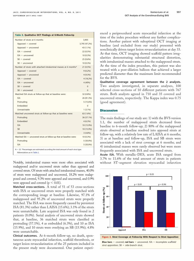
wtmmIwptp(w
3w
J A C C : C A R D I O V A S C U L A R I N T E R V E N T I O N S , V O L . 4 , N O . 9 , 2 0 1 1 Gomez-Lara et al.
S E P T E M B E R 2 0 1 1 : 9 9 2 – 1 0 0 1 OCT Analysis of the Everolimus-Eluting BVS
997
Notably, intraluminal masses were more often associated withmalapposed and/or uncovered struts rather than apposed andcovered struts. Of struts with attached intraluminal masses, 40.0%of struts were malapposed and uncovered, 18.2% were malap-posed and covered, 9.3% were apposed and uncovered, and 0.9%were apposed and covered (p � 0.01).Matched cross-sections. A total of 51 of 53 cross-sections
ith ISA or uncovered struts were properly matched withhe corresponding image at baseline. Likewise, 97.1% ofalapposed and 95.2% of uncovered struts were properlyatched. The ISA was more frequently caused by persistent
SA (81.3%) rather than LAISA (15.6%); 3.1% of the strutsere unmatchable. Late acquired ISA was only found in 2atients (8.0%). Serial analysis of uncovered struts showedhat, at baseline, 36 matched struts were classified asrotruding (57.1%), 4 as embedded (6.3%), and 10 as ISA15.9%), and 10 struts were overlying an SB (15.9%); 4.8%ere unmatchable.
Clinical outcomes. At 6-month follow-up, no death, spon-taneous acute myocardial infarction, scaffold thrombosis, ortarget lesion revascularization of the 25 patients included in
Table 3. Qualitative OCT Findings at 6-Month Follow-Up
Number of struts at 6 months 3,905
Apposed � covered 3,795 (97.2%)
Apposed � uncovered 43 (1.1%)
ISA � covered 22 (0.5%)
ISA � uncovered 10 (0.3%)
SB � covered 25 (0.6%)
SB � uncovered 10 (0.3%)
Number of struts with attached intraluminal masses at 6 months* 21 (0.5%)
Apposed � covered 9 (0.2%)
Apposed � uncovered 4 (9.3%)
ISA � covered 4 (18.2%)
ISA � uncovered 4 (40%)
SB � covered 0
SB � uncovered 0
Matched ISA struts at follow-up that at baseline were: 32 (0.8%)
ISA 26 (81.3%)
Protruding 5 (15.6%)
Embedded 0
Unmatchable 1 (3.1%)
Matched uncovered struts at follow-up that at baseline were: 63 (1.6%)
Protruding 36 (57.1%)
Embedded 4 (6.3%)
ISA 10 (15.9%)
SB 10 (15.9%)
Unmatchable 3 (4.8%)
Matched ISA � uncovered struts at follow-up that at baseline were: 10 (0.3%)
Protruding 5 (50.0%)
ISA 5 (50.0%)
N � 25. *Percentages are estimated according the total amount of each strut type.
Abbreviations as in Table 2.
the present study were documented. One patient experi-
enced a periprocedural acute myocardial infarction at thetime of the index procedure without any further complica-tions. Another patient with suboptimal OCT imaging atbaseline (and excluded from our study) presented withnonclinically driven target lesion revascularization at day 33.At that time, OCT imaging showed scaffold pattern irreg-ularities demonstrating substantial structural distortion,with intraluminal masses attached to the malapposed struts.At the time of the index procedure, this patient was alsotreated with a post-dilation balloon that achieved a largerpredicted diameter than the maximum limit recommendedfor the BVS.Qualitative coverage agreement between the 2 analysts.Two analysts investigated, in separate analyses, 100selected cross-sections of 10 different patients with 767struts. Both analysts agreed in 710 and 35 covered anduncovered struts, respectively. The Kappa index was 0.75(good agreement).
Discussion
The main findings of our study are: 1) with the BVS version1.1, the number of malapposed struts decreased frombaseline to 6-month follow-up; 2) 80% of the malapposedstruts observed at baseline resolved into apposed struts atfollow-up, with a relatively low rate of LAISA at 6 months;3) as at baseline and follow-up, ISA and SB struts wereassociated with a lack of strut coverage at 6 months; and4) intraluminal masses were rarely observed but were morefrequently associated with ISA and uncovered struts.Acute ISA. With metallic-DES, acute ISA ranged from.7% to 11.6% of the total amount of struts in patientsithout ST-segment elevation myocardial infarction
Figure 3. Strut Coverage at Follow-Up With Respect to Strut Apposition
Blue bars � covered; red bars � uncovered. ISA � incomplete scaffold/
strut apposition; SB � side-branch strut.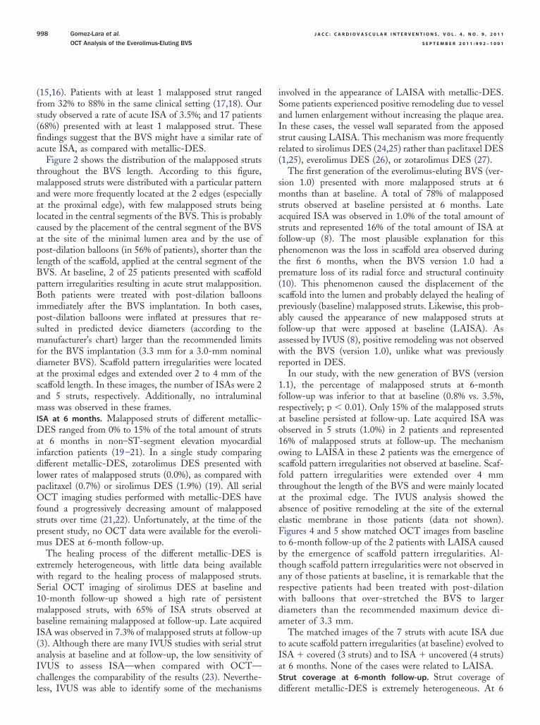
J A C C : C A R D I O V A S C U L A R I N T E R V E N T I O N S , V O L . 4 , N O . 9 , 2 0 1 1
S E P T E M B E R 2 0 1 1 : 9 9 2 – 1 0 0 1
Gomez-Lara et al.
OCT Analysis of the Everolimus-Eluting BVS
998
(15,16). Patients with at least 1 malapposed strut rangedfrom 32% to 88% in the same clinical setting (17,18). Ourstudy observed a rate of acute ISA of 3.5%; and 17 patients(68%) presented with at least 1 malapposed strut. Thesefindings suggest that the BVS might have a similar rate ofacute ISA, as compared with metallic-DES.
Figure 2 shows the distribution of the malapposed strutsthroughout the BVS length. According to this figure,malapposed struts were distributed with a particular patternand were more frequently located at the 2 edges (especiallyat the proximal edge), with few malapposed struts beinglocated in the central segments of the BVS. This is probablycaused by the placement of the central segment of the BVSat the site of the minimal lumen area and by the use ofpost-dilation balloons (in 56% of patients), shorter than thelength of the scaffold, applied at the central segment of theBVS. At baseline, 2 of 25 patients presented with scaffoldpattern irregularities resulting in acute strut malapposition.Both patients were treated with post-dilation balloonsimmediately after the BVS implantation. In both cases,post-dilation balloons were inflated at pressures that re-sulted in predicted device diameters (according to themanufacturer’s chart) larger than the recommended limitsfor the BVS implantation (3.3 mm for a 3.0-mm nominaldiameter BVS). Scaffold pattern irregularities were locatedat the proximal edges and extended over 2 to 4 mm of thescaffold length. In these images, the number of ISAs were 2and 5 struts, respectively. Additionally, no intraluminalmass was observed in these frames.ISA at 6 months. Malapposed struts of different metallic-DES ranged from 0% to 15% of the total amount of strutsat 6 months in non–ST-segment elevation myocardialinfarction patients (19–21). In a single study comparingdifferent metallic-DES, zotarolimus DES presented withlower rates of malapposed struts (0.0%), as compared withpaclitaxel (0.7%) or sirolimus DES (1.9%) (19). All serialOCT imaging studies performed with metallic-DES havefound a progressively decreasing amount of malapposedstruts over time (21,22). Unfortunately, at the time of thepresent study, no OCT data were available for the everoli-mus DES at 6-month follow-up.
The healing process of the different metallic-DES isextremely heterogeneous, with little data being availablewith regard to the healing process of malapposed struts.Serial OCT imaging of sirolimus DES at baseline and10-month follow-up showed a high rate of persistentmalapposed struts, with 65% of ISA struts observed atbaseline remaining malapposed at follow-up. Late acquiredISA was observed in 7.3% of malapposed struts at follow-up(3). Although there are many IVUS studies with serial strutanalysis at baseline and at follow-up, the low sensitivity ofIVUS to assess ISA—when compared with OCT—challenges the comparability of the results (23). Neverthe-
less, IVUS was able to identify some of the mechanisms dinvolved in the appearance of LAISA with metallic-DES.Some patients experienced positive remodeling due to vesseland lumen enlargement without increasing the plaque area.In these cases, the vessel wall separated from the apposedstrut causing LAISA. This mechanism was more frequentlyrelated to sirolimus DES (24,25) rather than paclitaxel DES(1,25), everolimus DES (26), or zotarolimus DES (27).
The first generation of the everolimus-eluting BVS (ver-sion 1.0) presented with more malapposed struts at 6months than at baseline. A total of 78% of malapposedstruts observed at baseline persisted at 6 months. Lateacquired ISA was observed in 1.0% of the total amount ofstruts and represented 16% of the total amount of ISA atfollow-up (8). The most plausible explanation for thisphenomenon was the loss in scaffold area observed duringthe first 6 months, when the BVS version 1.0 had apremature loss of its radial force and structural continuity(10). This phenomenon caused the displacement of thescaffold into the lumen and probably delayed the healing ofpreviously (baseline) malapposed struts. Likewise, this prob-ably caused the appearance of new malapposed struts atfollow-up that were apposed at baseline (LAISA). Asassessed by IVUS (8), positive remodeling was not observedwith the BVS (version 1.0), unlike what was previouslyreported in DES.
In our study, with the new generation of BVS (version1.1), the percentage of malapposed struts at 6-monthfollow-up was inferior to that at baseline (0.8% vs. 3.5%,respectively; p � 0.01). Only 15% of the malapposed strutsat baseline persisted at follow-up. Late acquired ISA wasobserved in 5 struts (1.0%) in 2 patients and represented16% of malapposed struts at follow-up. The mechanismowing to LAISA in these 2 patients was the emergence ofscaffold pattern irregularities not observed at baseline. Scaf-fold pattern irregularities were extended over 4 mmthroughout the length of the BVS and were mainly locatedat the proximal edge. The IVUS analysis showed theabsence of positive remodeling at the site of the externalelastic membrane in those patients (data not shown).Figures 4 and 5 show matched OCT images from baselineto 6-month follow-up of the 2 patients with LAISA causedby the emergence of scaffold pattern irregularities. Al-though scaffold pattern irregularities were not observed inany of those patients at baseline, it is remarkable that therespective patients had been treated with post-dilationwith balloons that over-stretched the BVS to largerdiameters than the recommended maximum device di-ameter of 3.3 mm.
The matched images of the 7 struts with acute ISA dueto acute scaffold pattern irregularities (at baseline) evolved toISA � covered (3 struts) and to ISA � uncovered (4 struts)at 6 months. None of the cases were related to LAISA.Strut coverage at 6-month follow-up. Strut coverage of
ifferent metallic-DES is extremely heterogeneous. At 6
J A C C : C A R D I O V A S C U L A R I N T E R V E N T I O N S , V O L . 4 , N O . 9 , 2 0 1 1 Gomez-Lara et al.
S E P T E M B E R 2 0 1 1 : 9 9 2 – 1 0 0 1 OCT Analysis of the Everolimus-Eluting BVS
999
months, sirolimus DES presented with a rate of uncoveredstruts from 8.7% to 15.0% (19–22). Uncovered struts withpaclitaxel DES ranged from 2.7% to 5.0% (19,20), andzotarolimus DES presented with 0% of uncovered struts ina single report (19).
In our study, with the new generation of BVS (version1.1), a total of 1.6% of struts were uncovered at 6-monthfollow-up. The distribution of uncovered struts throughoutthe BVS length did not show any particular pattern (Fig. 2).However, uncovered struts at follow-up were relatively morefrequently found in struts that at baseline were ISA and SBstruts rather than apposed struts. Similar results have beenobtained with serial OCT imaging of sirolimus DES. Ozaki
Figure 4. Late Acquired ISA
Consecutive, matched OCT images at baseline (A to E) and 6 months (A‘ to Efold pattern is regular. B, C, and D show 1 apposed strut at baseline (arrows)pattern irregularities. Abbreviations as in Figure 2.
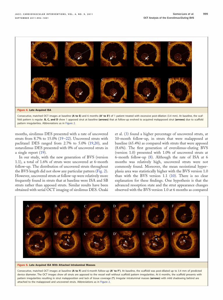
Figure 5. Late Acquired ISA With Attached Intraluminal Masses
Consecutive, matched OCT images at baseline (A to F) and 6-month follow-updevice diameter. The OCT images show all struts are apposed to the vessel wapattern irregularities resulting in strut malapposition and lack of tissue coverag
attached to the malapposed and uncovered struts. Abbreviations as in Figure 2.et al. (3) found a higher percentage of uncovered struts, at10-month follow-up, in struts that were malapposed atbaseline (65.4%) as compared with struts that were apposed(8.6%). The first generation of everolimus-eluting BVS(version 1.0) presented with 1.0% of uncovered struts at6-month follow-up (8). Although the rate of ISA at 6months was relatively high, uncovered struts were notcommonly found. Moreover, the mean neointimal hyper-plasia area was statistically higher with the BVS version 1.0than with the BVS version 1.1 (10). There is no clearexplanation for these findings. One hypothesis is that theadvanced resorption state and the strut appearance changesobserved with the BVS version 1.0 at 6 months as compared
patient treated with excessive post-dilation (3.4 mm). At baseline, the scaf-t follow-up evolved to acquired malapposed strut (arrows) due to scaffold
o F‘). At baseline, the scaffold was post-dilated up to 3.4 mm of predictedout scaffold pattern irregularities. At 6 months, the scaffold presents withIrregular intraluminal masses (arrows) with mild shadowing behind are
‘) of 1that a
(A‘ tll withe (*).
pfspw
J A C C : C A R D I O V A S C U L A R I N T E R V E N T I O N S , V O L . 4 , N O . 9 , 2 0 1 1
S E P T E M B E R 2 0 1 1 : 9 9 2 – 1 0 0 1
Gomez-Lara et al.
OCT Analysis of the Everolimus-Eluting BVS
1000
with the BVS 1.1 could trigger a higher neointimal responseand strut coverage.Intraluminal masses at 6-month follow-up. Lack of neointi-mal coverage of different DES was proposed as the bestpredictor of late stent thrombosis in pathological studies (6).However, although sirolimus DES presented with a higherpercentage of uncovered struts compared with paclitaxelDES, thrombi were more frequently found with paclitaxelDES (20). This observation supports the concept of amulticausal pathogenesis of late stent thrombosis, includingmalapposition, expansive vessel remodeling, and other fac-tors that can initiate thrombus formation (28,29). Due to itshigh resolution, OCT is able to visualize, with great detail,many of the proposed predictors of late stent thrombosis invivo (3,30). With sirolimus DES, Ozaki et al. (3) found4.1% of frames with thrombi at 10-month follow-up;thrombi were more frequently observed in frames with ISAthan in those without this feature (21% vs. 2%, respectively,p � 0.01). Our study found 14 cross-sections (2.9%) in 6
atients (24%) containing intraluminal masses at 6-monthollow-up. Intraluminal masses were more frequently ob-erved in cross-sections with ISA than without (39% vs. 2%;� 0.01) and in cross-sections with uncovered struts thanithout (13% vs. 2%; p � 0.01), supporting that malappo-
sition and absence of coverage can initiate thrombosis.However, whether this is of clinical importance needs to befurther investigated. Figure 5 shows intraluminal massesattached to malapposed and uncovered struts in 1 patientwith acquired scaffold pattern irregularities and LAISA at 6months.Clinical implications. In our study, the new generation ofBVS presented with a lower rate of ISA, uncovered struts,and intraluminal masses as compared with sirolimus DES at6-month follow-up. The polymeric nature of this newtechnology, however, could render it susceptible to iatro-genically induced scaffold pattern irregularities. These canlead to ISA and lack of strut coverage at 6-month follow-up.The 5 cases reported in our study were treated withaggressive post-dilations, probably resulting in overstretch-ing of the scaffold at the time of the implantation. A thirdgeneration of the device is intended to raise the limit ofdeployment of the 3.0-mm nominal diameter device to 3.8mm. Nevertheless, the previous generation of the BVS(version 1.0) showed a complete resorption of its compo-nents at 2-year follow-up without any malapposed oruncovered struts (7). Therefore, in this case, the treatmentwith dual antiplatelet therapy can become unnecessary. It is,however, currently uncertain whether the new generation ofthe BVS (version 1.1) will exhibit similar resorption char-acteristics at 2 years.Study limitations. The first limitation of the present study isthe limited number of patients. However, our report is 1 ofthe largest OCT studies with serial imaging of the same
intracoronary device at baseline and at follow-up. Thesecond limitation is the lack of statistical adjustment forclinical and anatomic covariables. The clustering essence ofthe OCT data (patient level, stent level, frame level, andstrut level) needs a multilevel regression analysis not appliedin our study. Moreover, the different OCT pullback speed,frame/rate, and quality of the image of the 2 OCT systemsused in the present study hamper the matching of cross-sections between baseline and follow-up. A total of 12cross-sections were unmatchable from baseline to follow-upor vice versa: 8 cross-sections were imaged with the M3OCT system and 4 cross-sections were imaged with the C7OCT system in both baseline and follow-up. Finally, thelast limitation is the lack of results at very-long-termfollow-up. The extrapolation of the 2-year follow-up resultsobserved with the first generation of BVS (version 1.0) withthe new generation of BVS (version 1.1) is purely specula-tive. However, all patients included in the ABSORB cohortB study will be reinvestigated at 2-year follow-up withinvasive imaging techniques.
Conclusions
The new generation of BVS exhibited a low rate of acute,persistent, and late acquired incomplete strut/scaffoldmalapposition as well as uncovered struts. At baseline andfollow-up, malapposed and SB struts were related to a lackof tissue coverage at 6 months. Scaffold pattern irregularitieswere the only cause observed in our study of late acquiredmalapposed struts and were also related to a lack of tissuecoverage. Attached intraluminal masses occurred more fre-quently in malapposed and uncovered struts at 6 months. Amore careful device implantation, with appropriate sizing ofthe vessel and respecting the deployment limits of inflation,will reduce the rates of acute ISA and scaffold patternirregularities and, therefore, could circumvent most of theISA, uncovered struts, and intraluminal masses observed atmidterm follow-up.
Reprint requests and correspondence: Dr. Patrick W. Serruys,Thoraxcenter, Erasmus University Medical Center, ‘s-Gravendijkwal230, Ba-583, 3015 CE Rotterdam, the Netherlands. E-mail:[email protected].
REFERENCES
1. Tanabe K, Serruys PW, Degertekin M, et al. Incomplete stentapposition after implantation of paclitaxel-eluting stents or bare metalstents: insights from the randomized TAXUS II trial. Circulation2005;111:900–5.
2. Degertekin M, Serruys PW, Tanabe K, et al. Long-term follow-up ofincomplete stent apposition in patients who received sirolimus-elutingstent for de novo coronary lesions: an intravascular ultrasound analysis.Circulation 2003;108:2747–50.
3. Ozaki Y, Okumura M, Ismail TF, et al. The fate of incomplete stentapposition with drug-eluting stents: an optical coherence tomography-
based natural history study. Eur Heart J 2010;31:1470–6.
J A C C : C A R D I O V A S C U L A R I N T E R V E N T I O N S , V O L . 4 , N O . 9 , 2 0 1 1 Gomez-Lara et al.
S E P T E M B E R 2 0 1 1 : 9 9 2 – 1 0 0 1 OCT Analysis of the Everolimus-Eluting BVS
1001
4. Matsumoto D, Shite J, Shinke T, et al. Neointimal coverage ofsirolimus-eluting stents at 6-month follow-up: evaluated by opticalcoherence tomography. Eur Heart J 2007;28:961–7.
5. Cook S, Wenaweser P, Togni M, et al. Incomplete stent appositionand very late stent thrombosis after drug-eluting stent implantation.Circulation 2007;115:2426–34.
6. Finn AV, Joner M, Nakazawa G, et al. Pathological correlates of latedrug-eluting stent thrombosis: strut coverage as a marker of endothe-lialization. Circulation 2007;115:2435–41.
7. Serruys PW, Ormiston JA, Onuma Y, et al. A bioabsorbableeverolimus-eluting coronary stent system (ABSORB): 2-year outcomesand results from multiple imaging methods. Lancet 2009;373:897–910.
8. Ormiston JA, Serruys PW, Regar E, et al. A bioabsorbable everolimus-eluting coronary stent system for patients with single de-novo coronaryartery lesions (ABSORB): a prospective open-label trial. Lancet 2008;371:899–907.
9. Okamura T, Garg S, Gutierrez-Chico JL, et al. In vivo evaluation ofstent strut distribution patterns in the bioabsorbable everolimus-elutingdevice: an OCT ad hoc analysis of the revision 1.0 and revision 1.1stent design in the ABSORB clinical trial EuroIntervention 2010;5:932–8.
10. Gomez-Lara J, Brugaletta S, Diletti R, et al. A comparative assessmentby optical coherence tomography of the performance of the first andsecond generation of the everolimus-eluting bioresorbable vascularscaffolds. Eur Heart J 2011;32:294–304.
11. Brugaletta S, Garcia-Garcia HM, Diletti R, et al. Comparison betweenthe first and second generation bioresorbable vascular scaffolds: a sixmonth virtual histology study. EuroIntervention 2010;6:1110–6.
12. Serruys PW, Onuma Y, Ormiston JA, et al. Evaluation of the secondgeneration of a bioresorbable everolimus drug-eluting vascular scaffoldfor treatment of de novo coronary artery stenosis: six-month clinicaland imaging outcomes. Circulation 2010;122:2301–12.
13. Cohen J. A coefficient of agreement for nominal scales. Educ PsycholMeas 1960;20:37–46.
14. Fleiss J. Statistical Methods for Rates and Proportions. 2nd edition.New York, NY: John Wiley, 1981.
15. Tanigawa J, Barlis P, Dimopoulos K, Dalby M, Moore P, Di Mario C.The influence of strut thickness and cell design on immediate apposi-tion of drug-eluting stents assessed by optical coherence tomography.Int J Cardiol 2009;134:180–8.
16. Kim JS, Jang IK, Fan C, et al. Evaluation in 3 months duration ofneointimal coverage after zotarolimus-eluting stent implantation byoptical coherence tomography: the ENDEAVOR OCT trial. J AmColl Cardiol Intv 2009;2:1240–7.
17. Kubo T, Imanishi T, Kitabata H, et al. Comparison of vascularresponse after sirolimus-eluting stent implantation between patients
with unstable and stable angina pectoris: a serial optical coherencetomography study. J Am Coll Cardiol Img 2008;1:475–84.18. Tanigawa J, Barlis P, Dimopoulos K, Di Mario C. Optical coherencetomography to assess malapposition in overlapping drug-eluting stents.EuroIntervention 2008;3:580–3.
19. Guagliumi G, Musumeci G, Sirbu V, et al. Optical coherencetomography assessment of in vivo vascular response after implantationof overlapping bare-metal and drug-eluting stents. J Am Coll CardiolIntv 2010;3:531–9.
20. Murakami D, Takano M, Yamamoto M, et al. Advanced neointimalgrowth is not associated with a low risk of in-stent thrombus. Opticalcoherence tomographic findings after first-generation drug-elutingstent implantation. Circ J 2009;73:1627–34.
21. Katoh H, Shite J, Shinke T, et al. Delayed neointimalization onsirolimus-eluting stents: 6-month and 12-month follow up by opticalcoherence tomography. Circ J 2009;73:1033–7.
22. Ishigami K, Uemura S, Morikawa Y, et al. Long-term follow-up ofneointimal coverage of sirolimus-eluting stents: evaluation with opticalcoherence tomography. Circ J 2009;73:2300–7.
23. Bouma BE, Tearney GJ, Yabushita H, et al. Evaluation of intracoro-nary stenting by intravascular optical coherence tomography. Heart2003;89:317–20.
24. Ako J, Morino Y, Honda Y, et al. Late incomplete stent appositionafter sirolimus-eluting stent implantation: a serial intravascular ultra-sound analysis. J Am Coll Cardiol 2005;46:1002–5.
25. Hong MK, Mintz GS, Lee CW, et al. Late stent malapposition afterdrug-eluting stent implantation: an intravascular ultrasound analysiswith long-term follow-up. Circulation 2006;113:414–9.
26. Tsuchiya Y, Lansky AJ, Costa RA, et al. Effect of everolimus-elutingstents in different vessel sizes (from the pooled FUTURE I and IItrials). Am J Cardiol 2006;98:464–9.
27. Miyazawa A, Ako J, Hongo Y, et al. Comparison of vascular responseto zotarolimus-eluting stent versus sirolimus-eluting stent: intravascu-lar ultrasound results from ENDEAVOR. Am Heart J 2008;III:155:108–13.
28. Oikawa Y, Yajima J, Costa M, et al. Intravascular ultrasound, angio-scopic and histopathological characterization of heterogeneous patternsof restenosis after sirolimus-eluting stent implantation: insights intopotential “thromborestenosis” phenomenon. EuroIntervention 2010;6:380–7.
29. Joner M, Finn AV, Farb A, et al. Pathology of drug-eluting stents inhumans: delayed healing and late thrombotic risk. J Am Coll Cardiol2006;48:193–202.
30. Otake H, Shite J, Ako J, et al. Local determinants of thrombusformation following sirolimus-eluting stent implantation assessed byoptical coherence tomography. J Am Coll Cardiol Intv 2009;2:459–66.
Key Words: bioresorbable scaffolds � incomplete stent �late acquired incomplete stent � optical coherence tomog-
raphy � strut apposition � uncovered struts.