kissing balloon inflation in percutaneous coronary...
TRANSCRIPT
J A C C : C A R D I O V A S C U L A R I N T E R V E N T I O N S V O L . 5 , N O . 8 , 2 0 1 2
© 2 0 1 2 B Y T H E A M E R I C A N C O L L E G E O F C A R D I O L O G Y F O U N D A T I O N I S S N 1 9 3 6 - 8 7 9 8 / $ 3 6 . 0 0
P U B L I S H E D B Y E L S E V I E R I N C . h t t p : / / d x . d o i . o r g / 1 0 . 1 0 1 6 / j . j c i n . 2 0 1 2 . 0 6 . 0 0 5
STATE-OF-THE-ART PAPER
Kissing Balloon Inflation inPercutaneous Coronary Interventions
Gregory A. Sgueglia, MD, PHD,* Bernard Chevalier, MD†
Latina, Italy; and Massy, France
Bifurcation lesions are the most frequently approached complex coronary lesions in everyday interven-
tional practice. Bifurcations complexity relies essentially on their very specific anatomy that is imper-
fectly handled by current coronary devices and, despite dedicated techniques and drug-eluting stents,
percutaneous coronary interventions directed toward the treatment of bifurcations are technically de-
manding and require proper execution. Kissing balloon (KB) inflation was the first specific bifurcation
technique to have been developed for percutaneous bifurcation interventions and continues to cur-
rently play an important role. Indeed, KB has been proposed to optimize stent apposition, improve
side branch access while correcting stent deformation or distortion. Over the years, the KB technique
has been deeply investigated by many different methods, from bench testing and computer simula-
tions to in vivo intravascular imaging and clinical studies, producing a large amount of data pointing
out the benefits and limitations of the technique. We sought to provide here a comprehensive over-
view of all those aspects. (J Am Coll Cardiol Intv 2012;5:803–11) © 2012 by the American College of
Cardiology Foundation
Among complex coronary lesions, bifurcations arethose most frequently encountered by every inter-ventional cardiologist. Bifurcation complexity es-sentially relies on their specific anatomic configu-ration, which is imperfectly handled by currentcoronary devices.
Until the advent of drug–eluting stents (DES)and dedicated techniques, percutaneous bifurcationinterventions were associated with very high ratesof unfavorable outcomes (1,2). Nevertheless, pro-cedures directed to bifurcation treatment are oftentechnically demanding and require proper execu-tion. When implementing dedicated percutaneousbifurcation approaches, kissing balloon (KB) hasbeen variably recommended to optimize stent ap-position, correct stent deformation or distortion,and improve side branch (SB) access. Over the
From *Interventional Cardiology, Ospedale Santa Maria Goretti, Latina,Italy; and †Interventional Cardiology, Institut Cardiovasculaire ParisSud, Massy, France. Dr. Sgueglia has reported that he has no relation-ships relevant to the contents of this paper to disclose. Dr. Chevalier isa consultant for Abbott Vascular, Medtronic, and Terumo.
Manuscript received March 8, 2012; revised manuscript received May 8,2012, accepted June 7, 2012.
years, KB has been deeply investigated by manydifferent methods, from bench testing and com-puter simulations to in vivo intravascular imagingand clinical studies that have produced a largeamount of data.
We review the rationale of KB and findings fromdedicated studies, aiming to provide an updated andcomprehensive overview of this technique.
Anatomy of Bifurcation Lesions
A coronary bifurcation is a branching artery con-stituted by a main vessel (MV) and a SB. Thesegment before the origin of the SB is referred asproximal MV, whereas the one that is distal to it isreferred as distal MV (Fig. 1). The tissue mem-brane separating the origins of the 2 bifurcationarms is called the flow divider or carina.
Operative definitions of bifurcation lesions havebeen based on the SB diameter, either arbitrarily orin relation to potential blood supply. Actually, abifurcation stenosis is defined as a coronary arterynarrowing occurring adjacent to and/or involving
the origin of a significant SB (3). To be significant,
J A C C : C A R D I O V A S C U L A R I N T E R V E N T I O N S , V O L . 5 , N O . 8 , 2 0 1 2
A U G U S T 2 0 1 2 : 8 0 3 – 1 1
Sgueglia and Chevalier
Kissing Balloon Inflation
804
the SB has to be considered important in the individualpatient according to symptoms, location of ischemia, vital-ity, collateral vessels, and left ventricular function.
Morphology classification is mainly based on plaquedistribution. Indeed, plaque distribution can variably involvethe proximal MV, the distal MV, or the SB. This hasengendered at least 6 different classification schemes (4–9).Sometimes, branching arteries are called “true” rather than“false” bifurcations according to the mere presence orabsence of significant SB stenosis. Pathological examinationof coronary arteries reveals that the atherosclerotic plaquesare mainly located in areas of low shear stress, such as thelateral walls of the MV and SB, whereas they are lesscommon at the carina level, which is characterized by highshear stress.
The spatial relation between the 2 arms of the bifurcationcan be defined by 3 angles (Fig. 1) that have been recently
named A (the angle between theproximal MV and the SB), B(the angle between the SB andthe distal MV), and C (the anglebetween the proximal and distalsegment of the MV). At times,bifurcations are defined as V- orT-type according to angle B be-ing �70° or �70°, respectively.Moreover, the proximal and dis-tal branches of a bifurcation of-ten do not lie on a single plane,thus posing significant chal-lenges to quantitative coronaryangiography software.
A recent ex vivo study of poly-mer casts of human coronaryarteries has revealed a complexcurvilinear transition zone be-tween MV and SB, mainly char-
acterized by an elliptical and asymmetrical configuration ofthe SB ostium and brief tapering of the SB origin (10).Moreover, it has been previously pointed out that SBostium asymmetry increases with increasing bifurcationangles (11). In bifurcations, there is also an asymmetricalgeometric reduction according to the law of conservationof energy (12).
The complex interaction among different factors makesevery bifurcation lesion quite unique (Fig. 1), althoughcertain lesion characteristics have been associated withtreatment success when using currently accepted techniquesand DES platforms (13).
The Need for KB
Bifurcation lesions, by their anatomy, expose the patient to
Abbreviationsand Acronyms
CI � confidence interval
DES � drug-eluting stent(s)
FFR � fractional flowreserve
IVUS � intravascularultrasound
KB � kissing balloon(s)
MV � main vessel
OCT � optical coherencetomography
SB � side branch(es)
TIMI � Thrombolysis InMyocardial Infarction
TLR � target lesionrevascularization
the risk of SB damage, defined as worsening of percent
stenosis, and in some cases, SB occlusion (14). Differentmechanisms have been suggested to explain SB damage,such as plaque or carina shift, refractory spasm, or dissectionof the ostium. In the case of SB occlusion, myocardialnecrosis could ensue, being associated with a worse short-and long-term clinical outcome with elevation of bothcreatine kinase–myocardial band isoform and cardiactroponin levels (15–17). Despite the fact that mostacutely occluded SBs undergo late spontaneous reperfu-sion (18), temporary occlusion causes myocardial enzymeelevation.
In the case of SB stenosis, myocardial ischemia mightensue with persistence of symptoms or mechanical dysfunc-tion. In a recent prospective study of patients with bifurca-tion lesions successfully treated by DES implantation ac-cording to the provisional approach, significant SB stenosiswas present in about 20% of patients as assessed by3-dimensional quantitative coronary angiography. Thesepatients had a significantly increased rate of late inducibleischemia and minor adverse coronary events (19). Angio-graphic (20) and intravascular ultrasound (IVUS) (21)predictors of SB damage have been described, with furtherinsights recently provided by 3-dimensional optical coher-ence tomography (OCT) (22).
Figure 1. The Complexity of Bifurcation Lesions
Main aspects of anatomic complexity of bifurcation lesions include variabledistribution of atherosclerosis, variable spatial relation between thebranches defined by angles A, B, and C, the tapered nature of the sidebranch as reflected by a bigger ostial diameter, and the asymmetrical geo-metric reduction of the vessel diameter at the bifurcation site. MV � main
vessel; SB � side branch.
dsfipacrdtssP(1raosceiswwbsca
mipgg
G
J A C C : C A R D I O V A S C U L A R I N T E R V E N T I O N S , V O L . 5 , N O . 8 , 2 0 1 2 Sgueglia and Chevalier
A U G U S T 2 0 1 2 : 8 0 3 – 1 1 Kissing Balloon Inflation
805
History of KB
The term kissing balloon was first used by Gruentzig toescribe the percutaneous treatment of an iliac bifurcationtenosis (23). In 1980, Velasquez et al. (24) published therst report of this technique for distal aorta angioplasty in aatient with Leriche syndrome. One year later, Gruentzigpplied the KB technique to percutaneous coronary revas-ularization (25). At that time, 2 guiding catheters wereequired, each inserted through a single vascular access, andespite the name of the technique, the simultaneous infla-ion of the 2 balloons was not the routine: rather, repeatedequential inflation of the MV and SB balloons was deemedafer in regard to the risk of vessel dissection (26,27).ioneering experiences were positively reported by Meier
25), Zack et al. (26), and Pinkerton et al. (28) in 3, 8, and3 patients, respectively. In 1986, George et al. (29)eported their experience with KB through a brachiofemoralpproach in 52 selected patients, with a procedural successbtained in 98% of them. To avoid a dual guiding catheterystem, a single-guide, two-wire technique, sometimesalled kissing wire, has been advocated as a simpler, butqually effective, approach to SB preservation (30). Advanc-ng technology has rapidly made KB possible through aingle guiding catheter using either 2 balloons with fixedires (31), a balloon with a fixed wire and a balloon over theire (23), 2 balloons over the wire (32), or 2 rapid exchangealloons (33). In 1996, Krikorian et al. (34) proposed aimplification of the technique with a single inflation deviceonnected to the 2 balloons through a 3-way stopcock,llowing for single-operator interventions.
Following the introduction of coronary stents and refine-ent of the technology, the rate of KB progressively
ncreased (6). Actually, KB can be performed with noncom-liant balloons (35) and drug-eluting balloons (36) in a 6-Fuiding catheter and with special equipment in a 5-Fuiding catheter (37).
Figure 2. Main Vessel Stent Distortion Without Kissing Balloon
Bench testing has shown that balloon dilation through the side of themain vessel stent to open a cell toward the side branch determinesmarked distortion of the stent itself (arrow).
aining Insight Into KB
KB modifies the geometry of the implanted stent dependingon many factors, including balloon size, inflation pressure,and deflation sequence.Bench testing. One of the most important contributions ofbench testing to the better understanding of bifurcationstenting is the demonstration by Ormiston et al. (38,39)that balloon dilation through the side of the MV stent toopen a cell toward the SB results in marked distortion of thestent itself (Fig. 2). This important issue has been shown tobe either prevented or corrected by KB. Accordingly, if theballoons chosen for the kissing inflation are too small, theMV stent will be distorted. Moreover, this finding under-scores that the SB balloon should be deflated at the sametime as the MV balloon to avoid MV stent deformation.However, the sensitivity to SB dilation in terms of MV stentdistortion might vary according to specific designs (40,41).
KB can also provide optimal scaffolding of the SB ostiumwhen care is taken to properly rewire the SB. In theprovisional technique, bench testing has shown that wirecrossing through the cell closest to the carina provides betterscaffolding than proximal crossing (Fig. 3).
By contrast, when implementing the crush technique, it ishighly advisable to cross the proximal cell. Indeed, benchtests have shown that when stents are crushed, there is atrough between the MV and SB stents on the side oppositeto the crushed portion (42–45). If a wire recrosses the MVstent through a distal strut, it may pass outside the stentsthrough the trough before entering the SB stent. If inflated,a post-dilation balloon would push the struts aside, produc-ing a gap in coverage between the stents at the level of the
Figure 3. Influence of Main Vessel Stent Cell Rewiring on Stent Deforma-tion Following Kissing Balloon
Access to the side branch through the strut of a stent is usually possiblethrough 2 or 3 different cells. The cell choice affects stent deformation.Bench testing has shown that wire crossing through the strut closest to thecarina (C and D) provides better scaffolding of the origin of the sidebranch than proximal crossing that pushes the struts inward towards themain vessel lumen (A and B).
carina. Moreover, a 2-step post-dilation involving a high-

mrepsstpsSa
hthttifpcc
ttd(c
I1Pcg
ttcuoK
t
J A C C : C A R D I O V A S C U L A R I N T E R V E N T I O N S , V O L . 5 , N O . 8 , 2 0 1 2
A U G U S T 2 0 1 2 : 8 0 3 – 1 1
Sgueglia and Chevalier
Kissing Balloon Inflation
806
pressure post-dilation in the SB followed by final KBsignificantly reduced the ostial stenosis as compared with a1-step post-dilation by KB. This is especially true for sharpSB angles (42–44).
Bench tests have also provided evidence on the limita-tions linked to a specific stent design (40) and to the KBtechnique itself. Indeed, it has been shown that KB deter-mines coating damage to first-generation DES and ellipticaldeformation of the stent proximally to the SB (46) and thatis corrected by final post-dilation of the proximal part of theMV stent (47). Overlapping configuration of the KB hasalso been shown to influence the stent deformation (45).
Finally, bench testing has recently been used to gaininsight into the influence of flow patterns in stentedcoronary bifurcations with a silicone bifurcation modelpositioned within a closed-flow loop system mimicking theflow conditions of human arterial circulation (48). In thismodel, KB corrected the systolic flow disturbance inducedby stent implantation.Finite element analysis. Computer simulation allows assess-
ent of physical structures through the building of geomet-ic models incorporating realistic material behavior. Finitelement analysis has recently provided valuable inside intoercutaneous bifurcation interventions. Indeed, it has beenhown that the relative position of the deployed MV stenttrongly affects the occurring strut deformations, with op-imal SB access being obtained only if a cell was centrallylaced with respect to the SB ostium (49,50). Moreover, thetent cell design significantly affects strut apposition afterB dilation, pointing toward mandatory KB when dilatingn open-cell stent (49).
Recently, a very elegant simulation by Mortier et al. (51)as highlighted that KB induces elliptical deformation ofhe proximal segment of the MV stent with consequentigh vessel wall stress and possible direct vessel wall injury athe entry of the SB. However, KB simulations with aapered balloon for the SB have shown a significant decreasen the MV stent overexpansion (52). Finally, it has beenound that despite KB, a high proportion of struts at theroximal MV stent edge remained incompletely apposed asompared with simple MV stenting without opening theell toward the SB (53).Computational flow dynamics. Computers can also applynumerical methods and algorithms to analyze the interac-tion of fluid with definite surfaces.
In a computed model of a 90° bifurcation treated byT-stenting, flow features were characterized by flow stasisand recirculation areas downstream from the bifurcation,depending on the way the cell facing the SB was openedaccording to its variable position with respect to the SBitself. In absence of final KB, the stent struts protruding intothe lumen of the MV induced high values of shear stress at
the stent wall (54). fiRecently, an innovative approach consisting in the devel-opment of a sequential model in which the structuralsimulations are used to build the fluid domains highlightedthe advantages of final KB in terms of better flow pattern(52). Indeed, by removing the stent struts from the bloodflow, final KB freed the access to the SB and lowered thehemodynamic disturbance that were present after the mereimplantation of a stent on the MV. Of note, flow alterationin stented bifurcations has been shown to significantlyinfluence the interaction between the eluted drug and thevessel wall (55).
Success and Safety of KB
The BBC ONE study (British Bifurcation Coronary Study:Old, New and Evolving Strategies) randomized 500 pa-tients to either a simple stenting procedure with optionalKB or a complex procedure (either crush or culotte) withmandatory KB (56). The reported rate of attempted andsuccessful final KB is 31% and 29% in the provisional groupand 90% and 76% in the crush group, respectively. Overall,KB success was 95% in the simple approach and 85% in thecomplex approach (p � 0.01).
So far, only 1 complication possibly related to the KBprocedure has been described in the published reports.Indeed, an intramural hematoma was reported in a patienton warfarin therapy (international normalized ratio: 3.3)treated by KB after a stent implantation on the left maincoronary artery across the left circumflex artery (57).
Imaging Assessment of KB
One-stent strategy. In a serial IVUS study on 23 patientsreated by a 1-stent strategy followed by SB dilation andhen final KB, dilation of the SB introduced geometricistortion of the distal MV stent and a 12% loss in stent area58). After KB, stent geometry was not fully restored, andomplete recovery of the stent area did not occur.
In the CORPAL (Cordoba & Las Palmas) Kiss trial,VUS findings were assessed in 101 patients treated by a-stent technique for coronary bifurcation disease (59).atients randomized to KB showed a larger proximal stentross-sectional area than did the patients from the non-KBroup, suggesting overexpansion of the proximal MV stent.
Recently, OCT has been used to point out the impor-ance of KB after MV stenting (60) and to confirm in vivohe importance of recrossing the MV stent through the celllosest to the carina (61). Importantly, OCT has recentlynderlined a high rate of uncovered struts across the SBstium when simple MV stenting is performed without finalB (62).
2-stent strategy. Twenty-five patients treated by crushingechnique underwent IVUS analysis, and in 23 of them,
nal KB was performed. At IVUS, most SB lesions showed
fgwsFt6
lae
Bp
pfi(tp
C
bfsm(sr
Ka
J A C C : C A R D I O V A S C U L A R I N T E R V E N T I O N S , V O L . 5 , N O . 8 , 2 0 1 2 Sgueglia and Chevalier
A U G U S T 2 0 1 2 : 8 0 3 – 1 1 Kissing Balloon Inflation
807
angiographically unsuspected stent underexpansion, withthe smallest minimal stent area found at the SB ostium andfrequent incomplete stent apposition in the crush area (63).
Another serial IVUS study compared the results ofclassical crush and double-crush technique at the end of theprocedure and at 8-month follow-up (64). Incomplete crushwas observed in 81.3% of the patients in the classical crushgroup compared with 38.5% in the double-crush group (p �0.004). The post-procedure symmetry index was higher inthe double-crush technique than in classical crush, both atthe level of the MV stent and at the SB ostium.
A recent IVUS study has shown that the quality of theKB technique, in addition to its simple performance, sig-nificantly impacts the clinical outcome following crushstenting (65). Indeed, rewiring proximal rather than distalto the carina significantly predicted SB restenosis (hazardratio: 2.34, 95% confidence interval [CI]: 1.78 to 4.32, p �0.001).
Functional Assessment of KB
In patients treated by a 1-stent technique, fractional flowreserve (FFR) measured in the jailed SB was compared withquantitative coronary angiography results showing a nega-tive correlation between percent stenosis and FFR (r ��0.41, p � 0.001). However, there was a wide variation ofunctional significance even among lesions with angio-raphically significant stenosis, with only 27% of lesionsith �75% stenosis being functionally significant (66). In a
ubsequent study, KB has been performed in 26 lesions withFR �0.75 showing achievement of FFR �0.75 in 92% of
hem (67). Notably, this functional gain was maintained at-month follow-up.In a study of 60 patients treated by provisional stenting,
ack of KB inflation was the only technical factor associatedt univariate analysis with post-procedural inducible isch-mia as assessed by exercise stress test (19).
Very recently, a FFR substudy of the Nordic-Balticifurcation Study III showed that among 75 participatingatients, FFR measured in the SB at the end of the
Table 1. Summary of Clinical Trials Assessing the Clinical Utility of KB Infl
First Author/Study(Ref. #)
StentingStrategy n
Follow-UpLength
Cardiac Dea(KB vs. Non-K
Ge et al. (75) Complex KB (n � 116) vs.non-KB (n � 65)
9 months 1.7% vs. 0%
THUEBIS (70) Simple KB (n � 56) vs.non-KB (n � 54)
6 months 0% vs. 3.7%
Nordic III (71) Simple KB (n � 238) vs.non-KB (n � 239)
6 months 0.8% vs. 0%
CORPAL Kiss (59) Simple KB (n � 124) vs.non-KB (n � 120)
12 months 0.8% vs. 1.7%
*p � 0.008; ††definite stent thrombosis.
KB � kissing balloon; MACE � major adverse cardiac events; PCI � percutaneous coronary intervention
rocedure showed a significantly higher mean value in thenal KB group as compared with the non-final KB group0.92 vs. 0.85, respectively, p � 0.011) (68). Interestingly,he absence of final KB was a strong predictor of post-rocedural FFR �0.75 (p � 0.006).
linical Assessment of KB
One-stent strategy. In the bare-metal stent era, KB haseen shown to be associated with improved outcomesollowing provisional stenting (6). Moreover, in a smalltudy on 59 patients undergoing MV stenting, SB compro-ise defined as Thrombolysis In Myocardial Infraction
TIMI) flow grade �3 was significantly higher usingequential balloon inflation than after KB (33% vs. 0%,espectively, p � 0.003), although the rate of target lesion
revascularization (TLR) at 6-month follow-up was notdifferent between the 2 groups (69).
THUEBIS (Thueringer Bifurcation Study) compared astrategy of percutaneous bifurcation intervention by provi-sional stenting and final KB with an approach consisting ofprovisional stenting, with SB dilation only in case of TIMIflow grade �2 in 110 patients (70). Paclitaxel-eluting stentswere implanted in all patients, and dual antiplatelet therapywas prescribed for at least 6 months. At 6-month follow-up,no significant differences in the incidence of major adversecardiac events was observed between the 2 groups (Table 1).Notably, in 10 patients randomized to final KB per proto-col, SB could not be rewired, and in 7 of 54 patientsrandomized to final KB, balloon inflation was actuallysequential rather than simultaneous. Overall, 31% of pa-tients randomized to KB did not receive this treatment, thusimpairing results interpretation.
In the Nordic-Baltic Bifurcation Study III, 477 patientswith a bifurcation lesion were randomized to KB (n � 238)or non-KB (n � 239) after MV stenting with sirolimus-eluting stent (71). At 6-month follow-up, the rates of majoradverse cardiac events were 2.1% and 2.5% (p � 1.00) in the
B and non-KB groups, respectively (Table 1). At 8-monthngiographic follow-up in 326 patients, a trend was ob-
in PCI
yocardial Infarction(KB vs. Non-KB)
Target LesionRevascularization(KB vs. Non-KB)
MACE(KB vs. Non-KB)
Definite/ProbableStent Thrombosis(KB vs. Non-KB)
10.3% vs. 13.9% 9.5% vs. 24.6%* 19.8% vs. 38.5%* 2.6% vs. 3.1%
3.6% vs. 1.9% 17.9% vs. 14.8% 23.2% vs. 24.1% 3.6% vs. 1.9%
0.4% vs. 1.3% 1.3% vs. 1.7% 2.1% vs. 2.5% 0.4% vs. 0.4%†
3.2% vs. 1.7% 4.0% vs. 1.7% 9% vs. 6% 0.8% vs. 0.8%
ation
thB)
M
.
oto2afi
ssglm
itmK
cds
(cd
neb8p
S
J A C C : C A R D I O V A S C U L A R I N T E R V E N T I O N S , V O L . 5 , N O . 8 , 2 0 1 2
A U G U S T 2 0 1 2 : 8 0 3 – 1 1
Sgueglia and Chevalier
Kissing Balloon Inflation
808
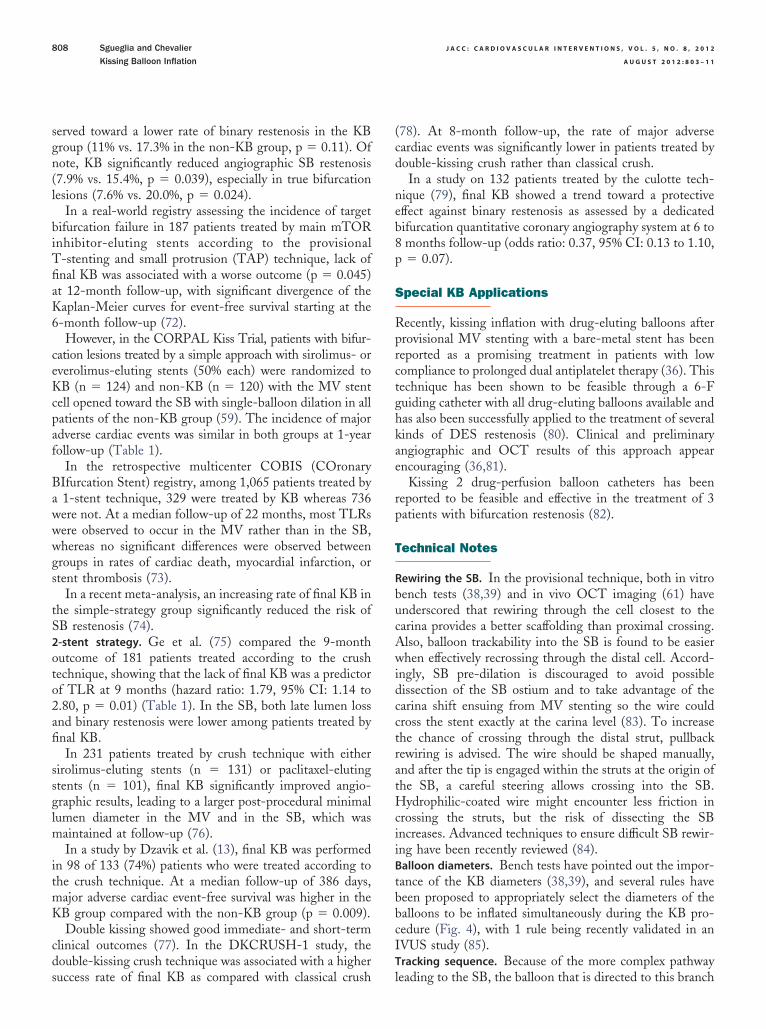
served toward a lower rate of binary restenosis in the KBgroup (11% vs. 17.3% in the non-KB group, p � 0.11). Ofnote, KB significantly reduced angiographic SB restenosis(7.9% vs. 15.4%, p � 0.039), especially in true bifurcationlesions (7.6% vs. 20.0%, p � 0.024).
In a real-world registry assessing the incidence of targetbifurcation failure in 187 patients treated by main mTORinhibitor-eluting stents according to the provisionalT-stenting and small protrusion (TAP) technique, lack offinal KB was associated with a worse outcome (p � 0.045)at 12-month follow-up, with significant divergence of theKaplan-Meier curves for event-free survival starting at the6-month follow-up (72).
However, in the CORPAL Kiss Trial, patients with bifur-cation lesions treated by a simple approach with sirolimus- oreverolimus-eluting stents (50% each) were randomized toKB (n � 124) and non-KB (n � 120) with the MV stentcell opened toward the SB with single-balloon dilation in allpatients of the non-KB group (59). The incidence of majoradverse cardiac events was similar in both groups at 1-yearfollow-up (Table 1).
In the retrospective multicenter COBIS (COronaryBIfurcation Stent) registry, among 1,065 patients treated bya 1-stent technique, 329 were treated by KB whereas 736were not. At a median follow-up of 22 months, most TLRswere observed to occur in the MV rather than in the SB,whereas no significant differences were observed betweengroups in rates of cardiac death, myocardial infarction, orstent thrombosis (73).
In a recent meta-analysis, an increasing rate of final KB inthe simple-strategy group significantly reduced the risk ofSB restenosis (74).2-stent strategy. Ge et al. (75) compared the 9-monthutcome of 181 patients treated according to the crushechnique, showing that the lack of final KB was a predictorf TLR at 9 months (hazard ratio: 1.79, 95% CI: 1.14 to.80, p � 0.01) (Table 1). In the SB, both late lumen lossnd binary restenosis were lower among patients treated bynal KB.In 231 patients treated by crush technique with either
irolimus-eluting stents (n � 131) or paclitaxel-elutingtents (n � 101), final KB significantly improved angio-raphic results, leading to a larger post-procedural minimalumen diameter in the MV and in the SB, which was
aintained at follow-up (76).In a study by Dzavik et al. (13), final KB was performed
n 98 of 133 (74%) patients who were treated according tohe crush technique. At a median follow-up of 386 days,ajor adverse cardiac event-free survival was higher in theB group compared with the non-KB group (p � 0.009).Double kissing showed good immediate- and short-term
linical outcomes (77). In the DKCRUSH-1 study, theouble-kissing crush technique was associated with a higher
uccess rate of final KB as compared with classical crush l78). At 8-month follow-up, the rate of major adverseardiac events was significantly lower in patients treated byouble-kissing crush rather than classical crush.In a study on 132 patients treated by the culotte tech-
ique (79), final KB showed a trend toward a protectiveffect against binary restenosis as assessed by a dedicatedifurcation quantitative coronary angiography system at 6 tomonths follow-up (odds ratio: 0.37, 95% CI: 0.13 to 1.10,� 0.07).
pecial KB Applications
Recently, kissing inflation with drug-eluting balloons afterprovisional MV stenting with a bare-metal stent has beenreported as a promising treatment in patients with lowcompliance to prolonged dual antiplatelet therapy (36). Thistechnique has been shown to be feasible through a 6-Fguiding catheter with all drug-eluting balloons available andhas also been successfully applied to the treatment of severalkinds of DES restenosis (80). Clinical and preliminaryangiographic and OCT results of this approach appearencouraging (36,81).
Kissing 2 drug-perfusion balloon catheters has beenreported to be feasible and effective in the treatment of 3patients with bifurcation restenosis (82).
Technical Notes
Rewiring the SB. In the provisional technique, both in vitrobench tests (38,39) and in vivo OCT imaging (61) haveunderscored that rewiring through the cell closest to thecarina provides a better scaffolding than proximal crossing.Also, balloon trackability into the SB is found to be easierwhen effectively recrossing through the distal cell. Accord-ingly, SB pre-dilation is discouraged to avoid possibledissection of the SB ostium and to take advantage of thecarina shift ensuing from MV stenting so the wire couldcross the stent exactly at the carina level (83). To increasethe chance of crossing through the distal strut, pullbackrewiring is advised. The wire should be shaped manually,and after the tip is engaged within the struts at the origin ofthe SB, a careful steering allows crossing into the SB.Hydrophilic-coated wire might encounter less friction incrossing the struts, but the risk of dissecting the SBincreases. Advanced techniques to ensure difficult SB rewir-ing have been recently reviewed (84).Balloon diameters. Bench tests have pointed out the impor-tance of the KB diameters (38,39), and several rules havebeen proposed to appropriately select the diameters of theballoons to be inflated simultaneously during the KB pro-cedure (Fig. 4), with 1 rule being recently validated in anIVUS study (85).Tracking sequence. Because of the more complex pathway
eading to the SB, the balloon that is directed to this branch
J A C C : C A R D I O V A S C U L A R I N T E R V E N T I O N S , V O L . 5 , N O . 8 , 2 0 1 2 Sgueglia and Chevalier
A U G U S T 2 0 1 2 : 8 0 3 – 1 1 Kissing Balloon Inflation
809
should be tracked at first. Indeed, in a simple stentingstrategy, easy navigability of the balloon to the SB is oftena marker of optimal rewiring. Sequential removal startingfrom the last balloon tracked is advised.Inflation duration. A recent study has demonstrated thatprolonged inflation times up to 60 s result in optimal stentexpansion (86). Therefore, a 2-step strategy consisting of30-s delivery balloon inflation followed by another 30-s KBinflation should be recommended.Deflation sequence. Bench-testing results suggest that theSB balloon should be deflated at the same time as the MVballoon to avoid MV stent deformation (38,39). A usefulmethod to ensure simultaneous deflation of both balloons isthe use of a 3-way stopcock by which the 2 balloons areconnected to a single inflation device.
Final Remarks
Owing to its important role in most approaches to percu-taneous bifurcation intervention, KB has been deeply inves-tigated by several different methods. However, despite theamount of data favoring KB, clinical studies have supportedthe value of this technique only in patients undergoingpercutaneous bifurcation intervention by a complex 2-stentstrategy (75–79). In patients treated by a 1-stent technique,published trials to date do not allow the endorsement ofsystematic KB owing to the lack of significant advantage or
Figure 4. Rules Guiding the Choice of the Balloon Diameters forKissing Inflation
Among the several rules that have been proposed to select the diametersof the balloons to inflate simultaneously during the kissing balloon proce-dure, these reported in the figure are the most frequently applied. PROX �
proximal; RVD � reference vessel diameter; other abbreviations as in Figure 1.
penalty (59,70,71). Surely, KB is a complex procedure
influenced by a number of parameters that can be modifiedby the operator. Bench tests in coronary models andcomputer simulations have shown how small differences inthese parameters could translate into significantly differentresults (38–41,49–54), leading toward the endeavor ofoptimal procedural performance in vivo. Although whethersuch an attempt might be effective and could provide betterclinical outcomes has not been explored. More importantly,in the assessment of the value of KB in the simple 1-stenttechnique, follow-up data extending over 1 year are actuallylacking. Since 1 year corresponds to the typical length ofdual antiplatelet therapy after DES implantation, this is anespecially critical issue because bifurcation lesions are sig-nificantly predictive of very late stent thrombosis after DESimplantation (87). Notably, recent OCT data have pointedout the lack of coverage of stent struts facing the SB ostiumwhen KB is not performed (62), thus suggesting an in-creased risk of very late stent thrombosis (88). Moreover,the finding that bifurcation stent thrombosis is associatedwith a higher in-hospital and long-term mortality than stentthrombosis occurring at non-bifurcation lesions (89) urgesone to ascertain the possible impact of KB on the long-termsafety of percutaneous coronary interventions.
Therefore, if the advantage of KB in 2-stent bifurcationtechniques is undoubtful, its role in a simple bifurcationapproach cannot be definitely ruled out until longer clinicalfollow-up data are available.
Reprint requests and correspondence: Dr. Gregory A. Sgueglia,UOC Emodinamica e Cardiologia Interventistica, Ospedale SantaMaria Goretti, Via Canova, 3, 04100 Latina, Italy. E-mail:[email protected].
REFERENCES
1. Al Suwaidi J, Berger PB, Rihal CS, et al. Immediate and long-termoutcome of intracoronary stent implantation for true bifurcation le-sions. J Am Coll Cardiol 2000;35:929–36.
2. Al Suwaidi J, Yeh W, Cohen HA, Detre KM, Williams DO, HolmesDR Jr. Immediate and one-year outcome in patients with coronarybifurcation lesions in the modern era (NHLBI dynamic registry). Am JCardiol 2001;87:1139–44.
3. Louvard Y, Thomas M, Dzavik V, et al. Classification of coronaryartery bifurcation lesions and treatments: time for a consensus! Cath-eter Cardiovasc Interv 2008;71:175–83.
4. Popma JJ, Leon MB, Topol EJ. Atlas of Interventional Cardiology.Philadelphia, PA: W. B. Saunders, 1994:77.
5. Spokojny AM, Sanborn TM. The bifurcation lesion. In: Ellis SG,Holmes DR Jr., editors. Strategic Approaches in Coronary Interven-tion. Baltimore, MD: Williams and Wilkins, 1996:288.
6. Lefèvre T, Louvard Y, Morice MC, et al. Stenting of bifurcationlesions: classification, treatments, and results. Catheter CardiovascInterv 2000;49:274–83.
7. Safian RD. Bifurcation lesions. In: Safian RD, Freed MS, editors. TheManual of Interventional Cardiology. 3rd edition. Royal Oak, MI:Physician’s Press, 2001:222.
8. Movahed MR, Stinis CT. A new proposed simplified classification ofcoronary artery bifurcation lesions and bifurcation interventional tech-
niques. J Invasive Cardiol 2006;18:199–204.
J A C C : C A R D I O V A S C U L A R I N T E R V E N T I O N S , V O L . 5 , N O . 8 , 2 0 1 2
A U G U S T 2 0 1 2 : 8 0 3 – 1 1
Sgueglia and Chevalier
Kissing Balloon Inflation
810
9. Medina A, Suarez de Lezo J, Pan M. [A new classification of coronarybifurcation lesions]. Spanish. Rev Esp Cardiol 2006;59:183.
10. Russell ME, Binyamin G, Konstantino E. Ex vivo analysis of humancoronary bifurcation anatomy: defining the main vessel-to-side-branchtransition zone. EuroIntervention 2009;5:96–103.
11. Louvard Y, Lefèvre T, Morice MC. Percutaneous coronary interven-tion for bifurcation coronary disease. Heart 2004;90:713–22.
12. Murray CD. The physiological principle of minimum work. The vascularsystem and the cost of blood volume. Proc Natl Acad Sci U S A1926;12:207–14.
13. Dzavik V, Kharbanda R, Ivanov J, et al. Predictors of long-termoutcome after crush stenting of coronary bifurcation lesions: impor-tance of the bifurcation angle. Am Heart J 2006;152:762–9.
14. Prasad N, Seidelin PH. Sidebranch compromise during percutaneouscoronary interventions. J Invasive Cardiol 2002;14:138–45.
15. Ioannidis JP, Karvouni E, Katritsis DG. Mortality risk conferred bysmall elevations of creatine kinase-MB isoenzyme after percutaneouscoronary intervention. J Am Coll Cardiol 2003;42:1406–11.
16. Nienhuis MB, Ottervanger JP, Bilo HJ, Dikkeschei BD, Zijlstra F.Prognostic value of troponin after elective percutaneous coronaryintervention: A meta-analysis. Catheter Cardiovasc Interv 2008;71:318–24.
17. Popma JJ, Mauri L, O’Shaughnessy C, et al. Frequency and clinicalconsequences associated with sidebranch occlusion during stent im-plantation using zotarolimus-eluting and paclitaxel-eluting coronarystents. Circ Cardiovasc Interv 2009;2:133–9.
18. Poerner TC, Kralev S, Voelker W, et al. Natural history of small andmedium-sized side branches after coronary stent implantation. AmHeart J 2002;143:627–35.
19. Burzotta F, Trani C, Todaro D, et al. Prospective evaluation ofmyocardial ischemia related to post-procedural side-branch stenosis inbifurcated lesions treated by provisional approach with drug-elutingstents. Catheter Cardiovasc Interv 2012;79:351–9.
20. Vassilev D, Gil R. Clinical verification of a theory for predicting sidebranch stenosis after main vessel stenting in coronary bifurcationlesions. J Interv Cardiol 2008;21:493–503.
21. Suarez de Lezo J, Medina A, Martın P, et al. Predictors of ostial sidebranch damage during provisional stenting of coronary bifurcationlesions not involving the side branch origin: an ultrasonographic study.EuroIntervention 2012;7:1147–54.
22. Farooq V, Serruys PW, Heo JH, et al. New insights into the coronaryartery bifurcation hypothesis-generating concepts utilizing 3-dimensionaloptical frequency domain imaging. J Am Coll Cardiovasc Intv 2011;4:921–31.
23. Myler RK, McConahay DR, Stertzer SH, et al. Coronary bifurcationstenoses: the kissing balloon probe technique via a single guidingcatheter. Cathet Cardiovasc Diagn 1989;16:267–78.
24. Velasquez G, Castaneda-Zuniga W, Formanek A, et al. Nonsurgicalaortoplasty in Leriche syndrome. Radiology 1980;134:359–60.
25. Meier B. Kissing balloon coronary angioplasty. Am J Cardiol 1984;54:918–20.
26. Zack PM, Ischinger TM. Experience with a technique for coronaryangioplasty of bifurcational lesions. Cathet Cardiovasc Diagn 1984;10:433–43.
27. Pinkerton CA, Slack JD. Complex coronary angioplasty: a techniquefor dilatation of bifurcation stenoses. Angiology 1985;36:543–8.
28. Pinkerton CA, Slack JD, Van Tassel JW, Orr CM. Angioplasty fordilatation of complex coronary artery bifurcation stenoses. Am JCardiol 1985;55:1626–8.
29. George BS, Myler RK, Stertzer SH, et al. Balloon angioplasty ofcoronary bifurcation lesions: the kissing balloon technique. CathetCardiovasc Diagn 1986;12:124–38.
30. Oesterle SN, McAuley BJ, Buchbinder M, Simpson JB. Angioplasty atcoronary bifurcations: single-guide, two-wire technique. Cathet Car-diovasc Diagn 1986;12:57–63.
31. van Leeuwen K, Blans W, Pijls NH, van der Werf T. Kissing balloonangioplasty of a circumflex artery bifurcation lesion. A new approachutilizing two balloon-on-wire probes and a single guiding catheter.Chest 1989;95:1144–5.
32. den Heijer P, Bernink PJ, van Dijk RB, Twisk SP, Lie KI. The kissing
balloon technique with two over-the-wire balloon catheters through asingle 8-French guiding catheter. Cathet Cardiovasc Diagn1991;23:47–9.
33. Castriz JL, Canales ML, Reynolds DW. Kissing balloon technique incomplex PTCA: single guiding catheter and dual wire rapid exchangesystem. Cathet Cardiovasc Diagn 1993;28:358–60.
34. Krikorian RK, Vacek JL, Beauchamp GB. “Kissing balloon” techniquein percutaneous transluminal coronary angiography: single-guide cath-eter, dual-wire, dual-balloon system with single inflation device. CathetCardiovasc Diagn 1996;37:331–3.
35. Mylotte D, Hovasse T, Ziani A, et al. Non-compliant balloons for finalkissing inflation in coronary bifurcation lesions treated with provisionalside branch stenting: a pilot study. EuroIntervention 2012;7:1162–9.
36. Sgueglia GA, Todaro D, Bisciglia A, Conte M, Stipo A, Pucci E.Kissing inflation is feasible with all second-generation drug-elutingballoons. Cardiovasc Revasc Med 2011;12:280–5.
37. Yoshimachi F, Masutani M, Matsukage T, Saito S, Ikari Y. Kissingballoon technique within a 5 Fr guiding catheter using 0.010 inchguidewires and 0.010 inch guidewire-compatible balloons. J InvasiveCardiol 2007;19:519–24.
38. Ormiston JA, Webster MW, Ruygrok PN, Stewart JT, White HD,Scott DS. Stent deformation following simulated side-branch dilata-tion: a comparison of five stent designs. Catheter Cardiovasc Interv1999;47:258–64.
39. Ormiston JA, Webster MW, El Jack S, et al. Drug-eluting stents forcoronary bifurcations: bench testing of provisional side-branch strate-gies. Catheter Cardiovasc Interv 2006;67:49–55.
40. Hikichi Y, Inoue T, Node K. Benefits and limitations of cypherstent-based bifurcation approaches: in vitro evaluation using micro-focus CT scan. J Interv Cardiol 2009;22:128–34.
41. Rux S, Sonntag S, Schulze R, et al. Acute and long-term results ofbifurcation stenting (from the Coroflex registry). Am J Cardiol 2006;98:1214–7.
42. Ormiston JA, Currie E, Webster MW, et al. Drug-eluting stents forcoronary bifurcations: insights into the crush technique. CatheterCardiovasc Interv 2004;63:332–6.
43. Ormiston JA, Webster MW, Webber B, Stewart JT, Ruygrok PN,Hatrick RI. The “crush” technique for coronary artery bifurcationstenting: insights from micro-computed tomographic imaging of benchdeployments. J Am Coll Cardiol Intv 2008;1:351–7.
44. Murasato Y. Impact of three-dimensional characteristics of the leftmain coronary artery bifurcation on outcome of crush stenting. Cath-eter Cardiovasc Interv 2007;69:248–56.
45. Murasato Y, Hikichi Y, Horiuchi M. Examination of stent deforma-tion and gap formation after complex stenting of left main coronaryartery bifurcations using microfocus computed tomography. J IntervCardiol 2009;22:135–44.
46. Guérin P, Pilet P, Finet G, et al. Drug-eluting stents in bifurcations:bench study of strut deformation and coating lesions. Circ CardiovascInterv 2010;3:120–6.
47. Foin N, Secco GG, Ghilencea L, Krams R, Di Mario C. Finalproximal post-dilatation is necessary after kissing balloon in bifurcationstenting. EuroIntervention 2011;7:597–604.
48. Nakazawa G, Yazdani SK, Finn AV, Vorpahl M, Kolodgie FD,Virmani R. Pathological findings at bifurcation lesions: the impact offlow distribution on atherosclerosis and arterial healing after stentimplantation. J Am Coll Cardiol 2010;55:1679–87.
49. Mortier P, De Beule M, Van Loo D, Verhegghe B, Verdonck P. Finiteelement analysis of side branch access during bifurcation stenting. MedEng Phys 2009;31:434–40.
50. Gastaldi D, Morlacchi S, Nichetti R, et al. Modelling of the provisionalside-branch stenting approach for the treatment of atheroscleroticcoronary bifurcations: effects of stent positioning. Biomech ModelMechanobiol 2010;9:551–61.
51. Mortier P, De Beule M, Dubini G, Hikichi Y, Murasato Y, OrmistonJA. Coronary bifurcation stenting: insights from in vitro and virtualbench testing. EuroIntervention 2010;6 Suppl J:J53–60.
52. Morlacchi S, Chiastra C, Gastaldi D, Pennati G, Dubini G, Miglia-vacca F. Sequential structural and fluid dynamic numerical simulationsof a stented bifurcated coronary artery. J Biomech Eng 2011;133:
121010.
J A C C : C A R D I O V A S C U L A R I N T E R V E N T I O N S , V O L . 5 , N O . 8 , 2 0 1 2 Sgueglia and Chevalier
A U G U S T 2 0 1 2 : 8 0 3 – 1 1 Kissing Balloon Inflation
811
53. Foin N, Torii R, Mortier P, et al. Kissing balloon or sequential dilationof the side branch and main vessel for provisional stenting of bifurca-tions: lessons from micro-computed tomography and computationalsimulations. J Am Coll Cardiol Intv 2012;5:47–56.
54. Deplano V, Bertolotti C, Barragan P. Three-dimensional numericalsimulations of physiological flows in a stented coronary bifurcation.Med Biol Eng Comput 2004;42:650–9.
55. Kolachalama VB, Levine EG, Edelman ER. Luminal flow amplifiesstent-based drug deposition in arterial bifurcations. PLoS ONE 2009;4:e8105.
56. Hildick-Smith D, de Belder AJ, Cooter N, et al. Randomized trial ofsimple versus complex drug-eluting stenting for bifurcation lesions: theBritish bifurcation coronary study: old, new, and evolving strategies.Circulation 2010;121:1235–43.
57. Shirodaria C, Van Gaal WJ, Banning AP. A bleeding kiss: intramuralhaematoma secondary to balloon angioplasty. Cardiovasc Ultrasound2007;5:21.
58. de Lezo JS, Medina A, Martın P, et al. Ultrasound findings duringpercutaneous treatment of bifurcated coronary lesions. Rev Esp Cardiol2008;61:930–5.
59. Pan M, Medina A, Suarez de Lezo J, et al. Coronary bifurcation lesionstreated with simple approach (from the Cordoba & Las Palmas[CORPAL] Kiss trial). Am J Cardiol 2011;107:1460–5.
60. Foin N, Viceconte N, Chan PH, Lindsay AC, Krams R, Di Mario C.Jailed side branches: fate of unapposed struts studied with 3Dfrequency-domain optical coherence tomography. J Cardiovasc Med(Hagerstown) 2011;12:581–2.
61. Di Mario C, Iakovou I, van der Giessen WJ, et al. Optical coherencetomography for guidance in bifurcation lesion treatment. EuroInter-vention 2010;6 Suppl J:J99–J106.
62. Her AY, Lee BK, Shim JM, et al. Neointimal coverage on drug-elutingstent struts crossing side-branch vessels using optical coherence tomog-raphy. Am J Cardiol 2010;105:1565–9.
63. Costa RA, Mintz GS, Carlier SG, et al. Bifurcation coronary lesionstreated with the “crush” technique: an intravascular ultrasound analysis.J Am Coll Cardiol 2005;46:599–605.
64. Chen SL, Mintz G, Kan J, et al. Serial intravascular ultrasound analysiscomparing double kissing (DK) and classical crush stenting for coro-nary bifurcation lesions. Catheter Cardiovasc Interv 2011;78:729–36.
65. Zhang JJ, Chen SL, Ye F, et al. Mechanisms and clinical significanceof quality of final kissing balloon inflation in patients with truebifurcation lesions treated by crush stenting technique. Chin Med J(Engl) 2009;122:2086–91.
66. Koo BK, Kang HJ, Youn TJ, et al. Physiologic assessment of jailed sidebranch lesions using fractional flow reserve. J Am Coll Cardiol2005;46:633–7.
67. Koo BK, Park KW, Kang HJ, et al. Physiological evaluation of theprovisional side-branch intervention strategy for bifurcation lesionsusing fractional flow reserve. Eur Heart J 2008;29:726–32.
68. Kumsars I, Narbute I, Thuesen L, et al. Side branch fractional flowreserve measurements after main vessel stenting: a Nordic-BalticBifurcation Study III substudy. EuroIntervention 2012;7:1155–61.
69. Brueck M, Scheinert D, Flachskampf FA, Daniel WG, Ludwig J.Sequential vs. kissing balloon angioplasty for stenting of bifurcationcoronary lesions. Catheter Cardiovasc Interv 2002;55:461–6.
70. Korn HV, Yu J, Ohlow MA, et al. Interventional therapy of bifurcationlesions: a TIMI flow-guided concept to treat side branches in bifurca-tion lesions—a prospective randomized clinical study (ThueringerBifurcation Study, THUEBIS study as pilot trial). Circ CardiovascInterv 2009;2:535–42.
71. Niemelä M, Kervinen K, Erglis A, et al. Randomized comparison offinal kissing balloon dilatation versus no final kissing balloon dilatation inpatients with coronary bifurcation lesions treated with main vessel stenting:the Nordic-Baltic bifurcation study III. Circulation 2011;123:79–86.
72. Sgueglia GA, Burzotta F, Trani C, et al. Comparative assessment of
mammalian target of rapamycin inhibitor-eluting stents in the treat-ment of coronary artery bifurcation lesions: the CASTOR-Bifurcationregistry. Catheter Cardiovasc Interv 2011;77:503–9.
73. Gwon HC, Hahn JY, Koo BK, et al. Final kissing ballooning andlong-term clinical outcomes in coronary bifurcation lesions treated with1-stent technique: results from the COBIS registry. Heart 2012;98:225–31.
74. Niccoli G, Ferrante G, Porto I, et al. Coronary bifurcation lesions: tostent one branch or both? A meta-analysis of patients treated with drugeluting stents. Int J Cardiol 2010;139:80–91.
75. Ge L, Airoldi F, Iakovou I, et al. Clinical and angiographic outcomeafter implantation of drug-eluting stents in bifurcation lesions with thecrush stent technique: importance of final kissing balloon post-dilation.J Am Coll Cardiol 2005;46:613–20.
76. Hoye A, Iakovou I, Ge L, et al. Long-term outcomes after stenting ofbifurcation lesions with the “crush” technique: predictors of an adverseoutcome. J Am Coll Cardiol 2006;47:1949–58.
77. Jim MH, Ho HH, Chan AO, Chow WH. Stenting of coronarybifurcation lesions by using modified crush technique with doublekissing balloon inflation (sleeve technique): immediate procedure resultand short-term clinical outcomes. Catheter Cardiovasc Interv 2007;69:969–75.
78. Chen SL, Zhang JJ, Ye F, et al. Study comparing the double kissing(DK) crush with classical crush for the treatment of coronary bifurca-tion lesions: the DKCRUSH-1 Bifurcation Study with drug-elutingstents. Eur J Clin Invest 2008;38:361–71.
79. Adriaenssens T, Byrne RA, Dibra A, et al. Culotte stenting techniquein coronary bifurcation disease: angiographic follow-up using dedicatedquantitative coronary angiographic analysis and 12-month clinicaloutcomes. Eur Heart J 2008;29:2868–76.
80. Sgueglia GA, Todaro D, Stipo A, Pucci E. Simultaneous inflation oftwo drug-eluting balloons for the treatment of coronary bifurcationrestenosis: a concept series. J Invasive Cardiol 2011;23:474–6.
81. Sgueglia GA, Todaro D, Pucci E. Drug-eluting balloon offers a newopportunity in percutaneous bifurcation interventions. EuroInterven-tion 2011;7:764–6.
82. Herdeg C, Geisler T, Goehring-Frischholz K, et al. Catheter-basedlocal antiproliferative therapy in kissing balloon technique for in-stentstenosis of coronary artery bifurcation lesions. Can J Cardiol 2008;24:309–11.
83. Stankovic G, Darremont O, Ferenc M, et al. Percutaneous coronaryintervention for bifurcation lesions: 2008 consensus document from thefourth meeting of the European Bifurcation Club. EuroIntervention2009;5:39–49.
84. Burzotta F, De Vita M, Sgueglia G, Todaro D, Trani C. How to solvedifficult side branch access? EuroIntervention 2010;6 Suppl J:J72–80.
85. Morino Y, Yamamoto H, Mitsudo K, et al. Functional formula todetermine adequate balloon diameter of simultaneous kissing balloontechnique for treatment of bifurcated coronary lesions: clinical valida-tion by volumetric intravascular ultrasound analysis. Circ J 2008;72:886–92.
86. Kawasaki T, Koga H, Serikawa T, et al. Impact of a prolonged deliveryinflation time for optimal drug-eluting stent expansion. CatheterCardiovasc Interv 2009;73:205–11.
87. Baran KW, Lasala JM, Cox DA, et al. A clinical risk score for theprediction of very late stent thrombosis in drug eluting stent patients.EuroIntervention 2011;6:949–54.
88. Guagliumi G, Sirbu V, Musumeci G, et al. Examination of the in vivomechanisms of late drug-eluting stent thrombosis: findings fromoptical coherence tomography and intravascular ultrasound imaging.J Am Coll Cardiol Intv 2012;5:12–20.
89. Armstrong EJ, Yeo KK, Javed U, et al. Angiographic stent thrombosisat coronary bifurcations: short- and long-term prognosis. J Am CollCardiol Intv 2012;5:57–63.
Key Words: bench tests � bifurcations � functional assessment
� imaging � kissing balloon.