progress in treatment by percutaneous coronary ... · in treatment by percutaneous coronary...
TRANSCRIPT

Rev Esp Cardiol. 2013;66(6):483–496
Update: Innovation in cardiology (V)
Progress in Treatment by Percutaneous Coronary Intervention:The Stent of the Future
Takashi Muramatsu,a Yoshinobu Onuma,a Yao-Jun Zhang,a Christos V. Bourantas,a
Alexander Kharlamov,a Roberto Diletti,a Vasim Farooq,a Bill D. Gogas,b Scot Garg,c
Hector M. Garcıa-Garcıa,a Yukio Ozaki,d and Patrick W. Serruysa,*a Thoraxcenter, Erasmus Medical Center, Rotterdam, The Netherlandsb Department of Interventional Cardiology, Andreas Gruentzig Cardiovascular Center, Emory University School of Medicine, Atlanta, Georgia, United Statesc Department of Cardiology, East Lancashire, NHS Trust, Lancashire, United Kingdomd Department of Cardiology, Fujita Health University, Toyoake, Japan
Article history:
Available online 4 May 2013
Keywords:
Bioresorbable scaffold
Coronary artery disease
Drug-eluting stent
Vascular reparative therapy
A B S T R A C T
First generation drug-eluting stents have considerably reduced in-stent restenosis and broadened
the applications of percutaneous coronary interventions for the treatment of coronary artery disease.
The polymer is an integral part of drug-eluting stents in that, it controls the release of an antiproliferative
drug. The main safety concern of first generation drug-eluting stents with permanent polymers—stent
thrombosis—has been caused by local hypersensitivity, delayed vessel healing, and endothelial
dysfunction. This has prompted the development of newer generation drug-eluting stents with
biodegradable polymers or even polymer-free drug-eluting stents. Recent clinical trials have shown the
safety and efficacy of drug-eluting stents with biodegradable polymer, with proven reductions in very
late stent thrombosis as compared to first generation drug-eluting stents. However, the concept of using
a permanent metallic prosthesis implies major drawbacks, such as the presence of a foreign material
within the native coronary artery that causes vascular inflammation and neoatherosclerosis, and also
impedes the restoration of the vasomotor function of the stented segment. Bioresorbable scaffolds have
been introduced to overcome these limitations, since they provide temporary scaffolding and then
disappear, liberating the treated vessel from its cage. This update article presents the current status of
these new technologies and highlights their future perspectives in interventional cardiology.
� 2012 Sociedad Espanola de Cardiologıa. Published by Elsevier Espana, S.L. All rights reserved.
Avances en el tratamiento mediante intervencion coronaria percutanea: el stentdel futuro
Palabras clave:
Estructura bioabsorbible
Enfermedad coronaria
Stent liberador de farmaco
Tratamiento de reparacion vascular
R E S U M E N
Los stents liberadores de farmacos de primera generacion han reducido considerablemente las
reestenosis en el stent y han ampliado las aplicaciones de las intervenciones coronarias percutaneas en el
tratamiento de la enfermedad coronaria. El polımero es parte integrante de los stents liberadores de
farmacos, ya que controla la liberacion de un farmaco antiproliferativo. La principal preocupacion
respecto a los stents liberadores de farmacos de primera generacion con polımeros permanentes—la
trombosis del stent—se ha debido a la hipersensibilidad local, la cicatrizacion tardıa del vaso y
la disfuncion endotelial. Esto ha llevado al desarrollo de stents liberadores de farmacos de nueva
generacion con polımeros biodegradables o incluso sin polımero. En ensayos clınicos recientes se ha
observado la seguridad y la eficacia de los stents liberadores de farmacos con polımero biodegradable,
que han mostrado una reduccion demostrada de la trombosis de stent muy tardıa, comparados con los
de primera generacion. Sin embargo, el concepto de utilizar protesis metalicas permanentes tiene
importantes inconvenientes, como la presencia de un cuerpo extrano en el interior de la arteria coronaria
nativa, que causa inflamacion vascular y neoaterosclerosis e impide tambien el restablecimiento de la
funcion vasomotora del segmento tratado con el stent. Para superar esas limitaciones, se han introducido
las estructuras de base bioabsorbible, que proporcionan un armazon temporal y luego al desaparecer
liberan el vaso tratado de la jaula que le imponıan. En este artıculo de puesta al dıa se presenta el estado
actual de estas nuevas tecnologıas y se resaltan sus perspectivas futuras en cardiologıa intervencionista.
� 2012 Sociedad Espanola de Cardiologıa. Publicado por Elsevier Espana, S.L. Todos los derechos reservados.
* Corresponding author: Department of Interventional Cardiology, Thoraxcenter, Erasmus Medical Center, Ba583, Dr. Molewaterplein 40, 3015 GD, Rotterdam,
The Netherlands.
E-mail address: [email protected] (P.W. Serruys).
1885-5857/$ – see front matter � 2012 Sociedad Espanola de Cardiologıa. Published by Elsevier Espana, S.L. All rights reserved.
http://dx.doi.org/10.1016/j.rec.2012.12.009

Abbreviations
BMS: bare metal stent
BRS: bioresorbable scaffold
DES: drug-eluting stent
LLL: late lumen loss
MACE: major adverse cardiac events
MI: myocardial infarction
SES: sirolimus-eluting stent
ST: stent thrombosis
T. Muramatsu et al. / Rev Esp Cardiol. 2013;66(6):483–496484
INTRODUCTION
Coronary stents were first developed in the mid 1980s toovercome the inherent limitations of balloon angioplasty, includingelastic recoil and vessel closure in the acute phase, as well asconstrictive remodeling and restenosis in the late phase.1–3 In the1990s, this technology became widely accepted as a promisingtreatment strategy for patients with coronary artery disease afterthe landmark Belgian Netherlands Stent trial, which demonstratedthe superiority of the bare metal stent (BMS) over balloonangioplasty.4 Although coronary stenting improved angiographicresults and clinical outcomes, neointimal hyperplasia and restenosiscontinued to be major limitations of this technology.5 In order tominimize neointimal hyperplasia and thereby reduce repeatrevascularization, drug-eluting stents (DES) were developed. Earlypivotal trials of the first generation DES showed excellent resultswith respect to the reduction of in-stent restenosis, such that theyrapidly replaced BMS.6,7 In the year 2006, safety concerns wereraised with DES following reports linking their use to an increasedrisk of stent thrombosis (ST).8,9 First generation DES, withpermanent polymers, have been associated with delayed endothe-lialization, endothelial dysfunction, and local hypersensitivityreactions, resulting in an increased risk of ST and the need forprolongation of dual antiplatelet therapy.10,11
Newer generation DES, with thinner struts and more biocom-patible polymers, have considerably improved their safetyprofile.12–15 However, concerns still persist over the presence ofdurable polymers, as evidence from animal and human studies stillsuggest that these durable polymers may cause persistent arterialwall inflammation and delayed vascular healing, both of whichmay subsequently have a potential role in precipitating ST anddelayed in-stent restenosis (ie, late catch-up phenomenon).16
Newer generation DES, coated with biodegradable polymers, offerthe attractive combination of controlled drug elution in parallelwith biodegradation of the polymer into inert monomers. After thecompletion of biodegradation, only a ‘‘BMS’’ remains, therebyreducing the long-term risks associated with the presence of apermanent polymer.17 An extension of this concept has broughtthe development of newer DES that are completely free of polymer,or come with novel coatings. In addition, bioresorbable metallic(ie, magnesium) and polymeric scaffolds have been developed,which initially safeguard the patency of the treated vessel and thendisappear. The aim of this article is to review new stenttechnologies that are currently undergoing clinical investigationand discuss their future perspectives in interventional cardiology.
NEW GENERATION METALLIC DRUG ELUTING STENT
Drug Eluting Stent With Biodegradable Polymers
Biodegradable polymeric coatings facilitate drug delivery to thevessel wall and are then resorbed without any long-term sequelae.
Since their introduction in the year 2004,18 many DES withbiodegradable polymers have been developed, particularly after itwas hypothesized that this technology would potentially reducethe risk of very late ST (VLST), an adverse event which has beenassociated with durable-polymer DES. The randomized ISAR-TEST 4trial was conducted to test the noninferiority of a biodegradable-polymer rapamycin-eluting stent (RES; Yukon Choice PC, Translu-mina; Hechingen, Germany) to a durable-polymer DES (ie, the firstgeneration Cypher sirolimus-eluting stent [SES] or the secondgeneration Xience V everolimus-eluting stent [EES]), with respect toclinical outcomes. A total of 2603 patients were enrolled in the trial.At 3-year follow-up, there were no significant differences in acomposite of cardiac death, target-vessel myocardial infarction (MI),and target lesion revascularization (TLR) (RES 20.1% vs DES 20.9%,P=.59), as well as the incidence of definite/probable ST (RES 1.4% vsDES 1.9%, P=.51).19 Longer-term clinical follow-up is required toevaluate the potential superiority of RES over the traditional DES inreducing the risk of VLST.
Biolimus-eluting Stent With Biodegradable Polymer
Biolimus A9 is a semisynthetic limus-drug designed for stentapplication which has a similar potency to sirolimus, but is10 times more lipophilic. It is immersed at a concentration of15.6 mg/mm into a polylactic acid biodegradable polymer thatcovers the abluminal stent surface. Polylactic acid is coreleasedwith biolimus and completely metabolized into carbon dioxideand water over 6 months to 9 months. The stainless steel stentplatform has a strut thickness of 112 mm, with a quadrature linkdesign. Currently, the stent platforms utilizing this technology arethe BioMatrix Flex (Biosensors Inc.; Singapore), NOBORI (TerumoCorp.; Tokyo, Japan), and Axxess (Biosensors Inc.).
In the LEADERS trial, the BioMatrix stent was shown to benoninferior to the first generation durable-polymer Cypher SES,with respect to a composite end point of cardiac death, MI, andischemia-driven target vessel revascularization at 12-monthfollow-up (BioMatrix 10.6% vs Cypher 12.0%, P=.37).20 Thisnoninferiority has recently been confirmed at 5-year follow-up.21 Importantly, the BioMatrix stent showed a significantlylower incidence of definite VLST at 5-year follow-up (hazardratio=0.26 [0.10-0.68]). A pooled data analysis of the randomizedISAR-TEST 3, ISAR-TEST 4, and LEADERS trials also showed that theDES with biodegradable polymers were associated with a lowerrisk of VLST as well as MI compared to the Cypher SES.22 TheLEADERS trial not only provided the first evidence of improvedclinical outcomes compared to the first generation DES, but is alsothe proof of concept in terms of biodegradable-polymer DES.
Everolimus-eluting Stent With Biodegradable Polymer: SYNERGY
Stent
The SYNERGY stent (Boston Scientific; Natick, Massachusetts,United States) consists of a thin-strut (74 mm), platinum-chromi-um platform that delivers everolimus from a bioabsorbable poly-lactide-co-glycolide polymer applied to the abluminal surface.In the randomized, EVOLVE trial, the safety and efficacy of2 dose formulations (standard dose [SD], 113 mg/20 mm, and halfdose [HD], 56 mg/20 mm) of the SYNERGY stent were compared tothe durable-polymer PROMUS Element EES (Boston Scientific).23 Atotal of 291 patients were randomly assigned in a 1:1:1 ratio toSYNERGY, SYNERGY HD, and EES. The primary clinical endpointwas the 30-day rate of target lesion failure (TLF), defined as acomposite of cardiac death, MI related to the target vessel, and TLR.TLF occurred in 3.1%, 1.1%, and 0% of patients in the SYNERGY,SYNERGY HD, and EES groups, respectively. The 6-month in-stent

Table 1Overview of Drug-eluting Stents With a Biodegradable Polymer Under Clinical Investigation or Already Available Outside the United States
Drug-eluting stent,
manufacturer
Drug, dosage Stent
platform
Strut/coating
thickness,
mm
Polymer Biodegradation
of polymer,
months
Drug release
kinetics %
(days)
Study, no.
of patients
Angiographic
follow-up,
months
In-stent late
loss, mm
Binary
restenosis, %
Current status
BioMatrix Flex
(Biosensors)
Biolimus A9
(15.6 mg/mm)
SS 112/10 Abluminal PLA 6-9 45 (30) LEADERS (857) 9 0.13 20.9 CE approved
NOBORI (Terumo) Biolimus A9
(15.6 mg/mm)
SS 112/10 Abluminal PLA 6-9 45 (30) NOBORI 1 (153) 9 0.11 0.7 CE approved
Axxess (Biosensors) Biolimus A9
(22 mg/mm)
Nitinol 152/15 Abluminal PLA 6-9 45 (30) DIVERGE (302) 9 MB, 0.29;
SB 0.29
MB, 2.3;
SB, 4.8
CE approved
Supralimus (Sahajanand
Medical)
Sirolimus
(125 mg/19 mm)
SS 80/4-5 PLLA-PLGA -
PCL-PVP
7 100 (48) SERIES I (100) 6 0.09 0.0 CE approved
Infinnium (Sahajanand
Medical)
Paclitaxel
(122 mg/19 mm)
SS 80/4-5 PLLA-PLGA -
PCL-PVP
7 50 (9-11) SIMPLE II (111) 9 0.54 8.3 CE approved
BioMime (Meril Life
Science)
Sirolimus
(1.25 mg/mm2)
Co-Cr 65/2 PLLA+PLGA N/A 100 (30) MERIT II (242) 8 0.11 5.0 CE approved
Orsiro (Biotronik) Sirolimus
(1.4 mg/mm2)
Co-Cr 60/7 PLLA with silicon
carbide layer
N/A 50 (30) BIOFLOW I (30) 9 0.05 0.0 CE approved
DESyne BD (Elixir
Medical)
Novolimus
(65 mg/14 mm)
Co-Cr 81/<3 Abluminal PLA 6-9 90 (90) EXCELLA BD
(115)
6 0.12 0.0 CE approved
SYNERGY (Boston
Scientific)
Everolimus
(SD, 113 mg/20 mm;
HD, 56 mg/20 mm)
Pt-Cr 71/3 Abluminal
PLGA rollcoat
3 50 (60) EVOLVE
(SD 92; HD, 99)
6 SD, 0.10;
HD, 0.13
SD, 2.3;
HD, 1.1
CE approved
MiStent (Micell) Sirolimus (N/A) Co-Cr 64/3-5 (luminal),
10-15 (abluminal)
PLGA 3 50 (30) DESSOLVE II
(121)
9 0.27 4.9 CE approval
submitted
Excel (JW Medical
Systems)
Sirolimus
(195-376 mg/stent)
SS 119/15 PLLA 6-9 N/A Registry (2077) 6 0.21 3.8 Ongoing
evaluation
Firehawk (MicroPort
Medical)
Sirolimus
(3 mg/mm)
Co-Cr N/A Abluminal PDLLA
(groove-filled)
9 90 (90) TARGET I (199) 9 0.13 1.0 Ongoing
evaluation
NOYA (Medfavor
Beijing Medical)
Sirolimus
(8.8 mg/mm)
Co-Cr 81/6 PDLLA N/A 80 (30) NOYA I (150) 9 0.11 4.2 Ongoing
evaluation
Inspiron (Sctech) Sirolimus
(56 mg/13mm)
Co-Cr 75/5 Abluminal
PLLA+PLGA
6-9 80 (30) INSPIRON I (38) 6 0.22 3.9 Ongoing
evaluation
Tivoli (Essen
Technology)
Sirolimus
(8 mg/mm)
Co-Cr 80/6 PLGA 3-6 80 (28) I-LovE-IT (168) 8 0.25 5.7 Ongoing
evaluation
BuMA (SinoMed) Sirolimus
(1.4 mg/mm2)
SS 100 PLGA N/A 100 (30) PANDA-1 (113) 9 0.24 N/A Ongoing
evaluation
Svelte (Svelte) Sirolimus
(130 mg/18mm)
Co-Cr 81/6 Amino acid-based
carrier coating
12 N/A DIRECT FIM (30) 6 0.15 N/A Ongoing
evaluation
CE, Conformite Europeenne; Co-Cr, cobalt-chromium; HD, half dose; MB, main branch; N/A, not applicable; PCL, poly-L-lactide-co-e-caprolactone; PDLLA, poly-D, L-lactic acid; PLA, polylactic acid; PLGA, poly-lactide-co-glycolide;
PLLA, poly-L-lactic acid; Pt-Cr, platinum-chromium; PVP, poly-vinyl-pyrrolidone; SB, side branch; SD, standard dose; SS, stainless steel.
T.
Mu
ram
atsu
et a
l. /
Rev
Esp
Ca
rdio
l. 2
01
3;6
6(6
):48
3–
49
6
48
5

Figure 1. The surface of the polymer-free BioFreedom stent. A scanningelectron microscopy image shows the micropores impregnated with biolimusA9 only in the albuminal side of the strut.
T. Muramatsu et al. / Rev Esp Cardiol. 2013;66(6):483–496486
late lumen loss (LLL) was 0.10 mm for SYNERGY, 0.13 mm forSYNERGY HD, and 0.15 mm for EES (Pnoninferiority<.001). There wereno ST events in any group at up to 6-month follow-up. Recently, theSYNERGY stent acquired the Conformite Europeenne (CE) markapproval; a pivotal EVOLVE II trial aiming a head-to-headcomparison of 12-month TLF with SYNERGY (842 patients) andEES (842 patients) is currently ongoing.
Other Drug Eluting Stents With Biodegradable Polymers
Currently, many DES with biodegradable polymers are com-mercially available or under clinical investigation (Table 1).Preliminary studies have shown comparable results at 6 monthsto 9 months to that of aforementioned DES with biodegradablepolymers. Although biodegradable polymers appear to havebecome a promising drug-delivery technology in the newergeneration DES platform, there are issues remaining to beaddressed before their widespread clinical application.24 Furtherresearch is needed in order to optimize the composition andrelease kinetics of these polymers.
Porous Polymer-free Drug Eluting Stent
The next major step forward may be metallic stent structureswhich allow for appropriate drug-elution kinetics without theuse of polymers. Several devices have been designed to test thisapproach by incorporating drugs into a microporous ornanoporous surface of the stent (Table 2). The efficacy of apolymer-free SES (SES-PF; Yukon Choice, Translumina) wasinvestigated in the ISAR-TEST 3 trial.25,26 The SES-PF(201 patients) was compared to a SES with a biodegradablepolymer (202 patients, SES-BP; Yukon Coice PC, Translumina)and a SES with a permanent polymer (202 patients, SES-PP;Cypher, Cordis; Miami Lakes, Florida, United States). At 2 years,there were no significant differences in death, MI (SES-PF 6.5%,SES-BP 5.9%, and SES-PP 6.4%), TLR (SES-PF 13.4%, SES-BP 8.4%,and SES-PP 10.4%), and definite/probable ST (SES-PF 1.0%, SES-BP0.5%, and SES-PP 1.0%). Patients undergoing paired angiographyat 6 months to 8 months and at 2 years (302 patients),demonstrated a lower delayed LLL in the SES-PF (�0.01 mm)group. as compared to both the SES-BP (0.17 mm) and theSES-PP (0.16 mm) (P<.001) groups. The absence of delayed LLLin the SES-PF group may indicate a lower propensity for stent-vessel wall interactions, owing to less inflammatory orhypersensitive reactions. Recently, the 5-year clinical outcomesin the ISAR-TEST trial have been reported.27 There were nostatistically significant differences in ST events between SES-PFand the first generation TAXUS paclitaxel-eluting stent (PES)(SES-PF 0.5% vs PES 1.6%, P=.32). Extended follow-up data mayfurther support the durability, safety, and efficacy of the SES-PF.
BioFreedom Stent
The BioFreedom stent (Biosensors Inc.). is a 316L stainlesssteel, polymer-free stent that is coated with biolimus A9 (Fig. 1).Preclinical studies have reported lower injury scores; lowernumbers of struts with fibrin, granulomas, and giant cells;significantly lower percentage of diameter stenosis; and greaterendothelialization with the BioFreedom stent at 180-day follow-up as compared to the Cypher SES.28 The first-in-man (FIM) trialenrolled 182 patients who were randomized to receive eitherBioFreedom with SD sirolimus (15.6 mg/mm), BioFreedom withlow dose (LD) sirolimus (7.8 mg/mm), or TAXUS Liberte PES. At12 months, the in-stent LLL was 0.17 mm in the BioFreedom SD
arm (P<.0001 vs PES), 0.22 mm in the BioFreedom LD arm (P=.21vs PES), and 0.35 mm in the PES arm. There were no ST eventsand no differences in major adverse cardiac events (MACE)including all-cause death, MI, and emergent bypass surgery orTLR at up to 36 months (BioFreedom SD 11.9%; BioFreedom LD18.1%, and PES 10.0%).29 Currently, a randomized LEADERS FREEtrial has been planned to examine the noninferiority (acomposite of cardiac death, MI, and ST) and superiority(clinically driven TLR) of the BioFreedom stent to BMS in>2400 elderly patients with dual antiplatelet therapy for 1month after stent implantation.
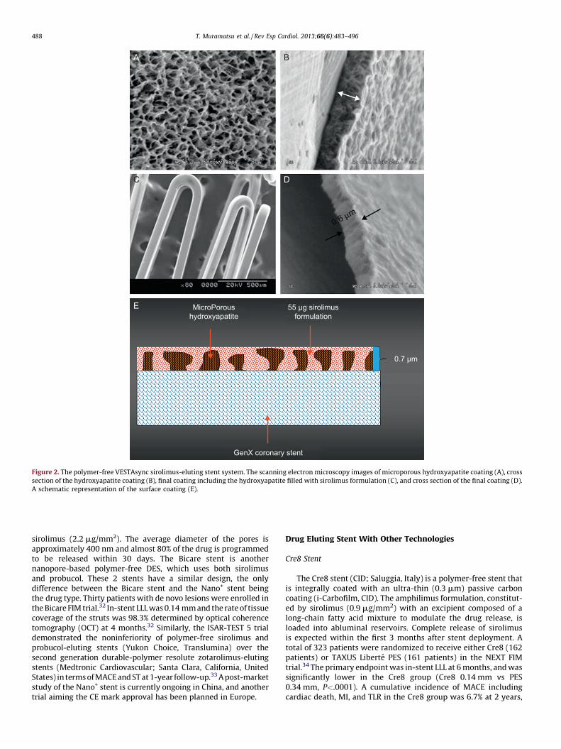
VESTAsync Stent
The VESTAsync stent (MIV Therapeutics; Atlanta, Georgia,United States) combines a stainless steel platform with ananoporous, hydroxyapatite (biocompatible crystalline derivativeof calcium phosphate) surface coating that is impregnated with55 mg of sirolimus mixture (Fig. 2). It is expected that sirolimuswill be completely released within the first 3 months after theimplantation, and that the hydroxyapatite will be stable over4 months. The safety and efficacy of the VESTAsync stent wasevaluated in the VESTAsync I FIM trial. A total of 15 patients withsingle de novo coronary artery lesions were enrolled. In-stent LLLwas 0.36 mm at 9 months, with no MACE reported at up to 1-yearfollow-up.30 Recently a randomized VESTAsync II trial has beenreported.31 The patients treated with the VESTAsync stent(50 patients) showed a significantly lower in-stent LLL comparedto those treated with BMS (25 patients) at 8 months (VESTAsync0.39 mm vs BMS 0.74 mm, P=.03). No evidence of ST was reportedat up to 2-year follow-up.
Nano+ Stent
The Nano+ stent (Lepu Medical; Beijing, China) is a stainlesssteel, polymer-free stent with a nanoporus surface coated with

Table 2Overview of Polymer-free and Novel-coating Drug-eluting Stents Under Clinical Investigation or Already Available Outside the United States
Drug-eluting stent,
manufacturer
Drug, dosage Stent
platform
Strut
thickness,
mm
Surface
modification
Drug release
kinetics
Study, no.
of patients
Angiographic
follow-up,
months
In-stent late
loss, mm
Binary
restenosis, %
Current status
Porous polymer-free DES
Yukon Choice
(Translumina)
Sirolimus (11.7-21.9 mg) SS 87 Abluminal microporous
surface
100%, 25 days ISAR-TEST 1 (225) 9 0.48 14.2 CE approved
BioFreedom
(Biosensors)
Biolimus A9 (SD, 15.6 mg/mm;
HD, 7.8 mg/mm)
SS 119 Abluminal microporous
surface
90%, 50 h FIM (SD, 31; HD, 35) 12 SD, 0.17;
HD, 0.22
N/A CE approval
submitted
VESTAsync
(MIV Therapeutics)
Sirolimus (55 mg) SS 65 Nanoporous surface
with hydroxyapatite
100%, 90 days VESTAsync II (15) 8 0.39 0 Ongoing
evaluation
Nano+
(Lepu Medical)
Sirolimus (2.2 mg/mm2) SS 100 Abluminal nanoporous
surface
80%, 30 days N/A N/A N/A N/A Ongoing
evaluation
DES with other technologies
Cre8 (CID) Sirolimus (0.9 mg/mm2) Co-Cr 80 Abluminal reservoirs 100%, 90 days NEXT-Cre8 (162) 6 0.14 3.2 CE approved
Combo
(OrbusNeich Medical)
EPC+sirolimus (5 mg/mm) SS 100/3-5 Abliminal biodegradable
polymer and luminal
CD34 antibody layer
N/A REMEDEE (124) 9 0.39 8.3 Ongoing
evaluation
FOCUS np
(Envision Scientific)
Sirolimus (108 mg/16mm) Co-Cr 73 Abluminal coating with
encapsulated drug
by nanoparticles
100%, 28 days N/A N/A N/A N/A Ongoing
evaluation
CE, Conformite Europeenne; Co-Cr, cobalt-chromium; SD, standard dose; DES, drug-eluting stent; EPC, endotheloal progenitor cell (capture technology); HD, half dose; N/A, not applicable; SS, stainless steel.
T.
Mu
ram
atsu
et a
l. /
Rev
Esp
Ca
rdio
l. 2
01
3;6
6(6
):48
3–
49
6
48
7
A B
C
E
D
MicroPoroushydroxyapatite
55 µg sirolimusformulation
0.7 µm
GenX coronary stent
0.6 µm
Figure 2. The polymer-free VESTAsync sirolimus-eluting stent system. The scanning electron microscopy images of microporous hydroxyapatite coating (A), cross
section of the hydroxyapatite coating (B), final coating including the hydroxyapatite filled with sirolimus formulation (C), and cross section of the final coating (D).A schematic representation of the surface coating (E).
T. Muramatsu et al. / Rev Esp Cardiol. 2013;66(6):483–496488
sirolimus (2.2 mg/mm2). The average diameter of the pores isapproximately 400 nm and almost 80% of the drug is programmedto be released within 30 days. The Bicare stent is anothernanopore-based polymer-free DES, which uses both sirolimusand probucol. These 2 stents have a similar design, the onlydifference between the Bicare stent and the Nano+ stent beingthe drug type. Thirty patients with de novo lesions were enrolled inthe Bicare FIM trial.32 In-stent LLL was 0.14 mm and the rate of tissuecoverage of the struts was 98.3% determined by optical coherencetomography (OCT) at 4 months.32 Similarly, the ISAR-TEST 5 trialdemonstrated the noninferiority of polymer-free sirolimus andprobucol-eluting stents (Yukon Choice, Translumina) over thesecond generation durable-polymer resolute zotarolimus-elutingstents (Medtronic Cardiovascular; Santa Clara, California, UnitedStates) in terms of MACE and ST at 1-year follow-up.33 A post-marketstudy of the Nano+ stent is currently ongoing in China, and anothertrial aiming the CE mark approval has been planned in Europe.
Drug Eluting Stent With Other Technologies
Cre8 Stent
The Cre8 stent (CID; Saluggia, Italy) is a polymer-free stent thatis integrally coated with an ultra-thin (0.3 mm) passive carboncoating (i-Carbofilm, CID). The amphilimus formulation, constitut-ed by sirolimus (0.9 mg/mm2) with an excipient composed of along-chain fatty acid mixture to modulate the drug release, isloaded into abluminal reservoirs. Complete release of sirolimusis expected within the first 3 months after stent deployment. Atotal of 323 patients were randomized to receive either Cre8 (162patients) or TAXUS Liberte PES (161 patients) in the NEXT FIMtrial.34 The primary endpoint was in-stent LLL at 6 months, and wassignificantly lower in the Cre8 group (Cre8 0.14 mm vs PES0.34 mm, P<.0001). A cumulative incidence of MACE includingcardiac death, MI, and TLR in the Cre8 group was 6.7% at 2 years,

Low dose sirolimus inbiodegradable polymer matrix
Luminal surface
Stent strut
Abluminal surface
Anti CD34antibody coatingfor EPC capture
EPC
CD34 Cellsurface antigen
Anti-CD34antibody
Prosthetic surface
BA
Figure 3. The Combo dual therapy stent system. A: The Combo stent consists of an abluminal biodegradable polymer matrix with a sirolimus and a luminal CD-34antibody layer. B: A schematic representation of the endothelial progenitor cells capture technology. The CD-34 antigens on the surface of the endothelialprogenitor cells attach to the anti-CD-34 antibodies on the stent’s surface, promoting endothelialization. EPC, endothelial progenitor cells.
T. Muramatsu et al. / Rev Esp Cardiol. 2013;66(6):483–496 489
showing no differences compared to PES (7.1%). Only 1 case ofdefinite late ST was observed in each group at up to 2-year follow-up.35 An all-comers registry (1000 patients) is currently ongoingand is expected to complete enrollment of patients by early 2013.
Combo Stent
The Combo stent (OrbusNeich Medical; Hong Kong, China)applies the endothelial progenitor cell capture technology toenhance vessel healing (ie, immobile CD34 antibodies on theluminal surface of the strut), incorporating abluminal LD sirolimusand a biodegradable polymer into the current DES technology(Fig. 3). Data from OCT and histology at 28 days in a porcine modelindicated that this hybrid stent promotes endothelialization, andreduces neointimal formation and inflammation when comparedto the Cypher SES and the first generation Genous endothelialprogenitor cell stent.36 The REMEDEE FIM trial randomized180 patients to treatment with either the Combo stent (124patients) or the TAXUS Liberte PES (59 patients). The in-stent LLLat 9 months was 0.39 mm in the Combo group and 0.44 mm inthe PES group (Pnoninferiority=.0012). Binary restenosis was observedin 8.3% patients in the Combo group and in 13.5% patients in thePES group (P=.30). No cases of ST were reported in both groups atup to 9-months follow-up.37 Further investigation is required todetermine the prohealing effect and clinical efficacy of this device.
FOCUS np Stent
The FOCUS np stent (Envision; Surat, India) platform has a novelcarrier; a phospholipid 2-layer nanoparticle that encapsulatessirolimus (Fig. 4). The encapsulated sirolimus is coated on thesurface of the stent and the balloon (108 mg of sirolimus on a3.0�16.0 mm system). Sirolimus is programmed to be completelyreleased within 28 days, however, the tissue concentration ofsirolimus peakes within the first 24 h. A preclinical study with theFOCUS np stent showed similar LLL and inflammation scores tothat seen in the Cypher SES at 28 days and at 90 days. A FIM trialwill be completed in early 2013.38
BIORESORBABLE SCAFFOLDS
Fully bioresorbable scaffolds (BRS) are a novel approach asthey provide transient vessel support in contrast to the
permanent caging caused by metallic stents. The concept ofBRS was introduced by Stack et al. in the year 1988.39 Zidar andcolleagues first implanted BRS made of poly-L-lactic acid (PLLA)into canine femoral arteries.40 Despite significant scaffolddegradation with low-grade vascular inflammation at 9-monthfollow-up, this technology failed to develop because of the inabilityto manufacture an ideal polymer that could limit inflammation andrestenosis.41,42 In the year 2000, Tamai and colleagues reported theirFIM experience with BRS implantation for the treatment of humancoronary arteries.43 This ‘‘Igaki-Tamai’’ PLLA stent had a uniquezigzag helical coil design, with a strut thickness of 170 mm. Thissystem was self-expanding but also required balloon inflation withheated contrast for expansion. The FIM study of the Igaki-Tamai stent (15 patients) demonstrated no MACE or ST eventswithin 30 days, and 1 repeat percutaneous coronary interventionat 6-month follow-up. Our group reported the findings of OCT at10 years after Igaki-Tamai stent implantation, showing absence ofvisible struts, with endoluminal lining of the vessel wall.44 Recently,Nishio et al. reported >10-year clinical outcomes of the first50 patients treated with Igaki-Tamai stents. Autopsy specimensshowed interesting histological findings, that indicated healingwith thickened neointima at the previously stented segment,without inflammatory cell infiltration or foreign body reactions.As measured by quantitative coronary angiography, LLL decreasedfrom 0.91 mm at 6 months to 0.59 mm at 3 years, whilstintravascular ultrasound (IVUS) showed an increased externalelastic lamina area (15.0 mm2 postprocedurally and 16.9 mm2 at3 years). These findings suggest that the artery restored its capabilityto respond to expansive remodeling and late lumen enlargementonce the scaffold degraded.
Currently, numerous BRS are being tested in clinical orpreclinical studies. An overview of this technology has beenshown in Table 3 and Figure 5.
Absorbable Magnesium Stent
Magnesium (Mg) is the fourth commonest cation withinthe human body. It is essential for the synthesis of over300 enzymes, and is a cofactor for ATPase. A high dose infusionof Mg can cause vasodilatation and the development ofcollaterals during ischaemia. The degradation of Mg producesan electronegative charge that results in the stent beinghypothrombogenic.45

Phospholipid bilayer
Hydrophilic
A B
C D
Hydrophobic
Figure 4. The nanocarrier-based FOCUS np sirolimus-eluting stent. The scanning electron microscopy (SEM) images of the crimped stent (A) and the magnifiedsurface of the strut and balloon (B) coated with encapsulated sirolimus (C, yellow arrows). The nanocarrier consists of a lipid bilayer with a hydrophilic head andtwo lipophilic/hydrophobic tails (D), and the drug is released on pH change.
Igaki-Tamai
AMS 1.0
REVA
BVS 1.0
Absorb BVS(BVS 1.1) On-ABS
Xinsorb
Amaranth
AMS 3.0(Dreams 1st generation)
AMS 4.0(Dreams 2nd generation)
ART18Z(ART 2nd generation)
IDEAL(BTI 2nd generation)
BTI
DESolve
ART
ReZolve(REVA 2nd generation)
Figure 5. Bioresorbable scaffolds under clinical or preclinical investigation.
T. Muramatsu et al. / Rev Esp Cardiol. 2013;66(6):483–496490

Table 3Overview of Bioresorbable Scaffolds Under Current Pre-clinical or Clinical Investigation
Bioresorbable
scaffold,
manufacturer
Strut
material
Coating
material
Drug Radio-
opacity
Strut
thickness,
mm
Crossing
profile, mm
Stent-to-
artery
coverage, %
Duration of
radial support
Resorption
time, months
Angiographic
late loss, mm
(months)
Target lesion
revascularization
rate, % (months)
Current
status
Igaki-Tamai
(Kyoto Medical)
PLLA None None Gold
markers
170 N/A 24 6 months 24 0.48 (6) 6.7 (6) CE approved
(PAD)
AMS-1.0
(Biotronik)
Mg None None None 165 1.2 10 Days or weeks <4 1.08 (4) 45 (12) FIM
completed
AMS-3.0
(Biotronik)
Mg None Paclitaxel None 125 N/A (6 Fr
compatible)
N/A Weeks >4 0.64 (6)
0.52 (12)
4.3 (6)
4.7 (12)
FIM
(BIOSOLVE-I)
completed
AMS-4.0
(Biotronik)
Mg PLLA Sirolimus Metallic
markers
N/A N/A (6 Fr
compatible)
N/A N/A N/A N/A N/A Used in
BIOSOLVE-I
BVS 1.0
(Abbott Vascular)
PLLA PDLLA Everolimus Platinum
markers
157 1.4 26 Weeks 24 0.44 (6) 0.0 (60) FIM
completed
Absorb BVS 1.1
(Abbott Vascular)
PLLA PDLLA Everolimus Platinum
markers
157 1.4 26 6 months 24 0.19 (6)
0.27 (12)
3.6 (12) CE
approved
REVA
(REVA Medical)
Poly-tyrosine-
derived
polycaronate
polymer
None None Scaffold
itself
200 1.7 55 3-6 months 24 1.81 (6) 67 (12) FIM
completed
ReZolve
(REVA Medical)
Poly-tyrosine-
derived
polycaronate
polymer
None Sirolimus Scaffold
itself
114-228 1.5 N/A 4-6 months 24 N/A N/A FIM planned
in 2013
DESolve
(Elixir Medical)
PLLA PLLA Mvolimus Metallic
markers
150 1.5 N/A N/A 12-24 0.19 (8) 6.7 (12) FIM
completed
IDEAL BioStent
(Xenogenics)
polymer
salicylate+
linker
Salicylate Sirolimus None 175 1.5-1.7 57 3 months >12 N/A N/A FIM
completed
ART18Z (Arterial
Remodeling
Technologies)
PDLLA None None None 170 N/A (6 Fr
compatible)
<25 3-6 months 18 None None FIM
initiated
Xinsorb (Huaan
Biotechnology)
PLLA+PCL+PLGA None Sirolimus Metallic
markers
160 N/A N/A N/A N/A None None Pre-clinical
underway
Amaranth PLLA
(Amaranth Medical)
PLLA None None None 150-200 N/A (6 Fr
compatible)
N/A 3-6 months N/A None None FIM initiated
On-ABS
(OrbusNeich Medical)
PLLA+PCL+PDLLA None EPC+
sirolimus
None 150 N/A N/A N/A N/A None None Pre-clinical
underway
CE, Conformite Europeenne; EPC, endothelial progenitor cell (capture technology); FIM, first-in-man; Mg, magnesium; N/A, not applicable; PAD, peripheral artery disease; PCL, poly-L-lactide-co-e-caprolactone; PDLLA, poly-D, L-
lactic acid; PLGA, poly-lactide-co-glycolide; PLLA, poly-L-lactic acid.
T.
Mu
ram
atsu
et a
l. /
Rev
Esp
Ca
rdio
l. 2
01
3;6
6(6
):48
3–
49
6
49
1
T. Muramatsu et al. / Rev Esp Cardiol. 2013;66(6):483–496492
The first generation absorbable metallic stent (AMS-1,Biotronik; Berlin, Germany) was composed of 93% Mg and 7%rare earth metals. In the porcine model, the AMS-1 was shown tobe rapidly endothelialized, and largely degraded into inorganicsalts at 60 days, with little associated inflammatory response.46
The PROGRESS AMS trial was a signle-arm FIM study, whichassessed the efficacy and safety of this stent in 63 patients withsingle de novo lesions.47 No evidence of death, MI, or ST wasreported at up to 12-month follow-up. Disappointingly, the TLR ratewas 23.8% at 4 months and 45% at 12 months. The in-stent LLL was1.08 mm and the vasodilator function, after the nitroglycerinadministration, appeared to be restored in the stented segment at4-month angiographic follow-up.48 IVUS data suggested that theincreased LLL was attributed to an increased neointimal formationand insufficient radial strength of the Mg alloy, due to rapid stentdegradation resulting in vessel recoil. Consequently, new stentshave been developed, namely AMS-2 and AMS-3. The AMS-2 stentwas designed to address excessive vessel recoil seen with AMS-1. Itprovided prolonged mechanical integrity by using a different Mgalloy, which not only had a higher collapse pressure, but also aslower degradation time. In addition, the strut thickness wasreduced from 165 mm to 125 mm, and the cross-sectional shape ofthe strut altered from rectangular to square. These modificationsfacilitated prolonged mechanical integrity, improved radialstrength, and resulted in reduced neointimal proliferation in animalstudies. The AMS-3 stent (ie, drug-eluting AMS [DREAMS]) wasdesigned to incorporate a bioresorbable matrix for the controlledrelease of paclitaxel with the AMS-2 platform. This devicewas evaluated in the BIOSOLVE-I trial (46 patients), and demon-strated an in-stent LLL of 0.64 mm at 6 months and 0.52 mm at12 months. The rate of TLF was 7.0% at up to 12-month follow-up,due to 2 clinically driven TLRs and 1 periprocedural MI.49 Thesecond generation DREAMS has a modified stent platform andsirolimus as its antiproliferative drug. The BIOSOLVE-II study aimedto assess the safety and efficacy of this device will be initiated in theyear 2013.
Everolimus-eluting Poly-L-lactic Acid Scaffold: Absorb BVS
The backbone of Absorb BVS (Abbott Vascular; Santa Clara,California, United States) is made of PLLA. The coating consists ofpoly-D, L-lactide (PDLLA), which is a random copolymer of D-lacticacid and L-lactic acid with lower crystallinity than the backbonePLLA. The PDLLA coating controls the release of the antiprolifera-tive drug everolimus. The first generation Absorb BVS (1.0) wastested in 30 patients who were enrolled in the ABSORB FIM (cohortA) trial. Multiple modality imaging was assessed in this trial, andthe results can be summarized as follows: a) partial bioresorptionof the polymeric struts; b) late lumen enlargement between6 months and 2 years; c) restoration of vasomotion and endothelialfunction at 2 years; d) sustained scaffolding of plaque deform-ability documented with palpography, and e) feasibility ofnoninvasive imaging with multislice computed tomography.50,51
Five-year clinical follow-up is available in 29 patients.52 Only1 patient experienced a non-Q wave MI related to the treatment ofa non-flow-limiting stenosis at 46 days after Absorb BVSimplantation. There were no ST events in the entire period andno MACE between 6 months and 5 years, resulting in an overallMACE rate of 3.4% at 5 years. Late scaffold shrinkage was the primaryreason for an increased in-stent LLL (0.44 mm) at 6 months. Lumenarea was reduced by 16.6%, whilst the late recoil was 11.7%.53 Inorder to enhance the radial strength of the struts and to reduce laterecoil, the strut design and the manufacturing process of thepolymer were modified in the second generation Absorb BVS (1.1).Firstly, the new design had in-phase zigzag hoops linked by bridgesthat allowed for a more uniform strut distribution. This new scaffold
design reduced maximum circular unsupported surface area thatprovided for more uniform vessel wall support and drug delivery.Secondly, a modified manufacturing process resulted in a slowerhydrolysis (in vivo degradation) rate of the polymer, thus allowingfor prolongation of its mechanical integrity.54
The Absorb BVS 1.1 was evaluated in 101 patients in theABSORB cohort B trial. This cohort was divided in 2 subgroups:the first group (B1) underwent invasive imaging with quantitativecoronary angiography, IVUS, and OCT postprocedurally, at6 months, and at 24 months; whereas the second group (B2)underwent invasive imaging postprocedurally, at 12 months, andat 36 months. In the entire cohort B population, the overall MACErate was 9.0%, including 3 non-Q wave MIs and 6 ischemia-drivenTLRs, without cardiac death during the 2-year follow-up. Therewere no possible, probable, or definite scaffold thromboses despitedual antiplatelet therapy rates of 97% at 6 months, 81.2% at12 months, and 24.8% at 24 months.
For the cohort B1 population, serial multimodality imagingresults are currently available.55 Serial angiographic analysesshowed that in-scaffold LLL of 0.16 mm at 6 months increased to0.27 mm at 2 years. Notably, serial IVUS analyses demonstratedthat the mean lumen area increased, whereas the minimum lumenarea remained stable between 6 months and 2 years (Fig. 6).Percentage hyperechogenic area, a more sensitive parameter tomeasure degradation of polymeric material, decreased from 25.3%postprocedurally to 20.4% at 6 months and to 13.8% at 2 years.Similar to IVUS, serial OCT investigation confirmed the progressiveincrease in mean scaffold area from 7.47 mm2 postprocedurally, to7.70 mm2 at 6 months, and 8.24 mm2 at 24 months.
The promising results of Absorb BVS constitute the proof ofconcept that this device can adequately revascularize coronaryvessels and prevent restenosis. The Absorb BVS acquired the CEmark approval in January 2011, and since September 2012 it iscommercially available in different diameters (2.5 mm, 3.0 mm,and 3.5 mm) and lengths (12 mm, 18 mm, and 28 mm). Thisdevice is now being evaluated in the ABSORB-EXTEND registry(�800 patients). A pivotal, randomized trial (ABSORB II), compar-ing Absorb BVS with Xience Prime EES (Abbott Vascular) in500 patients, is simultaneously ongoing in Europe.
Tyrosine Polycarbonate Stent
The REVA stent (REVA Medical, San Diego, California, UnitedStates) consists of a tyrosine-derived poly carbonate thatdegrades into water, carbon dioxide, and ethanol. In addition toits radio-opacity, the REVA stent also has a unique ‘‘slide and lock’’design that provides flexibility. This design maintains the acutelumen gain following stent deployment, and provides additionalsupport to the scaffold during vessel remodeling. The RESORB FIMtrial enrolled 27 patients. The in-stent LLL was disappointinglyhigh (1.81 mm) and IVUS data showed no vessel recoil as indicatedby external elastic lamina area (15.5 mm2 postprocedurally and15.3 mm2 at 6 months). There was a high rate of TLR (66.7%)between 4 months and 6 months, mostly due to excessiveneointimal hyperplasia.56 The second generation ReZolve stenthad a more robust polymer, a ‘‘spiral’’ slide and lock system, and acoating of sirolimus. Furthermore, the ReZolve2 stent had asmaller profile (1.52 mm) and achieved approximately 30%increase in radial strength. The safety and efficacy of the ReZolveor ReZolve2 stent is currently under investigation in the RESTOREstudy (50 patients) that was initiated in December 2011.Preliminary data (26 patients) showed that technical successrate was 85%, due to the delivery failure seen in 4 patients. Twocases with TLR as a primary endpoint were reported at 6-monthsfollow-up.57

Post-procedure(n=33)
IVUS measurements
Mean vessel area, (mm2)
Mean scaffold area, (mm2)
Mean plaque area, (mm2)
Mean lumen area, (mm2)
Mean NIH area, (mm2)
14.04 (3.80) 14.44 (3.82)
6.42 (1.17)
8.02 (2.89)
6.36 (1.18)
0.08 (0.13)
15.35 (4.05)
7.08 (1.73)
8.27 (2.87)
6.85 (1.78)
0.25 (0.27)
P=.008
P=.06
P=.02
P<.001
P<.001
P<.001
P=.01
P<.001
P=.03
6.53 (1.23)
6.53 (1.24)
7.51 (2.85)
6 months(n=33)
2 years(n=33)
Figure 6. Serial changes in intravascular ultrasound measurements over postprocedure, 6 months, and 2 years after Absorb BVS implantation. NIH, neointimalhyperplasia. IVUS, intravascular ultrasound.
T. Muramatsu et al. / Rev Esp Cardiol. 2013;66(6):483–496 493
Myolimus-eluting Poly-L-Lactic Acid Scaffold: DESolve
DESolve BRS (Elixir Medical; Sunnyvale, California, UnitedStates) has a similar PLLA backbone to the Absorb BVS, but it iscoated with myolimus (3 mg/mm), an mTOR inhibitor macrocy-clic lactone, and a sirolimus analogue. Sufficient radial strengthwas achieved over 3 months and the bioresorption of the scaffoldwas observed between 1 year and 2 years. The DESolve-I FIM trial(16 patients) demonstrated that the rate of acute recoil was 6.4%,and in-scaffold LLL was 0.19 mm at 6 months.58 In IVUSinvestigation, the respective mean scaffold area and lumen areawas 5.35 mm2 and 5.35 mm2 postprocedurally, and 5.61 mm2
and 5.10 mm2 at 6 months (P=no significant). OCT revealed that98.7% of the struts were covered at 6 months. All patients wereclinically followed up to 1 year, and 3 patients experienced MACEincluding 1 cardiac death, 1 target vessel MI, and 1 TLR. Therewere no patients with the evidence of ST. The DESolve Nx trial iscurrently enrolling 120 patients treated with the next generationDESolve Nx stent with novolimus (5 mg/mm), which is an activemetabolite of sirolimus.59
Poly Salicylic Acid Stent: IDEAL
The IDEAL BRS (Xenogenics Corp.; Canton, Massachusetts,United States) has 2 components: the backbone and the drug layer.The backbone of the device is made of polylactide anhydride mixedwith a polymer of salicylic acid and sebacic acid linker. The druglayer consists of salicylate that controls the release of theantiproliferative drug sirolimus. The presence of salicylic acidprovides the device with anti-inflammatory properties, whichhave been confirmed in preclinical studies.60 The IDEAL BRS wastested in a small number of humans (11 patients) in 2008.Although this study has not been fully reported, there wasinsufficient neointimal suppression and a reduction in lumen areadue to inadequate drug dosing and rapid release of the sirolimus.61
The second generation IDEAL BioStent has a higher drug dose,slower drug-release kinetics, and a smaller system profile. Thedevice is currently undergoing preclinical evaluation.
Arterial Remodeling Technologies Bioresorbable Scaffold
The ART BRS (Arterial Remodeling Technologies; Noisy le Roi,France) is manufactured from a PDLLA amorphous polymer
without an antiproliferative drug. This device is 6 Fr compatible,and provides transient scaffolding for 5 months to 7 months. Fullresorption occurs within 18 months. The performance of the ARTBRS was compared with the BMS in rabbit and porcine models, andno MACE was reported. The acute recoil was comparable to BMS.Interestingly, angiographic analyses demonstrated the phenome-non of late lumen enlargement, as well as increased external elasticlamina area detected by IVUS at 9 months. Based on the results ofpreclinical studies, the ARTDIVA FIM trial has already commencedand is recruiting patients at 5 sites in France. It aims to evaluateclinical outcomes at 6 months.62
Xinsorb Bioresorbable Scaffold
The Xinsorb BRS (Huaan Biotechnology; Laiwu, China) is a fullybioresorbable sirolimus-loaded scaffold that consists of PLLA, poly-lactide-co-glycolide, and poly-L-lactide-co-e-caprolactone. Anexperimental study evaluated the feasibility of Xinsorb BRS incomparison with the Excel DES (JW Medical; Shandong, China).Sixteen Xinsorb scaffolds and 16 Excel stents were implanted inthe coronary arteries of porcine models.63 In vitro drug-elutionkinetics indicated that 78% of sirolimus was released from XinsorbBRS within the first 14 days. Histomorphometry demonstrated asignificantly lower percentage diameter restenosis in the XinsorbBRS compared to the Excel DES (18.6% vs 21.4% at 30 days and24.5% vs 27.7% at 90 days, respectively). In addition, the struts ofthe Xinsorb BRS were completely covered by neointima at90 days.64 Although these preliminary results are encouraging,further extensive preclinical studies are necessary to investigatethe safety and efficacy of this device. The company is expecting toorganize a FIM trial in the year 2013.
Other Brioresorbable Scaffolds
The Amaranth PLLA scaffold (Amaranth Medical; MountainView, California, United States) and the On-ABS (OrbusNeich, HongKong, China) are currently under preclinical evaluation. Inaddition, there are several other devices that are still underdevelopment. These include the Sahajanand BRS (SahajanandMedical Technologies; Surat, India), the Avatar BRS (S3 V;Hyderabad, India), the MeRes BRS (Meril Life Sciences; Vapi,Gujarat, India), and the Zorion BRS (Zorion Medical; Indianapolis,Indiana, United States).

Acute occlusion
Acute ST
Subacute ST
Acute recoil
Constrictive remodelling
Neointimal hyperplasia
Expansive remodeling
Late luminal enlargement
Late or very late ST
–
BA BMS DES VRT
N/A
N/A
–
–
–
+
+
N/A
+
–/+
+
+
+
–
–
–
–
+
+
+
+
+
+
–
–
–
+
+
+
+
+
+
+
+
+/?
Figure 7. Schematic illustration presenting the evolution of percutaneous coronary interventions. BA, balloon angioplasty; BMS, bare metal stents; DES, drug-eluting metallic stents; N/A, not applicable because of the absence of stent; ST, stent thrombosis; VRT, vascular reparative therapy. ‘‘+’’ implies prevented or notrestricted, whilst ‘‘�’’ implies not prevented or restricted. Modified with permission from Serruys et al.67
T. Muramatsu et al. / Rev Esp Cardiol. 2013;66(6):483–496494
FUTURE PERSPECTIVES
The new enemy in the DES era—ST—has accelerated techno-logical evolution in interventional cardiology. Newer generationDES, with biodegradable polymers, have shown an impressivereduction in VLST, contributing to improved long-term outcomes,as compared to first generation DES. The polymer-free DES or BRSare relatively new technologies, with many trials still in progress.The currently available angiographic, intravascular imaging, andclinical data, suggest acceptable safety and efficacy of these newtechnologies. However, it remains unclear as to whether biode-gradable-polymer DES or polymer-free DES can minimize late STevents, particularly as these late events have also been observed inpatients receiving BMS.65,66 Furthermore, considering the fatalconsequences of ST, focus should be maintained on the eradication,rather than the minimization of this serious complication. There isa fundamental difference in concept between the DES and the BRStechnologies, with the latter having a capability of liberating thevessel from a permanent metallic cage. Therefore, BRS technologyhas a theoretical advantage in reducing ST by means ofendoluminal prosthesis elimination. BRS also facilitates therestoration of vasomotor function, which indirectly results inthe completeness of vessel healing. The entire process of thistreatment has been hence named as vascular reparative therapy(Fig. 7).67
One possible fate of the stenotic lesion treated with metallicstents is the development of in-stent neointimal tissue (even seenwith DES), where the antiproliferative drug slows down orpostpones the phenomenon. This neointimal tissue may in turnbecome atherosclerotic, degenerate to a vulnerable plaque, andfinally rupture inside the cage of the stent (ie, neoatherosclero-sis).68,69 A stiff metallic stent can also alter vessel geometry andbiomechanics, which may result in long-term flow disturbancesand chronic irritation, in addition to the risk of late strut fractures,which potentially contribute to restenosis and adverse clinicalevents.70,71 From a physiological perspective, the absence of a rigidmetallic cage facilitates the restoration of vasomotor function,
adaptive shear stress, and late luminal enlargement. Afterbioresorption, there would be no triggers for thrombosis, suchas uncovered struts or durable polymers. The absence of foreignmaterials may also reduce concerns about future treatmentoptions, such as precluding bypass-graft surgery, and the require-ments for long-term dual antiplatelet therapy with a potentialreduction in associated bleeding complications. Since BRS haveonly been evaluated in limited patients with noncomplex lesions,the feasibility of these devices in complex lesions requires furtherclinical evaluation. In addition, future investigations are requiredto establish if BRS technology is superior to permanent metallicDES.
CONCLUSIONS
Newer metallic DES technology has proven to decrease the riskof revascularization and ST events. The optimal design, however, ofscaffolds, polymers, antiproliferative drugs and their degradation/release kinetics is still under investigation. BRS technology isanticipated not only to eliminate the risk of VLST, but also tocontribute to the restoration of physiological function of treatedvessels. Although further technical development and clinicalevaluation are required before BRS can be accepted as the ultimatedevice for the treatment of coronary artery disease, this newtechnology looks promising and could be the next revolution ininterventional cardiology.
CONFLICTS OF INTEREST
None declared.
REFERENCES
1. Gruntzig AR, Senning A, Siegenthaler WE. Nonoperative dilatation of coronary-artery stenosis: percutaneous transluminal coronary angioplasty. N Engl J Med.1979;301:61–8.
T. Muramatsu et al. / Rev Esp Cardiol. 2013;66(6):483–496 495
2. Sigwart U, Puel J, Mirkovitch V, Joffre F, Kappenberger L. Intravascular stents toprevent occlusion and restenosis after transluminal angioplasty. N Engl J Med.1987;316:701–6.
3. Sigwart U, Urban P, Golf S, Kaufmann U, Imbert C, Fischer A, et al. Emergencystenting for acute occlusion after coronary balloon angioplasty. Circulation.1988;78(5 Pt 1):1121–7.
4. Serruys PW, De Jaegere P, Kiemeneij F, Macaya C, Rutsch W, Heyndrickx G, et al.A comparison of balloon-expandable-stent implantation with balloon angio-plasty in patients with coronary artery disease. Benestent Study Group. N Engl JMed. 1994;331:489–95.
5. Hoffmann R, Mintz GS, Dussaillant GR, Popma JJ, Pichard AD, Satler LF, et al.Patterns and mechanisms of in-stent restenosis. A serial intravascular ultra-sound study. Circulation. 1996;94:1247–54.
6. Morice MC, Serruys PW, Sousa JE, Fajadet J, Ban Hayashi E, Perin M, et al. Arandomized comparison of a sirolimus-eluting stent with a standard stent forcoronary revascularization. N Engl J Med. 2002;346:1773–80.
7. Stone GW, Ellis SG, Cox DA, Hermiller J, O’Shaughnessy C, Mann JT, et al. Apolymer-based, paclitaxel-eluting stent in patients with coronary artery dis-ease. N Engl J Med. 2004;350:221–31.
8. Nordmann AJ, Briel M, Bucher HC. Mortality in randomized controlled trialscomparing drug-eluting vs. bare metal stents in coronary artery disease: ameta-analysis. Eur Heart J. 2006;27:2784–814.
9. Camenzind E, Steg PG, Wijns W. Stent thrombosis late after implantation offirst-generation drug-eluting stents: a cause for concern. Circulation. 2007;115:1440–55. discussion 55.
10. Joner M, Finn AV, Farb A, Mont EK, Kolodgie FD, Ladich E, et al. Pathology ofdrug-eluting stents in humans: delayed healing and late thrombotic risk. J AmColl Cardiol. 2006;48:193–202.
11. Virmani R, Guagliumi G, Farb A, Musumeci G, Grieco N, Motta T, et al. Localizedhypersensitivity and late coronary thrombosis secondary to a sirolimus-elutingstent: should we be cautious? Circulation. 2004;109:701–5.
12. Rasmussen K, Maeng M, Kaltoft A, Thayssen P, Kelbaek H, Tilsted HH, et al.Efficacy and safety of zotarolimus-eluting and sirolimus-eluting coronarystents in routine clinical care (SORT OUT III): a randomised controlled superi-ority trial. Lancet. 2010;375:1090–9.
13. Stone GW, Rizvi A, Newman W, Mastali K, Wang JC, Caputo R, et al. Everolimus-eluting versus paclitaxel-eluting stents in coronary artery disease. N Engl J Med.2010;362:1663–74.
14. Kedhi E, Joesoef KS, McFadden E, Wassing J, Van Mieghem C, Goedhart D, et al.Second-generation everolimus-eluting and paclitaxel-eluting stents in real-lifepractice (COMPARE): a randomised trial. Lancet. 2010;375:201–9.
15. Stefanini GG, Kalesan B, Serruys PW, Heg D, Buszman P, Linke A, et al. Long-termclinical outcomes of biodegradable polymer biolimus-eluting stents versusdurable polymer sirolimus-eluting stents in patients with coronary arterydisease (LEADERS): 4 year follow-up of a randomised non-inferiority trial.Lancet. 2011;378:1940–8.
16. Finn AV, Nakazawa G, Joner M, Kolodgie FD, Mont EK, Gold HK, et al. Vascularresponses to drug eluting stents: importance of delayed healing. ArteriosclerThromb Vasc Biol. 2007;27:1500–10.
17. De la Torre Hernandez JM, Windecker S. Trombosis muy tardıa con nuevosstents farmacoactivos:
?
ha dejado de ser un asunto relevante? Rev Esp Cardiol.2012;65:595–8.
18. Grube E, Sonoda S, Ikeno F, Honda Y, Kar S, Chan C, et al. Six- and twelve-monthresults from first human experience using everolimus-eluting stents withbioabsorbable polymer. Circulation. 2004;109:2168–71.
19. Byrne RA, Kastrati A, Massberg S, Wieczorek A, Laugwitz KL, Hadamitzky M,et al. Biodegradable polymer versus permanent polymer drug-eluting stentsand everolimus- versus sirolimus-eluting stents in patients with coronaryartery disease: 3-year outcomes from a randomized clinical trial. J Am CollCardiol. 2011;58:1325–31.
20. Windecker S, Serruys PW, Wandel S, Buszman P, Trznadel S, Linke A, et al.Biolimus-eluting stent with biodegradable polymer versus sirolimus-elutingstent with durable polymer for coronary revascularisation (LEADERS): a ran-domised non-inferiority trial. Lancet. 2008;372:1163–73.
21. Serruys PW, Buszman P, Linke A, Ischinger T, Antoni D, Klauss V, et al. TCT-44LEADERS: 5-year follow-up from a prospective, randomized trial of biolimusA9-eluting stents with a biodegradable polymer vs. sirolimus-eluting stentswith a durable polymer- final report of the LEADERS study. J Am Coll Cardiol.2012;60:B13–4.
22. Stefanini GG, Byrne RA, Serruys PW, De Waha A, Meier B, Massberg S, et al.Biodegradable polymer drug-eluting stents reduce the risk of stent thrombosisat 4 years in patients undergoing percutaneous coronary intervention: a pooledanalysis of individual patient data from the ISAR-TEST 3, ISAR-TEST 4, andLEADERS randomized trials. Eur Heart J. 2012;33:1214–22.
23. Meredith IT, Verheye S, Dubois CL, Dens J, Fajadet J, Carrie D, et al. Primaryendpoint results of the EVOLVE trial: a randomized evaluation of a novelbioabsorbable polymer-coated, everolimus-eluting coronary stent. J Am CollCardiol. 2012;59:1362–70.
24. Farooq V, Gogas BD, Serruys PW. Restenosis: delineating the numerous causesof drug-eluting stent restenosis. Circ Cardiovasc Interv. 2011;4:195–205.
25. Mehilli J, Byrne RA, Wieczorek A, Iijima R, Schulz S, Bruskina O, et al.Randomized trial of three rapamycin-eluting stents with different coatingstrategies for the reduction of coronary restenosis. Eur Heart J. 2008;29:1975–82.
26. Byrne RA, Kufner S, Tiroch K, Massberg S, Laugwitz KL, Birkmeier A, et al.Randomised trial of three rapamycin-eluting stents with different coating
strategies for the reduction of coronary restenosis: 2-year follow-up results.Heart. 2009;95:1489–94.
27. King L, Byrne RA, Mehilli J, Schomig A, Kastrati A, Pache J. Five-year clinicaloutcomes of a polymer-free sirolimus-eluting stent versus a permanent poly-mer paclitaxel-eluting stent: final results of the intracoronary stenting andangiographic restenosis—test equivalence between two drug-eluting stents(ISAR-TEST) trial. Catheter Cardiovasc Interv. 2013;81:E23–8.
28. Tada N, Virmani R, Grant G, Bartlett L, Black A, Clavijo C, et al. Polymer-freebiolimus a9-coated stent demonstrates more sustained intimal inhibition,improved healing, and reduced inflammation compared with a polymer-coatedsirolimus-eluting cypher stent in a porcine model. Circ Cardiovasc Interv.2010;3:174–83.
29. Grube E, Mueller R, Schuler G, Hauptmann KE, Schofer J. TCT-46: Comparisonof polymer-free BioFreedom stents with durable polymer Taxus Libere stents:3-year results from the BioFreedom first-in-man trial. J Am Coll Cardiol.2012;60:B14.
30. Costa Jr JR, Abizaid A, Costa R, Feres F, Tanajura LF, Maldonado G, et al. 1-yearresults of the hydroxyapatite polymer-free sirolimus-eluting stent for thetreatment of single de novo coronary lesions: the VESTASYNC I trial. JACCCardiovasc Interv. 2009;2:422–7.
31. Costa Jr JR. VESTAsync (MIV Therapeutics) program update. TranscatheterCardiovascular Therapeutics. 2012.
32. Yu M, Xu B, Wu Y, Yan H, Chen J, Qian J, et al. TCT-229: First report of anovel polymer-free dual-drug eluting stent in de novo coronary artery dis-ease: results of the first in man BICARE trial. J Am Coll Cardiol. 2011;58(20S1):B62.
33. Massberg S, Byrne RA, Kastrati A, Schulz S, Pache J, Hausleiter J, et al. Polymer-free sirolimus- and probucol-eluting versus new generation zotarolimus-elut-ing stents in coronary artery disease: the intracoronary stenting and angio-graphic results: test efficacy of sirolimus- and probucol-eluting versuszotarolimus-eluting stents (ISAR-TEST 5) trial. Circulation. 2011;124:624–32.
34. Carrie D, Berland J, Verheye S, Hauptmann KE, Vrolix M, Violini R, et al. Amulticenter randomized trial comparing amphilimus- with paclitaxel-elutingstents in de novo native coronary artery lesions. J Am Coll Cardiol.2012;59:1371–6.
35. Carrie D. Cre8 program update. Transcatheter Cardiovascular Therapeutics.2012.
36. Aoki J, Serruys PW, Van Beusekom H, Ong AT, McFadden EP, Sianos G, et al.Endothelial progenitor cell capture by stents coated with antibody againstCD34: the HEALING-FIM (healthy endothelial accelerated lining inhibits neoin-timal growth-first in man) registry. J Am Coll Cardiol. 2005;45:1574–9.
37. Haude M. REMEDEE COMBO program update. Transcatheter CardiovascularTherapeutics. 2012.
38. Seth A. Nanoparticle based stents FOCUSnp program update. TranscatheterCardiovascular Therapeutics. 2012.
39. Stack RS, Califf RM, Phillips HR, Pryor DB, Quigley PJ, Bauman RP, et al.Interventional cardiac catheterization at Duke Medical Center. Am JCardiol.1988;62(10 Pt 2):F3–24.
40. Zidar J, Lincoff A, Stack R. Biodegradable stents. In: Topol EJ, editor. Textbook ofinterventional cardiology. 2nd ed. Philadelphia: Saunders; 1994. p. 787–802.
41. Waksman R. The disappearing stent: when plastic replaces metal. Circulation.2012;125:2291–4.
42. Van der Giessen WJ, Lincoff AM, Schwartz RS, Van Beusekom HM,Serruys PW, Holmes Jr DR, et al. Marked inflammatory sequelae to implan-tation of biodegradable and nonbiodegradable polymers in porcine coronaryarteries. Circulation. 1996;94:1690–7.
43. Tamai H, Igaki K, Kyo E, Kosuga K, Kawashima A, Matsui S, et al. Initial and6-month results of biodegradable poly-l-lactic acid coronary stents in humans.Circulation. 2000;102:399–404.
44. Onuma Y, Garg S, Okamura T, Ligthart J, Van Geuns RJ, De Feyter PJ, et al.Ten-year follow-up of the IGAKI-TAMAI stent. A posthumous tribute to thescientific work of Dr. Hideo Tamai EuroIntervention. 2009;5 Suppl. F:F109–11.
45. Heublein B, Rohde R, Kaese V, Niemeyer M, Hartung W, Haverich A. Biocorro-sion of magnesium alloys: a new principle in cardiovascular implant technolo-gy? Heart. 2003;89:651–6.
46. Waksman R, Pakala R, Kuchulakanti PK, Baffour R, Hellinga D, Seabron R, et al.Safety and efficacy of bioabsorbable magnesium alloy stents in porcinecoronary arteries. Catheter Cardiovasc Interv. 2006;68:607–17. discussion18-9.
47. Erbel R, Di Mario C, Bartunek J, Bonnier J, De Bruyne B, Eberli FR, et al.Temporary scaffolding of coronary arteries with bioabsorbable magnesiumstents: a prospective, non-randomised multicentre trial. Lancet. 2007;369:1869–75.
48. Ghimire G, Spiro J, Kharbanda R, Roughton M, Barlis P, Mason M, et al. Initialevidence for the return of coronary vasoreactivity following the absorption ofbioabsorbable magnesium alloy coronary stents. EuroIntervention. 2009;4:481–4.
49. Haude M, Erbel R, Erne P, Verheye S, Degen H, Bose D, et al. Safety andperformance of the drug-eluting absorbable metal scaffold (DREAMS) inpatients with de-novo coronary lesions: 12 month results of the prospective,multicentre, first-in-man BIOSOLVE-I trial. Lancet. 2013;12:61765–6. http://dx.doi.org/10.1016/S0140-6736.
50. Ormiston JA, Serruys PW, Regar E, Dudek D, Thuesen L, Webster MW, et al. Abioabsorbable everolimus-eluting coronary stent system for patients withsingle de-novo coronary artery lesions (ABSORB): a prospective open-labeltrial. Lancet. 2008;371:899–907.

T. Muramatsu et al. / Rev Esp Cardiol. 2013;66(6):483–496496
51. Serruys PW, Ormiston JA, Onuma Y, Regar E, Gonzalo N, Garcia-Garcia HM, et al.A bioabsorbable everolimus-eluting coronary stent system (ABSORB): 2-yearoutcomes and results from multiple imaging methods. Lancet. 2009;373:897–910.
52. Onuma Y, Nieman K, Webster M, Thuesen L, Dudek D, Ormiston JA, et al.TCT-37: Five-year clinical outcomes and non-invasive angiographic imagingresults with functional assessment after bioresorbable everolimus-elutingscaffold implantation in patients with de novo coronary artery disease. J AmColl Cardiol. 2012;60:B11.
53. Tanimoto S, Bruining N, Van Domburg RT, Rotger D, Radeva P, Ligthart JM, et al.Late stent recoil of the bioabsorbable everolimus-eluting coronary stentand its relationship with plaque morphology. J Am Coll Cardiol. 2008;52:1616–20.
54. Serruys PW, Onuma Y, Dudek D, Smits PC, Koolen J, Chevalier B, et al. Evaluationof the second generation of a bioresorbable everolimus-eluting vascular scaf-fold for the treatment of de novo coronary artery stenosis: 12-month clinicaland imaging outcomes. J Am Coll Cardiol. 2011;58:1578–88.
55. Ormiston JA, Serruys PW, Onuma Y, Van Geuns RJ, De Bruyne B, Dudek D, et al.First serial assessment at 6 months and 2 years of the second generation ofabsorb everolimus-eluting bioresorbable vascular scaffold: a multi-imagingmodality study. Circ Cardiovasc Interv. 2012;5:620–32.
56. Grube E. Bioabsorbable stent. The Boston Scientific and REVA technology.EuroPCR. 2009. Barcelona; 2009.
57. Costa RA. REVA ReZolve clinical program update. Transcatheter CardiovascularTherapeutics. 2012.
58. Yan J, Bhat VD. Elixir Medical’s bioresorbable drug eluting stent (BDES) pro-gramme: an overview. EuroIntervention. 2009;5 Suppl. F:80–2.
59. Verheye S. First-in-man results with a myolimus-eluting bioresorbablePLLA-based vascular scaffold. Transcatheter Cardiovascular Therapeutics.2012.
60. Jabara R, Chronos N, Robinson K. Novel bioabsorbable salicylate-based polymeras a drug-eluting stent coating. Catheter Cardiovasc Interv. 2008;72:186–94.
61. Jabara R, Chronos N, Robinson K. Novel fully bioabsorbable salicylate-basedsirolimus-eluting stent. EuroIntervention. 2009;5 Suppl. F:F58–64.
62. Fajadet J. The ART stent: design and early first-in-man experiences. Transcath-eter Cardiovascular Therapeutics. 2012.
63. Shen L, Wang Q, Wu Y, Xie J, Zhang F, Ge L, et al. Preliminary evaluation of fullybioabsorbable PLLA sirolimus eluting stents in a porcine model. Chin J InterventCardiol. 2009;19:301–5.
64. Shen L, Wang Q, Wu Y, Xie J, Ge J. Short-term effects of sirolimus eluting fullybioabsorbable polymeric coronary stents in a porcine model. TranscatheterCardiovascular Therapeutics. 2011.
65. Daemen J, Wenaweser P, Tsuchida K, Abrecht L, Vaina S, Morger C, et al. Earlyand late coronary stent thrombosis of sirolimus-eluting and paclitaxel-elutingstents in routine clinical practice: data from a large two-institutional cohortstudy. Lancet. 2007;369:667–78.
66. Stettler C, Wandel S, Allemann S, Kastrati A, Morice MC, Schomig A, et al.Outcomes associated with drug-eluting and bare-metal stents: a collaborativenetwork meta-analysis. Lancet. 2007;370:937–48.
67. Serruys PW, Garcia-Garcia HM, Onuma Y. From metallic cages to transientbioresorbable scaffolds: change in paradigm of coronary revascularization inthe upcoming decade? Eur Heart J. 2012;33:16–25.
68. Ramcharitar S, Garcia-Garcia HM, Nakazawa G, Kukreja N, Ligthart J, Virmani R,et al. Ultrasonic and pathological evidence of a neo-intimal plaque rupture inpatients with bare metal stents. EuroIntervention. 2007;3:290–1.
69. Nakazawa G, Otsuka F, Nakano M, Vorpahl M, Yazdani SK, Ladich E, et al. Thepathology of neoatherosclerosis in human coronary implants bare-metal anddrug-eluting stents. J Am Coll Cardiol. 2011;57:1314–22.
70. Wentzel JJ, Whelan DM, Van der Giessen WJ, Van Beusekom HM, Andhyiswara I,Serruys PW, et al. Coronary stent implantation changes 3-D vessel geometryand 3-D shear stress distribution. J Biomech. 2000;33:1287–95.
71. Wentzel JJ, Gijsen FJ, Schuurbiers JC, Van der Steen AF, Serruys PW. Theinfluence of shear stress on in-stent restenosis and thrombosis. EuroInterven-tion. 2008;4 Suppl. C:C27–32.