selective nuclear transport of the drosophila toll and...
TRANSCRIPT

Selective nuclear transport of the Drosophila morphogen dorsal can be established by a signaling pathway involving the transmemlirane protein Toll and protein kinase A Jacqueline L. Norris and James L. Manley
Department of Biological Sciences, Columbia University, New York, New York 10027 USA
Establishment of dorsal-ventral polarity in the early Drosophila embryo requires a concentration gradient of the maternal morphogen dorsal (d/). This concentration gradient is established by selective nuclear transport of d / so that dl protein is present only in ventral nuclei. The activity of 11 genes is required for d/nuclear localization. One of these genes, Toll, encodes a transmembrane protein that appears to play the most direct role in regulating dl localization. We have examined the effects of Toll on dl in cotransfected Schneider cells to gain insight into the nature of the interaction between these proteins. We have found that Toll can enhance the nuclear localization of d/and, independently, the ability of d / to activate transcription once in the nucleus. We present evidence that the signaling pathway from Toll to d/ involves protein kinase A (PKA) and that nuclear transport and activation of d/results from phosphorylation of d /by PKA. We discuss the significance of these results with respect both to Drosophila embryogenesis and to the regulation of the mammalian transcription factor NF-KB.
[Key Words: Drosophila morphogen; dorsal; Toll; protein kinase A]
Received December 30, 1991; revised version accepted June 24, 1992.
Establishment of dorsal-ventral (D/V} polarity m the Drosophila embryo requires the activities of several ma- ternal genes, including 11 that comprise the dorsal group (for review, see Anderson 1989; Rushlow and Arora 1990). One member of this group is the D/V morphogen dorsal (dl), a protein that shares homology, over the amino-terminal 300 amino acids, with the proto-onco- gene c-rel and the mammalian transcription factor NF- KB. This region includes a putative DNA-binding do- main, a nuclear localization sequence (NLS), and a po- tential protein kinase A (PKA) phosphorylation site (Steward 1987; Ghosh et al. 1990; Kieran et al. 1990}. The dl protein is distributed in a concentration gradient over the embryo, with the highest levels present in ven- tral regions and progressively decreasing levels in dorsal regions (Steward et al. 1988). This gradient results from the selective nuclear transport of the dl protein, which is uniformly cytoplasmic in the early embryo until cleav- age cycle 10 when it is transported into ventral but not dorsal nuclei (Roth et al. 1989; Rushlow et al. 1989; Steward 1989). Following its accumulation in the nu- cleus, dI functions to influence the expression of several zygotic genes required for D/V polarity (for review, see Anderson 1987; Rushlow and Arora 1990).
The activity of the other dorsal group genes is essential
for the proper localization of the dl protein (Anderson and Nfisslein-Volhard 1986). In embryos that carry null mutations in any of these genes, ventral cells follow the developmental pathway of dorsal cells. The dorsalization of these embryos is the result of the absence of the dl protein in ventral nuclei, which results in the improper expression of zygotic genes. The function of the dorsal group gene products is to establish the dl nuclear protein gradient. Two of these genes, easter and snake, encode serine proteases (DeLotto and Spierer 1986; Chasan and Anderson 1989), suggesting that a cascade of interactions involving post-translational processing could result in the formation of the signal for dl movement into ventral nuclei. This signal is most likely localized in the peri- vitelline space on the ventral side of the embryo and is transmitted to dl through the product of the Toll gene (Hashimoto et al. 1988; Stein et al. 1991}, which is lo- calized uniformally in the embryo plasma membrane (Hashimoto et al. 1991). Genetic studies have shown that Toll is one of the last genes in the D/V cascade, acting upstream of dl, and cytoplasmic injection experi- ments have shown that Toll is responsible for establish- ing polarity in the embryo by defining the position of the D/V axis (Anderson et al. 1985a, b). These observations, along with the isolation of both dominant-ventralizing
1654 GENES & DEVELOPMENT 6:1654-1667 © 1992 by Cold Spring Harbor Laboratory ISSN 0890-9369/92 $3.00
Cold Spring Harbor Laboratory Press on August 22, 2019 - Published by genesdev.cshlp.orgDownloaded from

Nuclear transport of the dorsal protein
as well as recessive-dorsalizing alleles of Toll (Anderson et al. 1985a; Schneider et al. 1991), suggest that the Toll gene product plays a particularly critical role in regulat- ing dl localization. However, nothing is known about the mechan i sm by which Toll enhances nuclear trans- port of dl.
The Toll protein has a large extracellular domain that contains two blocks of leucine-rich repeats with adjacent cysteine-containing motifs (Hashimoto et al. 1988; Schneider et al. 1991} and a small cytoplasmic domain that is s imilar to the cytoplasmic domain of the inter- leukin-1 receptor (IL-1R)(Schneider et al. 1991). The overall structure of the Toll protein is most s imilar to the e~ chain of h u m a n platelet glycoprotein lb (GPlb), a receptor for yon Willebrand factor and thrombin (Lopez et al. 1987; Hashimoto 1988). The leucine-rich repeat and cysteine-containing motif in GP lb are required for thrombin binding, suggesting that these regions in the Toll protein may serve as ligand-binding sites (Keith and Gay 1990). The importance of these regions in Toll is indicated by the finding that three of the strongest Toll- dominant alleles contain point mutat ions in one of the cysteine-containing motifs (Schneider et al. 1991).
To study the mechan i sm of dl nuclear transport we have used transient cotransfection assays in Drosophila Schneider cells. These cells contain no detectable endog- enous dl protein so the localization and activity of dl produced from transfected templates can be readily ob- served (Rushlow et al. 1989}. In this system, exogenously produced dl protein was shown previously to be local-
ized primari ly in the cytoplasm at low concentrations of expression vector, but increasingly in the nucleus at higher concentrations. The dl protein was also shown to activate expression of several reporter genes in a highly concentration-dependent manner (Rushlow et al. 1989). Here, we have used the cotransfection system as an assay to show that Toll can bring about selective nuclear trans- port of dl in Schneider cells, ruling out an absolute re- quirement of factors unique to the early embryo. Addi- tional experiments indicate that the mechan i sm appears to involve phosphorylation of dl by activated PKA.
Results
We have used transient cotransfection assays in Droso- phila Schneider cells to study the activity and nuclear transport of the dl protein. Figure 1 describes the princi- pal plasmid constructs used in these studies. The cDNAs encoding dl and Toll, as well as PKA, were each cloned into an actin 5C expression vector so that their expres- sion was driven by the actin 5C promoter, a strong pro- moter in Schneider cells (Han et al. 1989). The activity of the dl protein was measured by its abil i ty to activate expression of the chloramphenicol acetyltransferase (CAT) gene from a 200-bp fragment ( - 196 to +39) of the Drosophila zen promoter (zen-CAT200). The dl protein was shown previously to activate CAT expression from this promoter in a strongly concentration-dependent manner, from 2- to >1000-fold (Rushlow et al. 1989). This promotor fragment contains no detectable dl-bind-
d o r s a l PKA
NLS
t£) C.-LN GLN
678
poly (A)
T o l l Extracellular Domain
166 452 659 729
+1 Actin 5C signal LEU CYS IFU CYS / / promoter sequence
804 828 Membrane Spanning Domain
Cytoplasmic Domain
1097
Actin 5C poly (A)
z e n - C A T 2 0 0
-196 +1 +39 SV40 poly (A)
Figure 1. Structures of plasmid con- structs. The dl protein shares homology with the rel and NF-KB proteins over the amino-terminal 300 amino acids (solid area). This region contains a conserved PKA phosphorylation site, a NLS, and a putative DNA-binding domain. The carboxyl termi- nus contains proline (PRO)- and glutamine (GLNJ-rich regions. Numbers refer to amino acid residues. The Toll protein has a large extracellular domain of 804 amino ac- ids, with two blocks of leucine-rich repeats (LEU), adjacent cysteine motifs (CYS), and a small (269-amino-acid) cytoplasmic do- main. Solid vertical rectangles indicate the locations of signal sequence and mem- brane-spanning domain. Numbers refer to amino acid residues. The zen-CAT200 re- porter plasmid contains -200 bp from the zen promoter (- 196 to + 39) controlling ex- pression of the CAT gene. The pUC vector also contains SV40 poly(A) sequences.
GENES & DEVELOPMENT 1655
Cold Spring Harbor Laboratory Press on August 22, 2019 - Published by genesdev.cshlp.orgDownloaded from

Norris and Manley
ing sites, and the mechanism by which dl mediates ac- tivation is not known (see Discussion). However, for the purposes of the current study, this reporter construct provided an excellent assay for dl activity.
Activi ty of the dl protein in the presence of Toll
Genetic studies have identified the Toll protein as the most likely candidate for receiving and transmitting the signal that results in movement of the dl protein into ventral nuclei (for review, see Anderson 1989). To inves- tigate the underlying mechanism, we wished to deter- mine whether a functional interaction between these two proteins could be observed in a simplified system, that is, cultured cells. To this end, we cotransfected dif- ferent concentrations of expression vectors containing Toll (Act-T/) or dl (Act-d/) cDNAs, along with zen- CAT200, into Schneider cells and assayed the resultant CAT activities. All increases in CAT activity are pre- sented relative to that measured from transfections that contained an equal amount of the actin 5C expression vector without a cDNA insert and were also normalized to an internal-control plasmid (see Materials and meth- ods). Transfections containing the dl expression vector alone showed that, as observed previously (Rushlow et al. 1989), dl activates CAT expression in a concentra- tion-dependent manner, with strong activations ob- served at concentrations > 1.0 ~g and weaker, more grad- ually increasing activations at concentrations from 0.1 to 1.0 ~g (Fig. 2, and results not shown).
Cotransfection of Act-T/wi th Act-d/resulted in sig- nificant enhancement of CAT expression (Fig. 2). The greatest effects were seen at moderate dl concentrations, where CAT activities were correspondingly modest. Thus, an activation of approximately ninefold was ob- served with 0.3 ~g of Act-d/, and activation was approx- imately sixfold with 0.4 ~g. In numerous independent experiments, we have always detected activations be- tween 6- and 12-fold with optimal concentration of Act- d/. At lower amounts of Act-d/, activations induced by the expression of Toll were reproducibly weaker, perhaps reflecting a subthreshold concentration of dl protein in the transfected cells. At high dl concentrations (>1.0 I~g), no significant activation was observed with cotrans- fection of Act-T/(results not shown), perhaps reflecting in part an inability to introduce enough Act-T/to further enhance the strong activations brought about by Act-d/ alone.
Several controls indicated that the observed increases resulted from a specific interaction involving dl and To//. For example, cotransfection of Act-T1 with zen-CAT200 alone did not affect CAT activity {Fig. 2). Likewise, cotransfections of Act-T/along with expression vectors encoding other Drosophila transcriptional activators (prd, z2, zen), together with their appropriate promoter- CAT reporter constructs (Han et al. 1989), did not alter the CAT expression observed with these expression vec- tors alone {results not shown). Thus, the enhancement of CAT activity induced by Toll appears to reflect a specific interaction with d/. Also, analysis of d/protein concen-
d o r s a l
0.1
0.2
0.3
0.4
0.6
1.0
- T o I I ÷ T o l l
1 1.1
2.2 3.3
4.1 16
3.6 32
5.6 34
16 54
50 168
Figure 2. Activity of wild-type dl in the presence of Toll. Schneider cells were cotransfected with the indicated amount of dl expression vector (~tg), 3.0 ~g of the zen-CAT200 reporter plasmid, and either pAct5C (-Toll) or Toll expression vector ( + Toil) to bring the final concentration of expression vector to 5.0 ~tg. All transfections were normalized for differences in transfection efficiency by using copia fPgal as an internal con- trol. The CAT activities shown are expressed relative to cotransfections containing 5.0 ~xg of actin 5C expression vector without an insert.
tration by Western blotting showed that cotransfection of Toll did not affect the accumulation of dl protein (data not shown), ruling out the possibility that the effects of Toll were the result of increased levels of dl.
Expression of Toll results in increased nuclear localization of the dl protein
To determine whether the increased activation of CAT expression by dl in the presence of Toll reflected in- creased nuclear localization of the d/protein, cells were transfected with 0.4 ~g of Act-d/alone or 0.4 p,g of Act- dl plus 4.6 p.g of Act-T/, fixed, and stained with anti-d/ antibodies. Representive cells from each transfection are shown in Figure 3a. Cells that received Act-d/alone pro- duced the dl protein that was localized predominantly in the cytoplasm (Fig. 3a, A). In contrast, dl protein was found in both the nucleus and cytoplasm, or in some cells predominantly in the nucleus, in cells cotrans- fected with Act-d/and Act-T/(Fig. 3a, B). A population of stained cells that were transfected with 0.4 ~g of Act- dl and increasing amounts of Act-T/were counted for cytoplasmic, nuclear and cytoplasmic, or nuclear local- ization, and the results are presented in Figure 3b. As the concentration of Act-T/ was increased, the number of cells with protein in both the cytoplasm and nucleus, or predominantly in the nucleus, increased by almost
1656 GENES & DEVELOPMENT
Cold Spring Harbor Laboratory Press on August 22, 2019 - Published by genesdev.cshlp.orgDownloaded from

I
A B a
Nuclear transport o| the dorsal protein
b z a t i o n
Cytoplasm
0 7 5
CytopLasm an( Nucleus Nucleus
20 5
1.0 78 22
3.0 45 33 22
4.0 50 38 12
4.7 32 44 24
Figure 3. Subcellular localization of dl in the pres- ence of Toll (a) Schneider cells were transfected with 0.3 ~g of dl (A) or 0.3 v-g of dl plus 4.7 v-g of Toll {B) and stained with anti-d/ antibodies and TRITC-conju- gated secondary antibodies. The dl protein accumu- lates predominantly in the cytoplasm of transfected cells in the absence of Toll. The dl protein is found in the nucleus as well as the cytoplasm of cells trans- fected with Toll. {b) Schneider cells were cotrans- fected with 0.3 ~g of dl and the indicated amounts of Toll expression vector (~g) and stained with anti-d/ and TRITC-conjugated secondary antibodies. At least 50 dl-expressing cells from each transfection were counted and scored for staining of the cytoplasm, the nucleus, or both the cytoplasm and nucleus. The per- centage of cells in each category is shown.
threefold. These findings suggest that Toll functions to aid in relocalization of the dl protein from the cytoplasm to the nucleus of Schneider cells, analogous to its role in the early embryo.
The effect of Toll on dl mutan t s
Several dl mutants were analyzed to identify regions of the protein required for activity and regulation of nuclear localization. Figure 4a describes these mutants and lists the activity and localization observed in transfections containing 1.0 ~,g of Act-d/, but in the absence of Toll. One of these mutants, d13, has been described previously as dl-561, and transfections with this mutant showed that the carboxyl terminus is essential for cytoplasmic retention of dl (Rushlow et al. 1989). Deletion of (d17), or point mutations in (d18), the nuclear localization signal (NLS), resulted in restriction of dl to the cytoplasm, showing that the NLS is required for nuclear localiza- tion. Deletion of the carboxy-terminal unique region (dI4), or part of the rel homology region (d16), resulted in a loss of dl activity even though these proteins were constitutively localized in the nucleus, indicating that these regions are required both for activity and for cyto- plasmic retention. In contrast, the subcellular localiza- tion of d/5, which lacks the DNA-binding region (Ip et al. 1991) and is essentially inactive, is similar to wild type,
indicating that these sequences are not necessary for cy- toplasmic retention.
These mutants were then used to examine regions of dl required for response to Toll. Figure 4b presents the results of cotransfection of several of the dl mutant ex- pression vectors with Act-T/. In cotransfections contain- ing any of the dl mutants that were inactive by them- selves, no increase in CAT activity was seen when Toll was present (d14, d15, (:116, d17, d18). These results show, for example, that the NLS is necessary for Toll respon- siveness. Unexpected results were obtained with the d13 mutant, which lacks 117 carboxy-terminal amino acids but retains some activity (-30%) and is constitutively nuclear (Rushlow et al. 1989). Cotransfections of 0.4 ~g of Act-dl3 with 4.6 ~g of Act -T/ resu l ted in enhance- ment of CAT expression by -15-fold, an activation sig- nificantly greater than that observed for wild-type dl. As d13 is tightly localized in the nucleus in the absence of Toll, this finding indicates that Toll can affect dl in a manner independent of its ability to direct d / t o the nu- cleus, and raises the possibility that Toll expression can result in direct modification of d/. As mentioned above, further deletion of the carboxyl terminus (d14) resulted in a protein that, by itself, was totally nuclear, inactive, and not influenced by the addition of Toil. However, when d14 was fused to the activation domain from the Drosophila z2 homeo box protein (Han et al. 1989), a
GENES & DEVELOPMENT 1657
Cold Spring Harbor Laboratory Press on August 22, 2019 - Published by genesdev.cshlp.orgDownloaded from

Norris and Manley
w t
I B
a PKA
NLS
A B 678
I 50 cyto
561
I nuc b
379
146 244
245 325
|
336 341
379
1.4 nuc
2.1 c y t o
1.2 nuc
1.1 cyto
1.1 cyto
z2 ] 16 nuc
d o r s a l
w t 0.4
1.0
0.4
t .0
0.4
1.0
- T o l l
4,8
50
2.3
15
1.2
+ T o l l
38
168
33
50
1.5
1.4 2.3
5 1.0 21 2.2
6 1_0 1.2 1.3
16
7,8 0.4
1.0
0.4
1.0
13
1.1
4.4
2.1
1.6
10
29
Figure 4. Subcellular localization and activity of dl mutants alone and in the presence of Toll. (a} The top line indicates the region of rel homology (solid area), the NLS, the conserved PKA phosphorylation site, and the approximate position of proline (PRO) and glutamine (GLN) regions of the wild-type dl protein. The regions deleted in the mutants are shown. Numbers refer to amino acid residues. Schneider cells were cotransfected with 1.0 lag of the indicated dl expression vector, 4.0 lag of the actin 5C expression vector without the dl-coding region, and 3.0 lag of the zen-CAT200 reporter plasmid. The predominant location of the protein in the cytoplasm {CYTO), the nucleus (NUC), or both is shown in column A. The ability of the protein to activate CAT expression is shown in column B. The CAT activities are expressed relative to cotransfections containing 5.0 lag of actin 5C expression vector without the dl-coding region. (b) Schneider cells were cotransfected with the indicated amount of dl expression vector (lag), 3.0 lag of the zen- CAT200 reporter plasmid, and either pActSC ( - Toll) or Toll expression vector ( + Toll) to bring the final concentration of expression vector to 5.0 t~g. The dl mutants are referred to by the numbers used in a. Mutants 7 and 8 behave essentially identically, and representive values are shown.
protein displaying low but significant activity was pro- duced (dlg; see Fig. 4a). Cotransfection of Act-dl9 with Act-T/ enhanced the activity of the fusion protein (which is constitutively nuclear), but only by approxi- mately twofold, suggesting that sequences in the dl unique region are in some way required for maximal ac- tivation by Toil.
Effect of Toll mutations on activation of dl
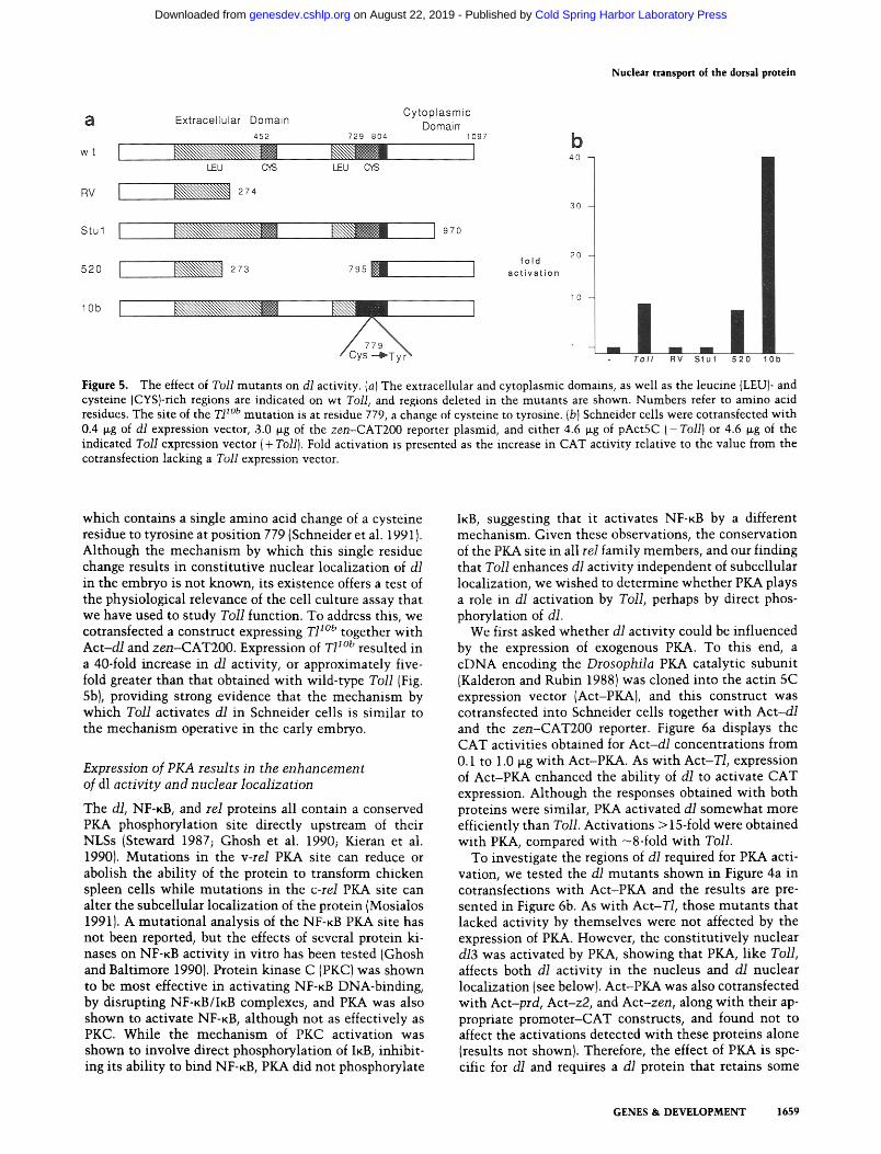
To begin to determine regions of Toll that are necessary for its function, we constructed and analyzed several Toll deletion mutants. Figure 5a details the structures of these mutants, and Figure 5b displays the results of cotransfections with Act-d/. A deletion of all but the amino-terminal 274 amino acids (RV) of Toll resulted in a total loss of Toll activity as measured by its ability to enhance the activation of CAT expression of dl from zen-CAT200. A small deletion (122 amino acids) into the carboxyl terminus (StuI) also abolished Toll activity, indicating that the intracytoplasmic domain is essential for activity. Because some receptors can be activated by
deletion of their extracellular domains (see Discussion), we deleted most of the extracellular domain of Toll (T1520) to determine whether this might activate Toll. However, this deletion had no effect on Toll activity, as approximately equal CAT activities were detected in cotransfections with Act-T1520 and Act-T/. This finding suggests that ligand binding is not required for Toll ac- tivation in Schneider cells (see Discussion).
Both dominant-ventralizing and recessive-dorsalizing alleles of Toll have been identified {Anderson et al. 1985al, and several of these alleles have been cloned and grouped into three classes (Schneider et al. 1991). One class consists of recessive alleles that result from point mutations in the cytoplasmic domain. This is consistent with our observation that an intact cytoplasmic domain is required for Toll activity. Another class contains dom- inant alleles that are amino-terminal truncations, al- though these ventralize only in the presence of wild-type Toll. The final class are dominant alleles that ventralize the embryo by facilitating nuclear transport of dl throughout the embryo (Roth et al. 1989; Steward 1989). The strongest of these is T11°b (Erd61yi and Szabad 1989),
1658 GENES & DEVELOPMENT
Cold Spring Harbor Laboratory Press on August 22, 2019 - Published by genesdev.cshlp.orgDownloaded from
Nuclear transport of the dorsal protein
a Ex t race l l u l a r Doma in C y t o p l a s m i c DomairT
452 729 804 1097 b w t I I ,o
LEU G'~ LEU ~ S
RV I 274 30
Stul I ~ ~ ~ ~ 1,7o
fo ld 520 I 273 7, 1 ] ac,,v.,,o.
20
10 lOb I
/ Cys - ' ~ T y r ~ T o l l RV S t u l 520 lOb
Figure 5. The effect of Toll mutants on dl activity. (a) The extracellular and cytoplasmic domains, as well as the leucine {LEU)- and cysteine (CYS}-rich regions are indicated on wt Toll, and regions deleted in the mutants are shown. Numbers refer to amino acid residues. The site of the T1 l°b mutat ion is at residue 779, a change of cysteine to tyrosine. {b) Schneider cells were cotransfected wi th 0.4 p.g of dl expression vector, 3.0 p.g of the zen-CAT200 reporter plasmid, and either 4.6 ~g of pActSC ( -Tol l ) or 4.6 ~g of the indicated Toll expression vector ( + Toll). Fold activation is presented as the increase in CAT activity relative to the value from the cotransfection lacking a Toll expression vector.
which contains a single amino acid change of a cysteine residue to tyrosine at position 779 (Schneider et al. 1991). Although the mechanism by which this single residue change results in constitutive nuclear localization of dl in the embryo is not known, its existence offers a test of the physiological relevance of the cell culture assay that we have used to study Toll function. To address this, we cotransfected a construct expressing T1 l°b together with Act -d /and zen-CAT200. Expression of T1 l°b resulted in a 40-fold increase in dl activity, or approximately five- fold greater than that obtained with wild-type Toll (Fig. 5b), providing strong evidence that the mechanism by which Toll activates dl in Schneider cells is similar to the mechanism operative in the early embryo.
Expression of PKA results in the enhancement of dl activity and nuclear localization
The dl, NF-~B, and rel proteins all contain a conserved PICA phosphorylation site directly upstream of their NLSs (Steward 1987; Ghosh et al. 1990; Kieran et al. 1990). Mutations in the v-re/ PKA site can reduce or abolish the ability of the protein to transform chicken spleen cells while mutations in the c-rel PKA site can alter the subceUular localization of the protein (Mosialos 1991). A mutational analysis of the NF-KB PKA site has not been reported, but the effects of several protein kio nases on NF-KB activity in vitro has been tested (Ghosh and Baltimore 1990). Protein kinase C (PKC) was shown to be most effective in activating NF-KB DNA-binding, by disrupting NF-KB/IKB complexes, and PKA was also shown to activate NF-KB, although not as effectively as PKC. While the mechanism of PKC activation was shown to involve direct phosphorylation of IKB, inhibit- ing its ability to bind NF-KB, PKA did not phosphorylate
IKB, suggesting that it activates NF-KB by a different mechanism. Given these observations, the conservation of the PKA site in all tel family members, and our finding that Toll enhances dl activity independent of subcellular localization, we wished to determine whether PKA plays a role in dl activation by Toll, perhaps by direct phos- phorylation of dl.
We first asked whether dl activity could be influenced by the expression of exogenous PKA. To this end, a cDNA encoding the Drosophila PKA catalytic subunit (Kalderon and Rubin 1988) was cloned into the actin 5C expression vector (Act-PKA), and this construct was cotransfected into Schneider cells together with Act-d/ and the zen-CAT200 reporter. Figure 6a displays the CAT activities obtained for Act-d/concentrat ions from 0.1 to 1.0 ~g with Act-PKA. As with Act-T/, expression of Act-PKA enhanced the ability of dl to activate CAT expression. Although the responses obtained with both proteins were similar, PKA activated dl somewhat more efficiently than Toll. Activations > 15-fold were obtained with PKA, compared with -8ofold with Toll.
To investigate the regions of d~ required for PKA acti- vation, we tested the d~ mutants shown in Figure 4a in cotransfections with Act-PICA and the results are pre- sented in Figure 6b. As with Act-T/, those mutants that lacked activity by themselves were not affected by the expression of PKA. However, the constitutively nuclear d/3 was activated by PICA, showing that PKA, like Toll, affects both dl activity in the nucleus and d/ nuclear localization (see below). Act-PKA was also cotransfected with Act-prd, Act-z2, and Act-zen, along with their ap- propriate promoter-CAT constructs, and found not to affect the activations detected with these proteins alone (results not shown). Therefore, the effect of PICA. is spe- cific for d / and requires a d /pro te in that retains some
GENES & DEVELOPMENT 1659
Cold Spring Harbor Laboratory Press on August 22, 2019 - Published by genesdev.cshlp.orgDownloaded from

Norris and Manley
a
dorsa l
0.1
0.2
0.3
0.4
0.6
1.0
-PKA +PKA
1 1.3
1.6 16
2.4 20
2.7 47
3.5 37
18 65
35 140
b - P K A +PKA
dorsal
w t 35 140
3 8.7 71
4 2.5 2.1
5 0.5 1 5
7,8 1.1 1 2
9 12 13
Figure 6. The effect of PKA on wild-type and mutant d] activ- ity. (a) Schneider cells were cotransfected with the indicated amount of dl expression vector, 3.0 ~g of the zen-CAT200 re- porter plasmid, and either pActSC (-PKA) or PKA expression vector (+ PKA) to bring the final concentration of expression vector to 5.0 p~g. The activation values are expressed relative to cotransfections containing the actin 5C expression vector with- out an insert. (bl Schneider ceils were transfected with 1.0 ~g of dl, 3.0 ~g of the zen-CAT200 reporter plasmid, and either 4.0 }ag of pActSC ( - PKA) or 4.0 ~g of PKA expression vector ( + PKA). The dl mutants are referred to by the numbers used in Fig. 4a.
activity and has an intact rel homology region, that is, the requirements for dl activation by PICA and Toll ap- pear to be identical.
To determine whether expression of PKA also in- creased the nuclear localization of d/, cells were trans- fected wi th 0.4 ~g of A c t - d / a l o n e (Fig. 7, panel A) or 0.4 ~g of A c t - d / p l u s 4.6 ~g of Act-PKA (panel B), fixed, and stained wi th anti-d/ antibodies. Figure 7 presents pic- tures of representive cells from each transfection and shows that the expression of PKA resulted in an in- creased nuclear localization of d/. d~ nuclear localization induced by PKA was more substantial than that observed
with Toll. In the presence of PKA, - 5 0 % of the trans- fected cells showed strong nuclear staining compared with 25% showing predominant ly nuclear staining in the presence of Toll.
Expression of a protein kinase inhibitor prevents activation of dl by Toll
The above results support the hypothesis that Toll acti- vates dl by a mechan i sm involving PKA. To address this idea further, we tested the effect of the inhibi tor protein of PKA (PKi) on dl activity in the presence of Toil. PKi has been analyzed in some detail, and peptide fragments with inhibitory activity have been isolated (Cheng et al. 1985; for review, see Kemp et al. 1988). The PKi peptides specifically inhibi t cAMP-dependent protein kinase by binding to the catalytic subunit (for review, see Walsh and Glass 1991). An actin 5C construct containing 26 amino acid residues of PKi fused to lacZ-coding se- quences, Act-lacZPKi, was cotransfected wi th Act-d/ and Act-T/, and the resultant CAT activities were mea- sured (see Materials and methods). This PKi construct encodes a fusion protein containing the 20-amino-acid peptide that has been shown to be the most potent in- hibitor of PKA (Cheng et al. 1985). Coexpression of Ac t - lacZPKi nearly abolished the enhancement of d~ activity brought about by Toll (Fig. 8). This inhibi t ion was spe- cific for d~ as the CAT activities induced by z2 or prd were not altered by the addition of PKi (results not shown). Inhibit ion was the result of the PKi moiety in the lacZ fusion protein, as identical amounts of an Ac t - lacZ expression vector did not affect d~ activity (Fig. 8). These results provide strong evidence that enhancement of dl activity induced by Toll involves activation of PKA.
Mutations in the PKA phosphorylation site of dl affect dl protein localization and activity in the presence of Toll and PKA
To determine whether activation of d~ by Toll and PKA is dependent on the dI PKA site, we mutated the pre- sumed site of phosphorylation, the serine (S) residue at position 312. We first changed this S residue to glu- tamine (Q) to create a site that should not be phospho- rylated by PKA. This mutant , Act-d/Q, was then cotransfected with Act -T/or Act-PKA, and the resulting CAT activities are shown in Figure 9a. Mutat ion of the PKA phosphorylation site resulted in only a slight reduc- tion (less than twofold) in the activity of d~ alone. How- ever, the activation of CAT expression by d/Q in the presence of either Toll or PKA was reduced significantly relative to wild-type dl, in each case by as much as a factor of 10, providing strong evidence that the d~ PKA site plays an important roll in Toll-mediated regulation of d~ activity.
To determine whether the decreased activity of d/Q in the presence of PKA or Toll reflected an inabi l i ty of the mutan t protein to be translocated to the nucleus, cells transfected wi th Act-d/Q alone (Fig. 9b, panel A), or with Act-T/(not shown) or Act-PKA (panel B) were fixed
1660 GENES & DEVELOPMENT
Cold Spring Harbor Laboratory Press on August 22, 2019 - Published by genesdev.cshlp.orgDownloaded from

Nuclear transport of the dorsal protein
A B
O o
Figure 7. Subcellular localization of dl in the pres- ence of PICA. Schneider cells were transfected with 0.3 ~g of dl (A) or 0.3 ~g of dl plus 4.7 tag of PKA (B} and stained with anti-d/and TRITC-conjugated secondary antibodies. The dl protein accumulates predominantly in the cytoplasm of transfected cells in the absence of PKA (A}. The dl protein is found predominantly in the nucleus of cells transfected with PKA (B).
and stained wi th ant i -d/ant ibodies , and pictures of rep- resentative cells are shown. These experiments show that dlQ, unl ike wild-type dl, was not transported effec- tively to the nucleus in the presence of Toll or PKA (panel B), indicating that the dl PKA site is critical for regulated nuclear transport of the dl protein.
We then wished to determine the effect of placing a
1 0 -
fo ld a c t i v a t i o n
5 -
2 - / | d l d l + T o l l d l+To l l+ d l+To l l+
lacZ PKi l acZ
Figure 8. Activity of dl in the presence of Toll and PKi. Schnei- der cells were cotransfected with the indicated amounts of dl and Toll expression vectors (ixg), 3.0 lag of the zen-CAT200 reporter plasmid, and either 3.6 lag of lacZPKi or 3.6 txg of actin lacZ. The final concentration of expression vector was adjusted to 7.0 lag with the actin 5C expression vector. Fold activation is presented as the increase in CAT activity relative to a cotrans- fection containing 7.0 lag of actin 5C expression vector without an insert.
negative charge at residue 312. Might this be sufficient to enhance nuclear localization in the absence of Toll? To address this, the S residue was changed to aspartic acid (D), creating the mutan t Act-diD. A similar muta- tion has been tested in the rel protein and was found to increase nuclear localization (Mosialos et al. 1991). Schneider cells were transfected wi th 0.4 Ixg of Act-d/D, fixed, and stained wi th anti-d/ antibodies as described above. In contrast to cells transfected wi th Act-d/, which had primari ly cytoplasmic staining, cells trans- fected with Act -d iD displayed diffuse staining of both the cytoplasm and the nucleus (Fig. 9b, panel C). This pattern was unaffected by cotransfection wi th PICA or Toll (results not shown). We also assayed the activity of the diD protein both alone and in the presence of Toll or PICA. The CAT activities presented in Figure 9a show that this mutat ion results in a protein that was not ac- tivated by Toll or PKA and whose activity alone was substantially reduced. This latter result is consistent with that observed with v-rel, where the corresponding S --* D change also reduced the activity of the protein in a CAT assay (Mosialos et al. 1991). These findings suggset that a negative charge at position 312 can en- hance nuclear localization, and strengthen the view that Toll (and PKA)funct ion through serine-312.
Finally, to rule out the possibil i ty that any change made in this region might alter d/act ivi ty, the proline (P) residue at position 311 was mutated to lysine (K). In the PKA consensus sequence this position can be any amino
GENES & DEVELOPMENT 1661
Cold Spring Harbor Laboratory Press on August 22, 2019 - Published by genesdev.cshlp.orgDownloaded from

Norris and Manley
a . , ]
d l Q Ser - ~ Gin
d l D Ser ~ Asp
dorsa l
0.4 wt
1.o
0.4 Q
1.o
D o.4
lO
- T O I I
5.6
40
2.8
16
2,1
2.3
+ T o l l
40
121
51
1.6
2.5
+ P K A
130
6.1
35
2.4
3.2
b A B C
Figure 9. Activity and subcellular localization of a dl phosphorylation site mutan t in the presence of Toll and PKA. (a) Schneider cells were cotransfected wi th the indicated amount of wild-type and mutant dl expression vectors (lag), 3.0 lag of the zen-CAT200 reporter plasmid, and pAct5C, Toll expression vector ( + Toil), or PKA expression vector ( + PKA) to bring the final concentrat ion of expression vector to 5.0 Izg. The activation values are expressed relative to cotransfections containing the actin 5C vector wi thout an insert. (b) Schneider cells were transfected wi th 0.4 ~g of dlQ CA), 0.4 lag of dlQ plus 4.6 lag of PKA (B), or 0.4 lag of diD {C) and stained with anti-d/ antibodies and TRITC-conjugated secondary antibodies. The dlQ protein accumulates predominant ly in the cytoplasm of transfected cells in the presence or absence of Toll or PKA; the diD protein accumulates in both the cytoplasm and nucleus.
acid {Kemp and Pearson 1990) so changing it should not affect the localization or activity of dl if phosphorylation is the underlying mechanism. This mutant, Act- d/RRKS, was cotransfected with zen-CAT200 with or without Ac t -T /o r Act-PKA, and the resulting CAT ac-
tivities were determined. These results (not shown) re- vealed that the activity of the dlRRKS protein was iden- tical to wild-type dl protein, providing additional sup- port for the idea that phosphorylation of serine-312 by PKA regulates dl.
1662 GENES & DEVELOPMENT
Cold Spring Harbor Laboratory Press on August 22, 2019 - Published by genesdev.cshlp.orgDownloaded from

Discussion
We have described a cell culture system in which the regulated nuclear transport and activity of the dl protein can be examined. We found that expression of either Toll or PKA together with dl resulted in an increased nuclear localization of dl and an enhancement of the ability of dl to activate CAT expression. We also observed that dl is somewhat more active, and more cells have strong nu- clear dl staining when cotransfected with dl plus PKA than when cotransfected with d/plus Toll. This obser- vation supports the notion, discussed below, that PKA is a downstream step in a signal transduction pathway that requires Toll to transmit the signal for dl nuclear local- ization to d/through PKA.
A model for nuclear localization and activation of dl
Our results support the model shown in Figure 10 for dl nuclear localization and activation. In this model Toll receives an extracellular signal that is ventrally localized in the perivitelline space of the embryo (Stein et al. 1991). Although the identity of this signal is unknown, it most likely functions by binding to or cleavage of the extracellular domain. Deletion of extracellular domains is a mechanism that can activate some receptors. The epidermal growth factor (EGF) receptor (c-erbB) can be activated by deletion of its extracellular domain and the oncogene v-erbB encodes only the transmembrane and cytoplasmic domain of c-erbB (Downward et al. 1984; Nilsen et al. 1985). Two of the Drosophila dorsal group genes, snake and easter, encode serine proteases that act upstream of Toll (DeLotto and Spierer 1986; Chasan and Anderson 1989), and either could activate Toll by cleav- ing the extracellular domain. However, the fact that many Toll dominant alleles contain point mutations in the extracellular domain (Schneider et al. 1991) perhaps supports ligand binding as the mechanism for Toll acti- vation. Point mutations may mimic ligand binding by inducing the aggregation of receptors, resulting in their
Nuclear transport o[ the dorsal protein
activation. For example, some oncogenic forms of the neu (erbB-2) proto-oncogene contain point mutations in the extracellular domain, and at least one has been shown to lead to an increased aggregation of neu recep- tors (Weiner et al. 1989). Overexpression has also been shown to activate erbB-2 (DiFiore et al. 1987). We sug- gest that in Schneider cells, Toll is also activated by over- expression and subsequent aggregation.
Once activated, Toll signals the nuclear localization and activation of dl through PKA. Toll most likely uses cAMP as a second messenger to activate PICA. Although we have no direct evidence for this, work with IL-1R supports this view. The IL-1R protein is a receptor for the interleukin 1 (IL-1) hormone, which is involved in me- diating immune and inflammatory responses (Sims et al. 1989). The IL-1R and Toll cytoplasmic domains share extensive sequence similarity (Schneider et al. 1991), suggesting that they could transmit their signals by a similar mechanism. IL-1 has been shown to induce the expression of interleukin-2 receptors (IL-2R) and to in- duce thymocyte proliferation, in cells that express IL-1 R, through stimulation of cAMP production (Shirakawa et al. 1988). IL-1, as well as cAMP and cAMP analogs, has also been shown to activate K light-chain expression by activation of a NF-KB like DNA-binding protein (Shirakawa et al. 1989). Together, these observations suggest that the signaling pathway from Toll to dl also involves stimulation of cAMP production, leading to the activation of PKA and the phosphorylation of dl. Phos- phorylated dl is free to move into the nucleus and has an enhanced ability to activate transcription.
Genetic studies have placed two dorsal group genes, tube and pelle, downstream of Toll (Govind and Steward 1991). The proteins encoded by these two genes may be used to help transmit the signal from Toll to dl. The tube gene has been cloned, but its sequence reveals nothing about its possible function (Letsou et al. 1991). Because we do not know whether tube and/or pelle are expressed in Schneider cells, we cannot say whether they are ab- solutely required for this signaling pathway. No genetic
AFTER ACTIVATION
BEFORE ACTIVATION
pc,u, (,1
(75 PKA
Extracellular Signal
C ~ Toll
Figure 10. A model for the activation of dl by Toll. Before activation, dl is held in the cytoplasm, and is therefore inactive, through an association with cactus. After Toll receives an extracellular signal, dl is freed from the cytoplasm and activated as a result of phosphorylation by PKA.
GENES & DEVELOPMENT 1663
Cold Spring Harbor Laboratory Press on August 22, 2019 - Published by genesdev.cshlp.orgDownloaded from

Norris and Manley
evidence exists implicating PKA in D/V patterning, al- though this is not surprising given the multiple func- tions of this protein.
Anchoring of dl in the cytoplasm
Like dl, NF-KB is regulated by its subcellular localiza- tion, inactive when in the cytoplasm and active when localized in the nucleus (Baeuerle and Baltimore 1988a). NF-KB is retained in the cytoplasm by association with IKB; and upon disruption of the complex, NF-KB moves into the nucleus (Baeuerle and Baltimore 1988b). Pro- teins that belong to the IKB family contain ankyrin re- peats (Haskill et al. 1991), and members of the ankyrin family can regulate interactions between membrane and cytoskeletal elements (Lux et al. 1990). Therefore, NF-KB could be held in the cytoplasm owing to its ability to interact with IKB, which could be anchored in the cyto- plasm through association with the cytoskeleton. The NF-KB/IKB complex can be destabilized by the phospho- rylation of IKB by PKC (Ghosh and Baltimore 1990), in- dicating that phosphorylation can disrupt this protein- protein interaction, resulting in the activation of NF-KB in vitro. Such an interaction with a protein anchored in the cytoplasm may also be used to retain dl in the cyto- plasm.
NF-KB can be activated by a variety of agents including viruses, T-cell mitogens, cytokines, bacterial lipo- polysaccharides, and DNA-damaging agents (for review, see Baeuerle and Baltimore 1990). NF-KB is involved in activating the expression of a variety of genes that are required for immune, infection, inflammatory, and acute phase responses in a variety of cell types (Lenardo and Baltimore 1989; Baeuerle and Baltimore 1990; Liber- mann and Baltimore 1990). Therefore, NF-KB should be responsive to a number of signaling pathways, including PKC. In contrast, dl activity is only required once, for the establishment of polarity in the early embryo; therefore, dl activation probably depends on only one signaling pathway, and the data presented here suggest that this involves phosphorylation of dl by activated PKA. We suggest that a similar activation pathway can be used to activate NF-KB under certain conditions, for example, in response to IL-1. For dl, phosphorylation of dt itself is required and alone may be responsible for activating dl by leading to its release from its inhibitor, presumably the cactus protein (see below). However, we cannot rule out that modification of cactus also occurs and plays a role, as we found that the dlQ mutant was still weakly activated by PKA or Toll. The exact mechanism of acti- vation of dl by PKA will not be known until the inter- action between dl and cactus is better understood.
The cactus gene is one that is required for establish- ment of D/V polarity in the early embryo tAnderson 1987). Unlike the dorsal group genes that give rise to dorsalized embryos when mutated, cactus mutations re- sult in partially ventralized embryos, suggesting that cactus is in some way responsible for inhibiting dl ac- tivity {Roth et al. 1989, 1991; Steward 1989). Therefore, cactus has been proposed to perform a function analo-
gous to IKB, anchoring dl in the cytoplasm by forming a dl-cactus complex. This idea is further supported by the finding that cactus, like IKB, contains ankyrin repeats (S. Kidd, pets. comm.).
In Schneider cells, dl protein is localized in the cyto- plasm at low concentrations but is increasingly found in the nucleus as the amount of transfected dl is increased (Rushlow et al. 1989). This observation can be explained in terms of an interaction between dl and cactus because cactus is known to be expressed in Schneider cells (S. Kidd, pers. comm.). Increasing the amount of dl protein expressed in these cells could saturate the endogenous cactus so that at low dl concentrations all dl is bound by cactus. As the amount of dl is increased, there is no free cactus to interact with dl, so dl is free to move into the nucleus. In our model for Toll-mediated activation of dl, phosphorylation of dl by PKA disrupts the dl-cactus complexes so that dl is free to move into the nucleus even at low concentrations.
Activation of transcription by dl
In the early embryo, dl influences the expression of sev- eral zygotic genes (for review, see Anderson 1987; Rushlow and Arora 1990). Functional binding sites, sim- ilar to NF-KB consensus sites, have been identified up- stream of zen (Ip et al. 1991} and twist (Jiang et al. 1991; Thisse et al. 1991) genes. Expression of zen is repressed in ventral regions of the embryo (Rushlow et al. 1987) while twist is activated in the ventral-most regions of the embryo (Thisse et al. 19871, suggesting that dl can act as both an activator and repressor. In Schneider cells dl can activate expression from a variety of promoters that do not appear to contain dl-binding sites, including, par- adoxically, the zen -CAT200 reporter used here [note that the dl sites in zen are located far upstream of the basal promoter (Ip et al. 1991)]. One reason for this could be the presence of dl-binding sites in the CAT reporter plasmid. A possible all-binding site has been found in the pUC vector used here (Thisse et al. 1991). However, a reporter construct containing a deletion of this region is still strongly activated by dl (J.L. Norris and J.L. Manley, unpubl.). A possible explanation for the ability of dl to activate a wide variety of promoters is that the function of dl involves a strong interaction with its target, per- haps a component of the general transcription machin- ery. This could conceivably allow dl to activate tran- scription in the absence of d/-binding sites, especially when expressed at high levels by transfection. In support of this idea, we have observed a strong and specific func- tional interaction involving TFIID and dl in cotransfec- tion experiments (J.L. Norris, J. Colgan, and J.L. Manley, unpubl.).
Whatever the mechanism by which dl activates tran- scription in cultured cells, it is intriguing that both PKA and Toll can enhance this activity independent of their effects on localization. This suggests that phosphoryla o tion of dl increases the ability of the protein to interact either with DNA or with other factors required to acti- vate transcription. Determining the precise effects of
1664 GENES & DEVELOPMENT
Cold Spring Harbor Laboratory Press on August 22, 2019 - Published by genesdev.cshlp.orgDownloaded from

Nuclear transport of the dorsal protein
phosphory la t ion on dl wil l require fur ther inves t iga t ion into the prote ins w i t h w h i c h d / i n t e r a c t s , in the cyto- p lasm as wel l as the nucleus .
Materials and m e t h o d s
Recom bin an t plasmids
All expression vectors were derived from a plasmid that con- tains the Drosophila actin 5C promoter and poly(A) site, pAct5CSRS (pActSC), which has been described in detail (Han et al. 1989}. The zen-CAT200 reporter plasmid, pAct-d/, and pAct-dl3 (dl-561) have been described previously by Rushlow et al. (1989). The above constructs and pAct-all4, pAct-diS, pac t - d16, and pAct-all9 were provided by K. Han (Columbia Univer- sity, New York). The dl NTS mutants, pAct-all7 and pAct-d/& were provided by S. Small. The dlQ, diD, and dlRRKS mutants were constructed by site-directed mutagenesis. A 0.65-kb SacI- EcoRI fragment was isolated from pAct-d/and cloned into pB- luescript SK{ + ) (Stratagene), which had been cleaved with SacI and EcoRI. The following oligonucleotides were made: 5'-GC- GACGTCCCCAGGATGGAG-3' for dlQ; 5'-CGACGTCCC- GATGATGGAGTTACC-3' for diD; 5'-CTGCGACG- TAAATCGGATGGA-3' for dlRRKS. The oligonucleotides were annealed to uracil containing single-stranded DNA, and synthesis of the second strand was done with T4 DNA poly- merase. The resulting clones were screened for sequence that encoded the appropriate amino acid change. Clones containing the desired mutations were isolated and digested with StuI and BstXI to generate a fragment, containing the mutated region, that was used to replace the corresponding wild-type fragment in pAct-d/.
The pAct-T/expression vector was constructed by isolating a NsiI-KpnI fragment, containing the Toll-coding region, from the Toll cDNA clone [kindly provided by K. Anderson (Schnei- der et al. 1991)]. The 3' overhang that resulted from cleavage with NsiI was digested with the Klenow fragment of DNA poly- merase (Klenow) before cleavage by KpnI. This fragment was inserted into the pActSC polylinker, adjacent to the actin 5C promoter, by a filled-in BamHI site and a KpnI site. The pac t - T1RV mutant was constructed by digesting pAct-T/ with EcoRV, which cleaves at one site within Toll and another in the pAct5C polylinker, and removing the 3.6-kb EcoRV-EcoRV fragment to create an in-frame stop after amino acid 274. To construct pAct-T/520, pAct-T/RV was digested with EcoRV and BglII and ligated with a 2.1-kb SalI-BglII fragment from pAct-T/to restore the Toll membrane-spanning and cytoplas- mic domains. The 5' overhang generated by digestion with SalI was filled in with Klenow to generate an in-frame deletion of amino acids 274-793. The pAct-TIStuI mutant was constructed by digesting pAct-T/wi th BglII, NcoI, and StuI to generate a 1.4-kb NcoI-BglII fragment, containing actin 5C promoter se- quences and Toll-coding sequence for amino acids 1-322, and a 1.9-kb BglII-StuI fragment that contains Toll-coding sequence for amino acids 323-970. These two fragments were ligated with a 7.0-kb BglII-NcoI fragment from pAct5C, containing ac- tin 5C promoter, actin 5C poly(A), and pBR322 sequences, which had been cleaved with BglII. The 5' overhang generated by cleavage with BglII was filled-in with Klenow before diges- tion with NcoI. The pAct-T1 ~°b mutant was constructed by di- gesting pAct-T/with BglII and StuI to generate a 7.3-kb BglII- BglII fragment, containing actin 5C promoter and poly(A) se- quences, as well as Toll-coding sequence for amino acids 1-322, and a 1.6-kb BglII-StuI fragment that contains Toll-coding se- quence for amino acids 970-1097. These fragments were ligated
with a BglII-StuI fragment, containing Toll-coding sequence for amino acids 323-969, from the T/lob eDNA clone (kindly pro- vided by K. Anderson) to replace the wild-type sequence with the sequence encoding a cysteine to tyrosine change at amino acid 779.
The pActPKA expression vector was constructed by cleaving the PKA cDNA clone (kindly provided by D. Kalderon (Colum- bia University) with XbaI, filling in the 5' overhang with Kle- now, and digesting with KpnI. A 1.1-kb KpnI-XbaI fragment, containing the PKA-coding sequence, was inserted into the pActSC polylinker by digesting pActSC with KpnI and EcoRV. The pAct-lacZPKi expression vector was provided by M.E. Lane (Columbia University).
DNA transfection and transient expression assay
Drosophila Schneider L2 cells were grown and transfected as described previously (Han et al. 1989). Each transfection con- tained 5.0 ~g of expression vector consisting of the indicated amounts of dl, Toil, or PKA expression vectors and variable amounts of pAct5C to bring the total amount of expression vector to 5.0 ~g. A total of 10 lag of DNA was used for each transfection, so the remaining 5.0 lag of DNA consisted of 2.0 ~g copia long terminal repeat (LTR}-IacZ as an internal control and 3.0 p-g of the zen-CAT200 reporter plasmid. All transfec- tions were performed in duplicate, and ~-galactosidase and CAT activities were measured as described previously {Han et al. 1989). The CAT activities presented represent the average of several independent transfections.
Transfections that included the lacZPKi expression vector contained 0.4 ~g of dl expression vector, 3.0 ~g of Toll expres- sion vector, and 2.0-3.6 lag of lacZPKi expression vector. The total amount of expression vector was adjusted to 7.0 ~g with pActSC. A total of 12.0 ~g of DNA, including 2.0 lag of copia LTR-lacZ and 3.0 lag of zen-CAT200, was used in each trans- fection. Cells were harvested and protein extracts were prepared as described (Han et al. 1989). The same amount of extract from each sample was used in a CAT assay. A ~-galactosidase assay was performed to ensure that protein was being expressed in approximately equal amounts. Three independent transfections were performed in duplicate, and all gave similar results.
Staining of cells
Schneider cells were transfected as described above, and 48 hr after transfection the cells were fixed on plates with formalde- hyde. The cells were blocked with 10% BSA, stained with anti ° dl primary antibodies [provided by C. Rushlow and M. Levine (Rushlow et al. 1989)[, washed twice with washing and dilution buffer (1% BSA, 0.5 M NaC1, and 0.1% Tween 80 in PBS), and stained with TRITC-conjugated secondary antibodies.
A c k n o w l e d g m e n t s
We are grateful to K. Anderson, K. Han, D. Kalderon, M.E. Lane, and S. Small for providing plasmids, and to M. Levine and C. Rushlow for providing antibodies. We thank D. Kaldron, M. Levine, K. Han, J. Colgan, D. Read, M.E. Lane, and J. Wu for advice and discussion, D. Kalderon for comments on the manu- script, and S. Kidd for communicating results prior to publica- tion. This work was supported by a predoctoral training grant from the National Institutes of Health (NIH) to J.L.N. and NIH grant GM37971 to J.L.M.
The publication costs of this article were defrayed in part by payment of page charges. This article must therefore be hereby
GENES & DEVELOPMENT 1665
Cold Spring Harbor Laboratory Press on August 22, 2019 - Published by genesdev.cshlp.orgDownloaded from

Norris and Manley
marked "advertisement" in accordance with 18 USC section 1734 solely to indicate this fact.
R e f e r e n c e s
Anderson, K.V. 1987. Dorsal-ventral embryonic pattern genes of Drosophila. Trends Genet. 3: 91-97.
• 1989. Drosophila: The maternal contribution. In Genes and embryos (ed. D.M. Glover and B.D. Hanes), pp. 1-37. IRL Press, Oxford, England.
Anderson, K.V. and C. Nfisslein-Volhard. 1986. Dorsal-group genes of Drosophila. In Gametogenesis and the early em- bryo (ed. J.Gall), pp. 177-194. Alan R. Liss, New York.
Anderson, K.V., G. Jiirgens, and C. N~sslein-Volhard. 1985a. Establishment of dorsal-ventral polarity in the Drosophila embryo: Genetic studies on the role of the Toll gene product. Cell 42: 779-789.
Anderson, K.V., L. Bokla, and C. Ntisslein-Volhard. 1985b. Es- tablishment of dorsal-ventral polarity in the Drosophila em- bryo: The induction of polarity by the Toll gene product. Cell 42: 791-798.
Baeuerle, P.A. and D. Baltimore. 1988a. Activation of DNA- binding activity in an apparently cytoplasmic precursor of the NF-KB transcription factor. Cell 53:211-217.
~ . 1988b. IKB: A specific inhibitor of the NF-KB transcrip- tion factor• Science 242: 540-546.
~ . 1990. The physiology of the NF-KB transcription factor. Hormonal regulation of transcription. Mol. Aspects Cell. Regul. 6: 409-432.
Chasan, R. and K.V. Anderson. 1989. The role of easter, an apparent serine protease, in organizing the dorsal-ventral pattern of the Drosophila embryo. Cell 56:391-400.
Cheng, H.C., S.M. Van Pattern, A.J. Smith, and D.A. Walsh. 1985. A active twenty-amino-acid-residue peptide derived form the inhibitor protein of the cyclic AMP-dependent pro- tein kinase. Biochem. J. 231: 655-661.
DeLotto, R. and P. Spierer. 1986. A gene required for the spec- ification of dorsal-ventral pattern in Drosophila appears to encode a serine protease. Nature 323: 688-692.
DiFiore, P.P., J.H. Pierce, M.H. Kraus, O. Segatto, C.R. King, and S.A. Aaronson. 1987. erbB-2 is a potent oncogene when over- expressed in NIH/3T3 cells. Science 237:178-182.
Downward, J., Y. Yorden, E. Mayes, G. Scrace, N. Totly, P. Stockwell, A. Ullrich, J. Schlessinger, and M.D. Waterfield. 1984. Close similarity of epidermal growth factor receptor and v-erb-B oncogene protein sequences. Nature 307: 521- 527.
Erd41yi, M. and J. Szabad. 1989. Isolation and characterization of dominant female sterile mutations of Drosophila melano- gaster I. Mutations on the third chromosome. Genetics 122: 111-127.
Ghosh, S. and D. Baltimore. 1990. Activation in vitro of NF-KB by phosphorylation of its inhibitor IKB. Nature 344: 678- 682.
Ghosh, S., A.M. Gifford, L.R. Riviere, P. Tempst, G.P. Nolan, and D. Baltimore. 1990. Cloning of the p50 DNA binding subunit of NF-KB: Homology to rel and dorsal. Cell 62: 1019-1029.
Govind, S. and R. Steward. 1991. Dorsoventral pattern forma- tion in Drosophila: Signal transduction and nuclear target- ing. Trends Genet. 7: 119-125.
Han, K., M.S. Levine, and J.L. Manley. 1989. Synergistic activa- tion and repression of transcription by Drosophila ho- meobox proteins. Cell 56: 573-583.
Hashimoto, C., K.L. Hudson, and K.V. Anderson. 1988. The Toll
gene of Drosophila, required for dorsal-ventral embryonic polarity, appears to encode a transmembrane protein. Cell 52: 269-279.
Hashimoto, C., S. Gerttula, and K.V. Anderson. 1991. Plasma membrane localization of the Toll protein in the syncytial Drosophila embryo: Importance of transmembrane signaling for dorsal-ventral pattern formation. Development 111: 1021-1028.
Haskill, S., A.A. Beg, S.M. Tompkins, J.S. Morris, A.D. Yuro- chko, A. Johannes-Sampson, K. Mondal, P. Ralph, and A.S. Baldwin. 1991. Characterization of an immediate-early gene induced in adherent monocytes that encodes IKB-like activ- ity. Cell 65: 1281-1289.
Ip, Y.T., R. Kraut, M. Levine, and C.A. Rushlow. 1991. The dorsal morphogen is a sequence-specific DNA-binding pro- tein that interacts with a long-range repression element in Drosophila. Cell 64: 439--446.
Jiang, J., D. Kosman, Y.T. Ip, and M. Levine. 1991. The dorsal morphogen gradient regulates the mesoderm determinant twist in early Drosophila embryos. Genes & Dev. 5: 1881- 1891.
Kalderon, D. and G.M. Rubin. 1988. Isolation and characteriza- tion of Drosophila cAMP-dependent protein kinase genes. Genes & Dev. 2: 1539-1556.
Keith, F.J. and N.J. Gay. 1990. The Drosophila membrane re- ceptor Toll can function to promote cellular adhesion. EMBO J. 9: 4299--4306.
Kemp, B.E. and R.B. Pearson. 1990. Protein kinase recognition sequence motifs. Trends Biochem. Sci. 15: 342-346.
Kemp, B.E., H. Cheng, and D.A. Walsh. 1988. Peptide inhibitors of cAMP-dependent protein kinase. Methods Enzymol. 159: 173-185.
Kieran, M., V. Blank, F. Logeat, I. Vandekerckhove, F. Lott- speich, O. Le Bail, M.B. Urban, P. Kourilsky, P.A. Baeuerle, and A. IsraEl. 1990. The DNA binding subunit of NF-KB is identical to factor KBF 1 and homologous to the tel oncogene product. Cell 62: 1007-1018.
Lenardo, M.J. and D. Baltimore. 1989. NF-KB: A pleiotropic me- diator of inducible and tissue-specific gene control. Cell 58: 227-229.
Letsou, A., S. Alexander, K. Orth, and S.A. Wasserman. 1991. Genetic and molecular characterization of tube, a Droso- phila gene maternally required for embryonic dorsoventral polarity. Proc. Natl. Acad. Sci. 88: 810-814.
Libermann, T.A. and D. Baltimore. 1990. Activation of inter- leukin-6 gene expression through the NF-KB transcription factor. MoI. Cell. Biol. 10: 2327-2334.
Lopez, J.A., D.W. Chung, K. Fujikawa, F.S. Hagen, T. Papayan- nopoulou, and G.J. Roth. 1987. Cloning of the a chain of human platelet glycoprotein Ib: A transmembrane protein with homology to leucine-rich ~2-glycoprotein. Proc. Natl. Acad. Sci. 84: 5615-5619.
Lux, S.E., K.M. John, and V. Bennett. 1990. Analysis of cDNA for human erythrocyte ankyrin indicates a repeated struc- ture with homology to tissue-differentiation and cell-cycle control proteins. Nature 344: 36-42.
Mosialos, G., P. Hamer, A.J. Capobianco, R.A. Laursen, and T.D. Gilmore. 1991. A protein kinase-A recognition se- quence is structurally linked to transformation by p59 v-r~i and cytoplasmic retention of p68 crel. Mol. Cell. Biol. 11: 5867-5877.
Nilsen, T.W., P.A. Maroney, R.G. Goodwin, F.W. Rottman, L.B. Crittenden, M.A. Raines, and H. Kung. 1985. c-erbB activa- tion in ALV-induced erythroblastosis: Novel RNA process- ing and promotor insertion result in expression of an amino- truncated EGF receptor. Cell 41: 719-726.
1666 GENES & DEVELOPMENT
Cold Spring Harbor Laboratory Press on August 22, 2019 - Published by genesdev.cshlp.orgDownloaded from

Roth, S., D. Stein, and C. Niisslein-Volhard. 1989. A gradient of nuclear localization of the dorsal protein determines dor- soventral pattern in the Drosophila embryo. Cell 59:1189- 1202.
Roth, S., Y. Hiromi, D. Godt, and C. Niisslein-Volhard. 1991. cactus a maternal gene required for proper formation of the dorsoventral morphogen gradient in Drosophila embryos. Development 112: 371-388.
Rushlow, C. and K. Arora. 1990. Dorsal-ventral polarity and pattern formation in Drosophila. Sere. Cell Biol. 1: 173-184.
Rushlow, C., M. Frasch, H. Doyle, and M. Levine. 1987. Mater- nal regulation of zenknffflt: A homeobox gene controlling differentiation of dorsal tissues in Drosophila. Nature 330: 583-586.
Rushlow, C.A., K. Han, J.L. Manley, and M. Levine. 1989. The graded distribution of the dorsal morphogen is initiated by selective nuclear transport in Drosophila. Cell 59: 1165- 1177.
Schneider, D.S., K.L. Hudson, T. Lin, and K.V. Anderson. 1991. Dominant and recessive mutations define functional do- mains of Toll, a transmembrane protein required for dorsal- ventral polarity in the Drosophila embryo. Genes & Dev. 5: 797-807.
Shirakawa, F., U. Yamashita, M. Chedid, and S.B. Mizel. 1988. Cyclic AMP-an intracellular second messenger for interleu- kin 1. Proc. Natl. Acad. Sci. 85: 8201-8205.
Shirakawa, F., M. Chedid, I. Suttleo, B.A. Pollok, and S.B. Mizel. 1989. Interleukin 1 and cyclic AMP induce K immunoglob- ulin light-chain expression via activation of an NF-KB-like DNA binding protein. Mol. Cell. Biol. 9: 959-964.
Sims, J.E., B. Acres, C.E. Grubin, C.J. McMahan, J.M. Wignall, C.J. March, and S.K. Dower. 1989. Cloning the interleukin 1 receptor form human T cells. Proc. Natl. Acad. Sci. 86: 8946-8950.
Stein, D., R. Siegfried, E. Vogelsang, and C. Ntisslein-Volhard. 1991. The polarity of the dorsoventral axis in the Drosophila embryo is defined by an extracellular signal. Cell 65: 725- 735.
Steward, R. 1987. Dorsal, an embryonic polarity gene in Droso- phila, is homologous to the vertebrate proto-oncogene, c-tel. Science 238: 692-694.
~ . 1989. Relocalization of the dorsal protein from the cy- toplasm to the nucleus correlates with its function. Cell 59: 1179-1188.
Steward, R., S.B. Zusman, L.H. Huang, and P. Schedl. 1988. The dorsal protein is distributed in a gradient in early Drosophila embryos. Cell 55: 487-495.
Thisse, B., C. Stoetzel, M. E1 Messal, and F. Perrin-Schmitt. 1987. Genes of the Drosophila matemal dorsal group control the specific expression of the zygotic gene twist in presump- tive mesodermal cells. Genes & Dev. 1: 709-715.
Thisse, C., F. Perrin-Schmitt, C. Stoetzel, and B. Thisse. 1991. Sequence-specific transactivation of the Drosophila twist gene by the dorsal gene product. Cell 65:1191-1201.
Walsh, D.A. and D.B. Glass. 1991. Utilization of the inhibitor protein of adenosine cyclic monophosphate-dependent pro- tein kinase, and peptides derived from it, as tools to study adenosine cyclic monophosphate mediated cellular pro- cesses. Methods Enzymol. 201: 304-317.
Weiner, D.B., J. Liu, J.A. Cohen, W.V. Williams, and M.I. Green. 1989. A point mutation in the neu oncogene mimics ligand induction of receptor aggregation. Nature 339: 230-231.
Nuclear transport of the dorsal protein
GENES & DEVELOPMENT 1667
Cold Spring Harbor Laboratory Press on August 22, 2019 - Published by genesdev.cshlp.orgDownloaded from

10.1101/gad.6.9.1654Access the most recent version at doi: 6:1992, Genes Dev.
J L Norris and J L Manley protein Toll and protein kinase A.be established by a signaling pathway involving the transmembrane Selective nuclear transport of the Drosophila morphogen dorsal can
References
http://genesdev.cshlp.org/content/6/9/1654.full.html#ref-list-1
This article cites 50 articles, 18 of which can be accessed free at:
License
ServiceEmail Alerting
click here.right corner of the article or
Receive free email alerts when new articles cite this article - sign up in the box at the top
Copyright © Cold Spring Harbor Laboratory Press
Cold Spring Harbor Laboratory Press on August 22, 2019 - Published by genesdev.cshlp.orgDownloaded from