sarah atchison dr. michael pilant - texas a&m...
TRANSCRIPT

SarahAtchison
Dr.MichaelPilant
MATH614.700
13May2009
TheRoleofFractalGeometryintheBiologicalSciences
Introduction
Fractalscanbeseenallthroughoutthenaturalworld.Infact,“wherevera
chaoticprocesshasshapedanenvironment,afractalstructureisleftbehind”
(KenkelandWalker1996).FromthecoastlinesofGreatBritaintotheperimeterof
acancercell,fractalsareapparentinwaysMandelbrotneverthoughtpossible.In
thepast,scientiststriedtouseclassicEuclideangeometrytoanalyzegeographical
structuresandbiologicalsystems.However,wehavesincerealizedthatamuch
morecomplextypeofgeometry,fractalgeometry,isneededtocompletethesetasks.
Euclideangeometrydoes“notyieldfiniteanswersforcertainobjects”inthenatural
world(BauerandMackenzie1995).KenkelandWalker(1996)notethatthe
“importanceoffractalscalinghasbeenrecognizedatvirtuallyeverylevelof
biologicalorganization”.Probablyoneofthegreatestobservationsoffractal
geometryinbiologyisthat,byanalyzingthefractaldimensionsoftumorsandcells,
scientistswillbeabletodetectcancergrowthinpatientsearlierandinamore
efficientmanner.Theeffectsofresearchingfractalsinbiologicalsystemsofall
levelsare,inasense,limitless.

SarahAtchisonTheRoleofFractalGeometryintheBiologicalSciences
2
SurveyofRelatedWorks
BenoitMandelbrotfirstdemonstratedtheconceptoffractaldimensionby
describingmeasuringthecoastlineofGreatBritainwitharuler.Asthelengthofthe
rulerapproacheszero,theperimeterofGreatBritainapproachesinfinity.Studies
showthatislandswithsmallerperimetershavealowermeanD,fractaldimension,
thanislandswithalargerperimeter.MandelbrotusedthedataLewisRichardson
hadfoundstudyingtheslopesofcoastlinestofindtheequationneededtocalculate
thefractaldimensionofanygivencoastline.Theequationis
N(d)=M/dD
whereN(d)isthenumberofsegmentsoflengthdneededtomeasuretheperimeter,
Misaconstant,andDisthefractaldimensionofthecoastline.
Notonlydocoastlineshavefractaldimension,sodoclouds.Whenflyingin
anairplane,“acloudtwentyfeetawaycanbeindistinguishablefrom[acloud]two
thousandfeetaway(Gleick,1987).Fractalpropertiesarealsoveryapparentin
plants.Fractaldimensionofleafedgesinplantscanbehighlyvariableinsome
species.Oaksareknowntobeanexampleofonesuchspecies.However,fractal
dimensionisthoughttobeapossibletoolfortaxonomicallyclassifyingaplant.Root
systemsalsohavefractaldimension.Rootsystemshave“beenexaminedusingthe
box‐countingmethod”andDshowstoincreaseovertime,varyingbetweenspecies
(KenkelandWalker,1996).Area‐perimeterrelationshipsarealsousedtoexamine
fractalpropertiesofmountains.Thesurfacesofrealmountainshavebeensculpted
byweatherandatmosphericchanges;however,researcherscanoftenmanufacture

SarahAtchisonTheRoleofFractalGeometryintheBiologicalSciences
3
thesameeffectsthemselvestorecreateandanalyzethesamefractaldimensionsof
mountains(Frameetal,2001).
Bacterialcolonies,liketreesandferns,growinafractalmannerunder
stressedconditions.Normally,bacteriagrowsinsoftagarwithabundantnutrient
concentration,leadingtosoftedgesandcompactness.Conversely,ifthebacteria
growsinstressfulconditionswheretheagarishardandnutrientsaremorescarce,
thecoloniestakeonafractalgrowingpatternresemblingthatofDLA(Diffusion‐
LimitedAggregation)clusters.DLAisacomputersimulationthatformsclustersby
“particlesdiffusingthroughamediumthatjostlestheparticlesastheymove”
(Frameetal.,2001).Tmorphotypecoloniesareanexampleofthisbehavior(see
Figure1).WhenTmorphotypecoloniesgrowinenvironmentswherenutrientsare
lowandagarissoft,Cmorphotypecoloniesarisethroughchiralgrowth(seeFigure
2).Consequently,whenCmorphotypesgrowunderextremelystressedconditions,
Vmorphotypesarisethroughvortexgrowth(seeFigure3).Bacteriaisnottheonly
organismthatfollowsthissamebehaviorinaharshenvironment;fungusalsogrow
fractallywhennutrientconcentrationsaredecreasing(seeFigure4).
Fractalscalingisfoundinvarioussystemsinhumananatomy.DNA
sequencesdemonstrateself‐similarityandthe“twistingsofDNAbindingproteins
havefractalproperties(KenkelandWatson,1996).Chromosomalstructure
consistsofa“concatenationof‘mini‐chromosomes’”,makingittree‐like(Kenkeland
Watson,1996).EvolutionarybiologistscanusethefractalpropertiesofDNAto
characterizeandclassifyrelationshipsbetweenorganismsthroughthemultifractal
spectra.Researchershavefoundthatcellsofalltypeshavefractalproperties.

SarahAtchisonTheRoleofFractalGeometryintheBiologicalSciences
4
Fractaldimensionhasbeenusedtomeasurecellularcomplexityandcontour
complexityofimagesofneuralcells(KenkelandWatson,1996).Thereiseven
fractalbehaviorpresentinthehumaneye.Scientistshavefoundthisbehaviorwhen
studying”homeostasisinthecornea,maintainedbyosmoticintegrityprovidedby
thecornealepithelium”(IannacconeandKhokha,1996).
Proteinandenzymeshavebeenextensivelystudiedfortheirfractal
propertiesanddimension.Enzymologistsandbiologistsstudyingmacromolecules
areactivelyresearchingfractalsinenzymesandproteins.Thefractalanalysisof
proteinshasproducedapowerlawintheformof
p~vα
wherepistheproperty,visthevariable,andαistheexponent.Variablesvandα
canberelatedtotheoreticallyorexperimentallyobtainedfractaldimensions.
Proteinsarefatfractals“sincethesurfaceoftheproteinisafractalbutatfinite
volume”(IannacconeandKhokha,1996).Liebovitchandhiscolleaguesexamined
the“kineticsofproteinionchannelsinthephospholipidbilayer”findingthatthe
fractalpropertiesofthe“timingofopeningsandclosingsofionchannels”meant
that“processesoperatingatdifferenttimescalesarerelated,notindependent”
(KenkelandWatson,1996).Proteinfractalshavebeenusedincreatingsynthetic
materials.Keratin,themainproteinfoundingoosedown,wasfoundtohavefractal
nodesandbranchesthatarethekeytodown’sabilitytotrapair(Gleick,1987).
Fractalpropertiesarefoundinthemanydichotomousbranchingsystemsof
thehumanbody,namelytherespiratory,digestive,andcirculatorysystems.Fractal
scalinginphysiologymeans“moresurfaceareaforabsorptionandtransfer”(Frame

SarahAtchisonTheRoleofFractalGeometryintheBiologicalSciences
5
etal,2001).Thelungsdemonstratemorefault‐toleranceduringgrowthduetothe
fractalscaling.Theyneedtohavetheabilitytofillupthebiggestsurfacepossible
intothesmallamountofspacetheyaregiven.Thesurfaceareaofananimal’slungs
isapproximatelyproportionaltoits“abilitytoabsorboxygen”(Gleick,1987).
Humanlungshaveasurfaceareaaboutthesizeofatenniscourt.Tissuesinthe
digestivetractshowfoldswithinfolds,givingthedigestivesystemitsfractalnature.
Theurinarycollectingsystemandthebiliaryductarealsoexamplesoffractal
structures.
Thecirculatorysystemisprobablythemostfractallycomplexsysteminthe
body.Liketherespiratorysystem,itmustfitanenormoussurfaceareaintoavery
tinyvolume.Bloodvesselsdivideandbranchoffsomanytimesthatthevesselsare
narrowenoughthebloodcellscanonlygothroughonebehindtheother.Inmost
tissues“nocellisevermorethanthreeorfourcellsawayfromavessel”,soitis
amazinghow“vesselsandbloodtakeupnomorethanaboutfivepercentofthe
body”(Gleick,1987).Theheartisfilledwithfractalnetworks.Examplesofthese
networksarethecoronaryarteriesandveins,thefibersbindingthevalvestothe
heartwall,andthecardiacmusclesthemselves.TheHis‐Purkinjesystem,”a
networkofspecialfibersinthehearthatcarrypulsesofelectriccurrenttothe
contractingmuscles”,iskeyinthefractalfrequencyspectrumofheartbeattiming
anditselfhasfractalproperties(Gleick,1987;Frameetal,2001).Theiterationof
fractalstructuresinthecirculatorysystemmakeitsostrongagainstinjurythatthe
heart“continuestofunctionevenaftertheHis‐Purkinjesystemhassuffered
considerabledamage”(Frameetal,2001;KenkelandWatson,1996).Theoretical
SarahAtchisonTheRoleofFractalGeometryintheBiologicalSciences
6
biologistshaveconcludedthatfractalscalingis“universalinmorphogenesis”
(Gleick,1987).
Manydiseasescausecellstofollowfractallaws.Histopathologistshave
turnedtofractalgeometryandmicroscopicimageprocessingtomoreeffectively
studythefractaleffectsdiseaseshaveontissueandcellgrowth.Usingthese
techniques,histopathologistscanmoreaccuratelygivediagnosesandprognoses.A
majorproblemhistopathologistshavewithdiagnosisisuniformity.Notonlywill
fractalgeometryaidinachievingartificialintelligence‐baseddiagnosis,itmightalso
provide“insightintosomediseaseprocesses”(IannacconeandKhokha,1996).Not
onlydosuchdiseasesastheherpessimplexvirushavefractalproperties,butsodo
most,ifnotall,typesofcancer.
InJanuaryof1998,itwasreleasedthatDrAndrewEinsteinandhis
colleaguesattheMountSinaiSchoolofMedicinehadbeenanalyzingthefractal
patternsofbreastcancercells.Theseresearchersanalyzedthenucleiofthesecells
andthedistributionandfractalpatternsofthechromatininside.Bymeasuring
differenceinlacunarityandbyexaminingthedifferencesinfractaldimension
betweenbenignandmalignantcells,theywereabletodeliverthecorrectdiagnosis
to39outof41patientsinablindstudy(ScheweandStein,1998).
Outlinesoftumorsrevealthelocalgrowthbehavioroftumorsto
histopathologists.Benigntumorshave“expansive”,smoothoutlineswhile
malignanttumorshave“localaggressivefeatureswithinvasionofsurrounding
tissue”(IannacconeandKhokha,1996).Theinvadingedgesaretypicallyirregular
andfragmented“withdetachmentofislandsfromthetumor”(Iannacconeand

SarahAtchisonTheRoleofFractalGeometryintheBiologicalSciences
7
Khokha,1996).Histopathologistshaveattemptedtoquantifythesepatterninto
classificationcategoriestoincreaseprognosisinoral,esophageal,andlaryngeal
carcinomas(IannacconeandKhokha,1996).Scientistsresearchedcanceroustumor
growthinmiceandfoundthatthetumorvesselsyieldedfractaldimensions1.89±
0.04whilethenormalvesselshaddimensionsof1.70±0.03.Tumorvesselshave
manysmallerbendsinsideoflargerbends.Beingableto“quantifytheirregular
structuresthatarepresentintumorshelpstoclarifywhytreatmentisso
frustratinglydifficult”(BaishandJain,2000).Thereare,however,limitationsto
applyingfractallawstocancerresearch.Whileincreasedfractaldimensionis
commoninmalignanttumorgrowth,itisnotuniversal.Normalbonemarrowcells
exhibitanincreasedfractaldimensionwhilethemalignantcellshaveadecreased
fractaldimension(BaishandJain,2000).
SummaryandConclusions
Fractalsaretheresultofchaoticprocesses.Notonlyarefractalsfoundin
mathematics,butalsointhereal,naturalworld.Fractalgeometryhasoftenbeen
dubbed“thegeometryofnature”,butitseffectsstretchmuchfurtherintobiologyas
well.Notonlydocoastlines,mountains,clouds,andplantshavefractaldimension,
thereismorecomplexfractaldimensiontobefoundinthebloodvessels,alveoliof
thelungs,andcellsinthehumanbody.Studyingfractalpropertiesofbiological
systemshasleadtomanyrevolutionarydiscoveries.Amongtheseepitomesisthe
knowledgethatfractaldimensioncanassistoncologistsindetecting,andtherefore
treating,severaltypesofcancerearlierandmoreeffectively.Ifsuchconclusionscan
befoundfromstudyingthesefractalsnow,withthecurrenttechnology,whatdoes

SarahAtchisonTheRoleofFractalGeometryintheBiologicalSciences
8
thefutureholdwithnewtechnologyandcontinuedstudyintofractalgeometryin
biologicalsciences?
MATLAB
Afterreading“CancerDetectionviaDeterminationofFractalCellDimension”
byWolfgangBauerandCharlesD.Mackenzie(1995),Idecidedtotrytoreplicate
theirfindingsfromtheirresearchofhumanlymphocytesaffectedbyhairy‐cell
leukemiausingthebox‐countingmethodandMatlab.PaulFrenchwrotetheMatlab
codeIusedandmodified.Itusesthebox‐countingmethodtofindtheHausdorff
dimensionofuploadedimages.IusedittofindtheHausdorffdimensionofan
“electronicmicroscopeimageofasectionofahumanlymphocyte,affectedwith
hairy‐cellleukemia,digitizedwith256greylevels”(BauerandMackenzie,1995).
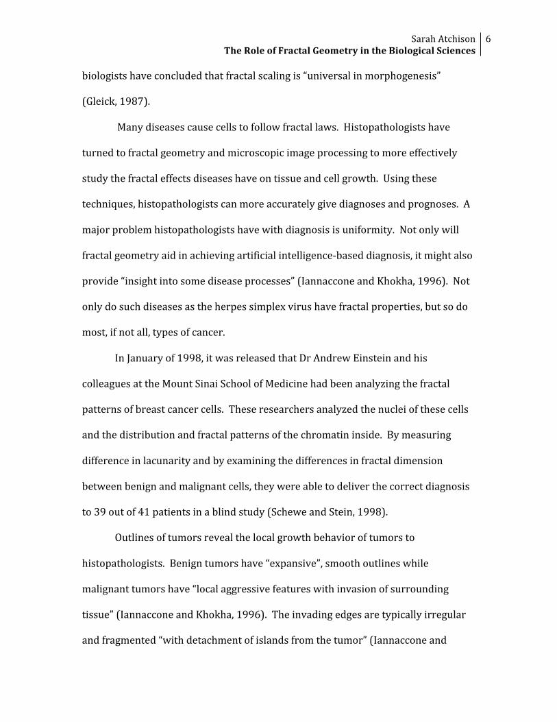
ThearticleBauerandMackenziepublishedalreadyhadanimageoftheperimeterof
thelymphocyte,soIuploadedthatimagetopreventpossibleerrorsinconverting
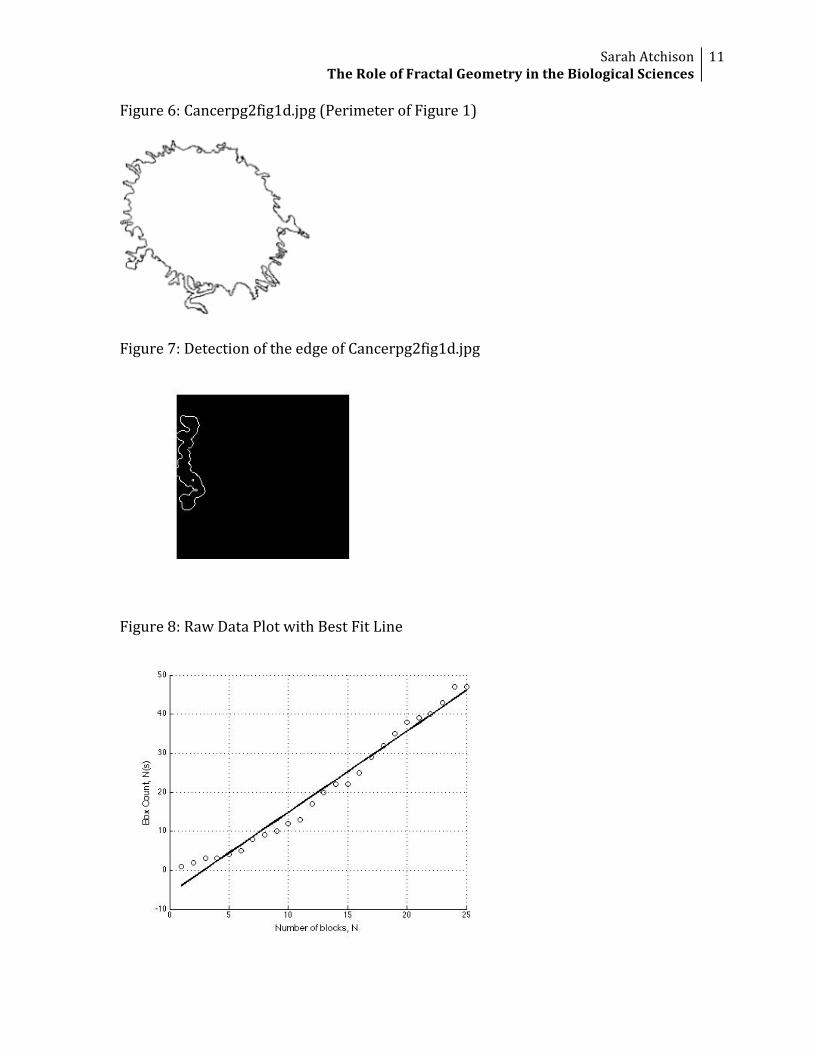
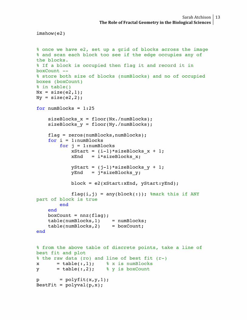
theoriginal(Figure5)intothedesiredgrayscaleimage(Figure6).Ithenranthe
programandcalculatedtheHausdorffdimensiontobe1.3041(also,seefigures7‐
9).BauerandMackenzie’smethodismoreaccurateandcanbeusedonmanytypes
ofcellsororganisms,couldpossibly“deriveaquantitativemeasureforthe
raggednessofcellsorsmallbiologicalorganisms”,andalso“showeditispossibleto
distinguishbetweenhealthypersonsandhairy‐cellleukemiacancerpatients”
(BauerandMackenzie,2000).

SarahAtchisonTheRoleofFractalGeometryintheBiologicalSciences
9
Graphics
Figure1:Tmorphotypecoloniesunderincreasingstressedconditions
Nutrient=15(g/l),Agar=2.25%Nutrient=4(g/l),Agar=2.25%
Nutrient=2(g/l),Agar=1.75%Nutrient=0.01(g/l),Agar=1.75%

SarahAtchisonTheRoleofFractalGeometryintheBiologicalSciences
10
Figure2:Cmorphotypesinstressedenvironments
Figure3:Vmorphotypesinstressedenvironments
Figure4:Fungalgrowthinstressedenvironments
Figure5:Cancerpg2fig1a.jpg
SarahAtchisonTheRoleofFractalGeometryintheBiologicalSciences
11
Figure6:Cancerpg2fig1d.jpg(PerimeterofFigure1)
Figure7:DetectionoftheedgeofCancerpg2fig1d.jpg
Figure8:RawDataPlotwithBestFitLine

SarahAtchisonTheRoleofFractalGeometryintheBiologicalSciences
12
Figure9:CalculationofHausdorffDimension
AppendixA–MatlabCode
Fractaldimensionmeasurement.m(withinputfilenameschanged)%%%%%%%%%%% % this is used to find the hausdorff dimension via the box counting method % email: [email protected] % web: www.ee.ucl.ac.uk/~pfrench %%%%%%%%%%% clear clc table =[,2]; % load up original image and convert to gray-scale p = imread('Cancerpg2fig1d.jpg'); %p = rgb2gray(P); figure(1) imshow(p) % detect the edge of image 'p' using the Canny algorithm % this gives edge as 'e2' bw = im2bw(p, graythresh(p)); e = edge(double(bw)); fi = imfill(bw, 'holes'); op = imerode(fi,strel('disk',4)); e2 = edge(double(op)); figure(2)
SarahAtchisonTheRoleofFractalGeometryintheBiologicalSciences
13
imshow(e2) % once we have e2, set up a grid of blocks across the image % and scan each block too see if the edge occupies any of the blocks. % If a block is occupied then flag it and record it in boxCount -- % store both size of blocks (numBlocks) and no of occupied boxes (boxCount) % in table() Nx = size(e2,1); Ny = size(e2,2); for numBlocks = 1:25 sizeBlocks_x = floor(Nx./numBlocks); sizeBlocks_y = floor(Ny./numBlocks); flag = zeros(numBlocks,numBlocks); for i = 1:numBlocks for j = 1:numBlocks xStart = (i-1)*sizeBlocks_x + 1; xEnd = i*sizeBlocks_x; yStart = (j-1)*sizeBlocks_y + 1; yEnd = j*sizeBlocks_y; block = e2(xStart:xEnd, yStart:yEnd); flag(i,j) = any(block(:)); %mark this if ANY part of block is true end end boxCount = nnz(flag); table(numBlocks,1) = numBlocks; table(numBlocks,2) = boxCount; end % from the above table of discrete points, take a line of best fit and plot % the raw data (ro) and line of best fit (r-) x = table(:,1); % x is numBlocks y = table(:,2); % y is boxCount p = polyfit(x,y,1); BestFit = polyval(p,x);

SarahAtchisonTheRoleofFractalGeometryintheBiologicalSciences
14
figure(3) hold on grid on plot(x,y, 'ko','LineWidth',1) plot(x,BestFit, 'k-','LineWidth',2) xlabel('Number of blocks, N','FontSize',12) ylabel('Box Count, N(s)','FontSize',12) % calculate Hausdorff Dimension x2 = log(x); y2 = log(y); p2 = polyfit(x2,y2,1); BestFit2 = polyval(p2,x2); figure(4) hold on grid on plot(x2,y2, 'bo','LineWidth',1) plot(x2,BestFit2, 'b-','LineWidth',2) xlabel('Number of blocks, log N','FontSize',12) ylabel('Box Count, log N(s)' ,'FontSize',12) HausdorffDimension = p2(:,1)
AppendixB–References
Baish,J.W.,&R.K.Jain(2000).FractalsandCancer.CancerResearch.60,3683‐3688.Bauer,W.,&C.D.Mackenzie(1995).Cancerdetectionviadeterminationoffractalcelldimension.RetrievedApril20,2009,fromhttp://www.pa.msu.edu/~bauer/cancer/cancer.pdfFrame,M.,B.Mandelbrot,&N.Neger(2001).Fractalgeometrypanorama.RetrievedMay11,2009,fromFractalgeometryWebsite:http://classes.yale.edu/Fractals/Panorama/welcome.htmlFrench,P.(2007,August13).MATLABcentral‐Filedetail‐Hausdorffdimensionbytheboxcountingmethod.RetrievedMay9,2009,fromMATLABCentralWebsite:http://www.mathworks.com/matlabcentral/fileexchange/15918Gleick,J.(1987).Chaos:Makinganewscience.NewYork,NY:PenguinBooks.

SarahAtchisonTheRoleofFractalGeometryintheBiologicalSciences
15
Iannaccone,P.M.,&M.Khokha(Ed.).(1996).Fractalgeometryinbiologicalsystems:Ananalyticalapproach.BocaRaton,FL:CRCPress,Inc.Kenkel,N.C.,&D.J.Walker(1996).Fractalsinthebiologicalsciences.Coenoses,11,RetrievedApril20,2009,fromhttp://www.umanitoba.ca/faculties/science/botany/LABS/ECOLOGY/FRACTALS/fractal.htmlSchewe,P.F.,&B.Stein(1998,January,05).InsideScienceResearch.PhysicsNewsUpdate,353,RetrievedApril20,2009,fromhttp://www.aip.rg/pnu/1998/physnews.353.htm