sanjay v, chaudhari t, shambhu mg, kusum p. inhibitory ... · pdf file1. section: genome...
TRANSCRIPT

Licensee OA Publishing London 2013. Creative Commons Attribution License (CC-BY)
Sanjay V, Chaudhari T, Shambhu MG, Kusum P. Inhibitory activity of hemagglutinin and neuraminidase protein casepase activity in swine flu [H1N1]. OA Bioinformatics 2013 Sep 01;1(1):4.
Com
petin
g in
tere
sts:
non
e de
clar
ed. C
onfli
ct o
f int
eres
ts: n
one
decl
ared
. Al
l aut
hors
con
trib
uted
to c
once
ptio
n an
d de
sign,
man
uscr
ipt p
repa
ratio
n, re
ad a
nd a
ppro
ved
the
final
man
uscr
ipt.
Al
l aut
hors
abi
de b
y th
e As
soci
atio
n fo
r Med
ical
Eth
ics (
AME)
eth
ical
rule
s of d
isclo
sure
.
1
Section: Genome Bioinformatics
Inhibitory activity of hemagglutinin and neuraminidase protein
casepase activity in swine flu [H1N1]
Sanjay V*, Chaudhari T, Shambhu MG, Kusum P
Department of Biotechnology, The Oxford college of Engineering, Bommanahalli, Hosur road,
Bangalore – 560 068, Karnataka state, India.
Corresponding author: [email protected]
ABSTRACT
Influenza A virus (H1N1) currently prevailing in Asia causes fatal pneumonia and
multiple organ failure in humans. The two principle polypeptides, the Hemagglutinin (HA) and
the Neuraminidase (NA), which are the target for the neutralizing antibodies immune response.
Despite intensive research, understanding of the characteristics of influenza A virus that
determine its virulence is incomplete. There are various immune cells inactivation is causing
swine flu. Therefore, current, hot task of influenza virus research is to look for a way how to get
us closer to a universal vaccine.
In this study aims to identify the 3D structure of H1N1-M2 channels, HA2 gp and eM2
protein structures of patient samples from swine flu. With an explicit water-membrane
environment, the molecular docking studies were performed for Oseltamivir and Zanamivir,
these two commercial drugs generally used to treat influenza A virus infection. It was found that
their binding affinity to the H1N1-M2 channel is significantly lower than that to the H5N1-HA2
gp, M2 and eM2 protein channel, fully consistent with the recent report that the H1N1 swine
virus was resistant to the 2 drugs. The findings the relevant analysis reported here might provide
useful structural insights for developing effective drugs against the new swine flu virus.
Keywords: Swine Flu, Hemagglutinin, Neuraminidase, Influenza A virus, Molecular Docking.

2
1. INTRODUCTION
The Swine flu is an infectious disease of swine and human, caused by influenza A virus
subtype H1N1 [1]. The World Health Organization shows that worldwide more than 214 countries
have reported laboratory confirmed cases of H1N1, including over 18,439 deaths. The virus was
first detected in India in May 2009[2]. Since then outbreaks have been reported from many parts
of the country. As of September 18, 2009, the total number of confirmed cases in India was
20,632 with 521 deaths.
Swine influenza A virus belongs to the viral family of Orthomyxoviridae. RNA virus
with a segmented genome that is comprised of eight negative-sense and single-stranded RNA
segments. These 8 segments encode eleven proteins [3] in which two are surface glycoproteins,
Hemagglutinin (HA) and neuraminidase (NA). Hemagglutinin has 16 subtypes (H1, H2,
H3,...H16) and neuraminidase has 9 subtypes (N1, N2, N3,...N9) and this novel virus consists of
subtype Hl and N1[4][5].
HA binds with sialic acid located on the surface of the targeted host cell to initiate virus
infection and sialic acid was removed from virus by NA[6]. By the above two steps process,
Hemagglutinin and Neuraminidase improve virus releasing and the spread of infection to new
cells, respectively [7][8]. By blocking Hemagglutinin or Neuraminidase could prevent virus from
invading into host cells [9][10]. Both Zanamivir (Relenza) and Oseltamivir (Tamiflu) are
neuraminidase inhibitors [11]. All the Indian isolates possessed residue H274 (position 275 in NA
numbering) a known marker for sensitivity to the Neuraminidase inhibitor, Oseltamivir [12].
Hence, a new drug is required against this epidemic.
In this study, Homology models were built for Hemagglutinin (Accession No:
ACZ97508) and Neuraminidase (Accession No:ACZ97471) proteins of influenza A virus
subtype H1N1 of A/Pune/NIV6196/2009(H1N1) isolated from a patient of Pune, India on
August 16, 2009[12]. Reliability of models was checked by Ramachandran plot. The 131
compounds were screened from ZINC database[13] using the criteria drug like compounds having
structural similarity greater than 80% with existing inhibitors Oseltamivir and Zanamivir of
Neuraminidase protein. These screened compound are docked with homology model of HA and

3
NA protein respectively. The aim was to find out potent candidates for HA and NA proteins for
the 2009 outbreak of influenza A virus sub-type H1N1.
2. MATERIALS AND METHODS
2.1 SELECTION OF TARGET PROTEIN SEQUENCE:
The sequences of the nucleocapsid protein that is Hemagglutinin (HA), and
Neuraminidase (NA) were taken from influenza database of NCBI [14]. The NCBI influenza virus
sequence database contains protein sequences as well as nucleotide sequences, and their
encoding regions derived from the nucleotide sequences. The Neuraminidase (NA) protein
sequences were retrieved by putting the keywords Type: A, Host: Human, Country: India,
Subtype: H1 and N1, Protein: NA, Sequence type: Protein. We got a list of 360 protein sequence
of Neuraminidase (NA) of different regions of Indian strain from which sequences of eastern
India were considered for research. Similar search was performed for HA (Hemagglutinin) and a
list of 95 protein sequences from different regions of India of which sequences of Eastern India
were considered.
About 15 sequences of Neuraminidase and 95 protein sequences of Hemagglutinin were
downloaded in FASTA format for analysis. For model development, the sequences were
analyzed in the Bio Edit program. It was found that all sequences were approximately of the
same length. The longest sequence of NA with Accession No: ACZ97471 was selected for 3D
model development that contains 469 amino acid residues with molecular weight 49954.19
Daltons. Similarly, the longest sequence of HA with Accession No: ACZ97508 was selected for
model development that contains 566 amino acid residues with a molecular weight of 37190.18
Daltons.
2.2 TEMPLATE IDENTIFICATION
Hemagglutinin (HA) and Neuraminidase (NA) phylogenetic analyses were performed by
the neighbor-joining method with the coding regions of HA and NA nucleotide sequences
containing only unambiguous sequences by using MEGA5.10 software as shown in Figure-1.
We have to find the template sequences for more similar sequences with >30% similarity. The
4
templates of HA and NA were downloaded from protein databank with PDB ID 2WR1 and
3B7E. The sequence alignment of target protein with corresponding templates was performed by
using Clustal W program.
2.3 HOMOLOGY MODELING
Homology Model of HA (Hemagglutinin) and NA (Neuraminidase) proteins were
constructed by using Swiss Model program. After aligning queries with templates of HA protein
2WR1-A and template of NA protein 3B7E-A was used as input in Swiss model program and
five comparative models were generated for each target. The model of HA (Hemagglutinin) and
NA (Neuraminidase) was validated with the help of Molprobity program. Molprobity provides a
detail of all atom contact analysis of any steric problems within the molecules and can calculate
and display the H-bond and Vander Waals contacts in the interfaces between components. The
validated HA (Hemagglutinin) and NA (Neuraminidase) models were chosen for further studies
and refinement.
2.4 STRUCTURAL VALIDATION AND ANALYSIS
The newly built homology models often produce bond angles, unfavorable atomic
distances and Vander Waal’s radius overlapping and undesirable torsion angles. Therefore, it is
more essential to minimize the energy to regularize local angle geometry and bond as well as to
relax close contacts in a geometric chain [15]. Among the above models, the acceptable model
was finalized by Ramachandran Plot, which provides the residue’s position, in particular,
segments based on PHI (φ) and PSI (ψ) angles between N-Ca and Ca-C atom of residues. After
the optimization procedure, the stereo chemical qualities of the models are checked by
PROCHECK [16].
2.5 ACTIVE SITE PREDICTION
The Ligand binding site of HA (Hemagglutinin) and NA (Neuraminidase) proteins were
predicted using Q-Site Finder program [17]. Q-Site Finder uses the interaction energy between the
simple Vander Waal’s probe and protein to locate energetically favorable binding sites.

5
2.6 ZINC DATABASE SCREENING
ZINC database contains over 15 million commercially available compounds in ready to
dock, three-dimensional formats for structure based virtual screening. ZINC databases was
screened using criteria drugs like,(XlogP should be less than or equal to 5, Hydrogen Bond
Donor should be less than or equal to 5, Hydrogen Bond Accepter should be less than or equal to
10, Rotatable Bonds= 8, Polar surface Area= 150, Molecular Weight is less than 500). These
compounds having similarity values from 80 to 99% with existing antiflu drug Oseltamivir and
Zanamivir 3D structures. A total of 131compounds (77 compounds which are similar to
Oseltamivir and 54 compounds, which are similar to Zanamivir) was screened using the above
criteria for docking studies.
2.7 VIRTUAL SCREENING
Virtual screening of the entire all 131 compounds screened against HA (Hemagglutinin)
and NA (Neuraminidase) model structures were done using molecular docking program
AutoDock4 [18][19]. Gasteiger charges are added to the Ligand and maximum six numbers of
active torsions are given to the lead compounds using AutoDock4 tool. The salvation term and
kollman charges were added to the modeled protein structure using AutoDock4 tool. A grid box
was generated that was large enough to cover all protein catalytic sites and accommodate ligands
to move freely. The genetic search algorithm was employed, and thirty search attempts were
performed for each ligand with a population size of 150. Remaining docking parameters were set
to the software default values. After docking as been done, the ligands were ranked according to
their docked energy as implemented into the AutoDock4 program.
3. RESULTS AND DISCUSSION
3.1 HOMOLOGY MODELING OF HEMAGGLUTININ PROTEIN
The sequence alignment of the query HA sequence (ACZ97508) of 2009-H1N1 virus and
template HA (2WR1-A) of Indian influenza A virus [20]. The query HA sequence of 2009-H1N1
virus was consisting of 566 residues. However, the structure of template HA protein 2WR1-A

6
was a segment containing 488 residues. Query sequence is modeled from 18 to 511 residues as
shown in Figure-2.
The sequence identity and similarity of HA protein was 63% and 79% respectively. The
result of alignment was employed to build new homology model. Reliability of new homology
model for Hemagglutinin was identified by Ramachandran Plot. After optimization and energy
minimization process, the best model was selected from 3D models generated for HA protein on
the basis of Swiss Model.
Energy minimization of 3D structure is vital for providing the maximum stability to the
protein. Ramachandran Plot drawn through Swiss Pdb Viewer program [16]validated the model
with 90.7% of the total residues in most favoured region and residues in additional allowed
region was 7.1 and 1.6% in the generously allowed region. This stipulates that protein that
backbone dihedral angle PHI (φ) and PSI (ψ) occupied reasonably accurate position in the
selected 3D model as shown in Figure-3.
Only 3 residues, ALA138, GLN193 and TYR362 were located in the disallowed region,
which constituted 0.6% of the total protein.
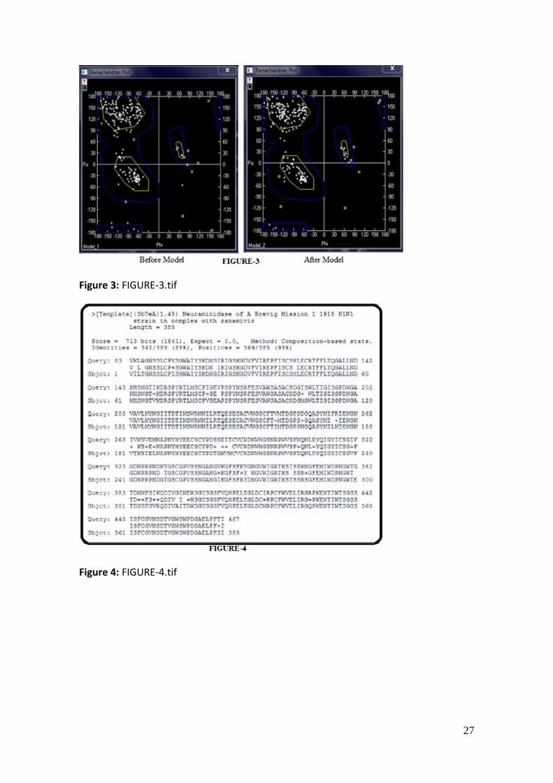
3.2 HOMOLOGY MODELING OF NEURAMINIDASE (NA) PROTEIN
The sequence alignment of the query NA sequence (ACZ97471) of 2009-H1N1 virus and
template NA (3B7E-A) of 1918 H1N1 virus [21]. The query NA sequence of 2009-H1N1 virus
was consisting of 469 residues. However, the structure of template NA protein 3B7E-A is a
segment containing 385 residues. Query sequence is modeled from 83 to 467 residues. The
sequence identity and similarity of NA protein were 88% and 95% respectively as shown in
Figure-4. The result of alignment was employed to build new homology model. The reliability of
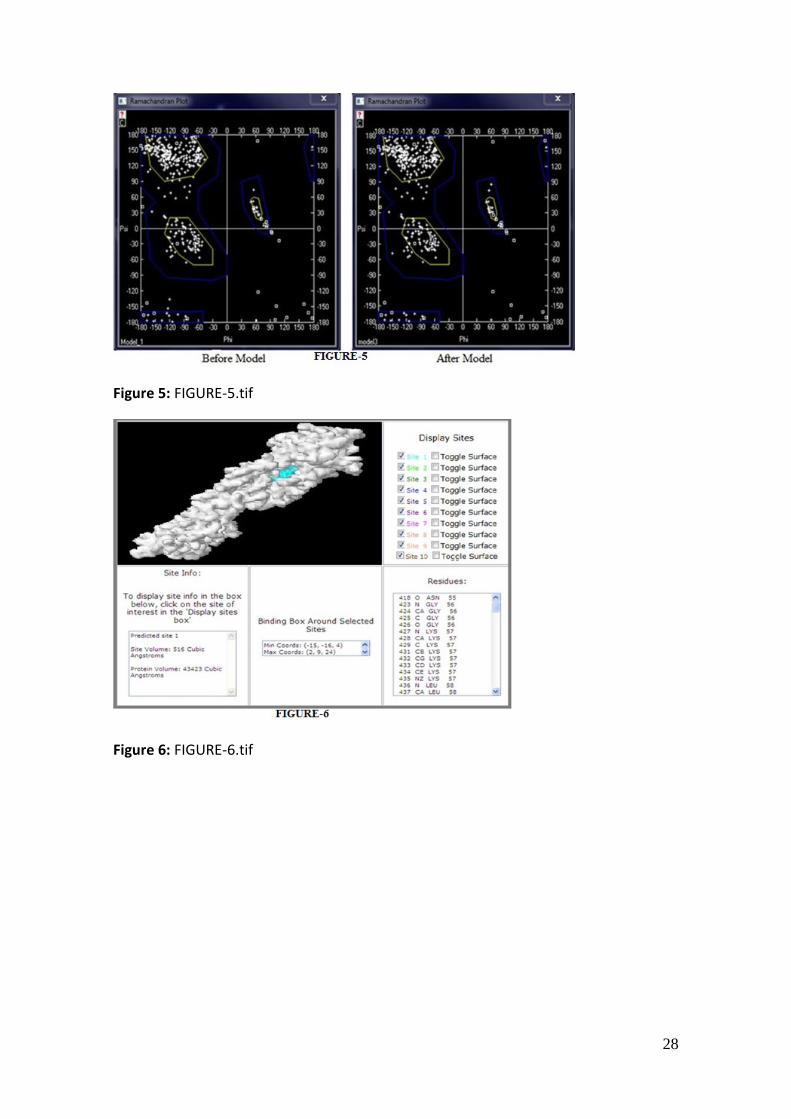
new homology model for Neuraminidase (NA) was identified by Ramachandran Plot. After
optimization and energy minimization process the best model was selected from 3D models
generated for NA protein on the basis of Swiss Model.
Energy minimization of three dimensional structures is vital for providing the maximum
stability to the protein. Ramachandran Plot drawn through Swiss Pdb Viewer program[16]
validated the model with 87.3% of the total residues in most favoured region and 1.2% in the

7
generously allowed region and residues in additional allowed region was 11.1 as shown in
Figure-5. This stipulates that protein that backbone dihedral angle PHI (φ) and PSI (ψ) occupied
reasonably accurate position in the selected 3d model.
Only 2 residues, THR156 and SER330 were located in the disallowed region, which
constituted 0.4% of the total protein.
3.3 ACTIVE SITE AND LIGAND BINDING SITE PREDICTION
The Ligand binding site of Hemagglutinin and Neuraminidase proteins were predicted
using Q-Site Finder program which is energy based sites [22]. It uses the energy interaction
between the simple Vander Waals probe and protein to locate energetically favorable binding
sites.
3.3.1 BINDING SITE ANALYSIS OF HA PROTEIN
The result of Q-site finder shows that predicted binding site cavity volume modeled HA
protein was 516 cubic angstroms and the coordinates of the binding box around predicted site
had minimum coordinates is [-15,-16,4] and maximum coordinates is [2,9,24]. Binding site of
HA was constituted by amino acid residues ASN55, GLY56, LYS57, LEU58, CYS59, LEU79,
ASN81, PRO82, GLU83, GLU85, SER88, GLU98, THR99, SER102, ARG155, GLU207, ALA261,
THR262, ASN264, GLY275, ARG298, ILE389, ASP390 as shown in Figure-6.
3.3.2 BINDING SITE ANALYSIS OF NA PROTEIN
The result of Q-site finder shows that predicted binding site cavity volume modeled NA
protein was 539 cubic angstroms and the coordinates of the binding box around predicted site
had minimum coordinates is [-42,6,-30] and maximum coordinates is [-22,33,-6].
Binding site of NA was constituted by amino acid residues SER110, ALA111, SER112,
ILE125, GLN152, ILE153, LEU154, ARG155, THR156, GLN157, GLU158, SER159, GLU160,
CYS161, VAL162, PHE169, THR170, ILE171, MET172, THR173, ASP174, PRO176, SER177,
TYR205, TYR206, GLU207, GLU208, CYS209, SER210, CYS211, LYS277 as shown in Figure-7.

8
3.4 RESULTS OF VIRTUAL SCREENING
Docking results predicted the interaction of ligands with protein and residues involved in
this complex. For such interaction, the most important requirement was the proper orientation
and conformation of ligand which fitted to the enzymes binding site appropriately and formed
protein ligand complex. Therefore, optimal interactions and best AutoDock score were used as
criteria to interpret the best conformation among the 30 conformations, generated by AutoDock
program.
All the 131compounds were docked into structures of HA and NA. The docking results
of 77 drug compounds and one known drug Oseltamivir with HA and NA models were shown in
Appendix-A. Among the 77 compounds ZINC039295508 and ZINC36451498 had the lowest
docking energy with both HA and NA respectively as shown in Table-1.
The docking results of 54 drug compounds and one known drug Zanamivir with HA and
NA models were shown in Appendix-B. Among the 54 drug compounds ZINC34532900,
ZINC34466451, ZINC02043006 and ZINC39158384 had the lowest docking energy with both
HA and NA models than other docked compounds as shown in Table-2.
On screening the docked results on the basis of the docking energy, it predicts that there
are 6 drug candidates which inhibit both HA and NA structures. These compounds had lower
docked energy and even lower than the standard controls, Zanamivir and Oseltamivir. In fact
Zanamivir and Oseltamivir were commonly used as inhibitors for NA drug for previous H1N1.
The chemical structure of all six compounds as shown in Figure-8.
Docking poses of the best conformation of 6 compounds ZINC03929508,
ZINC02043006, ZINC36451498, ZINC34466451, ZINC34532900 and ZINC29158384 in the
binding site of modeled HA proteins were shown in Figure-9.
Residues of Hemagglutinin protein involved in the formation of hydrogen bonds with 6
compounds are ARG155, GLU207, ASN225, GLY275, and ARG298.
Docking poses of the best conformation of 6 compounds ZINC03929508,
ZINC02043006, ZINC36451498, ZINC34466451, ZINC34532900 and ZINC29158384 in the
binding site of modeled NA proteins were shown in Figure-10.

9
Residues of Neuraminidase protein involved in the formation of hydrogen bonds with 6
compounds are ILE153, ARG155, and GLU207.
CONCLUSION
The Hemagglutinin (HA) and Neuraminidase (NA) of influenza A virus are two drug
targeting proteins for the drug discovery fighting with current influenza virus pandemic.
Homology model built for HA [Accession No: ACZ97508] and NA [Accession No: ACZ97471]
proteins of influenza A virus subtypes H1N1.Models built had high reliability show by
Ramachandran Plot. There are 131 compounds screened from ZINC database were docked with
homology model of HA and NA proteins respectively. After docking 6 compounds
ZINC039295508, ZINC36451498, ZINC02043006, ZINC34466451, ZINC39158384 and
ZINC34532900, ZINC39158384 were predicted as potent dual target candidate drug for H1N1.
Hopefully we have proposed some useful candidates for H1N1 diseases. Yet finally
pharmacological studies have to confirm, which is the best target candidate drug is for H1N1
disease.
Acknowledgement
I extend my sincere thanks to the management of The Oxford College of Engineering for
their support. I thank our principal Dr. Nagaraj. R and our HOD Dr. Kusum Paul, Department
of Biotechnology for providing necessary resources.
I would like to express my sincere gratitude to my guide, Mrs. Tanusree Chaudhari,
Assistant Professor, Dept.of BT, for her guidance, encouragement and constantly monitoring the
progress of the project. She provided me the chance to do my project work under her guidance
and gave advice at times. Her constructive and critical questions were an incomparable stimulus
and always helped me to consider issues in greater depth.
I would like to express my sincere gratitude to Mr. Shambu. M.G, Assistant professor,
Dept.of BT, for his continuous inspiration and encouragement throughout my year of work.
10
LIST OF TABLES
Table-1: Two compounds had lowest docked energy comparing with one known drug
Oseltamivir.
Table-2: Four compounds had lowest docked energy comparing with 1 known drug Zanamivir.
APPENDIX-A: All the 131compounds were docked into structures of HA and NA. The
docking results of 77 drug compounds and one know drug Oseltamivir with HA and NA models
were shown below.
APPENDIX-B: The docking results of 54 drug compounds and one know drug Zanamivir with
HA and NA models were shown in Table- below.
FIGURE LEGEND
Figure-1: The Evolutionary analysis of HA and NA using NJ method.
Figure-2: The sequence Alignment of the query HA sequence (ACZ97508) of influenza A
virus and the template HA (2WR1) of 1918-H1N1 virus. Secondary Structures of the query HA
protein was predicted using program SOPMA at ExPASy Server.
Figure-3: Ramachandran Plot of modeled HA protein of influenza A virus subtypes H1N1.

11
Figure-4: The sequence Alignment of the query NA sequence (ACZ97471) of influenza A
virus and the template NA (3B7E-A) of 1918-H1N1 virus. Secondary Structures of the query NA
protein was predicted using program SOPMA at ExPASy Server.
Figure-5: Ramachandran Plot of modeled NA protein of influenza A virus subtypes H1N1.
Figure-6: Binding Site Analysis of HA Protein.
Figure-7: Binding Site Analysis of NA Protein.
Figure-8: The chemical structures of all six candidates.
Figure-9: The docking poses of 6 candidates in HA.
Figure-10: The docking poses of 6 candidates in NA.

12
REFERENCES
[1] Y.K.Shinya, T. Watanabe and Y. Sakoda. In vitro and in vivo characterization of new swine-
origin H1N1 influenza viruses. Nature (2009), 460 (8), 1021-1025.
[2] MHFW, 2009. Ministry of Health and Family Welfare. Update on influenza a (H1N1) as on
17th May 2009.
[3] Brock well, R.Webster and R.J.Webby. Diversity of influenza virus in swine and the
emergence of a novel human pandemic influenza A (H1N1). Influenza other Respi. Viruses,2009
vol-3: 207-213.
[4] Mukhtar, D. Song, C. Zhu, Y. Zhu and J. Wu. Origin of highly pathogenic H5N1 avian
influenza virus in China and genetic characterization of donor and recipient viruses. J. Gen.
Virol. 2007, vol- 88: 3094-3099.
[5] Shirvan, AN., M. Moradi, M. Aminian and R. Madam. Preparation of Neuraminidase-
specific antiserum from the H9N2 subtype of avian influenza virus. Turk. J. Vet. Anim. Sci.
2007,vol-31: 219-223.
[6] Chen, C.Y. Huang, F.J. Tsai and C.Y.Chen,. Drug design for influenza A virus Subtypes
H1N1, J.Taiwan Inst. Chem.Eng 2010, 41:8-15.
[7] Raymond, L.W. and L. Leach, Treatment of post-influenza pneumonia in health care
workers. J. Occup. Environ. Med 2007, 49: 1181-1183.
[8] Takabatake, N., M. Okamura, K. Okubo, Y. Ikehara, I. Igarashi. Involvement of host
erythrocyte sialic acid content in Babesia bovis infection. J. Vet. Med. Sci 2009, 69: 999-1004.
[9] Russell, R.J., L.F. Haire, D.J. Stevens, P.J. Collins and Y.P. Lin et al., 2006. The structure of
H5N1 avian influenza neuraminidase suggests new opportunities for drug design. Nature, 443:
45-49.

13
[10] Shimbo, T., M. Kawachi, K. Saga, H. Fujita, T. Yamazaki, K. Tamai and Y. Kaneda, 2007.
Development of a transferrin receptor-targeting HVJ-E vector. Biochem. Biophys. Res.
Commun., 364: 423-428.
[11] Collins, P.J., L.F. Haire, Y.P. Lin, J. Liu and R.J. Russell et al.,2008. Crystal structures of
Oseltamivir-resistant influenza virus neuraminidase mutants. Nature, 453: 1258-1261.
[12] Potdar, V.A., M.S. Chadha, S.M. Jadhav, J. Mullick, S.S. Cherian and A.C. Mishra, 2010.
Genetic characterization of the influenza A pandemic (H1N1) 2009 virus isolates from India.
PLoS One, Vol. 5. 10.1371/journal.pone.0009693.
[13] Irwin, J.J. and B.K. Shoichet, 2005. ZINC-a free database of commercially available
compounds for virtual screening. J. Chem. Inf. Model., 45: 177-182.
[14] Bao, D.Dornovoy, B.Kirytin and Zaslavsky, The influenza virus resources at the national
center for biotechnology information J.Virol, 82:596-601.
[15] Sali, A. and T.L. Blundell, 1993. Comparative protein modeling by satisfaction of spatial
restraints.J. Mol. Biol., 234: 779-815
[16] Laskowski, D.S. Moss and J.M. Thornton, 1993. PROCHECK: A program to check the
stereo chemical quality of protein structures. J. Applied Cryst., 26: 283-291.
[17] Laurie, A.T.R. and R.M. Jackson, 2005. Q-Site Finder: An energy-based method for the
prediction of protein-ligand binding sites. Bioinformatics, 21: 1908-1916.
[18] Goodsell, D.S. and A.J. Olson, 1990. Automated docking of substrates to proteins by
simulated annealing. Proteins Structure, Function Bioinformatics, 8: 195-202.
[19] Morris, G.M., D.S. Goodsell, R.S. Halliday, R. Huey, WE. Hart, R.K. Belew and A.J.
Olson, 1998. Automated docking using a Lamarckian genetic algorithm and empirical binding
free energy function. J. Computat. Chem., 19: 1639-1662.

14
[20] Liu, J., D.J. Stevens, L.F. Haire, P.A. Walker and P.J. Coombs et al., 2009. Structures of
receptor complexes formed by Hemagglutinin from the Asian influenza pandemic of 1957. Proc.
Natl. Acad. Sci. USA., 106: 17175-17180.
[21] Xu, X., X. Zhu, R.A. Dwek, J. Stevens and I.A. Wilson, 2008. Structural characterization of
the 1918 influenza virus H1N1 neuraminidase. J. Virol., 82: 10493-10501.
[22] Laurie, A.T.R. and R.M. Jackson, 2005. Q-Site Finder: An energy-based method for the
prediction of protein-ligand binding sites. Bioinformatics, 21: 1908-1916.

15
Table 1: Table-1.docx

16
Drug
Compounds
Mol Wt X
LogP
HBD
HBA PSA Net
Cha
rge
RB Docked Energy Ref RMS
HA NA HA NA
Oseltamivir 312.40 1.10 2 5 90.6 0 8 -02.61 -03.95 34.13 24.78
ZINC03929508 313.41 0.85 4 6 92 1 8 -16.65 -17.45 27.69 32.97
ZINC36451498 299.39 0.48 4 6 92 1 7 -15.96 -17.75 26.98 33.14
TABLE-1: Two compounds had lowest docked energy comparing with one known drug
Oseltamivir.

17
Table 2: Table-2.docx

18
Drug
Compounds
Mol Wt X
LogP
HBD
HBA PSA Net
Cha
rge
RB Docked Energy Ref RMS
HA NA HA NA
Zanamivir 332.30 -3.20 7 10 201 0 6 -14.6 -16.77 24.19 31.08
ZINC02043006 290.29 -1.22 9 9 174 1 5 -15.97 -18.00 23.93 34.96
ZINC34466451 318.32 -2.04 6 9 151 0 7 -15.17 -17.31 25.49 37.55
ZINC34532900 318.32 -2.64 5 9 143 0 6 -15.10 -17.74 25.68 37.11
ZINC39158384 330.33 -2.27 5 9 143 0 6 -16.02 -18.09 26.21 38.29
TABLE-2: Four compounds had lowest docked energy comparing with 1 known drug
Zanamivir.

19
Table 3: Table-3.docx
20
APPENDIX-A
All the 131compounds were docked into structures of HA and NA. The docking results
of 77 drug compounds and one know drug Oseltamivir with HA and NA models were shown
below.
Drug
Compounds
Mol Wt X
LogP
HBD
HBA PSA Net
Cha
rge
RB Docked Energy Ref RMS
HA NA HA NA
Oseltamivir 312.40 1.10 2 5 90.6 0 8 -02.61 -03.95 34.13 24.78 ZINC11592802 313.41 0.85 4 6 92 1 8 -16.23 -16.99 23.63 32.41
ZINC03874568 313.41 0.85 4 6 92 1 8 -16.10 -15.81 27.00 32.77
ZINC03874569 313.41 0.85 4 6 92 1 8 -15.96 -17.74 26.09 34.22
ZINC03874570 313.41 0.85 4 6 92 1 8 -15.64 -16.57 27.97 33.93
ZINC03874571 313.41 0.85 4 6 92 1 8 -15.03 -16.95 26.43 34.25
ZINC03929508 313.41 0.85 4 6 92 1 8 -16.65 -17.45 27.69 32.97
ZINC36451498 299.39 0.48 4 6 92 1 7 -15.96 -17.75 26.98 33.14 ZINC04134497 298.38 0.41 4 6 106 0 6 -11.25 -10.72 27.02 34.39
ZINC06777828 284.35 -0.14 4 6 106 0 6 -9.28 -9.56 23.41 32.42
ZINC06777829 284.35 -0.14 4 6 106 0 6 -11.19 -8.85 27.50 32.75
ZINC06777830 284.35 -0.14 4 6 106 0 6 -10.13 -9.21 20.85 37.65
ZINC34817612 270.32 -0.64 4 6 106 0 5 -9.96 -10.07 25.19 37.05
ZINC03929509 284.35 -0.14 4 6 106 0 6 -11.21 -10.96 22.68 35.06
ZINC04134486 270.32 -0.64 4 6 106 0 5 -10.19 -10.71 25.10 36.40
ZINC34083570 270.32 -0.64 4 6 106 0 5 -10.27 -10.03 24.42 38.05
ZINC34083568 270.32 -0.64 4 6 106 0 5 -8.44 -10.09 27.07 33.34
ZINC06411782 270.32 -0.64 4 6 106 0 5 -10.45 -10.71 22.71 34.27
ZINC06777826 284.35 -0.14 4 6 106 0 6 -8.53 -9.24 22.18 34.08
ZINC14944898 282.34 -0.48 4 6 106 0 4 -10.97 -10.86 23.91 35.01
ZINC34083579 296.36 0.02 4 6 106 0 4 -11.11 -10.49 25.52 35.75
ZINC12404506 296.36 0.02 4 6 106 0 4 -11.26 -10.82 25.06 36.63
ZINC04134490 296.36 -0.10 4 6 106 0 7 -11.42 -11.27 22.50 37.33
ZINC04134489 296.36 -0.10 4 6 106 0 7 -11.36 -10.94 27.52 30.47
ZINC40865651 282.34 -0.37 4 6 106 0 6 -11.16 -10.97 21.15 39.15

21
ZINC34083576 298.38 0.56 4 6 106 0 8 -11.19 -10.70 28.18 34.01
ZINC04134488 298.38 0.56 4 6 106 0 8 -12.11 -10.97 21.16 36.57
ZINC04134483 256.30 -1.01 4 6 106 0 5 -10.38 -10.24 27.27 33.61
ZINC04134484 270.32 -0.45 4 6 106 0 6 -11.17 -9.99 22.27 35.94
ZINC34083573 256.30 -1.01 4 6 106 0 5 -10.12 -9.94 24.46 38.66
ZINC04134487 284.35 0.06 4 6 106 0 7 -11.94 -10.68 22.07 36.80
ZINC14944926 300.35 -1.38 5 7 126 0 7 -10.66 -10.70 21.74 36.78
ZINC14944928 300.35 -1.38 5 7 126 0 7 -10.00 -10.36 22.48 35.82
ZINC14944900 270.32 -0.17 4 6 106 0 6 -11.11 -10.83 21.46 38.25
ZINC04134482 242.27 -1.51 4 6 106 0 4 -9.86 -9.57 25.88 36.19
ZINC14944922 313.33 -1.50 4 8 146 -1 7 -6.23 -4.09 21.14 35.98
ZINC14944924 313.33 -1.50 4 8 146 -1 7 -6.57 -4.63 21.27 36.69
ZINC14944897 254.28 -1.24 4 6 106 0 5 -10.27 -10.27 27.67 33.63
ZINC13780043 366.50 1.99 4 6 106 0 8 -12.82 -12.88 26.72 33.47
ZINC13780042 352.47 1.72 4 6 106 0 7 -12.80 -11.17 22.76 36.24
ZINC04134485 270.32 -0.76 4 6 106 0 5 -9.53 -10.10 26.76 33.09
ZINC35963009 243.28 -1.51 5 6 103 1 4 -15.19 -16.36 27.82 36.77
ZINC03833967 298.38 0.34 4 6 106 0 6 -9.95 -11.27 20.89 37.94
ZINC14944899 310.27 -0.12 4 6 106 0 6 -10.98 -10.23 27.84 33.39
ZINC04134481 228.24 -1.88 4 6 106 0 3 -9.4 -8.94 24.41 34.96
ZINC14944896 310.27 -0.69 4 6 106 0 6 -10.86 -10.03 26.81 34.41
ZINC14944902 283.32 -0.49 4 7 116 0 7 -4.79 -2.29 26.25 32.90
ZINC32062406 268.33 1.27 1 5 78 -1 6 -3.34 -1.44 23.86 36.34
ZINC03833965 360.45 1.30 4 6 106 0 8 -12.46 -12.81 25.34 31.70
ZINC34083583 360.45 1.30 4 6 106 0 8 -11.75 -10.57 21.82 36.42
ZINC08552722 360.45 1.30 4 6 106 0 8 -11.50 -12.15 24.88 31.05
ZINC38392952 253.34 2.48 1 4 57 0 7 -14.49 -16.16 28.14 30.40
ZINC38340093 253.34 2.48 1 4 57 0 7 -15.35 -15.89 27.80 33.90
ZINC03833966 408.49 2.23 4 6 106 0 8 -10.68 -11.88 23.11 36.74
ZINC03833964 304.34 -0.29 4 6 106 0 5 -12.46 -10.66 25.34 34.16
ZINC14944917 318.80 0.19 4 6 106 0 6 -11.40 -10.81 26.88 32.34
ZINC13780047 312.37 -0.66 6 8 142 0 7 -10.87 -10.27 22.14 37.88
ZINC13780049 326.39 -0.15 6 8 142 0 8 -8.78 -9.48 25.15 32.43

22
ZINC13780048 312.37 -0.66 6 8 142 0 7 -10.56 -10.06 22.73 38.56
ZINC40863125 346.42 0.79 4 6 106 0 7 -11.46 -12.22 21.60 38.78
ZINC13780050 408.49 1.71 4 6 106 0 8 -9.62 -11.07 20.14 35.81
ZINC04134495 312.37 -0.79 6 8 144 0 6 -10.15 -11.02 23.53 34.11
ZINC04134496 312.37 -0.79 6 8 144 0 6 -10.56 -9.34 22.04 36.41
ZINC38393066 256.27 -0.65 2 6 99 -1 4 -3.01 -0.92 26.56 32.62
ZINC13780045 298.34 -1.02 6 8 142 0 7 -10.66 -10.34 22.23 39.68
ZINC13780046 312.37 -0.46 6 8 142 0 8 -10.01 -10.57 26.41 34.74
ZINC04134493 298.34 -1.15 6 8 144 0 6 -10.40 -10.52 22.25 36.46
ZINC34031776 298.34 -1.15 6 8 144 0 6 -11.27 -10.42 25.71 36.99
ZINC34031775 298.34 -1.15 6 8 144 0 6 -10.13 -10.95 24.56 34.21
ZINC34031774 298.34 -1.15 6 8 144 0 6 -10.00 -9.48 21.77 33.89
ZINC34031773 298.34 -1.15 6 8 144 0 6 -10.37 -9.98 22.73 35.16
ZINC04134494 312.37 -0.59 6 8 144 0 7 -10.62 -10.99 26.87 33.34
ZINC04134498 302.34 -0.11 4 6 106 0 6 -11.34 -11.05 26.95 32.39
ZINC14944895 258.27 -0.91 4 7 115 0 5 -9.37 -9.85 23.09 37.29
ZINC13860569 312.34 0.19 1 7 105 -1 7 -2.81 -0.24 24.53 32.49
ZINC38393596 281.35 1.62 1 5 65 0 6 -9.95 -8.41 25.87 31.36
ZINC14944903 312.37 -0.77 5 8 130 0 7 -9.55 -9.81 20.10 35.88
ZINC03833958 214.22 -2.5 5 6 117 0 2 -8.92 -8.34 26.57 34.75
Table-A: Docking results of 77 drug compounds and one known drug with HA and NA model.
Mol Wt: Molecular weight, HBD: Hydrogen Bond Donors, HBA: Hydrogen Bond Acceptors,
PSA: Polar Surface Area, HA: Hemagglutinin, NA: Neuraminidase, RB: Rotatable Bonds, Ref
RMS: Ref root mean square deviation.

23
Table 4: Table-4.docx

24
APPENDIX-B
The docking results of 54 drug compounds and one know drug Zanamivir with HA and
NA models were shown in Table- below.
Drug
Compounds
Mol Wt X
LogP
HBD
HBA PSA Net
Cha
rge
RB Docked Energy Ref RMS
HA NA HA NA
Zanamivir 332.30 -3.20 7 10 201 0 6 -14.6 -16.77 24.19 31.08
ZINC34126688 302.28 -3.21 8 10 185 0 5 -10.00 -9.84 26.92 36.54
ZINC13443807 316.31 -2.81 8 10 183 0 7 -10.48 -9.53 24.51 33.53
ZINC13487809 326.35 -0.93 6 9 151 0 5 -11.32 10.56 28.06 27.76
ZINC13443835 342.39 -0.08 6 9 151 0 9 -9.86 10.67 20.63 36.79
ZINC13443833 328.36 -0.58 6 9 151 0 8 -9.48 10.49 25.08 36.41
ZINC34124979 272.26 -2.57 7 9 165 0 4 -9.07 -9.60 26.76 37.80
ZINC13604075 290.27 -3.50 7 9 167 0 5 -9.63 -8.86 26.65 31.43
ZINC03870992 290.27 -3.50 7 9 166 0 5 -10.14 -9.39 22.69 35.17
ZINC03870991 290.27 -3.50 7 9 166 0 5 -10.22 -9.36 25.07 33.60
ZINC02043007 290.27 -3.50 7 9 166 0 5 -9.71 -9.35 23.87 34.55
ZINC12503458 290.27 -3.50 7 9 167 0 5 -9.24 -9.37 23.75 35.07
ZINC12503456 290.27 -3.50 7 9 167 0 5 -9.40 -10.47 21.75 34.75
ZINC12503454 290.27 -3.50 7 9 167 0 5 -9.05 -9.99 22.92 35.35
ZINC12503452 290.27 -3.50 7 9 167 0 5 -9.89 -9.90 24.07 34.02
ZINCO3590792 290.27 -3.50 7 9 166 0 5 -8.98 -9.46 25.43 34.43
ZINC40747111 290.27 -3.50 7 9 167 0 5 -9.77 -9.01 26.31 33.19
ZINC40747113 290.27 -3.50 7 9 167 0 5 -10.17 -9.53 26.80 31.96
ZINCO5884083 290.27 -3.50 7 9 166 0 5 -9.43 -10.17 22.88 35.55
ZINC39156769 304.29 -2.66 7 9 167 0 6 -9.52 -10.3 27.33 35.76
ZINC02043006 290.29 -1.22 9 9 174 1 5 -15.97 -18.00 23.93 34.96 ZINC05884077 290.29 -1.22 9 9 174 1 5 -8.91 -17.56 23.74 35.55
ZINC39086001 304.29 -3.25 6 9 158 0 5 -9.22 -10.51 25.57 34.09
ZINC39176066 316.31 -2.89 6 9 158 0 5 -10.80 -10.91 23.68 38.82
ZINC34369778 331.30 -3.23 5 10 168 -1 6 -3.90 -1.83 26.33 32.68
ZINC34369779 331.30 -3.23 5 10 168 -1 6 -4.16 -1.98 25.73 34.71
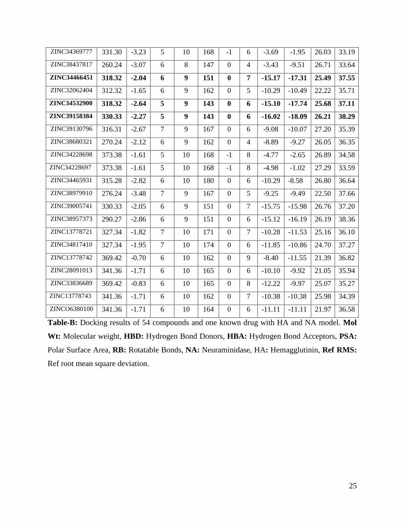
25
ZINC34369777 331.30 -3.23 5 10 168 -1 6 -3.69 -1.95 26.03 33.19
ZINC38437817 260.24 -3.07 6 8 147 0 4 -3.43 -9.51 26.71 33.64
ZINC34466451 318.32 -2.04 6 9 151 0 7 -15.17 -17.31 25.49 37.55 ZINC32062404 312.32 -1.65 6 9 162 0 5 -10.29 -10.49 22.22 35.71
ZINC34532900 318.32 -2.64 5 9 143 0 6 -15.10 -17.74 25.68 37.11
ZINC39158384 330.33 -2.27 5 9 143 0 6 -16.02 -18.09 26.21 38.29 ZINC39130796 316.31 -2.67 7 9 167 0 6 -9.08 -10.07 27.20 35.39
ZINC38680321 270.24 -2.12 6 9 162 0 4 -8.89 -9.27 26.05 36.35
ZINC34228698 373.38 -1.61 5 10 168 -1 8 -4.77 -2.65 26.89 34.58
ZINC34228697 373.38 -1.61 5 10 168 -1 8 -4.98 -1.02 27.29 33.59
ZINC34465931 315.28 -2.82 6 10 180 0 6 -10.29 -8.58 26.80 36.64
ZINC38979910 276.24 -3.48 7 9 167 0 5 -9.25 -9.49 22.50 37.66
ZINC39005741 330.33 -2.05 6 9 151 0 7 -15.75 -15.98 26.76 37.20
ZINC38957373 290.27 -2.86 6 9 151 0 6 -15.12 -16.19 26.19 38.36
ZINC13778721 327.34 -1.82 7 10 171 0 7 -10.28 -11.53 25.16 36.10
ZINC34817410 327.34 -1.95 7 10 174 0 6 -11.85 -10.86 24.70 37.27
ZINC13778742 369.42 -0.70 6 10 162 0 9 -8.40 -11.55 21.39 36.82
ZINC28091013 341.36 -1.71 6 10 165 0 6 -10.10 -9.92 21.05 35.94
ZINC33836689 369.42 -0.83 6 10 165 0 8 -12.22 -9.97 25.07 35.27
ZINC13778743 341.36 -1.71 6 10 162 0 7 -10.38 -10.38 25.98 34.39
ZINCO6380100 341.36 -1.71 6 10 164 0 6 -11.11 -11.11 21.97 36.58
Table-B: Docking results of 54 compounds and one known drug with HA and NA model. Mol
Wt: Molecular weight, HBD: Hydrogen Bond Donors, HBA: Hydrogen Bond Acceptors, PSA:
Polar Surface Area, RB: Rotatable Bonds, NA: Neuraminidase, HA: Hemagglutinin, Ref RMS:
Ref root mean square deviation.

26
Figure 1: FIGURE-1.tif
Figure 2: FIGURE-2.tif
27
Figure 3: FIGURE-3.tif
Figure 4: FIGURE-4.tif
28
Figure 5: FIGURE-5.tif
Figure 6: FIGURE-6.tif

29
Figure 7: FIGURE-7.tif
Figure 8: FIGURE-8.tif
30
Figure 9: FIGURE-9.tif

31
Figure 10: FIGURE-10.tif