s-adenosyl methionine is necessary for inhibition of the ... · methionine histone mutants using...
TRANSCRIPT

S-adenosyl methionine is necessary for inhibition ofthe methyltransferase G9a by the lysine 9 tomethionine mutation on histone H3Hariharan Jayarama,1, Dominik Hoelperb,c,1, Siddhant U. Jainb,c, Nico Cantonea, Stefan M. Lundgrenb,c, Florence Poya,C. David Allisd,2, Richard Cummingsa,2, Steven Bellona,2, and Peter W. Lewisb,c,2
aConstellation Pharmaceuticals, Cambridge, MA 02142; bWisconsin Institute of Discovery, University of Wisconsin, Madison, WI 53715; cBiomolecularChemistry, School of Medicine and Public Health, University of Wisconsin, Madison, WI 53715; and dLaboratory of Chromatin Biology and Epigenetics, TheRockefeller University, New York, NY 10065
Contributed by C. David Allis, April 8, 2016 (sent for review March 16, 2016; reviewed by Cheryl H. Arrowsmith, Sharon Y. R. Dent, and Ronen Marmorstein)
Lysine to methionine (K-to-M) mutations in genes encoding histoneH3 are thought to drive a subset of pediatric brain and bone cancers.These high-frequency K-to-M mutations occur at sites of methylationon histone H3, and tumors containing the mutant histones exhibit aglobal loss of specific histone methylation marks. Previous studiesshowed that K-to-M mutant histones, also known as oncohistones,are potent orthosteric inhibitors of specific Su(var)3-9, Enhancer-of-zeste, Trithorax (SET) domain methyltransferases. However, the bio-chemical and biophysical details of the interaction between K-to-Mmutant histones and the respective SET domain methyltransferasesare currently unknown. Here, we use the histone H3K9-directedmethyltransferase G9a as a model to explore the mechanism ofinhibition by K-to-M oncohistones. X-ray cocrystal structuresrevealed that the K9M residue of histone H3 occupies the activesite cavity of G9a, and kinetic analysis indicates competitive inhibi-tion of G9a by histone H3K9M. Additionally, we find that the co-factor S-adenosyl methionine (SAM) is necessary for stableinteraction between G9a and H3K9M histone. Consistent withthe formation of a ternary complex, we find that the inhibitorypeptide is uncompetitive with regard to SAM. These data andothers indicate that K-to-M oncohistones promote global loss ofspecific lysine methylation through sequestration and inhibition ofSAM-bound SET domain methyltransferases.
oncohistone | G9a | EHMT2 | H3K9me3 | K to M
Covalent modifications to both DNA and histone proteinsturn chromatin into a dynamic information hub that inte-
grates diverse biochemical stimuli to regulate genomic DNAaccess to the transcription machinery and ultimately, establishand maintain cellular phenotypes. Moreover, there is increasingappreciation that alterations of the chromatin landscape, in-cluding DNA and histone modifications, are involved in thepathogenesis of cancer. Nowhere is this finding better supportedthan with the groundbreaking discoveries of high-frequency so-matic mutations in histones that are drivers of tumorigenesis.Monoallelic missense mutations in genes encoding for histone
H3 were recently found in bone and brain tumors that affectchildren and young adults. Approximately 80% of diffuse intrinsicpontine glioma contain a lysine 27-methionine (K27M) mutation(1, 2), and 95% of chondroblastoma samples contain a K36Mmutation in genes encoding either histone H3.1 or H3.3 (3). TheK27M and K36M mutations in histone H3 are the known lysine tomethionine (“K-to-M”) histone H3 missense mutations found inhuman disease thus far. Although these oncohistones represent asmall population of total histone H3 in tumor cells (3–17% ofhistone H3), they remarkably lead to global loss of the associatedmethylation mark on the WT complement of histone H3. Thenearly invariant nature of the K-to-Mmutation strongly suggests thatthis specific amino acid substitution imparts a unique gain of functionto the mutant histone. We and others previously showed that K-to-Moncohistones transform the histone H3 proteins into potent
inhibitors of specific lysine methyltransferase enzymes (4–7). Theunprecedented finding that mutant histones act as enzyme in-hibitors and alter downstream epigenomic landscapes implicatesa direct effect of epigenetic misregulation driven by oncohistonesin tumorigenesis.Polycomb Repressive Complex 2 (PRC2) is one component of
the two main Polycomb group protein complexes that function ina collaborative epigenetic cross-talk with H3K27me3 to initiateand maintain transcriptional repression, and the EHZ2 subunitof PRC2 catalyzes mono-, di-, and trimethylation of H3K27.Previous studies showed that the K27M mutation found in pe-diatric glioma is sufficient to transform histone H3 into a potentinhibitor of enzymatic activity of PRC2 (4, 5, 7). Consequently,tumor cells with the K27M mutation exhibit low levels ofH3K27me3 and aberrant gene silencing (4, 7–9). Previously, wefound that only long, unbranched hydrophobic side chains con-taining methionine (M) and isoleucine (I) at lysine 27 (K27Mand K27I) were capable of inhibiting PRC2 in vitro and de-creasing H3K27me3 when expressed ectopically in cultured cells(7). The recent identification of K27I histone H3 mutations in asmall number of diffuse intrinsic pontine glioma samples furtherlinks the inhibition of PRC2 and loss of K27me3 to the likely on-cogenic activity of histone H3K27 mutations (10). Additionally,histone H3 peptides that contained unbranched hydrophobic lysine
Significance
Recent exome sequencing studies have uncovered high-frequencyhistone H3 driver mutations in pediatric cancers. Previous studieshave shown that lysine to methionine histone mutations arepotent inhibitors of their respective lysine methyltransferases.However, an in-depth understanding of this inhibition was limitedby the lack of structural and kinetic information. This study in-vestigates the biochemical and biophysical parameters of lysine tomethionine histone mutants using the methyltransferase G9a andH3K9M as a model system. Structural and functional experimentsconclude that the methyltransferase cofactor S-adenosyl methio-nine is required for binding of G9a to the mutant histone.
Author contributions: H.J., D.H., S.U.J., C.D.A., R.C., S.B., and P.W.L. designed research; H.J., D.H.,S.U.J., N.C., S.M.L., F.P., R.C., and P.W.L. performed research; H.J., D.H., S.U.J., R.C., S.B., andP.W.L. analyzed data; and H.J., D.H., R.C., S.B., and P.W.L. wrote the paper.
Reviewers: C.H.A., University of Toronto; S.Y.R.D., The University of Texas MD AndersonCancer Center; and R.M., Perelman School of Medicine, University of Pennsylvania.
The authors declare no conflict of interest.
Freely available online through the PNAS open access option.1H.J. and D.H. contributed equally to this work.2To whom correspondence may be addressed. Email: [email protected], [email protected], [email protected], [email protected].
This article contains supporting information online at www.pnas.org/lookup/suppl/doi:10.1073/pnas.1605523113/-/DCSupplemental.
6182–6187 | PNAS | May 31, 2016 | vol. 113 | no. 22 www.pnas.org/cgi/doi/10.1073/pnas.1605523113
Dow
nloa
ded
by g
uest
on
May
28,
202
0
isosteres at position 27 are potent orthosteric inhibitors of PRC2activity (5). Despite these findings, a detailed biochemical andbiophysical understanding of the interaction of K-to-M mutanthistones with Su(var)3-9, Enhancer-of-zeste, Trithorax (SET) do-main methyltransferases is currently lacking.In seeking to better understand the mechanism of K-to-M
mutant histones, we chose to study the H3K9 methyltransferaseG9a as a model system because of the availability of high-resolutionG9a crystals described in the literature. G9a is an ∼120-kDa proteinand contains an SET domain at its C terminus that catalyzes mono-and dimethylation of histones at lysine 9 (11), and we previouslyshowed that H3K9M peptides are capable of inhibiting activity of
SUV39H1 and G9a, two H3K9-directed methyltransferases, in vitro(7). Recent studies identified a role for G9a in heterochromatinmaintenance and transcriptional repression, such as the silencing ofrepeat elements, including long interspersed nuclear elementsand endogenous retroviruses (12–14). Up-regulation of both G9aand G9a like protein (GLP) has implications in a variety of hu-man cancers, including solid tumors as well as acute myeloidleukemia (15–17). Here, we present biochemical and structuralevidence that the histone H3K9M peptide inhibits G9a activity byeffectively competing with WT histone H3 substrate peptide forbinding to the active site. Additionally, we find that G9a bound toS-adenosyl methionine (SAM) is necessary to form a stable ter-nary complex with the K9M peptide. Together, these data pro-vide important mechanistic insights into how oncohistones alterthe chromatin landscape in tumorigenesis.
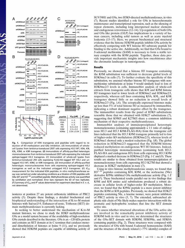
ResultsPreviously, we showed that a histone H3 transgene containingthe K9M substitution was sufficient to decrease global levels ofH3K9me3 in cells (7). To further evaluate the specificity of thissubstitution, we assessed whether histone H3 transgenes with non-methionine substitutions at lysine 9 would similarly lead to loss ofH3K9me2/3 levels in cells. Immunoblot analysis of whole-cellextracts from transgenic cells shows that K9I and K9M histoneH3 transgenes lead to lower levels of H3K9me2 and H3K9me2/3,respectively, whereas cell lines containing K9A, K9L, and K9Rtransgenes showed little or no reduction in overall levels ofH3K9me2/3 (Fig. 1A). The ectopically expressed histones makeup less than 5% of total histone H3 as measured by immunoblot,indicating a robust dominant negative effect by the transgenes.The immunoblot results from the panel of H3K9 substitutionsresemble those that we obtained with H3K27 substitutions (7),suggesting that K9M/I and K27M/I share a common inhibitionmechanism of their respective methyltransferases.Immunoblot analyses of immunoprecipitated heterotypic
mononucleosomes (containing approximately a 1:1 ratio of endoge-nous H3.3 and H3.3 K9M-FLAG-HA) from the transgenic celllines indicated that the H3.3 K9M transgene primarily led to lossof higher-order K9 methylation (H3K9me2/3), whereas levels ofH3K9me1 showed only a modest reduction (Fig. 1B). The globalreduction in H3K9me2/3 suggested that the H3K9M histonereduced methylation on endogenous WT H3 histones. Indeed,purified heterotypic mononucleosomes (containing both H3.2-FLAG-HA and endogenous H3) from cells expressing H3.3 K9Mdisplayed a marked decrease in H3K9me2/3 (Fig. 1C andD). Theseresults are similar to those obtained from immunoprecipitation ofmononucleosomes from cells expressing H3.3 K27M that showed adecrease in H3K27 methylation in trans (7).Using in vitro histone methyltransferase assays, we found that
H31–20 peptides containing K9I, K9M, or the norleucine (Nle)derivative K9Nle inhibited G9a methyltransferase activity (Fig. 1 Eand F). These biochemical results parallel our in vivo findings thatK9M and K9I amino acid substitutions produced a robust de-crease in cellular levels of higher-order K9 methylation. More-over, we found that the K9Nle peptide is a more potent inhibitorthan the K9M or K9I peptides. Previously, we showed that H3K27peptides containing Nle, a leucine isomer, were potent inhibitorsof PRC2 activity in vitro (5, 7). The extended, unbranched ali-phatic side chain of Nle likely makes superior interactions with thearomatic and hydrophobic residues that line the SET domainactive site.To assess whether structural alterations of the G9a active site
are involved in the remarkably potent inhibitory activity ofH3K9M both in vitro and in vivo, we determined the structuresof the G9a–SET domain with H3K9M, H3K9Nle, and H3K9Acontaining peptides (Fig. 2). These structures were compared withthe structures of G9a with bound S-adenosyl homocysteine (SAH)and the structure of the closely related (∼77% identity) complex of
D
A
E
B
C
FpeptideK9me3K9AK9LK9IK9MK9Nle
n. d. 0.1785n. d.n. d.
9.3 ± 0.82.3 ± 0.2
0.54 ± 0.04
0.81310.86330.99210.99080.9960
R2 valueIC50 [μM]
αH3K9me3
Ponceau
αHA
H3.2-FLAG-HAbackground
H3.3 (untagged)
H3.2-FLAG-HA
H3.2-FLAG-HA
H3
H3H2BH2AH4
WT K9M K9R
H4
αH3K9me2
αH3K9me3
αH3K9me1
αH3K27me3
WT K9M K9R
αK9M
αH3.3
αHA
Ponceau
H3.3-FLAG-HA NUCs
H3.3-FLAG-HAH3.3
H3.3-FLAG-HA
H3.3-FLAG-HA
H3.3-FLAG-HA
H3.3
H3.3-FLAG-HAH3.3
H3.3-FLAG-HA
H3.3
H3.3-FLAG-HA
H3.3
H3.3-FLAG-HA
H3.3
H2BH2A
αH3K9me3
αH3K9me1
αH3K9me2
αHA
αH3.1/H3.2
Ponceau
H3.3 (untagged)
H3.2-FLAG-HA
H3
H3
H3.2-FLAG-HA
H3.2-FLAG-HA
H3.2-FLAG-HA
H3
αH3K27me3H3.2-FLAG-HA
H3
H3.2-FLAG-HA
H3
H3.2-FLAG-HA
H3
H2BH2AH4
H3.2-FLAG-HA NUCs
WT K9M K9R
H3H2BH2AH4
WT K9A K9I K9L K9M K9R
αH3K9me3
αH3K9M
αHA
Ponceau
αH3K9me2
αH3 general
αH3K9me1
αH3K9ac
αH3S10ph
-1.0 -0.5 0.0 0.5 1.0 1.50
25
50
75
100
log[K9X peptide] ( M)
% G
9a A
ctiv
ity
K9me3K9AK9LK9IK9MK9Nle
Fig. 1. Comparison of K9X transgenes and peptides with regard to re-duction of K9 methylation and G9a inhibition. (A) Immunoblots of whole-cell lysates from lentivirus-transduced 293T cells expressing H3.3 WT, K9A, K9I,K9L, K9M, or K9R transgenes. (B) Immunoblots of affinity-purified heterotypicmononucleosomes from lentivirus-transduced 293T cells expressing the indicatedepitope-tagged H3.3 transgenes. (C) Immunoblot of whole-cell lysates fromlentivirus-transduced 293 cells expressing FLAG-HA–tagged WT H3.2 and un-tagged H3.3 WT, K9M, or K9R transgenes. (D) Immunoblot of affinity-purifiedheterotypic mononucleosomes from cells expressing epitope-tagged H3.2transgenes as well as the indicated untagged H3.3 transgenes. (E) IC50
measurement for the indicated K9X peptides. In vitro methyltransferase as-say was carried out under saturating conditions as a titration of K9X peptides withG9a–SET and H31–20 unmodified peptide. Methyltransferase activity was quantifiedby scintillation and normalized. Error bars represent the SD of two replicates.(F) Summary of IC50 and R2 values determined for experiment described in E. n. d.,not determined.
Jayaram et al. PNAS | May 31, 2016 | vol. 113 | no. 22 | 6183
BIOCH
EMISTR
Y
Dow
nloa
ded
by g
uest
on
May
28,
202
0

G9a-like protein with bound mono- and dimethylated histonepeptide. The cocrystal structure of G9a with the H3K9M-containingpeptide at 1.85-Å resolution (Table S1) showed clear electrondensity for both the mutant methionine residue and the surroundingpeptide. Comparison with the structure of G9a with bound SAH[Protein Data Bank (PDB) ID code 2O8J] revealed surprisingly fewdifferences (0.343 Å2) overall as well as with regard to the residuessurrounding the cofactor and substrate binding sites (0.445 Å2).Comparison with the product peptides bound to GLP [PDB IDcodes 3HNA (GLP-H3K9me1) and 2RFI (GLP-H3K9me2)] re-veals an almost identical conformation of the peptide backbones,with overall rmsd of ∼0.6 Å2 (Fig. 2A). Examination of the con-formation of the mutant methionine residue bound to G9a with theproduct dimethyl-lysine showed that the two adopt a virtuallyidentical conformation in the substrate binding site with their Cαthrough Ce carbon atoms superimposed perfectly, suggesting thatthe mechanism for methionine inhibition results from a directcompetition for the peptide substrate binding site.Strikingly, despite an overall similarity in the conformation of
peptide and surrounding residues, the crystallized G9a–K9Mcomplex exhibited complete occupancy of SAM within the co-factor binding pocket. The structures of G9a and GLP used inour comparisons consistently show occupancy of the cofactorbinding pocket by SAH, the expended cofactor product that
results after methylation. The surprising complete occupancy forSAM was even more notable, because no additional SAM wasadded at any stage of the protein production, purification, orcrystallization from bacterially expressed G9a protein. We hy-pothesize that our G9a protein preparation contains a mixtureof SAH- and SAM-bound G9a populations and that, despitethe inferred low abundance of SAM-bound G9a used in thecocrystallography experiments, the structure of G9a with the mutantmethionine histone peptide results from an enrichment of theSAM-bound population in the cofactor binding pocket.In addition to observing full SAM occupancy in the H3K9M
mutant cocrystal structures, we also observed full SAM occu-pancy in the H3K9Nle and H3K9A cocrystal structures (Fig. 2B).Furthermore, adding a high concentration of SAH duringcocrystallization of G9a with the H3K9M mutant did not decreasethe SAM occupancy of the structure. A comparison of the peptidecomplex structures with each other showed that they were highlysimilar to each other (0.18–0.21 Å2 rmsd), and all showed completeoccupancy for SAM in the active site. The bound SAM moleculeswere superimposable across all four structures. The cooccupancy ofSAM in the G9a–H3K9M structure suggested that SAM may benecessary for stable interaction between G9a and the K9M peptide.To explore whether SAM is required for K9M peptide interac-tion with G9a, a variety of in vitro assays were performed. First, we
G9a with H3M9 peptide G9a without peptide
F1158
Y1067 SAM
Y1085
M-pep
Y1154
F1152
F1087 Di-meth pep
A
B
G9a + H3M9 peptide
G9a + H3A9 peptide
G9a + H3Nle9 peptide
G9a + H3M9 peptide (SAH swamp)
SAM
SAM
SAM
SAM
Met
Met
Nle
Ala
Dimethyl-lysine product peptide
Fig. 2. Cocrystal structures of mutant H3 peptides with G9a. (A) Comparison of G9a protein cocrystallized with H3K9M peptide (blue) with G9a bound withSAH and no peptide (2O9S) (gray). Also superimposed for purposes of comparison is the dimethyl-lysine product peptide alone (green) from GLP (ID code2RFI). (B) Experimental electron density (2mFo-Dfc) maps calculated using Sigmaa for the H3K9M, H3K9Nle, and H3K9A crystallization experiments. Alsoshown in Lower Right is an experiment where, in addition to the H3K9M peptide, a 50-fold molar excess of SAH was added to the crystallization experimentto determine if the bound SAM could be “swamped” out by SAH.
6184 | www.pnas.org/cgi/doi/10.1073/pnas.1605523113 Jayaram et al.
Dow
nloa
ded
by g
uest
on
May
28,
202
0

characterized the relative binding affinities of SAM-bound G9a tothe mutant peptide by differential scanning fluorimetry, where anincrease in the melting temperature (Tm) for a protein ligandcomplex correlates with the tightness of binding. Fig. 3A shows thetitration of G9a at constant peptide concentration and varyingSAM. The nonlinear increase of melting temperature (ΔTm) withincreasing SAM concentrations implies a level of cooperativity inthe interaction of SAM and mutant histone peptides. The Tm vs.SAM concentration curve for the K9Nle peptide was the steepestfollowed by K9M, whereas the K9A peptide curve showed only amodest upward slope. This finding suggests that the Nle peptide hadthe most favorable interaction with G9a and SAM and that thealanine peptide had the least SAM dependence. This trend isconsistent with their respective inhibitory activities in vitro andin vivo.Next, we used isothermal titration calorimetry to assess the
SAM and SAH dependency of the G9a–K9Nle peptide interactionand determine the dissociation constants for the interaction. Beforeanalysis, G9a was treated with substrate peptide to convert boundSAM into SAH and subsequently dialyzed. Although G9a boundSAM alone with a Kd of 2.6 μM (Fig. 3B, Left), G9a did not bindK9Nle peptide after dialysis (Fig. 3B, Center). However, a robustinteraction (Kd = 0.032 μM) between G9a and the K9Nle peptidewas observed in the presence of added SAM (Fig. 3B, Right). Incontrast, G9A did not bind SAH when assessed as for SAM bind-ing, and even when present at 1 mM, SAH did not promote bindingof K9Nle peptide (Fig. S1A). These data indicate that SAM is anecessary cofactor in the stable association between G9a andK9M/Nle peptides. Isothermal titration calorimetry analysis ofK9 substrate also suggested the necessity of SAM for binding but
was complicated by turnover at the high substrate/enzymeconcentrations (Fig. S1B). These data are consistent with aproposed model where SAM facilitates folding of the G9a post-SET domain, which forms part of the peptide binding site forH3K9 (18). We, furthermore, performed peptide pulldown as-says to assess the SAM dependency of interaction between var-ious histone H3K9X peptides and the SET domain of G9a (Fig.3C). In this experiment, histone H3 peptides that inhibited G9aactivity in vitro (K9I, M, and Nle) bound to relatively morerecombinant G9a–SET than peptides that displayed weak in-hibitory activity (K9L and K9A). Additionally, all K9-substitutedpeptides displayed increased binding to G9a–SET when SAMwas present during both binding and washes as opposed to SAMbeing absent.We next determined the detailed mechanism of action and
Michaelis–Menten kinetic parameters associated with both thepeptide and SAM as a function of K9Nle concentration. In pastwork, oxidation of methionine was found to decrease the potencyof K27M peptides (5). In an effort to maintain consistency be-tween experiments, we chose to use K9Nle in our kinetic assays,because it is a methionine isostere that is not oxidized. Theseexperiments confirmed that K9Nle was competitive with thepeptide substrate, because the Km of the peptide increased lin-early with the K9Nle concentration (Fig. 4 A–C and Fig. S2). TheK9Nle peptide was uncompetitive with cofactor SAM, becauseboth the apparent Km of SAM and the Vmax decreased as thepeptide concentration increased (Fig. 4 D–F). Previous studiesidentified small molecules that inhibited G9a and other SETdomain methyltransferases in an uncompetitive manner withrespect to SAM (19–22). A detailed analysis of available
0.0 0.5 1.0 1.5 2.0 3.02.5Molar Ratio
kcal
mol
-1 o
f inj
ecta
ntμc
al/s
ec
Time (min)0 10 20 30 400.1
0.0- 0.1- 0.2- 0.3- 0.4- 0.5
0.00-2.00- 4.00- 6.00- 8.00
- 10.00- 12.00- 14.00
2.00
0.0 0.5 1.0 1.5 2.0 2.5Molar Ratio
kcal
mol
-1 o
f inj
ecta
ntμc
al/s
ec
Time (min)0 10 20 30 400.1
0.0- 0.1- 0.2- 0.3- 0.4- 0.5
0.00-2.00- 4.00- 6.00- 8.00
- 10.00- 12.00- 14.00
2.00
0.0 0.5 1.0 1.5 2.0 3.02.5Molar Ratio
kcal
mol
-1 o
f inj
ecta
ntμc
al/s
ec
Time (min)0 10 20 30 400.1
0.0- 0.1- 0.2- 0.3- 0.4- 0.5
0.00-2.00- 4.00- 6.00- 8.00
- 10.00- 12.00- 14.00
2.00
A
B
C
D
G9a + K9Nle G9a + K9Nle + SAMG9a + SAM
HeLa N
E
K9un
K9M K9Nle
SAM
αG9a
K9me3
- + - + - + - +
InputK9A K9I K9L K9N
Le
K9me3
SAMK9M
G9a-SET+ - + - + - + - + - + -
Log[SAM] (μM)
ΔT m
2.5
2.0
1.5
1.0
0.5
0.00.8 1.0 1.2 1.4 1.6 1.8
Kd = 0.032 ± 0.016 μM (N = 1.2)
Kd = 2.6 ± 0.3 μM (N = 1.7)
Kd = n. d.
Ala
Met
NleHN
O
HN
O
HN
O S
Fig. 3. SAM dependence of binding of inhibitory K9X peptides to G9a. (A) Plot of differential scanning fluorimetry-determined melting temperature changewith respect to apo-G9a (ΔTm) for H3K9Nle, H3K9M, and H3K9A peptides at varying concentrations of SAM. (B) Isothermal titration calorimetry analysis of Nlebinding to G9A. (Left) G9A after turnover and dialysis binds SAM with low-micromolar affinity (the divergence of N from one is likely caused by being doneunder “low-C” conditions). (Center) After dialysis, G9A does not bind Nle peptide in the absence of SAM. (Right) Addition of 1 mM SAM causes potent Nlebinding. (C) Peptide pulldown assay was performed using recombinant G9a–SET and streptavidin agarose beads coupled to the indicated C-terminallybiotinylated H31–20 K9X peptides. Where indicated, SAM was present throughout the binding and washing process. Bands were visualized by Coomassiestaining. (D) Peptide pulldown assay was performed using HeLa nuclear extract and streptavidin agarose beads coupled to the indicated C-terminally biotinylatedH31–20 K9X peptides. Where indicated, SAM was present throughout the binding and washing process. Immunoblot using anti-G9a antibody is shown.
Jayaram et al. PNAS | May 31, 2016 | vol. 113 | no. 22 | 6185
BIOCH
EMISTR
Y
Dow
nloa
ded
by g
uest
on
May
28,
202
0

crystallography data revealed that these uncompetitive com-pounds also selectively bound to SAM-occupied G9a like thepeptides used in our study (Fig. S3). These data are consistentwith the necessity of SAM for interaction between G9a and K9peptides. The competitive mode of inhibition with regard to H3peptide suggests that K-to-M oncohistones need to mimic theendogenous substrate to effectively bind to SET domain en-zymes. Together with K9, arginine 8 (R8) on histone H3 peptideswas reported to be necessary for both binding and catalysis byG9a (23). We assessed whether R8 was required for inhibition ofG9a by K9M peptides. We found that K9M peptides containingan R8A substitution failed to both inhibit and interact with G9ain an SAM-dependent manner (Fig. S4). These findings stronglysupport the competitive inhibition model for H3K9M withregard to the peptide substrate.
DiscussionMultiple lines of biochemical and cellular evidence converge onthe idea that K-to-M oncohistones function as potent orthostericinhibitors of SET domain-containing lysine methyltransferases. Ourstudies to date indicate that K-to-M oncohistones decrease levels ofhistone methylation through a trans mechanism, suggesting that theoncohistone concentration rather than the genomic location is im-portant for oncogenic potential. Indeed, the K27M mutation hasbeen observed to occur in genes encoding all forms of histone H3(H3.1, H3.2, and H3.3) in human glioma (1, 10, 24). Our studyreveals a critical role for SAM in the sequestration of lysinemethyltransferases by these inhibitory mutant histones.Our G9a inhibitory peptide cocrystal structures reveal a striking
similarity to the WT dimethylated end product peptide (however,with SAM instead of SAH bound in the cofactor binding pocket).The presence of SAM in our G9a–peptide complex structures was
striking compared with the SAH occupying the WT structure withbound peptide. Taken together, these results suggest that K-to-Moncohistones interact with G9a and likely other lysine methyl-transferases in their SAM-occupied state with a far greater affinitythan in their SAH-occupied state. This stable ternary complex couldalso nucleate and crystallize readily, which possibly explains ourfailure to observe SAH-bound complex structures, despite growingcrystals with the inhibitory peptide in the presence of a large excessof SAH (Fig. 2B). Indeed, a recent publication describing the firsthigh-resolution X-ray crystal structure of PRC2 revealed that ad-dition of the K27M peptide was necessary for crystallization (25).Our structural and biochemical data suggest that G9a binds
the inhibitory peptides in an SAM-dependent manner with acooperativity with a trend that matches the inhibitory activity ofthe peptides with Nle > Met >> Ala. The resulting cooperativeinteraction of SAM-bound G9a with the K9 peptides leads us topostulate a model, in which sequestration of SAM-bound methyl-transferases may drive the reduction in the levels of methylation onthe WT copy (Fig. 5).The extraordinary cooperative binding for SAM and the in-
hibitory peptides indicates that G9a bound to SAM has a signifi-cantly higher affinity for inhibitory peptides bearing the K-to-Mmutation. We propose that the increased affinity of the K-to-Mhistone to specific SET domain methyltransferases in the presenceof SAM likely leads to sequestration of the enzymes onto oncohistone-containing nucleosomes in tumor cells (Fig. 5). The cooperativitycoupled with the IC50 for the inhibitory peptides make it likelythat a small population of oncohistone could exert a strikingreduction in the levels of SAM-bound methyltransferase mole-cules, resulting in a marked reduction in histone methylation levels.As previously noted, the low-micromolar concentrations that
0.0 2.5 5.0 7.5 10.0 12.50
1000
2000
3000
4000
[H3] ( M)
Rat
e (c
.p.m
. x m
in-1
)
0 M0.005 M0.025 M0.1 M0.5 M2.5 M
0 1 2
0.00
0.05
0.10
1/[H3] ( M-1)
1/ra
te (c
.p.m
.-1 x
min
)
0 M0.005 M0.025 M0.1 M0.5 M2.5 M
-0.1 0.0 0.1 0.20.000
0.001
0.002
0.003
1/[SAM] ( M-1)
1/ra
te (c
.p.m
.-1 x
min
)0 M0.005 M0.025 M0.1 M0.5 M2.5 M
0 10 20 30 40 500
1000
2000
3000
4000
[SAM] (uM)
Rat
e (c
.p.m
. x m
in-1
)
0 M0.005 M0.025 M0.1 M0.5 M2.5 M
A
B
H3 titration
C
D
E
F
SAM titrationK9Nle
K9Nle
K9Nle
K9Nle
VmaxKmKis 1.6 ± 0.2 nM
0.55 ± 0.06 µM
3730 ± 75 c.p.m. x min-1 VmaxKmKii 66 ± 5 nM
17 ± 1.5 µM
4797 ± 150 c.p.m. x min-1
Fig. 4. Kinetic analysis of G9a inhibition by K9Nle with regard to H3 andSAM dependency. (A–C) Steady-state kinetics of G9a–SET with constant SAMand titration of H31–20 unmodified peptide at different K9Nle peptideconcentrations. Data are represented as a Michaelis–Menten plot (A) and aLineweaver–Burk plot (B). Data were fitted (using nonlinear least squares) tothe equation for competitive inhibition (black lines) and yielded a Kis of 1.6 ±0.2 nM (C). Error bars represent SD of two replicates. (D–F) Steady-state ki-netics of G9a–SET with constant H3 and titration of SAM at different K9Nleconcentrations. Data are represented as a Michaelis–Menten plot (D) and aLineweaver–Burk plot (E). Data were fitted (using nonlinear least squares) tothe equation for uncompetitive inhibition (black lines) and yielded a Kii of66 ± 5 nM (F). Error bars represent SD of two replicates.
Normal
Disease
= wildtype histones throughout
= K-to-M heterozygous mutation in small subpopulation of histones
HMT
SAM
SAM
SAM
SAM
SAM
SAM
“free” enzyme
lysine methylation
KK
KK
K KK
KK
K
KK
KK
K KK
KK
K
meme
meme
me
me
SAM SAH
HMTSAM
KM
KK
sequestered enzyme
KK
KK
K KK
KK
K
KK
KK
K KK
KK
K
SAM SAH
lysine methylation
Fig. 5. Model for sequestration and inhibition of histone lysine methyl-transferases (HMTs) by oncohistones. Under normal conditions, HMTs cata-lyze the transfer of methyl groups from SAM to specific lysine residues onhistones. In cells expressing K-to-M oncohistones, the high affinity of SAM-bound HMTs for oncohistone tails results in sequestration of the enzymesonto the mutant histone, whereas HMT cannot bind the K-to-M histone inthe absence of SAM. The effective decrease of catalytically active HMTs re-sults in a global reduction of histone methylation.
6186 | www.pnas.org/cgi/doi/10.1073/pnas.1605523113 Jayaram et al.
Dow
nloa
ded
by g
uest
on
May
28,
202
0

oncohistones can achieve in tumor cell nuclei exceed the ob-served in vitro IC50 for these inhibitory peptides (5, 7, 26).The H3K27M mutation, which is observed in midline gliomas,
may be even more susceptible to such inhibition, because PRC2is the sole methyltransferase that methylates histones at lysine27. The cooperativity of the SAM–PRC2–K27M interaction mayallow a small population of mutant histones to effectively sequesteractive enzyme and result in a global reduction of H3K27me2/3. Ourprevious work indicates that the presence of several histone post-translational modifications in the H3 tail, both proximal and distalto K27, was able to neutralize the toxic effect of the K27Mmutation(5). Our observation of SAM-bound enzyme for both mutantpeptides and previously reported inhibitory compounds suggeststhat engaging the substrate channel allows for an uncompetitiveinteraction with SAM and possibly, helps enhance the cellular po-tency of some SET domain binding inhibitors reported in the lit-erature. Based on our studies modeled with G9a, we predict thataltering the SAM:SAH ratio or lowering cofactor levels in the nu-cleus may lower the inhibitory potency of K-to-M oncohistones.Importantly, this strategy may provide an alternative interventionstrategy for oncohistone-driven cancers brought about, in part, bythe sequestration of active methyltransferases.
Materials and MethodsStructure Determination. G9a–SET at 33 mg/mL was mixed with peptides at a1:3 stoichiometric ratio of protein:peptide. The protein mixture was crys-tallized against a mother liquor containing 0.2 M triammonium citrate(pH 7) and 20% (wt/vol) PEG 3350 using the sitting drop methodology. Datawere collected at the Argonne National Laboratory Advanced PhotonSource, Life Science Collaborative Access Team Beamline 21-ID-G and the
Canadian Light Source CMCF-BM Beamline. The data processing statistics aregiven in SI Materials and Methods. Diffraction data were phased by mo-lecular replacement using the apo-G9a as a model (PDB ID code 2O8J).Crystallographic analysis was carried out using the CCP4 Suite of crystallo-graphic programs followed by model building in Coot and visualizationin PyMol.
In Vitro Histone Methyltransferase Assay for G9a Enzymatic Activity in thePresence of Different K9X Peptides. Unmodified H31–20 peptide (23 μM) wasincubated with 25 nM G9a–SET in the presence of 50 μM SAM (radioactive:nonradioactive molar ratio = 0.016) in a buffer containing 50 mM Hepes(pH 7.9), 0.5 mM DTT, 0.25 mM PMSF, and 2 mM MgCl2. Where applicable,different H31–20 K9X peptides (Tufts University Peptide Synthesis Core) werepresent in the reaction. Inhibitor peptides were present together with en-zyme and substrate peptide before the reaction start, and reactions wereinitiated by the addition of SAM. Reactions were carried out at 30 °C for 1 h.Reactions were stopped by spotting on P81 nitrocellulose filters (Whatman),and filters were washed three times with 100 mM sodium bicarbonate,dipped into acetone, and air-dried. For assay quantification, radioactivityretained on the filter was counted using a Tri-Carb 2910 TR Liquid Scintil-lation Analyzer (PerkinElmer). IC50 data were obtained by logarithmic con-version of [K9X] fitting to the log(inhibitor) vs. normalized response functionin GraphPad Prism 6.
Detailed information regarding the reagent assembly and assay conditionscan be found in SI Materials and Methods.
ACKNOWLEDGMENTS. The authors thank Jessica Feldman and John Denufor productive discussions. This work was funded by Starr Cancer ConsortiumGrant SCC I6-A614 (to C.D.A.), NIH Grant P01CA196539 (to C.D.A. and P.W.L.),Sidney Kimmel Foundation Kimmel Scholar Award (to P.W.L.), GreaterMilwaukee Foundation Shaw Scientist Award (to P.W.L), and startup fundsprovided by theWisconsin Institute for Discovery (to P.W.L.). D.H. is supported bya Boehringer Ingelheim Fonds Predoctoral Fellowship.
1. Schwartzentruber J, et al. (2012) Driver mutations in histone H3.3 and chromatinremodelling genes in paediatric glioblastoma. Nature 482(7384):226–231.
2. Wu G, et al.; St. Jude Children’s Research Hospital–Washington University PediatricCancer Genome Project (2014) The genomic landscape of diffuse intrinsic pontineglioma and pediatric non-brainstem high-grade glioma. Nat Genet 46(5):444–450.
3. Behjati S, et al. (2013) Distinct H3F3A and H3F3B driver mutations define chondro-blastoma and giant cell tumor of bone. Nat Genet 45(12):1479–1482.
4. Bender S, et al. (2013) Reduced H3K27me3 and DNA hypomethylation are majordrivers of gene expression in K27M mutant pediatric high-grade gliomas. Cancer Cell24(5):660–672.
5. Brown ZZ, et al. (2014) Strategy for “detoxification” of a cancer-derived histonemutant based on mapping its interaction with the methyltransferase PRC2. J AmChem Soc 136(39):13498–13501.
6. Brown ZZ, Müller MM, Kong HE, Lewis PW, Muir TW (2015) Targeted histone pep-tides: Insights into the spatial regulation of the methyltransferase PRC2 by using asurrogate of heterotypic chromatin. Angew Chem Int Ed Engl 54(22):6457–6461.
7. Lewis PW, et al. (2013) Inhibition of PRC2 activity by a gain-of-function H3 mutationfound in pediatric glioblastoma. Science 340(6134):857–861.
8. Bechet D, et al. (2014) Specific detection of methionine 27 mutation in histone3 variants (H3K27M) in fixed tissue from high-grade astrocytomas. Acta Neuropathol128(5):733–741.
9. Venneti S, et al. (2014) A sensitive and specific histopathologic prognostic marker forH3F3A K27M mutant pediatric glioblastomas. Acta Neuropathol 128(5):743–753.
10. Castel D, et al. (2015) Histone H3F3A and HIST1H3B K27M mutations define twosubgroups of diffuse intrinsic pontine gliomas with different prognosis and pheno-types. Acta Neuropathol 130(6):815–827.
11. Tachibana M, Sugimoto K, Fukushima T, Shinkai Y (2001) Set domain-containingprotein, G9a, is a novel lysine-preferring mammalian histone methyltransferase withhyperactivity and specific selectivity to lysines 9 and 27 of histone H3. J Biol Chem276(27):25309–25317.
12. Di Giacomo M, Comazzetto S, Sampath SC, Sampath SC, O’Carroll D (2014) G9a co-suppresses LINE1 elements in spermatogonia. Epigenetics Chromatin 7:24.
13. Leung DC, et al. (2011) Lysine methyltransferase G9a is required for de novo DNAmethylation and the establishment, but not the maintenance, of proviral silencing.Proc Natl Acad Sci USA 108(14):5718–5723.
14. Maksakova IA, et al. (2013) Distinct roles of KAP1, HP1 and G9a/GLP in silencing of thetwo-cell-specific retrotransposon MERVL in mouse ES cells. Epigenetics Chromatin6(1):15.
15. Chen MW, et al. (2010) H3K9 histone methyltransferase G9a promotes lung cancerinvasion and metastasis by silencing the cell adhesion molecule Ep-CAM. Cancer Res70(20):7830–7840.
16. Huang J, et al. (2010) G9a and Glp methylate lysine 373 in the tumor suppressor p53.J Biol Chem 285(13):9636–9641.
17. Lehnertz B, et al. (2014) The methyltransferase G9a regulates HoxA9-dependenttranscription in AML. Genes Dev 28(4):317–327.
18. Wu H, et al. (2010) Structural biology of human H3K9 methyltransferases. PLoS One5(1):e8570.
19. Kubicek S, et al. (2007) Reversal of H3K9me2 by a small-molecule inhibitor for the G9ahistone methyltransferase. Mol Cell 25(3):473–481.
20. Lin Y, et al. (2012) Detecting S-adenosyl-L-methionine-induced conformationalchange of a histone methyltransferase using a homogeneous time-resolved fluores-cence-based binding assay. Anal Biochem 423(1):171–177.
21. Sweis RF, et al. (2014) Discovery and development of potent and selective inhibitors ofhistone methyltransferase g9a. ACS Med Chem Lett 5(2):205–209.
22. Barsyte-Lovejoy D, et al. (2014) (R)-PFI-2 is a potent and selective inhibitor of SETD7methyltransferase activity in cells. Proc Natl Acad Sci USA 111(35):12853–12858.
23. Rathert P, et al. (2008) Protein lysine methyltransferase G9a acts on non-histonetargets. Nat Chem Biol 4(6):344–346.
24. Wu G, et al.; St. Jude Children’s Research Hospital–Washington University PediatricCancer Genome Project (2012) Somatic histone H3 alterations in pediatric diffuseintrinsic pontine gliomas and non-brainstem glioblastomas. Nat Genet 44(3):251–253.
25. Jiao L, Liu X (2015) Structural basis of histone H3K27 trimethylation by an activepolycomb repressive complex 2. Science 350(6258):aac4383.
26. Lewis PW, Allis CD (2013) Poisoning the “histone code” in pediatric gliomagenesis.Cell Cycle 12(20):3241–3242.
Jayaram et al. PNAS | May 31, 2016 | vol. 113 | no. 22 | 6187
BIOCH
EMISTR
Y
Dow
nloa
ded
by g
uest
on
May
28,
202
0