root xylem in three woody angiosperm species is not more
TRANSCRIPT

REGULAR ARTICLE
Root xylem in three woody angiosperm species is not morevulnerable to embolism than stem xylem
MinWu &YaZhang &Thais Oya &CarmenReginaMarcati & Luciano Pereira & Steven Jansen
Received: 13 November 2019 /Accepted: 3 April 2020# The Author(s) 2020
AbstractAims Since plants are compartmentalised organisms,failure of their hydraulic transport system could differbetween organs. We test here whether xylem tissue ofstems and roots differ in their drought-induced embo-lism resistance, and whether intact roots are equallyresistant to embolism than root segments.
Methods Embolism resistance of stem and root xylemwas measured based on the pneumatic technique forAcer campestre, A. pseudoplatanus and Corylusavellana, comparing also intact roots and root segmentsof C. avellana. Moreover, we compared anatomicalfeatures such as interconduit pit membrane betweenroots and stems.Results We found a higher embolism resistance forroots than stems, although a significant difference wasonly found for A. pseudoplatanus. Interconduit pitmembrane thickness was similar for both organs of thetwo Acer species, but pit membranes were thicker inroots than stems of C. avellana. Also, embolism resis-tance of an intact root network was similar to thick rootsegments for C. avellana.Conclusion Our observations show that root xylem isnot more vulnerable to embolism than stem xylem,although more species need to be studied to test if thisfinding can be generalised. We also demonstrated thatthe pneumatic method can be applied to non-terminalplant samples.
Keywords Pneumatic method . Roots . Stems .Woodanatomy. Xylem embolism resistance
Introduction
The plant hydraulic system is known to form asoil-plant-atmosphere continuum (Taiz and Zeiger1998). Plants transport water in the xylem vascularsystem, pulling water from the soil through roots,
https://doi.org/10.1007/s11104-020-04525-0
Responsible Editor: Rafael S. Oliveira.
Electronic supplementary material The online version of thisarticle (https://doi.org/10.1007/s11104-020-04525-0) containssupplementary material, which is available to authorized users.
M. WuKey Laboratory Breeding Base for the Protection and Utilizationof Rare Economic Species in Southeastern Guangxi, College ofBiology and Pharmacy, Yulin Normal University, Yulin, GuangxiZhuang Autonomous Region, People’s Republic of China
M. Wu :Y. Zhang : S. JansenInstitute of Systematic Botany and Ecology, Ulm University,Albert-Einstein-Allee 11, 89081 Ulm, Germany
Y. Zhang (*)College of Life Sciences, Anhui Normal University, BeijingdongRoad 1, Wuhu 241000, Chinae-mail: [email protected]
T. Oya : C. R. MarcatiDepartamento de Ciência Florestal, Solos Ambiente, UniversidadeEstadual Paulista (UNESP), Faculdade de Ciências Agronômicas,Avenida Universitária, 3780, Botucatu, SP 18.610-034, Brazil
L. PereiraLaboratory of Plant Physiol “CoaracyM. Franco”, Center R&D inEcophysiology and Biophysics, Agronomic Institute (IAC),Campinas, SP, Brazil
Plant Soil (2020) 450:479–495
/Published online: 20April 2020
stems, and leaves (Dixon 1896). The continuouswater column in the xylem vascular system can beinterrupted by embolism (i.e. the entry of large gasbubbles in conduits) (Jansen and Schenk 2015;Lens et al. 2011). Although embolism can occurin the xylem of any plant organ, an interestingquestion is whether or not different organs areequally vulnerable to embolism. The answer to thisquestion is especially relevant because of thecompartmentalised nature of plants, which affectsnot only anatomy, but also functional processessuch as transport and defence systems (Morriset al. 2016a, 2019; Schenk et al. 2008).
The “hydraulic vulnerability segmentation hypoth-esis” (HVSH) suggests that distal organs (e.g. leavesand roots) are less resistant to xylem embolism thanthe proximal organs (e.g. stems and trunks) by meansof providing hydraulic safety and protecting vital,meristematic tissues on proximal organs from dehy-dration (Johnson et al. 2016; Tyree and Ewers 1991;Zhu et al. 2016). This hypothesis is supported bysome s t ud i e s on ang i o s p e rms ( e . g . Ace rpseudoplatanus, Fagus sylvatica, Juglans regia ×nigra and Juglans regia; Cochard et al. 2002;Hochberg et al. 2016; Losso et al. 2019; Tyree et al.1993 ) and gymnospe rms ( e . g . Cupre s su ssempervirens and Pinus halepensis; Domec et al.2006; Froux et al. 2005; Sperry and Ikeda 1997).Based on these studies, leaves and roots are found tobe less resistant to embolism than branches and trunks(Creek et al. 2018; Johnson et al. 2016). However,other studies show clear evidence against the HVSH.For instance, similar xylem embolism resistance be-tween l e ave s and b r anche s was found inAllocasuarina verticillata, Betula pendula, Eucalyp-tus pulchella, Liriodendron tulipifera, Melaleucapustulata and Pinus pinaster (Bouche et al. 2016;Klepsch et al. 2018; Smith-Martin et al. 2020). Sim-ilar xylem embolism resistance was also found inxylem from roots, stem and leaves of tomato plants(Skelton et al. 2017). Moreover, embolism resistanceof roots was found to be higher than stems instead oflower for Juniperus ashei, Quercus fusiformis,Q. sinuata, Olea europaea, and several poplar species(Hukin et al. 2005; McElrone et al. 2004; Rodriguez-Dominguez et al. 2018). The discrepancy betweenstudies may be due to variation among species, po-tential differences in habitats and growth patterns,and/or methodological differences in the protocol
applied (Choat et al. 2016; Lamarque et al. 2018;Wheeler et al. 2013).
Variation in xylem embolism resistance is typi-cally reflected in xylem anatomy (Choat et al. 2008;Jansen et al. 2009; Tyree and Zimmermann 2002).The characteristics of interconduit pits, especiallythe pit membrane thickness, are suggested to beone of the major determinants of xylem embolismresistance (Lazzarin et al. 2016; Li et al. 2016a;Prendin et al. 2018; Tissier et al. 2004). There isstrong and convincing evidence that drought-induced embolism is determined by pit membranes,which control embolism spreading betweenneighbouring conduits (Kaack et al. 2019). Interest-ingly, interconduit pit membranes showed a similarthickness in leaves and branches of Liriodendrontulipifera and Laurus nobilis, but were found to bethicker in leaves than in branches of Betula pendula(Klepsch et al. 2018). Quantitative variation ininterconduit pit membrane thickness among variousorgans within a single tree of Acer pseudoplatanusshowed considerable variation (Kotowska et al.2020), but it is unknown whether pit membranethickness may determine differences in xylem em-bolism resistance among organs within a singleplant. Moreover, xylem anatomical features areknown to show various quantitative differences be-tween stems and roots, which reflect also their dif-ferent mechanical properties (Plavcová et al. 2019).
This study investigates xylem embolism resis-tance in stems and roots of three angiosperm species(Acer campestre, A. pseudoplatanus, and Corylusavellana) based on the manual pneumatic method(Pereira et al. 2016; Zhang et al. 2018). Moreover,anatomical observations of stem and root xylemwere carried out to test for potential differences inxylem anatomy between these organs. Our mainobjectives are 1) to test whether or not stems androots of the species selected differ in their xylemembolism resistance, and whether or not this differ-ence is associated with xylem anatomical character-istics; 2) to examine whether embolism resistance issimilar between intact root systems and non-terminalroot segments. Finally, an automated pneumatrondevice was applied to intact roots of C. avellanaand compared with the manual pneumatic approach.These tests are essential to develop straightforwardprotocols to estimate embolism vulnerability inroots, allowing comparisons among plant organs.
Plant Soil (2020) 450:479–495480
Materials and methods
Plant material
All measurements were performed on three commontemperate angiosperm trees: Acer campestre ,A. pseudoplatanus, andCorylus avellana. These specieswere selected based on availability of plant material andaccess to their roots. Five to ten saplings, which were ca.0.75 to 1 m tall and 4 to 5 years old, were grown in 3 Lpots at the Botanical Garden of Ulm University (48°25′9.84”N, 9°57′ 59.76″E). The measurements were con-ducted between April (after leaf flushing) and October2018. The saplingswere growing in amixture of organicsoil / sand / loam peat / turf (5:2:2:1, v/v/v/v) andwatered on a regular basis during the growing season.The stem and the intact root system of each sapling werecollected for our measurements.
Moreover, five saplings of C. avellana, which wereca. 2 m in height and 4 to 5 years old, were selected fortheir root segments. These sapling were growing in theforest of the Botanical Garden of Ulm University.
The pneumatic method (Pereira et al. 2016; Zhanget al. 2018) was applied to potted saplings of threespecies, and forest saplings of C. avellana (Fig. 1). Thismethod measures the amount of gas extracted fromxylem tissue over time, while the sample is left to dryat room temperature (Bittencourt et al. 2018; Fig. 2).Based on a striking correlation between the amount ofgas extracted in pneumatic measurements and measure-ments on the loss of hydraulic conductivity for morethan 20 tropical and temperate angiosperm species(Pereira et al. 2016; Zhang et al. 2018), there is verygood empirical evidence that the pneumatic methodquantifies embolism resistance (Barros et al. 2019;Brum et al. 2019; Lima et al. 2018; Oliveira et al. 2019).
Xylem vulnerability curves were conducted for (1)stems (terminal branch ends), (2) intact root networks ofpotted saplings, and (3) root segments of C. avellanasaplings growing in the forest. For each species, fivepotted saplings were wrapped with moist paper towelsin the early morning, and transported in sealed blackplastic bags to the laboratory. Saplings were taken out ofthe pots and carefully washed with tap water to clean theroots. The stem and the entire root network of eachsapling were separated under water from the stem-rootjunction. Both stems and root networks were kept underwater to rehydrate for 1 h. Then, the stem was recutunder water at the proximal end and shortened into 50–
80 cm long branches, which had a diameter of 0.7–1.0 cm at the cut end. The root network was kept intact,including many fine, highly curled roots, and had a rootdiameter of 0.8–1.7 cm at the cut surface. The stem-rootjunction that was cut off was ca. 15–20 cm long.
For the five saplings of C. avellana that were collect-ed in the forest, one root segment per sapling wascarefully dug out in the early morning in August 2018.Segments of 50–80 cm long and 2.0–2.6 cm thick werewrapped in wet paper tissue, enclosed in a black plasticbag, and transferred to the laboratory. Root segmentswere kept under water to rehydrate for 1 h, carefullytrimmed with a razor blade, and the proximal end wasconnected to the pneumatic apparatus for embolismresistance measurements. The other (distal) end of theroot segments was sealed with super glue (Loctite 431).
Vulnerability to embolism
The manual pneumatic approach
The pneumatic apparatus (Fig. 2a) consists of a syringe(0.06 L) as a vacuum generator, a rigid tube (0.0082 L)as sample-vacuum reservoir, and a pressure sensor(PX26-015GV, Omega Engineering, NJ, USA) and adata logger (CR850, Campbell Scientific, Logan, USA).A pressure of 40 kPa was applied in the tube via thesyringe and kept for 2 min so that any potential leakage(0–1 kPa) could be recorded. Then, the proximal end ofhydrated stems or roots were connected to the tube byopening a three-way stopcock to the sample-vacuumreservoir pathway and the initial pressure (Pi, kPa) inthe tube was recorded immediately. After 2 min, thefinal pressure (Pf, kPa) was also recorded. This proce-dure was repeated several times until the samples wereseverely dehydrated, which occurred at a xylem waterpotential (Ψ, MPa) of −9.5 MPa for stems, and −9.0 MPa for roots. An important observation of thepneumatic method is that the final pressure value Pf atthe beginning of a measurement differs only slightlyfrom the initial pressure Pi when there is hardly anyembolism, but rises to a maximum difference whenmost vessels are embolised. This observation makesclear that in between pneumatic measurements theamount of gas inside a xylem sample is restored to itsoriginal concentration by equilibration under atmo-spheric pressure. In between pneumatic measurements,samples were left drying on a bench and bagged up in ablack plastic bag for 60 min to obtain a water potential
Plant Soil (2020) 450:479–495 481

equilibrium within the sample. The drying time was20 min for the first measurements, and one to four hoursfor the last measurements.
The xylem water potential of each sample was deter-mined with a pressure chamber (PMS 1000, PMS In-struments, Albany, USA) immediately after conductinga pneumatic measurement. We took the mean value ofthe water potential of two leaves cut from a stem, or twolateral roots from an intact root network. The cut tissuewas immediately sealed with super glue (Loctite 431) toavoid air leakage from the cut surface. For the rootsegments of C. avellana, which showed little to no sideroots, the xylem water potential was determined with astem psychrometer (PSY1, ICT International, Armidale,NSW, Australia), which was attached near the middle ofthe root segment.
Based on the ideal gas law, the amount of moles of airdischarged (Δn, mol) for each pneumatic measurementwas computed as follows:
Δn ¼ P f−Pið Þ � VRT
ð1Þ
where V was the volume of the sample-vacuum reser-voir (0.0082 L in our pneumatic apparatus), R was the
gas constant (8.314 kPa L mol−1 K−1), and T was theroom temperature in the laboratory. Then, the volume ofthe air discharged (AD, μl) was calculated based on theideal gas law by transforming Δn to an equivalentvolume of air at atmospheric pressure (Patm, 94 kPa at618 m, the altitude of Ulm University). Any potentialleakage from the apparatus over 2 min was thensubtracted from AD (the volume of air discharged).The percentage of air discharged (PAD, %) was calcu-
lated as follows:
PAD ¼ ADi−ADmin
ADmax−ADminð2Þ
where ADi was the volume of air discharged for eachmeasurement, ADmin was the minimum volume of airdischarged when the sample was fresh, and ADmax wasthe maximum volume of air discharged when the sam-ple was at its lowest xylem water potential.
Vulnerability curves were then generated by plottingPAD against their corresponding water potential (Ψ)values with the following equation (Pammenter andVan der Willigen 1998):
Fig. 1 Scheme for sample preparation. For each potted seedling,the stem and the intact root were separated at the stem-rootjunction, where a 15–20 cm transition part was removed. The stemwas then further trimmed to a length of 50–80 cm, with a stemdiameter of 0.7–1.0 cm at the proximal end, and the diameter of theintact root was 0.8–1.7 cm (a). For saplings of C. avellana, a root
segment of 50–80 cm long with a diameter of 2.0–2.6 cm at theproximal end was dug out manually (b). d = sample diameter, L =sample length. Pictures show an intact root network ofA. pseudoplatanus (c), and a mature root segment of C. avellana(d). Scale bars in (c) and (d) = 15 mm
Plant Soil (2020) 450:479–495482

PAD ¼ 100
1þ exp S=25ð Þ Ψ−P50ð Þð Þ ð3Þ
where S showed the slope of the curve and P50 was thexylem water potential at 50% of air discharged fromsamples. P88, which represented the xylem water poten-tial at 88% of air discharged from samples, was calcu-lated using the following equation (Domec and Gartner2001):
P88 ¼ −2S=25
þ P50 ð4Þ
The pneumatron method
A pneumatron device (Fig. 2b; Pereira et al. 2020) wasapplied to five potted saplings of C. avellana to obtainxylem vulnerability curves of intact roots. Thepneumatron is an automated pneumatic apparatus, in-cluding a microcontroller (Arduino Uno board; AdafruitIndustries, NY, USA), a pressure sensor (PX26-015GV,Omega Engineering, NJ, USA), a mini-vacuum pump
(DQB380-FB2, Dyx, Shenzhen, China), two mini-solenoid valves (DSF2-A, Dyx, Shenzhen, China), anda data logger shield (Adafruit Industries, NY, USA).
Briefly, a vacuum (absolute pressure = 45 kPa) creat-ed by the pump was applied to a rigid tube that wasconnected with the proximal end of an intact root. Thepressure in the tube was recorded every 0.5 s and savedon a SD card by a data logger module. After 2.5 min, themeasurements were finished. The next measurementstarted automatically after 12.5 min. The intact rootwas put on a lab bench and gradually desiccating duringthe measurements. The pneumatron measurements werestopped manually when the intact root showed severedehydration. The xylem water potential of an intact rootwas determined by measuring the water potential of twolateral roots using the pressure chamber. The xylemwater potential was monitored every 20 min at thebeginning of dehydration, and every 1–4 h as dehydra-tion proceeded. The volume of air discharged (AD) andthe percentage of air discharged (PAD) were calculatedin a similar way as described above. Finally, vulnerabil-ity curves were built to obtain P50 and P88 values.
Fig. 2 Schematic diagrams of the manual pneumatic (a) and theautomated pneumatron method (b). Both approaches measure theamount of air discharged from plant samples over time. a A plantsample (e.g. stem, intact root, or root segment) was connected to athree-way stopcock, which was linked to a syringe and a rigiddischarge tube. The syringe acted as a vacuum generator and thetube as a vacuum reservoir. Increasing pressure in the dischargetube were recorded by a pressure sensor (PX26-015GV) and savedin a data logger (CR850). Before opening the sample-vacuumreservoir pathway, a pressure of 40 kPa was applied in the dis-charge tube by carefully pulling the syringe. Then, the sample-
vacuum reservoir pathway was opened and air from the samplewas discharged into the rigid tube with a known volume. Thisdiagram was modified from Pereira et al. (2016). b The automatedpneumatron method was generally similar to the setup of themanual pneumatic approach. The syringe was replaced by a vac-uum pump together with solenoid valves. Increasing pressure inthe discharge tube was recorded by the same pressure sensor andautomatically saved in a data logger shield. An Arduino board wasadded to take pneumatic measurements at a constant time interval(e.g. each 15 min)
Plant Soil (2020) 450:479–495 483
Anatomical measurements
Light microscopy (LM), scanning (SEM) and transmis-sion electronmicroscopy (TEM)were conducted at UlmUniversity. For LM and SEM, one stem and one lateralwoody root segment from a single potted sapling perspecies were collected after conducting embolism resis-tance measurements. Five individuals were included foreach species. TEM observations were based on onefresh (i.e. not drought stressed) stem xylem and onefresh lateral root from a single sapling per species. TheTEM sample was not taken from the branches used forpneumatic measurements, because pit membraneswould then be aspirated and partly or completelyshrunken.
Light microscopy (LM)
To determine the conduit diameter (D, μm), smallblocks (ca. 10 × 10 × 10 mm) of stem and lateral rootsegments were softened with a 25% (v/v) glycerin for 8–10 h. Then, transverse sections of 10 μm thick wereobtained from each block with a sliding microtome(Schenkung Dapples Mikrol). Sections were stainedwith a 1% safranin solution for 5 min, and then washedtwice with distilled water for 30 s. Then, they weredehydrated through an ethanol series (50%, 70% and96% EtOH) for 2 min each. The stained sections weretransferred to a microscope slide and embedded withNeo-Mount (Merck Millipore). The slides were dried inan oven at 60 °C for 15 h and observed under a lightmicroscope. Photographs of transverse sections weretaken with a digital camera (Zeiss Axio Zoom V16,Göttingen, German) and the conduit diameter was man-ually measured using ImageJ (version 1.48v, NationalInstitutes of Health, Bethesda, MD, USA) (Schindelinet al. 2012) based on 250 counts for each organ perspecies (Scholz et al. 2013a).
Scanning electron microscopy (SEM)
Xylem segments (5–10mm in length) of stems and rootswere split with a sharp blade to expose radial surfaces.These samples were air-dried at room temperature forone week, fixed to aluminium stubs and sputter-coatedwith gold-palladium (FL-9496 Balzers, FürstentumLiechtenstein) for 2 min. Finally, these samples wereexamined under a SEM (Phenom-XL-0067-L, Nether-lands) at an accelerating voltage of 5 kV.
SEM images were used for measuring the borderedpit membrane area (APM, μm
2), pit aperture area (APA,μm2) and interconduit pitfield fraction (FPF) (the ratio ofinterconduit surface area occupied by interconduit pitsto the total interconduit wall area) with a minimum of 50pits included for each organ per species. The pit aperturefraction (FPA) was calculated as the ratio of pit aperturearea (APA) to pit membrane area (APM) (Lens et al.2011). The pit density (PD, No. per 100 μm2) wasdefined as the number of pits per 100 μm2 interconduitarea, and was measured for a minimum of 10 conduitsfor each organ per species (Scholz et al. 2013a; Zhanget al. 2017). Special care was taken to distinguish vessel-vessel and vessel-tracheid pits from vessel-parenchymapits. The latter could be identified based on their spatialarrangement.
Transmission electron microscopy (TEM)
Fresh blocks (1 to 2 mm3) of xylem from the currentgrowth ring were cut under distilled water, and imme-diately stored in a standard fixative solution (2.5% glu-taraldehyde, 0.1 mol phosphate, 1% sucrose, pH 7.3) ina fridge overnight. Samples were washed in 0.1 Mphosphate buffer, post-fixated with 2% buffered osmi-um tetroxide for 2 h, and stained with uranyl acetate for30 min at 37 °C. Then, samples were dehydrated with agradual ethanol series (30%, 50%, 70%, 90% and100%), immersed in 1.2-propylenoxide (CAS260 Nr.75–56-9, Fontenay-sous-Bois cedex), and gradually em-bedded in Epon resin (Sigma-Aldrich, Steinheim, Ger-many), which was polymerized at 60 °C over 48 h.Semi-thin sections (ca. 500 nm thick) were preparedfrom embedded samples with an ultramicrotome (LeicaUltracut UCT, Leica Microsystems, Vienna, Austria),stained with 0.5% toluidine blue in 0.1 M phosphatebuffer, and mounted on slides with Eukitt. Ultra-thinsections (70–90 nm thick) were cut using a diamondknife and mounted on 300 mesh hexagonal copper grids(Agar Scientific, Stansted, U.K.). Observations wereconducted with a JEOL1400 TEM (JEOL, Tokyo, Ja-pan) at 120 kV accelerating voltage, and images weretaken with a MegaView III camera (Soft Imaging Sys-tem, Münster, Germany).
Since pit membranes showed a rather homogeneousthickness in TEM images, pit membrane thickness(TPM, nm) was determined as the mean value of threemeasurements, i.e. at opposite sides near the pit mem-brane annulus and in the centre (Zhang et al. 2017). At
Plant Soil (2020) 450:479–495484
least 10 interconduit pit membranes were measuredusing ImageJ (Schindelin et al. 2012) for each organper species. Interconduit wall thickness (TCW, μm) wasdefined as the double wall thickness of twoneighbouring conduits and measured for at least 50replicates based on semi-thin sections for each organper species. In most cases, these conduits included ves-sels, but since the imperforate nature of some conduitscould not be determined in transverse sections withabsolute certainty, we used the term interconduit wallthickness to include both vessels and tracheids.
Statistics
SPSS software (version 21, IBMCorp. Armonk,NewYork) was used for statistical analyses. Comparisonof xylem embolism resistance and anatomical featuresbetween stems and roots at the intraspecific level wereconducted using independent sample t-tests after testingfor normal distribution (Sharpiro-Wilk test) and homo-geneity of variance (Levene-test). Similar t-tests wereapplied to determine differences in xylem embolismresistance traits between the manual pneumatic methodand the automated pneumatron method for intact rootsof C. avellana, and between intact root networks androot segments of C. avellana. All figures were madewith SigmaPlot 12.5 (Systat Software Inc., Erkrath,Germany).
Results
Variation in stem and root vulnerability to xylemembolism
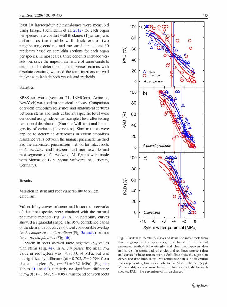
Vulnerability curves of stems and intact root networksof the three species were obtained with the manualpneumatic method (Fig. 3). All vulnerability curvesshowed a sigmoidal shape. The 95% confidence bandsof the stem and root curves showed considerable overlapfor A. campestre and C. avellana (Fig. 3a and c), but notfor A. pseudoplatanus (Fig. 3b).
Xylem in roots showed more negative P50 valuesthan stems (Fig. 4a). In A. campestre, the mean P50
value in root xylem was −4.86 ± 0.84 MPa, but wasnot significantly different (t(6) = 0.702, P = 0.509) fromthe stem xylem P50 (−4.21 ± 0.38 MPa) (Fig. 4a;Tables S1 and S2). Similarly, no significant differencein P50 (t(8) = 1.882, P = 0.097) was found between roots
Fig. 3 Xylem vulnerability curves of stems and intact roots fromthree angiosperm tree species (a, b, c) based on the manualpneumatic method. Blue triangles and blue lines represent dataand curves for stems, and red circles and red lines represent dataand curves for intact root networks. Solid lines show the regressioncurves and dash lines show 95% confidence bands. Solid verticallines represent xylem water potential at 50% embolism (P50).Vulnerability curves were based on five individuals for eachspecies. PAD= the percentage of air discharged
Plant Soil (2020) 450:479–495 485

and stems of C. avellana, which showed a mean valueof −3.52 ± 0.33MPa and − 2.77 ± 0.22MPa, respective-ly (Fig. 4a; Tables S1 and S3). For A. pseudoplatanus,however, the mean P50 value of root xylem was −4.17 ±0.31 MPa, which was significantly more negative(t(4) = 3.924, P = 0.004) than the stem xylem P50
(−2.63 ± 0.24 MPa; Fig. 4a; Tables S1 and S2).A comparison of P88 values between xylem of stems
and roots showed no significant difference (t(6) =−2.182, P = 0.072) for A. campestre, with the stem P88
(−9.15 ± 0.74 MPa) being more negative than the rootP88 (−6.49 ± 0.97MPa) (Fig. 4b; Tables S1 and S2). Theopposite was shown for A. pseudoplatanus andC. avellana, with root P88 values (−6.26 ± 0.32 MPaand − 5.49 ± 0.33MPa, respectively) being significantlymore negative than stem P88 values (−4.51 ± 0.49 MPaand − 4.36 ± 0.34MPa, respectively) (Fig. 4b; Tables S1and S2).
Variation in root vulnerability to xylem embolism inC. avellana.
There was no significant difference (t(6) = −0.225,P = 0.830) in embolism resistance between intact rootnetworks from potted saplings and thick root segmentsfrom forest saplings, with a mean P50 value of −3.52 ±0.33MPa and − 3.41 ± 0.30 MPa, respectively (Fig. 5a–d; Tables S1 and S3). Similarly, no significant differencein P88 values (t(6) = 0.123, P = 0.906) was found be-tween intact roots and thick roots segments ofC. avellana (Fig. 5e; Tables S1 and S3).
No significant difference (t(6) = −0.435, P =0.679) was found between P50 values based on themanual approach and the automated pneumatronmethod (Fig. 5a–d; Tables S1 and S3), with P50
values of −3.52 ± 0.33 MPa and − 3.30 ± 0.38 MPa,respectively. A similar trend was shown in P88
values, with no significant difference (t(6) = 0.142,
Fig. 4 Comparisons of the P50 (a) and P88 (b) values (n = 5)between stems and intact root networks of three angiosperm treespecies based on the manual pneumatic method. Different smallletters indicate significant differences. Data of stems and roots
were present in blue and red, respectively. Box plots show themedian (horizontal line inside the box), average (square inside thebox), 90th percentile (upper bar), 10th percentile (lower bar), 75thpercentile (upper box line), and 25th percentile (lower box line)
Plant Soil (2020) 450:479–495486

P = 0.579) based on these two methods (Fig. 5e;Tables S1 and S3).
Conduit anatomy
Conduit diameter (D) was significantly wider in rootsthan in stems of the species studied (P < 0.001)(Table 1). Significant differences were found ininterconduit cell wall thickness (TCW) between stem
xy l em and r oo t x y l em o f A . c ampe s t re ,A. pseudoplatanus, and C. avellana (P < 0.01), withthe mean interconduit cell wall thickness being higherin stems than in roots (Table 1).
Bordered pit characteristics
There were both similarities and differences in thebordered pit structure between stem and root xylem
Fig. 5 Root xylem vulnerability curves ofC. avellana, with intactroot networks based on the manual approach (a), thick root seg-ments based on the manual pneumatic method (b), and intact rootnetworks based on the automated pneumatic method (c). Solidlines show the fitting curves and the short dash lines represent the95% confidence bands. Solid and dash vertical lines represent P50
and P88 values, which are shown in the boxplots d and e, respec-tively. No difference in P50 (d) and P88 (e) values (n = 3 or 5) were
found. I = application of themanual method to intact root networks(red), II = the manual pneumatic method applied to thick rootssegments (blue), and III = application of the automatedpneumatron to intact root networks (green). PAD= the percentageof air discharged. Box plots show the median (horizontal lineinside the box), average (square inside the box), 90th percentile(upper bar), 10th percentile (lower bar), 75th percentile (upper boxline), and 25th percentile (lower box line)
Plant Soil (2020) 450:479–495 487

Table 1 Wood anatomical features related to conduit and pit characteristics for stem and root xylem in three angiosperm tree species
Species Organ D (μm) APM
(μm2)APA (μm2) PD (No. per
100 μm2)FPA (%) FPF (%) TPM (nm) TCW (μm)
A. campestre Stem 19.6 ± 0.4 26.6 ± 0.9 2.0 ± 0.2 2.2 ± 0.2 8.0 ± 0.8 63.1 ± 1.4 235 ± 12 3.1 ± 0.1
Root 26.2 ± 0.6* 24.9 ± 1.1 1.4 ± 0.1* 2.1 ± 0.2 6.0 ± 0.4 53.9 ± 5.8 248 ± 11 2.7 ± 0.1*
A. pseudoplatanus Stem 24.4 ± 0.5 29.2 ± 1.1 2.0 ± 0.1 1.7 ± 0.2 7.3 ± 0.5 55.4 ± 6.7 256 ± 14 3.2 ± 0.1
Root 27.7 ± 0.6* 28.2 ± 1.5 2.3 ± 0.2 1.7 ± 0.2 10.0 ± 1.0* 54.9 ± 4.7 266 ± 12 2.9 ± 0.1*
C. avellana Stem 25.8 ± 0.4 23.9 ± 1.8 3.1 ± 0.2 3.2 ± 0. 3 16.2 ± 1.5 59.4 ± 2.0 241 ± 10 4.0 ± 0.1
Root 29.7 ± 0.6* 26.3 ± 1.6 3.0 ± 0.1 3.1 ± 0.6 14.3 ± 1.0 57.6 ± 4.4 304 ± 14* 3.2 ± 0.1*
D conduit diameter, APM bordered pit area, APA pit aperture area, PD pit density, FPA pit aperture fraction, FPF interconduit pit field fraction,TPM interconduit pit membrane thickness, TCW interconduit double wall thickness of conduit
Values are means ± SE, n = 5. * = statistical significance (P < 0.05) between stems and roots
Fig. 6 SEM images of interconduit pits in stem (a, c, e) and root (b, c, f) xylem of A. campestre (a, b), A. pseudoplatanus (c, d), andC. avellana (e, f). The axial orientation of the conduits is in a horizontal position for all images. All scale bars = 5 μm
Plant Soil (2020) 450:479–495488

(Table 1; Fig. 6). The pit border surface area (APM)showed no significant difference between stem androot xylem (P > 0.05, Table 1; Fig. 6). Pit bordershad a slightly larger surface area in stems than rootsfor A. campestre and A. pseudoplatanus, but theopposite was found for C. avellana (Table 1; Fig.6). The pit aperture surface area (APA) showed nodifference between stems and roots (P > 0.05), ex-cept for A. campestre (t(122) = −2.945, P = 0.005).For A. campestre, pit aperture areas were larger inthe stem than in the root (Table 1).
The ratio between the pit aperture surface areaand pit membrane surface area (FPA) was slightlyhigher in stems than roots for A. campestre andC. avellana (P > 0.05, Table 1). The pit aperture arearepresented about 7% and 15% of the total pit bor-der area for stems and roots of A. campestre andC. avellana, respectively. This ratio was significant-ly lower in stems than roots for A. pseudoplatanus(t(107) = 2.281, P = 0.025), with pit apertures occu-pying 7% of the pit border area in the stem, and10% in root xylem (Table 1).
There was no significant difference in the meanpit density (PD) and the interconduit pit field frac-tion (FPF) between stem and root xylem (P > 0.05,Table 1) . Mean values of pi t densi ty andinterconduit pit field fraction were higher in stemthan in root xylem, except for pit density values inA. pseudoplatanus (1.73 ± 0.17 and 1.74 ± 0.15 per100 μm2 of interconduit area for stem and rootxylem, respectively).
TEM observations of intercoduit pit membranesof fresh samples showed considerably darker (i.e.more electron dense) particles in roots than in stemsfor A. pseudoplatanus and C. avellana, but not forA. campestre (Fig. 7). Most interconduit pit mem-branes showed a homogeneous appearance in elec-tron density (Fig. 7b–e). Pit membranes with bothtransparent and electron dense parts were observedin roots of A. pseudoplatanus and C. avellana, withdarker particles generally at the outermost layers ofthe pit membrane (Fig. 7d and f). The interconduitpit membranes were slightly thicker in root xylemthan stem xylem for the three species studied(Table 1). The difference in pit membrane thickness,however, was only significant between stem androot xylem for C. avellana (t(67) = −3.704,P < 0.001) (Table 1).
Discussion
Embolism resistance of intact roots was found to besimilar to stems for A. campestre and C. avellana, buthigher than stems for A. pseudoplatanus. This finding isin line with previous studies that show no universaloccurrence of the hydraulic vulnerability segmentationhypothesis (Hukin et al. 2005; McElrone et al. 2004;Rodriguez-Dominguez et al. 2018; Skelton et al. 2017),and raises some important points. Firstly, whether or notembolism occurs more quickly in a distant organ than ina proximal organ depends on the potential difference inxylemwater potential that these organs experience in thefield. Although we did not take xylem water potentialmeasurements in the field, stems have a typically morenegative water potential than roots (Jackson et al. 2000).Therefore, even without any pronounced difference inembolism resistance, hydraulic safety margins (definedas the difference between the xylem water potentialexperience and the P50 value) might be lower for stemsthan for roots.
Secondly, the discrepancy between our results andearlier studies that report more vulnerable xylem inroots than in stems (Domec et al. 2006; Pratt et al.2007, 2015) could at least be partly affected by thedifferent methods applied to measure xylem embolismresistance. Low root embolism resistance, for instance,has especially been found for angiosperms and conifersbased on the air-injection method with double-endedcavitation chambers (Domec et al. 2006; Froux et al.2005; Johnson et al. 2016; Martínez-Vilalta et al. 2002;Sperry and Ikeda 1997). This method may underesti-mate embolism resistance due to effervescence (i.e. theescape of gas bubbles from xylem sap when stemsegments are exposed to reduced gas pressure accord-ing to Henry’s law, resulting in artificial embolism) orother methodological concerns (Choat et al. 2010;Torres-Ruiz et al. 2014, 2015; Yin and Cai 2018). Highvulnerability to embolism may also be found for cen-trifuge methods, especially when vessels are at leasthalf as long as the rotor diameter (Du et al. 2018;Hacke et al. 2015; Lamarque et al. 2018; Sperry et al.2012; Torres-Ruiz et al. 2017; Wang et al. 2014).Observations based on X-ray tomography and the op-tical method suggest that embolism resistance of intactroots is similar to stems (Losso et al. 2019; Skeltonet al. 2017), or even higher than stems (Rodriguez-Dominguez et al. 2018). The P50 values based on the
Plant Soil (2020) 450:479–495 489

pneumatic method for stems of A. campestre andA. pseudoplatanus are 1 to 2.2 MPa more negativethan P50 values obtained with the air-injection method(Tissier et al. 2004). However, our P50 values for stemsof the three species differed only by 0.4 to 0.7 MPa tothose reported by Li et al. (2016b), which were basedon the cavitron method using samples from the samepopulation of trees. Moreover, our mean P50 value forstems of A. pseudoplatanus (−2.63MPa) is also similarto the −2.51 MPa P50 based on micro-CT observations(Losso et al. 2019). These similarities are interestingand confirm earlier comparison of the pneumatic meth-od with the bench dehydration approach (Pereira et al.2016; Zhang et al. 2018).
One major advantage of the pneumatic method is thatits measurements are based on the kinetics of gas diffu-sion from embolised, non-cut open conduits viainterconduit pit membranes to the discharge tube. How-ever, extraction of gas from alternative sources (e.g.extraxylary tissue or intercellular air spaces in xylem)is much slower (Sorz and Hietz 2006), and can thereforebe ignored. This also means that the pneumatic methoddoes not measure any change in root hydraulic conduc-tivity, which may be strongly affected by extraxylarytissue prior to embolism formation in xylem conduits(Cuneo et al. 2016; Rodriguez-Dominguez andBrodribb 2020). When both xylem and extraxylary con-ductivity were considered (Creek et al. 2018), roots were
Fig. 7 TEM images of interconduit pit membranes in stem (a, c, e) and root (b, d, f) xylem of A. campestre (a, b), A. pseudoplatanus (c, d),and C. avellana (e, f). All scale bars = 0.5 μm. Black arrows show the pit membranes
Plant Soil (2020) 450:479–495490

found to be more vulnerable than stems. However, ourstudy showed an opposite trend based on the pneumaticmeasurements where the extraxylary tissue were notmeasured. P50 methods that are unable to distinguishextraxylary conductivity from xylem conductivity couldoverestimate xylem embolism resistance.
The finding that root xylem was not more vulnerableto embolism than stem xylem based on our pneumaticmeasurements was further supported by data on pitmembrane thickness. The interconduit pit membranethickness (TPM) was suggested to be more important toembolism resistance than bordered pit area and pit ap-erture area (Lens et al. 2011; Li et al. 2016a). Based on athree dimensional view of pit membranes, it has beensuggested that pit membrane thickness is related to thenumber of pore constrictions within a single pore path-way (Kaack et al. 2019; Zhang et al. 2020). Since themost narrow pore constriction within a pore pathwaydetermines embolism resistance, the likelihood that thesmallest pore constriction is very narrow will increasewith the number of pore constrictions. Although thismechanistic link between pit membrane thickness andembolism resistance needs further research, higher em-bolism resistance in roots was found to correspond tothicker interconduit pit membranes in this study. Signif-icant difference in pit membrane thickness betweenroots and stems, however, was only found forC. avellana. In fact, thicker pit membranes in roots ofC. avellana may explain its more negative P50 and P88
values, which were 0.75 and 1.13 MPa more negativethan stems of this species. Although interconduit pitmembrane thickness is repeatedly shown to be relatedto embolism resistance (Jansen et al. 2009, 2018; Lenset al. 2011; Li et al. 2016a), this relationship cannot betreated as very tight, and might be blurred by potentialartefacts in measuring embolism and pit membranethickness (Kotowska et al. 2020).
We also do not fully understand the functionalsignificance of the electron density of pit mem-branes between roots and stems. Since it is knownthat OsO4 treatment results in visualisation of unsat-urated fatty acid chains of lipids (Riemersma 1986),differences in electron density may reflect differentconcentrations of lipids associated with pit mem-branes, which are likely to affect air-seeding andembolism resistance (Jansen et al. 2018; Schenket al. 2017, 2018; Yang et al. 2020). Besides thehypothesis that xylem sap lipids represent remnants
of vessel element cytoplasm (Esau 1965; Esau et al.1966), it is possible that a high amount of vessel-associated parenchyma cells may contribute to ahigher production of xylem sap lipids in roots thanin stems (Morris et al. 2018; Schenk et al. 2018).Although the amount of parenchyma tissue in thespecies studied was not quantified, roots were foundto show higher tissue fractions of ray and axialparenchyma than stems (Morris et al. 2016b;Plavcová et al. 2019), and it is also possible thatthe wide vessels in roots are more surrounded byparenchyma cells then narrow vessels in stem xylem(Morris et al. 2017).
Could the proximity of the stem and root samples,which was 15 to 20 cm in the saplings tested, explainthe similarity in embolism resistance between bothorgans? Although this distance appears to be rathersmall, the samples selected were clearly root andstem samples from small saplings, with clear differ-ences in conduit diameter and interconduit wallthickness between stem and root xylem. Since mostvessels in the species studied are much shorter than20 cm, with a mean vessel length below 6 cm (datanot shown), it is unlikely that a few, long vesselscould interconnect the root and stem samples thatwere used for our measurements. Moreover, the lackof any significant difference in embolism resistancebetween thick, mature root segments from forest sap-lings and the terminal root networks from pottedsaplings indicate that the proximity of the xylemtissue does not affect xylem embolism resistance inC. avellana.
Roots exhibited a wider conduit diameter andthinner interconduit wall thickness than stems inthe three species studied, which is in line with pre-vious studies (e.g. Aloni 1987; Anfodillo et al.2012). Smaller conduits and thicker intervessel wallsare suggested to increase hydraulic safety and me-chanical support (Corcuera et al. 2004; Hacke et al.2001; Mauseth and Stevenson 2004; Plavcová et al.2019), although there is also evidence that drought-induced embolism is not directly related to conduitsize (e.g. Choat et al. 2016; Klepsch et al. 2018;Skelton et al. 2018; Wason et al. 2018). Since rootswere equally or even more resistant to embolismthan stems in this study, this may indicate that con-duit diameter and intervessel wall thickness do notplay a major, direct role in embolism resistance.
Plant Soil (2020) 450:479–495 491

Although intervessel wall thickness was suggestedto be linked with pit membrane thickness (Jansenet al. 2009; Li et al. 2016a), this relationship couldnot be supported for stems and roots of A. campestreand A. pseudoplatanus (Table 1). No clear relation-ship between these two features has also been re-ported in other studies (Klepsch et al. 2018; Scholzet al. 2013b).
In conclusion, this study shows that embolism resis-tance is higher for roots than for stems ofA. pseudoplatanus, while no difference was found be-tween roots and stems of A. campestre and C. avellana.This finding was supported by data on the interconduitpit membrane thickness, and suggests that the hydraulicvulnerability segmentation hypothesis does not apply toroots and stems of the three temperate tree speciesstudied. Moreover, thick root segments of C. avellanashow a similar embolism resistance to intact root net-works, indicating that the pneumatic method can beapplied to non-terminal plant material. We also demon-strate that the pneumatron approach shows promisingresults for establishing a high-throughput platform on alarge number of samples, especially when this approachis combined with stem psychrometers (Pereira et al.2020).
Acknowledgements We thank the Botanical Garden and theElectron Microscopy Section of Ulm University for technicalsupport. Daniel Glöckler and Peter Zindl are acknowledged forassistance with collecting plant material. M.W. acknowledgesfinancial support from the Guangxi Education Department(GED). Y.Z. acknowledges financial support from the ChinaScholarship Council (CSC). This research was financially support-ed by the National Natural Science Foundation of China (No.31800205, 31560124), and Guangxi Natural Science FoundationProgram (No. 2015GXNSFBA139113)
Funding Information Open Access funding provided byProjekt DEAL.
Open Access This article is licensed under a Creative CommonsAttribution 4.0 International License, which permits use, sharing,adaptation, distribution and reproduction in anymedium or format,as long as you give appropriate credit to the original author(s) andthe source, provide a link to the Creative Commons licence, andindicate if changes were made. The images or other third partymaterial in this article are included in the article's Creative Com-mons licence, unless indicated otherwise in a credit line to thematerial. If material is not included in the article's Creative Com-mons licence and your intended use is not permitted by statutoryregulation or exceeds the permitted use, you will need to obtainpermission directly from the copyright holder. To view a copy ofthis licence, visit http://creativecommons.org/licenses/by/4.0/.
References
Aloni R (1987) Differentiation of vascular tissues. Annu Rev PlantPhysiol 38:179–204. https://doi.org/10.1146/annurev.pp.38.060187.001143
Anfodillo T, Deslauriers A,Menardi R, Tedoldi L, Petit G, Rossi S(2012) Widening of xylem conduits in a conifer tree dependson the longer time of cell expansion downwards along thestem. J Exp Bot 63:837–845. https://doi.org/10.1093/jxb/err309
Barros FV, Bittencourt PRL, BrumM, Restrepo-Coupe N, PereiraL, Teodoro GS, Saleska SR, Borma LS, Christoffersen BO,Penha D, Alves LF, Lima AJN, Carneiro VMC, Gentine P,Lee JE, Aragão LEOC, Ivanov V, Leal LSM, Araujo AC,Oliveira RS (2019) Hydraulic traits explain differential re-sponses of Amazonian forests to the 2015 El Niño-induceddrought. New Phytol 223:1253–1266. https://doi.org/10.1111/nph.15909
Bittencourt PR, Pereira L, Oliveira RS (2018) Pneumatic methodto measure plant xylem embolism. Bio-protocol 8:1–14.https://doi.org/10.21769/BioProtoc.3059
Bouche PS, Jansen S, Sabalera JC, Cochard H, Burlett R, Delzon S(2016) Low intra-tree variability in resistance to embolism infour Pinaceae species. Ann For Sci 73:681–689. https://doi.org/10.1007/s13595-016-0553-6
Brum M, Vadeboncoeur MA, Ivanov Vet al (2019) Hydrologicalniche segregation defines forest structure and drought toler-ance strategies in a seasonal Amazon forest. J Ecol 107:318–333. https://doi.org/10.1111/1365-2745.13022
Choat B, Cobb AR, Jansen S (2008) Structure and function ofbordered pits: new discoveries and impacts on whole-planthydraulic function. New Phytol 177:608–626. https://doi.org/10.1111/j.1469-8137.2007.02317.x
Choat B, DraytonWM,Brodersen C,MatthewsMA, Shackel KA,Wada H, McElrone AJ (2010) Measurement of vulnerabilityto water stress-induced cavitation in grapevine: a comparisonof four techniques applied to a long-vesseled species. PlantCell Environ 33:1502–1512. https://doi.org/10.1111/j.1365-3040.2010.02160.x
Choat B, Badel E, Burlett R, Delzon S, Cochard H, Jansen S(2016) Noninvasive measurement of vulnerability todrought-induced embolism by X-ray microtomography.Plant Physiol 170:273–282. https://doi.org/10.1104/pp.15.00732
Cochard H, Coll L, Le Roux X, Améglio T (2002) Unraveling theeffects of plant hydraulics on stomatal closure during waterstress in walnut. Plant Physiol 128:282–290. https://doi.org/10.2307/4280289
Corcuera L, Camarero JJ, Gil-Pelegrín E (2004) Effects of a severedrought on Quercus ilex radial growth and xylem anatomy.Trees 18:83–92. https://doi.org/10.1007/s00468-003-0284-9
Creek D, Blackman C, Brodribb TJ, Choat B, Tissue DT (2018)Coordination between leaf, stem, and root hydraulics and gasexchange in three arid-zone angiosperms during severedrought and recovery. Plant Cell Environ 41:2869–2881.https://doi.org/10.1111/pce.13418
Cuneo IF, Knipfer T, Brodersen CR, McElrone AJ (2016)Mechanical failure of fine root cortical cells initiates plant
Plant Soil (2020) 450:479–495492
hydraulic decline during drought. Plant Physiol 172:1669–1678. https://doi.org/10.1104/pp.16.00923
Dixon HH (1896) Transpiration into a saturated atmosphere. ProcR Ir Acad 4:627–635. https://doi.org/10.2307/20490525
Domec J-C, Gartner BL (2001) Cavitation and water storagecapacity in bole xylem segments of mature and youngDouglas-fir trees. Trees 15:204–214. https://doi.org/10.1007/s004680100095
Domec J-C, Lachenbruch B, Meinzer FC (2006) Bordered pitstructure and function determine spatial patterns of air-seeding thresholds in xylem of Douglas-fir (Pseudotsugamenziesii; Pinaceae) trees. Am J Bot 93:1588–1600.https://doi.org/10.2307/4123175
Du GY, Feng F, Wang YJ, Tyree MT (2018) Do nano-particlescause recalcitrant vulnerability curves in Robinia? Testingwith a four-cuvette Cochard rotor and with water extractioncurves. Tree Physiol 39:156–165. https://doi.org/10.1093/treephys/tpy051
Esau K (1965) Plant anatomy, 2nd edn. Wiley, New YorkEsau K, Cheadle VI, Gill RH (1966) Cytology of differentiating
tracheary elements. II. Structures associated with cell sur-faces. Am J Bot 53:765–771. https://doi.org/10.1002/j.1537-2197.1966.tb06832.x
Froux F, Ducrey M, Dreyer E, Huc R (2005) Vulnerability toembolism differs in roots and shoots and among threeMediterranean conifers: consequences for stomatal regula-tion of water loss? Trees 19:137–144. https://doi.org/10.1007/s00468-004-0372-5
Hacke UG, Sperry JS, Pockman WT, Davis SD, McCulloh KA(2001) Trends in wood density and structure are linked toprevention of xylem implosion by negative pressure.Oecologia 126:457–461. https:/ /doi.org/10.1007/s004420100628
Hacke UG, Venturas MD, MacKinnon ED, Jacobsen AL, SperryJS, Pratt RB (2015) The standard centrifuge method accu-rately measures vulnerability curves of long-vesselled olivestems. New Phytol 205:116–127. https://doi.org/10.1111/nph.13017
Hochberg U, Albuquerque C, Rachmilevitch S, Cochard H,David-Schwartz R, Brodersen CR, McElrone A, Windt CW(2016) Grapevine petioles are more sensitive to droughtinduced embolism than stems: evidence from in vivo MRIand microcomputed tomography observations of hydraulicvulnerability segmentation. Plant Cell Environ 39:1886–1894. https://doi.org/10.1111/pce.12688
Hukin D, Cochard H, Dreyer E, Thiec DL, Bogeat-Triboulot MB(2005) Cavitation vulnerability in roots and shoots: doesPopulus euphratica Oliv., a poplar from arid areas ofCentral Asia, differ from other poplar species? J Exp Bot56:2003–2010. https://doi.org/10.1093/jxb/eri198
Jackson RB, Sperry JS, Dawson TE (2000) Root water uptake andtransport: using physiological processes in global predic-tions. Trends Plant Sci 5:482–488. https://doi.org/10.1016/S1360-1385(00)01766-0
Jansen S, SchenkHJ (2015) On the ascent of sap in the presence ofbubbles. Am J Bot 102:1561–1563. https://doi.org/10.3732/ajb.1500305
Jansen S, Choat B, Pletsers A (2009) Morphological variation ofintervessel pit membranes and implications to xylem functionin angiosperms. Am J Bot 92:409–419. https://doi.org/10.2307/27793098
Jansen S, Klepsch M, Li S, Kotowska MM, Schiele S, Zhang Y,Schenk HJ (2018) Challenges in understanding air-seeding inangiosperm xylem. Acta Hortic 1222:13–20
Johnson DM, Wortemann R, McCulloh KA, Jordan-Meille L,Ward E, Warren JM, Palmroth S, Domec JC (2016) A testof the hydraulic vulnerability segmentation hypothesis inangiosperm and conifer tree species. Tree Physiol 36:983–993. https://doi.org/10.1093/treephys/tpw031
Kaack L, Altaner CM,Carmesin C et al (2019) Function and three-dimensional structure of intervessel pit membranes in angio-sperms: a review. IAWA J 40:673–702. https://doi.org/10.1163/22941932-40190259
Klepsch M, Zhang Y, Kotowska MM, Lamarque LJ, Nolf M,Schuldt B, Torres-Ruiz JM, Qin DW, Choat B, Delzon S,Scoffoni C, Cao KF, Jansen S (2018) Is xylem of angiospermleaves less resistant to embolism than branches? Insightsfrom microCT, hydraulics, and anatomy. J Exp Bot 69:5611–5623. https://doi.org/10.1093/jxb/ery321
Kotowska MM, Thom R, Zhang Y, Schenk HJ, Jansen S (2020)Within-tree variability and sample storage effects of borderedpit membranes in xylem of Acer pseudoplatanus. Trees 34:61–71. https://doi.org/10.1007/s00468-019-01897-4
Lamarque LJ, Corso D, Torres-Ruiz JM, Badel E, Brodribb TJ,Burlett R, Charrier G, Choat B, Cochard H, Gambetta GA,Jansen S, King A, Lenoir N, Martin-StPaul N, Steppe K,Bulcke JVD, Zhang Y, Delzon S (2018) An inconvenienttruth about xylem resistance to embolism in the model spe-cies for refilling Laurus nobilis L. Ann For Sci 75:1–15.https://doi.org/10.1007/s13595-018-0768-9
Lazzarin M, Crivellaro A, Williams CB, Dawson TE, Mozzi G,Anfodillo T (2016) Tracheid and pit anatomy vary in tandemin a tall Sequoiadendron giganteum tree. IAWA J 37:172–185. https://doi.org/10.1163/22941932-20160129
Lens F, Sperry JS, Christman MA, Choat B, Rabaey D, Jansen S(2011) Testing hypotheses that link wood anatomy to cavita-tion resistance and hydraulic conductivity in the genus Acer.New Phytol 190:709–723. https://doi.org/10.1111/j.1469-8137.2010.03518.x
Li S, Klepsch M, Jansen S, Schmitt M, Lens F, Karimi Z, SchuldtB, Espino S, Schenk HJ (2016a) Intervessel pit membranethickness as a key determinant of embolism resistance inangiosperm xylem. IAWA J 37:152–171. https://doi.org/10.1163/22941932-20160128
Li S, Feifel M, Karimi Z, Schuldt B, Choat B, Jansen S (2016b)Leaf gas exchange performance and the lethal water potentialof five European species during drought. Tree Physiol 36:179–192. https://doi.org/10.1093/treephys/tpv117
Lima TRA, Carvalho ECD,Martins FR, Oliveira RS,Miranda RS,Müller CS, Pereira L, Bittencourt PRL, Sobczak JCMSM,Gomes-Filho E, Costa RC, Araújo FS (2018) Lignin compo-sition is related to xylem embolism resistance and leaf lifespan in trees in a tropical semiarid climate. New Phytol 219:1252–1262. https://doi.org/10.1111/nph.15211
Losso A, Bär A, Dämon B, Dullin C, Ganthaler A, Petruzzellis F,Savi T, Tromba G, Nardini A, Mayr S, Beikircher B (2019)Insights from in vivomicro-CTanalysis: testing the hydraulicvulnerability segmentation in Acer pseudoplatanus andFagus sylvatica seedlings. New Phytol 221:1831–1842.https://doi.org/10.1111/nph.15549
Martínez-Vilalta J, Prat E, Oliveras I, Piñol J (2002) Xylemhydraulic properties of roots and stems of nine
Plant Soil (2020) 450:479–495 493

Mediterranean woody species. Oecologia 133:19–29.https://doi.org/10.1007/s00442-002-1009-2
Mauseth JD, Stevenson JF (2004) Theoretical considerations ofvessel diameter and conductive safety in populations of ves-sels. Int J Plant Sci 165:359–368. https://doi.org/10.1086/382808
McElrone AJ, Pockman WT, Martínez-Vilalta J, Jackson RB(2004) Variation in xylem structure and function in stemsand roots of trees to 20 m depth. New Phytol 215:466–471.https://doi.org/10.1111/j.1469-8137.2004.01127.x
Morris H, Brodersen C, Schwarze FMWR, Jansen S (2016a) Theparenchyma of secondary xylem and its critical role in treedefense against fungal decay in relation to the CODITmodel.Front Plant Sci 7:1665. https: / /doi.org/10.3389/fpls.2016.01665
Morris H, Plavcová L, Cvecko P, Fichtler E, Gillingham MAF,Martínez-Cabrera HI, McGlynn DJ, Wheeler E, Zheng J,Ziemińska K, Jansen S (2016b) A global analysis of paren-chyma tissue fractions in secondary xylem of seed plants.New Phytol 209:1553–1565. https://doi.org/10.1111/nph.13737
Morris H, GillinghamMAF, Plavcová L, Gleason SM, Olson ME,Coomes DA, Fichtler E, Klepsch MM,Martínez-Cabrera HI,McGlinn DJ, Wheeler EA, Zheng J, Ziemínska K, Jansen S(2017) Vessel diameter is related to amount and spatial ar-rangement of axial parenchyma in woody angiosperms. PlantCell Environ 41:245–260. https://doi.org/10.1111/pce.13091
Morris H, Plavcová L, GoraiM, KlepschM, KotowskaM, SchenkHJ, Jansen S (2018) Vessel-associated cells in angiospermxylem: highly specialized living cells at the symplast-apoplast boundary. Am J Bot 105:153–162. https://doi.org/10.1002/ajb2.1030
Morris H, Hietala AM, Jansen S, Ribera J, Rosner S, Salmeia K,Schwarze F (2019) Using the CODIT model to explainsecondary metabolites of the xylem in defence systems oftemperate trees to decay fungi. Ann Bot-London. https://doi.org/10.1093/aob/mcz138
Oliveira RS, Costa FR, van Baalen E, Jonge AD, Bittencourt PR,Almanza Y, Barros FDV, Cordoba EC, FagundesMV, GarciaS, Guimaraes HM, Schietti J, Rodrigues-Souza J, Poorter L(2019) Embolism resistance drives the distribution ofAmazonian rainforest tree species along hydro-topographicgradients. New Phytol 221:1457–1465. https://doi.org/10.1111/nph.15463
Pammenter NW, van der Willigen C (1998) A mathematical andstatistical analysis of the curves illustrating vulnerability ofxylem to cavitation. Tree Physiol 18:589–593. https://doi.org/10.1093/treephys/18.8-9.589
Pereira L, Bittencourt PR, Oliveira RS, Junior MB, Barros FV,Ribeiro RV, Mazzafera P (2016) Plant pneumatics: stem airflow is related to embolism – new perspectives onmethods inplant hydraulics. New Phytol 211:357–370. https://doi.org/10.1111/nph.13905
Pereira L, Bittencourt PR, Pacheco VS, Miranda MT, Zhang Y,Oliveira RS, Groenendijk P, Machado EC, Tyree MT, JansenS, Rowland L, Ribeiro RV (2020) The Pneumatron: anautomated pneumatic apparatus for estimating xylem vulner-ability to embolism at high temporal resolution. Plant CellEnviron 43:131–142. https://doi.org/10.1111/pce.13647
Plavcová L, Gallenmüller F, Morris H, Khatamirad M, Jansen S,Speck T (2019) Mechanical properties and structure-function
trade-offs in secondary xylem of young roots and stems. JExp Bot 70:3679–3691. https://doi.org/10.1093/jxb/erz286
Pratt RB, Jacobsen AL, Golgotiu KA, Sperry JS, Ewers FW, DavisSD (2007) Life history type and water stress tolerance in nineCalifornia chaparral species (Rhamnaceae). EcolMonogr 77:239–253. https://doi.org/10.1890/06-0780
Pratt RB, MacKinnon ED, Venturas MD, Crous CJ, Jacobsen AL(2015) Root resistance to cavitation is accurately measuredusing a centrifuge technique. Tree Physiol 35:185–196.https://doi.org/10.1093/treephys/tpv003
Prendin AL, Mayr S, Beikircher B, von Arx G, Petit G (2018)Xylem anatomical adjustments prioritize hydraulic efficiencyover safety as Norway spruce trees grow taller. Tree Physiol38:1088–1097. https://doi.org/10.1093/treephys/tpy065
Riemersma JC (1968) Osmium tetroxide fixation of lipids forelectron microscopy. A possible reaction mechanism.Biochim Biophys Acta 152:718−727. https://doi.org/10.1016/0005-2760(68)90118-5
Rodriguez-Dominguez CM, Brodribb TJ (2020) Declining rootwater transport drives stomatal closure in olive under mod-erate water stress. New Phytol 225:126–134. https://doi.org/10.1111/nph.16177
Rodriguez-Dominguez CM, Carins Murphy MR, Lucani C,Brodribb TJ (2018) Mapping xylem failure in disparate or-gans of whole plants reveals extreme resistance in olive roots.New Phytol 218:1025–1035. https://doi.org/10.1111/nph.15079
Schenk HJ, Espino S, Goedhart CM, Nordenstahl M, Martinez-Cabrera HI, Jones CS (2008) Hydraulic integration and shrubgrowth form linked across continental aridity gradients. ProcNatl Acad Sci U S A 105:11248–11253. https://doi.org/10.1073/pnas.0804294105
Schenk HJ, Espino S, Romo DM, Nima N, Do AY, Michaud JM,Papahadjopoulos-Sternberg B, Yang J, Zuo YY, Steppe K,Jansen S (2017) Xylem surfactants introduce a new elementto the cohesion-tension theory. Plant Physiol 173:1177–1196.https://doi.org/10.1104/pp.16.01039
Schenk HJ, Espino S, Rich-Cavazos SM, Jansen S (2018) Fromthe sap’s perspective: the nature of vessel surfaces in angio-sperm xylem. Am J Bot 105:172–185. https://doi.org/10.1002/ajb2.1034
Schindelin J, Carreras A, Frise E et al (2012) Fiji: an open-sourceplatform for biological-image analysis. Nat Methods 9:676–682. https://doi.org/10.1038/nmeth.2019
Scholz A, Klepsch M, Karimi Z, Jansen S (2013a) How to quan-tify conduits in wood? Front Plant Sci 4:56–67. https://doi.org/10.3389/fpls.2013.00056
Scholz A, Rabaey D, Stein A, Cochard H, Smets E, Jansen S(2013b) The evolution and function of vessel and pit charac-ters with respect to cavitation resistance across 10 Prunusspecies. Tree Physiol 33:684–694. https://doi.org/10.1093/treephys/tpt050
Skelton RP, Brodribb TJ, Choat B (2017) Casting light on xylemvulnerability in an herbaceous species reveals a lack of seg-mentation. New Phytol 214:561–569. https://doi.org/10.1111/nph.14450
Skelton RP, Dawson TE, Thompson SE, Shen Y, Weitz AP,Ackerly D (2018) Low vulnerability to xylem embolism inleaves and stems of north American oaks. Plant Physiol 177:1066–1077. https://doi.org/10.1104/pp.18.00103
Plant Soil (2020) 450:479–495494
Smith-Martin C, Skelton RP, Johnson KM, Lucani C, Brodribb TJ(2020) Lack of vulnerability segmentation among woodyspecies in a diverse dry sclerophyll woodland community.Funct Ecol 00:1–11. https://doi.org/10.1111/1365-2435.13519
Sorz J, Hietz P (2006) Gas diffusion through wood: implicationsfor oxygen supply. Trees 20:34–41. https://doi.org/10.1007/s00468-005-0010-x
Sperry JS, Ikeda T (1997) Xylem cavitation in roots and stems ofDouglas-fir and white fir. Tree Physiol 17:275–280.https://doi.org/10.1093/treephys/17.4.275
Sperry JS, Christman MA, Torres-Ruiz JM, Taneda H, Smith DD(2012) Vulnerability curves by centrifugation: is thereanopen vessel artefact, and are ‘r’ shaped curvesnecessarilyinvalid. Plant Cell Environ 35:601–610. https://doi.org/10.1111/j.1365-3040.2011.02439.x
Taiz L, Zeiger E (1998) Cell walls: structure, biogenesis, andexpansion. In: Taiz L, Zeiger E (eds) Plant physiology, 2ndedn. Sinauer Associates, Sunderland, Massachusetts, pp409–443
Tissier J, Lambs L, Peltier J, Marigo G (2004) Relationshipsbetween hydraulic traits and habitat preference for six Acerspecies occurring in the French Alps. Ann For Sci 61:81–86.https://doi.org/10.1051/forest:2003087
Torres-Ruiz JM, Cochard H, Mayr S, Beikircher B, Diaz-EspejoA, Rodriguez-Dominguez CM, Badel E, Fernández GE(2014) Vulnerability to cavitation in Olea europaea current-year shoots: further evidence of an open-vessel artifact asso-ciated with centrifuge and air-injection techniques. PhysiolPlantarum 152:465–474. https://doi.org/10.1111/ppl.12185
Torres-Ruiz JM, Jansen S, Choat B, McElrone AJ, Cochard H,Brodribb TJ, Badel E, Burlett R, Bouche PS, Brodersen CR,Li S, Morris H, Delzon S (2015) Direct X-raymicrotomography observation confirms the induction of em-bolism upon xylem cutting under tension. Plant Physiol 167:40–43. https://doi.org/10.1104/pp.114.249706
Torres-Ruiz JM, Cochard H, Choat B, Jansen S, López R,Tomášková I, Padilla-Díaz CM, Badel E, Burlett R, KingA, Lenoir N, Martin-StPaul NK, Delzon S (2017) Xylemresistance to embolism: presenting a simple diagnostic testfor the open vessel artefact. New Phytol 215:489–499.https://doi.org/10.1111/nph.14589
Tyree MT, Ewers FW (1991) The hydraulic architecture of treesand other woody plants. New Phytol 119:345–360.https://doi.org/10.1111/j.1469-8137.1991.tb00035.x
Tyree MT, Zimmermann MH (2002) Xylem structure and theascent of sap. Springer-Verlag, Berlin
Tyree MT, Cochard H, Cruiziat P, Sinclair B, Ameglio T (1993)Drought-induced leaf shedding in walnut: evidence for
vulnerability segmentation. Plant Cell Environ 16:879–882.https://doi.org/10.1111/j.1365-3040.1993.tb00511.x
Wang R, Zhang L, Zhang S, Cai J, Tyree MT (2014) Waterrelations of Robinia pseudoacacia L.: do vessels cavitateand refill diurnally or are R-shaped curves invalid inRobinia? Plant Cell Environ 37:2667–2678. https://doi.org/10.1111/pce.12315
Wason JW, Anstreicher KS, Stephansky N, Huggett BA,Brodersen CR (2018) Hydraulic safety margins and air-seeding thresholds in roots, trunks, branches and petioles offour northern hardwood trees. New Phytol 219:77–88.https://doi.org/10.1111/nph.15135
Wheeler JK, Huggett BA, Tofte AN, Rockwell FE, Holbrook NM(2013) Cutting xylem under tension or supersaturated withgas can generate PLC and the appearance of rapid recoveryfrom embolism. Plant Cell Environ 36:1938–1949.https://doi.org/10.1111/pce.12139
Yang J, Michaud J, Jansen S, Schenk HJ, Zuo Y (2020) Dynamicsurface tension of xylem sap lipids. Tree Physiol. https://doi.org/10.1093/treephys/tpaa006
Yin P, Cai J (2018) New possible mechanisms of embolismformation when measuring vulnerability curves by air injec-tion in a pressure sleeve. Plant Cell Environ 41:1361–1368.https://doi.org/10.1111/pce.13163
Zhang Y, Klepsch M, Jansen S (2017) Bordered pits in xylem ofvesselless angiosperms and their possible misinterpretationas perforation plates. Plant Cell Environ 40:2133–2146.https://doi.org/10.1111/pce.13014
Zhang Y, Lamarque LJ, Torres-Ruiz JM, Schuldt B, Karimi Z, LiS, Qin DW, Bittencourt P, Burlett R, Cao KF, Delzon S,Oliveira R, Pereira L, Jansen S (2018) Testing the plantpneumatic method to estimate xylem embolism resistancein stems of temperate trees. Tree Physiol 38:1016–1025.https://doi.org/10.1093/treephys/tpy015
Zhang Y, Matei T, Kotowska M, Weber M, Klepsch MM, SchenkHJ, Walter P, Schmidt V, Jansen S (2020) High porosity withtiny pore constrictions and unbending pathways characterisethe 3D structure of intervessel pit membranes in angiospermxylem. Plant Cell Environ 43:116–130. https://doi.org/10.1111/pce.13654
Zhu SD, Liu H, Xu QY, Cao KF, Ye Q (2016) Are leaves morevulnerable to cavitation than branches? Funct Ecol 30:1740–1744. https://doi.org/10.1111/1365-2435.12656
Publisher’s note Springer Nature remains neutral with regard tojurisdictional claims in published maps and institutionalaffiliations.
Plant Soil (2020) 450:479–495 495