role of vascular diseases and cerebral emboli in causation and progression of alzheimer’s disease...
TRANSCRIPT

Role of vascular diseases and cerebral emboli in causation
and progression of Alzheimer’s disease and
vascular dementia
Nitin Purandare Peninsula College of Medicine and Dentistry
Devon Partnership Mental Health Trust

Outline
• Epidemiology
• Vascular diseases and causation of dementia
• Role of cerebral emboli
• Vascular diseases and progression of dementia

Epidemiology
• 24.3 million people worldwide– 81.1 million affected by 2040
• 821,884 people with dementia in the UK – the annual cost of care = £23 billion
• 21 million people in UK know close friend of family member with dementia
• 1 in 3 people over 65 will have dementia by the time they die
• About 50% remain undiagnosed
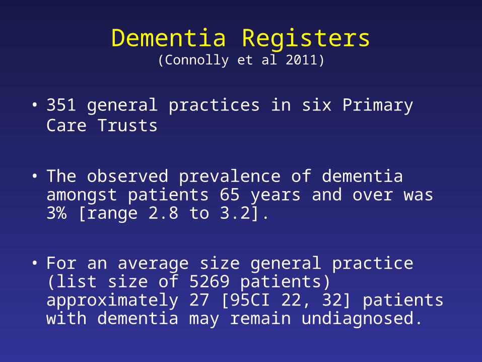
Dementia Registers(Connolly et al 2011)
• 351 general practices in six Primary Care Trusts
• The observed prevalence of dementia amongst patients 65 years and over was 3% [range 2.8 to 3.2].
• For an average size general practice (list size of 5269 patients) approximately 27 [95CI 22, 32] patients with dementia may remain undiagnosed.
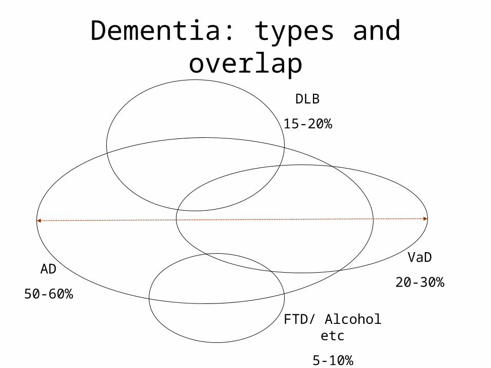
Dementia: types and overlap
AD
50-60%
FTD/ Alcohol etc
5-10%
DLB
15-20%
VaD
20-30%

Nun study
• 102 college educated nuns aged 76-100 years• Clinical and neuropathological assessment of
dementia and Alzheimer’s disease• 61 met neuropathological criteria for AD
Fewer neuropathologic lesions of AD required for the clinical manifestation of dementia in those who had infarcts in basal ganglia, thalamus or deep white matter
Snowdon et al, JAMA, 1997

Mixed AD and VaD
• 156 brains of AD patients
• Braak NFT, Aβ deposition, cortical microinfarcts (CMI) and thalamic and basal ganglia lacunes (TBGL) predicted 27% of CDR variability and 49% of presence of dementia
• Classification of dementia– NFT > II and CMI + TBGL > 2 mixed dementia– NFT ≤ II and CMI + TBGL > 2 pure VaD– NFT > II and CMI + TBGL ≤ 2 pure AD
Gold et al, Brain, 2007

Role of vascular diseasesin causation of dementia
• Epidemiological evidence
• Evidence from RCTs

Honolulu Asia Aging Study(White and Launer, 2006)
• HHP 1965 n = 8006HAAS 1991 n = 3731Follow ups 5 over 14 yearsAutopsy 2006 n = 650
• Hypertension in midlife is a risk factor for dementia in late life
– Systolic BP >= 160 HR for dementia 4.8 (2.0-11.0)– HT Brain wt; SP count, hippo atrophy – SP + lacunar infarcts risk of clinical dementia
(Launer et al, ‘00; Petrovitch et al, ’00; Korf et al, ’04; Petrovitch et al, 2005)

Decline in BP over time and risk of dementia
• Kungsholmen project• Age ≥ 75 yrs and no dementia
(n=947)• Two follow ups approx 3 years
apart– 186 dementia at FU1– 118 dementia at FU2– 643 no dementia
Results• No drop in BP prior to 3 years
before diagnosis of dementia
• BP markedly decreased over 3 years before dementia diagnosis and afterwards
• If baseline SBP<160, drop of ≥15 mm Hg at FU1 associated with increased risk of dementia at FU2 (RR 3.1)- dose effect in subjects with vascular disease?
Qiu et al, Stroke, 2004
Mechanism
• Risk Factor: Drop in BP cerebral hypoperfusion neurodegeneration clinical dementia
• Risk Marker: Neurodegeneration in strategic areas drop in BP cerebral hypoperfusion clinical dementia
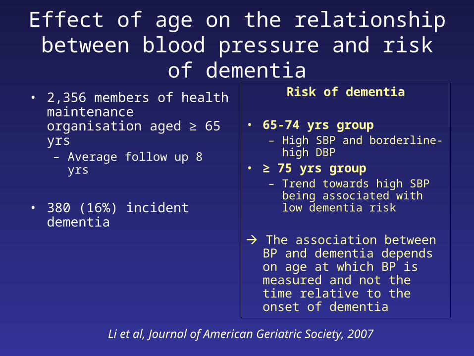
Effect of age on the relationship between blood pressure and risk of dementia
• 2,356 members of health maintenance organisation aged ≥ 65 yrs– Average follow up 8 yrs
• 380 (16%) incident dementia
Risk of dementia
• 65-74 yrs group– High SBP and borderline-high
DBP
• ≥ 75 yrs group– Trend towards high SBP being
associated with low dementia risk
The association between BP and dementia depends on age at which BP is measured and not the time relative to the onset of dementia
Li et al, Journal of American Geriatric Society, 2007
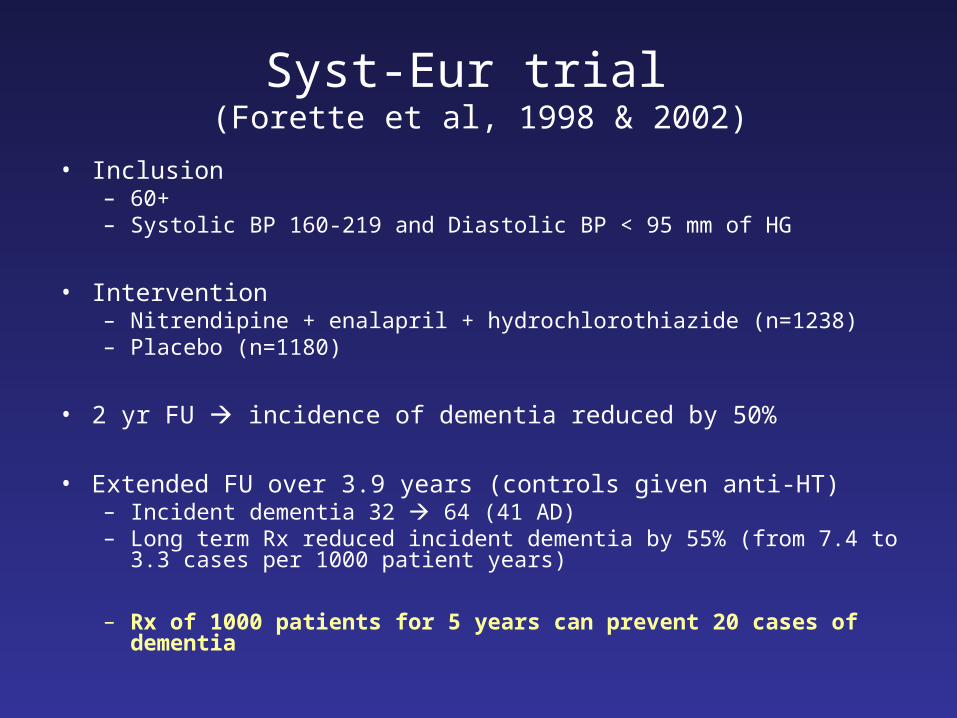
Syst-Eur trial (Forette et al, 1998 & 2002)
• Inclusion– 60+– Systolic BP 160-219 and Diastolic BP < 95 mm of HG
• Intervention– Nitrendipine + enalapril + hydrochlorothiazide (n=1238)– Placebo (n=1180)
• 2 yr FU incidence of dementia reduced by 50%
• Extended FU over 3.9 years (controls given anti-HT)– Incident dementia 32 64 (41 AD)– Long term Rx reduced incident dementia by 55% (from 7.4 to 3.3 cases
per 1000 patient years)
– Rx of 1000 patients for 5 years can prevent 20 cases of dementia

Randomised Controlled Trials
Peters et al. 2008 (HYVET-COG) indapamide, perindopril
Forette et al 1998 (Syst-Eur) nitrendipine
Forette et al 2002 (Extended follow-up) nitrendipine +
Applegate et al 1994(Systolic Hypertension in the Elderly)
chlorthalidone
Prince et al 1996 (MRC Older people with HT)
atenolol, hydrochlorothiazide
Bosch et al 2002 (HOPE) ramipril, vitamin E
The PROGRESS Collaborative Group (2003)
perindopril, indapamide
Lithell et al 2003(SCOPE)
Candesartan

Cholesterol and risk of AD• Raised cholesterol in midlife is a risk factor for dementia (most cohort studies)
• Decreasing cholesterol in late-life is risk marker (Mielke et al, 2005; Solomon et al, 2009)
• Cohort studies on statins and other LLA– Mixed results – Statins > LLA?– Simavastatin associated with reduced risk (Wolozin et al, BMC Medicine, 2007)
• RCTs– No effect HPS collaborative study, PROSPER– +ve effect? ADCLT (Sparks et al, Arch Neurol, 2005)– CLASP, LEADE, ESPRIT, PIT-ROAD
• Chole, 24SOHC, 27OHC and BBB
• Low cholesterol in cell membranes in AD
Blood Brain
BBB
Chol Chol
24SOHC
27OHC

Multiple risk factors and WMH• Midlife raised systolic BP + midlife raised cholesterol + APOE4
increased risk of dementia (Kivipelto et al, 2002)
• Metabolic syndrome risk factor in those with high inflammation (Yaffe K, 2007) only in those with MCI (Solfrizzi et al. 2011)
• VRF increase risk of AD in those with MCI and those treated appear to have reduced conversion (Li et al, 2011)
• Subcortical WMH increase risk of AD in those with MCI (Prasad et al, 2011) Only non-AD dementia (Staekenborg et al 2009)
• Metaanlyses HR 1.9 (1.3, 2.8) (Debette & Markus, 2010)
• Calculating risk of dementia in an individual– Mitnitski et al, European J of Neurology, 2006– Kivipelto et al, Lancet Neurol, 2006

Predicting dementia risk
• Population based CAIDE study– 1409 people seen in midlife
and 20 years later– 61 (4%) developed
dementia
• Predictors– High age– low education– Hypertension– Hypercholesterolaemia– obesity
Score Risk
0-5 1%
6-7 1.9%
8-9 4.2%
10-11 7.4%
12-15 16.4%
Cut-off score ≥9• AUC 0.77 (95%CI 0.71-0.83)• Sensitivity 0.77• Specificity 0.63• Negative predictive value 0.98
Kivipelto et al, Lancet Neurology, 2006

RCT evidence
• General population or with a specific risk factor– Hypertension No (Staessen et al, 2010)– Statins NO (HPS 2002, Shepherd et al 2002)– Folic acid small, positive effect on cognition (Durga et al
2007)– Cognitive training no effect (Owen et al 2010)
• Memory problems: “at risk” of dementia– NSAID, Cox-2 inhibitors NO (Martin et al 2008; Meinert et al
2009)– Statins NO? (ADCLT; Sparks et al 2005)– Vitamin E NO (Petersen et al 2005)– Cholinesterase inhibitors NO (Petersen et al 2005 + others)– Physical activity small positive effect on cognition (van Uffelen
et al 2007; Lautenschlager et al 2008)

Potential impact of delaying onset of dementia: Australian perspective
Number of cases with dementia
Year 2000 2010 2050
No prevention 171,950 219,200 587,650
Onset delayed by 5 years
80,800 fewer case
249,810 fewer cases
Onset delayed by 6 months
10,330 fewer cases
31,920 fewer cases
Younger adults to dementia ratio
75 65 26
Jorm et al, Australian & New Zealand Journal of Psychiatry, 2005
Will decrease prevalence of dementia in 2050 by 44%
Will decrease prevalence of dementia in 2050 by 6%

Questions for future research?
• Therapeutic time window between midlife and late life?
• Precise mechanism of any protective agent?
• Is ‘lower the better’ doctrine about BP true for preventing and slowing progression of dementia?
• How to account for the interaction and additive effects of multiple risk factors?
• Who should be the target patients?
• Better understanding of mechanisms involved in vascular brain damage?

Spontaneous cerebral emboli (SCE)
• Emboli cause stroke
• Micro-emboli may be asymptomatic but are– common in severe/ symptomatic carotid disease– predict future risk of strokes in at risk patients
• Sources of SCE– Carotid arteries & aorta – Heart (valvular disease and AF)– Venous emboli (paradoxical embolisation)
Research questions
Could asymptomatic spontaneous cerebral emboli cause progressive brain damage or dementia?
Could some of such emboli originate via paradoxical embolisation through a patent foramen ovale?

85 Alzheimer’s disease (AD)
85 vascular dementia (VaD)
85 controls for AD
85 controls for VaD
Wellcome Trust
Cross-sectional, case-control study
• to investigate spontaneous cerebral emboli and paradoxical embolisation through venous to arterial circulation shunt (v-aCS) in Alzheimer’s disease and vascular dementia
150 controls recruited (20 duplicated for the main analyses)

Consensus criteria: CCNICHS, 1995
• “Embolic signals should be transient (lasting <300 milliseconds), at least 3 dB higher than the background blood flow signal, unidirectional, within the Doppler spectrum, and accompanied by an audible snap, chirp, or moan”
• Detection of one or more embolic signals was defined as “SCE positive”
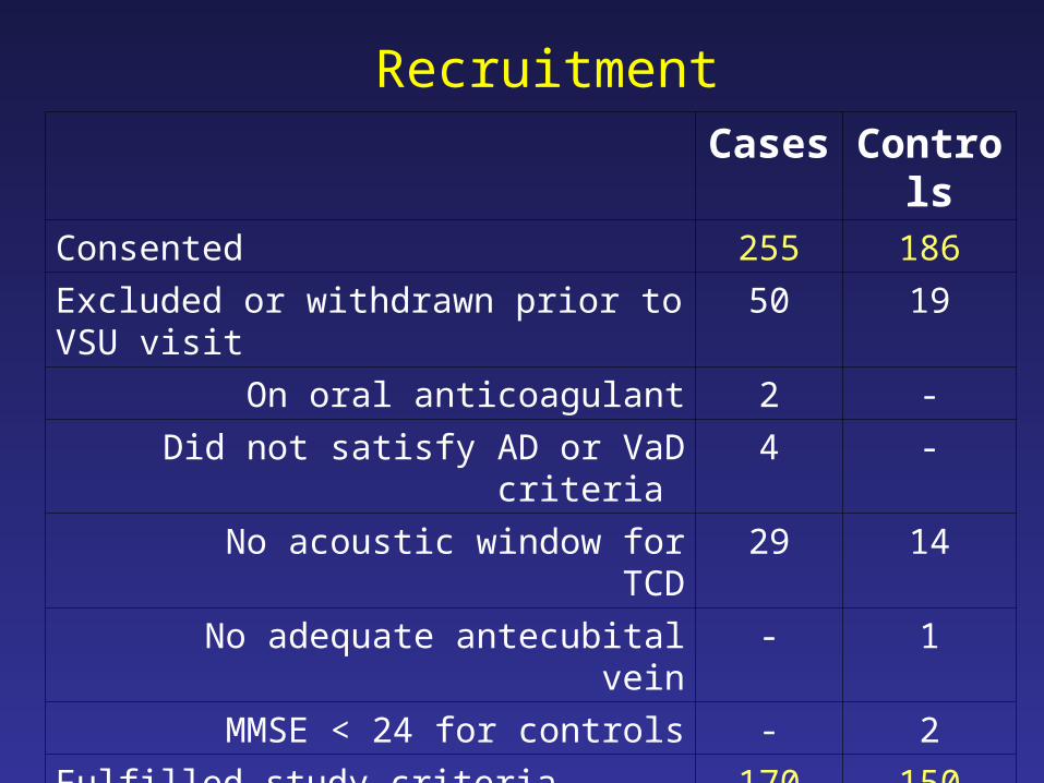
Cases Controls
Consented 255 186
Excluded or withdrawn prior to VSU visit 50 19
On oral anticoagulant 2 -
Did not satisfy AD or VaD criteria 4 -
No acoustic window for TCD 29 14
No adequate antecubital vein - 1
MMSE < 24 for controls - 2
Fulfilled study criteria 170 150
Controls for both AD and VaD patients - 20
Case-control pairs in matched analysis 170 170
Recruitment

ADn = 85
Controlsn = 85
VaDn = 85
Controlsn = 85
Male (%) 43 (51%) 45 (53%)
Age: mean (sd) 75.2 (7.6) 75.4 (7.2) 77.5 (6.3) 77.6 (6.2)
Dementia‘Probable’‘Possible’
73 (86%) 12 (14%)
60 (71%)25 (29%)
MMSE: mean (sd) 21.8 (4.5) 28.6 (1.4) 21.4 (5.1) 28.5 (1.4)

AD Controls VaD Controls
80 pairs 83 pairs
SCE +ve 32 (40%) 12 (15%) 31 (37%) 12 (14%)
OR (95% CI)
Adjusted*
2.7 (1.2 – 6.2)
P = 0.02
5.4 (1.2 -23.2)
P = 0.02
* Adjusted for cardiovascular risk factors
Spontaneous cerebral emboli in dementiaPurandare et al, BMJ, 2006
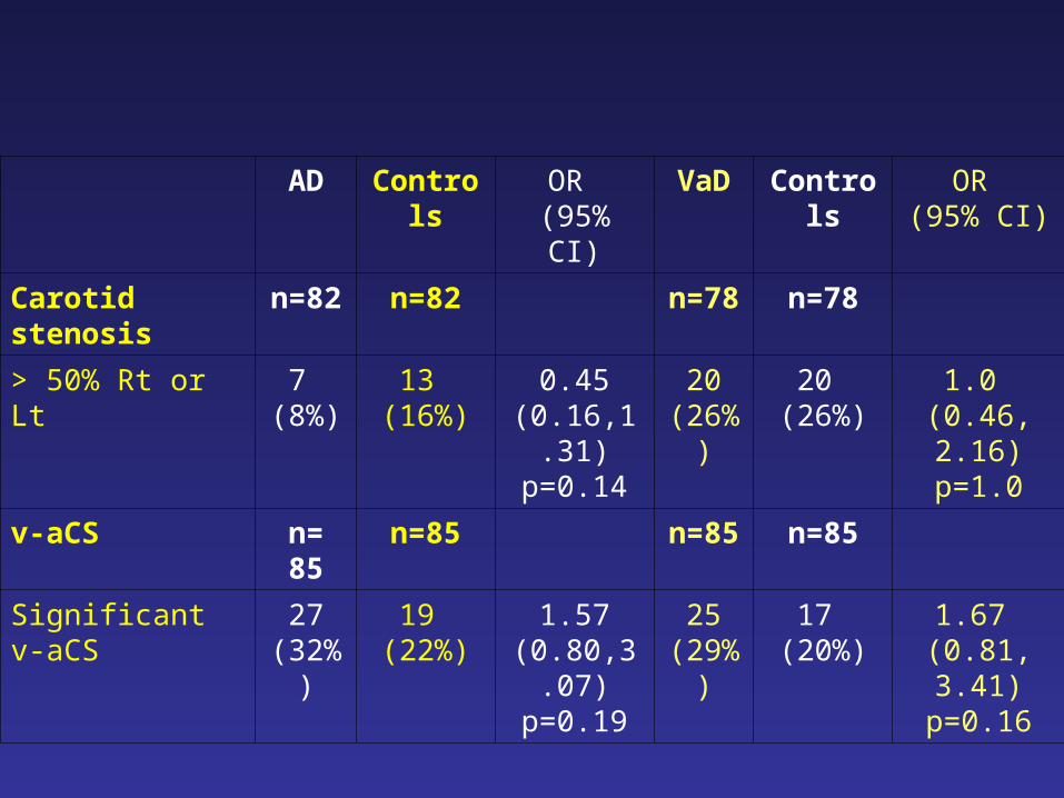
AD Controls OR (95% CI)
VaD Controls OR (95% CI)
Carotid stenosis n=82 n=82 n=78 n=78
> 50% Rt or Lt 7 (8%)
13 (16%)
0.45 (0.16,1.31)
p=0.14
20 (26%)
20 (26%)
1.0 (0.46, 2.16)
p=1.0
v-aCS n= 85 n=85 n=85 n=85
Significant v-aCS 27 (32%)
19 (22%)
1.57 (0.80,3.07)
p=0.19
25 (29%)
17 (20%)
1.67 (0.81, 3.41)
p=0.16
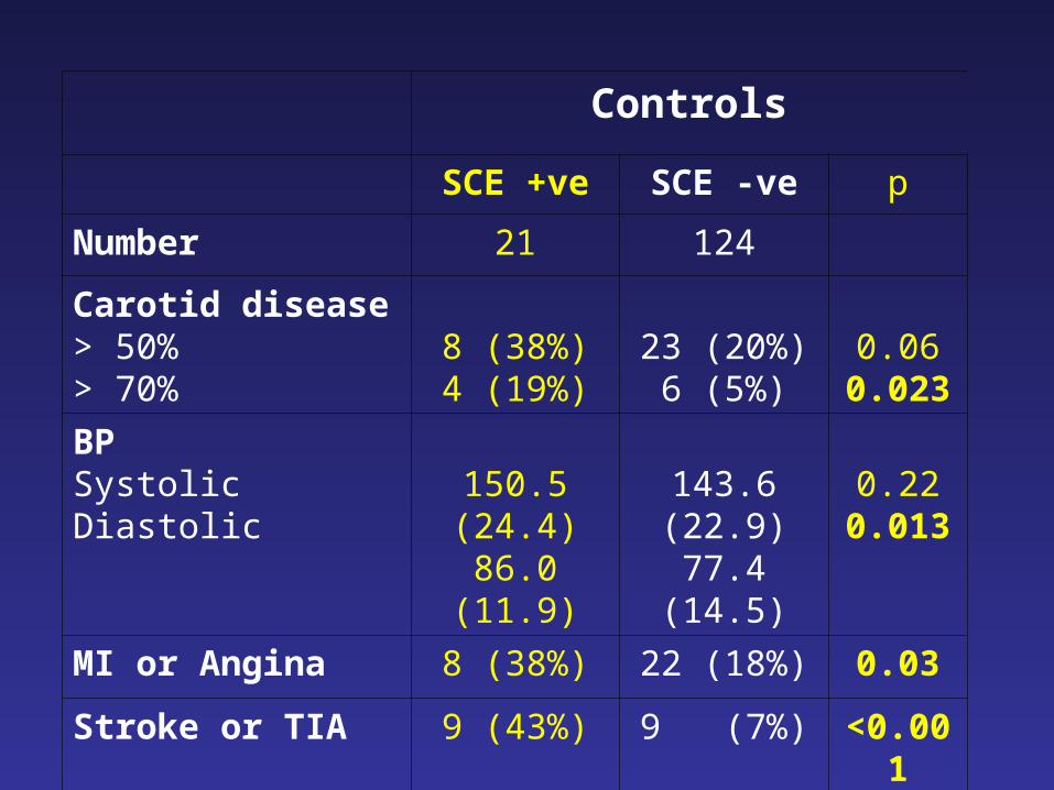
Controls
SCE +ve SCE -ve p
Number 21 124
Carotid disease> 50%> 70%
8 (38%)4 (19%)
23 (20%)6 (5%)
0.060.023
BPSystolicDiastolic
150.5 (24.4)86.0 (11.9)
143.6 (22.9)77.4 (14.5)
0.220.013
MI or Angina 8 (38%) 22 (18%) 0.03
Stroke or TIA 9 (43%) 9 (7%) <0.001
Anti-platelet Rx 12 (57%) 32 (26%) 0.004

• SCE are more frequent in dementia but are they associated with any of the behavioural and psychological symptoms of dementia?
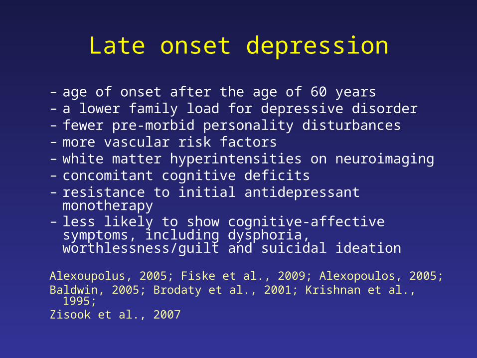
Late onset depression
– age of onset after the age of 60 years – a lower family load for depressive disorder– fewer pre-morbid personality disturbances– more vascular risk factors– white matter hyperintensities on neuroimaging– concomitant cognitive deficits– resistance to initial antidepressant monotherapy– less likely to show cognitive-affective symptoms,
including dysphoria, worthlessness/guilt and suicidal ideation
Alexoupolus, 2005; Fiske et al., 2009; Alexopoulos, 2005; Baldwin, 2005; Brodaty et al., 2001; Krishnan et al., 1995; Zisook et al., 2007
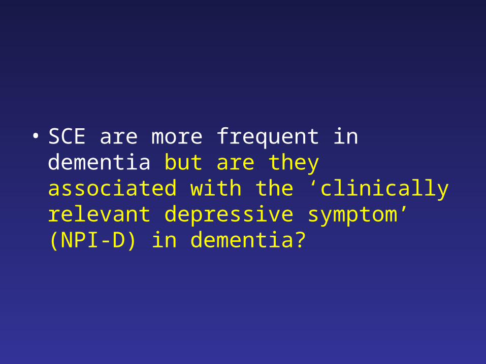
• SCE are more frequent in dementia but are they associated with the ‘clinically relevant depressive symptom’ (NPI-D) in dementia?

NPI – depression ≥ 4SCE
absent present
All dementia 5 / 85 (6%) 10 / 57 (18%)*
Alzheimer Disease 4 / 43 (9%) 5 / 29 (17%)
Vascular dementia 1 / 42 (2%) 5 / 28 (18%)*
* p < 0.05
Emboli are associated with depressive symptoms in dementia
Purandare et al, 2006, BJP
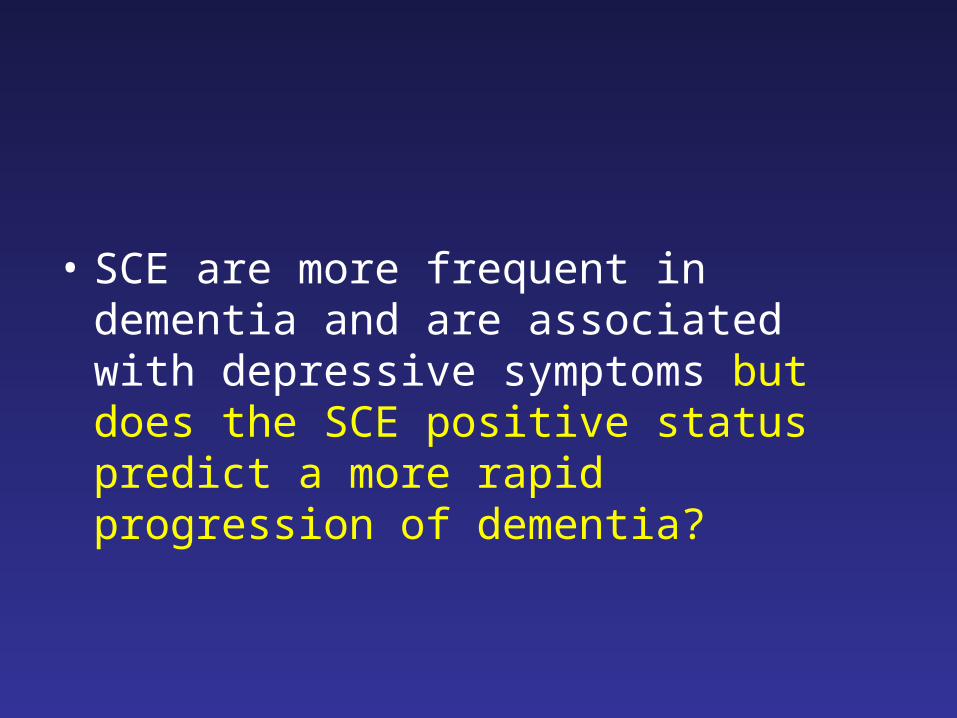
• SCE are more frequent in dementia and are associated with depressive symptoms but does the SCE positive status predict a more rapid progression of dementia?
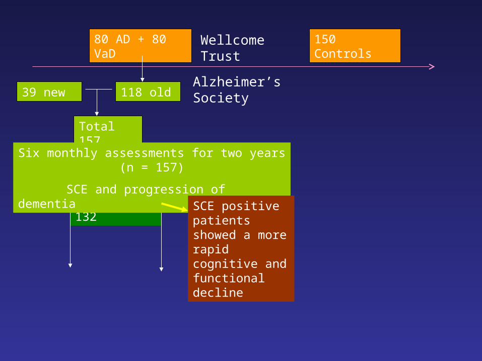
80 AD + 80 VaD 150 Controls
39 new 118 old
6 mth = 132
Total 157
Six monthly assessments for two years (n = 157)
SCE and progression of dementia
Wellcome Trust
Alzheimer’s Society
SCE positive patients showed a more rapid cognitive and functional decline

SCE absent SCE present
ADAS-cog (n=124)
- initial 23.2 [21.2 – 25.2] 22.9 [20.1 – 25.6]
- 6 months 26.5 [23.9 – 29.0] 30.0 [26.5 – 33.4]
- difference 3.3 7.1 (p=0.006)
MMSE (n=128)
- initial 21.4 [20.0 – 22.8] 20.4 [18.4 – 22.3]
- 6 months 20.2 [18.6 – 21.8] 17.6 [15.4 – 19.9]
- difference 1.2 2.8 (p=0.032)
IDDD (n=120)
- initial 85.2 [85.2 – 85.2] 85.2 [85.2 – 85.2]
- 6 months 96.0 [89.6 – 102.3] 109.6 [101.0 – 118.3]
- difference 10.8 24.4 (p=0.014)
* corrected for age, gender, type of dementia, cardiovascular risk factors, and other baseline characteristics associated with the change score over time (p<0.15).
Emboli status predicts progression of dementia over 6 monthsPurandare et al, Biol Psychiatry, 2007
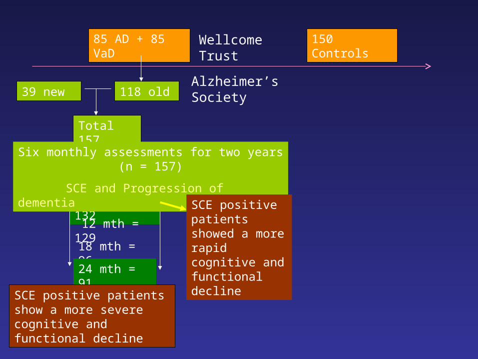
85 AD + 85 VaD 150 Controls
39 new 118 old
6 mth = 132
12 mth = 129
18 mth = 96
24 mth = 91
Total 157
SCE positive patients show a more severe cognitive and functional decline
Six monthly assessments for two years (n = 157)
SCE and Progression of dementia
Wellcome Trust
Alzheimer’s Society
SCE positive patients showed a more rapid cognitive and functional decline

Cerebral emboli and progression of dementia over two years (AJP, 2012)
• 144 patients with dementia (84 AD, 60 VaD)
• SCE were detected (SCE+ve) in 63 (44%) dementia patients, 36 (43%) in AD and 27 (45%) in VaD patients.
• ADAS-Cog score increased by a mean of 13.8 (95% CI: 9.1, 18.5) in SCE+ve and by 6.7 (2.8, 10.5) in SCE-ve patients (p=0.007).
• IDDD scores increased by 57.8 (39.4, 76.2) in SCE +ve patients compared with 20.2 (8.7, 31.8) for the SCE –ve patients (p=0.068).
• NPI scores increased in SCE+ve patients by 12.7 (6.7, 18.6) compared with -3.8 (-7.3, -0.3) in SCE-ve patients (p<0.001).
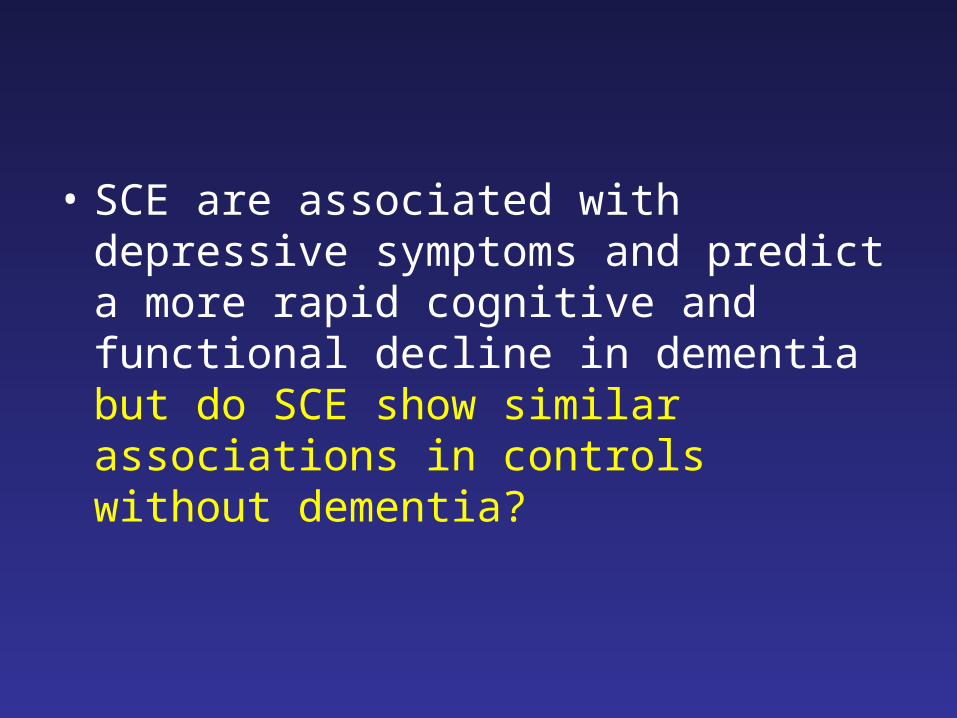
• SCE are associated with depressive symptoms and predict a more rapid cognitive and functional decline in dementia but do SCE show similar associations in controls without dementia?

85 AD + 85 VaD 150 Controls
39 new 118 old
6 mth = 132
12 mth = 129
18 mth = 96
24 mth = 91
Total 157
SCE positive patients show a more severe cognitive and functional decline
96 controls followed
After 2 years from baseline:
- Cognitive function
- DepressionSix monthly assessments for two years (n = 157)
SCE and Progression of dementia
Wellcome Trust
Alzheimer’s Society
SCE positive patients showed a more rapid cognitive and functional decline
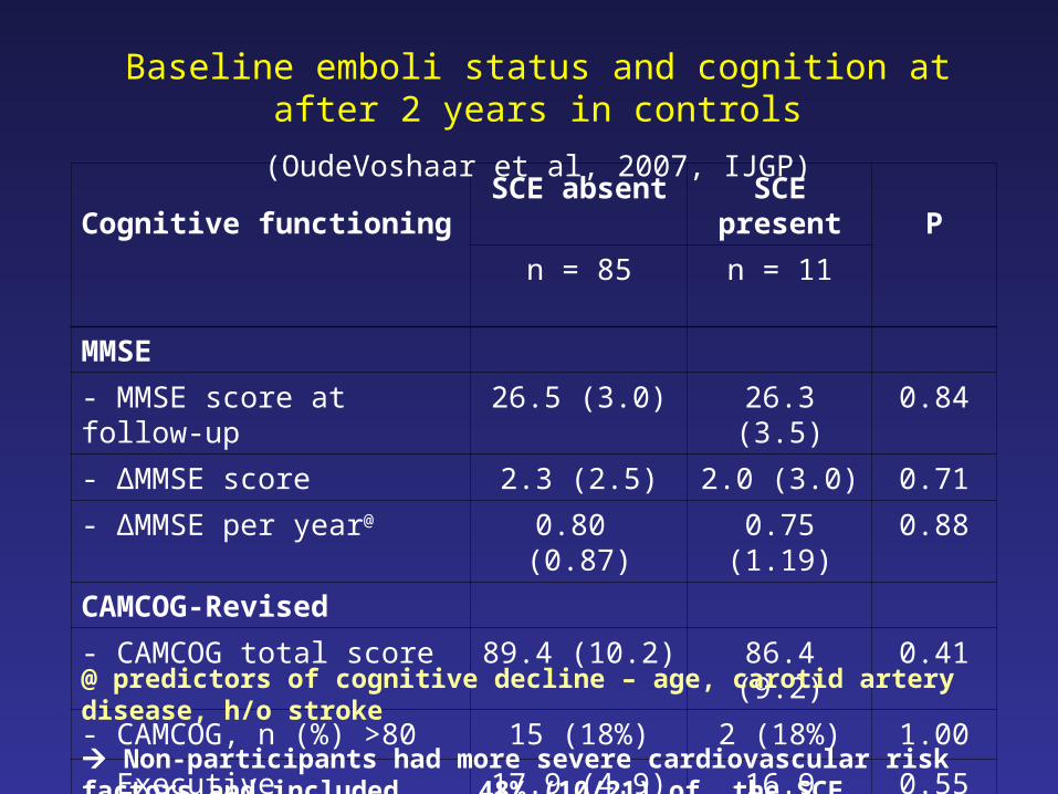
Cognitive functioningSCE absent SCE present
Pn = 85 n = 11
MMSE
- MMSE score at follow-up 26.5 (3.0) 26.3 (3.5) 0.84
- ∆MMSE score 2.3 (2.5) 2.0 (3.0) 0.71
- ∆MMSE per year@ 0.80 (0.87) 0.75 (1.19) 0.88
CAMCOG-Revised
- CAMCOG total score 89.4 (10.2) 86.4 (9.2) 0.41
- CAMCOG, n (%) >80 15 (18%) 2 (18%) 1.00
- Executive functioning 17.9 (4.9) 16.9 (5.2) 0.55
Baseline emboli status and cognition at after 2 years in controls
(OudeVoshaar et al, 2007, IJGP)
@ predictors of cognitive decline – age, carotid artery disease, h/o stroke
Non-participants had more severe cardiovascular risk factors and included 48% (10/21) of the SCE positive controls

Depression
SCE negative
SCE positive
P(n=85) (n=11)
GDS (SD) 2.3 (2.9) 4.5 (4.0) 0.031
MADRS (SD) 4.4 (7.9) 9.7 (11.2) 0.017
DSM-IV, n (%) 10 (12%) 3 (27%) 0.17
Baseline emboli status predicts depressive symptoms at 2 year follow-up in older controls
(OudeVoshaar et al, AJGP, 2007)
* SCE were independent predictors of MADRS after correction for age, gender, baseline cognitive status, and cardiovascular risk factors

85 AD + 85 VaD 150 Controls
39 new 118 old
6 mth = 132
12 mth = 129
18 mth = 96
24 mth = 91
Total 157
SCE positive patients show a more rapid progression over two years
96 controls followed
After 2 years from baseline:
- Cognitive function
- Depression
Aetiology & mechanism?
Six monthly assessments for two years (n = 157)
SCE and Progression of dementia
Wellcome Trust
Alzheimer’s Society
SCE positive patients showed a more rapid cognitive and functional decline
SCE positive controls showed more depressive symptoms, but not cognitive decline
• 48% drop out of SCE positive controls?
• Depression > Dementia?
• Depression Dementia?

Asymptomatic spontaneous cerebral emboli are frequent in patients with Alzheimer’s disease and vascular dementia and are associated with clinically relevant depressive symptoms and a more rapid progression of clinical dementia over two years compared to those who do not have emboli.

Role of vascular diseases in progression of dementia
• Evidence
• Current clinical practice
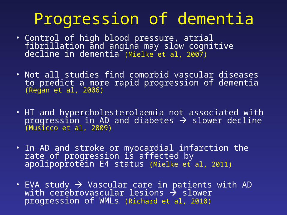
Progression of dementia• Control of high blood pressure, atrial fibrillation and
angina may slow cognitive decline in dementia (Mielke et al, 2007)
• Not all studies find comorbid vascular diseases to predict a more rapid progression of dementia (Regan et al, 2006)
• HT and hypercholesterolaemia not associated with progression in AD and diabetes slower decline (Musicco et al, 2009)
• In AD and stroke or myocardial infarction the rate of progression is affected by apolipoprotein E4 status (Mielke et al, 2011)
• EVA study Vascular care in patients with AD with cerebrovascular lesions slower progression of WMLs (Richard et al, 2010)
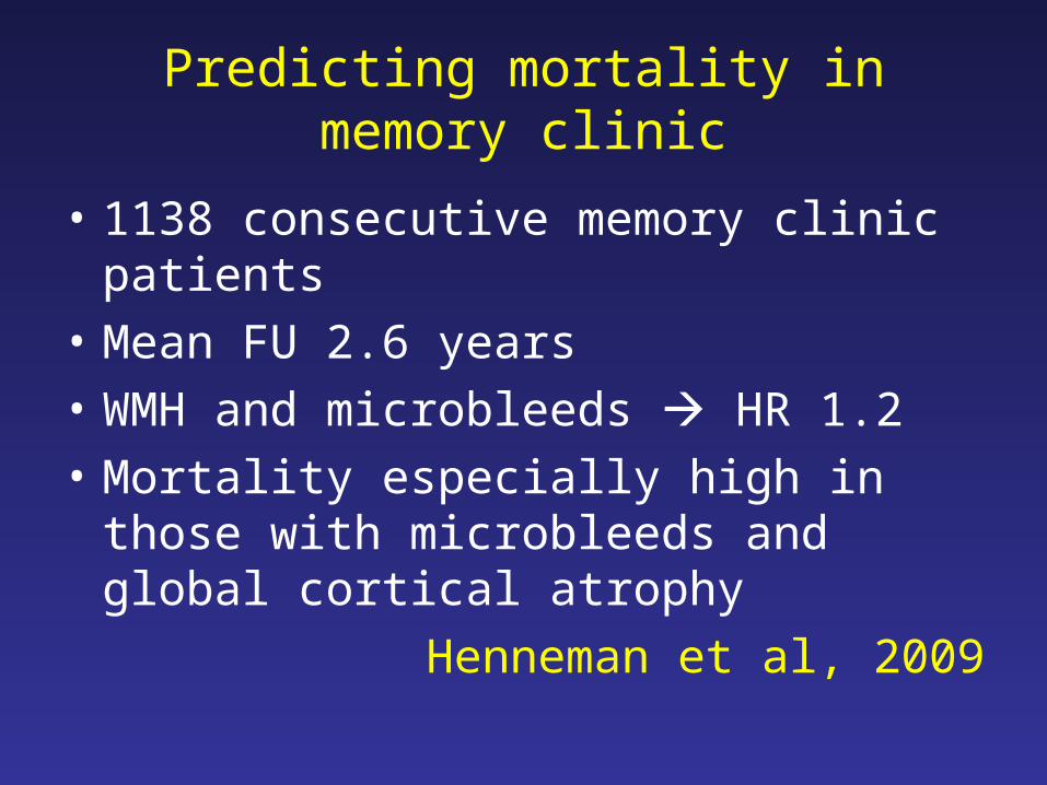
Predicting mortality in memory clinic
• 1138 consecutive memory clinic patients
• Mean FU 2.6 years
• WMH and microbleeds HR 1.2
• Mortality especially high in those with microbleeds and global cortical atrophy
Henneman et al, 2009

Participating Primary Care Trusts
Ashton, Leigh & WiganAshton, Leigh & Wigan
BoltonBolton
BuryBury
ManchesterManchester
OldhamOldham
StockportStockport

Number of medications mean (SD) [range 0-25]
N = 994
6.8 (3.9)
HT (%) 440 (44)
Ischemic Heart Disease (%) 197 (20)
Stroke or TIA (%) 169 (17)
Diabetes (%) 137 (14)
Atrial Fibrillation (%) 99 (10)
Heart Failure (%) 46 (5)
Arthritis (%) 313 (32)
Cancer, current or past (%) 118 (12)
Respiratory disease (%) 129 (13)
Kidney disease (%) 162 (16)
Comorbidity in dementia (BJGP 2012)
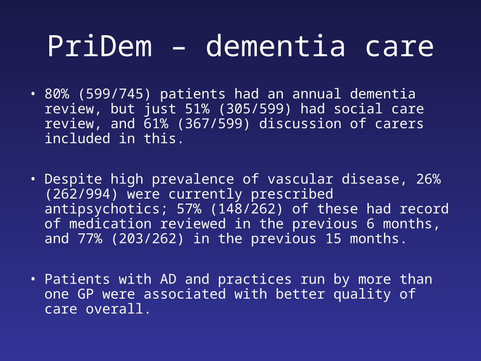
PriDem – dementia care
• 80% (599/745) patients had an annual dementia review, but just 51% (305/599) had social care review, and 61% (367/599) discussion of carers included in this.
• Despite high prevalence of vascular disease, 26% (262/994) were currently prescribed antipsychotics; 57% (148/262) of these had record of medication reviewed in the previous 6 months, and 77% (203/262) in the previous 15 months.
• Patients with AD and practices run by more than one GP were associated with better quality of care overall.

PriDem - vascular care
• Care was (significantly) poorer for PWD compared to those without dementia on 22/30(73%) indicators.
• Peripheral pulses check, neuropathy testing, retinal screening, and BMI monitoring for diabetes; cholesterol records for Stroke and CHD patients; and smoking cessation advice received the lowest provision of care.
• After multivariate adjustment, better quality of vascular care was found for males, those living in the community rather than care homes, and those with more comorbid physical conditions and medications.

Diabetes: QOF indicatorsDementia
No Dementia
DM 2 BMI 61.3 92.2
DM 11 BP 83.9 97.0
DM 12 Last BP is 145/85 or less 84.7 78.6
DM 5 HbA1c 73.7 94.2
DM 20 Last HbA1c is 7.5 or less 54.0 64.6
DM 7 Last HbA1c is 10 or less 71.5 88.4
DM 21 Retinal screening 38.7 84.9
DM 9 Examined for peripheral pulse 38.7 83.1
DM 10 Neuropathy testing 37.2 82.0
DM 22 eGFR or serum creatinine testing 81.8 94.6
DM 16 Total cholesterol 82.5 94.0
DM 17 Last total cholesterol ≤ 5mmol/l 70.1 77.5

Conclusion
• Vascular risk factors are involved in causation and progression of vascular dementia and Alzheimer’s disease.
• Cerebral emboli may be a target to slow progression of dementia.
• Dementia remains under-diagnosed and management of vascular diseases is not equitable.