role of the serotoninergic system in inhibition of jaw-opening reflex induced by stimulating the...
TRANSCRIPT

6. A. Dahlstrom and K. Fuxe, "Evidence of the existence of monoamine neurons in the cen- tral nervous system. 3. The distribution of monoamine terminals in the central nervous system," Acta Physiol. Scand., 64, No. 247, 1-85 (1965).
7. R. Freedman, B. Hoffer, D. Woodward, et al., "Interaction of norepinephrine with care- bellar activity evoked by mossy and climbing fibers," Exp. Neurol., 55, No. i, 269-288 (1977).
8. K. Fuxe, "Evidence for the existence of monoamine neurons in the CNS. 3. The mono- amine nerve terminals," Z. Zellforsch., 65, No. 3, 573-596 (1965).
9. B. Hoffer, G. Siggins, A. Oliver, et el., "Activation of the pathway from locus coeru- leus to rat cerebellar neurons. Pharmacological evidence of noradrenergic central inhibition," J. Pharmacol. Exp. Ther., 184, No. 3, 553-569 (1973).
i0. R. Kobayashi, M. Palkovits, I. Kopin, et al., Biochemical mapping of noradrenergic nerves arising from the rat locus coeruleus," Brain Res., 77, No. 2, 269-279 (1974).
ii. H. G. Moises and D. J. Woodward, "Potentiation of GABA inhibitory action in cerebellum by locus coeruleus stimulation," Brain Res., 182, No. 2, 327-344 (1980).
12. L. Olson and K. Fuxe, "On the projections from the locus coeruleus noradrenaline neurons," Brain Res., 28, No. i, 165-172 (1971).
13. D. H. Versteed, J. Van der Gugten, W. Jong, et al., "Regional distribution of noradrena- line and dopamine in rat brain," Brain Res., i13, No. 3, 563-574 (1976).
ROLE OF THE SEROTONINERGIC SYSTEM IN INHIBITION OF JAW-OPENING
REFLEX INDUCED BY STIMULATING THE MIDBRAIN CENTRAL GRAY MATTER
E. V. Gura, V. A. Yakhnitsa, and Yu. P. Limanskii UDC 612.826.5:616-003.725
The effects were studied in waking cats of brief stimulation (20 stimuli at a rate of 400 Hz) of the central gray matter (CGM) and dorsal raphe nucleus (DRN) on high-threshold jaw-opening reflex (HJOR) evoked by tooth pulp stimu- lation during blockade of serotonin synthesis produced by application of 300 mg/kg parachlorophenylalanine (PCPA) i.p. Inhibitory effects of CGM and DRN stimulation had already declined in comparison with post-stimulation (but pre- PCPA) level within 24 h after PCPA application; 96 h afterwards, inhibition of HJOR induced by CGM and DRN stimulation had become only minimal: ampli- tude of the reflex had declined to 30-35% and duration of inhibitory effects ws 200-250 msec° It is therefore deduced that serotonin contributes to the HJOR depression induced by CGM and DRN stimulation and the possible involve- ment of other neuromodulators in this effect is discussed.
INTRODUCTION
Neurons of the midbrain central gray matter (CGM) and dorsal raphe (DR) are known to form at least five afferent serotoninergic and nonserotoninergic pathways with their endings distributed throughout different structures of the forebrain, diencephalon, midbrain, and hindbrain. Descending influences produced by electrical stimulation of the CGM and DR pro- duce a marked inhibitory effect on jaw-opening reflex induced by activation of high-thres- hold trigeminal nerve afferents (HJOR) but modulate evolution of the reflex evoked by low- threshold activation (LJOR) to a lesser extent [i].
Duration of HJOR and LJOR depend largely on parameters of the stimuli used. Brief CGM and DR stimulation consisting of trains of 10-12 stimuli at the rate of 200-400 Hz in- hibits jaw-opening reflex for 450-1000 msec, whereas prolonged stimulation of these struc- tures (stimuli at the rate of 50 Hz over 30 sec) inhibits HJOR and LJOR for 60 min [i, 16].
A, A. Bogomolets Institute of Physiology, Academy of Sciences of the Ukrainian SSR, Kiev. Translated from Neirofiziologiya, Vol. 21, No. i, pp. 45-52, January-February, 1989. Original article submitted January 6, 1988.
0090-2977/89/2101-0037512.50 © 1989 Plenum Publishing Corporation 37

In view of the serotoninergic and nonserotoninergic efferent systems proceeding from the CGM and DR as well as the difference between the inhibitory effects produced by stimulating the latter structures on HJOR and LJOR, depending on duration of stimulation, we postulated the existence of two inhibitory mechanisms operating on different time scales in the CGM and DR. In support of this hypothesis we adduced the fact that neurons belonging to other neuroregulatory systems projecting to brainstem and spinal cord nuclei are also concentrated in the CGM and DR (nuclei of the serotoninergic system). Neurons of other neuroregulatory systems have been shown to use substance P, thyroliberin, leu- and met-enkephalins, somato- statin, and some aminoacids as neurotransmitters.
These compounds exist side by side (and together with serotonin) in various combinations in a number of CGM and DR neurons.
The question naturally arises, when reviewing our findings on "multitransmitter" neurons of the CGM and DR, of the part played by the serotoninergic system in inhibition of jaw-open- ing reflexes induced by CGM and DR stimulation. Parachlorophenylaianine (PCPA), a selective serotonin depletor which acts as inhibitor of tryptophan hydroxylase (an enzyme converting l-tryptophan to 5-hydroxytryptophan, the precursor of serotonin) was used to block the sero- toninergic system.
METHODS
Experiments were conducted on eight male cats weighing between 2.5 and 3.5 kg. Prepara- tory surgery was performed on the animals under Nembutal-induced anesthesia (60 mg/kg, i.p.) and sterile conditions. A bipolar stimulating electrode made of stainless steel wire was placed in the tooth pulp of the upper canine. Two openings were made into the tooth so as to preserve a slender wall of bone between tooth pump and electrode. Electrodes were fas- tened to the tooth by an acrylic crown which did not impede mastication. The tooth pulp was stimulated at an intensity of 1 1/2-2 thresholds.
A bipolar electrode consisting of a ring of nichrome wire was placed on the digastric muscle to record electrical activity (EMG). Amplitude of this EMG response served as a mea- sure for the jaw-opening reflex.
A bipolar nichrome electrode was inserted into the midbrain CGM or dorsal PuN (DRN) ac- cording to the coordinates of a stereotaxic atlas [5]. Leads from all electrodes were passed under the skin and linked up to a connector fastened to the skull with acrylic, where two tubes for steadying the animal's head were also fastened during trials. Experimentation began a week later, when the animals were recovering from surgery, Animals were placed in a sling permitting freedom of limb movement during the course of experiments; the heads were (painlessly) held still in a device of our own construction and a disk stuck round with porous resin was introduced below the lower jaw.
Control values of HJOR induced by tooth pulp stimulation were first determined, and the extent of depression exerted on these values by CGM and DR stimulation. Trains of 20 stimuli were used following on the rate of 400 Hz within trains to stimulate these brain structures. Stimulating current ranged between 120 and 175 ~A. Straight afterwards the animals were each injected with 300 mg/kg PCPA, diluted to uniform suspension state in sterile physiological saline. Readings were taken at day two, three, and so on after PCPA injection. Animals were put to sleep and the stimulation site coagulated once the experi- mental series had ended.
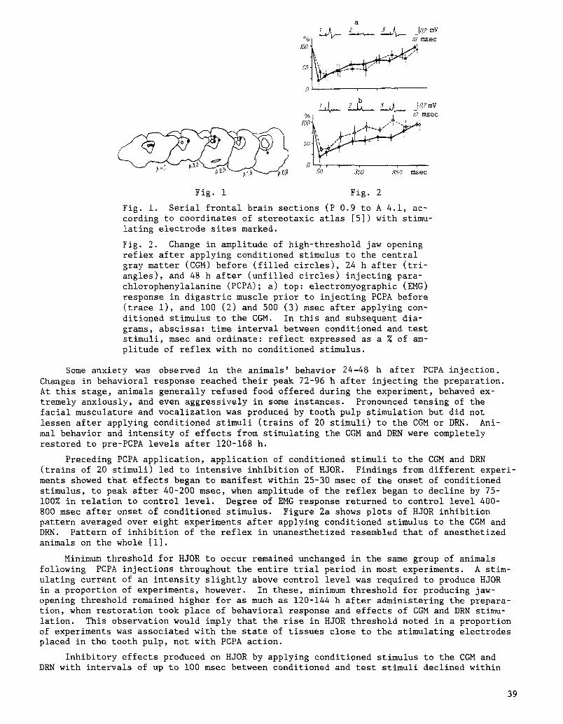
Location of the stimulating electrode tip was verified on serial brain slices i00 ~m thick prepared on a freezing microtome. A coagulation marker was found in the CGM in one animal (at level A 1.6-A 4.1) and in the DRN of another (level P 0.9 - see Fig. I).
Amplitude of EMG response in the digastric muscle was determined from averaged response (over 5-10 events). Averaging was performed using a nerve fiber spike analyzer.
RESULTS
Animals showed no distress at having been fastened into a stand for investigations. They actively partook of food when offered. Electrical tooth pulp stimulation led to acute contraction of the entire facial musculature and vocalization in a proportion of experi- ments -responses either suppressed or considerably reduced by applying conditioning stimuli to the CGM or DRN.
38
a
% | - - /0 msec
5_4_ %| 1 /8 msec
foot • .-?-.. i
170 .750 850 msec
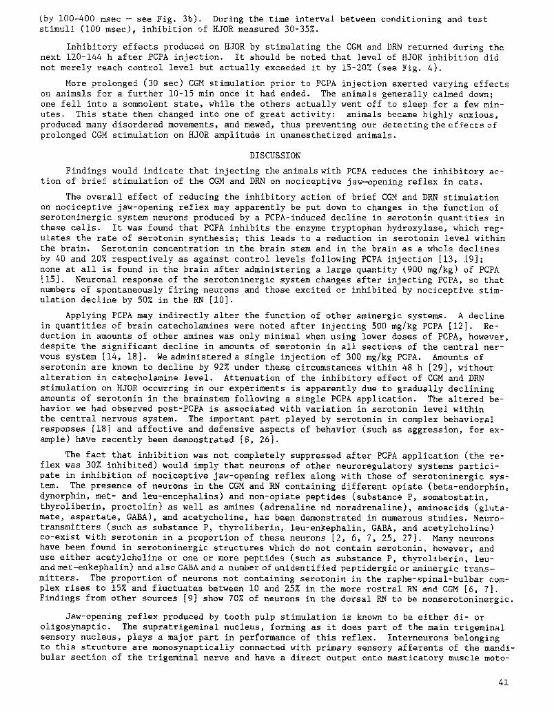
Fig. i Fig. 2
Fig. i. Serial frontal brain sections (P 0.9 to A 4.1, ac- cording to coordinates of stereotaxic atlas [5]) with stimu- lating electrode sites marked.
Fig. 2. Change in amplitude of high-threshold jaw opening reflex after applying conditioned stimulus to the central gray matter (CGM) before (filled circles), 24 h after (tri- angles), and 48 h after (unfilled circles) injecting para- chlorophenylalanine (PCPA); a) top: electromyographic (EMG) response in digastric muscle prior to injecting PCPA before (trace i), and i00 (2) and 500 (3) msec after applying con- ditioned stimulus to the CGM. In this and subsequent dia- grams, abscissa: time interval between conditioned and test stimuli, msec and ordinate: reflect expressed as a % of am- plitude of reflex with no conditioned stimulus.
Some anxiety was observed in the animals' behavior 24-48 h after PCPA injection. Changes in behavioral response reached their peak 72-96 h after injecting the preparation. At this stage, animals generally refused food offered during the experiment, behaved ex- tremely anxiously, and even aggressively in some instances. Pronounced tensing of the facial musculature and vocalization was produced by tooth pulp stimulation but did not lessen after applying conditioned stimuli (trains of 20 stimuli) to the CGM or DRN. Ani- mal behavior and intensity of effects from stimulating the CGM and DRN were completely restored to pre-PCPA levels after 120-168 h.
Preceding PCPA application, application of conditioned stimuli to the CGM and DRN (trains of 20 stimuli) led to intensive inhibition of HJOR. Findings from different experi- ments showed that effects began to manifest within 25-30 msec of the onset of conditioned stimulus, to peak after 40-200 msec, when amplitude of the reflex began to decline by 75- 100% in relation to control level. Degree of EMG response returned to control level 400- 800 msec after onset of conditioned stimulus. Figure 2a shows plots of HJOR inhibition pattern averaged over eight experiments after applying conditioned stimulus to the CGM and DRN. Pattern of inhibition of the reflex in unanesthetized resembled that of anesthetized animals on the whole [i].
Minimum threshold for HJOR to occur remained unchanged in the same group of animals following PCPA injections throughout the entire trial period in most experiments. A stim- ulating current of an intensity slightly above control level was required to produce HJOR in a proportion of experiments, however. In these, minimum threshold for producing jaw- opening threshold remained higher for as much as 120-144 h after administering the prepara- tion, when restoration took place of behavioral response and effects of CGM and DRN stimu- lation. This observation would imply that the rise in HJOR threshold noted in a proportion of experiments was associated with the state of tissues close to the stimulating electrodes placed in the tooth pulp, not with PCPA action.
Inhibitory effects produced on HJOR by applying conditioned stimulus to the CGM and DRN with intervals of up to I00 msec between conditioned and test stimuli declined within
39

a
% I0- msec
(9 i , , , . 1 ,
b
,0%1 + , + mseo
50 350 650 msec
a
,oo i
b
60 J60 gSO msec
Fig. 3 Fig. 4
Fig. 3. Changed amplitude of high-threshold jaw-opening re- flex after a conditioned stimulus applied to the central gray matter (CGM) before (filled circles), 72 h after (un- filled triangles), and 96 h after (filled squares) parachlo- rophenylaianine (PCPA) injection; a), top: electromyographic (EMG) response in digastric muscle 72 h after PCPA injection, before (trace i), I00 msec after (2), and 500 msec after (3) conditioned stimulus applied to the CGM; b) top: EMG response in digastric muscle 96 h after PCPA injection before (trace I), i00 msec after (2) and 500 msec after (3) conditioned stimulus to the CGM.
Fig. 4. Change in amplitude of high-threshold jaw-opening reflex after applying conditioned stimulus to the central gray matter (CGM) before (filled circles), 120 h after (un- filled squares), and 144 h after (crosses) parachlorophenyl- alanine (PCPA) application; a) top: electromyographic (EMG) response in digastric muscle 120 h after injecting PCPA be- fore (trace I), i00 msec after (2), and 500 msec after (3) conditioned stimulus applied to the CGM; b) top: EMG response in digastric muscle 144 h after PCPA injection before (trace I), i00 msec after (2), and 500 msec after (3) conditioned stimulus applied to the CGM.
24 h after PCPOA injection. Amplitude of HJOR increased in relation to its pre-PCPA level by -20% during this time interval. Overall duration of depressed HJOR arising from apply- ing conditioned stimulus to the CGM was the same as prior to PCPA administration (see Fig. 2a).
At 48 h after PCPA administration, the decline in HJOR inhibition persisted at the level observed at 24 h after injection during the first 100 msec after conditioned stimulus applied to the CGM and DRN but the inhibitory effect of this CGM stimulus fell off consider- ably during the time interval between i00 and 500-700 msec (see Fig. 2b). Overall duration of HJOR depression produced by applying conditioned stimulus to the CGM or DRN scarcely changed in relation to control levels (Fig. 2b).
Action of PCPA continued to rise thereafter, manifesting in increased amplitude of HJOR after conditioned stimulus applied to the CGM or DRN in comparison with control levels. De- pression of HJOR 72 h after PCPA injection averaged less than 50% during the time interval between conditioned stimuli applied to the CGM and DRN on the one hand and test stimulation of the tooth pulp on the other, at 50-1000 msec, whereas amplitude of HJOR declined by over 75% in control experiments. Overall duration of HJOR inhibition produced by conditioned stimulus applied to the CGM or DRN did not decrease (see Fig. 3a).
The biggest decline in HJOR level produced by conditioned stimulus applied to the CGM or DRN was seen 96 h after PCPA injection. Overall duration of the inhibitory action pro- duced on HJOR by CGM and DRN stimulation declined especially noticeably during this period
40
(by 100-400 msec - see Fig. 3b). During the time interval between conditioning and test stimuli (i00 msec), inhibition of HJOR measured 30-35%.
Inhibitory effects produced on HJOR by stimulating the CGM and DRN returned during the next 120-144 h after PCPA injection. It should be noted that level of HJOR inhibition did not merely reach control level but actually exceeded it by 15-20% (see Fig. 4).
More prolonged (30 sec) CGM stimulation prior to PCPA injection exerted varying effects on animals for a further 10-15 min once it had ended. The animals generally calmed down; one fell into a somnolent state, while the others actually went off to sleep for a few min- utes. This state then changed into one of great activity: animals became highly anxious, produced many disordered movements, and mewed, thus preventing our detecting the effects of prolonged CGM stimulation on HJOR amplitude in unanesthetized animals.
DISCUSSION
Findings would indicate that injecting the animals with PCPA reduces the inhibitory ac- tion of brief stimulation of the CGM and DRN on nociceptive jaw-opening reflex in cats.
The overall effect of reducing the inhibitory action of brief CGM and DRN stimulation on nociceptive jaw-opening reflex may apparently be put down to changes in the function of serotoninergic system neurons produced by a PCPA-induced decline in serotonin quantities in these cells. It was found that PCPA inhibits the enzyme tryptophan hydroxylase, which reg- ulates the rate of serotonin synthesis; this leads to a reduction in serotonin level within the brain. Serotonin concentration in the brain stem and in the brain as a whole declines by 40 and 20% respectively as against control levels following PCPA injection [13, 19]; none at all is found in the brain after administering a large quantity (900 mg/kg) of PCPA [15]. Neuronal response of the serotoninergic system changes after injecting PCPA, so that numbers of spontaneously firing neurons and those excited or inhibited by nociceptive stim- ulation decline by 50% in the RN [i0].
Applying PCPA may indirectly alter the function of other aminergic systems. A decline in quantities of brain catecholamines were noted after injecting 500 mg/kg PCPA [12]. Re- duction in amounts of other amines was only minimal when using lower doses of PCPA, however, despite the significant decline in amounts of serotonin in all sections of the central ner- vous system [14, 18]. We administered a single injection of 300 mg/kg PCPA. Amounts of serotonin are known to decline by 92% under these circumstances within 48 h [29], without alteration in catecholamine level. Attenuation of the inhibitory effect of CGM and DRN stimulation on HJOR occurring in our experiments is apparently due to gradually declining amounts of serotonin in the brainstem following a single PCPA application. The altered be- havior we had observed post-PCPA is associated with variation in serotonin level within the central nervous system. The important part played by serotonin in complex behavioral responses [18] and affective and defensive aspects of behavior (such as aggression, for ex- ample) have recently been demonstrated [8, 26].
The fact that inhibition was not completely suppressed after PCPA application (the re- flex was 30% inhibited) would imply that neurons of other neuroregulatory systems partici- pate in inhibition of nociceptive jaw-opening reflex along with those of serotoninergic sys- tem. The presence of neurons in the CGM and RN containing different opiate (beta-endorphin, dynorphin, met- and leu-encephalins) and non-opiate peptides (substance P, somatostatin, thyroliberin, proctolin) as well as amines (adrenaline nd noradrenaline), aminoacids (gluta- mate, aspartate, GABA), and acetycholine, has been demonstrated in numerous studies. Neuro- transmitters (such as substance P, thyroliberin, leu-enkephalin, GABA, and acetylcholine) co-exist with serotonin in a proportion of these neurons [2, 6, 7, 25, 27]. Many neurons have been found in serotoninergic structures which do not contain serotonin, however, and use either acetylcholine or one or more peptides (such as substance P, thyroliberin, leu- and met-enkephalin) and also GABA and a number of unidentified peptidergic or aminergic trans- mitters. The proportion of neurons not containing serotonin in the raphe-spinal-bulbar com- plex rises to 15% and fluctuates between i0 and 25% in the more rostral RN and CGM [6, 7]. Findings from other sources [9] show 70% of neurons in the dorsal RN to be nonserotoninergic.
Jaw-opening reflex produced by tooth pulp stimulation is known to be either di- or oligosynaptic. The supratrigeminai nucleus, forming as it does part of the main trigeminal sensory nucleus, plays a major part in performance of this reflex. Interneurons belonging to this structure are monosynaptical!y connected with primary sensory afferents of the mandi- bular section of the trigeminal nerve and have a direct output onto masticatory muscle moto-
41
neurons [3, 20]. Numerous serotoninergic terminals are spread among neurons of the main trigeminal sensory nucleus, those of the spinal trigeminal tract caudal nucleus, and cells of the trigeminal motor nucleus [24]. About 5% of these form clearly defined synaptic con- tacts in the rat and the remainder are represented by varicose endings containing small light-colored and large granular vesicles [21].
It has been established [ii, 22] that stimulation of the CGM and RN can produce pre- synaptic depolarization of high-threshold tooth pulp afferents and either inhibit or excite interneurons belonging to the trigeminal sensory nuclei. Serotonin application is also known to facilitate response in motoneurons of the facial nerve nucleus to afferent impul- ses. It might be supposed that impulses from the CGM and DRN reach neuronal elements in- volved in jaw-opening reflex both directly, via such serotoninergic system structures as the greater raphe nucleus, as well as indirectly, through the magno- and gigantocellular reti- cular nuclei, where neurons have close bilateral connections with the CGM and DRN [4].
Effects of the serotoninergic system are mediated by the aid of two or three types of serotoninergic receptors which have been found in very low numbers at the neuronal mem- brane of the trigeminal nerve sensory and motor nuclei as well as at primary afferent ter- minals [17]. Sensitivity of postsynaptic receptors has long been known to increase within a certain interval after functional or structural neuronal denervation. This could be the reason for the increased action on HJOR of applying conditioned stimuli to the CGM and DRN observed on the 5th-6th day after PCPA application during our study.
LITERATURE CITED
i. E. V. Gura, V. A. Yakhnitsa, and Yu. P. Limanskii, "Inhibition of jaw-opening reflex in the cat induced by stimulating the central gray matter and raphe nucleus," Neiro- fiziologiya, 16, No. 3, 374-384 (1984).
2. V. E. Klusha, Peptides as Regulators of Cerebral Function [in Russian], Zinatne, Riga (1984).
3. Yu. P. Limanskii, Structure and Function of the Trigeminal Nerve System [in Russian], Nauk. Dumka, Kiev (1976).
4. A. I. Basbaum and A. H. Fields, "Endogeneous pain control systems: brainstem spinal pathways and endorphin circuitry," Annu. Rev. Neurosci., Z, 309-338 (1984).
5. A. L. Berman, The Brain Stem of the Cat, Univ. Wisconsin Press, Madison (1968). 6. R. Bowker, K. Westlund, M. Sullivan, et al., Transmitters of the raphe-spinal complex:
immunocytochemical studies," Peptides, ~, No. 3, 291-298 (1982). 7. R. Bowker, K. Westlund, M. Sullivan, and J. Coulter, "Organization of descending sero-
tonergic projections to the spinal cord," in: Descending Pathways to the Spinal Cord, H. Kuypers and G. Martin (eds.), Elsevier Biomedical Press, Amsterdam (1982), pp. 239- 265.
8. J. H. Daruna and E. W. Kent, "Comparison of regional serotonin levels and turnover in the brain of naturally high and low aggressive rats," Brain Res., I01, No. 3, 489- 503 (1976).
9. L. Descarries, A. Beaudet, K. C. Watkins, and C. Garcia, "The serotonin neurons in nucleus raphe dorsalis of adult rat," Anat. Rec., 193, No. 3, 520 (1979).
I0. A. H. Dickenson and G. Goldsmith, "Evidence for a role of 5-hydroxytryptamine in the responses of rat raphe magnus neurones to peripheral noxious stimuli," Neuropharmacol- ogy, 25, No. 8, 863-868 (1986).
Ii. J. O. Dostrovsky, B. J. Sessle, and J. W. Hu, "Presynaptic excitability changes pro- duced in brain stem endings of tooth pulp afferents by raphe and other central and peripheral influences," Brain Res., 218, No. i, 141-160 (1981).
12. P. Gauthier and T. A. Reader, "Adrenomedullary secretory response to midbrain stimula- tion in rat: effects of depletion of brain catecholamines or serotonin," Can. J. Physiol. Pharmacol., 60, No. 8, 1467-1474 (1982).
13. H. H. Keller, "Depletion of cerebral monoamines by p-chlorophenyialanine in the cat," Experientia, 28, No. i, 177-178 (1972).
14. K. B. Koe and A. Weissman, "p-Chlorophenylalanine: a specific depletor of brain serotonin," J. Pharmacol. Exp. Ther., 154, No. 3, 499-516 (1966).
15. D. Le Bars, J. P. Rivot, G. Guilbaud, et al., "The depressive effect of morphine on the C fiber response of dorsal horn neurones in the spinal rat pretreated or not by PCPA," Brain Res., 176, No. 2, 337-353 (1979).
42

16. Yu. P. Limansky, E. V. Gura, and V. A. Yakhnitsa, "Effects of the central gray matter stimulation on nociceptive and non-nociceptive jaw-opening reflexes in a cat," in: The Second World Congress of Neuroscience (IBRO), Budapest (1987), p. I197P.
17. R. C. Meibach, "Serotonergic receptors," in: Handbook of Chemical Neuroananomy, Vol. 3, Part 2, A. Bjorklund, et al (eds.), Elsevier, Amsterdam (1984), pp. 304-324.
18. S. O. Ogren, "Evidence for role of brain serotonergic neurotransmission in avoidance learning," Acta Physiol. Scand., 125, Suppl. 544, 1-71 (1985).
19. J. F. Pujol, A. Buquet, J. Froment, et al°, "The central metabolism of serotonin in the cat during insomnia. A neurophysical and biochemical study after administration of p-chlorophenylalanine or destruction of the raphe system," Brain Res., 29, No. i, 195-212 (1971).
20. J. Rokx, J. D. Van Willigen, and P. Juch, "Bilateral brainstem connections of the rat supratrigeminal region," Acta Anat., 127, No. i, 16-21 (1986).
21. N. Schaffar, A. Jean, and A. Calas, "Radioautographic study of serotonergic axon ter- minals in the rat trigeminal motor nucleus," Neurosci. Lett., 44, No. i, 31-36 (1984).
22. Y. Shah and J. O. Dostrovsky, "Postsynaptic inhibition of cat medullary dorsal horn neurons by stimulation of nucleus raphe magnus and other brainstem sites," Exp. Neurol., 77, No. 3, 419-435 (1982).
23. H. Steinbusch, "Serotonin-immunoreactive neurons and their projections in the CNS," in: Handbook of Chemical Neuroanatomy, Vol. 3, Part 2, A. Bjorklund, et al. (eds.), Elsevier, Amsterdam (1984), pp. 68-125.
24. H. W. M. Steinbusch, "Distribution of serotonin-immunoreactivity in the central ner- vous system of the rat cell bodies and terminals," Neuroscience, 6, No. 4, 557-618 (1981).
25. H. Steinbusch and R. Nieuwenhuys, "The raphe nuclei of the rate brainstem: a cyto- architectonic and immunohistochemical study," in: Chemical Neuroanatomy, P. Emson (ed.), Raven Press, New York (1983), pp. 131-205.
26. L. Valzelli, "Serotonergic inhibitory control of experimental aggression," Pharmacol. Res. Commun., 14, No. i, 1-13 (1982).
27. C. P. Vandermaelen, "Serotonin," in: Neurotransmitter Actions, M. Rogawski and J. Barker (eds.), Plenum Press, New York (1985), pp. 201-240.
28. C. P. Vandermaelen and G. K. Aghajanian, "Serotonin-induced depolarization of rat facial motoneurons in vivo: Comparison with amino acid transmitters," Brain Res., 239, No. i, 139-152 (1982).
29. M. Vogt, "The effect of lowering the 5-hydroxytryptamine content in the rat spinal cord on analgesia produced by morphine," J. Physiol., 236, No. 2, 483-498 (1974).
43