role of notch/rbpj signaling pathway in embryonic...
TRANSCRIPT

Universitat de Barcelona
Facultat de Farmàcia
Departament de Bioquímica i Biologia Molecular
Role of Notch/RBPjκ signaling pathway in embryonic
hematopoiesis
Alexandre Robert i Moreno
2007


Universitat de Barcelona
Facultat de Farmàcia
Departament de Bioquímica i Biologia Molecular
Doctorat en Biomedicina
Bienni 2001-03
“Role of Notch/RBPjκ signaling pathway in embryonic hematopoiesis”
Memòria presentada per Alexandre Robert i Moreno per optar al títol de doctor europeu per la Universitat de
Barcelona.
Thesis presented by Alexandre Robert i Moreno to obtain the title of PhD (European Doctorate) by the
Universitat de Barcelona.
This thesis has been supervised by doctor:
Anna Bigas Salvans, PhD
PhD student: Alexandre Robert Moreno Tutor: Carme Caelles Franch, PhD
Alexandre Robert i Moreno
2007

5
Contents Presentation 7
Acknowledgements 8
Abbreviations 9
Chapter 1. General introduction
Section 1. Embryonic and adult hematopoiesis 1.1 The hematopoietic system 15
1.1.1 Introduction to the hematopoietic system 15
* Definition of hematopoietic stem cell
1.1.2 Regulation of the hematopoietic system 18 1.1.2.1 The hematopoietic stem cell niche
1.1.2.2 Cytokines and cytokine receptor signaling
1.1.2.3 Hematopoietic transcription factors
1.1.2.4 MicroRNAs as hematopoietic regulators
1.1.2.5 Transdifferentiation / Reprogramming
1.1.3 Methodological approaches to study HSCs and hematopoietic progenitors 23
1.2 Ontogeny of the hematopoietic system in the mouse embryo 25
1.2.1 Brief introduction to mammalian embryonic development 25
1.2.2 Brief introduction to the hematopoietic system development
in the mouse embryo 26
1.2.3 Primitive hematopoiesis 28 1.2.3.1 The yolk sac
1.2.3.2 Primitive hematopoiesis 1.2.4 Definitive hematopoiesis 29 1.2.4.1 Generation of the AGM region
1.2.4.2 Hematopoietic activity in the AGM region
1.2.4.3 Direct relationship between hematopoietic and endothelial lineages
1.2.4.4 Proposed models for AGM-derived HSCs emergence 1.2.5 Regulators of embryonic hematopoiesis 33
1.2.5.1 Developmental signaling pathways
1.2.5.2 Hematopoietic transcription factors
1.2.5.3 Cytokines
1.3 Hematologic disorders 38
1.3.1 Leukemias 38
1.3.2 Myelodysplastic syndromes 39
Section 2. Erythropoiesis 2.1 Erythroid differentiation 40
2.1.1 Primitive and definitive erythropoiesis 40
2.1.2 Erythroid markers 40

6
2.2 Regulation of embryonic and adult erythropoiesis 41
2.2.1 Cytokine-mediated regulation 41
2.2.2 Erythropoietic transcription factors 42
2.2.3 Apoptosis 43
2.3 Erythroid pathologies 45
Section 3. The Notch signaling pathway
3.1 Notch signaling and the control of cell fate 46
3.1.1 Key players and mechanism 46
3.1.2 Control of cell-fate decisions 48
3.2 Role of the Notch signaling pathway in hematopoiesis 50
3.2.1 Expression of Notch members in the hematopoietic system 50
3.2.2 Role of Notch in HSC self-renewal 50
3.2.3 Notch regulation of lymphoid cell-fate decisions 51
3.2.4 Notch in myeloid differentiation 51
3.2.5 Notch in apoptosis 51
3.2.6 Notch implication in the ontogeny of the hematopoietic system 52
3.3 Altered Notch signaling and disease 52
3.4 Animal models 53
Chapter 2. Aims 57
Chapter 3. Results Section 1. RBPjκ-dependent Notch function regulates Gata2 and is essential for the
formation of intra-embryonic hematopoietic cells 61
Section 2. The Notch ligand Jagged1 is required for intra-embryonic hematopoiesis 77 Section 3. The notch pathway positively regulates programmed cell death during
erythroid differentiation 95
Chapter 4. General discussion and future prospects 113
Chapter 5. Conclusions 123
Chapter 6. Resum de la tesi 127
Chapter 7. About the author 159
Chapter 8. References 163
Chapter 9. Publications 173

____________________________________________________________ Presentation
7
Presentation The work presented in this thesis has been developed in the laboratory of Transcriptional Regulation of
Stem Cells and Cancer sited in the Molecular Oncology Department in the Institut de Investigació Biomèdica
de Bellvitge (IDIBELL) in Barcelona, Spain. This laboratory is co-directed by Dr. Anna Bigas Salvans and Dr.
Lluís Espinosa Blai.
This study was the continuation of the work initiated in the lab about the role of the Notch signaling
pathway in the hematopoietic system although it signified the initiation of the in vivo studies of Notch
implication in the regulation of hematopoietic stem cells.
The present thesis exposes the implication of the Notch signaling pathway in the generation of
hematopoietic cells during the first stages of embryonic development. This work gave rise to two
publications: one about the role of Notch in intra-embryonic AGM hematopoiesis (published in Development)
and the other about Notch involvement in extra-embryonic yolk sac hematopoiesis (published in Leukemia).
Thus, this thesis has been written in the papers compilation format beginning with a General Introduction
about embryonic and adult hematopoiesis and the Notch signaling pathway; the Results chapter that
contains the two published papers (each one with their own Material and Methods description and
Discussion) and the more recent results (that are not published yet) and finally, a General Discussion that
integrates the whole work.
This thesis has been written in English plus a summary in catalan with the aim of obtaining the title of
PhD with the European Doctorate mention by the Universitat de Barcelona.

Acknowledgements _
8
Acknowledgements Now that the work is done and looking back I would like to thank the support to those people who
contributed to this thesis:
Dr. Anna Bigas and Dr. Lluís Espinosa for the supervision of the whole work during these years; specially
to Dr. Anna Bigas, my supervisor. Also to Dr. Carme Caelles, my thesis tutor.
Dr. Júlia Inglés-Esteve for all the help in the diary work and all the questions about techniques,
products…
To all my lab collegues, the ones that left (Dr. Lluís Riera, Dr. Cristina Aguilera…), the ones that remain
(Vanessa Fernández-Majada, Cristina Ruiz, Verónica Rodilla) and the new ones (Tiago Guimaraes, Mª
Eugènia López). It has been a pleasure to share the lab with all of you and good luck with your work! Also to
Jessica González, Irene Mérida and to all the technicians that were “in practices”, for their technical support.
Thanks as well to many of the people in the Molecular Oncology Department of IRO-IDIBELL.
Thanks to the people in the animal facility for their advisement, explanations and help. Specially to Mila
González, Blanca Luena, Rosa Bonavia and Joana Visa. I would not like to forget the people in the Serveis
Científico-Tècnics in Bellvitge: Ester Castaño for her teaching in the “hard world” of Flow Cytometry and
Benjamín Castrejón for the confocal assistance.
Many thanks to the people that act as “godmothers” when I was alone in such a rainy country as The
Netherlands during my 3 months stage in the Erasmus Medical College in Rotterdam. Specially to Dr.
Claudia Orelio and Prof. Dr. Elaine Dzierzak, who supervised my work there. Thanks to the rest of Elaine’s
lab for their dairly support.
Finally, thanks to the Department of Research and Universities (AGAUR) from the Catalan Government
(Generalitat de Catalunya) and Institut de Recerca Oncològica (IRO-IDIBELL) for their financial support
[CIRIT predoctoral fellowship (2002-SI00791) and other grants].
Last but not the least I would like to specially thank to those people who helped me outside the lab,
specially to my girlfriend Mireia Mercé, my sister Mª del Mar Robert and my parents.
Abbreviations
9
Abbreviations (mi)RNA or miR: micro-interfering ribonucleic acid
(si)RNA: small-interfering ribonucleic acid
[E(spl)-C]: enhancer of split complex
7-AAD: 7-aminoactinomicin-D AGM: aorta-gonad-mesonephros
AHSP: α-hemoglobin stabilizing protein
Alad: 5-aminolevulinic acid dehydratase
ALAS-E: δ-amino levulinic acid synthase-erythroid
ALL: acute lymphoblastic leukemia
AMKL: acute megakaryoblastic leukemia
AML: acute myeloid leukaemia
ANK: ankyrin repeats domain
APC: antigen presenting cells
B-ALL: B-cell acute lymphoblastic leukemia
bFGF: basic fibroblast growth factor
BFU-e: burst-forming unit-erythroid
b-HLH: basic helix-loop-helix
BM: bone marrow
BMP: bone morphogenetic protein
C/EBPα: CCAAT/Enhancer-Binding Protein-α
CADASIL: cerebral autosomal dominant arteriopathy with subcortical infarcts and leukoencephalopathy
CFC: colony-forming cell
CFU-C: colony-forming unit-culture
CFU-e: colony-forming unit-erythroid
CFU-S: spleen colony-forming unit
ChIP: chromatin immunoprecipitation
CLL: chronic lymphocytic leukemia
CLP: common lymphoid progenitor
CML: chronic myeloid leukemia
CMP: common myeloid progenitor
CSL: CBF1, Supressor of hairless, Lag-1
CXCR4: C-X-C chemokine (SDF1) receptor 4
DAPI: 4’6-diamidino-2-phenylindole
DC: dendritic cells
DD: death domain
DISC: death-inducing signaling complexe
DISH: double in-situ hybridization
DMSO: dimethyl sulfoxide
DNA: deoxy-ribonucleic acid
DSL: Delta, Serrate and Lag-2
E10.5: embryonic day 10.5
EB: embryoid bodies
EBF: early B-cell factor
EBP: early B-cell factor
EGF: epidermal growth factor
EKLF: erythroid Krüppel-like factor
EPO: erythropoietin
EpoR: erythropoietin receptor
EryP: primitive erythroid progenitors
ESC: embryonic stem cell
ETP: early T-lineage progenitor
FACS: fluorescence-activated cell sorting
FasL: Fas ligand
FasR: Fas receptor
FBS: fetal bovine serum
Abbreviations _
10
FGFR-1: fibroblast growth factor receptor-1
FOG-1: Friend of Gata-1
FTOC: fetal thymic organ culture
G-CFC: granulocyte colony-forming cell
G-CSF: granulocyte colony-stimulating factor
G-CSFR: granulocyte colony-stimulating factor receptor
GFP: green fluorescent protein
GM-CFC: granulocyte-macrophage colony-forming cell
GM-CSF: granulocyte-macrophage colony-stimulating factor
GMP: granulocyte-macrophage progenitor
GSK3β: glycogen-synthetase kinase 3β
HDAC: histone deacetylases
Herp: Hes-related protein
Hes: Hairy and Enhancer of Split
HMBA: hexametinelene-bisacetamide
HPC: hematopoietic progenitor cell
HPP-CFC: high proliferative potential colony-forming cell
Hrt: Hes-related
HSC: hematopoietic stem cell
IFN-γ: interferon-γ
IgM: immunoglobulin M
Ihh: indian hedgehog
IL: interleukin
Jag1: Jagged1
Jag2: Jagged2
JAK2: Janus kinase 2
KLF: Krüppel-like factor
KSL: c-Kit+ Sca-1+Lin- cells
LEF: lymphoid enhancer factor
LIF: leukemia inhibitory factor
LNR: LIN/Notch repeats
LTR-HSC: long-term repopulating hematopoietic stem cell
Mac-CFC: macrophage colony-forming cell
M-CSF: macrophage colony-stimulating factor
MDS: myelodysplastic syndrome
MEF: murine embryonic feeder
Meg-CFC: megakaryocyte colony-forming unit
MEL: murine erythroleukemia cell line
MEP: megakaryocyte-erythroid progenitor
Mix-CFC: erythroid and myeloid colony-forming cell
MPP: multipotent progenitor
mRNA: messenger ribonucleic acid
NK: natural killer cells
NOD-SCID: non-obese diabetic/severely compromised immunodeficient mice
NotchIC/NICD: intracel.lular domain of Notch
OSMR: oncostatin M receptor
PBS: phosphate-buffered saline
PECAM-1: platelet endothelial cell adhesion molecule-1
PI3K: phosphoinositide-3-kinase
P-Sp: para-aortic splanchnopleura
Ptch: patched
PV: polycythemia vera
qRT-PCR: quantitative reverse transcriptase-polymerase chain reaction
RBPjκ: recombinant binding protein-Jκ
RNAi: interference ribonucleic acid
SAPs: subaortic patches
Sca-1: stem cell antigen-1
SCF: stem cell factor
Scl: stem cell leukemia

Abbreviations
11
SDF1: stromal cell derived factor 1
Shh: sonic hedgehog
Smo: smoothened
Sp: somite pairs
STR-HSC: short-term repopulating hematopoietic stem cell
Su[H]: Supressor of Hairless
TACE: TNF-α-converting enzyme
Tal-1: T-cell acute leukemia-1
T-ALL: T-cell acute lymphoblastic leukaemia
TCF: T-cell factor
TCR: T-cell receptor
TGFβ: transforming growth factor-β
TMD: transient myeloproliferative disorder
TNF-R: tumor necrosis factor receptor
TNF-α: tumor necrosis factor-α
TPO: thrombopoietin
UGR: urogenital ridges
UTR: untranslated region
VE-C: vascular endothelial cadherin
VEGF: vascular endothelial growth factor
WISH: whole mount in-situ hybridization
WT: wild-type
YS: yolk sac
Z-VAD-FMK:Z-Val-Ala-DL-Asp-fluoromethylketone


CHAPTER 1: GENERAL INTRODUCTION


Section 1. Embryonic and adult hematopoiesis
15
Section 1. Embryonic and adult hematopoiesis 1.1 The hematopoietic system
1.1.1 Introduction to the hematopoietic system The hematopoietic system has developed through evolution to ensure nutrient supply and protection from
external challenges in multicellular organisms. The blood is composed of a large variety of mature cell types
with a limited life-span (i.e two days for neutrophils, thirty days for erythrocytes), thus blood cells need
constantly to be replenished from a pool of hematopoietic stem cells (HSCs). This process is known as
hematopoiesis [reviewed in (Godin and Cumano, 2002)].
The different hematopoietic cell types play different physiological functions throughout life. For example,
red blood cells or erythrocytes are specialized in gas exchange within the different tissues (providing oxygen
and eliminating carbon dioxide), whereas platelets are responsible for clotting processes in vessel fissures or
wounds [reviewed in (Godin and Cumano, 2002)]. The other hematopoietic cell types are components of the
immune system.
The innate immune system consists of a physical and chemical barrier formed by the epithelium with its
secreted antimicrobial substances and a pool of specialized cells: macrophages, neutrophils, eosinophils,
basophils and natural killer cells. Macrophages are phagocytic cells that clear exogenous particles and
cellular debris; neutrophils are specialized in destroying bacteria, whereas eosinophils mainly act on
parasites. Neutrophils and eosinophils are called granulocytes since they are filled with granules and
lysosomes that contain cytotoxic agents. Mast cells and basophils are also granulocytes and help to the
immune response against pathogens. Finally, the lymphoid-derived natural killer cells (NK) induce
cytotoxicity in the antigen presenting target cells and the dendritic cells are also involved in the innate
immune response.
The adaptive immune system is responsible for the specific defence against pathogens and the
elimination of abnormal cells. This function is carried out by lymphocytes, a group that includes B and T
lymphoid cells and cytotoxic T cells (Abas, Lichtman & Pober, “Immunología Celular y Molecular”, McGraw-
Hill, 1999).
During embryonic development, the major site of hematopoiesis shifts from one organ to another in a
dynamic temporal and spatial manner. However, the bone marrow (BM) is the main hematopoietic organ
after birth and is responsible for the generation of all the hematopoietic hierarchy in the adult [reviewed in
(Orkin, 2000)]. Other important hematopoietic organs in the adult are the spleen and thymus (responsible for
the final maturation of B and T cells, respectively), together with the lymph nodes.
For many years, hematopoiesis was conceived as a cascade of binary decisions, resumed in the Akashi-
Kondo-Weissman model (see Figure 1A) (Akashi et al., 2000; Kondo et al., 1997). A limited number of HSCs
are at the basis of the hematopoietic hierarchy and they have the ability to self-renew and differentiate into
the common lymphoid or the common myeloid progenitors (CLP and CMP). These progenitors give rise to
specific lineage committed progenitors that differentiate into mature cells. Once a cell is committed along a
lineage the ability to self-renew and its plasticity decreases. However, recent experimental data suggest that
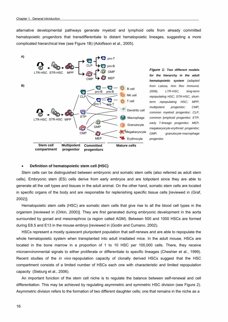
Chapter 1. General introduction _
16
alternative developmental pathways generate myeloid and lymphoid cells from already committed
hematopoietic progenitors that transdifferentiate to distant hematopoietic lineages, suggesting a more
complicated hierarchical tree (see Figure 1B) (Adolfsson et al., 2005).
Figure 1: Two different models
for the hierarchy in the adult
hematopoietic system (adapted
from Laiosa, Ann Rev Immunol,
2006). LTR-HSC, long-term
repopulating HSC; STR-HSC, short-
term repopulating HSC; MPP,
multipotent progenitor; CMP,
common myeloid progenitor; CLP,
common lymphoid progenitor; ETP,
early T-lineage progenitor; MEP,
megakaryocyte-erythroid progenitor;
GMP, granulocyte-macrophage
progenitor.
• Definition of hematopoietic stem cell (HSC) Stem cells can be distinguished between embryonic and somatic stem cells (also referred as adult stem
cells). Embryonic stem (ES) cells derive from early embryos and are totipotent since they are able to
generate all the cell types and tissues in the adult animal. On the other hand, somatic stem cells are located
in specific organs of the body and are responsible for replenishing specific tissue cells [reviewed in (Graf,
2002)].
Hematopoietic stem cells (HSC) are somatic stem cells that give rise to all the blood cell types in the
organism [reviewed in (Orkin, 2000)]. They are first generated during embryonic development in the aorta
surrounded by gonad and mesonephros (a region called AGM). Between 500 and 1000 HSCs are formed
during E8.5 and E13 in the mouse embryo [reviewed in (Godin and Cumano, 2002).
HSCs represent a mostly quiescent pluripotent population that self-renews and are able to repopulate the
whole hematopoietic system when transplanted into adult irradiated mice. In the adult mouse, HSCs are
located in the bone marrow in a proportion of 1 to 10 HSC per 100,000 cells. There, they receive
microenvironmental signals to either proliferate or differentiate to specific lineages (Cheshier et al., 1999).
Recent studies of the in vivo repopulation capacity of clonally derived HSCs suggest that the HSC
compartment consists of a limited number of HSCs each one with characteristic and limited repopulation
capacity (Sieburg et al., 2006).
An important function of the stem cell niche is to regulate the balance between self-renewal and cell
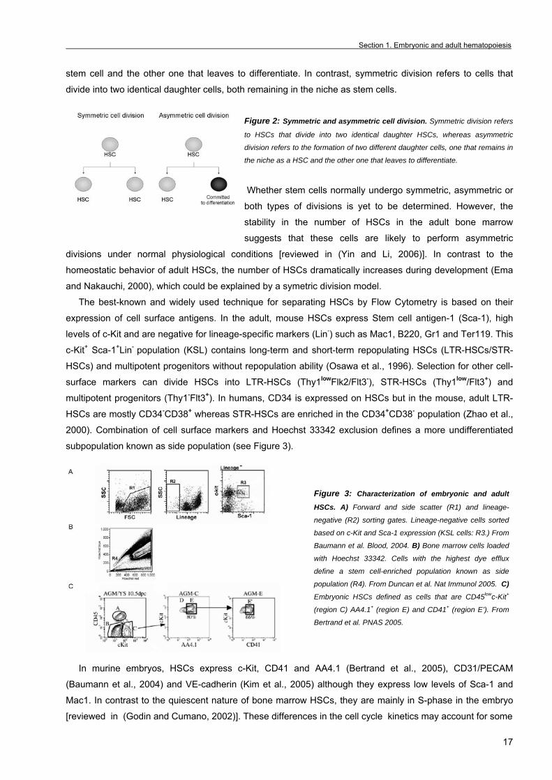
differentiation. This may be achieved by regulating asymmetric and symmetric HSC division (see Figure 2).
Asymmetric division refers to the formation of two different daughter cells; one that remains in the niche as a
A)
LTR-HSC MPP
CLP
STR-HSC
CMP
pro-B
pro-T
GMP
MEP
B)
LTR-HSC MPP STR-HSC
CLP
CMP
ETP
pro-B pre-BB cell
NK cell
pro-T pre-TT cell
Dendritic cell
GMP
MEP
Macrophage
Granulocyte
Erythrocyte
Stem cell compartment
Multipotent progenitor
Committed progenitors
Mature cells
Megakaryocyte
Section 1. Embryonic and adult hematopoiesis
17
stem cell and the other one that leaves to differentiate. In contrast, symmetric division refers to cells that
divide into two identical daughter cells, both remaining in the niche as stem cells.
Figure 2: Symmetric and asymmetric cell division. Symmetric division refers
to HSCs that divide into two identical daughter HSCs, whereas asymmetric
division refers to the formation of two different daughter cells, one that remains in
the niche as a HSC and the other one that leaves to differentiate.
Whether stem cells normally undergo symmetric, asymmetric or
both types of divisions is yet to be determined. However, the
stability in the number of HSCs in the adult bone marrow
suggests that these cells are likely to perform asymmetric
divisions under normal physiological conditions [reviewed in (Yin and Li, 2006)]. In contrast to the
homeostatic behavior of adult HSCs, the number of HSCs dramatically increases during development (Ema
and Nakauchi, 2000), which could be explained by a symetric division model.
The best-known and widely used technique for separating HSCs by Flow Cytometry is based on their
expression of cell surface antigens. In the adult, mouse HSCs express Stem cell antigen-1 (Sca-1), high
levels of c-Kit and are negative for lineage-specific markers (Lin-) such as Mac1, B220, Gr1 and Ter119. This
c-Kit+ Sca-1+Lin- population (KSL) contains long-term and short-term repopulating HSCs (LTR-HSCs/STR-
HSCs) and multipotent progenitors without repopulation ability (Osawa et al., 1996). Selection for other cell-
surface markers can divide HSCs into LTR-HSCs (Thy1lowFlk2/Flt3-), STR-HSCs (Thy1low/Flt3+) and
multipotent progenitors (Thy1-Flt3+). In humans, CD34 is expressed on HSCs but in the mouse, adult LTR-
HSCs are mostly CD34-CD38+ whereas STR-HSCs are enriched in the CD34+CD38- population (Zhao et al.,
2000). Combination of cell surface markers and Hoechst 33342 exclusion defines a more undifferentiated
subpopulation known as side population (see Figure 3).
Figure 3: Characterization of embryonic and adult
HSCs. A) Forward and side scatter (R1) and lineage-
negative (R2) sorting gates. Lineage-negative cells sorted
based on c-Kit and Sca-1 expression (KSL cells: R3.) From
Baumann et al. Blood, 2004. B) Bone marrow cells loaded
with Hoechst 33342. Cells with the highest dye efflux
define a stem cell-enriched population known as side
population (R4). From Duncan et al. Nat Immunol 2005. C)
Embryonic HSCs defined as cells that are CD45lowc-Kit+
(region C) AA4.1+ (region E) and CD41+ (region E’). From
Bertrand et al. PNAS 2005.
In murine embryos, HSCs express c-Kit, CD41 and AA4.1 (Bertrand et al., 2005), CD31/PECAM
(Baumann et al., 2004) and VE-cadherin (Kim et al., 2005) although they express low levels of Sca-1 and
Mac1. In contrast to the quiescent nature of bone marrow HSCs, they are mainly in S-phase in the embryo
[reviewed in (Godin and Cumano, 2002)]. These differences in the cell cycle kinetics may account for some

Chapter 1. General introduction _
18
of the differences in the cell-surface markers whereas other proteins such as CD41 and Mac1 receptors are
related with the ability of embryonic HSCs to migrate within the embryo (Sanchez et al., 1996).
1.1.2 Regulation of the hematopoietic system Interactions between cells and their environment regulate development of hematopoietic cells either
through cell-cell interactions or by secreted factors such as cytokines or growth factors. Altogether
characterizes the hematopoietic stem cell niche.
1.1.2.1 The hematopoietic stem cell niche
The bone marrow from the major long bones is the most important hematopoietic organ during adult-life.
Hematopoietic cells are retained within the bone cavity until they achieve the appropriate stage of maturation
and then are released into the bloodstream. HSCs and progenitor cells are surrounded by different
mesenchymal-derived stromal cells including chondrocytes, endothelial cells, fibroblasts and osteoblasts and
by the extracellular matrix that these cells produce (which includes fibronectin, laminin, collagen and
proteoglycans) [reviewed in (Dazzi et al., 2006)]. Interaction of both hematopoietic and stromal cells together
with signals mediated by soluble and membrane-bound growth factors are important in the regulation of adult
bone marrow hematopoiesis. All these elements form the stem cell niche, which offer the proper
microenvironment for stem cells to either self-renew or differentiate into their progeny. In the last years it has
been demonstrated that osteoblasts (cells responsible for bone growth) and the endothelial cells of
sinusoidal vessels are required for proper HSCs function, leading to the notion that two different niches
support HSCs development, the osteoblastic and the vascular niche. The former may maintain the HSCs in a
quiescent state whereas the latter may promote proliferation and further differentiation into the different
hematopoietic lineages [reviewed in (Yin and Li, 2006)].
Ligands with their corresponding receptors that mediate the interaction between the HSC and the niche
include Notch [reviewed in (Li and Li, 2006)], Stem Cell Factor (SCF)/c-Kit [reviewed in (Linnekin, 1999)],
Wnt (Reya et al., 2003), basic Fibroblast Growth Factor (bFGF) or hedgehog [reviewed in (Yin and Li, 2006)]
signaling pathways.
For example, Notch receptor is expressed in HSCs whereas the Notch ligand Jagged1 is expressed in
osteoblasts and bone marrow stromal cells, supporting the hypothesis that activated Notch induces self-
renewal of HSCs and hematopoietic progenitors (Varnum-Finney et al., 2000) (see Notch section in page
46).
• Chemokines and CXC receptors In addition to the ligand-receptor interactions, HSC-niche specific chemokines (cytokines with chemotactic
activity) are important in regulating HSC behavior. Chemokines have conserved cysteine residues that allow
them to be assigned to four groups which are C-C chemokines (RANTES, MCP-1, MIP-1α, and MIP-1β), C-
X-C chemokines (SDF1), C chemokines (Lymphotactin), and CXXXC chemokines (Fractalkine). Stromal cell
derived factor 1 (SDF1/CXCL12) participates in the mobilization of HSC from bone marrow to the blood
stream. In one hand, SDF1 expressed in endothelial cells regulates the transendothelial migration of HSCs
that express CXCR4 receptor, whereas SDF1 expression in osteoblast mediates HSC homing to the bone
marrow. SDF1 is also involved in the HSCs mobilization to peripheral blood induced by granulocyte colony-
Section 1. Embryonic and adult hematopoiesis
19
stimulating factor (G-CSF) treatment. G-CSF treatment is clinically used for HSC mobilization and stem cell
transplantation for the treatment of leukemias [reviewed in (Juarez and Bendall, 2004)].
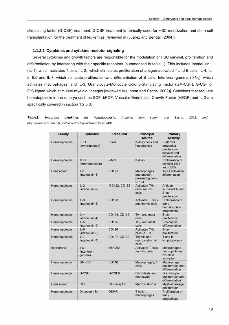
1.1.2.2 Cytokines and cytokine receptor signaling Several cytokines and growth factors are responsible for the modulation of HSC survival, proliferation and
differentiation by interacting with their specific receptors (summarized in table 1). This includes Interleukin 1
(IL-1), which activates T cells; IL-2, which stimulates proliferation of antigen-activated T and B cells; IL-4, IL-
5, IL6 and IL-7, which stimulate proliferation and differentiation of B cells; Interferon-gamma (IFNγ), which
activates macrophages; and IL-3, Granulocyte-Monocyte Colony-Stimulating Factor (GM-CSF), G-CSF or
Flt3 ligand which stimulate myeloid lineages [reviewed in (Lotem and Sachs, 2002)]. Cytokines that regulate
hematopoiesis in the embryo such as SCF, bFGF, Vascular Endothelial Growth Factor (VEGF) and IL-3 are
specifically covered in section 1.2.5.3.
Table1: Important cytokines for hematopoiesis. Adapted from Lotem and Sachs, 2002; and
htpp://www.ncbi.nlm.nih.gov/books/bv.fcgi?rid=imm.table.2499
Family Cytokine Receptor Principal source
Primary activity
Hematopoietins EPO (erythropoietin)
EpoR Kidney cells and hepatocytes
Erythroid progenitor proliferation, survival and differentiation
Hematopoietins TPO (thrombopoietin)
c-Mpl Kidney Proliferation of myeloid cells and HSCs
Unassigned IL-1 (interleukin-1)
CD121 Macrophages and antigen presenting cells (APC)
T-cell activation, inflammation
Hematopoietins IL-2 (interleukin-2)
CD122, CD132 Activated TH1 cells and NK cells
Antigen activated T- and B-cell proliferation
Hematopoietins IL-3 (interleukin-3)
CD123 Activated T cells and thymic cells
Proliferation of early hematopoietic progenitors
Hematopoietins IL-4 (interleukin-4)
CD124, CD132 TH2 and mast cells
B-cell proliferation
Hematopoietins IL-5 (interleukin-5)
CD125 TH2 and mast cells
Eosinophil differentiation
Hematopoietins IL-6 (interleukin-6)
CD126 Activated TH2 cells, APCs
B-cell proliferation
Hematopoietins IL-7 (interleukin-7)
CD127, CD132 Thymic and marrow stromal cells
T and B lymphopoiesis
Interferons IFNγ (interferon-gamma)
IFNGR2 Activated T cells and NK cells
Macrophages, neutrophils and NK cells activation
Hematopoietins GM-CSF
CD116 Macrophages, T cells
Macrophage proliferation and differentiation
Hematopoietins G-CSF G-CSFR Fibroblasts and monocytes
Granulocyte proliferation and differentiation
Unassigned Flt3 Flt3 receptor Marrow stroma Myeloid lineage proliferation
Hematopoietins Oncostatin M OSMR T cells, macrophages
Proliferation of early progenitors

Chapter 1. General introduction _
20
1.1.2.3 Hematopoietic transcription factors Specific transcription factors have been identified for their ability to control lineage specific gene programs
during hematopoietic development. Some of them are summarized in table2 and in the following section.
Table2: Important transcription factors for the regulation of the hematopoietic system
Transcription factor
Class Time of Death(E)
Primitive hematopoiesis
(yolk sac)
Definitive hematopoiesis
(fetal liver)
Hematopoietic defect
References
Pu.1 ETS E18.5 Normal Reduced -Defects in myeloid and lymphoid development
McKercher, 1996
Ikaros Zinc finger
Viable Normal Reduced -Decreased HSC/HPC generation or proliferation. -Lymphoid defects
Nichogiannopoulou, 1999
C/EBPα Basic leucine zipper
Perinathally Normal Reduced -Completely absence of GMP and mature neutrophils/macrophages -Increased number of HSCs and myeloblasts
Zhang, 1997 Zhang, 2004
E2A bHLH Viable Normal B-cell blockage -Defective B-cell commitment
Bain, 1994
Pax5 paired Alive but die within 3 weeks
Normal B-cell blockage -Defective B-cell differentiation
Urbánek, 1994 Nutt, 1997; Souabni, 2002
GATA3 Zinc finger
E11.5-E12.5
Normal Defective -T-cell blockage at the earliest stages
Ting, 1996
Runx1/AML1 bHLH E11.5-E12.5
Normal Blocked -Impaired HSC/HPC generation and/or proliferation
Okuda, 1996; North, 1999 and 2002; Burns, 2005; Lacaud, 2003
GATA2 Zinc finger
E10.5-E11.5
Reduced Markedly reduced -Impaired HSC/HPC generation and/or proliferation. -Reduced expansion of the various lineages
Tsai, 1994; Shivdasani, 1996; Minegishi, 2003; Ling, 2004
Scl/Tal-1 bHLH E9-E10.5 Markedly reduced Absent -Lack of precursor determination or maintenance.
Elefanty, 1999; Shivdasani, 1995; Robb, 1995 &1996
c-Myb Leucine zipper
E15 Normal Reduced -Impaired proliferation of hematopoietic progenitors
Mucensky, 1991; Emambokus, 2003; Mukouyama, 1999
GATA1 Zinc finger
E10.5-E11.5
Markedly reduced Absent -Erythrocytes arrested at the proerythroblast stage
Fujiwara, 1996; Weiss and Orkin, 1995; Takahashi, 1997; Suwabe, 1998
FOG-1 Zinc finger
E10.5-E11.5
Markedly reduced Absent -Erythroid and megakaryocytic development arrest
Tsang, 1998
• Common Myeloid and Lymphoid transcription factors Pu.1 belongs to the Ets family of transcription factors. Mice with a targeted mutation of Pu.1 display a
complete absence of granulocytic, monocytic and B- and T-cell lineages whereas erythroid and
megakaryocytic development is not affected. This indicates that this transcription factor plays a specific role
in myeloid and lymphoid differentiation (McKercher et al., 1996). Consistent with this, Pu.1 is expressed in
adult spleen, thymus and bone marrow and different myeloid and lymphoid cell lines [reviewed in (Ling and
Dzierzak, 2002)]. In early erythroblasts Pu.1 is expressed together with GATA1 and GATA2. Since GATA
factors promote erythroid proliferation and differentiation and Pu.1 mediates myeloid differentiation it is likely
that the previously reported mutual inhibition regulate the commitment of common hematopoietic progenitor
into erythroid or myeloid lineages (Zhang et al., 1999).

Section 1. Embryonic and adult hematopoiesis
21
The Ikaros transcription factor is a member of the Krüppel family of zinc-finger proteins and was first
described as a key factor in activating B- and T-cell specific gene expression. Moreover, Ikaros is expressed
in HSCs and lymphoid restricted progenitors (Klug et al., 1998) and null mice for Ikaros display reduced
number of HSCs (Nichogiannopoulou et al., 1999).
• Granulocytic-macrophage transcription factors
C/EBPα is required for granulocytic differentiation since null mice for this factor display a complete lack of
neutrophils and eosinophils (Zhang et al., 1997). Moreover, studies with conditional knockouts suggest that
C/EBPα is required for the transition from the Common Myeloid Progenitor to the Granulocyte-Macrophage
Progenitor (Zhang et al., 2004). In addition, C/EBPα-/- mice display increased numbers of HSCs and
myeloblasts (resembling human acute myeloid leukemia) in the bone marrow suggesting a role for this
protein in limiting self-renewal of HSCs and hematopoietic progenitors (Zhang et al., 2004).
• B-cell lineage transcription factor The basic helix-loop-helix (bHLH) transcription factor E2A and the early B-cell factor (EBF) are required
for the initiation of B lymphopoiesis since targeted mutations trigger B-cell blockage at the pro-B cell stage
(Bain et al., 1994). Both proteins cooperate to activate the expression of several B-lineage specific genes
such as those involved in the B-cell receptor rearrangements explaining the block of differentiation at the
pro-B stage [reviewed in (Laiosa et al., 2006)]. Pax5 is another B-cell specific transcription factor. The null mice for Pax5 display a similar effect than
E2A and EBF in arresting B-cell development in the bone marrow at the pro-B stage (Urbanek et al., 1994),
without affecting expression of E2A and EBF (Nutt et al., 1997). This suggests that Pax5 is acting
downstream of E2A and EBF in B-cell differentiation. Conversely, overexpression of Pax5 in bone marrow
HSCs and progenitors strongly induces B-cell development at the expense of the T-cell fate by repressing
Notch1 (Souabni et al., 2002).
• T-cell lineage transcription factors Notch1 is the most important inductor of the T-cell fate at expenses of the B-cell one and it functions not
only in T-cell specification but also in the whole hematopoietic system. Due to its relevance and because this
is the main subject of this work the role of Notch in hematopoiesis will be discussed in Chapter 1, Section 3.
GATA3 is a very recently characterized Notch-target that is involved in T-cell development (Dontje et al.,
2006). GATA3 null mice display blockage in T-cell generation at the earliest stages. Moreover, in chimeric
mice, GATA3-/- embryonic stem cells failed to contribute to the T-cell lineage (Ting et al., 1996).
• Natural killer transcription factors Natural killer cells represent a subtype of lymphoid cells that induces cytotoxicity in the target cells. The
phenotype of Ikaros-, Pu.1- and Id-2-null mice revealed that this three transcription factors are required for
NK cell development. Ikaros deficiency leads to the most severe phenotype with a complete absence of this
lymphoid cell lineage, indicating that this transcription factor plays a key role in the development of NK cells
[reviewed in (Laiosa et al., 2006)].
Chapter 1. General introduction _
22
• Dendritic cells Dendritic cells (DC) are responsible for the uptake, processing and presentation of antigens to T and B
cells. Many studies revealed that dendritic cells originate from both lymphoid and myeloid progenitors and
these two different DC populations require different transcription factors. In this sense, RelB and Pu.1 null
mice lack myeloid-derived DC, Id-2 null mice lack lymphoid-derived DCs and Ikaros mutants lack both DC
types [reviewed in (Laiosa et al., 2006)].
1.1.2.4 MicroRNAs as hematopoietic regulators Recent studies indicate that small non-coding RNAs regulate many different cellular and developmental
processes in animals and plants. This group of regulators called interference RNAs (RNAi) is composed by
the small-interfering (si)RNAs and the micro (mi)RNAs. Both types are small RNAs (22 nucleotides
approximately) and while the former bind to the target mRNA leading to its degradation, the latter leads to
both translational inhibition and mRNA degradation [reviewed in (Chen and Lodish, 2005)].
The first miRNAs were found in C. elegans but nowadays it is known that multi-cellular organisms express
hundreds of miRNAs in different cell types and their target sites are mainly found in the 3’-untranslated
region (UTRs) of the mRNA.
Emerging studies reveal that miRNAs regulate different developmental processes such as muscle
differentiation and limb formation. For example, miR-1 likely regulates Hand2 expression, a transcription
factor that controls cardiomyocyte expansion whereas miR-196 represses HoxB8 mRNA [reviewed in
(Shivdasani, 2006)]. In the last few years, many examples of miRNAs that regulate different hematopoietic
processes have been described. Ectopic expression of specific c-Kit microRNAs miR-221 and miR-222 on
CD34+ human cord blood cells leads to arrested proliferation and accelerated differentiation (Felli et al.,
2005). Moreover, the stem cell activity of these cells is abrogated as shown by transplantation experiments
in NOD-SCID immunodeficient mice (Felli et al., 2005). On the other hand, miR-223 that is activated by
C/EBPα is suggested to induce human granulocytic differentiation through inhibition of the transcription
factor NF1-A (Fazi et al., 2005).
Although not much is known about the genes regulated by miRNAs and most of the data comes from
bioinformatic predictions, it is tempting to speculate that the study of miRNAs will have an important
contribution in the understanding of hematopoiesis.
1.1.2.5 Transdifferentiation / Reprogramming Current models of hematopoietic differentiation suggest that stem cells become gradually restricted in
their differentiation potential by activating gene expression programs and simultaneously inactivating
alternative lineage choices. However, the notion of a strictly hierarchical branching model of hematopoiesis
has been challenged by many experimental data which suggest that both HSCs and committed
hematopoietic cells can transdifferentiate or switch into cells of another lineage [reviewed in (Graf, 2002)].
There are many examples of differentiated cells that can reprogramme their gene expression patterns
and switch into cells of a different lineage. For example, Pax5-/- B-cell progenitors can be induced to
differentiate into T cells when transplanted into immunodeficient Rag2-null mice. Alternatively, they can
differentiate into NK cells, dendritic cells, macrophages or neutrophils in response to different cytokines in
vitro. Moreover transdifferentiation within the myeloid and erythroid compartments can be achieved by
Section 1. Embryonic and adult hematopoiesis
23
overexpressing or inhibiting transcription factors such as Pu.1, GATA1, FOG-1 and C/EBPα [reviewed in
(Graf, 2002)]. Thus, it is likely that some hematopoietic progenitors maintain their plasticity and are able to
transdifferentiate into other lineages even when committed to a given one. However, these experiments rely
in alterations of physiological conditions and it is not known whether hematopoietic progenitors are able to
transdifferentiated into different lineages in vivo [reviewed in (Graf, 2002)].
More exciting are the recent experiments that suggest that HSCs can transdifferentiate into
nonhematopoietic cells such as myocytes, neurons, hepatocytes and endothelial cells following
transplantation of adult irradiated mice with bone marrow HSCs. All these experiments are controversial
since angiogenic precursors and mesenchymal stem cells from the bone marrow may be responsible for
these results. Moreover it has been shown that HSC can fuse to different cell types thus giving an alternative
explanation to transdifferentiation results [reviewed in (French et al., 2002)].
1.1.3 Methodological approaches to study HSCs and hematopoietic progenitors. Classification of HSCs and hematopoietic progenitors is based on their ability to long-term repopulate the
hematopoietic system of irradiated/chemically-ablated mice (LTR-HSCs), generate macroscopic colonies in
the spleen of adult irradiated mice (STR-HSCs and/or CFU-S) or form colonies in vitro (committed
hematopoietic progenitors or colony-forming cells CFC) [reviewed in (Dzierzak and Medvinsky, 1995)].
Considering the particularities of this methodology, a detailed explanation of the most common used
techniques to identify HSC and progenitors is included below.
• Short and Long-term multilineage repopulation assays This assay allows the detection of long-term repopulating hematopoietic stem cells (LTR-HSCs), the most
undifferentiated hematopoietic progenitor capable of generating the entire hematopoietic system (Muller et
al., 1994).
Two different approaches of this in vivo assay have been developed. The first one consists in the
intravenously transplantation of HSCs or progenitor cells (accompanied of more mature progenitors to
facilitate survival during the first weeks) from a donor mouse into an adult recipient whose hematopoietic
system has been depleted by lethal irradiation. One month after transplantation, the contribution from the
donor short-term repopulating HSCs (STR-HSCs) to the hematopoiesis of the recipient can be detected by
the presence of the donor specific marker in the peripheral blood. At four months post-transplantation,
detection of donor hematopoietic cells indicates the long-term repopulation ability of the transplanted cells
(LTR-HSC) (Muller et al., 1994). Limiting dilution and competitive long-term repopulation experiments can be
used to determine in a more quantitative and qualitative manner the repopulation ability of HSCs. In addition,
secondary and tertiary transplantations can be performed to assay self-renewal capacity that is characteristic
of the HSCs.
A modification of this methodology consists in the usage of a chemotherapeutic agent (busulfan) that
depletes the endogenous hematopoiesis of the recipient. This protocol is used when the experimental
approach requires the transplantation of donor cells into the liver of newborn pups. Such is the case for very
immature HSCs that are unable to engraft adult recipients (i.e. yolk sac HSCs) [reviewed in (Palis and Yoder,
2001)].
Chapter 1. General introduction _
24
• CFU-S and HPP-CFC Committed hematopoietic progenitors arise from multipotential precursors that are detected by two
different assays: 1) the spleen colony-forming assay (CFU-S assay) that allows the detection of multipotent
precursors, some of which are able to self-renew. In this case, progenitor cells are intravenously injected into
lethally irradiated mouse recipients and after 8-16 days the presence of macroscopic colonies in the spleen
determined (Medvinsky and Dzierzak, 1996) and 2) the high proliferative potential colony-forming cell assay
(HPP-CFC) permits the detection of multipotent precursors because they generate large macroscopic
colonies of myeloid cells in soft-agar in the presence of the appropriate cytokines (Bertoncello, 1992).
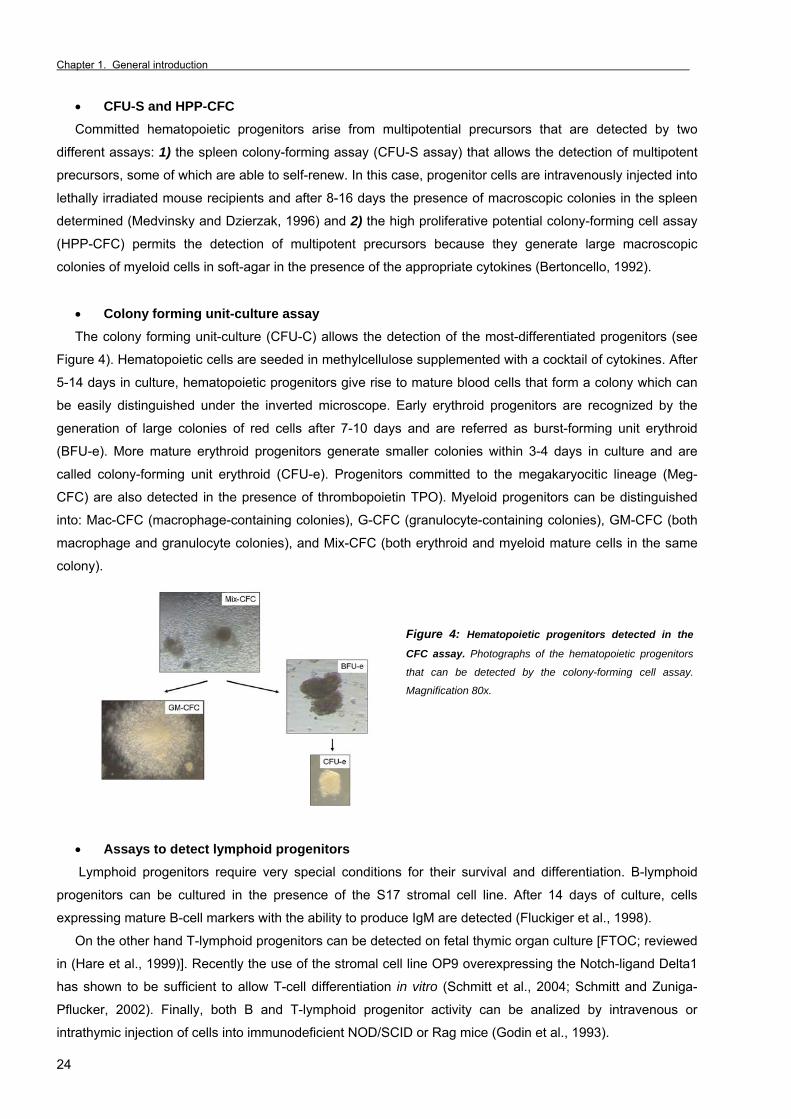
• Colony forming unit-culture assay The colony forming unit-culture (CFU-C) allows the detection of the most-differentiated progenitors (see
Figure 4). Hematopoietic cells are seeded in methylcellulose supplemented with a cocktail of cytokines. After
5-14 days in culture, hematopoietic progenitors give rise to mature blood cells that form a colony which can
be easily distinguished under the inverted microscope. Early erythroid progenitors are recognized by the
generation of large colonies of red cells after 7-10 days and are referred as burst-forming unit erythroid
(BFU-e). More mature erythroid progenitors generate smaller colonies within 3-4 days in culture and are
called colony-forming unit erythroid (CFU-e). Progenitors committed to the megakaryocitic lineage (Meg-
CFC) are also detected in the presence of thrombopoietin TPO). Myeloid progenitors can be distinguished
into: Mac-CFC (macrophage-containing colonies), G-CFC (granulocyte-containing colonies), GM-CFC (both
macrophage and granulocyte colonies), and Mix-CFC (both erythroid and myeloid mature cells in the same
colony).
Figure 4: Hematopoietic progenitors detected in the
CFC assay. Photographs of the hematopoietic progenitors
that can be detected by the colony-forming cell assay.
Magnification 80x.
• Assays to detect lymphoid progenitors Lymphoid progenitors require very special conditions for their survival and differentiation. B-lymphoid
progenitors can be cultured in the presence of the S17 stromal cell line. After 14 days of culture, cells
expressing mature B-cell markers with the ability to produce IgM are detected (Fluckiger et al., 1998).
On the other hand T-lymphoid progenitors can be detected on fetal thymic organ culture [FTOC; reviewed
in (Hare et al., 1999)]. Recently the use of the stromal cell line OP9 overexpressing the Notch-ligand Delta1
has shown to be sufficient to allow T-cell differentiation in vitro (Schmitt et al., 2004; Schmitt and Zuniga-
Pflucker, 2002). Finally, both B and T-lymphoid progenitor activity can be analized by intravenous or
intrathymic injection of cells into immunodeficient NOD/SCID or Rag mice (Godin et al., 1993).
Section 1. Embryonic and adult hematopoiesis
25
• Hematopoietic cell lines Many cell lines have been established from primary cells and they have become very important tools to
study hematopoietic regulation. Some of the most common cell lines used in hematopoietic research are
summarized.
Embrionic stem cells are derived from the inner cell mass of mouse blastocysts, which can self-renew or
differentiate into all adult tissues. ES cells are key tools to generate gene targeted mutant mice by
homologous recombination. In general, ES cells are maintained in vitro by co-culture with murine embryonic
feeder cell layer with leukemia inhibitory factor (LIF) for murine cells or bFGF for human cells. ES cells can
form aggregates of differentiated cells called embryoid bodies (EB) when LIF is removed from the media. EB
development recapitulates the hematopoietic ontogeny in vitro, including generation of hemangioblasts from
mesodermal precursors and the development of primitive and definitive hematopoiesis in the embryo. This
system has proved to be extremely powerful to study the effect of genetic manipulation in vitro (Keller et al.,
1993; Olsen et al., 2006).
Stromal cell lines are also useful tools for the study of hematopoiesis. Several cell lines have been
established that support the survival, proliferation and maintenance of HSCs. The most commonly used are
OP9, which was established from bone marrow of newborn macrophage colony-stimulating factor (M-CSF)-
deficient mice. AGM-S3 is another stromal endothelial cell line derived from murine embryonic day 10.5
(E10.5) AGM (aorta-gonad-mesonephros) region that is able to support hematopoiesis [reviewed in (Olsen et
al., 2006)]. Other stromal cell lines such as S17 and OP9-Delta1 are specifically used to support B-cell and
T-cell differentiation respectively (Fluckiger et al., 1998; Schmitt et al., 2004; Schmitt and Zuniga-Pflucker,
2002).
Finally, many different hematopoietic cell lines have been developed from leukemic cells or by
immortalizing hematopoietic progenitors and some of them can be induced to differentiate in vitro. For
example, 32D can be maintained as undifferentiated progenitors in the presence of IL-3 or induced to
granulocytic differentiation with G-CSF. Murine erythroleukemia (MEL) cells are spleen-derived cells
transformed by the Friend leukemia virus that are arrested at the proerythroblast stage. Differentiation can
be induced in vitro by hexamethylene bisacetamide (HMBA) [reviewed in (Marks and Rifkind, 1988)]. K562 is
a human erythroid cell line obtained from a chronic myeloid leukemia patient that can be induced to
differentiate by hemin or sodium butyrate treatment (Lozzio et al., 1981).
1.2 Ontogeny of the hematopoietic system in the mouse embryo
1.2.1 Brief introduction to mammalian embryonic development Once fertilization occurs, the zygote starts several mitotic divisions to form a compact morula with 16 to
64 pluripotent cells or blastomeres. In the stage of blastula (E4.5) an outer monolayer of cells called
trophoblasts generate the chorion and the amnion (fetal membranes) which line an inner cavity filled of fluid
that contains the inner cell mass or embryoblast. Gastrulation is the next morphogenic process and results in
the formation of the three germ layers: ectoderm, mesoderm and endoderm that give rise to the different
organs of the embryo.
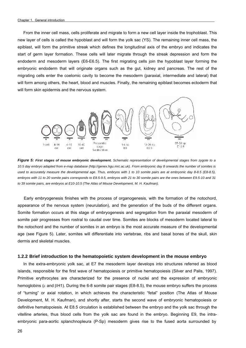
Chapter 1. General introduction _
26
From the inner cell mass, cells proliferate and migrate to form a new cell layer inside the trophoblast. This
new layer of cells is called the hypoblast and will form the yolk sac (YS). The remaining inner cell mass, the
epiblast, will form the primitive streak which defines the longitudinal axis of the embryo and indicates the
start of germ layer formation. These cells will later migrate through the streak depression and form the
endoderm and mesoderm layers (E6-E6.5). The first migrating cells join the hypoblast layer forming the
embryonic endoderm that will originate organs such as the gut, kidney and pancreas. The rest of the
migrating cells enter the coelomic cavity to become the mesoderm (paraxial, intermediate and lateral) that
will form among others, the heart, blood and muscles. Finally, the remaining epiblast becomes ectoderm that
will form skin epidermis and the nervous system.
Figure 5: First stages of mouse embryonic development. Schematic representation of developmental stages from zygote to a
10.5 day embryo adapted from e-map database (http://genex.hgu.mrc.ac.uk). From embryonic day 8 onwards the number of somites is
used to accurately measure the developmental age. Thus, embryos with 1 to 10 somite pairs are at embryonic day 8-8.5 (E8-8.5),
embryos with 11 to 20 somite pairs corresponds to E8.5-9.5, embryos with 21 to 30 somite pairs are the ones between E9.5-10 and 31
to 39 somite pairs, are embryos at E10-10.5 (The Atlas of Mouse Development, M. H. Kaufman).
Early embryogenesis finishes with the process of organogenesis, with the formation of the notochord,
appearance of the nervous system (neurulation), and the generation of the buds of the different organs.
Somite formation occurs at this stage of embryogenesis and segregation from the paraxial mesoderm of
somite pair progresses from rostral to caudal over time. Somites are blocks of mesoderm located lateral to
the notochord and the number of somites in an embryo is the most accurate measure of the developmental
age (see Figure 5). Later, somites will differentiate into vertebrae, ribs and basal bones of the skull, skin
dermis and skeletal muscles.
1.2.2 Brief introduction to the hematopoietic system development in the mouse embryo In the extra-embryonic yolk sac, at E7 the mesoderm layer develops into structures referred as blood
islands, responsible for the first wave of hematopoiesis or primitive hematopoiesis (Silver and Palis, 1997).
Primitive erythrocytes are characterized for the presence of nuclei and the expression of embryonic
hemoglobins (ε and βH1). During the 6-8 somite pair stages (E8-8.5), the mouse embryo suffers the process
of “turning” or axial rotation, in which achieves the characteristic “fetal” position (The Atlas of Mouse
Development, M. H. Kaufman), and shortly after, starts the second wave of embryonic hematopoiesis or
definitive hematopoiesis. At E8.5 circulation is established between the embryo and the yolk sac through the
vitelline arteries, thus blood cells from the yolk sac are found in the embryo. Beginning E9, the intra-
embryonic para-aortic splanchnopleura (P-Sp) mesoderm gives rise to the fused aorta surrounded by
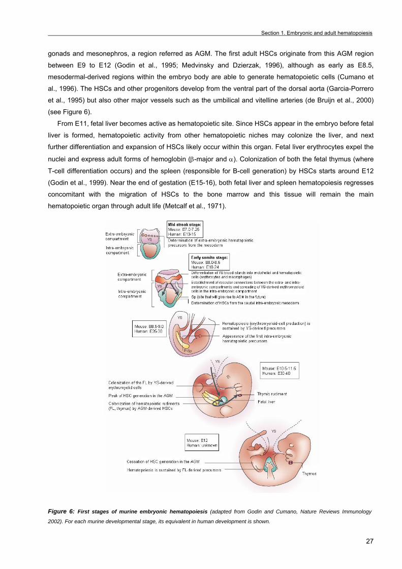
Section 1. Embryonic and adult hematopoiesis
27
gonads and mesonephros, a region referred as AGM. The first adult HSCs originate from this AGM region
between E9 to E12 (Godin et al., 1995; Medvinsky and Dzierzak, 1996), although as early as E8.5,
mesodermal-derived regions within the embryo body are able to generate hematopoietic cells (Cumano et
al., 1996). The HSCs and other progenitors develop from the ventral part of the dorsal aorta (Garcia-Porrero
et al., 1995) but also other major vessels such as the umbilical and vitelline arteries (de Bruijn et al., 2000)
(see Figure 6).
From E11, fetal liver becomes active as hematopoietic site. Since HSCs appear in the embryo before fetal
liver is formed, hematopoietic activity from other hematopoietic niches may colonize the liver, and next
further differentiation and expansion of HSCs likely occur within this organ. Fetal liver erythrocytes expel the
nuclei and express adult forms of hemoglobin (β-major and α). Colonization of both the fetal thymus (where
T-cell differentiation occurs) and the spleen (responsible for B-cell generation) by HSCs starts around E12
(Godin et al., 1999). Near the end of gestation (E15-16), both fetal liver and spleen hematopoiesis regresses
concomitant with the migration of HSCs to the bone marrow and this tissue will remain the main
hematopoietic organ through adult life (Metcalf et al., 1971).
Figure 6: First stages of murine embryonic hematopoiesis (adapted from Godin and Cumano, Nature Reviews Immunology
2002). For each murine developmental stage, its equivalent in human development is shown.
Chapter 1. General introduction _
28
The ontogeny of the human hematopoietic system is very similar to the mice. The first blood islands
appear in the human embryo around day 18 of gestation, and YS primitive erythropoiesis takes place from
weeks 3-6 of gestation. HSCs generation in the AGM occurs at weeks 5-7. From weeks 6-22, fetal liver acts
as the major hematopoietic site and finally the bone marrow becomes the lifelong site of blood-cell
production [reviewed in (Palis and Yoder, 2001)]. 1.2.3 Primitive hematopoiesis As mentioned before, generation of blood cell precursors in the embryo occurs in two different
hemogenic tissues, quite different in space and time, the YS and the AGM. Primitive hematopoiesis occurs in
the yolk sac and it is the first wave of hematopoietic cell production in the developing embryo.
1.2.3.1 The yolk sac
The murine yolk sac is composed by a layer of extra-embryonic mesoderm cells closely associated to a
layer of visceral endoderm cells. The endoderm functions as a supporting layer for mesoderm and
metabolizes maternally-derived molecules and synthesizes serum proteins. Furthermore, this layer seems to
be the source of some inductive signals necessary for both blood cell generation (Belaoussoff et al., 1998)
and endothelial network formation in the yolk sac (Palis et al., 1995). In agreement with this, embryoid
bodies derived from GATA4 null embryonic stem cells which lack visceral endoderm display reduced blood
island formation (Bielinska et al., 1996).
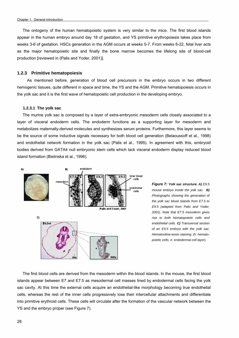
Figure 7: Yolk sac structure. A) E9.5
mouse embryo inside the yolk sac. B)
Photographs showing the generation of
the yolk sac blood islands from E7.5 to
E9.5 (adapted from Palis and Yoder,
2001). Note that E7.5 mesoderm gives
rise to both hematopoietic cells and
endothelial cells. C) Transversal section
of an E9.5 embryo with the yolk sac.
Hematoxiline-eosin staining. (h: hemato-
poietic cells; e: endodermal cell layer).
The first blood cells are derived from the mesoderm within the blood islands. In the mouse, the first blood
islands appear between E7 and E7.5 as mesodermal cell masses lined by endodermal cells facing the yolk
sac cavity. At this time the external cells acquire an endothelial-like morphology becoming true endothelial
cells, whereas the rest of the inner cells progressively lose their intercellular attachments and differentiate
into primitive erythroid cells. These cells will circulate after the formation of the vascular network between the
YS and the embryo proper (see Figure 7).
Section 1. Embryonic and adult hematopoiesis
29
1.2.3.2 Primitive hematopoiesis
Morphological studies revealed that blood cells generated in the YS resembled those found in lower
vertebrates and were called primitive blood cells. Primitive mature erythrocytes (also called megaloblasts)
are large nucleated cells that express embryonic globins (ε and βH1) and differ from definitive anucleated
adult-type erythrocytes that express adult-type globins (β-globin). During development, yolk sac produces
macrophages and megakariocytes that are different from their adult counterparts. More specifically, primitive
macrophages lack lysozyme and peroxidase activity, whereas megakaryocytes display reduced ploidy
[reviewed in (Godin and Cumano, 2002; Xu et al., 2001)]. In general, YS hematopoiesis produces red cells to
ensure oxygen supply, megakaryocytes to clot the new vessels that are being formed and macrophages to
clear the increasing amount of apoptotic cells present during early organogenesis in the developing embryo.
From E7 to E9, yolk sac erythroid progenitor cells are primitive erythroid progenitors (EryP) (Palis et al.,
1999), which can be distinguished in CFC assays because they generate colonies of about a hundred large
nucleated primitive erythroid cells expressing embryonic globins after 3 days of culture. From E9, definitive
hematopoietic progenitors are detected in the yolk sac as indicates the presence of BFU-e, CFU-e, GM-CFC
and Mix-CFC colonies. However, definitive erythrocytes at these stages are only detected in in vitro cultures
whereas in vivo, they are first evident by E12, when the liver is the most important hematopoietic organ. This
suggests that YS definitive erythroid progenitors do not physiologically differentiate in the YS (Palis et al.,
1999) and is in agreement with the observation that explants of YS required the coculture with liver
primordium to produced definitive erythrocytes (Cudennec et al., 1981).
Near 90% of YS-derived blood cells are erythrocytes. However, the yolk sac also contains Mac-CFC, GM-
CFC (Palis et al., 1999) and microglial progenitors which migrate to the developing central nervous system
(Kurz and Christ, 1998). Multipotent hematopoietic progenitors appear in the YS at E8.5 detected as HPP-
CFCs (Palis and Yoder, 2001) and at day E9.5 detected as CFU-S (Medvinsky et al., 1993). Finally, whether
the yolk sac has lymphoid potential is still controversial as to date no evidence of lymphopoiesis has been
found in yolk sac stromal co-cultures [reviewed in (Palis and Segel, 1998; Yokota et al., 2006)].
1.2.4 Definitive hematopoiesis Definitive hematopoiesis originates in an intra-embryonic region, formed by the aorta, gonads and
mesonephros and referred as the AGM region (Medvinsky and Dzierzak, 1996). The HSCs and other
progenitors generated in this region will move most likely to the fetal liver, where they proliferate and
differentiate into adult-type hematopoietic cells. Finally, near birth, HSCs migrate to the bone marrow niches
contributing to the adult-life hematopoietic system.
1.2.4.1 Generation of the AGM region
The AGM region extends from the forelimbs to the hindlimbs of the E9.5-E12.5 mouse embryo. It comes
from the mesoderm germ layer and is composed of the dorsal aorta, the genital ridges (which will form the
gonads) and the mesonephros (a mesodermally derived embryonic kidney) (see Figure 8). Around E8.5,
there is a pair of dorsal aortas that will connect to the yolk sac vasculature through the vitelline vessels.
These aortas fuse in a single dorsal aorta starting at E8.5 from caudal to rostral (Garcia-Porrero et al., 1995).
Simultaneously, the umbilical artery forms the connection between the dorsal aorta and the placenta.

Chapter 1. General introduction _
30
Figure 8: The AGM region. A) E10.5 mouse embryo. The discontinuous red line marks the section in B in the region of the trunk. B)
Transversal section through the region of the trunk of an E10.5 embryo. The black square marks the region represented in C;
Hematoxiline-eosin staining. C) Representation of the aorta in the AGM region. From E10 to E12.5, clusters of hematopoietic cells
appear as budding mainly from the ventral endothelium of the aorta in the AGM region.
1.2.4.2 Hematopoietic activity in the AGM region
The para-aortic splanchnopleura mesoderm will give rise to the AGM region and contains B- and T-
lymphoid progenitor activity at E8-9 (Godin et al., 1993) and CFU-spleen activity at E9 (Medvinsky et al.,
1993). However, no adult HSC activity (in the sense that they are able to repopulate adult irradiated mice
hematopoiesis) is found in the AGM region until day 10 (Muller et al., 1994). Historically, yolk sac was
assumed as the origin site of HSCs. It was until 1975 when Dieterlen-Lièvre showed that in chicken yolk sac
and quail embryo chimeras explanted before the establishment of the vascular network, only the quail cells
contributed to the adult hematopoietic system (Dieterlen-Lievre, 1975). Similarly, in amphibian embryos,
Turpen et al demonstrated that most of the HSCs come from the dorsal lateral plate, a homologous region to
the AGM, and that contribution to definitive hematopoiesis of the ventral blood islands (homologous to the
yolk sac) was less important (Turpen et al., 1997). Most recently, fate-mapping studies in Xenopus embryos
revealed that ventral blood islands and dorsal lateral plate regions develop from different blastomeres in the
blastula embryo and only the dorsal lateral plate contributes to the adult hematopoietic system (Ciau-Uitz et
al., 2000).
In mammalian embryos, it has been demonstrated that HSC are autonomously generated from E10 in the
AGM. From E11 HSCs most likely move to the fetal liver (Medvinsky and Dzierzak, 1996; Muller et al., 1994)
as previously mentioned. Further subdissection of E10.5 AGMs into aorta and urogenital ridges (UGR)
revealed that not only the aorta but also the umbilical and vitelline arteries generate definitive HSCs (de
Bruijn et al., 2000).
Although yolk sac and para-aortic splanchnopleura at E9-10 failed to engraft hematopoiesis of irradiated
adult recipients, these tissues engrafted in the liver of sublethally myeloablated newborn mice, being able to
long-term reconstitute all blood cell lineages, and bone marrow (Yoder and Hiatt, 1997; Yoder et al., 1997;
Yoder et al., 1997). These experiments indicate that immature pre-HSCs were present in the YS and early
splanchnopleura and they require an appropriated microenvironment supplied by the new-born liver. A
comparable result was obtained by Matsuoka et al by repopulating lethally irradiated adult mice with E8 yolk
sac or P-sp co-cultured with AGM-S3 cells, a clonal endothelial cell line from E10.5 AGM region (Matsuoka
et al., 2001).
Section 1. Embryonic and adult hematopoiesis
31
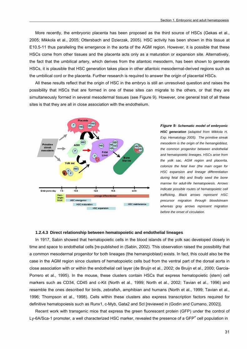
More recently, the embryonic placenta has been proposed as the third source of HSCs (Gekas et al.,
2005; Mikkola et al., 2005; Ottersbach and Dzierzak, 2005). HSC activity has been shown in this tissue at
E10.5-11 thus paralleling the emergence in the aorta of the AGM region. However, it is possible that these
HSCs come from other tissues and the placenta acts only as a maturation or expansion site. Alternatively,
the fact that the umbilical artery, which derives from the allantoic mesoderm, has been shown to generate
HSCs, it is plausible that HSC generation takes place in other allantoic mesodermal-derived regions such as
the umbilical cord or the placenta. Further research is required to answer the origin of placental HSCs.
All these results reflect that the origin of HSC in the embryo is still an unresolved question and raises the
possibility that HSCs that are formed in one of these sites can migrate to the others, or that they are
simultaneously formed in several mesodermal tissues (see Figure 9). However, one general trait of all these
sites is that they are all in close association with the endothelium.
Figure 9: Schematic model of embryonic
HSC generation (adapted from Mikkola H,
Exp. Hematology 2005). The primitive streak
mesoderm is the origin of the hemangioblast,
the common progenitor between endothelial
and hematopoietic lineages. HSCs arise from
the yolk sac, AGM region and placenta,
colonize the fetal liver (the main organ for
HSC expansion and lineage differentiation
during fetal life) and finally seed the bone
marrow for adult-life hematopoiesis. Arrows
indicate possible routes of hematopoietic cell
trafficking. Black arrows represent HSC
precursor migration through bloodstream
whereas gray arrows represent migration
before the onset of circulation.
1.2.4.3 Direct relationship between hematopoietic and endothelial lineages In 1917, Sabin showed that hematopoietic cells in the blood islands of the yolk sac developed closely in
time and space to endothelial cells [re-published in (Sabin, 2002). This observation raised the possibility that
a common mesodermal progenitor for both lineages (the hemangioblast) exists. In fact, this could also be the
case in the AGM region since clusters of hematopoietic cells bud from the ventral part of the dorsal aorta in
close association with or within the endothelial cell layer (de Bruijn et al., 2002; de Bruijn et al., 2000; Garcia-
Porrero et al., 1995). In the mouse, these clusters contain HSCs that express hematopoietic (stem) cell
markers such as CD34, CD45 and c-Kit (North et al., 1999; North et al., 2002; Tavian et al., 1996) and
resemble the ones described for birds, zebrafish, amphibian and humans (North et al., 1999; Tavian et al.,
1996; Thompson et al., 1998). Cells within these clusters also express transcription factors required for
definitive hematopoiesis such as Runx1, c-Myb, Gata2 and Scl [reviewed in (Godin and Cumano, 2002)].
Recent work with transgenic mice that express the green fluorescent protein (GFP) under the control of
Ly-6A/Sca-1 promoter, a well characterized HSC marker, revealed the presence of a GFP+ cell population in
Chapter 1. General introduction _
32
the AGM with long-term repopulating ability that resides in the endothelial layer lining the wall of the dorsal
aorta, strongly suggesting that HSCs come from the endothelium (de Bruijn et al., 2002). Hematopoietic and endothelial cells share a great number of cell surface markers: PECAM-1/CD31
(platelet endothelial cell adhesion molecule-1), angiopoietin receptor Tie-2, CD34 and the VEGF receptor-2,
Flk-1 (Baumann et al., 2004; Hamaguchi et al., 1999; Hsu et al., 2000; North et al., 1999; Young et al.,
1995). Moreover, several targeted mutations in endothelial genes strongly compromise the hematopoietic
development in mice. For example, mice deficient for Flk-1 (Shalaby et al., 1995) or the transcription factor
Scl (Robb et al., 1995) die at early stages of development due to severe hematopoietic and endothelial
disorders. Moreover, Flk-1 deficient ES cells failed to contribute to the vascular endothelium and to primitive
and definitive hematopoiesis as well (Shalaby et al., 1997). These results indicate that hematopoiesis and
endothelial vascular formation share a similar genetic program. In fact, there is strong evidence that
endothelial cells (characterized by the presence of exclusive endothelial markers) are able to generate
hematopoietic cells (Eichmann et al., 1997). Moreover, murine embryoid bodies contain blast-colony forming
cells that can differentiate into endothelial or hematopoietic cells upon secondary replating (Choi et al.,
1998). More recently, a cell population that expresses the Brachyury mesodermal marker and Flk-1, first
detected in the primitive streak of the mouse embryo, was shown to be the precursor of both endothelial and
hematopoietic cells of the yolk sac (Huber et al., 2004). Finally, Wang’s group identified an endothelial-like
subpopulation (PECAM-1, VE-cadherin, Flk-1 positive) within human ES cells with hemangioblastic
properties (Wang et al., 2004).
1.2.4.4 Proposed models for AGM-derived HSCs emergence
Endothelium of the dorsal aorta is formed before the emergence of adult repopulating HSCs, in contrast
with the simultaneous appearance of hematopoietic and endothelial cells in the yolk sac. Three different
models have been proposed to explain the generation of HSCs in the embryonic AGM [reviewed in (Godin
and Cumano, 2002)]:
1) HSCs are generated from cells in the ventral part of the dorsal aorta with an endothelial phenotype that
transdifferentiate into HSCs. This hypothesis would explain the great number of surface markers
coexpressed in endothelial and HSCs, although its expression is also found in mesodermal cells.
2) HSCs develop from different cell populations within the aortic endothelium either intra-embryonic
hemangioblasts or less differentiated mesodermal cells. Supporting this hypothesis Pardanaud et al
demonstrated in the avian model that aorta formation rises from two different mesodermal populations, one
of them with hemogenic potential and the other without (Pardanaud et al., 1996). Moreover, the use of
Ly6/Sca-1-GFP mice revealed that HSC activity arises within the endothelium lining the dorsal aorta (de
Bruijn et al., 2002).
3) HSCs develop from subaortic patches in the mesenchyme of the aortic floor and then migrate towards
the ventral endothelium of the aorta, to form the hematopoietic clusters and release into the bloodstream.
This hypothesis was based in the presence of CD31+CD41+ HSCs cells in these subaortic patches (which
may express Gata2) (Bertrand et al., 2005). Nowadays, all three models are still valid and more research is
needed to decipher how HSCs are generated.
Section 1. Embryonic and adult hematopoiesis
33
1.2.5 Regulators of embryonic hematopoiesis
1.2.5.1 Developmental signaling pathways Although not much is known, there is increasing evidence for a role of conserved developmental
pathways in the regulation of HSC formation and maintenance. Some of the current data is summarized
below.
• Wnt signaling pathway
Wnt proteins are secreted glycoproteins involved in cell fate determination, survival, proliferation and
migration in a wide variety of tissues [reviewed in (Khan and Bendall, 2006)]. Activation of the canonical
Wnt/β-catenin pathway by several Wnt molecules (including Wnt1, 3a, 8 or 8b) through Frizzled receptors
results in the inhibition of glycogen synthetase kinase 3β (GSK3β)-mediated phosphorylation of β-catenin.
This protects β-catenin from degradation by the proteosome and leads to an increase in protein levels and
nuclear translocation. In the nucleus, β-catenin binds to LEF/TCF family of transcription factors to specifically
activate gene expression. However, non-canonical Wnt pathways, independent of β-catenin have been
described and are mainly involved in motility, apoptosis and planar cell polarity [reviewed in (Khan and
Bendall, 2006)].
Several members of the Wnt family have been found to affect hematopoiesis in several species. Wnt5a
and 10b are expressed in the yolk sac and the fetal liver where they are supposed to promote progenitor cell
expansion (Austin et al., 1997). Wnt3a protein is able to induce self-renewal of HSC in vitro (Willert et al.,
2003) and activates the expression of brachyury, a mesodermal gene required for commitment of the
hematopoietic cell fate in ES cells (Arnold et al., 2000). Moreover, overexpression of an activated form of β-
catenin in HSCs increased self-renewal in vitro and reconstitution of lethally irradiated adult mice by
enhancing expression of both HoxB4 and Notch1 (Reya et al., 2003). It has been reported that Notch
signaling is required for Wnt-mediated maintenance of undifferentiated HSCs but not for their survival and
cell cycle entry (Duncan et al., 2005). All these reports indicate that Wnt signaling may be required for
promoting expansion and self-renewal of HSCs, although recent work shows that Wnt activation leads to
HSC differentiation but not self-renewal (Kirstetter et al., 2006).
Finally, increasing evidences that the Wnt signaling pathway is involved in T- and B-cell development
have been provided by the analysis of different mutant mice for different members of the Wnt signaling
pathway. In this sense, TCF1 knockout mice show a dramatic decrease in thymocyte number likely due to
impaired proliferation and enhanced apoptosis (reviewed in (Staal and Clevers, 2005).
• Smad-mediated signaling and hematopoiesis
The transforming growth factor-β (TGFβ) superfamily is composed by a wide variety of peptide growth
factors. Apart from TGFβ1, 2 and 3 other members are the activins and bone morphogenetic proteins
(BMPs). These ligands transduce their signals through transmembrane serine/threonine kinase receptors
which in turn, phosphorylate the intracellular mediators called Smad which heterodimerize to regulate gene
expression [reviewed in (Larsson and Karlsson, 2005)].
In the last years, many studies revealed a key role of BMPs and other TGFβ members in the
hematopoietic system. BMP4 mutant mice die at E7.5-9.5 displaying impaired mesoderm formation,

Chapter 1. General introduction _
34
defective generation of blood islands with reduced numbers of red blood cells (Winnier et al., 1995) similar to
the phenotype of TGFβ1 null mice (Dickson et al., 1995). In contrast, Smad5 null mice display increased
numbers of hematopoietic progenitors, indicating its negative role in either hematopoietic commitment or
progenitor expansion (Liu et al., 2003). Hence, TGFβ family members exert both positive and negative
regulation in the hematopoietic system.
• Hedgehog signaling Hedgehog is a family of secreted proteins composed by three members: Indian, Desert and Sonic.
Binding of Hedgehog to the receptor Patched (Ptch) results in the activation of a second transmembrane
protein, Smoothened (Smo) that transduces the signal through the zinc finger transcription factor Gli
[reviewed in (Baron, 2001)]. Coculture experiments of mesodermal tissue from early gastrulating embryo and
visceral endoderm tissue revealed that the latter is required for the mesoderm to generate primitive
hematopoietic red blood cells. This effect is likely mediated by secreted Indian Hedgehog (Ihh) that
upregulates BMP4 expression in the mesodermal tissue (Dyer et al., 2001). Consistent with this observation,
Ihh and Smo null mice display hemato-vascular defects (Byrd et al., 2002; Dyer et al., 2001). In zebrafish
embryos, the analysis of both Hedgehog mutants and cyclopamine-treated embryos (which is a Smo
inhibitor) revealed that Hedgehog signaling is required for definitive but not for primitive hematopoiesis
(Gering and Patient, 2005). Finally, Sonic hedgehog (Shh) induces proliferation of human CD34+CD38-Lin-
cord blood hematopoietic progenitors in vitro via BMP4 (Bhardwaj et al., 2001).
• The Notch signaling pathway Notch is an evolutionary conserved signaling pathway, widely used for the control of cell fate decisions
during development. In mammals, Notch regulates vasculogenesis, neurogenesis, miogenesis,
somitogenesis and many other processes. Since the Notch pathway is the main scope of this thesis, its role
in the control of hematopoiesis will be discussed in detail separately.
1.2.5.2 Hematopoietic transcription factors Transcription factors are key regulators of all biological processes including hematopoiesis since they
modulate the expression of downstream genes induced by (extracellular) signals. Several transcription
factors are responsible for the onset of the hematopoietic program in the embryo. The most important ones
are described in this section.
• Runx1
Runx1 (also referred as AML-1, CBFα2 and PEBPα2B) is one of the members of the conserved runt
homology domain transcription factor family. Runx1 is a helix-loop-helix protein that regulates the expression
of several hematopoietic genes such as IL-3, Pu.1, c-myb, Flk-2 and the GM-CSF and M-CSF receptors
(Okada et al., 1998).
Runx1 null mice die around E12.5 lacking definitive fetal liver hematopoiesis whereas primitive
hematopoiesis is not affected (Okuda et al., 1996; Wang et al., 1996), although Runx1 is expressed in
primitive erythrocytes and endothelial cells in the E8 yolk sac (North et al., 1999). In the embryo proper,
Runx1 is expressed from E8.5 in the endothelial cells at the ventral part of the dorsal aorta and other major

Section 1. Embryonic and adult hematopoiesis
35
arteries, and by E11.5 in the fetal liver. Interestingly, Runx1-/- embryos lack hematopoietic clusters in the
aorta (North et al., 1999) but also Runx1+/- embryos display changes in the distribution of HSCs (Cai et al.,
2000) indicating that Runx1 dosage is important for hematopoiesis. However, Runx1-/- ES cells-derived
embryoid bodies contain some blast-colonies progenitors indicating that hematopoiesis can occur in a
Runx1-independent manner (Lacaud et al., 2002). In addition, there is some evidence for Runx1 in
repressing VEGF receptor-2 (Flk-1) expression suggesting a possible role in the differentiation of
endothelial-like cells to hematopoietic cells (Hirai et al., 2005).
• GATA2 The GATA family of transcription factors is characterized by the presence of two homologous zinc-finger
domains: the C-terminus zinc-finger binds to the DNA GATA-consensus sequence (T/AGATAA/G), whereas
the N-terminus domain interacts with other transcription factors. GATA1 is exclusively found in the
hematopoietic system; GATA2 and GATA3 are expressed in the hematopoietic system but also in other
tissues.
GATA2 null mice display profound reduction in blast/mixed colonies in the yolk sac and die by E10.5 due
to severe anemia (Tsai et al., 1994). Analysis of GATA2-null ES chimeras revealed that these cells could not
contribute to neither primitive nor definitve hematopoiesis (Tsai et al., 1994). Interestingly, GATA2
haploinsufficiency results in a reduced number of HSCs in the AGM (Ling et al., 2004). In vitro, GATA2 null
ES cells generate multipotential progenitors that proliferate poorly and undergo massive apoptosis (Tsai and
Orkin, 1997). GATA2 is expressed in progenitor cells and progressively downregulated during erythroid
maturation and myeloid cell differentiation, in contrast with GATA1 that is progressively upregulated during
erythroid differentiation. Thus, a cross-regulatory mechanism by which GATA1 and GATA2 reciprocally
control their expression has been described possibly through GATA binding sites [reviewed in (Ohneda and
Yamamoto, 2002)].
• Scl / Tal-1 Scl (stem cell leukemia) or tal-1 (T-cell acute leukemia-1) gene encodes a basic-helix-loop-helix
transcription factor, which has been associated with many leukemogenic processes. In the developing
embryo it is expressed first in the visceral mesoderm that will form the yolk sac blood islands and next in
both hematopoietic and endothelial cells (Elefanty et al., 1999). Scl null embryos die between E8.5 and
E10.5 displaying absence of yolk sac hematopoiesis and vitelline vessel formation. Moreover, GATA1, c-myb
and embryonic βH1-globin expression is lost in this mutant indicating that Scl may be required for their
activation (Robb et al., 1995; Shivdasani et al., 1995). Chimeric mice with Scl null ES cells revealed that this
gene is required for both primitive and definitive hematopoiesis (Robb et al., 1996). In contrast with its crucial
role during development, disruption of scl in adult mice does not affect the repopulation ability of the bone
marrow HSCs (Mikkola et al., 2003).
• c-Myb The c-Myb transcription factor is expressed in immature blood progenitors and in embryonic
hematopoietic sites. c-Myb null mice die at E15 due to failure of fetal liver hematopoiesis whereas primitive
hematopoiesis is not affected (Mucenski et al., 1991). Moreover, primary cultures from P-Sp at E9.5 and
Chapter 1. General introduction _
36
AGM at E11.5 of c-Myb-/- embryos displayed a complete lack of hematopoiesis (Mukouyama et al., 1999)
suggesting that the absence of hematopoietic progenitors in the fetal liver of murine c-Myb-/- embryos is likely
due to a previous defect in the generation of HSCs in the AGM.
In addition, c-myb doses are important in the regulation of hematopoietic progenitor cell proliferation and
differentiation since a knockdown allele of c-myb, which expresses 5-10% of the wild-type c-Myb levels
allows in vitro progenitor expansion while blocking differentiation (Emambokus et al., 2003).
Finally and similar to GATA2, c-Myb is highly expressed in immature erythroid progenitors and its
downregulation is required to allow terminal erythroid maturation (Bartunek et al., 2003).
• HoxB4 Hox transcription factors were identified as main regulators of developmental patterning. However, more
recent work shows that they also function in adult tissues including hematopoiesis. There are four hox
clusters (A-D) located on different chromosomes, and a total number of 39 hox genes assigned to 13 paralog
groups in each cluster, based on homeobox sequence similarities. Although several hox genes may be
involved in hematopoiesis, hoxB4 is the best characterized. Retroviral overexpression of HoxB4 expands
HSCs in murine bone marrow (Antonchuk et al., 2002) and human cord blood cultures without affecting their
repopulating ability or terminal differentiation capacity (Buske et al., 2002). Moreover, hoxB4 infection of
primitive murine yolk sac hematopoietic progenitors (E8.25, prior to the onset of circulation) results in the
acquisition of definitive HSCs characteristics such as long-term repopulating capacity in irradiated adult mice
(Kyba et al., 2002). This also supports the idea that yolk sac contains cells that are capable of generating
definitive HSCs when the appropriate gene program is induced.
1.2.5.3 Cytokines
• Vascular endothelial growth factor The vascular endothelial growth factor is an important cytokine for both hematopoietic and endothelial
cells in the developing embryo. Studies with both mice and ES cells lacking the VEGF receptor-2 (Flk-1)
suggested that signaling through VEGF is required for hemangioblast commitment from mesoderm (Shalaby
et al., 1997; Shalaby et al., 1995). Conversly, injection of VEGF mRNA in Xenopus embryos results in
excessive production of endothelial cells at expense of blood cells indicating that proper levels of VEGF may
be required to maintain hematopoietic/angioblastic differentiation (Koibuchi et al., 2006).
In chimeras, Flk1-/- ES cells fail to contribute to primitive and definitive blood cells and they aberrantly
accumulate on the surface of the amnion (Shalaby et al., 1997). Thus, VEGF signaling may be involved in
migration of Flk1-positive precursors from the mesoderm to hematopoietic sites, a hypothesis supported by
studies in Drosophila which indicate that VEGF may be involved in migration of hemocytes during embryonic
life (Cho et al., 2002).
VEGF effects are dose dependent. Embryos with two VEGF hypomorphic alleles which reduce VEGF
expression to a 50% compared with the wild-type (VEGFlo/lo embryos) die at E9.5 displaying the same
hematopoietic and vascular defects than VEGF+/- and VEGF-/- embryos (Martin et al., 2004). VEGFlo/lo
embryos display increased apoptosis in the Ter119+ population in the yolk sac indicating that proper VEGF
dose is necessary for survival of primitive erythroid cells. Moreover, Scl overexpression in VEGFlo/lo embryos
Section 1. Embryonic and adult hematopoiesis
37
decreases the percentage of apoptotic Ter119+ cells indicating that Scl acts downstream of VEGF to ensure
survival of primitive erythroid cells (Martin et al., 2004).
• Stem cell factor / Stem cell factor receptor The Stem Cell Factor (SCF, also Kit ligand) is a hematopoietic cytokine that exerts its functions by binding
to its tyrosine kinase receptor c-Kit. Two natural occurring mutations in this locus (Steel and White spotting)
lead to severe anemia and perinatal lethality. Several studies revealed that c-Kit activation is required for
proliferation and maintenance of hematopoietic progenitors, mast cells and primordial germ cells [reviewed in
(Broudy, 1997)].
In the developing embryo, SCF and c-Kit receptor expression is found in the yolk sac, fetal liver and bone
marrow and other non-hematopoietic tissues such as the gut and the central nervous system. c-Kit
expression is also found in HSCs, progenitor cells and mature cell types [(Sanchez et al., 1996); reviewed in
(Broudy, 1997)]. Transplantation of c-Kit null fetal liver cells in Rag2-/- mice (which are deficient for B and T
cells) revealed that SCF/c-Kit are required for T lymphopoiesis but not for B-cell development (Takeda et al.,
1997).
SCF can be found in both soluble and membrane bound forms resulting from alternative splicing. The
soluble form is likely to be required for proliferation and differentiation of progenitors/stem cells in
combination with other cytokines such as IL-3, IL-1, IL-6 and erythropoietin (EPO). However, in the absence
of specific cytokines SCF promotes viability rather than proliferation [reviewed in (Broudy, 1997)].
Alternatively, the membrane bound form of SCF is thought to be required for adhesion of HSCs and
progenitor cells to bone marrow stromal elements through its binding to c-Kit [reviewed in (Linnekin, 1999)].
• Fibroblast growth factor In the last years it has been shown that the fibroblast growth factor (FGF) is also a regulator of
hematopoiesis. FGFR1 (fibroblast growth factor receptor-1) null ES cells do not generate hematopoietic cells
whereas bFGF enhances blast-CFC (or hemangioblast) generation (Faloon et al., 2000). Conversely,
different FGFs such as FGF-2 and FGF-4 or more recently FGF-1 have been shown to regulate lineage
differentiation [reviewed in (Kashiwakura and Takahashi, 2005)] and ex vivo self-renewal of HSCs (de Haan
et al., 2003), respectively.
• Interleukin-3 IL-3 has been widely used for both expansion of hematopoietic progenitors and colony growth in
semisolid media in vitro. Mice carrying IL-3 or IL-3 receptor mutations display minor alterations of adult
hematopoiesis (Lantz et al., 1998; Nishinakamura et al., 1996). However, recent studies indicate a possible
embryonic role of IL-3 as a proliferation and/or survival factor for the earliest HSCs in the embryo. IL-3 is a
putative target of Runx1 and it has been shown that IL-3 alone is able to rescue the Runx1+/- HSC defect in
the AGM, indicating that this gene is important for proper generation of embryonic HSCs (Robin et al., 2006).
Chapter 1. General introduction _
38
1.3 Hematologic disorders Regulation of the hematopoietic system is extremely complex. Thus, mutations and alterations in genes
regulating hematopoiesis often develop into life-threatening disorders, indicating that blood cell homeostasis
is essential for survival of an organism.
In the following section, an overview of the most frequent pathologies of the hematopoietic system
caused by known hematopoietic regulators is discussed. Erythroid pathologies will be analysed on Chapter
1, Section 2.
1.3.1 Leukemias Neoplasms of hematopoietic cells are termed leukemias. Transformation to malignancy likely occurs in a
single cell, which starts proliferating, clonal expanding and avoiding apoptosis. This fact usually occurs at
the pluripotent stem cell level but sometimes it may involve a committed stem cell. Leukemic cells
accumulate and/or replace bone marrow cells causing abnormal hematopoiesis, which leads to anemia,
thrombocytopenia and granulocytopenia. In addition, they can infiltrate organs such as liver, spleen, lymph
nodes and kidney leading to their dysfunction.
Leukemias are classified according to cell differentiation stages and the severity of the disease. Thus,
acute leukemia is usually a rapidly progressing disease characterized by replacement of normal bone
marrow and other organs by malignant blast cells. They are divided into acute lymphoblastic leukemias
(ALL) and acute myeloid leukemias (AML). Chronic leukemia shows a slower progression and many patients
can be asymptomatic for years. Chronic lymphocytic leukemia (CLL) is due to the clonal expansion of
mature-appearing lymphocytes in lymph nodes and other lymphoid tissues with progressive infiltration of
bone marrow and peripheral blood. On the other hand, chronic myeloid leukemia (CML) is caused by clonal
myeloid cell proliferation likely from a transformed pluripotent cell resulting in overproduction of granulocytes
in both bone marrow and extramedullary sites (spleen and/or liver).
• Abnormalities leading to leukemia Leukemogenesis needs a critical first step, which usually is an acquired genetic aberration or initiating
mutation which perturbs normal hematopoietic development. This “first hit” is necessary but not sufficient for
development of a leukemic process and frequently occurs in genes encoding regulators of normal
hematopoiesis. The progression to leukemia depends on “second hits” in additional pathways that control
survival and proliferation of the developmentally arrested leukemic cells [reviewed in (Izraeli, 2004)].
ALL is the most common malignancy in children but can also appear in adults. The t(1;14)(p32;q11)
chromosomal translocation causes T-cell acute lymphoblastic leukemia (T-ALL) due to the insertion of the scl
locus in the regulatory elements of the T-cell receptor locus leading to aberrant expression of Scl. Moreover,
a small deletion in the sil gene promoter, a gene upstream of scl, results in overexpression of Scl and
deregulation of cell cycle genes in proliferating T cell precursors [reviewed in (Izraeli, 2004)].
Other chromosomic rearrangements such as the Tel-Runx1 translocation and those affecting the E2A
locus cause B-cell acute lymphoblastic leukemia (B-ALL). The t(12;21) translocation generates a Tel-Runx1
fusion protein, involving two important regulators of the bone marrow HSCs population [reviewed in (Izraeli,
2004)]. Another frequent translocation associated with AML which involves Runx1 is the t(8;21) runx1-eto
(also known aml1-eto). Besides preventing Runx1-dependent activities, the Runx1-Eto protein also inhibits
Section 1. Embryonic and adult hematopoiesis
39
C/EBPα and Pu.1, thus blocking granulocytic differentiation (de Guzman et al., 2002). Gain-of-function
mutations on the hoxA9 gene (both inducing its constitutive expression or activity) also lead to AML
[reviewed in (Moore, 2005)].
Finally, in 95% of the patients with CML, a reciprocal translocation t(9;22) known as “Philadelphia
chromosome” between the oncogene c-abl and the gene bcr generates the aberrant fusion protein Bcr-Abl.
This chimeric protein inhibits C/EBPα translation, thus leading to a blockage in myeloid differentiation.
Inactivation of C/EBPα also leads to different types of AML [reviewed in (Leroy et al., 2005)].
1.3.2 Myelodysplastic syndromes (MDS) Myelodysplastic syndromes are hematopoietic stem cell disorders characterized by impaired
hematopoiesis which result in anemia, neutropenia and/or thrombocytopenia of variable severity. The first
stages of MDS are characterized by excessive apoptosis of progenitor cells leading to ineffective
hematopoiesis. In response to this defect, the hematopoietic system increases proliferation of progenitor
cells and as MDS progresses; cells become resistant to apoptosis and in most of the cases develop into
acute myeloid leukemia (AML) [reviewed in (Catenacci and Schiller, 2005)]. Recently it was shown that
GATA1 was mutated in Down Syndrome patients which develop a transient myeloproliferative disorder
(TMD) and acute megakaryoblastic leukemia (AMKL) suggesting a high implication of this transcription factor
in MDS and/or AMKL [reviewed in (Izraeli, 2004)].
On the other hand, a constitutively active form of human c-Kit (D816V) has been found with high
frequency in patients with mastocytosis and associated hematological disorders. This mutation results in
SCF-independent proliferation as well as transforming abilities leading to leukemia [reviewed in (Linnekin,
1999)].
Chapter 1. General introduction _
40
Section 2. Erythropoiesis
2.1 Erythroid differentiation 2.1.1 Primitive and definitive erythropoiesis Erythropoiesis is a multistep process that involves the differentiation from HSCs to mature erythrocytes. In
the adult hematopoietic system, erythropoiesis takes place in the bone marrow and first implies the
differentiation of HSC into the common myeloid progenitor, a multipotent progenitor that generates
granulocytic-macrophage and megakaryocytic-erythroid progenitors. In the CFU-culture assay, the CMP
generates mixed colonies of myeloid and erythroid cells (Mix-CFC). When commited to the erythroid lineage
CMPs give rise to burst-forming unit-erythroid (BFU-e), which generates large colonies of erythroid cells in
vitro and the late erythroid progenitor CFU-e [reviewed in (Testa, 2004)]. After several mitotic divisions, the
CFU-e differentiates into morphologically recognizable erythroid cells. During this differentiation process, the
cells gradually express erythropoietin receptor thus becoming sensitive to erythropoietin (EPO). This
cytokine regulates both proliferation and survival of erythroid cells. The last stages of erythroid differentiation
involve a proliferative-maturative compartment composed by proerythroblasts, basophilic erythroblasts and
polychromatophilic erythroblasts and a maturative non-proliferative compartment involving maturation of
orthochromatic erythroblast, reticulocytes and mature erythrocytes. During last stages of maturation,
erythroid cells decrease their size, accumulate hemoglobin and increase their chromatin density to exclude
the nucleus. Finally, they are released to the bloodstream and take part in the gas exchange for a half-period
of thirty days [reviewed in (Testa, 2004)].
During embryonic development, there are two major waves of erythroid cell production, the first one
starting at E7.5 in the yolk sac and the second one beginning at E11 in the fetal liver. Yolk sac erythropoiesis
is characterized by the appearance of primitive nucleated large erythrocytes (6-fold larger than adult ones)
expressing embryonic (ζ, βH1 and εy) and adult globins (α1, α2, β1 and β2) differentiated from primitive
erythroid progenitors (EryP-CFC). However, from E8.5 adult-like progenitors (BFU-e and CFU-e) are found in
the yolk sac, which in culture generate enucleated erythrocytes expressing adult globins (Palis et al., 1999).
Adult erythropoiesis is well characterized based on cell morphology, expression of cell-surface markers
and hemoglobin content. Nevertheless, not much is known about primitive erythroid development. Primitive
erythroid cells, generated in the blood islands of the yolk sac, enter the bloodstream once circulation
between embryo and yolk sac is established, at the stage of basophilic erythroblasts. While circulating
primitive erythroblasts undergo maturation characterized by a decrease in size, accumulation of hemoglobin
resulting in decreased cytoplasmic basophilia and chromatin condensation, similar to that occurring in fetal
liver or adult-life bone marrow erythropoiesis [reviewed in (McGrath and Palis, 2005)].
2.1.2 Erythroid markers Several cell surface markers have been identified in different erythroid subpopulations and are related to
specific maturation stages (see Figure 10).
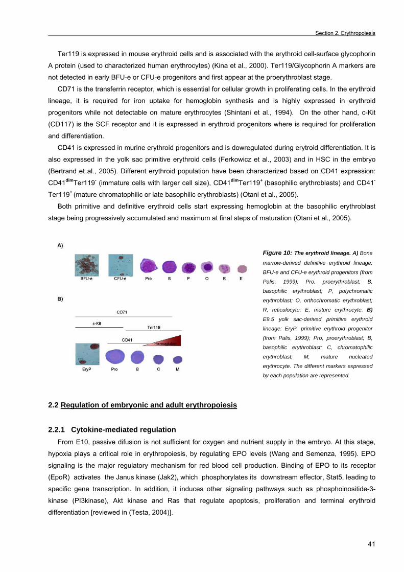
Section 2. Erythropoiesis
41
Ter119 is expressed in mouse erythroid cells and is associated with the erythroid cell-surface glycophorin
A protein (used to characterized human erythrocytes) (Kina et al., 2000). Ter119/Glycophorin A markers are
not detected in early BFU-e or CFU-e progenitors and first appear at the proerythroblast stage. CD71 is the transferrin receptor, which is essential for cellular growth in proliferating cells. In the erythroid
lineage, it is required for iron uptake for hemoglobin synthesis and is highly expressed in erythroid
progenitors while not detectable on mature erythrocytes (Shintani et al., 1994). On the other hand, c-Kit
(CD117) is the SCF receptor and it is expressed in erythroid progenitors where is required for proliferation
and differentiation.
CD41 is expressed in murine erythroid progenitors and is dowregulated during erytroid differentiation. It is
also expressed in the yolk sac primitive erythroid cells (Ferkowicz et al., 2003) and in HSC in the embryo
(Bertrand et al., 2005). Different erythroid population have been characterized based on CD41 expression:
CD41dimTer119- (immature cells with larger cell size), CD41dimTer119+ (basophilic erythroblasts) and CD41-
Ter119+ (mature chromatophilic or late basophilic erythroblasts) (Otani et al., 2005).
Both primitive and definitive erythroid cells start expressing hemoglobin at the basophilic erythroblast
stage being progressively accumulated and maximum at final steps of maturation (Otani et al., 2005).
Figure 10: The erythroid lineage. A) Bone
marrow-derived definitive erythroid lineage:
BFU-e and CFU-e erythroid progenitors (from
Palis, 1999); Pro, proerythroblast; B,
basophilic erythroblast; P, polychromatic
erythroblast; O, orthochromatic erythroblast;
R, reticulocyte; E, mature erythrocyte. B)
E9.5 yolk sac-derived primitive erythroid
lineage: EryP, primitive erythroid progenitor
(from Palis, 1999); Pro, proerythroblast; B,
basophilic erythroblast; C, chromatophilic
erythroblast; M, mature nucleated
erythrocyte. The different markers expressed
by each population are represented.
2.2 Regulation of embryonic and adult erythropoiesis
2.2.1 Cytokine-mediated regulation From E10, passive difusion is not sufficient for oxygen and nutrient supply in the embryo. At this stage,
hypoxia plays a critical role in erythropoiesis, by regulating EPO levels (Wang and Semenza, 1995). EPO
signaling is the major regulatory mechanism for red blood cell production. Binding of EPO to its receptor
(EpoR) activates the Janus kinase (Jak2), which phosphorylates its downstream effector, Stat5, leading to
specific gene transcription. In addition, it induces other signaling pathways such as phosphoinositide-3-
kinase (PI3kinase), Akt kinase and Ras that regulate apoptosis, proliferation and terminal erythroid
differentiation [reviewed in (Testa, 2004)].
Chapter 1. General introduction _
42
Targeted disruption of EPO signaling pathway components such as EPO, EpoR and Jak2 kinase (Lin et
al., 1996; Neubauer et al., 1998; Wu et al., 1995) leads to embryonic death with severe anemia due to a
huge reduction of primitive erythroblasts at E12.5. However, EPO signaling mutants develop normal
numbers of erythroid progenitors indicating that this cytokine is not required at the first stages of erythroid
commitment. The role of EPO in primitive erythropoiesis is still unclear although it has been shown that EPO
increases primitive red blood cell production and prevents from death in E8.5 yolk sac cultures (Kimura et al.,
2000).
The Stem cell factor (SCF) or Kit ligand is another important cytokine in erythropoiesis. Both SCF and c-
Kit are highly expressed in uncommitted CD34 positive cells and erythroid committed progenitors (BFU-e
and CFU-e) being downregulated with differentiation and absent in mature blood cells (Testa et al., 1996).
Thus, SCF is likely involved in the expansion and survival of erythroid progenitors.
2.2.2 Erythropoietic transcription factors GATA1 is the most important transcription factor in erythroid development although it is also required for
proper maturation of mast and megakaryocytic precursors and for specification of eosinophils. It recognizes
the WGATAR-binding motif present in the promoter and/or enhancers of all erythroid-specific genes
[reviewed in (Patient and McGhee, 2002)]. This zinc finger transcription factor is expressed at basal levels in
quiescent erythroid progenitors, but after EPO-induced differentiation, expression increases reaching its
maximum between CFU-e and proerythroblasts stages [reviewed in (Cantor and Orkin, 2002)].
GATA1 null mutation is embryonic lethal at E10.5-11.5 due to severe anemia. The presence of erythroid
progenitors in these embryos indicates that GATA1 is not essential for erythroid commitment, however they
display a maturational arrest at the level of proerythroblasts (Fujiwara et al., 1996). In agreement with this,
GATA1-/- erythroid precursors derived from embryoid bodies have a blockage at the proerythroblast stage
and undergo apoptosis (Weiss and Orkin, 1995). Surprisingly, in a mouse model in which GATA1 levels are less than 5% of the level present in wild-type,
the mutant embryos die at E12.5 as well (Takahashi et al., 1997). However, erythroid cells are resistant to
apoptosis and remain proliferative (Suwabe et al., 1998), leading to a defect that develops in leukemia
(Shimizu et al., 2004). Thus, GATA1 levels seem to be crucial to regulate erythroid homeostasis.
Friend of GATA1 (FOG-1) is another zinc-finger transcription factor that interacts with GATA1. FOG-1
mutant mice display a similar phenotype than GATA1 null animals indicating that both proteins are required
for erythroid differentiation. However, FOG-1 null phenotype is more severe compared to GATA1 as shown
by the complete lack of megakaryocytes, suggesting that FOG-1 exerts some GATA1-independent functions
(Tsang et al., 1998).
The basic helix-loop-helix transcription factor Scl /Tal-1 interacts with ubiquitously expressed E proteins to
bind the promoters of erythroid and megakaryocytic specific genes. Enforced Scl expression favours
erythroid commitment of hematopoietic progenitors (Valtieri et al., 1998), whereas Scl deficiency inhibits
proliferation and self-renewal of erythroleukemia cells (Green et al., 1991).
Other transcription factors involved in erythroid development are the family of Krüppel-like zinc finger
transcription factors or KLF. Among all the members of the family, KLF1/EKLF is required for the expression
of the β-globin gene, heme group synthesis enzymes (such as ALAS-E or Alad), the α-hemoglobin stabilizing
protein (AHSP) and other proteins involved in red cell membrane and cytoskeletal stability (Hodge et al.,
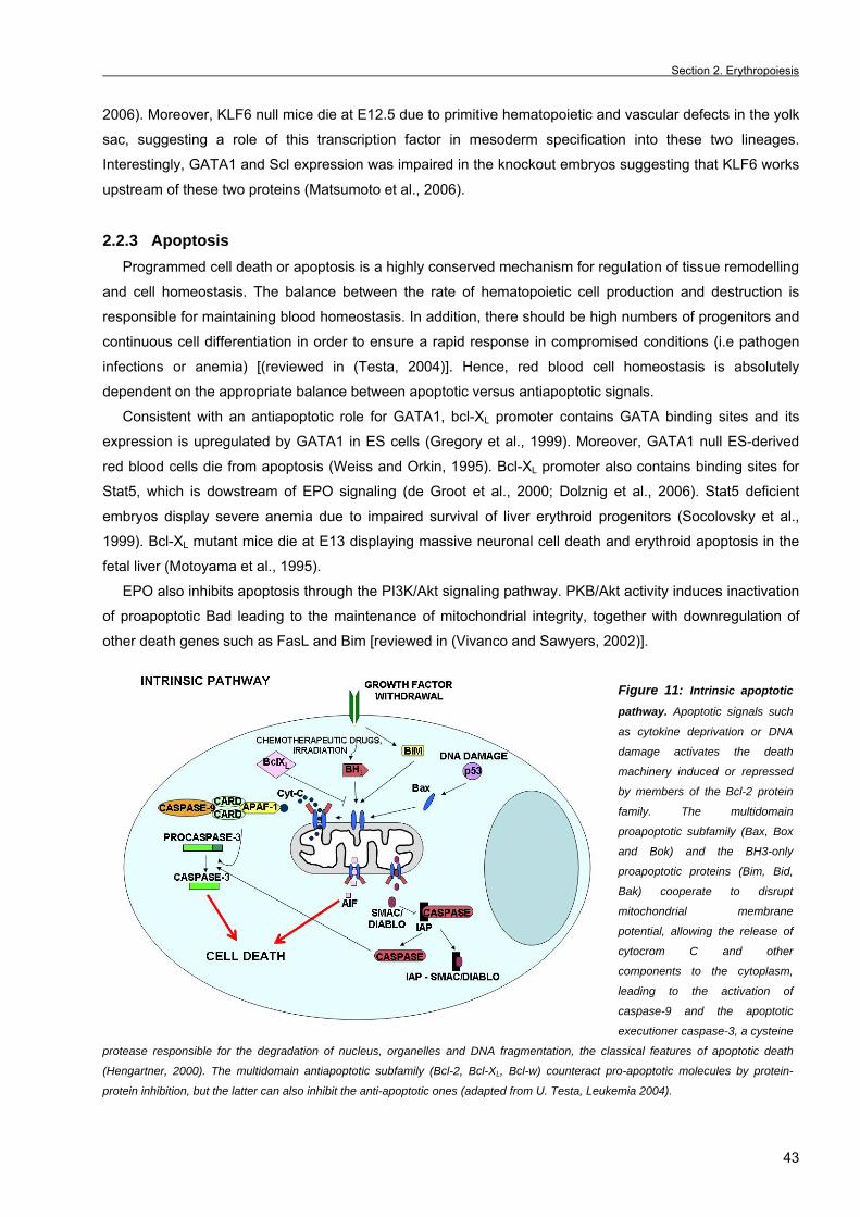
Section 2. Erythropoiesis
43
2006). Moreover, KLF6 null mice die at E12.5 due to primitive hematopoietic and vascular defects in the yolk
sac, suggesting a role of this transcription factor in mesoderm specification into these two lineages.
Interestingly, GATA1 and Scl expression was impaired in the knockout embryos suggesting that KLF6 works
upstream of these two proteins (Matsumoto et al., 2006).
2.2.3 Apoptosis Programmed cell death or apoptosis is a highly conserved mechanism for regulation of tissue remodelling
and cell homeostasis. The balance between the rate of hematopoietic cell production and destruction is
responsible for maintaining blood homeostasis. In addition, there should be high numbers of progenitors and
continuous cell differentiation in order to ensure a rapid response in compromised conditions (i.e pathogen
infections or anemia) [(reviewed in (Testa, 2004)]. Hence, red blood cell homeostasis is absolutely
dependent on the appropriate balance between apoptotic versus antiapoptotic signals.
Consistent with an antiapoptotic role for GATA1, bcl-XL promoter contains GATA binding sites and its
expression is upregulated by GATA1 in ES cells (Gregory et al., 1999). Moreover, GATA1 null ES-derived
red blood cells die from apoptosis (Weiss and Orkin, 1995). Bcl-XL promoter also contains binding sites for
Stat5, which is dowstream of EPO signaling (de Groot et al., 2000; Dolznig et al., 2006). Stat5 deficient
embryos display severe anemia due to impaired survival of liver erythroid progenitors (Socolovsky et al.,
1999). Bcl-XL mutant mice die at E13 displaying massive neuronal cell death and erythroid apoptosis in the
fetal liver (Motoyama et al., 1995).
EPO also inhibits apoptosis through the PI3K/Akt signaling pathway. PKB/Akt activity induces inactivation
of proapoptotic Bad leading to the maintenance of mitochondrial integrity, together with downregulation of
other death genes such as FasL and Bim [reviewed in (Vivanco and Sawyers, 2002)].
Figure 11: Intrinsic apoptotic
pathway. Apoptotic signals such
as cytokine deprivation or DNA
damage activates the death
machinery induced or repressed
by members of the Bcl-2 protein
family. The multidomain
proapoptotic subfamily (Bax, Box
and Bok) and the BH3-only
proapoptotic proteins (Bim, Bid,
Bak) cooperate to disrupt
mitochondrial membrane
potential, allowing the release of
cytocrom C and other
components to the cytoplasm,
leading to the activation of
caspase-9 and the apoptotic
executioner caspase-3, a cysteine
protease responsible for the degradation of nucleus, organelles and DNA fragmentation, the classical features of apoptotic death
(Hengartner, 2000). The multidomain antiapoptotic subfamily (Bcl-2, Bcl-XL, Bcl-w) counteract pro-apoptotic molecules by protein-
protein inhibition, but the latter can also inhibit the anti-apoptotic ones (adapted from U. Testa, Leukemia 2004).
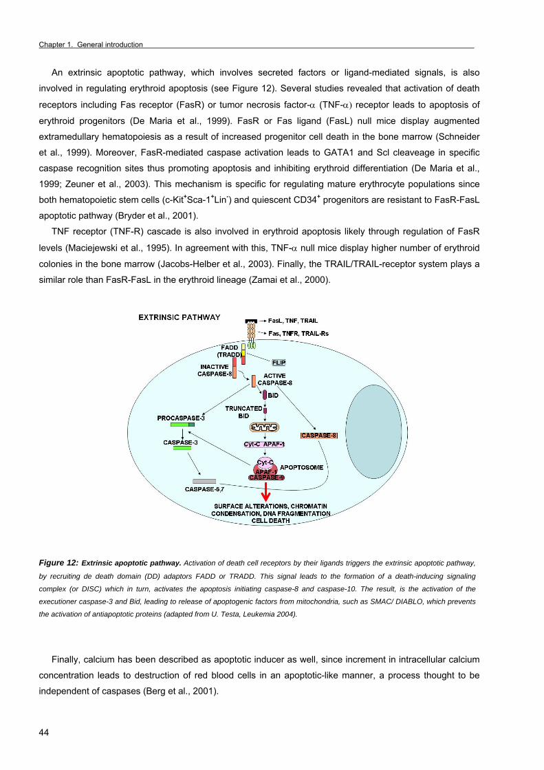
Chapter 1. General introduction _
44
An extrinsic apoptotic pathway, which involves secreted factors or ligand-mediated signals, is also
involved in regulating erythroid apoptosis (see Figure 12). Several studies revealed that activation of death
receptors including Fas receptor (FasR) or tumor necrosis factor-α (TNF-α) receptor leads to apoptosis of
erythroid progenitors (De Maria et al., 1999). FasR or Fas ligand (FasL) null mice display augmented
extramedullary hematopoiesis as a result of increased progenitor cell death in the bone marrow (Schneider
et al., 1999). Moreover, FasR-mediated caspase activation leads to GATA1 and Scl cleaveage in specific
caspase recognition sites thus promoting apoptosis and inhibiting erythroid differentiation (De Maria et al.,
1999; Zeuner et al., 2003). This mechanism is specific for regulating mature erythrocyte populations since
both hematopoietic stem cells (c-Kit+Sca-1+Lin-) and quiescent CD34+ progenitors are resistant to FasR-FasL
apoptotic pathway (Bryder et al., 2001).
TNF receptor (TNF-R) cascade is also involved in erythroid apoptosis likely through regulation of FasR
levels (Maciejewski et al., 1995). In agreement with this, TNF-α null mice display higher number of erythroid
colonies in the bone marrow (Jacobs-Helber et al., 2003). Finally, the TRAIL/TRAIL-receptor system plays a
similar role than FasR-FasL in the erythroid lineage (Zamai et al., 2000).
Figure 12: Extrinsic apoptotic pathway. Activation of death cell receptors by their ligands triggers the extrinsic apoptotic pathway,
by recruiting de death domain (DD) adaptors FADD or TRADD. This signal leads to the formation of a death-inducing signaling
complex (or DISC) which in turn, activates the apoptosis initiating caspase-8 and caspase-10. The result, is the activation of the
executioner caspase-3 and Bid, leading to release of apoptogenic factors from mitochondria, such as SMAC/ DIABLO, which prevents
the activation of antiapoptotic proteins (adapted from U. Testa, Leukemia 2004).
Finally, calcium has been described as apoptotic inducer as well, since increment in intracellular calcium
concentration leads to destruction of red blood cells in an apoptotic-like manner, a process thought to be
independent of caspases (Berg et al., 2001).
Section 2. Erythropoiesis
45
2.3 Erythroid pathologies
Many erythroid disorders are due to a deregulation of the apoptotic mechanisms that control red blood
cell compartment.
• Polycythemia vera (PV) Polycythemia vera (PV) patients display an acquired myelodysplastic syndrome (MDS) with erythroid,
megakaryocytic and granulocytic cell overproduction, that usually develops into acute leukemia [reviewed in
(Spivak, 2002)]. Some molecular alterations have been described in this syndrome that results in increased
apostosis resistance in the erythroid lineage. However, most PV patients harbour a point mutation that
activates Jak2 leading to a blockage of death receptor-mediated apoptosis concomitant with bcl-XL
overexpression. Moreover, PV erythroid cells also express high levels of FLICE-inhibitory protein (c-FLIP), a
specific inhibitor or caspase-8, which confers resistance to the extrinsic apoptotic pathway (Zeuner et al.,
2006).
• Anemia Multiple myeloma associated anemia as well as Fanconi anemia are due to spontaneous apoptosis due
to a higher sensitivity to FasL-mediated apoptosis (Silvestris et al., 2002), leading to GATA1 cleavage and
cell death. Moreover, point mutations in the GATA1 gene cause dyserythropoietic anemia and
thrombocytopenia due to a conformational change that impedes its binding to FOG-1 (Nichols et al., 2000).
On the other hand, rheumatoid arthritis-linked anemia is caused by an excessive TNF-α release.
• Hemoglobinopathies Abnormal globin expression leads to hemoglobinopathies commonly known as thalassemias.
Thalassemias are characterized by unbalanced globin chain synthesis leading to free α-chains (β-
thalassemias) or β-chains (α-thalassemias) that precipitate into the cytoplasm promoting hemolysis of the
more mature erythroblasts (cells with higher hemoglobinization). GATA1 and EKLF control the expression of
both β-globin and the α-haemoglobin stabilizing protein. Thus, the absence of EKLF in mice results in globin
imbalance and embryonic death by E16 due to a phenotype resembling β-thalassemia.
On the other hand, sickle cell disease is also caused by β-chain precipitation leading to an aberrant
erythroid morphology, which blocks small vessels [reviewed in (Testa, 2004)].
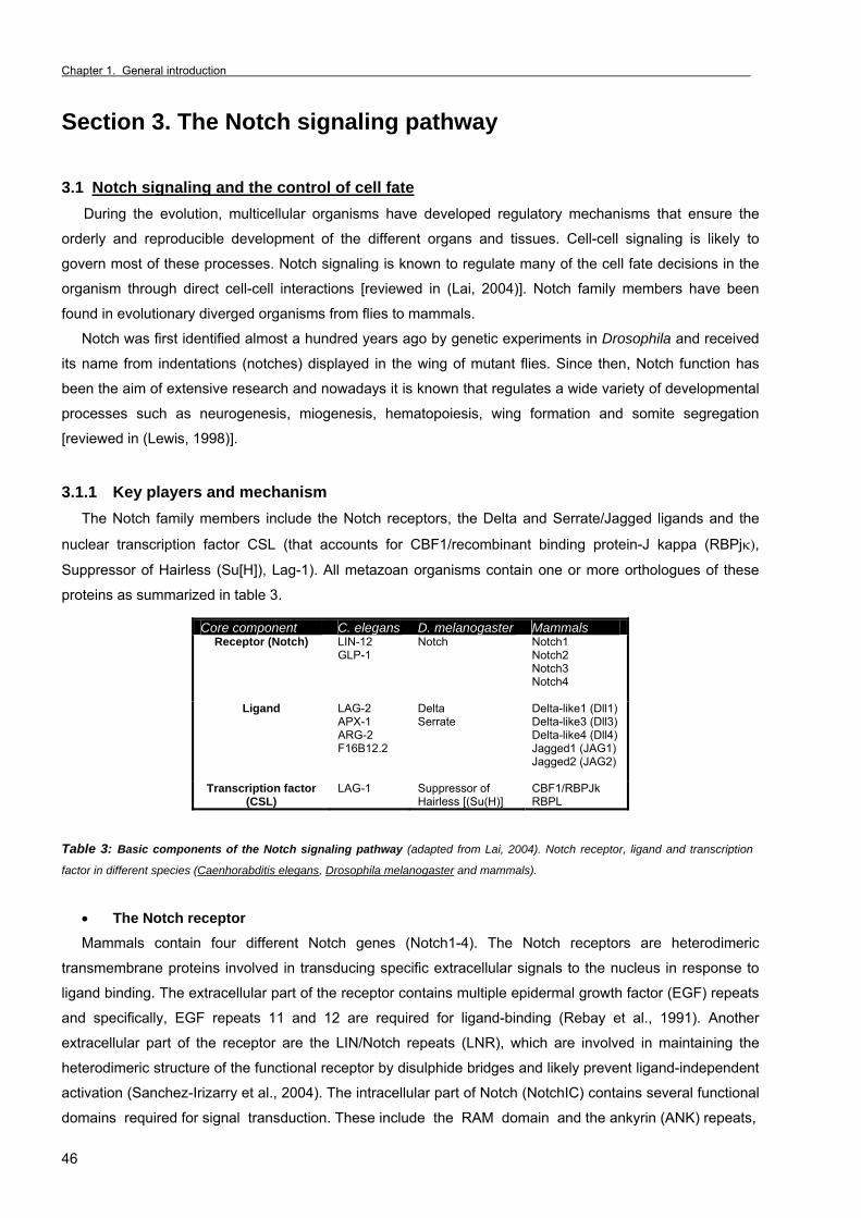
Chapter 1. General introduction _
46
Section 3. The Notch signaling pathway 3.1 Notch signaling and the control of cell fate During the evolution, multicellular organisms have developed regulatory mechanisms that ensure the
orderly and reproducible development of the different organs and tissues. Cell-cell signaling is likely to
govern most of these processes. Notch signaling is known to regulate many of the cell fate decisions in the
organism through direct cell-cell interactions [reviewed in (Lai, 2004)]. Notch family members have been
found in evolutionary diverged organisms from flies to mammals.
Notch was first identified almost a hundred years ago by genetic experiments in Drosophila and received
its name from indentations (notches) displayed in the wing of mutant flies. Since then, Notch function has
been the aim of extensive research and nowadays it is known that regulates a wide variety of developmental
processes such as neurogenesis, miogenesis, hematopoiesis, wing formation and somite segregation
[reviewed in (Lewis, 1998)]. 3.1.1 Key players and mechanism The Notch family members include the Notch receptors, the Delta and Serrate/Jagged ligands and the
nuclear transcription factor CSL (that accounts for CBF1/recombinant binding protein-J kappa (RBPjκ),
Suppressor of Hairless (Su[H]), Lag-1). All metazoan organisms contain one or more orthologues of these
proteins as summarized in table 3.
Table 3: Basic components of the Notch signaling pathway (adapted from Lai, 2004). Notch receptor, ligand and transcription
factor in different species (Caenhorabditis elegans, Drosophila melanogaster and mammals).
• The Notch receptor Mammals contain four different Notch genes (Notch1-4). The Notch receptors are heterodimeric
transmembrane proteins involved in transducing specific extracellular signals to the nucleus in response to
ligand binding. The extracellular part of the receptor contains multiple epidermal growth factor (EGF) repeats
and specifically, EGF repeats 11 and 12 are required for ligand-binding (Rebay et al., 1991). Another
extracellular part of the receptor are the LIN/Notch repeats (LNR), which are involved in maintaining the
heterodimeric structure of the functional receptor by disulphide bridges and likely prevent ligand-independent
activation (Sanchez-Irizarry et al., 2004). The intracellular part of Notch (NotchIC) contains several functional
domains required for signal transduction. These include the RAM domain and the ankyrin (ANK) repeats,
Core component C. elegans D. melanogaster Mammals Receptor (Notch) LIN-12
GLP-1 Notch Notch1
Notch2 Notch3 Notch4
Ligand LAG-2 APX-1 ARG-2 F16B12.2
Delta Serrate
Delta-like1 (Dll1) Delta-like3 (Dll3) Delta-like4 (Dll4) Jagged1 (JAG1) Jagged2 (JAG2)
Transcription factor (CSL)
LAG-1 Suppressor of Hairless [(Su(H)]
CBF1/RBPJk RBPL
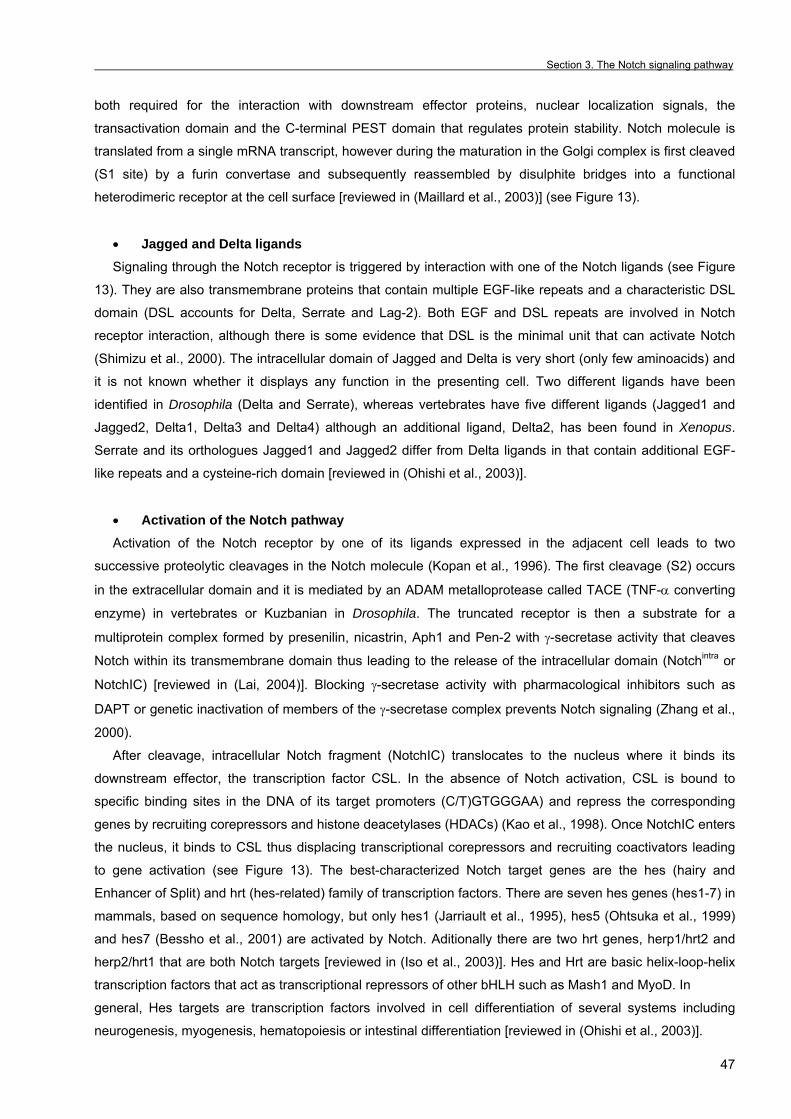
Section 3. The Notch signaling pathway
47
both required for the interaction with downstream effector proteins, nuclear localization signals, the
transactivation domain and the C-terminal PEST domain that regulates protein stability. Notch molecule is
translated from a single mRNA transcript, however during the maturation in the Golgi complex is first cleaved
(S1 site) by a furin convertase and subsequently reassembled by disulphite bridges into a functional
heterodimeric receptor at the cell surface [reviewed in (Maillard et al., 2003)] (see Figure 13).
• Jagged and Delta ligands Signaling through the Notch receptor is triggered by interaction with one of the Notch ligands (see Figure
13). They are also transmembrane proteins that contain multiple EGF-like repeats and a characteristic DSL
domain (DSL accounts for Delta, Serrate and Lag-2). Both EGF and DSL repeats are involved in Notch
receptor interaction, although there is some evidence that DSL is the minimal unit that can activate Notch
(Shimizu et al., 2000). The intracellular domain of Jagged and Delta is very short (only few aminoacids) and
it is not known whether it displays any function in the presenting cell. Two different ligands have been
identified in Drosophila (Delta and Serrate), whereas vertebrates have five different ligands (Jagged1 and
Jagged2, Delta1, Delta3 and Delta4) although an additional ligand, Delta2, has been found in Xenopus.
Serrate and its orthologues Jagged1 and Jagged2 differ from Delta ligands in that contain additional EGF-
like repeats and a cysteine-rich domain [reviewed in (Ohishi et al., 2003)].
• Activation of the Notch pathway Activation of the Notch receptor by one of its ligands expressed in the adjacent cell leads to two
successive proteolytic cleavages in the Notch molecule (Kopan et al., 1996). The first cleavage (S2) occurs
in the extracellular domain and it is mediated by an ADAM metalloprotease called TACE (TNF-α converting
enzyme) in vertebrates or Kuzbanian in Drosophila. The truncated receptor is then a substrate for a
multiprotein complex formed by presenilin, nicastrin, Aph1 and Pen-2 with γ-secretase activity that cleaves
Notch within its transmembrane domain thus leading to the release of the intracellular domain (Notchintra or
NotchIC) [reviewed in (Lai, 2004)]. Blocking γ-secretase activity with pharmacological inhibitors such as
DAPT or genetic inactivation of members of the γ-secretase complex prevents Notch signaling (Zhang et al.,
2000).
After cleavage, intracellular Notch fragment (NotchIC) translocates to the nucleus where it binds its
downstream effector, the transcription factor CSL. In the absence of Notch activation, CSL is bound to
specific binding sites in the DNA of its target promoters (C/T)GTGGGAA) and repress the corresponding
genes by recruiting corepressors and histone deacetylases (HDACs) (Kao et al., 1998). Once NotchIC enters
the nucleus, it binds to CSL thus displacing transcriptional corepressors and recruiting coactivators leading
to gene activation (see Figure 13). The best-characterized Notch target genes are the hes (hairy and
Enhancer of Split) and hrt (hes-related) family of transcription factors. There are seven hes genes (hes1-7) in
mammals, based on sequence homology, but only hes1 (Jarriault et al., 1995), hes5 (Ohtsuka et al., 1999)
and hes7 (Bessho et al., 2001) are activated by Notch. Aditionally there are two hrt genes, herp1/hrt2 and
herp2/hrt1 that are both Notch targets [reviewed in (Iso et al., 2003)]. Hes and Hrt are basic helix-loop-helix
transcription factors that act as transcriptional repressors of other bHLH such as Mash1 and MyoD. In
general, Hes targets are transcription factors involved in cell differentiation of several systems including
neurogenesis, myogenesis, hematopoiesis or intestinal differentiation [reviewed in (Ohishi et al., 2003)].
Chapter 1. General introduction _
48
Figure 13: The canonical Notch signaling pathway. Notch activation leads to a cascade of proteolytic events resulting in NotchIC
translocation to the nucleus and NotchIC/RBPjκ dependent target gene expression (adapted from Lai, 2004).
3.1.2 Control of cell-fate decisions During the first stages of development of a given tissue, most of the cells have an equivalent
developmental background and potentiality. In this homogeneous population, the interactions between Notch
and Jagged/Delta (located in neighboring cells) are responsible for generating cell diversity through
activating specific gene programs [reviewed in (Lewis, 1998)]. Two different mechanisms have been
proposed to explain how Notch regulates cellular diversity: lateral inhibition and lateral induction processes.
• Lateral inhibition model The lateral inhibition model implies that in a population of equivalent cells expressing low levels of both
receptor and ligand, the cell that first produces more ligand, activates the Notch receptor in the neighboring
cell that dowregulates ligand expression. This mechanism allows the maintenance and intensification of the
differences in expression of receptor and ligand, since the ligand-expressing cell does not receive inhibitory
signals from its surrounding cells. A salt-and-pepper mosaic of cells emerges and as a result, the ligand-
expressing cell differentiates into a distinct lineage than the surrounding cells (see Figure 14) [reviewed in
(Lewis, 1998)].
The classical example of lateral inhibition occurs during the neural-epidermal choice in Drosophila.
Specification to the neural lineage needs the expression of the achaete-scute bHLH transcriptional
activators. Notch activation results in the expression of the Enhancer of split Complex [E(spl)-C] which
represses the expression of these proneural genes and results in the inhibition of the neural fate (Parks et
al., 1997) leading to the epidermal fate. In mammals, Notch signaling represses neurogenesis and
myogenesis via homologous Hairy/E(spl)-related bHLH repressors known as Hes, which inhibit the bHLH
transcription factors Mash1 and MyoD [reviewed in (Artavanis-Tsakonas et al., 1999)].
Ligand Notch receptor
Notch-IC
RAM domain Ankyrin domain TAD domain PEST domain
EGF-like repeats
LIN12/Notch repeats
EGF-like repeats
DSL domain Extracellular
space
ADAM-mediated S2 cleavage
γ-secretase-mediated S3 cleavage
Signal receiving-cell
CSL/RBP-jκ
Co-repressors
CSL
Co-activators
Co-R
Notch-IC
nuclear translocation
Nucleus
GTGGGAA
Co-R CSL OFF
GTGGGAA CSL ON Co-A
Default repression by CSL
Notch-IC mediated target activation
Hes1,5,7 Herp1-2
…others?
Signal sending-cell
Co-A
Co-A

Section 3. The Notch signaling pathway
49
Figure 14: Lateral inhibition model. i) Equivalent cells expressing receptor and ligand exert mutual inhibitory signals. ii) One of
these cells expresses high levels of ligand, inhibiting the expression in the surrounding cells. iii) The ligand expressing cell switches on
a different genetic program than the neighboring cells and adopts a different cellular fate. iv) As a result, a salt-and-pepper pattern is
generated. Loss of Notch signaling drives all the cells to adopt the restricted cell fate. In Drosophila neuro-epidermis system, all the
cells would differentiate into neural cells. Gain of Notch signaling inhibits differentiation and all the cells would appear as epidermis
(adapted from Lai, 2004).
• Lateral induction model
Notch can also function in a lateral induction model. In this case, activation of Notch in a given cell
promotes the expression of the Notch ligands in the same cell. Thus, a ligand-expressing cell activates
Notch signaling in the adjacent cell preventing the salt-and-pepper pattern, inducing their cell-fate choices
cooperatively and forming defined boundaries of gene expression (see Figure 15) [reviewed in (Lewis,
1998)].
In Drosophila, the classical example of lateral induction occurs during the wing formation. Notch signaling
between the dorsal and ventral compartments of the wing imaginal disc results in the formation of the wing
margin. In this case, Notch activates the expression of vestigial, a transcriptional coactivator that is required
for proper wing development (Couso et al., 1995). In vertebrates the classical example is the generation of
boundaries during somitogenesis. Somites are regularly spaced blocks of mesoderm that split off from the
presomitic mesoderm in a periodic oscillatory manner. This is mediated by Notch through oscillatory
activation of hes genes (Jouve et al., 2000) and oscillatory inhibition of Notch activity by Lunatic fringe (Dale
et al., 2003).
Thus, Notch principal role during development is the control of cell-fate decisions generating cell diversity
in cells sending or receiving signals. Although these two models explain some developmental events, other
developmental systems are likely to be more complex.
Figure 15: Lateral induction model. i) A group of cells (in grey) signal to the adjacent white cells. ii) Subsequently, the Notch
receptor expressing cells adopt a new fate, in this case, to form a boundary (black cells). Loss of Notch signaling results in the absence
of the new fate, whereas excessive Notch signaling has the opposite effect (adapted from Lai, 2004).
Chapter 1. General introduction _
50
• Other key players in Notch signaling Glycosilation of the Notch receptor by Fringe glycosyltransferases (Radical, Lunatic and Manic Fringe) is
responsible for modulating ligand specificity. Fringe proteins are shown to regulate Notch activity and
contribute to generation of cell diversity [reviewed in (Irvine, 1999)]. Ubiquitination has been shown to be required for endocytosis of Notch and their ligands. Neuralized and
Mind bomb which belong to the RING-type E3 ubiquitin ligases ubiquitinate Delta ligands and mutations in
these proteins result in Notch-loss of function phenotypes (Itoh et al., 2003; Lai et al., 2001). Recent studies
suggest that Delta ubiquitination increases its affinity for Notch binding. Moreover, endocytosis of the Delta
ligand bound to extracellular Notch facilitates the S2 cleavage of Notch receptor (Nichols et al., 2007).
3.2 Role of the Notch signaling pathway in hematopoiesis Notch signaling pathway functions at various stages of hematopoietic development [reviewed in (Ohishi et
al., 2003)]. Notch is activated by interactions between precursors and stromal supporting cells but also by
interactions between hematopoietic cells.
3.2.1 Expression of Notch members in the hematopoietic system Notch1 and Notch2 expression is found in CD34+Lin- bone marrow hematopoietic precursors, suggesting
a role for Notch very early in mammalian blood cell development (Milner et al., 1994; Ohishi et al., 2000),
Conversely, Jagged1 expression is found in bone marrow stromal cells (Walker et al., 2001) as well as
Delta1 and Delta4 (Karanu et al., 2001). All these ligands are also expressed in thymic epithelial cells,
consistent with the important role of Notch in T-cell development (Felli et al., 1999; Mohtashami and Zuniga-
Pflucker, 2006).
In the myeloid lineage, monocytes express high levels of Notch1 and 2 but expression has not been
found in granulocytes (Ohishi et al., 2000). Finally, bone marrow erythroid progenitors express Notch1 but
expression decreases in more mature erythroid cells such as acidophilic normoblasts (Ohishi et al., 2000;
Walker et al., 2001).
3.2.2 Role of Notch in HSC self-renewal There is important evidence supporting the idea that Notch plays a crucial role in HSC self-renewal.
Recent work performed by Duncan et al, demonstrated that Notch signaling is active in the KSL
subpopulation located in the bone marrow niche and downregulated as these cells differentiate. Moreover,
Notch inhibition by a dominant negative CBF1 leads to accelerated differentiation of HSCs in vitro and
depletion of HSC activity in vivo indicating that Notch is required for the maintenance of the undifferentiated
state of HSCs (Duncan et al., 2005).
Retroviral transduction of the Notch intracellular domain in c-Kit+Sca1+Lin- murine hematopoietic
progenitors leads to immortalization of these cells and repopulation of both myeloid and lymphoid lineages
when transplanted into lethally irradiated mice (Varnum-Finney et al., 2000). Moreover, Notch1IC expands
the number of bone marrow repopulating cells in secondary transplants and promotes their lymphoid

Section 3. The Notch signaling pathway
51
differentiation (Stier et al., 2002). A similar result was obtained by expressing Notch4IC in human Lin- cord
blood cells (Vercauteren and Sutherland, 2004).
Ex vivo expansion of human hematopoietic cells is critical for clinical procedures that involve stem cell
transplantation. Addition of soluble Jagged1 to ex vivo cultures of human CD34+CD38-Lin- cord blood cells
expand human stem cells without loosing the ability to repopulate the hematopoiesis of NOD/SCID mice
(Karanu et al., 2000). A similar effect was found by incubating bone marrow KSL progenitors with a Delta1
fusion protein (Varnum-Finney et al., 2003). Many of these effects are reproduced in CD34-KSL cells
retrovirally transduced with hes1, suggesting that this protein is responsible for the effects of activating
Notch1 on HSCs self-renewal (Kunisato et al., 2003).
3.2.3 Notch regulation of lymphoid cell-fate decisions Notch signaling regulates several cell-fate decisions in the lymphoid lineage. There is strong evidence
from different studies that Notch activation promotes T- whereas inhibits B-cell fate. Deletion of RBPjκ in
hematopoietic cells results in increased B-cell differentiation and blockage of T-cell development (Han et al.,
2002). Conversely, expression of Notch1IC in progenitors blocks B-cell differentiation and leads to
generation of immature CD4+CD8+ T cells (Pui et al., 1999). It is still unclear whether this B- versus T-cell
decision is made in the CLP or in a recently described early T-cell progenitor (ETP) [reviewed in (Maillard et
al., 2003)]. In addition, Notch2 is required to generate the marginal zone B cells in the spleen which are
important for T-cell independent immune response [reviewed in (Maillard et al., 2003)].
Notch is also involved in the commitment of αβ versus γδ TCR (T-cell receptor) lineage. Notch1 activation
favours the αβ choice since decreased Notch1 levels results in an increase of γδ cells (Washburn et al.,
1997). Similarly, conditional inactivation of RBPjκ increases the number of γδ T cells (Tanigaki et al., 2004).
At the last stage of T-cell differentiation, Notch is also involved in the decision between CD4+ and CD8+
from CD4+CD8+ double positive cells since expression of Notch1IC in thymocytes favours the generation of
CD8+ cells with a corresponding decrease in CD4+ T cells (Robey et al., 1996).
3.2.4 Notch in myeloid differentiation Many experimental models have been used to determine the role of Notch in myelopoiesis from cell lines
to in vivo models. Early studies with myeloid cell lines showed that forced Notch1 or Notch2 activation could
inhibit differentiation in a cytokine-dependent manner (Bigas et al., 1998; Milner et al., 1996), likely through
expression of Gata2 (Kumano et al., 2001). Recently, a similar result has been reported with KSL
progenitors co-cultured on an OP9 stromal cell line expressing Delta1 (de Pooter et al., 2006). However,
several results suggest that Notch activity is mainly required for T and B lymphoid differentiation in vivo
(Radtke et al., 1999).
3.2.5 Notch in apoptosis There is increasing evidence that Notch1 activity is involved in regulating programmed cell death or
apoptosis. In most cases, Notch activation inhibits apoptosis as described in T cells (Jehn et al., 1999),
Kaposi’s sarcoma (Curry et al., 2005) and Hodgkin lymphoma cells (Jundt et al., 2002), however and likely
due to cell context specificity, there are several examples that show Notch as a positive regulator of

Chapter 1. General introduction _
52
apoptosis. In B-ALL cell lines, activation of Notch induces apoptosis, most likely through Hes1 (Zweidler-
McKay et al., 2005) comparable to human peripheral blood monocytes cultured in the presence of
immobilized Delta1 ligand and M-CSF (Ohishi et al., 2000).
Whether Notch induces apoptosis in the erythroid lineage remains controversial. In K562 cell line, Notch
inhibits erythroid differentiation and induces apoptosis, likely through Hes1, which inhibits GATA1 activity
and bcl-XL expression (Ishiko et al., 2005). However, Notch1 prevents apoptosis in the murine
erythroleukemic cell line during HMBA-induced differentiation (Jang et al., 2004; Shelly et al., 1999).
3.2.6 Notch implication in the ontogeny of the hematopoietic system During the recent years, the function of Notch in embryonic hematopoiesis has partially been elucidated.
Some of the work is the aim of this thesis and will be discused in detail; however, while this work was in
progress other important contributions came out and have been included in this section.
The first in vivo proof that Notch signaling plays a key role in the generation of HSCs during embryonic
development came from Kumano et al by studying the Notch1 null mice. Using P-sp explant culture on OP9
stromal cell line, they found that definitive hematopoiesis is impaired in the Notch1-/- embryos due to the lack
of HSC. In contrast, the number of CFC in the Notch1-/- yolk sacs was similar to wild-type, suggesting that
primitive hematopoiesis is at least partially preserved. However, HSC from Notch1-/- YS failed to reconstitute
myeloablated new-born mice. At E9.5 Notch1 null embryos display similar number of CD34+c-Kit+ and VE-
cadh+CD45- hemogenic endothelial cells in both YS and P-sp, suggesting that lack of definitive
hematopoiesis is due to impaired commitment of HSC from endothelial cells (Kumano et al., 2003).
Consistent with this finding, chimeric mice with Notch1-deficient ES cells do not contribute to long-term
definitive hematopoiesis (Hadland et al., 2004). In contrast, analysis of Notch2-/- embryos indicates that
Notch2 is dispensible for the generation of hematopoietic cells (Kumano et al., 2003).
Finally, important contributions for the role of Notch in the formation of HSC came from zebrafish studies
(Danio rerio). Mind bomb mutants display normal primitive hematopoiesis but impaired HSC development.
Consistent with this phenotype c-myb and runx1 expression is lost in the aorta of Mind bomb mutants.
Conversely, transient notch1 expression revealed an expansion in the number of HSCs, dependent on
Runx1 (Burns et al., 2005). Moreover, Runx1 but not GATA2 or Scl was able to reconstitute the generation
of CFC progenitors but not HSCs in embryonic Notch1-null cells in mice (Nakagawa et al., 2006). These
results indicate that Runx1 acts downstream of Notch signaling for HSC emergence in the AGM.
3.3 Altered Notch signaling and disease Three different congenital diseases due to mutations in Notch ligands have been described. Mutations in
the Jagged1 gene are responsible for Alagille sydrome, which results in impaired generation and function of
different organs such as heart, eye, liver and skeleton (Li et al., 1997). Delta3 mutations are responsible for
spondylocostal dysostosis, a developmental disease characterized by rib fusion and trunk dwarfism (Bulman
et al., 2000). Finally, mutations in the extracellular EGF-like repeats of Notch3 results in cerebral autosomal
dominant arteriopathy with subcortical infarcts and leukoencephalopathy (CADASIL), characterized by
migraines, strokes and dementia, further supporting that Notch is involved in neural development (Joutel et
al., 1996).

Section 3. The Notch signaling pathway
53
Notch also participates in tumorigenic processes in different tissues including mammary gland, skin,
cervix and prostate [reviewed in (Lai, 2004)], colon (Fernandez-Majada et al., 2007) or pancreas (Miyamoto
et al., 2003).
• Hematopoietic disorders The human Notch1 gene was first identified in a t(7;9)(q34;q34.3) chromosomal translocation in which
Notch1 fused to the TCR-β resulted in constitutive activation of Notch1 in T cells leading to human T-cell
leukemia (Ellisen et al., 1991). However, this rearrangment is a rare event in T-cell leukemias and point
mutations occurring in the heterodimerization domain (aberrantly facilitating Notch activation) or in the PEST
domain (resulting in a more stable Notch1IC protein) have been recently described in 50% of human T-ALL
(Weng et al., 2004).
In the last few years, some reports have suggested a crosstalk between the Ikaros transcription factor
(involved in T-cell development) and the Notch signaling pathway in T-cell tumorigenesis. In this sense,
homozygous mice for a hypomorphic mutation in the Ikaros gene which develop thymic lymphomas have
higher Notch activity and hes1 expression in thymocytes (Dumortier et al., 2006). Thus, in normal conditions
Ikaros may be repressing the Notch target genes likely by competing with RBPjκ for a similar DNA-binding
consensus.
In B cells, Epstein-Barr virus or Kaposi’s sarcoma-associated herpes viruses are known to activate Notch-
target genes by using viral proteins that bind to the RBPjκ consensus leading to cell immortalization and
transformation [reviewed in (Milner and Bigas, 1999)].
3.4 Animal models To study the role of the Notch in development and in adult organisms, transgenic and knockout mutants
of several Notch signaling elements have been generated in species such as Drosophila (fly), C. elegans
(nematodes), Danio rerio (zebrafish) and Mus musculus (mice).
The strongest Notch-loss-of-function mutation is obtained by deletion of the RBPjκ gene, the unique
effector of the Notch signaling pathway (Oka et al., 1995). RBPjκ-/- embryos display a complex phenotype
with impaired somitogenesis, defects in the neural tube formation and growth retardation (see Figure 16).
Moreover, the fusion of the embryonic allantois to the maternal placenta does not occur in RBPjκ null
embryos, thus umbilical vessels cannot form. All these features indicate that Notch signaling is required for
vasculogenesis, somitogenesis and neurogenesis (Oka et al., 1995).
Figure 16: The RBPjκ null mice. Photographs of E9.5 RBPjκ
wild-type and knockout embryos. RBPjκ disruption is embryonic
lethal by E10 and leads to growth retardation, defective
somitogenesis, pericardium dilatation, neural tube contorsion
and defective allantoid fusion.
E9.5 RBPjκwt E9.5 RBPjκ-/-
15x 30x

Chapter 1. General introduction _
54
The phenotype of the Notch1 null embryos is quite similar to the RBPjκ null ones, although embryonic
lethality occurs at E11.5. Notch1-/- mutants also display growth retardation, impaired neurogenesis and
somite formation, but the chorioallantoic fusion defect is not found (Conlon et al., 1995).
Notch2 mutants die at E11.5 and display a massive apoptosis in the neuroephitelium and otic and optic
vesicles (Hamada et al., 1999).
Notch3 null mutants are viable and fertile and only display some defects in vasculogenesis (Domenga et
al., 2004; Krebs et al., 2003).
Finally, Notch4 mutant embryos are viable and fertile although it cooperates with Notch1 mutation in
inducing vasculogenic defects (Krebs et al., 2000).
Jagged1 null mice die at E10. The mutant embryos can form the primitive blood vessels both in the yolk
sac and the embryo proper but they fail to remodel the vascular plexus to form the large vitelline blood
vessels, suggesting that angiogenesis but not vasculogenesis is impaired (Xue et al., 1999).
Jagged2 mutation is not lethal until birth and mutant fetuses display impaired craniophacial
morphogenesis and palatal clefting, defects in limb formation and abnormal thymic development with a
reduction of γδ T cells (Jiang et al., 1998).
Finally, Mind bomb mutant mice and double knockouts for presenilin1 and presenilin2 mice die at E10.5
and E9.5 respectively, and both display several Notch-associated defects, similar to RBPjκ-/- embryos
(impaired neurogenesis, somitogenesis and vasculature remodelling) suggesting that all these proteins acts
upstream of Notch and are required for Notch signaling (Donoviel et al., 1999; Koo et al., 2005).

CHAPTER 2: AIMS


Aims
57
AIMS Although Notch signaling pathway was previously reported to regulate many cell-fate decisions in the
hematopoietic system, its role in the ontogeny of embryonic hematopoiesis was completely unknown at the
beginning of this thesis. Thus, by using the RBPjκ-/- mice as a model, we aimed to study whether the Notch
signaling pathway was involved in the generation of both primitive and/or definitive hematopoiesis in the
mice embryo.
The following specific aims were proposed for this project:
• To study whether the Notch/RBPjκ signaling plays a specific role in the ontogeny of the primitive
hematopoietic system in the murine yolk sac from E7.5 to E9.5.
• To determine whether Notch/RBPjκ signaling is required for the generation of definitive HSCs and
other progenitors from the murine P-sp/AGM region at E9.5-11.5.

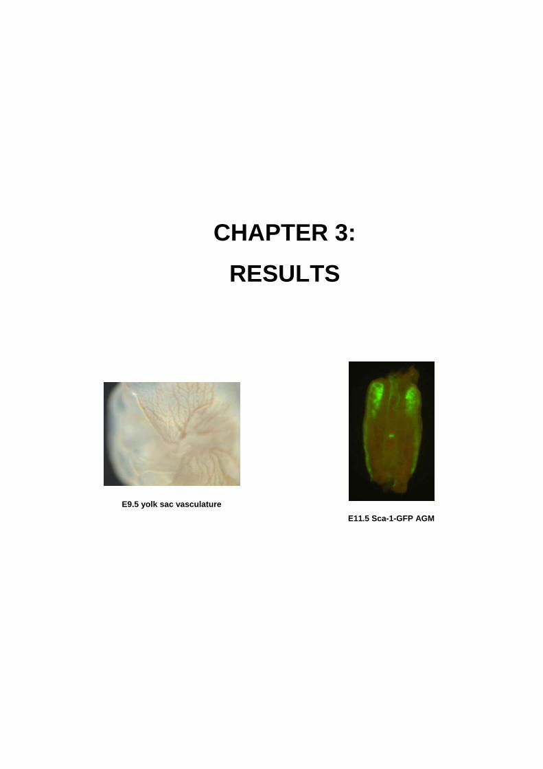
CHAPTER 3: RESULTS
E9.5 yolk sac vasculature E11.5 Sca-1-GFP AGM

Section 1
61
SECTION 1: RBPjκ-dependent Notch function regulates Gata2 and is essential
for the formation of intra-embryonic hematopoietic cells (This chapter was published in Development 132, 1117-1126, 2005). The intra-embryonic region encompassing the aorta surrounded by gonad and mesonephros tissues has
been described as one of the first embryonic hematopoietic organs involved in the generation of
hematopoietic progenitors (HP) and hematopoietic stem cells (HSC) that will later seed the bone marrow and
sustain hematopoiesis in the adulthood (Medvinsky, 1996; de Bruijn, 2000; de Bruijn, 2002). Previous to our
work there were many evidences that the Notch signaling pathway regulates different biological processes in
many hematopoietic cell lines (Bigas, 1998; Karanu, 2000; Varnum-Finney, 2000; Kumano, 2001) although
little work was done in vivo. Thus, taking advantage of the RBPjκ null mice (Oka, 1995), our aim was to
determine whether Notch signaling was required for the generation of HP/HSC from the AGM.
Here we show that in wild-type embryos, members of the Notch pathway including the Notch1 and
Notch4 receptor, and the Delta4, Jagged1 and Jagged2 ligands are expressed in endothelial cells from the
AGM aorta at E9.5-10.5. Moreover, Notch target genes such as hes1, hrt1 and hrt2 are also expressed
indicating that Notch pathway is active in this region. This expression pattern of Notch receptors and ligands
is lost in the RBPjκ null embryos (which lack the common nuclear effector thus impeding downstream
signaling from all four Notch receptors) demonstrating that positive and negative feed-back loops regulate
their expression.
We also demonstrate that RBPjκ-/- embryos display impaired hematopoietic potential from the AGM
region with a complete lack of hematopoietic progenitors (determined by CFC assay) and CD45+
hematopoietic cells after 6 days in culture. Absence of hematopoiesis correlates with a complete lack of the
critical hematopoietic transcription factor Gata2, Runx1 and Scl expression and with an increase of
endothelial cells suggesting the presence of a common progenitor (or hemangioblast) for hematopoietic and
endothelial lineages.
By double in-situ hybridization (DISH) and chromatin immunoprecipitation we describe that Notch directly
binds through RBPjκ to the gata2 promoter thus regulating its expression not only in the 32D cell line but
also in the embryo. Finally, by DISH we describe which ligands are expressed in the adjacent cell next to the
Notch1-expressing cell thus suggesting that Notch1-induced activation of gata2 expression in endothelial
cells from the ventral part of the AGM aorta and activation of the hematopoietic program in that cell may be
induced by Jagged2 or Delta4.

Chapter 3. Results _
62
RBPjκ-dependent Notch function regulates Gata2 and is essential for the formation
of intra-embryonic hematopoietic cells
AUTHORS: Àlex Robert-Moreno1, Lluís Espinosa1, José Luis de la Pompa2 and Anna Bigas1*
INSTITUTION: 1Centre Oncologia Molecular.
IDIBELL-Institut de Recerca Oncologica.
Hospitalet, Barcelona 08907, Spain. 2Dept. of Immunology and Oncology, Centro Nacional de
Biotecnología,
CSIC. Darwin, 3. Campus de Cantoblanco, Madrid 28049,
Spain
RUNNING TITLE: Notch regulates Gata2 in early hematopoiesis
*Corresponding author.
Mailing address: Centre Oncologia Molecular.
Institut de Recerca Oncològica
Gran Via Km 2.7.
08907 - Hospitalet, Barcelona. Spain
Phone: 932 607 404
Fax: 932 607 426
e-mail: [email protected]

Section 1
63
SUMMARY Definitive hematopoiesis in the mouse embryo originates from the aortic floor in the P-Sp/AGM region in
close association with endothelial cells. An important role for Notch1 in the control of hematopoietic ontogeny
has been recently established although its mechanism of action is poorly understood. Here we show detailed
analysis of Notch family gene expression in the aorta endothelium between embryonic day (E) 9.5 and
E10.5. Since Notch requires binding to RBPjκ transcription factor to activate transcription, we analyzed the
aorta of the Para-aortic Splanchnopleura/AGM in RBPjκ mutant embryos. We found specific patterns of
expression of Notch receptors, ligands and Hes genes that were lost in RBPjκ mutants. Analysis of these
mutants revealed the absence of hematopoietic progenitors, accompanied by the lack of expression of the
hematopoietic transcription factors Aml1/Runx1, Gata2 and Scl/Tal1. We show that in wild-type embryos, a
few cells lining the aorta endothelium at E9.5 simultaneously expressed Notch1 and Gata2 and demonstrate
by chromatin immunoprecipitation that Notch1 specifically associated with the Gata2 promoter in E9.5 wild-
type embryos and 32D myeloid cells, an interaction lost in RBPjκ mutants. Consistent with a role for Notch1
in regulating Gata2, we observe increased expression of this gene in 32D cells expressing activated Notch1.
Taken together these data strongly suggest that activation of Gata2 expression by Notch1/RBPjκ is a crucial
event for the onset of definite hematopoiesis in the embryo.
INTRODUCTION
Hematopoietic cells differentiate from mesoderm during embryogenesis, in close association with
endothelial cells. Definitive hematopoietic progenitors and stem cells originate in distinct sites in the embryo
including the yolk sac (YS)(Yoder et al., 1997), the umbilical and vitelline arteries (de Bruijn et al., 2000), the
para-aortic splanchnopleura (P-Sp) (Cumano et al., 2001) and the aorta/genital ridge/mesonephros (AGM)
region (Medvinsky and Dzierzak, 1996). The first hematopoietic cells detected during mouse embryonic
development are the primitive erythroid cells of the YS at embryonic day (E) 7. One day later, before
circulation between the embryo and YS is established, multipotent hematopoietic stem cells (HSC) have
been isolated from the intra-embryonic P-Sp (Cumano et al., 2001) indicating that intra-embryonic
hematopoietic cells can originate independently of the YS. In the mouse, the P-Sp forms from the splanchnic
mesoderm (the endoderm-associated mesoderm) and the whole region develops into aorta, gonads and
mesonephros and is subsequently called AGM. Around E10-11, the HSC activity is autonomously generated
in this region (reviewed by (Ling and Dzierzak, 2002).
The developmental origin and the genetic program of embryonic HSC emergence in the YS and the P-
Sp/AGM in some aspects are divergent. Yolk sac blood cells originate simultaneously with the surrounding
endothelial cells, consistent with the idea of developing from a common progenitor or hemangioblast (Palis
and Yoder, 2001). By contrast, P-Sp/AGM hematopoietic cells emerge in close association to the
presumably differentiated aortic endothelium. The lineage relationships and molecular events leading to their
differentiation are not completely understood. Immunohistochemical analyses of the AGM region reveal
overlapping expression of hematopoietic and endothelial markers in the clusters of cells that emerge from
the ventral wall of the aorta. However, Aml1/Cbfa2 (Runx1 – Mouse Genome Informatics) transcription factor
has been shown to specifically be involved in the development of intra-embryonic hematopoiesis without
Chapter 3. Results _
64
affecting the main vasculature (North et al., 1999). The analysis of recently developed transgenic mice,
which enable specific labeling of emerging HSC, provides supportive evidence that true HSCs originate
among the cells residing in the endothelial layer (Ma et al., 2002). Besides Aml1 (North et al., 2002), Gata2
(Tsai et al., 1994; Tsai and Orkin, 1997) and Scl (Tal1 – Mouse Genome Informatics) (Porcher et al., 1996;
Robb et al., 1996) are also expressed in hematopoietic clusters and endothelial-like cells lining the ventral
wall of the dorsal aorta at E10-11 and there is now strong evidence that all these transcription factors are
important for the onset of definitive hematopoiesis in the embryo.
Signaling through the Notch receptors is a widely used mechanism for cell fate specification and pattern
formation in embryonic development and adulthood (Artavanis-Tsakonas et al., 1999; Lai, 2004; Lewis,
1998). The interaction between Notch receptors and ligands results in the cleavage of the intracellular
domain of Notch that translocates to the nucleus and together with RBPjκ (Rbpsuh – Mouse Genome
Informatics) activates gene transcription. The best-characterized Notch-target genes are the orthologs of the
Hairy and enhancer of split (Hes) and Hes-related (Hrt) proteins (for a review, see (Iso et al., 2003). Notch
family members have been identified in several hematopoietic cell types from diverse origin and there is now
strong evidence that they participate in the control of hematopoietic differentiation in many different lineages
(Han et al., 2002; Radtke et al., 1999; Stier et al., 2002).
The first evidence showing the involvement of Notch in the onset of embryonic hematopoiesis has
recently been published, confirming that development of hematopoietic cells from the hemogenic
endothelium is a Notch1-regulated event and it is impaired in Notch1-deficient embryos (Hadland et al.,
2004; Kumano et al., 2003). We show here that this is an RBPjκ-dependent event, since RBPjκ mutant
embryos also lack intra-embryonic hematopoiesis. Endothelial cells are not affected, as previously seen in
the Notch1 mutant embryos. We identify several Notch family members showing distinct expression patterns
in presumptive E9.5 and E10.5 hemogenic endothelium suggesting that different Notch signals may operate
in this system. We also present evidence that Notch1 directly regulates the expression of Gata2, thus
suggesting that one of the first events in embryonic hematopoietic determination consists in the activation of
Gata2 expression by Notch1/RBPjκ.
MATERIALS AND METHODS
Animals
RBPjκ null mice have been previously described (Oka et al., 1995). Whole embryos were dissected from
the decidual tissue of timed-pregnant females (E9.5-10.5 gestation embryos) under a dissecting microscope.
Embryos were genotyped according to morphological criteria or by PCR (Oka et al., 1995).
Cell lines
32Dcl3 wild-type (32D-wt) and activated Notch1 expressing 32D cells (32D-N1IC) have been extensively
characterized (Bigas et al., 1998; Milner et al., 1996). Cells were maintained in Iscove’s 10% fetal bovine
serum (FBS) and 10% IL-3-conditioned media.

Section 1
65
RT-PCR
Total RNA from dissected wild-type and RBPjκ mutant embryonic P-Sp was isolated using TRIzol
Reagent (Invitrogen). Poly-AT Tract System IV (Promega) and RT-First Strand cDNA Synthesis kit
(Amersham Pharmacia Biotech) was used to obtain mRNA and cDNA respectively. PCR product was
analyzed at 35 and 40 cycles to avoid saturation. Quantity One software (Biorad) was used for densitometry.
Oligonucleotide sequences will be given under request.
Hematopoietic Colony assay
The P-Sp from E9.5 wild-type and RBPjκ mutant embryos was digested in 0.1% collagenase (Sigma) in
PBS, 10% FBS and 10% IL3- and stem cell factor (SCF)-conditioned medium for 1 hour at 37ºC. One
hundred thousand cells were plated in 1% methylcellulose (Stem Cell Technologies) plus Iscove’s with 10%
FBS, 10% IL3- and SCF-conditioned medium, 2.5% L-glutamine, 0.1% monothioglycerol (Sigma), 1%
Pen/Strep (Biological Industries), 2 IU/ml erythropoietin (Laboratorios Pensa), 20ng/ml GM-CSF (PeproTech)
and 100ng/ml of G-CSF (Aventis Pharma). After 7 days, the presence of hematopoietic colonies was scored
under a microscope. For liquid cultures, P-Sp region was dissected from embryos and dissociated by gentle
pipetting. One hundred thousand cells were plated in Iscove’s with 10% FBS, 10% IL3- and SCF-conditioned
medium, 0.1% monothioglycerol, 2.5% L-glutamine and 1% Pen/Strep. Non-adherent cells were recovered
and analyzed after 6 days.
Flow cytometry analysis
For flow cytometry (FACS) assay, 75,000 non-adherent cells were stained with anti-CD45-FITC or IgG-
FITC (Pharmingen). Cells were analyzed by FACScalibur (Becton&Dickinson) and WinMDI2.8 software.
Dead cells were excluded by 7-aminoactinomicin-D staining.
Immunostaining
Wild-type and RBPjκ null embryos (E9.5) were frozen in tissue-tek OCT (Sakura) and sectioned (10µm).
Slides were fixed with –20ºC methanol for 15 minutes and blocked-permeabilized in 10% FBS, 0.3% Surfact-
AmpsX100 (Pierce), 5% non-fat milk in PBS for 90 minutes at 4ºC. Samples were stained with rat anti-
PECAM (Pharmingen) at 1:50 in 10% FBS, 5% non-fat milk in PBS overnight and HRP-conjugated rabbit
anti-rat antibody (Dako) at 1:100 for 90 minutes and developed with Cy3-coupled tyramide (PerkinElmer).
Sections were mounted in Vectashield medium with 4’6-diamidino-2-phenylindole (DAPI) (Vector).
Chromatin immunoprecipitation assay
Chromatin immunoprecipitation (ChIP) analysis was performed as described previously (Aguilera, 2004).
In brief, crosslinked chromatin from 32D cells or whole E9.5 embryos was sheared by sonication with a
UP50H Ultrasonic Processor (2 minutes, four times), incubated overnight with anti-N1 antibody (sc-6014) or
anti-N1 (Huppert et al., 2000) and precipitated with protein G/A-Sepharose. Cross-linkage of the co-
precipitated DNA-protein complexes was reversed, and DNA was used as a template for semiquantitative
PCR to detect the mouse Gata2IG (from -435 to -326), Hes1 (from -175 to +13), β-globin (from +125 to +309)
promoters. PCR primers will be given under request.

Chapter 3. Results _
66
Whole-mount in-situ hybridization
Whole-mount in-situ hybridization (WISH) was performed according to standard protocols (de la Pompa et
al., 1997). For histological analysis, embryos were fixed overnight at 4ºC in 4% paraformaldehyde,
dehydrated and embedded in Paraplast (Sigma). Embryos were sectioned in a Leica-RM2135 at 7µm.
Double in-situ hybridization Wild-type embryos (E10.5) were frozen in OCT and sectioned (10µm). Sections were fixed in 4%
paraformaldehyde for 10 minutes, digested with 1 µg/ml proteinase K (Roche) in 50mmol/l TrisHcl ph 7.5,
5mmol/l EDTA buffer and permeabilized with 1%Surfact-Amps X100 (Pierce) in PBS. After incubation with
3% H2O2 (Sigma) in PBS, slides were prehybridized for 1 hour and hybridized overnight at 70ºC with
fluorescein-tagged or digoxigenin-tagged probes. Anti-fluorescein and anti-digoxigenin-POD antibodies
(Roche) were used at 1:1000 in Blocking reagent (Roche). Slides were developed using the tyramide
amplification system, TSA-Plus Cyanine3/Fluorescein System (PerkinElmer) and mounted in glycerol:water.
Image acquisition
Images were acquired with an Olympus BX-60 for embryonic sections and with a Leica MZ125 for whole
embryos using a Spot camera and Spot3.2.4 sofware (Diagnostic Instruments). Images for liquid cultures
were acquired with an Olympus IX-70 using a video camera and Image-Pro-Plus4.5.1 software. Adobe
Photoshop 6.0 software was used for photograph editing.
RESULTS
Notch1 and Notch4 are expressed in the endothelium of the P-Sp/AGM region In the embryo, hematopoietic cells originate from the aortic floor in the P-Sp/AGM region in close
association with endothelial cells. Hemogenic activity in this region is concentrated between E8.5-E12.5
(Cumano et al., 2001; Medvinsky and Dzierzak, 1996), and expression of genes that are critical for the
generation of hematopoietic cells are first detected in the endothelium of the P-Sp/AGM as early as E9.5
(North et al., 1999); (Minegishi et al., 1999). Thus, crucial decisions that specify the hematopoietic phenotype
and are likely to involve the Notch pathway are occurring at this embryonic stage. In order to identify the Notch family members that may be involved in the onset of definitive hematopoiesis, we studied their
expression in the endothelium of the aorta on transverse sections through the trunkal region of E9.5 and
10.5 mouse embryos (Fig. 1A). WISH revealed that Notch4 mRNA is widely distributed in the aorta
endothelium, whereas Notch1 was restricted to a few individual cells at the ventral wall of the dorsal aorta in
E9.5 and 10.5 embryos (Fig. 1B). Notch2 or Notch3 expression was not detected in the aorta, although there
was expression in other tissues, such as heart or neural tube. This is consistent with the lack of
hematopoietic defects in the Notch2 mutant embryos (Kumano et al., 2003). Interestingly, the Notch1
patched pattern was specifically detected in the aorta of sections containing mesonephric tissue, where
hematopoietic precursors are generated, whereas in other regions of the aorta its distribution was more
general and the patched pattern was lost (data not shown). Interestingly, this Notch1 patched expression
pattern was similar to that described for the transcription factors involved in the generation of the definitive
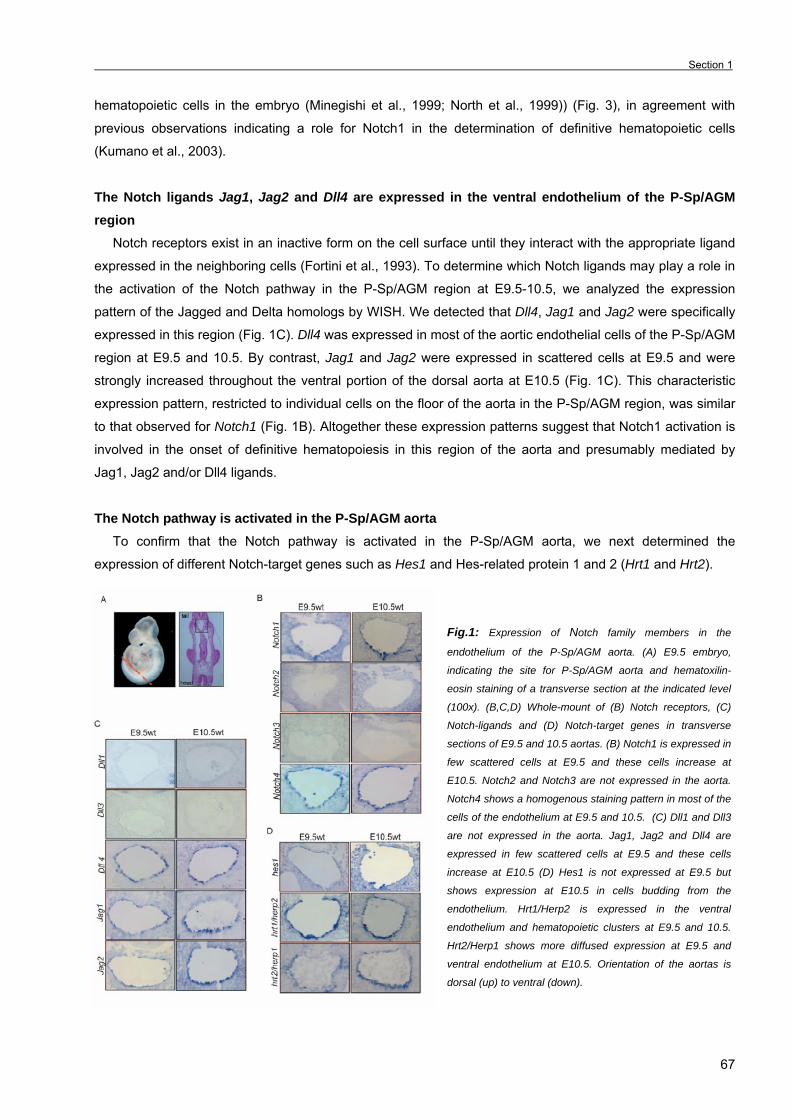
Section 1
67
hematopoietic cells in the embryo (Minegishi et al., 1999; North et al., 1999)) (Fig. 3), in agreement with
previous observations indicating a role for Notch1 in the determination of definitive hematopoietic cells
(Kumano et al., 2003).
The Notch ligands Jag1, Jag2 and Dll4 are expressed in the ventral endothelium of the P-Sp/AGM region
Notch receptors exist in an inactive form on the cell surface until they interact with the appropriate ligand
expressed in the neighboring cells (Fortini et al., 1993). To determine which Notch ligands may play a role in
the activation of the Notch pathway in the P-Sp/AGM region at E9.5-10.5, we analyzed the expression
pattern of the Jagged and Delta homologs by WISH. We detected that Dll4, Jag1 and Jag2 were specifically
expressed in this region (Fig. 1C). Dll4 was expressed in most of the aortic endothelial cells of the P-Sp/AGM
region at E9.5 and 10.5. By contrast, Jag1 and Jag2 were expressed in scattered cells at E9.5 and were
strongly increased throughout the ventral portion of the dorsal aorta at E10.5 (Fig. 1C). This characteristic
expression pattern, restricted to individual cells on the floor of the aorta in the P-Sp/AGM region, was similar
to that observed for Notch1 (Fig. 1B). Altogether these expression patterns suggest that Notch1 activation is
involved in the onset of definitive hematopoiesis in this region of the aorta and presumably mediated by
Jag1, Jag2 and/or Dll4 ligands.
The Notch pathway is activated in the P-Sp/AGM aorta To confirm that the Notch pathway is activated in the P-Sp/AGM aorta, we next determined the
expression of different Notch-target genes such as Hes1 and Hes-related protein 1 and 2 (Hrt1 and Hrt2).
Fig.1: Expression of Notch family members in the
endothelium of the P-Sp/AGM aorta. (A) E9.5 embryo,
indicating the site for P-Sp/AGM aorta and hematoxilin-
eosin staining of a transverse section at the indicated level
(100x). (B,C,D) Whole-mount of (B) Notch receptors, (C)
Notch-ligands and (D) Notch-target genes in transverse
sections of E9.5 and 10.5 aortas. (B) Notch1 is expressed in
few scattered cells at E9.5 and these cells increase at
E10.5. Notch2 and Notch3 are not expressed in the aorta.
Notch4 shows a homogenous staining pattern in most of the
cells of the endothelium at E9.5 and 10.5. (C) Dll1 and Dll3
are not expressed in the aorta. Jag1, Jag2 and Dll4 are
expressed in few scattered cells at E9.5 and these cells
increase at E10.5 (D) Hes1 is not expressed at E9.5 but
shows expression at E10.5 in cells budding from the
endothelium. Hrt1/Herp2 is expressed in the ventral
endothelium and hematopoietic clusters at E9.5 and 10.5.
Hrt2/Herp1 shows more diffused expression at E9.5 and
ventral endothelium at E10.5. Orientation of the aortas is
dorsal (up) to ventral (down).

Chapter 3. Results _
68
Consistent with previous reports, Hrt1 and Hrt2 are expressed in endothelial cells of the aorta (Nakagawa
et al., 2000), although their expression patterns are not completely homogenous, showing a preferential
ventral staining in the AGM region at E9.5 and 10.5 (Fig. 1D). We could not detect Hes1 expression in E9.5
aorta, whereas a strong upregulation was observed in few ventral cells and in hematopoietic clusters arising
from the endothelium at E10.5 (Fig. 1D). Thus, different Notch-target genes display specific temporal and
spatial expression patterns in the aorta, suggesting that they could be playing different roles in early
hematopoietic/endothelial decisions.
RBPjκ mutant embryos display an aberrant expression of Notch receptors and ligands in the P-
Sp/AGM region There is strong evidence from a variety of systems that Notch signaling participates in the transcriptional
regulation of several Notch receptors and ligands by positive (Barrantes et al., 1999; Timmerman et al.,
2004) or negative (Chitnis, 1995; de la Pompa et al., 1997; Heitzler et al., 1996) feedback mechanisms.
Since most of these regulatory networks depend on the RBPjκ transcription factor (Heitzler et al., 1996);
(Timmerman et al., 2004), we investigated whether the expression of the different Notch family members is
affected in the aorta of the RBPjκ mutant embryos (Oka et al., 1995). We first compared the expression by
semi-quantitative RT-PCR of Notch receptors and ligands in the dissected P-Sp/AGM region from wild-type
and mutant embryos at E9.5. We consistently observed a decrease in the expression of Notch1 in the RBPjκ
mutant embryos compared with the wild-type, while we did not detect important changes in the level of
expression of Notch4 or the different Notch ligands (Fig. 2A).
When we specifically studied the expression of these genes in the aorta endothelium using WISH, we
observed decreased Notch1 mRNA levels in the RBPjκ mutant embryos (Fig. 2B) compared with the
restricted but strong expression observed in the wild-type aortas (see Fig. 1B), as detected by RT-PCR. By
contrast, expression of Jag1 and Jag2 was specifically impaired in the aorta endothelial cells (Fig. 2B),
whereas their expression was not affected in adjacent tissues in this region (data not shown). These results
further confirm that expression and distribution of different Notch ligands and receptors depend on RBPjκ as
previously published (Heitzler et al., 1996) and points out the possibility that specific interactions between
these proteins may regulate the proper cellular specification in the P-Sp/AGM aorta.
Intra-embryonic hematopoiesis is impared in the RBPjκ mutant embryos
To investigate whether Notch/RBPjκ signaling plays a role in hematopoietic determination in the aorta, we
next assayed the hematopoietic activity contained in the P-Sp/AGM region of RBPjκ mutant embryos
compared with wild-type. Despite the presence of several developmental abnormalities and disorganized
vasculature, the majority of the RBPjκ mutant embryos (more than 80%) display a regular fused aorta in the
trunkal region at E9.5 (Oka et al., 1995). As RBPjκ mutants die at E10, we performed direct hematopoietic
colony assays with cells obtained from P-Sp/AGM at E9.5. Hematopoietic colony forming cells (CFCs) of the
different lineages were generated in cell cultures from wild-type embryos whereas few rare colonies were
obtained from the cultures from RBPjκ mutant littermates in the same conditions (Fig. 2C). We speculated
that RBPjκ mutant embryos contained lower numbers of HSC that may be undetectable in the direct CFC
cultures. To test this possibility, we expanded the number of progenitors by incubating cells from single wild-

Section 1
69
type P-Sp/AGM compared with pools of two or three mutant P-Sp/AGM in liquid cultures with cytokines for 6
days. As shown in Fig. 2D, liquid cultures from both wild-type and mutant embryos formed equivalent stromal
cell layers after 6 days, although only wild-type cultures contained non-adherent, round-shaped,
hematopoietic-like cells (Fig. 2D). By flow cytometry, we demonstrated that liquid cell cultures from wild-type
embryos contained 30-50% of CD45+ cells (Fig. 2E) that corresponded to the non-adherent population (data
not shown). In agreement with the absence of hematopoietic-like cells, this CD45+ population was not
detected in the mutant cultures (Fig. 2E). Cells from wild-type cultures generated CFCs with a predominant
granulo-monocytic morphology, although colonies from other lineages were also observed (Fig. 2F). By
contrast, we did not observe any hematopoietic colonies from the RBPjκ mutant cultures (Fig. 2F). These
results indicate that Notch signaling through RBPjκ is required for the generation of the hematopoietic
progenitors in the P-Sp/AGM.
Fig.2: Intra-embryonic hematopoiesis is impaired in the
RBPjκ mutant embryos and they display an aberrant
expression of Notch family members in the aorta. (A)
Semiquan-titative RT-PCR analysis in dissected P-Sp of
E9.5 wild-type and RBPjκ mutants. Representative PCR
products after 35 and 40 cycles of two independent
experiments are shown. (B) WISH, with the indicated probes
and transverse sections of E9.5 aortas of RBPjκ mutants.
Orientation of the aortas is dorsal (up) to ventral (down). (C)
Hematopoietic CFC from dissected P-Sp of E9.5 wild-type
and RBPjκ mutants. Bars represent the average number of
CFCs and standard deviation from three different embryos.
(D) Liquid cultures with IL3 and SCF-conditioned media from
P-Sp of E9.5 wild-type and RBPjκ mutants at day 0 and 6.
(E) After 6 days in culture, cells were assayed for the
expression of CD45 by flow cytometry and (F) the number of
CFCs generated. Bars represent the average number of
CFCs obtained from one wild-type embryo and pools of two
or three mutant embryos equivalent (e.e.) in three
independent different experiments.
Absence of hematopoietic cells and increase of endothelial cells in the P-Sp/AGM of RBPjκ mutant
embryos Difficulties in characterizing HSCs in the P-Sp/AGM endothelium reside in the lack of specific HSC
markers. In fact, endothelial markers were expressed in all the cells in the P-Sp/AGM endothelium, including
the cells that would generate the HSCs. Thus, specific hematopoietic transcription factors such as Aml1,
Gata2, and Scl are widely used to identify these endothelial-like cells that will generate the hematopoietic
clusters (Minegishi et al., 1999; North et al., 1999). These hemapotoietic markers are expressed in individual
rare cells in the floor of the dorsal aorta of the AGM region (North et al., 2002; Porcher et al., 1996; Tsai and
Orkin, 1997)) (Fig. 3B). To better understand the mechanisms by which definitive hematopoiesis is
abrogated in RBPjκ mutant embryos, we studied the expression of these genes together with endothelial
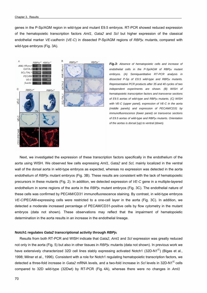
Chapter 3. Results _
70
genes in the P-Sp/AGM region in wild-type and mutant E9.5 embryos. RT-PCR showed reduced expression
of the hematopoietic transcription factors Aml1, Gata2 and Scl but higher expression of the classical
endothelial marker VE-cadherin (VE-C) in dissected P-Sp/AGM regions of RBPjκ mutants, compared with
wild-type embryos (Fig. 3A).
Fig.3: Absence of hematopoietic cells and increase of
endothelial cells in the P-Sp/AGM of RBPjκ mutant
embryos. (A) Semiquantitative RT-PCR analysis in
dissected P-Sp of E9.5 wild-type and RBPjκ mutants.
Representative PCR products after 35 and 40 cycles of two
independent experiments are shown. (B) WISH of
hematopoietic transcription factors and transverse sections
of E9.5 aortas of wild-type and RBPjκ mutants. (C) WISH
with VE-C (upper panel), expression of VE-C in the aorta
(middle panels) and expression of PECAM/CD31 by
immunofluorescence (lower panel) on transverse sections
of E9.5 aortas of wild-type and RBPjκ mutants. Orientation
of the aortas is dorsal (up) to ventral (down).
Next, we investigated the expression of these transcription factors specifically in the endothelium of the
aorta using WISH. We observed few cells expressing Aml1, Gata2 and Scl, mainly localized in the ventral
wall of the dorsal aorta in wild-type embryos as expected, whereas no expression was detected in the aorta
endothelium of RBPjκ mutant embryos (Fig. 3B). These results are consistent with the lack of hematopoietic
precursors in these mutants (Fig. 2). In addition, we detected expression of VE-C gene in a multiple-layered
endothelium in some regions of the aorta in the RBPjκ mutant embryos (Fig. 3C). The endothelial nature of
these cells was confirmed by PECAM/CD31 immunofluorescence staining. By contrast, in wild-type embryos
VE-C/PECAM-expressing cells were restricted to a one-cell layer in the aorta (Fig. 3C). In addition, we
detected a moderate increased percentage of PECAM/CD31-positive cells by flow cytometry in the mutant
embryos (data not shown). These observations may reflect that the impairment of hematopoietic
determination in the aorta results in an increase in the endothelial lineage.
Notch1 regulates Gata2 transcriptional activity through RBPjκ
Results from both RT-PCR and WISH indicate that Gata2, Aml1 and Scl expression was greatly reduced
not only in the aorta (Fig. 5) but also in other tissues in RBPjκ mutants (data not shown). In previous work we
have extensively characterized 32D cell lines stably expressing activated Notch1 (32D-N1IC) (Bigas et al.,
1998; Milner et al., 1996). Consistent with a role for Notch1 regulating hematopoietic transcription factors, we
detected a three-fold increase in Gata2 mRNA levels, and a two-fold increase in Scl levels in 32D-N1IC cells
compared to 32D wild-type (32Dwt) by RT-PCR (Fig. 4A), whereas there were no changes in Aml1
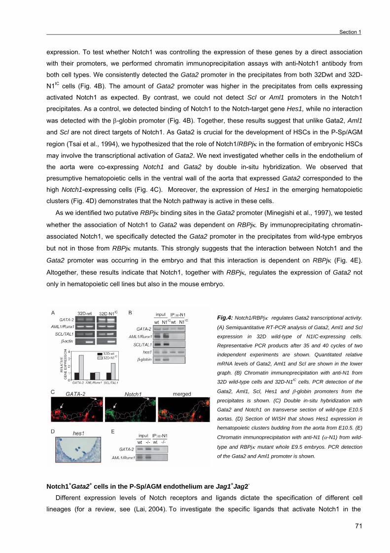
Section 1
71
expression. To test whether Notch1 was controlling the expression of these genes by a direct association
with their promoters, we performed chromatin immunoprecipitation assays with anti-Notch1 antibody from
both cell types. We consistently detected the Gata2 promoter in the precipitates from both 32Dwt and 32D-
N1IC cells (Fig. 4B). The amount of Gata2 promoter was higher in the precipitates from cells expressing
activated Notch1 as expected. By contrast, we could not detect Scl or Aml1 promoters in the Notch1
precipitates. As a control, we detected binding of Notch1 to the Notch-target gene Hes1, while no interaction
was detected with the β-globin promoter (Fig. 4B). Together, these results suggest that unlike Gata2, Aml1
and Scl are not direct targets of Notch1. As Gata2 is crucial for the development of HSCs in the P-Sp/AGM
region (Tsai et al., 1994), we hypothesized that the role of Notch1/RBPjκ in the formation of embryonic HSCs
may involve the transcriptional activation of Gata2. We next investigated whether cells in the endothelium of
the aorta were co-expressing Notch1 and Gata2 by double in-situ hybridization. We observed that
presumptive hematopoietic cells in the ventral wall of the aorta that expressed Gata2 corresponded to the
high Notch1-expressing cells (Fig. 4C). Moreover, the expression of Hes1 in the emerging hematopoietic
clusters (Fig. 4D) demonstrates that the Notch pathway is active in these cells.
As we identified two putative RBPjκ binding sites in the Gata2 promoter (Minegishi et al., 1997), we tested
whether the association of Notch1 to Gata2 was dependent on RBPjκ. By immunoprecipitating chromatin-
associated Notch1, we specifically detected the Gata2 promoter in the precipitates from wild-type embryos
but not in those from RBPjκ mutants. This strongly suggests that the interaction between Notch1 and the
Gata2 promoter was occurring in the embryo and that this interaction is dependent on RBPjκ (Fig. 4E) .
Altogether, these results indicate that Notch1, together with RBPjκ, regulates the expression of Gata2 not
only in hematopoietic cell lines but also in the mouse embryo.
Fig.4: Notch1/RBPjκ regulates Gata2 transcriptional activity.
(A) Semiquantitative RT-PCR analysis of Gata2, Aml1 and Scl
expression in 32D wild-type of N1IC-expressing cells.
Representative PCR products after 35 and 40 cycles of two
independent experiments are shown. Quantitated relative
mRNA levels of Gata2, Aml1 and Scl are shown in the lower
graph. (B) Chromatin immunoprecipitation with anti-N1 from
32D wild-type cells and 32D-N1IC cells. PCR detection of the
Gata2, Aml1, Scl, Hes1 and β-globin promoters from the
precipitates is shown. (C) Double in-situ hybridization with
Gata2 and Notch1 on transverse section of wild-type E10.5
aortas. (D) Section of WISH that shows Hes1 expression in
hematopoietic clusters budding from the aorta from E10.5. (E)
Chromatin immunoprecipitation with anti-N1 (α-N1) from wild-
type and RBPjκ mutant whole E9.5 embryos. PCR detection
of the Gata2 and Aml1 promoter is shown.
Notch1+Gata2+ cells in the P-Sp/AGM endothelium are Jag1+Jag2- Different expression levels of Notch receptors and ligands dictate the specification of different cell
lineages (for a review, see (Lai, 2004). To investigate the specific ligands that activate Notch1 in the

Chapter 3. Results _
72
presumptive hematopoietic cells in the aorta, we performed double in situ-hybridizations. It is well
established that the ligands responsible for activating Notch1 are expressed in cells adjacent to the Notch
expressing one. We analyzed transverse sections of E10.5 embryos simultaneously hybridized with specific
probes for Notch1 and the different ligands that are expressed in the aorta endothelium at this
developmental stage. We consistently observed that cells expressing Notch1 (Notch1+) also expressed Jag1
(Fig. 5A, upper panels), whereas Jag2 was specifically detected in cells adjacent to Notch1+ but not in the
Notch1+ themselves (Fig. 5A, middle panels). Dll4 showed a mixed pattern of co-expression with Notch1, in
which some cells simultaneously expressed both Notch1 and Dll4 and other cells only expressed one of
these genes (Fig. 5A, lower panels). Altogether, these results are consistent with a model in which Jag2 or
Dll4 activate Notch1 in the ventral wall of the aorta. This event would initiate the hematopoietic program in
the Notch1+ cells by activating the expression of Gata2 (Fig. 5B). However, and considering that multiple
ligands are simultaneously expressed in the endothelium of the aorta, it is tempting to speculate that back
and forward signals between different members may occur.
Fig.5: Notch1+/Gata2+ cells in the P-Sp/AGM endothelium
are Jag1+/Jag2-. Double in-situ hybridization on transverse
sections of wild-type E10.5 aortas. (A) Hybridization of
Notch1 with Jag1 (upper), Jag2 (middle) and Dll4 (lower
panels). Representative photographs of at least three
hybridizations are shown. (B) Model for Notch function in
the formation of hematopoietic clusters from the aorta
endothelium during development.
DISCUSSION
There is now evidence that Notch1 is required for the generation of intra-embryonic hematopoiesis
(Hadland et al., 2004; Kumano et al., 2003). Here we show that this function is dependent on the
transcription factor RBPjκ and several members of the Notch family are likely to be involved. Consistent with
the phenotype described for the Notch1 mutant embryos, RBPjκ mutants are deficient for intra-
embryonic/definitive hematopoiesis. We propose that Notch1 activation in individual cells of the hemogenic
endothelium regulates transcription of Gata2, which is essential for the generation and proliferation of HSCs
(Tsai et al., 1994).
RBPjκ-dependent Notch function in the generation of intra-embryonic hematopoiesis
The origin of definitive HSCs from an endothelial/hematopoietic common progenitor known as
hemangioblast is still controversial. While the yolk sac is a primary site of hematopoietic development,
Section 1
73
several lines of evidence support the idea that, under physiological conditions, HSCs are generated de novo
within the endothelium lining the ventral wall of the aorta of the P-Sp/AGM region (Cai et al., 2000; de Bruijn
et al., 2002). Our work demonstrates that intra-embryonic hematopoiesis is abolished in the RBPjκ mutant
embryos, presumably due to impaired hematopoietic progenitor determination from endothelial-like
precursors in the aorta. This correlates with the absence of expression of hematopoietic transcription factors
in this region in the mutant embryos compared with wild-type. Furthermore, expression of classical
endothelial markers, such as VE-cadherin and PECAM, is increased in the embryonic aortas of these
mutants, suggesting that in the absence of Notch signaling, the endothelial lineage is favored at the expense
of the hematopoietic one. While this work was in progress, it was reported that Notch1-deficient embryos
have impaired intra-embryonic hematopoiesis due to a defect in hematopoietic determination from
endothelial cells (Kumano et al., 2003), and that Notch1-deficient embryonic stem cells cannot contribute to
definitive hematopoiesis in chimeric embryos (Hadland et al., 2004). Our results are in agreement with a role
of Notch1 in the onset of definitive hematopoiesis through a transcriptional activation mechanism dependent
on RBPjκ. Although the expression of other hematopoietic genes such as Scl and Aml1 is severely affected
in the RBPjκ mutants, we showed that only Gata2 is a direct target of Notch1/RBPjκ signaling. As Gata2 is
required to maintain the pool of undifferentiated hematopoietic progenitors (Tsai and Orkin, 1997), we
speculate and present evidence that the absence of Gata2 in the RBPjκ mutants could be responsible for the
lack of hematopoietic progenitors in these mutants and is likely in the Notch1 mutants (Kumano et al., 2003).
In agreement with this, the maintenance of undifferentiated 32D myeloid progenitors by Notch1 has been
associated with Gata2 expression (Kumano et al., 2001). Our work demonstrates that most of the cells in the
aorta that express Notch1 simultaneously express Gata2. This result together with the demonstration by
chromatin precipitation assays that intracellular Notch1 associates with the Gata2 promoter, strongly
suggests that Notch1 may regulate the generation and maintenance of hematopoietic progenitors by directly
activating the expression of Gata2.
Using in-situ hybridization, we detected high levels of expression of the Hes1 gene in a few endothelial
cells as well as in the hematopoietic clusters of the aorta, thus suggesting that Notch activation is
concomitant with the formation of these clusters. The function of Hes1 in the maintenance of HSC has not
been studied in vivo; however, several pieces of evidence confirm that Hes1 is regulating cell differentiation
in different hematopoietic cell types (Kawamata et al., 2002; Kumano et al., 2001). These studies together
with our results suggest that Hes1 could be involved in maintaining the immature phenotype of the
hematopoietic precursors budding from the aorta and/or in repressing the expression of specific endothelial
markers in these cells. The detection of other Notch-target genes, such as Hrt1 and Hrt2 (E9.5), preceding
Hes1 expression confirms that Notch is active at this embryonic stage. However, the role of these Hes-
related proteins in the cellular specification of the aorta remains to be determined.
Lateral inhibition or lateral induction in P-Sp/AGM hematopoietic determination
During the development of complex multicellular organisms, numerous cell-cell signaling events are
required for proper cell-fate determination. Two different Notch signaling mechanisms have been proposed:
lateral inhibition and lateral induction (reviewed by (Lewis, 1998). Singling out an individual cell or group of
cells from initially equivalent cells is known as lateral inhibition, whereas lateral induction implies the

Chapter 3. Results _
74
adoption of cellular fates cooperatively. In lateral inhibition Notch activation leads to Delta downregulation,
while in lateral induction activation of Notch leads to Delta upregulation. A typical example of lateral inhibition
mediated by Notch is the process of neurogenesis in Drosophila (Artavanis-Tsakonas et al., 1999) and
vertebrates (Chitnis, 1995), while lateral induction occurs during wing margin development in Drosophila
(Panin et al., 1997), somite formation (reviewed by (Lewis, 1998) and endocardial development (Timmerman
et al., 2004). To define whether the determination of hematopoietic cells in the mid-gestation aorta is
compatible with one of these mechanisms, it is crucial to know the expression pattern of Notch receptors and
ligands at this stage, as well as the characterization of the aorta hematopoietic potential of the different
mutant embryos. Although Notch family members have been detected in many adult and embryonic
hematopoietic tissues, this is the first time that E9.5-10.5 P-Sp/AGM aorta endothelium has been studied by
single and double in-situ hybridization and the expression of these genes has been analyzed on transverse
sections through the trunkal region. Our analysis reveals co-expression of multiple Notch-family members in
these cells at this developmental stage, strongly suggesting that several Notch signals are likely to be
involved in hematopoietic determination. For example, Jag1 is co-expressed with Notch1 in most of the
endothelial cells, while the Jag2 transcript is absent from these cells and specifically expressed in the cells
neighboring the Notch1+ ones. Moreover, Jag1 is absent from the endothelium of RBPjκ mutant embryos,
strongly suggesting that its expression depends on Notch1 activation in this tissue.
An important question to be determined is how specific expression patterns of Notch family members are
acquired. For example, the endothelium covering the aorta outside the AGM region has a very homogenous
pattern of Notch1 or Dll4 expression in the majority of cells (data not shown), while the scattered expression
pattern is restricted to the AGM aorta. Considering this, it is tempting to speculate that the aorta endothelium
originates as a pool of equivalent Notch- and ligand-expressing cells and lateral inhibition events will
generate a “salt and pepper” expression pattern that is reminiscent of that described for Drosophila
neurogenesis (Artavanis-Tsakonas and Simpson, 1991). Once Notch1 expression pattern in the P-Sp/AGM
aorta is established, hemogenic endothelial cells have to undergo determination, proliferation and migration
events that may require multiple local interactions with the neighboring cells. Our results are consistent with
a model in which expression of Notch1 in individual cells in the ventral wall of the aorta leads to the
activation of Gata2 that is crucial for the generation of a pool of definitive HSCs. Loss of Gata2 expression in
the RBPjκ-deficient embryos results in the loss of the HSC pool and in the absence of definitive
hematopoiesis (Fig. 5B). This model implies that, similarly to the situation in other developmental systems
(de Celis et al., 1991), Notch1 acts cell-autonomously in promoting an HSC fate in the P-Sp/AGM aorta as
previously proposed (Kumano et al., 2003). Our results support a role for Notch in the maintenance of a
population of stem cells (HSCs) that are critical for the definitive hematopoiesis in the embryos and are
consistent with the finding that alterations in the Notch function are responsible for leukemias (reviewed by
(Radtke and Raj, 2003). Gaining insight into the mechanism of Notch action will help to design therapeutical
approaches for the treatment of such complex diseases.

Section 1
75
ACKNOWLEDGEMENTS
We thank R. Kageyama, N. A. Speck, D. Martin and D.Srivastava for cDNA probes. We sincerely thank Elaine
Dzierzak for helpful discussions in various aspects of this work. We thank all the members of the lab for many
constructive discussions, Irene Merida for technical support and Serveis Cientifico-Tècnics, UB-Bellvitge, for confocal
microscopy technical support. L.E. is an investigator from the Carlos III program (ISCIII/02/3027). A.R.M. is a recipient of
a CIRIT predoctoral fellowship (2002-SI00791). This work was supported by a grant from the Comisión Interministerial de
Ciencia y Tecnología, Plan Nacional de Salud (SAF2001-1191).
REFERENCES
• Aguilera, C., Hoya-Arias R., Haegeman, G., Espinosa, L., Bigas, A. (2004). Recruitment of IkBalpha to the hes1 promoter is associated with transcriptional repression. Proc Natl Acad Sci U S A, 101, 16537-16542.
• Artavanis-Tsakonas, S., Rand, M. D. and Lake, R. J. (1999). Notch signaling: cell fate control and signal integration in development. Science 284, 770-6.
• Artavanis-Tsakonas, S. and Simpson, P. (1991). Choosing a cell fate: a view from the notch locus. Trends Genet 7, 403-408. • Barrantes, I. B., Elia, A. J., Wunsch, K., De Angelis, M. H., Mak, T. W., Rossant, J., Conlon, R. A., Gossler, A. and de la
Pompa, J. L. (1999). Interaction between Notch signaling and Lunatic fringe during somite boundary formation in the mouse. Current Biology 9, 470-80.
• Bigas, A., Martin, D. I. and Milner, L. A. (1998). Notch1 and Notch2 inhibit myeloid differentiation in response to different cytokines. Mol.Cell Biol 18, 2324-2333.
• Cai, Z., de Bruijn, M., Ma, X., Dortland, B., Luteijn, T., Downing, R. J. and Dzierzak, E. (2000). Haploinsufficiency of AML1 affects the temporal and spatial generation of hematopoietic stem cells in the mouse embryo. Immunity 13, 423-31.
• Chitnis, A. B. (1995). The role of Notch in lateral inhibition and cell fate specification. Mol.Cell Neurosci., 311-321. • Cumano, A., Ferraz, J. C., Klaine, M., Di Santo, J. P. and Godin, I. (2001). Intraembryonic, but not yolk sac hematopoietic
precursors, isolated before circulation, provide long-term multilineage reconstitution. Immunity 15, 477-85. • de Bruijn, M. F., Ma, X., Robin, C., Ottersbach, K., Sanchez, M. J. and Dzierzak, E. (2002). Hematopoietic stem cells localize to
the endothelial cell layer in the midgestation mouse aorta. Immunity 16, 673-83. • de Bruijn, M. F., Peeters, M. C., Luteijn, T., Visser, P., Speck, N. A. and Dzierzak, E. (2000). Definitive hematopoietic stem cells
first develop within the major arterial regions of the mouse embryo. Blood 96, 2902-4. • de Celis, J. F., Mari-Beffa, M. and Garcia-Bellido, A. (1991). Cell-autonomous role of Notch, an epidermal growth factor
homologue, in sensory organ differentiation in Drosophila. Proc Natl Acad Sci U S A 88, 632-6. • de la Pompa, J. L., Wakeham, A., Correia, K. M., Samper, E., Brown, S., Aguilera, R. J., Nakano, T., Honjo, T., Mak, T. W.,
Rossant, J. et al. (1997). Conservation of the Notch signaling pathway in mammalian neurogenesis. Development 124, 1139-1148. • Fortini, M. E., Rebay, I., Caron, L. A. and Artavanis-Tsakonas, S. (1993). An activated notch receptor blocks cell-fate
commitment in the developing drosophila eye. Nature 365, 555-557. • Hadland, B. K., Huppert, S. S., Kanungo, J., Xue, Y., Jiang, R., Gridley, T., Conlon, R. A., Cheng, A. M., Kopan, R. and
Longmore, G. D. (2004). A requirement for Notch1 distinguishes 2 phases of definitive hematopoiesis during development. Blood 104, 3097-105.
• Han, H., Tanigaki, K., Yamamoto, N., Kuroda, K., Yoshimoto, M., Nakahata, T., Ikuta, K. and Honjo, T. (2002). Inducible gene knockout of transcription factor recombination signal binding protein-J reveals its essential role in T versus B lineage decision. Int Immunol 14, 637-45.
• Heitzler, P., Bourouis, M., Ruel, L., Carteret, C. and Simpson, P. (1996). Genes of the Enhancer of split and achaete-scute complexes are required for a regulatory loop between Notch and Delta during lateral signaling in Drosophila. Development, 161-171.
• Huppert, S. S., Le, A., Schroeter, E. H., Mumm, J. S., Saxena, M. T., Milner, L. A. and Kopan, R. (2000). Embryonic lethality in mice homozygous for a processing-deficient allele of Notch1. Nature 405, 966-70.
• Iso, T., Kedes, L. and Hamamori, Y. (2003). HES and HERP families: multiple effectors of the Notch signaling pathway. J Cell Physiol 194, 237-55.
• Kawamata, S., Du, C., Li, K. and Lavau, C. (2002). Overexpression of the Notch target genes Hes in vivo induces lymphoid and myeloid alterations. Oncogene 21, 3855-63.
• Kumano, K., Chiba, S., Kunisato, A., Sata, M., Saito, T., Nakagami-Yamaguchi, E., Yamaguchi, T., Masuda, S., Shimizu, K., Takahashi, T. et al. (2003). Notch1 but not Notch2 is essential for generating hematopoietic stem cells from endothelial cells. Immunity 18, 699-711.
• Kumano, K., Chiba, S., Shimizu, K., Yamagata, T., Hosoya, N., Saito, T., Takahashi, T., Hamada, Y. and Hirai, H. (2001). Notch1 inhibits differentiation of hematopoietic cells by sustaining GATA-2 expression. Blood 98, 3283-9.
• Lai, E. C. (2004). Notch signaling: control of cell communication and cell fate. Development 131, 965-73. • Lewis, J. (1998). Notch signaling and the control of cell fate choices in vertebrates. Semin Cell Dev Biol 9, 583-9. • Ling, K. W. and Dzierzak, E. (2002). Ontogeny and genetics of the hemato/lymphopoietic system. Curr Opin Immunol 14, 186-91. • Ma, X., Robin, C., Ottersbach, K. and Dzierzak, E. (2002). The Ly-6A (Sca-1) GFP transgene is expressed in all adult mouse
hematopoietic stem cells. Stem Cells 20, 514-21. • Medvinsky, A. and Dzierzak, E. (1996). Definitive hematopoiesis is autonomously initiated by the AGM region. Cell 86, 897-906. • Milner, L. A., Bigas, A., Kopan, R., Brashem-Stein, C., Bernstein, I. D. and Martin, D. I. (1996). Inhibition of granulocytic
differentiation by mNotch1. Proc.Natl.Acad.Sci.U.S.A. 93, 13014-13019. • Minegishi, N., Morita, S., Minegishi, M., Tsuchiya, S., Konno, T., Hayashi, N. and Yamamoto, M. (1997). Expression of GATA
transcription factors in myelogenous and lymphoblastic leukemia cells. Int J Hematol 65, 239-49.
Chapter 3. Results _
76
• Minegishi, N., Ohta, J., Yamagiwa, H., Suzuki, N., Kawauchi, S., Zhou, Y., Takahashi, S., Hayashi, N., Engel, J. D. and
Yamamoto, M. (1999). The mouse GATA-2 gene is expressed in the para-aortic splanchnopleura and aorta-gonads and mesonephros region. Blood 93, 4196-207.
• Nakagawa, O., McFadden, D. G., Nakagawa, M., Yanagisawa, H., Hu, T., Srivastava, D. and Olson, E. N. (2000). Members of the HRT family of basic helix-loop-helix proteins act as transcriptional repressors downstream of notch signaling. Proc Natl Acad Sci U S A 97, 13655-60.
• North, T., Gu, T. L., Stacy, T., Wang, Q., Howard, L., Binder, M., Marin-Padilla, M. and Speck, N. A. (1999). Cbfa2 is required for the formation of intra-aortic hematopoietic clusters. Development 126, 2563-75.
• North, T. E., de Bruijn, M. F., Stacy, T., Talebian, L., Lind, E., Robin, C., Binder, M., Dzierzak, E. and Speck, N. A. (2002). Runx1 expression marks long-term repopulating hematopoietic stem cells in the midgestation mouse embryo. Immunity 16, 661-72.
• Oka, C., Nakano, T., Wakeham, A., de la Pompa, J. L., Mori, C., Sakai, T., Okazaki, S., Kawaichi, M., Shiota, K., Mak, T. W. et al. (1995). Disruption of the mouse RBP-J kappa gene results in early embryonic death. Development 121, 3291-301.
• Palis, J. and Yoder, M. C. (2001). Yolk-sac hematopoiesis: the first blood cells of mouse and man. Exp Hematol 29, 927-36. • Panin, V. M., Papayannopoulos, V., Wilson, R. and Irvine, K. D. (1997). Fringe modulates Notch-ligand interactions. Nature 387,
908-912. • Porcher, C., Swat, W., Rockwell, K., Fujiwara, Y., Alt, F. W. and Orkin, S. H. (1996). The T cell leukemia oncoprotein SCL/tal-1 is
essential for development of all hematopoietic lineages. Cell, 47-57. • Radtke, F. and Raj, K. (2003). The role of Notch in tumorigenesis: oncogene or tumour suppressor? Nat Rev Cancer 3, 756-67. • Radtke, F., Wilson, A., Stark, G., Bauer, M., van Meerwijk, J., MacDonald, H. R. and Aguet, M. (1999). Deficient T cell fate
specification in mice with an induced inactivation of Notch1. Immunity 10, 547-58. • Robb, L., Elwood, N. J., Elefanty, A. G., Kontgen, F., Li, R., Barnett, L. D. and Begley, C. G. (1996). The scl gene product is
required for the generation of all hematopoietic lineages in the adult mouse. EMBO Journa 15, 4123-4129. • Stier, S., Cheng, T., Dombkowski, D., Carlesso, N. and Scadden, D. T. (2002). Notch1 activation increases hematopoietic stem
cell self-renewal in vivo and favors lymphoid over myeloid lineage outcome. Blood 99, 2369-78. • Timmerman, L. A., Grego-Bessa, J., Raya, A., Bertran, E., Perez-Pomares, J. M., Diez, J., Aranda, S., Palomo, S.,
McCormick, F., Izpisua-Belmonte, J. C. et al. (2004). Notch promotes epithelial-mesenchymal transition during cardiac development and oncogenic transformation. Genes Dev 18, 99-115.
• Tsai, F. Y., Keller, G., Kuo, F. C., Weiss, M., Chen, J., Rosenblatt, M., Alt, F. W. and Orkin, S. H. (1994). An early haematopoietic defect in mice lacking the transcription factor GATA-2. Nature 371, 221-6.
• Tsai, F. Y. and Orkin, S. H. (1997). Transcription factor GATA-2 is required for proliferation/survival of early hematopoietic cells and mast cell formation, but not for erythroid and myeloid terminal differentiation. Blood 89, 3636-43.
• Yoder, M. C., Hiatt, K., Dutt, P., Mukherjee, P., Bodine, D. M. and Orlic, D. (1997). Characterization of definitive lymphohematopoietic stem cells in the day 9 murine yolk sac. Immunity 7, 335-44.
Section 2
77
SECTION 2: The Notch ligand Jagged1 is required for intra-embryonic AGM
hematopoiesis (This chapter is unpublished).
We have described that Notch1/RBPjκ-mediated Gata2 expression is required for AGM hematopoiesis
(Robert-Moreno, 2005). Our results are in agreement with Kumano’s work with the Notch1 null mice
(Kumano, 2003). However, nothing is known about the specific ligand that activates Notch to induce AGM
hematopoiesis. Moreover, due to the strong vascular defects displayed by the Notch1 and RBPjκ mutants
(Oka, 1995; Krebs, 2000) and to preclude the possibility that lack of intra-embryonic hematopoiesis is due to
the lack of a previous specified artery, assessment of the Notch function in hematopoiesis of wild-type aortas
was required.
In this chapter, we describe that hematopoietic potential of AGM aortas is severely compromised (albeit
not totally impaired) in embryos lacking the Notch ligand Jagged1 but not Jagged2. Jagged1 null embryos
completely lack Gata2 expression, display reduced number of hematopoietic progenitors and reduced
number of Sca-1+ cells (a population of cells which includes HSCs). In addition and similar to the RBPjκ null
embryos, loss of intra-embryonic hematopoiesis in the Jagged1 mutant embryos correlates with an increase
of endothelial cells.
Moreover, γ-secretase inhibition of Notch signaling in wild-type E11 aortas cultured as explants, which
promotes HSC/HPC generation and/or expansion (Medvinsky, 1996) significantly reduces the number of
hematopoietic progenitors (determined by CFC assay) and a population of high repopulating HSCs
(determined by repopulating studies).
Since we also describe that Notch1, Jagged1 and Hes1 are highly expressed in hematopoietic clusters
budding from the ventral part of the AGM aorta we propose that Jagged1-induced activation leads to Gata2
and Hes1 expression and to an expansion of both HP and HSCs in the aortic endothelium and to a
maintenance of the stemness state in these cells.
Chapter 3. Results _
78
The Notch ligand Jagged1 is required for intra-embryonic AGM hematopoiesis
Àlex Robert-Moreno1, Claudia Orelio2, Ruiz-Herguido C, Lluis Riera1, Thomas Gridley3, Elaine
Dzierzak2, Lluis Espinosa1 and Anna Bigas1*
1Centre Oncologia Molecular, IDIBELL-Institut de Recerca Oncològica. Gran Via km 2.7 Hospitalet,
Barcelona, Spain. 2Department of Cell Biology and Genetics. Erasmus University Medical Center. Rotterdam, The
Netherlands. 3The Jackson Laboratory, Bar Harbor, Maine, USA.
Scientific heading: Hematopoiesis
* Corresponding author
Mailing address: Oncologia Molecular. IDIBELL
Gran Via Km 2.7.
08907 - Hospitalet, Barcelona. Spain
Phone: 932 607 404
Fax: 932 607 426
e-mail: [email protected]
Running title: Jagged1/Notch in intra-embryonic hematopoiesis
Section 2
79
SUMMARY The mechanisms that regulate the generation of both hematopoietic stem cells (HSC) and hematopoietic
progenitor cells (HPC) during the ontogeny of the hematopoietic system in the embryo have been
extensively studied in the last years. Among many others, the implication of the Notch signaling pathway in
regulating the emergence of HPC/HSC from the P-sp/AGM region has been recently pinpointed (Burns et al.,
2005; Hadland et al., 2004; Kumano et al., 2003; Robert-Moreno et al., 2005). In the present study we
describe by in-situ hybridization that certain Notch signaling members including the Notch1 receptor and the
Jagged1 and Jagged2 ligands, are expressed in hematopoietic clusters emerging from the AGM aortic
endothelium at E10.5. Moreover, Jagged1- (but not Jagged2-) mediated Notch activation is required to
induce the expression of the hematopoietic transcription factor Gata2 but not Runx1. In agreement with this,
AGM hematopoiesis is severely reduced in Jagged1 but not Jagged2 null embryos. Finally, HSC/HPC
emergence in aorta explants cultured in the presence of the γ-secretase inhibitor DAPT is also reduced,
indicating that Notch signaling is required for activating the hematopoietic program of cells budding from the
hemogenic aortic endothelium of the AGM region.
INTRODUCTION
Different embyronic hematopoietic sites such as the yolk sac (Yoder and Hiatt, 1997; Yoder et al., 1997;
Yoder et al., 1997), the intra-embryonic para-aortic splanchnopleura (P-sp)/AGM (aorta-gonad-
mesonephros) region (Cumano et al., 2001; Medvinsky and Dzierzak, 1996), other major vessels such as
the umbilical and vitelline arteries (de Bruijn et al., 2000) and more recently the placenta (Gekas et al., 2005;
Ottersbach and Dzierzak, 2005) are responsible for generating or amplifying the pool of Hematopoietic Stem
Cells (HSC) and more committed progenitors. HSC, defined as cells with the ability to reconstitute
hematopoiesis in immunodepleted mice, are present in all this sites. However there is compelling evidence
that niche-dependent- or cell-autonomous-induced signals confer different qualities to these generally called
HSC. For example, yolk sac cells are able to reconstitute busulfan-treated new-born mice, but not adult
irradiated mice (Yoder and Hiatt, 1997; Yoder et al., 1997; Yoder et al., 1997). In contrast, AGM-derived
HSC show identical qualities as adult/definitive HSC in terms of hematopoietic reconstitution of adult animals
(de Bruijn et al., 2000; Medvinsky and Dzierzak, 1996). Moreover, recent studies from several gene-targeted
mice suggest that hematopoiesis from different embryonic sites have specific gene expression requirements
(Kumano et al., 2003; Ling et al., 2004; Okuda et al., 1996; Robert-Moreno et al., 2005; Wang et al., 1996). The Notch pathway is generally involved in the regulation of cell fate decisions and it is activated through
cell-cell interaction in a variety of developmental systems including hematopoiesis. It has previously been
characterized that several Notch pathway mutant embryos cannot generate intra-embryonic HSC both in the
mouse and zebrafish (Burns et al., 2005; Kumano et al., 2003; Robert-Moreno et al., 2005). However, it is
remarkably interesting that no major hematopoietic defects have been found in the yolk sac hematopoiesis of
these mutants (Burns et al., 2005; Kumano et al., 2003; Robert-Moreno et al., 2005). This observation
suggests that Notch may be involved in the acquisition of specific traits of AGM-definitive HSC.
We further investigate the mechanism underlying Notch activation in the AGM and whether the
hematopoietic effects of Notch on this region depends on specific Notch/Notch-ligand interactions. Here we
show that Jagged1- but not Jagged2-deficient embryos fail to activate Gata2 in the AGM region but not
Chapter 3. Results _
80
Runx1 which results in a reduced number of HSC and hematopoietic progenitors. Moreover, blocking Notch
activation in E11 AGM wild-type cells by γ-secretase inhibitors highly compromised the capacity of AGM-
derived HSC to efficiently repopulate hematopoiesis in an irradiated recipient. The overall data reinforce the
idea that Notch signaling is required for the generation of HSC and HPC in the AGM aorta and for the first
time highlights the importance of the Jagged1 ligand to activate the Notch-induced establishment of the
hematopoietic program in this region.
MATERIALS AND METHODS
Animals Ly-6A-GFP (Sca-1-GFP) mice (CD1 or B10CBA background)(kindly given by Dr. E. Dzierzak) and
Jagged1dDSL and Jagged2dDSL null mutant mice (C57BL/6J background; kindly given by Dr. Thomas Gridley)
have been extensively characterized (de Bruijn et al., 2002; Jiang et al., 1998; Xue et al., 1999). Transgenic
embryos were typed based on the presence of GFP positive cells using an Olympus IX70 fluorescent
microscope and by PCR for the transgene as well. Jag1dDSL and Jag2dDSL were typed by PCR against the
mutant allele. Sca-1-GFP tg/+ mice were crossed with Jag1dDSL/+ or Jag2dDSL/+ mice and embryos were not
used for experimental procedures before the fifth generation. Wild-type CD1 embryos were used for whole
mount in-situ hybridization (WISH). Animals were kept under pathogen-free conditions and experiments
approved by the Animal Care Committee. Embryos were obtained from timed pregnant females and somite
pairs were counted for precisely timing.
Dorsal aorta explant culture
The P-sp/AGM region from E9.5 to E11.5 embryos was dissected and subsequently subdissected into the
aorta with the surrounding mesenchyme using 27G needles. Aortas were cultured as explants for 3 days as
previously described (Medvinsky and Dzierzak, 1996). Briefly, aortas were deposited on nylon filters
(Millipore) placed on metallic grids and cultured in myeloid long-term culture medium (Stem Cell
Technologies) supplemented with 10 µM hydrocortisone succinate (Sigma) in an air-liquid interphase culture
for 3 days in the presence of DMSO (as a control) or the γ-secretase inhibitor DAPT (50µM). Explanted
aortas were either used for hematopoietic colony assay or repopulation studies.
Hematopoietic colony assay
The 3-day explanted aortas were digested in 0.12% collagenase (Sigma) in PBS supplemented with 10%
fetal bovine serum (FBS) and penicillin/streptomycin (Biological Industries) for 1 hour at 37ºC and 3.5x10⁴
cells were plated in M-5323 semisolid medium (Stem Cell Technologies). After 7 days the presence of
hematopoietic colonies was scored under the microscope, a part from CFU-e progenitors that were scored at
day 4.
Hematopoietic liquid culture
The P-sp/AGM region from E9.5 wild-type embryos was dissociated by gentle pippeting and cells were
split and treated with DMSO or 50µM DAPT in Iscove’s with 10% FBS, 10% IL3- and SCF-conditioned
medium, 0.1% monothioglycerol (Sigma), 2.5% L-glutamine and 1% Pen/Strep for 6 days.
Section 2
81
Flow cytometry analysis Following the 6-day liquid culture, cells from E9.5 P-sp were stained with anti-cd45-FITC or IgG-FITC
(Pharmingen), for flow activated cell sorting (FACS) assay. Cells were analyzed by FACScalibur (Becton &
Dickinson) and WinMDI2.8 software. Dead cells were excluded by 7-aminoactinomicin-D staining (Molecular
Probes).
CD31 immunostaining
E10.5 embryos were fixed overnight in 4% paraformaldehyde (Sigma) at 4ºC, frozen in Tissue-tek
(Sakura) and sectioned (10µm). Slides were fixed with -20ºC methanol for 15 minutes and block-
permeabilized in 10% FBS, 0.3% Surfact-AmpsX100 (Pierce) and 5% non-fat milk in PBS for 90 minutes at
4ºC. Samples were stained with rat anti-CD31 (PECAM1; Pharmingen) at 1:50 in 10% FBS, 5% non-fat milk
in PBS overnight and HRP-conjugated rabbit anti-rat antibody (Dako) at 1:100 for 90 minutes and developed
with Cy3-coupled tyramide (PerkinElmer). Sections were mounted in Vectashield medium with 4’6-diamidino-
2-phenylindole (DAPI) (Vector).
Whole mount in-situ hybridization (WISH)
WISH was performed according to standard protocols (de la Pompa et al., 1997). For histological
analysis, precisely timed embryos were fixed overnight at 4ºC in 4% paraformaldehyde, dehydrated and
embedded in Paraplast (Sigma). Embryos were sectioned in a Leica-RM2135 at 7µm and mounted with DPX
(Roche).
Short-term and long-term multilineage repopulating activity
DMSO or DAPT-treated explanted aortas (genetically marked by the Ln72 transgene) were dissociated
and cell suspensions were assayed for the presence of definitive HSCs by intravenous injection into
irradiated adult recipients (de Bruijn et al., 2002; Medvinsky and Dzierzak, 1996). Briefly, C57BL/10 x CBA
male recipients were irradiated with a 9.5Gy split dose of γ-irradiation. Adult spleen cells (2x10⁵/mouse) were
coinjected with the aorta cells to promote survival. Transplanted mice were bled at 1 and 4 months after
injection to assay short- and long-term repopulation respectively by analyzing for donor contribution by donor
marker-specific PCR (hβ-globin) on peripheral blood. Percentage of donor-cell contribution
(engraftment/chimerism) was analyzed by normalizing for the myogenin gene.
Image acquisition
Images were acquired with an Olympus BX-60 for embryonic sections and with a Leica MZ125 for whole
embryos using a Spot camera and Spot3.2.4 software (Diagnostic Instruments). Adobe Photoshop 6.0
software was used for photograph editing.
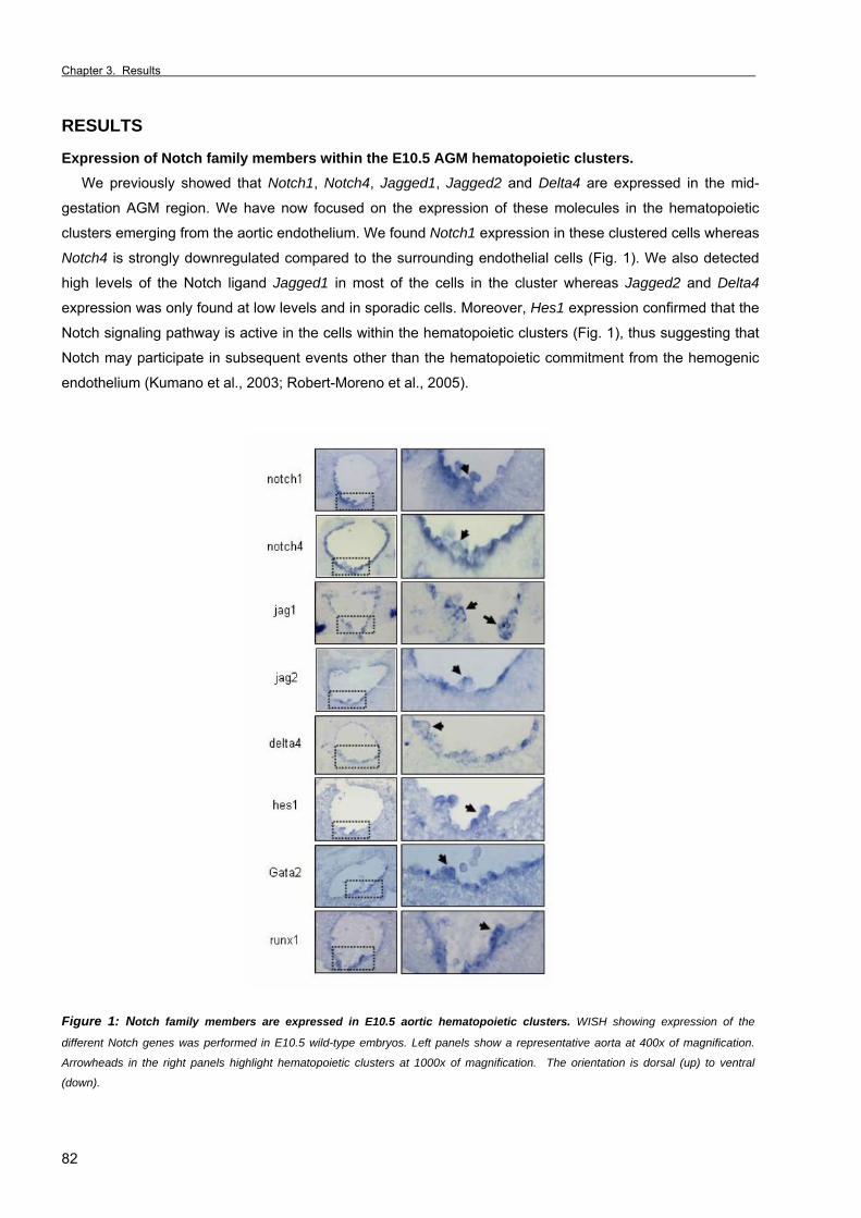
Chapter 3. Results _
82
RESULTS
Expression of Notch family members within the E10.5 AGM hematopoietic clusters. We previously showed that Notch1, Notch4, Jagged1, Jagged2 and Delta4 are expressed in the mid-
gestation AGM region. We have now focused on the expression of these molecules in the hematopoietic
clusters emerging from the aortic endothelium. We found Notch1 expression in these clustered cells whereas
Notch4 is strongly downregulated compared to the surrounding endothelial cells (Fig. 1). We also detected
high levels of the Notch ligand Jagged1 in most of the cells in the cluster whereas Jagged2 and Delta4
expression was only found at low levels and in sporadic cells. Moreover, Hes1 expression confirmed that the
Notch signaling pathway is active in the cells within the hematopoietic clusters (Fig. 1), thus suggesting that
Notch may participate in subsequent events other than the hematopoietic commitment from the hemogenic
endothelium (Kumano et al., 2003; Robert-Moreno et al., 2005).
Figure 1: Notch family members are expressed in E10.5 aortic hematopoietic clusters. WISH showing expression of the
different Notch genes was performed in E10.5 wild-type embryos. Left panels show a representative aorta at 400x of magnification.
Arrowheads in the right panels highlight hematopoietic clusters at 1000x of magnification. The orientation is dorsal (up) to ventral
(down).
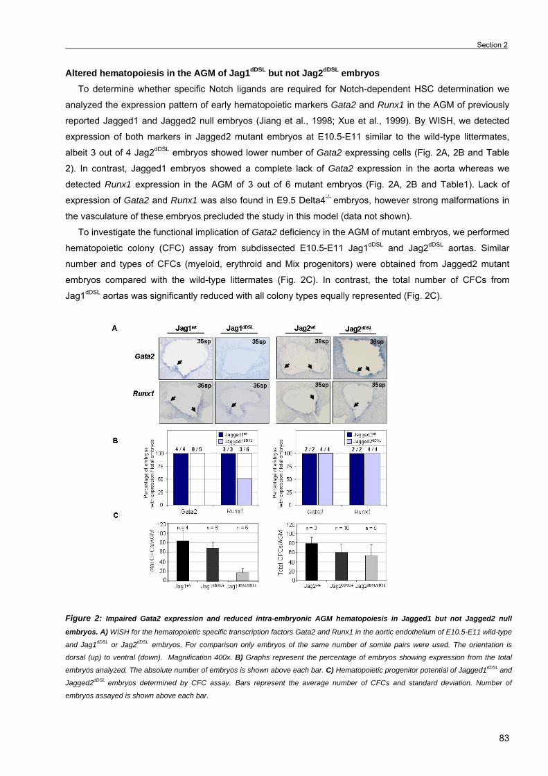
Section 2
83
Altered hematopoiesis in the AGM of Jag1dDSL but not Jag2dDSL embryos To determine whether specific Notch ligands are required for Notch-dependent HSC determination we
analyzed the expression pattern of early hematopoietic markers Gata2 and Runx1 in the AGM of previously
reported Jagged1 and Jagged2 null embryos (Jiang et al., 1998; Xue et al., 1999). By WISH, we detected
expression of both markers in Jagged2 mutant embryos at E10.5-E11 similar to the wild-type littermates,
albeit 3 out of 4 Jag2dDSL embryos showed lower number of Gata2 expressing cells (Fig. 2A, 2B and Table
2). In contrast, Jagged1 embryos showed a complete lack of Gata2 expression in the aorta whereas we
detected Runx1 expression in the AGM of 3 out of 6 mutant embryos (Fig. 2A, 2B and Table1). Lack of
expression of Gata2 and Runx1 was also found in E9.5 Delta4-/- embryos, however strong malformations in
the vasculature of these embryos precluded the study in this model (data not shown).
To investigate the functional implication of Gata2 deficiency in the AGM of mutant embryos, we performed
hematopoietic colony (CFC) assay from subdissected E10.5-E11 Jag1dDSL and Jag2dDSL aortas. Similar
number and types of CFCs (myeloid, erythroid and Mix progenitors) were obtained from Jagged2 mutant
embryos compared with the wild-type littermates (Fig. 2C). In contrast, the total number of CFCs from
Jag1dDSL aortas was significantly reduced with all colony types equally represented (Fig. 2C).
Figure 2: Impaired Gata2 expression and reduced intra-embryonic AGM hematopoiesis in Jagged1 but not Jagged2 null
embryos. A) WISH for the hematopoietic specific transcription factors Gata2 and Runx1 in the aortic endothelium of E10.5-E11 wild-type
and Jag1dDSL or Jag2dDSL embryos. For comparison only embryos of the same number of somite pairs were used. The orientation is
dorsal (up) to ventral (down). Magnification 400x. B) Graphs represent the percentage of embryos showing expression from the total
embryos analyzed. The absolute number of embryos is shown above each bar. C) Hematopoietic progenitor potential of Jagged1dDSL and
Jagged2dDSL embryos determined by CFC assay. Bars represent the average number of CFCs and standard deviation. Number of
embryos assayed is shown above each bar.
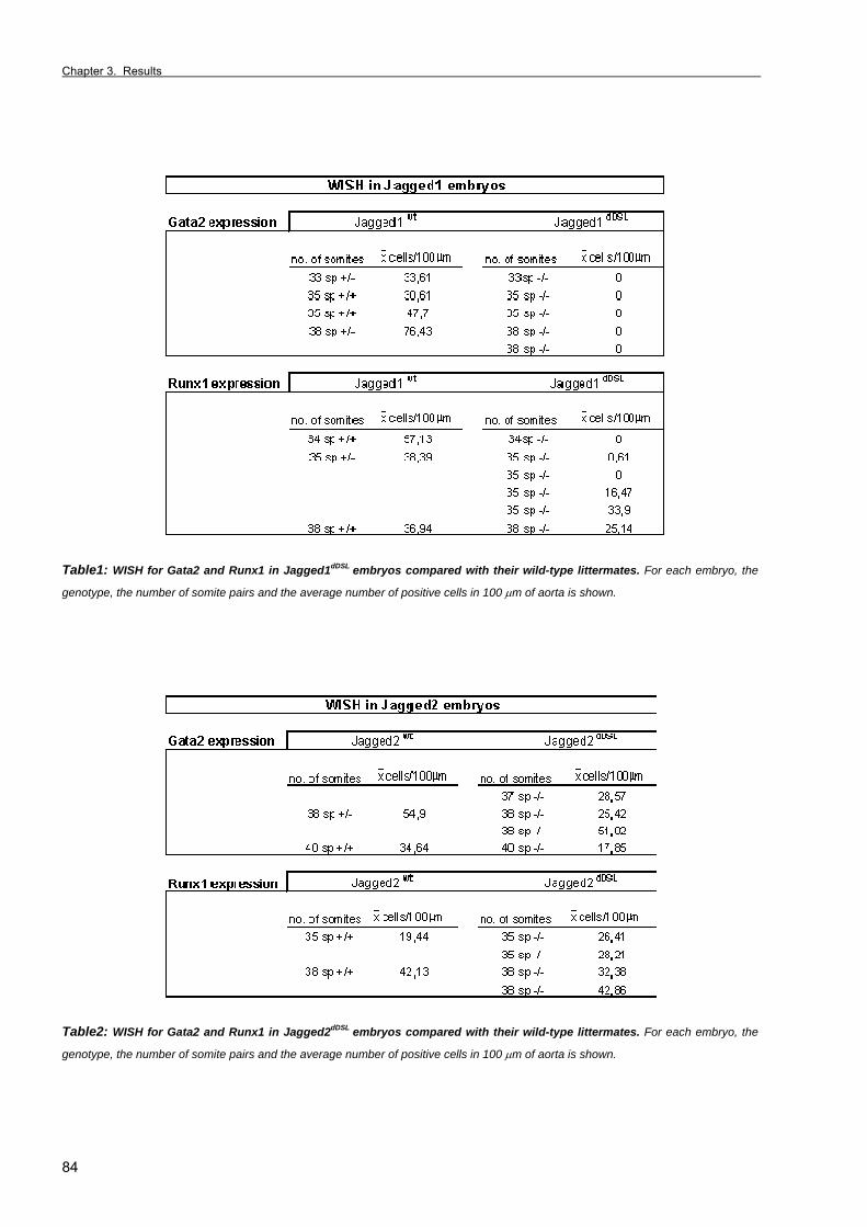
Chapter 3. Results _
84
Table1: WISH for Gata2 and Runx1 in Jagged1dDSL embryos compared with their wild-type littermates. For each embryo, the
genotype, the number of somite pairs and the average number of positive cells in 100 µm of aorta is shown.
Table2: WISH for Gata2 and Runx1 in Jagged2dDSL embryos compared with their wild-type littermates. For each embryo, the
genotype, the number of somite pairs and the average number of positive cells in 100 µm of aorta is shown.
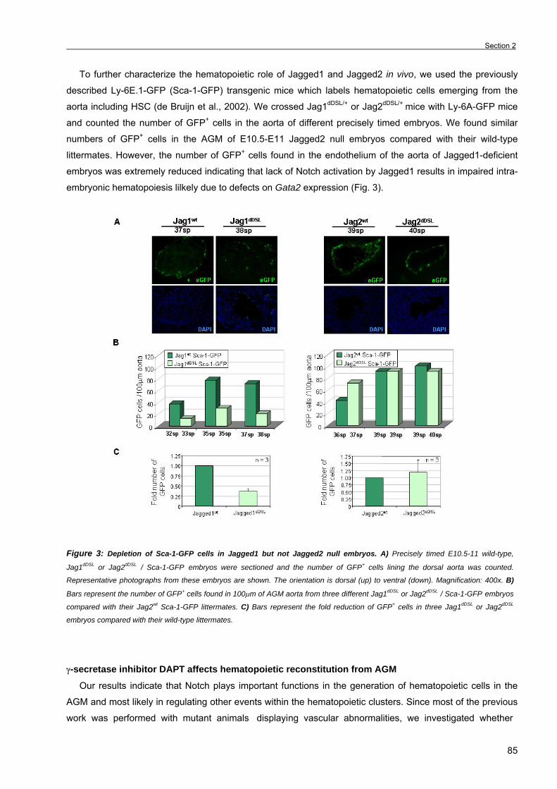
Section 2
85
To further characterize the hematopoietic role of Jagged1 and Jagged2 in vivo, we used the previously
described Ly-6E.1-GFP (Sca-1-GFP) transgenic mice which labels hematopoietic cells emerging from the
aorta including HSC (de Bruijn et al., 2002). We crossed Jag1dDSL/+ or Jag2dDSL/+ mice with Ly-6A-GFP mice
and counted the number of GFP+ cells in the aorta of different precisely timed embryos. We found similar
numbers of GFP+ cells in the AGM of E10.5-E11 Jagged2 null embryos compared with their wild-type
littermates. However, the number of GFP+ cells found in the endothelium of the aorta of Jagged1-deficient
embryos was extremely reduced indicating that lack of Notch activation by Jagged1 results in impaired intra-
embryonic hematopoiesis lilkely due to defects on Gata2 expression (Fig. 3).
Figure 3: Depletion of Sca-1-GFP cells in Jagged1 but not Jagged2 null embryos. A) Precisely timed E10.5-11 wild-type,
Jag1dDSL or Jag2dDSL / Sca-1-GFP embryos were sectioned and the number of GFP+ cells lining the dorsal aorta was counted.
Representative photographs from these embryos are shown. The orientation is dorsal (up) to ventral (down). Magnification: 400x. B)
Bars represent the number of GFP+ cells found in 100µm of AGM aorta from three different Jag1dDSL or Jag2dDSL / Sca-1-GFP embryos
compared with their Jag2wt Sca-1-GFP littermates. C) Bars represent the fold reduction of GFP+ cells in three Jag1dDSL or Jag2dDSL
embryos compared with their wild-type littermates.
γ-secretase inhibitor DAPT affects hematopoietic reconstitution from AGM
Our results indicate that Notch plays important functions in the generation of hematopoietic cells in the
AGM and most likely in regulating other events within the hematopoietic clusters. Since most of the previous
work was performed with mutant animals displaying vascular abnormalities, we investigated whether
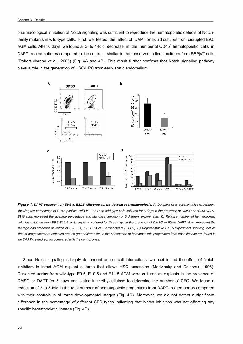
Chapter 3. Results _
86
pharmacological inhibition of Notch signaling was sufficient to reproduce the hematopoietic defects of Notch-
family mutants in wild-type cells. First, we tested the effect of DAPT on liquid cultures from disrupted E9.5
AGM cells. After 6 days, we found a 3- to 4-fold decrease in the number of CD45+ hematopoietic cells in
DAPT-treated cultures compared to the controls, similar to that observed in liquid cultures from RBPjκ-/- cells
(Robert-Moreno et al., 2005) (Fig. 4A and 4B). This result further confirms that Notch signaling pathway
plays a role in the generation of HSC/HPC from early aortic endothelium.
Figure 4: DAPT treatment on E9.5 to E11.5 wild-type aortas decreases hematopoiesis. A) Dot plots of a representative experiment
showing the percentage of CD45 positive cells in E9.5 P-sp wild-type cells cultured for 6 days in the presence of DMSO or 50µM DAPT.
B) Graphs represent the average percentage and standard deviation of 5 different experiments. C) Relative number of hematopoietic
colonies obtained from E9.5-E11.5 aorta explants cultured for three days in the presence of DMSO or 50µM DAPT. Bars represent the
average and standard deviation of 2 (E9.5), 1 (E10.5) or 3 experiments (E11.5). D) Representative E11.5 experiment showing that all
kind of progenitors are detected and no great differences in the percentage of hematopoietic progenitors from each lineage are found in
the DAPT-treated aortas compared with the control ones.
Since Notch signaling is highly dependent on cell-cell interactions, we next tested the effect of Notch
inhibitors in intact AGM explant cultures that allows HSC expansion (Medvinsky and Dzierzak, 1996).
Dissected aortas from wild-type E9.5, E10.5 and E11.5 AGM were cultured as explants in the presence of
DMSO or DAPT for 3 days and plated in methylcellulose to determine the number of CFC. We found a
reduction of 2 to 3-fold in the total number of hematopoietic progenitors from DAPT-treated aortas compared
with their controls in all three developmental stages (Fig. 4C). Moreover, we did not detect a significant
difference in the percentage of different CFC types indicating that Notch inhibition was not affecting any
specific hematopoietic lineage (Fig. 4D).
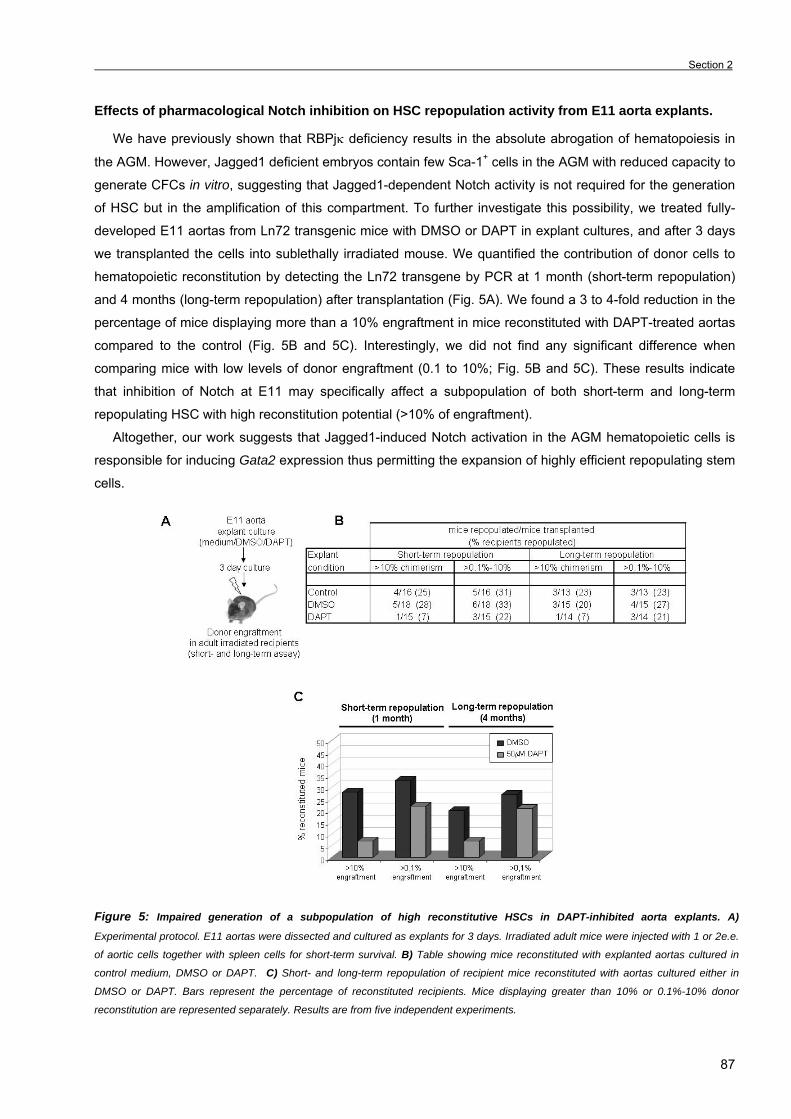
Section 2
87
Effects of pharmacological Notch inhibition on HSC repopulation activity from E11 aorta explants.
We have previously shown that RBPjκ deficiency results in the absolute abrogation of hematopoiesis in
the AGM. However, Jagged1 deficient embryos contain few Sca-1+ cells in the AGM with reduced capacity to
generate CFCs in vitro, suggesting that Jagged1-dependent Notch activity is not required for the generation
of HSC but in the amplification of this compartment. To further investigate this possibility, we treated fully-
developed E11 aortas from Ln72 transgenic mice with DMSO or DAPT in explant cultures, and after 3 days
we transplanted the cells into sublethally irradiated mouse. We quantified the contribution of donor cells to
hematopoietic reconstitution by detecting the Ln72 transgene by PCR at 1 month (short-term repopulation)
and 4 months (long-term repopulation) after transplantation (Fig. 5A). We found a 3 to 4-fold reduction in the
percentage of mice displaying more than a 10% engraftment in mice reconstituted with DAPT-treated aortas
compared to the control (Fig. 5B and 5C). Interestingly, we did not find any significant difference when
comparing mice with low levels of donor engraftment (0.1 to 10%; Fig. 5B and 5C). These results indicate
that inhibition of Notch at E11 may specifically affect a subpopulation of both short-term and long-term
repopulating HSC with high reconstitution potential (>10% of engraftment).
Altogether, our work suggests that Jagged1-induced Notch activation in the AGM hematopoietic cells is
responsible for inducing Gata2 expression thus permitting the expansion of highly efficient repopulating stem
cells.
Figure 5: Impaired generation of a subpopulation of high reconstitutive HSCs in DAPT-inhibited aorta explants. A)
Experimental protocol. E11 aortas were dissected and cultured as explants for 3 days. Irradiated adult mice were injected with 1 or 2e.e.
of aortic cells together with spleen cells for short-term survival. B) Table showing mice reconstituted with explanted aortas cultured in
control medium, DMSO or DAPT. C) Short- and long-term repopulation of recipient mice reconstituted with aortas cultured either in
DMSO or DAPT. Bars represent the percentage of reconstituted recipients. Mice displaying greater than 10% or 0.1%-10% donor
reconstitution are represented separately. Results are from five independent experiments.

Chapter 3. Results _
88
DISCUSSION There is evidence for a role of Notch signaling in the generation of HSC in the aortic endothelium (Kumano
et al., 2003; Robert-Moreno et al., 2005). In this work, we show that Jagged1 is responsible for Notch
activation in hematopoietic cells of the aorta in the AGM region. This is the first evidence about the specific
Notch ligand which is involved in the AGM hematopoiesis. The interaction between Notch and Jagged1
induces the transcriptional activation of Gata2 which likely regulates the proliferation of the stem cell pool in
the AGM region. In addition, DAPT-treatment of E11 AGM explants confirms the involvement of Notch in the
AGM hematopoiesis and reveals a new role for Notch in the acquisition of high repopulation capacity in HSC.
Our previous work suggested that Delta4, Jagged1 and Jagged2 were good candidates for Notch-
activating ligands, since they are all expressed in the mid-gestation aortic endothelium (Robert-Moreno et al.,
2005). Delta4 heterozigous embryos die at E10-10.5 displaying severe vasculogenic defects and loss of the
endothelial arterial cell fate (Duarte et al., 2004), which strongly compromised the study of hematopoiesis in
these animals. Thus, we focused in the study of the other two candidates, Jagged1 and Jagged2: Jag1dDSL/dDSL embryos display defects in the vasculature of the yolk sac and in the branching of blood vessels
in the embryo head but no defects in aorta formation have been reported (Xue et al., 1999) whereas
Jag2dDSL homozygous embryos die perinatally due to craniophacial, thymus and limb bud formation defects
(Jiang et al., 1998). We have now identified that Jagged1 ligand is responsible for specifically activating the
Notch receptor, switching on the hematopoietic program in endothelial cells of the AGM. This is supported by
the lower number of hematopoietic progenitors, Sca-1+ cells and the absence of Gata2 expression. By
contrast, these hematopoietic defects are not observed in the Jagged2-deficient embryos, although a lower
number of Gata2-expressing cells were found in these embryos.
We previously described that Notch1/RBPjκ signaling leads to the expression of the hematopoietic
transcription factor Gata2 and presumably to the commitment of HPC/HSCs from the AGM region (Robert-
Moreno et al., 2005). However, the Notch-Runx pathway establishes the commitment of the hematopoietic
stem cell fate in the zebrafish embryo (Burns et al., 2005) and Drosophila hemocytogenesis needs the
Notch-induced activation of the Runt family gene, Lozenge (Lebestky et al., 2003). Loss of Gata2 expression
in the Jag1dDSL null embryos reinforces the idea that Gata2 (but not Runx1) is a target gene of the
Jagged1/Notch1/RBPjκ signaling pathway. In contrast, Runx1 is expressed in the aorta in the 50% of the
Jagged1 embryos indicating that Jagged1 is not required at this stage for Runx1 expression. However, the
possibility that the expression of Runx1 may depend on another Notch ligand cannot be excluded.
Gene targeting studies revealed the importance of Gata2 for hematopoiesis since Gata2-/- embryos have
reduced numbers of hematopoietic cells (Tsai et al., 1994); there is no contribution of Gata2-/- ES-derived
cells to any hematopoietic tissue (Tsai et al., 1994) and Gata2 haploinsufficiency results in different HSC
abnormalities (Ling et al., 2004; Rodrigues et al., 2005). Interestingly, several similarities are found between
hematopoietic cells from Jagged1-deficient, DAPT-treated and Gata2-deficient hematopoietic cells since
they all show defects in HSC proliferation and/or expansion. Thus, a possible explanation is that Notch acts
on the first hematopoietic decision activating Runx1 and on a posterior committed cell to induce Gata2
resulting in the expansion of these cells. This interpretation is in agreement with our present work, since
Jagged1 mutants contain some hematopoiesis in the AGM and express Runx1 but not Gata2. Thus Jagged1

Section 2
89
would be responsible for a secondary Notch decision in these cells whereas another ligand may be
responsible for the first event that activates Runx1 expression.
To exclude the possibility that vascular abnormalities in Notch-mutants was responsible for the
hematopoietic defect (Kumano et al., 2003; Robert-Moreno et al., 2005), we have tested the effect of γ-
secretase inhibitors in the amplification of HSC in AGM explants. Our results suggest that Notch signaling is
required for the generation of HPC (as demonstrated by CFC assay of DAPT-treated explanted aortas) and
HSC (determined by the lower adult repopulation of γ-secretase inhibited explants). Hematopoietic colonies
found in the DAPT-treated aortas may come from cells generated previous to the γ-secretase treatment, thus
suggesting that Notch would not be required for cell-fate decisions within the hematopoietic hierarchical tree,
but may be required for de novo generation of a common undifferentiated hematopoietic progenitor from the
aortic endothelium. Thus, Notch signaling is required in endothelial cells from a previously specified artery in
order to generate HPC/HSCs. A very interesting observation is that Notch seems to be necessary for the
generation of a subpopulation of short- and long-term repopulating HSCs with high reconstitution ability,
suggesting that, different subsets of HSCs with distinct repopulation and self-renewal behavior are generated
from the AGM, as previously described in the bone marrow (Sieburg et al., 2006). This specific Jagged1
function may have an enormous relevance in cell therapy when trying to use Notch ligands for HSC
amplification.
Hematopoietic clusters emerging from the endothelium of the dorsal aorta are likely to be qualitatively
different since they express different levels of hematopoietic and endothelial markers (reviewed in (Dieterlen-
Lievre et al., 2006)]. We have now characterized the expression of Notch family members in these clusters
and have shown that their expression also differ among clustered cells. The Notch target gene hes1 is also
expressed in the hematopoietic clusters, which is a measure of Notch activity. It has been proposed that
Hes1 maintains the stemness of HSCs by inhibiting their differentiation (Kunisato et al., 2003). Consistent
with this, we propose a model in which Notch1 activates both hes1 and gata2 expression thus maintaining
the undifferentiated state and promoting proliferation of a subset of HSCs with high repopulating ability.
Chapter 3. Results _
90
SUPPLEMENTAL INFORMATION
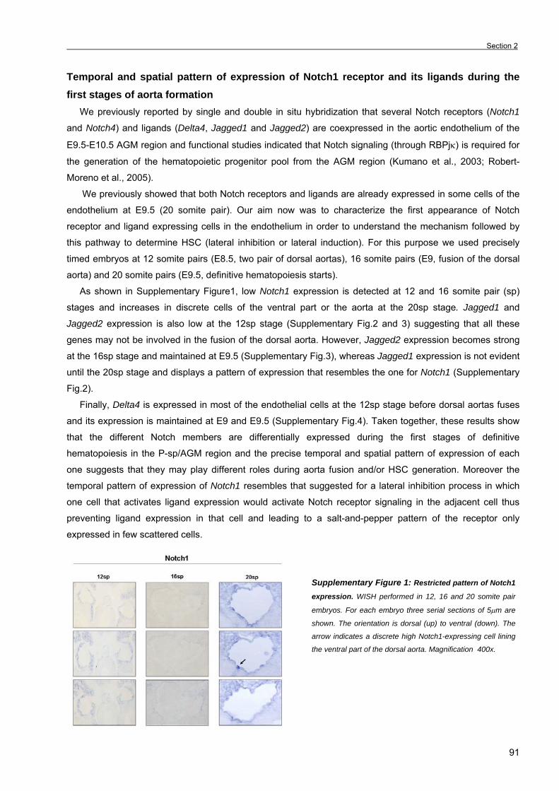
Section 2
91
Temporal and spatial pattern of expression of Notch1 receptor and its ligands during the first stages of aorta formation We previously reported by single and double in situ hybridization that several Notch receptors (Notch1
and Notch4) and ligands (Delta4, Jagged1 and Jagged2) are coexpressed in the aortic endothelium of the
E9.5-E10.5 AGM region and functional studies indicated that Notch signaling (through RBPjκ) is required for
the generation of the hematopoietic progenitor pool from the AGM region (Kumano et al., 2003; Robert-
Moreno et al., 2005).
We previously showed that both Notch receptors and ligands are already expressed in some cells of the
endothelium at E9.5 (20 somite pair). Our aim now was to characterize the first appearance of Notch
receptor and ligand expressing cells in the endothelium in order to understand the mechanism followed by
this pathway to determine HSC (lateral inhibition or lateral induction). For this purpose we used precisely
timed embryos at 12 somite pairs (E8.5, two pair of dorsal aortas), 16 somite pairs (E9, fusion of the dorsal
aorta) and 20 somite pairs (E9.5, definitive hematopoiesis starts).
As shown in Supplementary Figure1, low Notch1 expression is detected at 12 and 16 somite pair (sp)
stages and increases in discrete cells of the ventral part or the aorta at the 20sp stage. Jagged1 and
Jagged2 expression is also low at the 12sp stage (Supplementary Fig.2 and 3) suggesting that all these
genes may not be involved in the fusion of the dorsal aorta. However, Jagged2 expression becomes strong
at the 16sp stage and maintained at E9.5 (Supplementary Fig.3), whereas Jagged1 expression is not evident
until the 20sp stage and displays a pattern of expression that resembles the one for Notch1 (Supplementary
Fig.2).
Finally, Delta4 is expressed in most of the endothelial cells at the 12sp stage before dorsal aortas fuses
and its expression is maintained at E9 and E9.5 (Supplementary Fig.4). Taken together, these results show
that the different Notch members are differentially expressed during the first stages of definitive
hematopoiesis in the P-sp/AGM region and the precise temporal and spatial pattern of expression of each
one suggests that they may play different roles during aorta fusion and/or HSC generation. Moreover the
temporal pattern of expression of Notch1 resembles that suggested for a lateral inhibition process in which
one cell that activates ligand expression would activate Notch receptor signaling in the adjacent cell thus
preventing ligand expression in that cell and leading to a salt-and-pepper pattern of the receptor only
expressed in few scattered cells.
Supplementary Figure 1: Restricted pattern of Notch1
expression. WISH performed in 12, 16 and 20 somite pair
embryos. For each embryo three serial sections of 5µm are
shown. The orientation is dorsal (up) to ventral (down). The
arrow indicates a discrete high Notch1-expressing cell lining
the ventral part of the dorsal aorta. Magnification 400x.

Chapter 3. Results _
92
Supplementary Figure 2: Jagged1 displays a similar
temporal and spatial pattern than Notch1. WISH performed
in 12, 16 and 20 somite pair embryos. For each embryo three
serial sections of 5µm are shown. The orientation is dorsal
(up) to ventral (down). The arrow indicates a discrete cell
lining the ventral part of the dorsal aorta with high Jagged1
expression. Magnification 400x.
Supplementary Figure 3: Jagged2 expression
begins at the 16 somite pair stage. WISH performed in
12, 16 and 20 somite pair embryos. For each embryo three
serial sections of 5µm are shown. The orientation is dorsal
(up) to ventral (down). Magnification 400x.
Supplementary Figure 4: Delta4 is expressed before
the pair of aortas fusion. WISH performed in 12, 16 and 20
somite pair embryos. For each embryo three serial sections
of 5µm are shown. The orientation is dorsal (up) to ventral
(down). Magnification 400x.
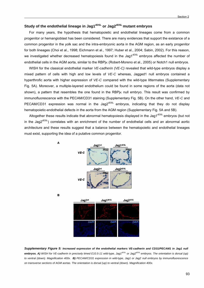
Section 2
93
Study of the endothelial lineage in Jag1dDSL or Jag2dDSL mutant embryos For many years, the hypothesis that hematopoietic and endothelial lineages come from a common
progenitor or hemangioblast has been considered. There are many evidences that support the existance of a
common progenitor in the yolk sac and the intra-embryonic aorta in the AGM region, as an early progenitor
for both lineages (Choi et al., 1998; Eichmann et al., 1997; Huber et al., 2004; Sabin, 2002). For this reason,
we investigated whether decreased hematopoiesis found in the Jag1dDSL embryos affected the number of
endothelial cells in the AGM aorta, similar to the RBPjκ (Robert-Moreno et al., 2005) or Notch1 null embryos.
WISH for the classical endothelial marker VE-cadherin (VE-C) revealed that wild-type embryos display a
mixed pattern of cells with high and low levels of VE-C whereas, Jagged1 null embryos contained a
hyperthrofic aorta with higher expression of VE-C compared with the wild-type littermates (Supplementary
Fig. 5A). Moreover, a multiple-layered endothelium could be found in some regions of the aorta (data not
shown), a pattern that resembles the one found in the RBPjκ null embryo. This result was confirmed by
immunofluorescence with the PECAM/CD31 staining (Supplementary Fig. 5B). On the other hand, VE-C and
PECAM/CD31 expression was normal in the Jag2dDSL embryos, indicating that they do not display
hematopoietic-endothelial defects in the aorta from the AGM region (Supplementary Fig. 5A and 5B). Altogether these results indicate that abnormal hematopoiesis displayed in the Jag1dDSL embryos (but not
in the Jag2dDSL) correlates with an enrichment of the number of endothelial cells and an abnormal aortic
architecture and these results suggest that a balance between the hematopoietic and endothelial lineages
must exist, supporting the idea of a putative common progenitor.
Supplementary Figure 5: Increased expression of the endothelial markers VE-cadherin and CD31/PECAM1 in Jag1 null
embryos. A) WISH for VE-cadherin in precisely timed E10.5-11 wild-type, Jag1dDSL or Jag2dDSL embryos. The orientation is dorsal (up)
to ventral (down). Magnification 400x. B) PECAM/CD31 expression in wild-type, Jag1 or Jag2 null embryos by immunofluorescence
on transverse sections of AGM aortas. The orientation is dorsal (up) to ventral (down). Magnification 400x.
Chapter 3. Results _
94
REFERENCES • Burns, C. E., Traver, D., Mayhall, E., Shepard, J. L. and Zon, L. I. (2005). Hematopoietic stem cell fate is established by the
Notch-Runx pathway. Genes Dev 19, 2331-42. • Cumano, A., Ferraz, J. C., Klaine, M., Di Santo, J. P. and Godin, I. (2001). Intraembryonic, but not yolk sac hematopoietic
precursors, isolated before circulation, provide long-term multilineage reconstitution. Immunity 15, 477-85. • Choi, K., Kennedy, M., Kazarov, A., Papadimitriou, J. C. and Keller, G. (1998). A common precursor for hematopoietic and
endothelial cells. Development 125, 725-32. • de Bruijn, M. F., Ma, X., Robin, C., Ottersbach, K., Sanchez, M. J. and Dzierzak, E. (2002). Hematopoietic stem cells localize to
the endothelial cell layer in the midgestation mouse aorta. Immunity 16, 673-83. • de Bruijn, M. F., Speck, N. A., Peeters, M. C. and Dzierzak, E. (2000). Definitive hematopoietic stem cells first develop within the
major arterial regions of the mouse embryo. Embo J 19, 2465-74. • de la Pompa, J. L., Wakeham, A., Correia, K. M., Samper, E., Brown, S., Aguilera, R. J., Nakano, T., Honjo, T., Mak, T. W.,
Rossant, J. et al. (1997). Conservation of the Notch signalling pathway in mammalian neurogenesis. Development 124, 1139-48. • Dieterlen-Lievre, F., Pouget, C., Bollerot, K. and Jaffredo, T. (2006). Are intra-aortic hemopoietic cells derived from endothelial
cells during ontogeny? Trends Cardiovasc Med 16, 128-39. • Duarte, A., Hirashima, M., Benedito, R., Trindade, A., Diniz, P., Bekman, E., Costa, L., Henrique, D. and Rossant, J. (2004).
Dosage-sensitive requirement for mouse Dll4 in artery development. Genes Dev 18, 2474-8. • Eichmann, A., Corbel, C., Nataf, V., Vaigot, P., Breant, C. and Le Douarin, N. M. (1997). Ligand-dependent development of the
endothelial and hemopoietic lineages from embryonic mesodermal cells expressing vascular endothelial growth factor receptor 2. Proc Natl Acad Sci U S A 94, 5141-6.
• Gekas, C., Dieterlen-Lievre, F., Orkin, S. H. and Mikkola, H. K. (2005). The placenta is a niche for hematopoietic stem cells. Dev Cell 8, 365-75.
• Hadland, B. K., Huppert, S. S., Kanungo, J., Xue, Y., Jiang, R., Gridley, T., Conlon, R. A., Cheng, A. M., Kopan, R. and Longmore, G. D. (2004). A requirement for Notch1 distinguishes 2 phases of definitive hematopoiesis during development. Blood 104, 3097-105.
• Huber, T. L., Kouskoff, V., Fehling, H. J., Palis, J. and Keller, G. (2004). Haemangioblast commitment is initiated in the primitive streak of the mouse embryo. Nature 432, 625-30.
• Jiang, R., Lan, Y., Chapman, H. D., Shawber, C., Norton, C. R., Serreze, D. V., Weinmaster, G. and Gridley, T. (1998). Defects in limb, craniofacial, and thymic development in Jagged2 mutant mice. Genes Dev 12, 1046-57.
• Kumano, K., Chiba, S., Kunisato, A., Sata, M., Saito, T., Nakagami-Yamaguchi, E., Yamaguchi, T., Masuda, S., Shimizu, K., Takahashi, T. et al. (2003). Notch1 but not Notch2 is essential for generating hematopoietic stem cells from endothelial cells. Immunity 18, 699-711.
• Kunisato, A., Chiba, S., Nakagami-Yamaguchi, E., Kumano, K., Saito, T., Masuda, S., Yamaguchi, T., Osawa, M., Kageyama, R., Nakauchi, H. et al. (2003). HES-1 preserves purified hematopoietic stem cells ex vivo and accumulates side population cells in vivo. Blood 101, 1777-83.
• Lebestky, T., Jung, S. H. and Banerjee, U. (2003). A Serrate-expressing signaling center controls Drosophila hematopoiesis. Genes Dev 17, 348-53.
• Ling, K. W., Ottersbach, K., van Hamburg, J. P., Oziemlak, A., Tsai, F. Y., Orkin, S. H., Ploemacher, R., Hendriks, R. W. and Dzierzak, E. (2004). GATA-2 plays two functionally distinct roles during the ontogeny of hematopoietic stem cells. J Exp Med 200, 871-82.
• Medvinsky, A. and Dzierzak, E. (1996). Definitive hematopoiesis is autonomously initiated by the AGM region. Cell 86, 897-906. • Ottersbach, K. and Dzierzak, E. (2005). The murine placenta contains hematopoietic stem cells within the vascular labyrinth
region. Dev Cell 8, 377-87. • Robert-Moreno, A., Espinosa, L., de la Pompa, J. L. and Bigas, A. (2005). RBPjkappa-dependent Notch function regulates
Gata2 and is essential for the formation of intra-embryonic hematopoietic cells. Development 132, 1117-26. • Rodrigues, N. P., Janzen, V., Forkert, R., Dombkowski, D. M., Boyd, A. S., Orkin, S. H., Enver, T., Vyas, P. and Scadden, D.
T. (2005). Haploinsufficiency of GATA-2 perturbs adult hematopoietic stem-cell homeostasis. Blood 106, 477-84. • Sabin, F. R. (2002). Preliminary note on the differentiation of angioblasts and the method by which they produce blood-vessels,
blood-plasma and red blood-cells as seen in the living chick. 1917. J Hematother Stem Cell Res 11, 5-7. • Sieburg, H. B., Cho, R. H., Dykstra, B., Uchida, N., Eaves, C. J. and Muller-Sieburg, C. E. (2006). The hematopoietic stem
compartment consists of a limited number of discrete stem cell subsets. Blood 107, 2311-6. • Tsai, F. Y., Keller, G., Kuo, F. C., Weiss, M., Chen, J., Rosenblatt, M., Alt, F. W. and Orkin, S. H. (1994). An early
haematopoietic defect in mice lacking the transcription factor GATA-2. Nature 371, 221-6. • Xue, Y., Gao, X., Lindsell, C. E., Norton, C. R., Chang, B., Hicks, C., Gendron-Maguire, M., Rand, E. B., Weinmaster, G. and
Gridley, T. (1999). Embryonic lethality and vascular defects in mice lacking the Notch ligand Jagged1. Hum Mol Genet 8, 723-30. • Yoder, M. C. and Hiatt, K. (1997). Engraftment of embryonic hematopoietic cells in conditioned newborn recipients. Blood 89,
2176-83. • Yoder, M. C., Hiatt, K., Dutt, P., Mukherjee, P., Bodine, D. M. and Orlic, D. (1997). Characterization of definitive
lymphohematopoietic stem cells in the day 9 murine yolk sac. Immunity 7, 335-44. • Yoder, M. C., Hiatt, K. and Mukherjee, P. (1997). In vivo repopulating hematopoietic stem cells are present in the murine yolk sac
at day 9.0 postcoitus. Proc Natl Acad Sci U S A 94, 6776-80.

Section 3
95
SECTION 3: The notch pathway positively regulates programmed cell death
during erythroid differentiation (This chapter was published in Leukemia, May 2007).
Several gene target mutations revealed that hematopoiesis in the yolk sac and in the intra-embryonic
AGM region are governed by two different genetic programs since some mutations affect both primitive and
definitive hematopoiesis whereas others affect only the definitive one (reviewed in Cumano and Godin,
2002).
In this paper, we show that in contrast to the absolute Notch/RBPjκ signaling requirement for definitive
AGM hematopoiesis, primitive erythropoiesis normally occurs in the yolk sac of RBPjκ mutants. All kind of
hematopoietic progenitors are generated in these mutant embryos and development and maturation of the
erythroid lineage does not display any defect. However, the percentage of Ter119+ erythroid cells is higher in
the RBPjκ-/- yolk sacs compared with their wild-type littermates and neither proliferation nor differentiation is
responsible for this increase.
In contrast, apoptosis is reduced specifically in erythroid cells from the mutant yolk sacs, indicating that
Notch signaling promotes programmed cell death in this lineage. By quantitative RT-PCR we demonstrate
that loss of RBPjκ correlates with an increase of Epo and its receptor (Epo-R) expression and in the levels of
the anti-apoptotic genes bcl-2 and bcl-xL in agreement with the higher survival of the Ter119+ population.
Finally we show that Notch induces apoptosis in the erythroid lineage not only during embryonic
development but in the adult as well, since bone marrow cells treated with the γ-secretase inhibitors DAPT
and L685,458 display low apoptotic cell death and Notch-induced apoptosis by incubating bone marrow with
Jagged1-expressing cells is abrogated by γ-secretase inhibitors treatment. In the same sense, murine
erythroleukemic cells overexpressing the activated form of Notch1 display higher programmed cell death in
hexametilene-bisacetamide-induced differentiation.
From all the data we suggest that Notch signaling is not essential for yolk sac hematopoiesis but
specifically induces programmed cell death in the erythroid lineage not only during embryonic life but in adult
tissues as well thus regulating homeostasis of this hematopoietic compartment.

Chapter 3. Results _
96
The notch pathway positively regulates programmed cell death during erythroid differentiation
Running title: Notch induces apoptosis in erythroid cells
Àlex Robert-Moreno1, Lluis Espinosa1, M.José Sanchez2, José L. de la Pompa3 and Anna Bigas1*
1Centre Oncologia Molecular, IDIBELL-Institut de Recerca Oncològica. Gran Via km 2.7 Hospitalet,
Barcelona, Spain. 2 Centro Andaluz Biología Desarrollo, CSIC, Universidad Pablo Olavide, Sevilla 3Dept. Immunology and Oncology, Centro Nacional de Biotecnología, CSIC. Darwin, 3. Campus de
Cantoblanco, Madrid 28049, Spain
* Corresponding author
Mailing address: Oncologia Molecular.
IDIBELL-Institut de Recerca Oncològica
Gran Via Km 2.7.
08907 - Hospitalet, Barcelona. Spain
Phone: 932 607 404
Fax: 932 607 426
e-mail: [email protected]

Section 3
97
SUMMARY
Programmed cell death plays an important role in erythropoiesis under physiological and pathological
conditions. In this study, we show that the Notch/RBPjκ signaling pathway induces erythroid apoptosis in
different hematopoietic tissues, including yolk sac and bone marrow as well as in murine erythroleukemia
cells. In RBPjκ-/- yolk sacs, erythroid cells have a decreased rate of cell death that results in increased
number of Ter119+ cells. A similar effect is observed when Notch activity is abrogated by incubation with the
γ-secretase inhibitors, DAPT or L685,458. We demonstrate that incubation with Jagged1-expressing cells
has a proapoptotic effect in erythroid cells from adult bone marrow that is prevented by blocking Notch
activity. Finally, we show that the sole expression of the activated Notch1 protein is sufficient to induce
apoptosis in hexametilene-bisacetamide-differentiating murine erythroleukemia cells. Together these results
demonstrate that Notch regulates erythroid homeostasis by inducing apoptosis.
INTRODUCTION Notch is a highly conserved signaling pathway that regulates cell fate specification during development
and adult tissue homeostasis. Physiological activation of the Notch pathway requires the interaction between
the Notch receptor and one of its ligands. This interaction leads to the cleavage of Notch receptor, releasing
the intracellular domain that translocates to the nucleus to bind RBPjκ and activate specific gene
transcription (reviewed by Bray1 and Lai2).
Notch function is required for the generation of definitive hematopoiesis as shown by the lack of
hematopoietic precursors in the aorta of Notch1-/- and RBPjκ-/- mouse embryos or in Mind bomb mutants in
zebrafish3-6. In contrast, primitive hematopoiesis occurs in different Notch pathway mutants in both mouse
and zebrafish3-5. In the mouse, primitive hematopoiesis originates in the blood islands of the yolk sac,
starting at embryonic day 7.5 (E7.5). The main component of this primitive hematopoiesis is erythroid
progenitor cells (EryP)7 that generate large nucleated primitive erythrocytes that contain embryonic globins
(βH1, ε-globin and ζ-globin) (reviewed by Palis and Segel8).
Erythropoiesis involves the progressive differentiation of uncommitted progenitors to mature erythrocytes.
However, not only differentiation but also apoptosis participates in the regulation of cell survival and mature
red cell turnover. The amount of erythropoietin (Epo), mainly dependent on hypoxia, is one of the key factors
in controlling the survival of erythroid cells (reviewed in Mulcahy9). Expression of the antiapoptotic members
of the Bcl-2 family, Bcl-2 and Bcl-x,10 are some of the downstream effects of EpoR activation in this system.
Consistent with this, bcl-x-/- embryos die of massive apoptosis in the nervous system and in fetal liver
erythroid cells11.
Other transcription factors including GATA1 and, more recently, p53 have been implicated in regulating
apoptosis at different stages of erythroid maturation12,13. In this sense, GATA1 plays a key role in
development and survival of erythroid cells since GATA1-deficient cells failed to develop beyond the
proerythroblast stage and undergo rapid apoptosis14.
Chapter 3. Results _
98
Notch pathway has previously been shown to induce apoptosis in cell lines from different hematopoietic
lineages most likely through the activation of its target gene hes115-17. However the overall data linking Notch
and erythroid apoptosis is controversial18,19.
In this work, we demonstrate that the Notch signaling pathway is a positive regulator of apoptosis in
primitive erythropoiesis in the yolk sac but also in erythroid cells from adult bone marrow (BM).
Complementary studies using the murine erythroleukemia (MEL) cell line indicate that Notch induces
erythroid-specific apoptosis.
MATERIALS and METHODS
Animals
RBPjκ-/- mice have been previously described20. Animals were kept under pathogen-free conditions and
experiments approved by the Animal Care Committee. Yolk sacs were obtained from timed pregnant females
at days 7.5 to 9.5 of gestation and dissected out from embryo and vitelline arteries. Embryos were
genotyped by polymerase chain reaction (PCR) and morphology. BM was obtained from 8-12 week wild-type
(WT) CD1 mice.
Cell lines and transfections
MEL cells21 were maintained in RPMI 10% fetal bovine serum (FBS), 1% L-glutamine and 1% Pen/Strep.
Stable clones of MEL cells expressing N1∆E22 or pcDNA.3 were obtained by electroporation and expression
was confirmed by western blot (9E10 antibody). NIH-3T3 cells were transfected by calcium phosphate with
Jagged1 construct23 and clones overexpressing Jagged1 were selected in G418. Differentiation of MEL cells
was performed with 5mM hexametilene-bisacetamide (HMBA; Sigma, St Louis, MI, USA) for 6 days.
Dianisidine staining
O-dianisidine (Sigma) was used to stain hemoglobin of both E9.5 WT and RBPjκ-mutant yolk sacs, and
MEL Friend cells to assay erythroid differentiation as previously described21.
Hematopoietic colony assay
Yolk sac from WT and RBPjκ-/- E7.5-9.5 embryos was digested in 0.1% collagenase (Sigma) in
phosphate-buffered saline (PBS), 10% FBS for 30 min at 37ºC. Cells (30,000) were plated in duplicates in
1% methylcellulose (Stem Cell Technologies, Vancouver, Canada) plus Iscove’s with 10% FBS, 10% IL3-
and stem cell factor (SCF)-conditioned medium, 2.5% L-glutamine, 0.1% monothioglycerol (Sigma), 1%
Pen/Strep (Biological Industries, Beit Haemek Kibbutz, Israel), 2 IU/ml erythropoietin (Laboratorios Pensa-
Esteve, Barcelona, Spain), 20 ng/ml granulocyte-macrophage colony-stimulating factor (PeproTech, Rocky
Hill, NJ, USA) and 100 ng/ml of granulocyte colony-stimulating factor (Avantis Pharma, Paris, France). After
7 days, the presence of hematopoietic colonies was scored under a microscope. EryP colonies were scored
at day 3.
Section 3
99
Flow cytometry analysis Collagenase-disrupted yolk sac cells were stained with fluorescein isothiocyanate FITC-conjugated
CD71, CD41, ckit, CD45 and mac1 and PE-conjugated Ter119 and CD31 antibodies (Pharmingen, BD
Biosciences, San Jose, CA, USA) or isotopic immunoglobulin G as a control. Cells were analyzed in a
FACScalibur (Becton & Dickinson, BD Biosciences, San Jose, CA, USA) and WinMDI 2.8 software. Dead
cells were excluded by 7-aminoactinomicin-D (7-AAD; Invitrogen, Carlsbad, CA, USA) staining. For the
Annexin V binding analysis, cells were stained with rh AnnexinV-FITC kit (Bender Medsystems, Burlingame,
CA, USA) and 7-AAD for 15 min according to the manufacturer’s instructions.
For cell cycle analysis of total yolk sac, cells were fixed in 70% EtOH at –200C overnight, treated with 50
µg DNAse-free RNAse and stained with 25 µg of propidium iodide (Sigma). Ter119+ cell-cycle analysis was
performed on fresh cells with 20 µM Draq5 (Biostatus Ltd, Leicestershire, UK). FlowJo 6.4.1 software was
used for cell-cycle analysis.
Yolk sac and BM cultures
Collagenase-disrupted yolk sacs were cultured for 6 days in Iscove’s with 10% FBS, 10% IL3- and SCF-
conditioned medium, 0.1% monothioglycerol in the presence of 50 µM DAPT (Calbiochem), 2 µM L685,458
(Sigma) or dimethyl sulfoxide (DMSO) as control. For BM culture, 1.5x10⁵ whole BM cells were incubated
with γ-secretase inhibitors in RPMI 10% FBS, 2IU/ml EPO for 2-3 days. Z-Val-Ala-DL-Asp-
fluoromethylketone (Z-VAD-FMK; Bachem, Budendorf, Switzerland) was used at 200µM.
Coculture on 3T3 or 3T3-Jag1 stromal cells was perfomed with 4x10⁵ whole BM cells in RPMI, 10% FBS
for 16h. Cells were assayed for AnnexinV binding and analyzed by flow cytometry.
Immunohistochemisty
Yolk sacs were fixed with 4% paraformaldehyde (Sigma), embedded in Paraplast (Sigma) and sectioned
(10 µm). Slides were dewaxed in xylene, antigen retrieval was performed by boiling for 2 min in sodium
acetate, rehydrated and blocked-permeabilized in 10% FBS, 0.3% Surfact-Amps X100 (Pierce, Aalst,
Belgium) and 5% non-fat milk in PBS for 90 min at 4ºC. Anti P-Ser10 H3 (Upstate, Charlottesville, VA, USA)
was used at 1:500 dilution and developed with Dakocytomation kit (Dako, Glostrup, DK, Denmark) following
manufacturer’s instructions. Hematoxilin (Merck, Whitehouse Station, NJ, USA) was used for
counterstaining.
For histological analysis, tissue samples were fixed overnight at 4ºC in 4% paraformaldehyde, dehydrated
and embedded in Paraplast (Sigma). Samples were sectioned in a Leica-RM2135 at 4µm and stained with
hematoxilin and eosin.
Images were acquired with an Olympus BX-60 using a Spot camera and Spot 3.2.4 software (Diagnostic
Instruments, Sterling Heights, MI, USA). Adobe Photoshop 6.0 software was used for photograph editing.
Semiquantitative reverse transcriptase-polymerase chain reaction
Total RNA from subdissected E9.5 WT and RBPjκ-/- yolk sacs was isolated using TRIzol Reagent
(Invitrogen). Poly-AT Tract System IV (Promega, Madison, WI, USA) and RT-First Strand cDNA Synthesis
Kit (GE Healthcare, Buckinghamshire, UK) were used to obtain mRNA and cDNA respectively. PCR product

Chapter 3. Results _
100
was analyzed at different cycles to avoid saturation. Quantity One software (Biorad, Hempstead, UK) was
used for densitometry. Primer pairs used in the experiments are listed in Supplementary Table S1.
Quantitative RT-PCR Ter119+ cells were sorted from E9.5 collagenase-treated embryos in MoFlo Cell Sorter (Dakocytomation,
BD Diagnostic, San Jose, CA, USA). mRNA was isolated with Rneasy minikit (Qiagen, Valencia, CA, USA)
following manufacture’s instructions. qRT-PCRs were performed with SYBR Green I Master (Roche, Basel,
Switzerland) in LightCycler480 system.
Statistical analysis Normal distribution of the samples was confirmed with one-sample Kolmogorow-Smirnoff test and
Student’s t-test was performed.
RESULTS
Absence of Notch signaling results in increased number of erythroid cells in the yolk sac
Notch signaling has previously been shown to influence differentiation and apoptosis of erythroid cells in
vitro although controversial observations have been reported15,18,19,24. For this reason, we aimed to
characterize the physiological role of the Notch pathway in erythropoiesis by comparing WT and RBPjκ-/-
embryos. Despite the absence of intra-embryonic hematopoiesis in the RBPjκ-/- embryos and the presence of
different angiogenic abnormalities in the yolk sac, we found that primitive hematopoiesis does occur in the
yolk sac of the RBPjκ-/- embryos (Figure 1a), similar to the Notch1-/- mutants25-27.
To determine whether Notch pathway plays a role in regulating hematopoiesis in the yolk sac, we first
analyzed the expression of different Notch receptors, ligands and Notch-target genes in the yolk sac of WT
and RBPjκ-/- embryos by semiquantitative RT-PCR and we observed that all Notch family genes are
expressed in the yolk sac at E9.5 (Figure 1b). In RBPjκ-/-, we found reduced expression of all ligands and
receptors, whereas Notch3 and Jagged2 were upregulated. We also tested the expression of Notch-target
genes and detected a consistent reduction in hes1 levels in RBPjκ-/- yolk sacs compared with WT (Figure
1b).
Since hematopoiesis in the yolk sac is mainly restricted to erythropoiesis, we determined the percentage
of cells expressing the erythroid marker Ter119 in collagenase-treated yolk sacs at E7.5, E8.5 and E9.5. We
detected a few positive cells in the yolk sac of E7.5 (Figure 1c) and E8.5 (data not shown). At day 9.5 the
percentage of Ter119+ in the yolk sac ranged from 20 to 40% in the WT and 40-60% in the RBPjκ-/- (Figure
1c and d) being similar the total number of cells per yolk sac (Figure 1a).

Section 3
101
Figure1: Increased number of erythroid cells in RBPjκ -/-yolk sacs. (a) Images of E9.5 WT and RBPjκ -/-embryos with the yolk sac
(upper panel) and hematoxilin/eosin staining of yolk sac sections. Total number of cells obtained from disrupted yolk sacs (right panel).
(b) Semiquantitative RT-PCR of Notch receptors, ligands and target genes from E9.5 WT and RBPjκ -/-yolk sacs. (c) Representative
analysis of Ter119+ cells from E7.5 and E9.5 WT and RBPjκ -/-yolk sacs. (d) Percentage of Ter119+ cells in the analyzed E9.5 WT and
RBPjκ -/-yolk sacs. Average and s.d. are represented.
Proliferation is not responsible for increased erythropoiesis in RBPjκ-/- yolk sacs
To investigate whether the higher number of Ter119+ cells in the RBPjκ-/- yolk sac was owing to an
increase in the number of progenitors, we performed colony-forming cell (CFC) assays with collagenase-
treated yolk sac cells at E7.5, E8.5 and E9.5 from RBPjκ+/+, +/- and -/- embryos. We detected a similar
percentage of myeloid, erythroid and mixed colonies in these cultures (Supplementary Figure S1), however;
at E9.5, there was a twofold increase in the total number of CFC in mutant embryos (Figure 2a). To test
whether this effect was due to increased proliferation, we analyzed the cell-cycle profile of WT and RBPjκ-/-
yolk sac cells by flow cytometry. Surprisingly, the percentage of cells in S/G2-M phase was slightly reduced
in RBPjκ-/- compared with WT cells (from 53 to 45%), and this reduction in S/G2-M phase was higher when
cell cycle was analyzed in the Ter119+ cells (Figure 2b). To confirm this observation, we performed the P-H3
staining on yolk sac sections to assess the number of cells undergoing mitosis inside the blood islands. P-H3
staining showed that hematopoietic cells from the WT yolk sacs have a similar mitotic rate (19.8 %) than
cells in RBPjκ-/- yolk sacs (12.9 %); (Figure 2c). Altogether these results indicate that proliferation is not
increased in the RBPjκ-/- yolk sac erythroid cells.
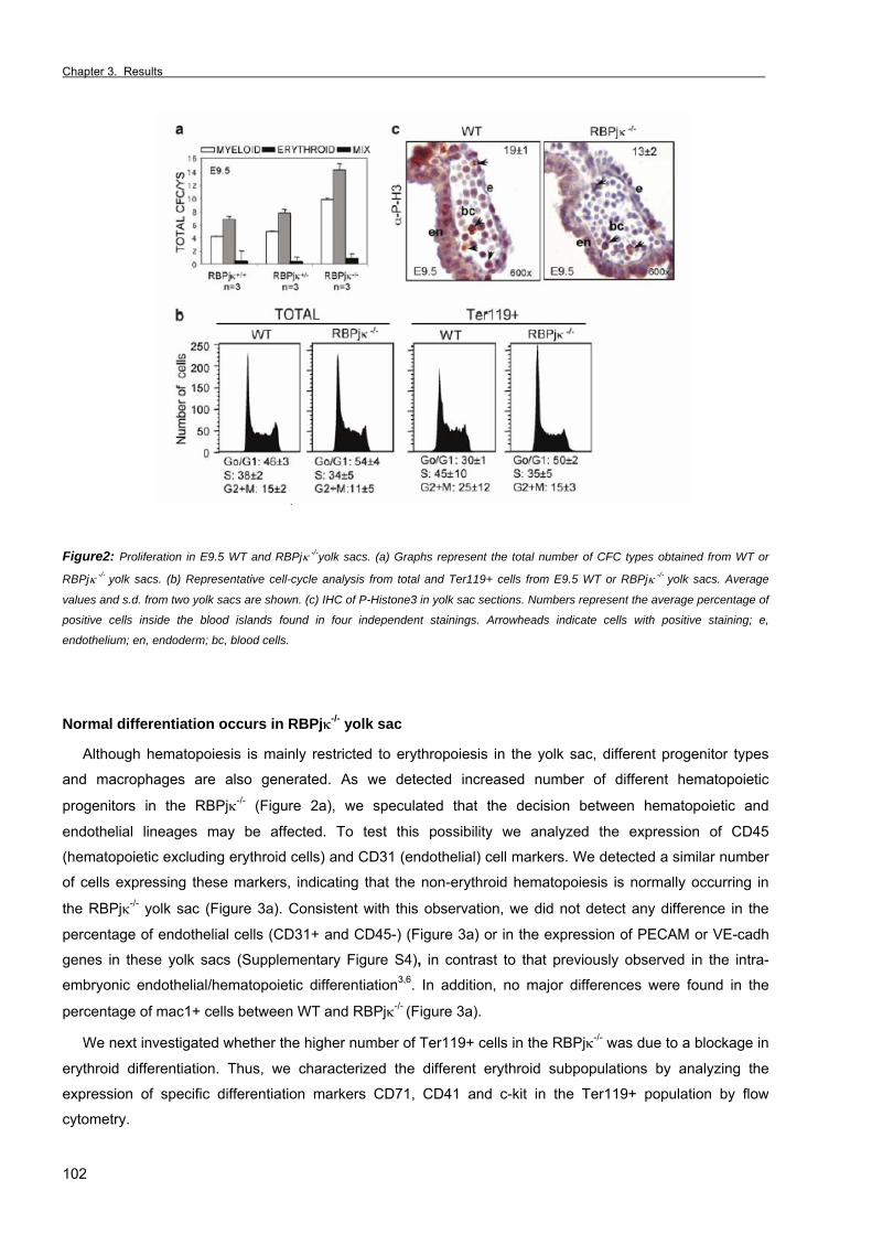
Chapter 3. Results _
102
Figure2: Proliferation in E9.5 WT and RBPjκ -/-yolk sacs. (a) Graphs represent the total number of CFC types obtained from WT or
RBPjκ -/- yolk sacs. (b) Representative cell-cycle analysis from total and Ter119+ cells from E9.5 WT or RBPjκ -/- yolk sacs. Average
values and s.d. from two yolk sacs are shown. (c) IHC of P-Histone3 in yolk sac sections. Numbers represent the average percentage of
positive cells inside the blood islands found in four independent stainings. Arrowheads indicate cells with positive staining; e,
endothelium; en, endoderm; bc, blood cells.
Normal differentiation occurs in RBPjκ-/- yolk sac
Although hematopoiesis is mainly restricted to erythropoiesis in the yolk sac, different progenitor types
and macrophages are also generated. As we detected increased number of different hematopoietic
progenitors in the RBPjκ-/- (Figure 2a), we speculated that the decision between hematopoietic and
endothelial lineages may be affected. To test this possibility we analyzed the expression of CD45
(hematopoietic excluding erythroid cells) and CD31 (endothelial) cell markers. We detected a similar number
of cells expressing these markers, indicating that the non-erythroid hematopoiesis is normally occurring in
the RBPjκ-/- yolk sac (Figure 3a). Consistent with this observation, we did not detect any difference in the
percentage of endothelial cells (CD31+ and CD45-) (Figure 3a) or in the expression of PECAM or VE-cadh
genes in these yolk sacs (Supplementary Figure S4), in contrast to that previously observed in the intra-
embryonic endothelial/hematopoietic differentiation3,6. In addition, no major differences were found in the
percentage of mac1+ cells between WT and RBPjκ-/- (Figure 3a).
We next investigated whether the higher number of Ter119+ cells in the RBPjκ-/- was due to a blockage in
erythroid differentiation. Thus, we characterized the different erythroid subpopulations by analyzing the
expression of specific differentiation markers CD71, CD41 and c-kit in the Ter119+ population by flow
cytometry.
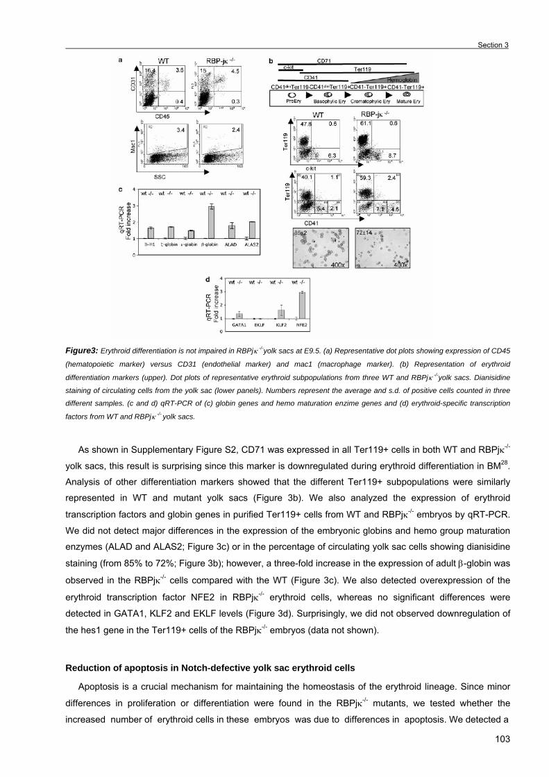
Section 3
103
Figure3: Erythroid differentiation is not impaired in RBPjκ -/-yolk sacs at E9.5. (a) Representative dot plots showing expression of CD45
(hematopoietic marker) versus CD31 (endothelial marker) and mac1 (macrophage marker). (b) Representation of erythroid
differentiation markers (upper). Dot plots of representative erythroid subpopulations from three WT and RBPjκ -/-yolk sacs. Dianisidine
staining of circulating cells from the yolk sac (lower panels). Numbers represent the average and s.d. of positive cells counted in three
different samples. (c and d) qRT-PCR of (c) globin genes and hemo maturation enzime genes and (d) erythroid-specific transcription
factors from WT and RBPjκ -/- yolk sacs.
As shown in Supplementary Figure S2, CD71 was expressed in all Ter119+ cells in both WT and RBPjκ-/-
yolk sacs, this result is surprising since this marker is downregulated during erythroid differentiation in BM28.
Analysis of other differentiation markers showed that the different Ter119+ subpopulations were similarly
represented in WT and mutant yolk sacs (Figure 3b). We also analyzed the expression of erythroid
transcription factors and globin genes in purified Ter119+ cells from WT and RBPjκ-/- embryos by qRT-PCR.
We did not detect major differences in the expression of the embryonic globins and hemo group maturation
enzymes (ALAD and ALAS2; Figure 3c) or in the percentage of circulating yolk sac cells showing dianisidine
staining (from 85% to 72%; Figure 3b); however, a three-fold increase in the expression of adult β-globin was
observed in the RBPjκ-/- cells compared with the WT (Figure 3c). We also detected overexpression of the
erythroid transcription factor NFE2 in RBPjκ-/- erythroid cells, whereas no significant differences were
detected in GATA1, KLF2 and EKLF levels (Figure 3d). Surprisingly, we did not observed downregulation of
the hes1 gene in the Ter119+ cells of the RBPjκ-/- embryos (data not shown).
Reduction of apoptosis in Notch-defective yolk sac erythroid cells
Apoptosis is a crucial mechanism for maintaining the homeostasis of the erythroid lineage. Since minor
differences in proliferation or differentiation were found in the RBPjκ-/- mutants, we tested whether the
increased number of erythroid cells in these embryos was due to differences in apoptosis. We detected a
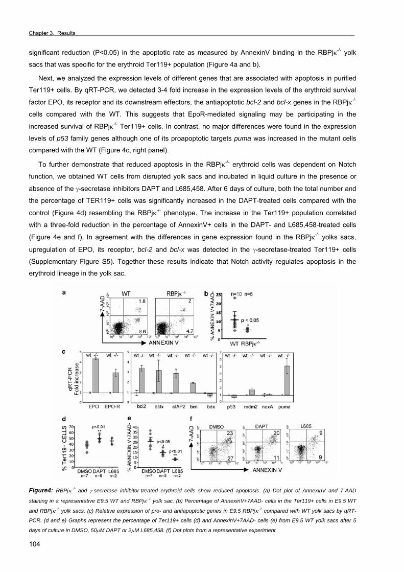
Chapter 3. Results _
104
significant reduction (P<0.05) in the apoptotic rate as measured by AnnexinV binding in the RBPjκ-/- yolk
sacs that was specific for the erythroid Ter119+ population (Figure 4a and b).
Next, we analyzed the expression levels of different genes that are associated with apoptosis in purified
Ter119+ cells. By qRT-PCR, we detected 3-4 fold increase in the expression levels of the erythroid survival
factor EPO, its receptor and its downstream effectors, the antiapoptotic bcl-2 and bcl-x genes in the RBPjκ-/-
cells compared with the WT. This suggests that EpoR-mediated signaling may be participating in the
increased survival of RBPjκ-/- Ter119+ cells. In contrast, no major differences were found in the expression
levels of p53 family genes although one of its proapoptotic targets puma was increased in the mutant cells
compared with the WT (Figure 4c, right panel).
To further demonstrate that reduced apoptosis in the RBPjκ-/- erythroid cells was dependent on Notch
function, we obtained WT cells from disrupted yolk sacs and incubated in liquid culture in the presence or
absence of the γ-secretase inhibitors DAPT and L685,458. After 6 days of culture, both the total number and
the percentage of TER119+ cells was significantly increased in the DAPT-treated cells compared with the
control (Figure 4d) resembling the RBPjκ-/- phenotype. The increase in the Ter119+ population correlated
with a three-fold reduction in the percentage of AnnexinV+ cells in the DAPT- and L685,458-treated cells
(Figure 4e and f). In agreement with the differences in gene expression found in the RBPjκ-/- yolks sacs,
upregulation of EPO, its receptor, bcl-2 and bcl-x was detected in the γ-secretase-treated Ter119+ cells
(Supplementary Figure S5). Together these results indicate that Notch activity regulates apoptosis in the
erythroid lineage in the yolk sac.
Figure4: RBPjκ -/- and γ-secretase inhibitor-treated erythroid cells show reduced apoptosis. (a) Dot plot of AnnexinV and 7-AAD
staining in a representative E9.5 WT and RBPjκ -/- yolk sac. (b) Percentage of AnnexinV+7AAD- cells in the Ter119+ cells in E9.5 WT
and RBPjκ -/- yolk sacs. (c) Relative expression of pro- and antiapoptotic genes in E9.5 RBPjκ -/- compared with WT yolk sacs by qRT-
PCR. (d and e) Graphs represent the percentage of Ter119+ cells (d) and AnnexinV+7AAD- cells (e) from E9.5 WT yolk sacs after 5
days of culture in DMSO, 50µM DAPT or 2µM L685,458. (f) Dot plots from a representative experiment.

Section 3
105
Notch activation induces apoptosis in adult BM erythroid and MEL cells
We next investigated whether Notch activation was regulating apoptosis not only in the yolk sac but also
in adult erythropoiesis. Since RBPjκ-/- mutants do not generate definitive hematopoiesis and die at E10.5, we
isolated BM from WT adult mice and performed cell cultures in the presence of DMSO, DAPT or L685,458. We observed a 30% decrease in the percentage of AnnexinV+ cells in the Ter119+ population after 2 days
of culture in γ-secretase inhibitors compared with the control (Figure 5a). Interestingly, Ter119-negative cells
were not protected from apoptosis by DAPT in these cultures (data not shown), indicating the specific
proapoptotic effect of Notch activity on the erythroid lineage. Conversely, incubation of total BM cells from
adult mouse on NIH-3T3-Jag1 cells resulted in a 2-3 fold increase in the percentage of AnnexinV+Ter119+
population compared with that incubated on NIH-3T3 control cells (P=0.04) (Figure 5b and c). Moreover, this
effect was prevented in the presence of γ-secretase inhibitors DAPT (P=0.01) or L685,458 (P=0.002; Figure
5c) and the caspase inhibitor Z-VAD-FMK (data not shown).
To further demonstrate that Notch was sufficient to induce apoptosis in erythroid cells, we generated
different clones of MEL cells expressing the active Notch1 fragment, N1∆E. Cells expressing N1∆E
differentiated and proliferated similar to the controls (Supplementary Figure S3); however, these cells
showed a higher percentage of Annexin V binding after 6 days in culture under differentiating conditions
(HMBA) as expected (Figure 5d).
Altogether our results indicate that Notch positively regulates apoptosis in embryonic and adult
erythropoiesis and in differentiating erythroleukemia cells.
Figure5: Notch activity induces
apoptosis in the Ter119+ cells
from adult bone marrow and
murine erythroleukemia cells. (a)
Graphs represent the percentage
of AnnexinV+7AAD- in the
Ter119+ population of BM
cultured in the presence of
DMSO, DAPT or L685,458.
Average percentage and s.d. of
three different experiments are
shown. (b and c) Representative
flow cytometry analysis of
AnnexinV binding in the Ter119+
cells from BM incubated on 3T3
or 3T3-Jag1 for 16h (b). Graphs
represent the average and s.d. of
3T3-Jag1-induced apoptosis
observed in three different
experiments incubated with DAPT
and L685,458 as measured by
AnnexinV+7AAD- in Ter119+ cells (c). (d) Graphs represent the average percentage of AnnexinV+7AAD- cells in Notch1∆E expressing
MEL clones (F1, F4) compared with control clones (D3, F3) in proliferation media (black bars) or after 6 days of differentiation in 5mM
HMBA (gray bars). Western blot shows the expression of Notch1∆E.

Chapter 3. Results _
106
DISCUSSION
Notch signaling had been linked previously to regulate apoptosis in erythroid cells, although it was
unclear whether Notch inhibits or promotes apoptosis. We have now used primary cells from yolk sac and
BM to elucidate the physiological effect of Notch activation on the erythroid lineage. We demonstrate that the
Notch pathway is a positive inducer of apoptosis in erythroid cells from different tissues.
Although different cytokines regulate the integrity of the erythroid compartment, production of definitive
erythroid cells is mainly dependent on Epo29. Signaling from EpoR through Jak2/Stat530,31 prevents apoptosis
on this cell lineage from late erythroid progenitors (CFU-E) until the onset of hemoglobinization32,33, and one
important target gene of this cascade is bcl-x34. Studies using bcl-x-null mice demonstrated a critical role for
this gene at the end of erythroid maturation when maximal hemoglobin synthesis occurs35. RBPjκ-/- erythroid
cells show a clear upregulation of genes involved in this antiapoptotic pathway, such as EPO, EPO-R, bcl-x
and bcl-2, suggesting that Notch may induce apoptosis by impinging on this pathway. Previous reports have
attempted to link Notch induction of apoptosis through the p53 pathway in other systems36,37. We have also
explored that possibility in erythroid cells, since there is increasing evidence of p53-mediated apoptosis at
different stages of erythroid differentiation13,38; however, our results show that crucial p53 pathway genes are
not affected in the RBPjκ−/− erythroid cells suggesting that this pathway is not responsible for the protection
of apoptosis in the RBPjκ mutants. In fact, we found an erythroid-specific upregulation of the proapoptotic
p53-target gene, puma, in the RBPjκ-/- Ter119+ cells, which is surprising since this population has a lower
apoptotic rate.
Although previous reports have attempted to decipher which is the Notch function in apoptosis of
erythroid cells15,19, this is the first time that this question is addressed in primary cells. The use of cell lines in
the previous published reports may explain the controversial data. Our data is in agreement with the results
from Ishiko et al15 using K562 cells in which Notch activity induces apoptosis. In this system, Notch activation
led to downregulation of bcl-x, likely through repression of GATA1 by hes1 under differentiation conditions15.
Similarly we found increased levels of bcl-x and bcl-2 in the Ter119+ cell population of RBPjκ-/-; however we
did not detect any change in hes1 and GATA1 levels in this model. The observation that hes1 is not affected
by the lack of RBPjκ in particular cell types has already been described and explained by the repressor
function of RBPjκ in the absence of Notch signaling (reviewed by Lai2).
We now have used different approaches including the Notch loss-of-function RBPjκ-/- embryos, γ-
secretase inhibitors and Jag1-expressing cell co-culture to investigate the role of Notch signaling in
regulating apoptosis on erythroid cells. Together, our results confirm that Notch activation favors apoptosis in
this particular lineage and this finding may be particularly relevant in some erythroid disorders. In fact,
erythropoiesis in the RBPjκ-/- embryos resembles the one observed in myelodysplastic syndrome (MDS)-
derived leukemias or polycytemia vera. These human myeloproliferative syndromes cause the
overproduction of erythroid cells, mainly because of activating mutations in the Jak2 kinase and this leads
not only to increased cell proliferation but also upregulation of bcl-x (reviewed by Campbell and Green39) and
protection from apoptosis. In this scenario and based on our results, it is particularly interesting to investigate
whether the Notch pathway is deregulated in these tumors. In this sense, higher expression of the Notch-like
ligand, dlk, in MDS compared with myeloid leukemia cells has been detected by microarray analysis40.
Section 3
107
Notch activation has previously been associated to inhibition of apoptosis in normal endothelial41 and T
cells42, likewise in neoplastic cells including Kaposi’s sarcoma43, glioma44 and Hodgkin’s lymphoma45 cells.
Conversely, Notch activation can induce apoptosis during the development of the retina46 and the serotonin
lineage47 in Drosophila. Notch also induces apoptosis in B-cell lymphomas16, normal monocytes48 or
hepatocellular carcinoma cells49. Based on this and our results, it is likely that apoptosis is another
mechanism used by Notch to control specific tissue homeostasis.
Altogether, our results indicate that the activation of the Notch pathway is able to regulate the erythroid
lineage in different hematopoietic tissues by inducing apoptosis.
ACKNOWLEDGEMENTS
We thank Liheng Li for Jagged1 plasmid, Raphael Kopan for N1∆E plasmid and M. Caudy for the hes1
plasmid. Thanks to I Fabregat and J Gil for extremely helpful suggestions and reagents. Many thanks to L
Riera and J Inglés-Esteve for critical comments of the manuscript. We thank E Castaño from Serveis
Cientifico-Tècnics, UB-Bellvitge, and J Petriz for flow cytometry technical support. LE is an investigator from
the Carlos III program (ISCIII/02/3027). ARM is a recipient of a CIRIT predoctoral fellowship (2002-SI00791).
This work was supported by the grants from Ministerio de Educación y Ciencia (SAF2004-03/98) and
(SAF205-23978-E) and (SAF2003-07214).
REFERENCES
1. Bray, SJ, (2006) Notch signaling: a simple pathway becomes complex. Nat Rev Mol Cell Biol 7: 678-89. 2. Lai, EC, (2004) Notch signaling: control of cell communication and cell fate. Development 131: 965-73. 3. Kumano, K, Chiba, S, Kunisato, A, Sata, M, Saito, T, Nakagami-Yamaguchi, E et al., (2003) Notch1 but not Notch2 is
essential for generating hematopoietic stem cells from endothelial cells. Immunity 18: 699-711. 4. Gering, M and Patient, R, (2005) Hedgehog signaling is required for adult blood stem cell formation in zebrafish embryos. Dev
Cell 8: 389-400. 5. Burns, CE, Traver, D, Mayhall, E, Shepard, JL and Zon, LI, (2005) Hematopoietic stem cell fate is established by the Notch-
Runx pathway. Genes Dev 19: 2331-42. 6. Robert-Moreno, A, Espinosa, L, de la Pompa, JL and Bigas, A, (2005) RBPjkappa-dependent Notch function regulates Gata2
and is essential for the formation of intra-embryonic hematopoietic cells. Development 132: 1117-26. 7. Palis, J, Robertson, S, Kennedy, M, Wall, C and Keller, G, (1999) Development of erythroid and myeloid progenitors in the
yolk sac and embryo proper of the mouse. Development 126: 5073-84. 8. Palis, J and Segel, GB, (1998) Developmental biology of erythropoiesis. Blood Rev 12: 106-14. 9. Mulcahy, L, (2001) The erythropoietin receptor. Semin Oncol 28: 19-23. 10. Silva, M, Grillot, D, Benito, A, Richard, C, Nunez, G and Fernandez-Luna, JL, (1996) Erythropoietin can promote erythroid
progenitor survival by repressing apoptosis through Bcl-XL and Bcl-2. Blood 88: 1576-82. 11. Motoyama, N, Wang, F, Roth, KA, Sawa, H, Nakayama, K, Nakayama, K et al., (1995) Massive cell death of immature
hematopoietic cells and neurons in Bcl-x-deficient mice. Science 267: 1506-10. 12. Ganguli, G, Back, J, Sengupta, S and Wasylyk, B, (2002) The p53 tumour suppressor inhibits glucocorticoid-induced
proliferation of erythroid progenitors. EMBO Rep 3: 569-74. 13. Peller, S, Frenkel, J, Lapidot, T, Kahn, J, Rahimi-Levene, N, Yona, R et al., (2003) The onset of p53-dependent apoptosis
plays a role in terminal differentiation of human normoblasts. Oncogene 22: 4648-55. 14. Weiss, MJ and Orkin, SH, (1995) Transcription factor GATA-1 permits survival and maturation of erythroid precursors by
preventing apoptosis. Proc Natl Acad Sci U S A 92: 9623-7. 15. Ishiko, E, Matsumura, I, Ezoe, S, Gale, K, Ishiko, J, Satoh, Y et al., (2005) Notch signals inhibit the development of
erythroid/megakaryocytic cells by suppressing GATA-1 activity through the induction of HES1. J Biol Chem 280: 4929-39. 16. Zweidler-McKay, PA, He, Y, Xu, L, Rodriguez, CG, Karnell, FG, Carpenter, AC et al., (2005) Notch signaling is a potent
inducer of growth arrest and apoptosis in a wide range of B-cell malignancies. Blood 106: 3898-906. 17. Romer, S, Saunders, U, Jack, HM and Jehn, BM, (2003) Notch1 enhances B-cell receptor-induced apoptosis in mature
activated B cells without affecting cell cycle progression and surface IgM expression. Cell Death Differ 10: 833-44. 18. Jang, MS, Miao, H, Carlesso, N, Shelly, L, Zlobin, A, Darack, N et al., (2004) Notch-1 regulates cell death independently of
differentiation in murine erythroleukemia cells through multiple apoptosis and cell cycle pathways. J Cell Physiol 199: 418-33.
Chapter 3. Results _
108
19. Shelly, LL, Fuchs, C and Miele, L, (1999) Notch-1 inhibits apoptosis in murine erythroleukemia cells and is necessary for
differentiation induced by hybrid polar compounds. J Cell Biochem 73: 164-75. 20. Oka, C, Nakano, T, Wakeham, A, de la Pompa, JL, Mori, C, Sakai, T et al., (1995) Disruption of the mouse RBP-J kappa gene
results in early embryonic death. Development 121: 3291-301. 21. Friend, C, Preisler, HD and Scher, W, (1974) Studies on the control of differentiation of murine virus-induced erythroleukemic
cells. Curr Top Dev Biol 8: 81-101. 22. Schroeter, EH, Kisslinger, JA and Kopan, R, (1998) Notch-1 signaling requires ligand-induced proteolytic release of
intracellular domain. Nature 393: 382-386. 23. Li, L, Milner, LA, Deng, Y, Iwata, M, Banta, A, Graf, L et al., (1998) The human homolog of rat jagged1 expressed by marrow
stroma inhibits differentiation of 32d cells through interaction with notch1. Immunity 8: 43-55. 24. Lam, LT, Ronchini, C, Norton, J, Capobianco, AJ and Bresnick, EH, (2000) Suppression of erythroid but not megakaryocytic
differentiation of human K562 erythroleukemic cells by notch-1. J Biol Chem 275: 19676-84. 25. Xue, Y, Gao, X, Lindsell, CE, Norton, CR, Chang, B, Hicks, C et al., (1999) Embryonic lethality and vascular defects in mice
lacking the Notch ligand Jagged1. Hum Mol Genet 8: 723-30. 26. Krebs, LT, Xue, Y, Norton, CR, Shutter, JR, Maguire, M, Sundberg, JP et al., (2000) Notch signaling is essential for vascular
morphogenesis in mice. Genes Dev 14: 1343-52. 27. Krebs, LT, Shutter, JR, Tanigaki, K, Honjo, T, Stark, KL and Gridley, T, (2004) Haploinsufficient lethality and formation of
arteriovenous malformations in Notch pathway mutants. Genes Dev 18: 2469-73. 28. Loken, MR, Shah, VO, Dattilio, KL and Civin, CI, (1987) Flow cytometric analysis of human bone marrow: I. Normal erythroid
development. Blood 69: 255-63. 29. Ihle, JN, Quelle, FW and Miura, O, (1993) Signal transduction through the receptor for erythropoietin. Semin Immunol 5: 375-
89. 30. Neubauer, H, Cumano, A, Muller, M, Wu, H, Huffstadt, U and Pfeffer, K, (1998) Jak2 deficiency defines an essential
developmental checkpoint in definitive hematopoiesis. Cell 93: 397-409. 31. Socolovsky, M, Nam, H, Fleming, MD, Haase, VH, Brugnara, C and Lodish, HF, (2001) Ineffective erythropoiesis in Stat5a(-/-
)5b(-/-) mice due to decreased survival of early erythroblasts. Blood 98: 3261-73. 32. Koury, MJ and Bondurant, MC, (1990) Control of red cell production: the roles of programmed cell death (apoptosis) and
erythropoietin. Transfusion 30: 673-4. 33. Yu, H, Bauer, B, Lipke, GK, Phillips, RL and Van Zant, G, (1993) Apoptosis and hematopoiesis in murine fetal liver. Blood 81:
373-84. 34. Dolznig, H, Habermann, B, Stangl, K, Deiner, EM, Moriggl, R, Beug, H et al., (2002) Apoptosis protection by the Epo target
Bcl-X(L) allows factor-independent differentiation of primary erythroblasts. Curr Biol 12: 1076-85. 35. Motoyama, N, Kimura, T, Takahashi, T, Watanabe, T and Nakano, T, (1999) bcl-x prevents apoptotic cell death of both
primitive and definitive erythrocytes at the end of maturation. J Exp Med 189: 1691-8. 36. Yang, X, Klein, R, Tian, X, Cheng, HT, Kopan, R and Shen, J, (2004) Notch activation induces apoptosis in neural progenitor
cells through a p53-dependent pathway. Dev Biol 269: 81-94. 37. Huang, Q, Raya, A, DeJesus, P, Chao, SH, Quon, KC, Caldwell, JS et al., (2004) Identification of p53 regulators by genome-
wide functional analysis. Proc Natl Acad Sci U S A 101: 3456-61. 38. Maetens, M, Doumont, G, De Clercq, S, Francoz, S, Froment, P, Bellefroid, E et al., (Epub 2006) Distinct Roles of Mdm2 and
Mdm4 in Red Cell Production. Blood. 39. Campbell, PJ and Green, AR, (2006) The myeloproliferative disorders. N Engl J Med 355: 2452-66. 40. Miyazato, A, Ueno, S, Ohmine, K, Ueda, M, Yoshida, K, Yamashita, Y et al., (2001) Identification of myelodysplastic
syndrome-specific genes by DNA microarray analysis with purified hematopoietic stem cell fraction. Blood 98: 422-7. 41. MacKenzie, F, Duriez, P, Wong, F, Noseda, M and Karsan, A, (2004) Notch4 inhibits endothelial apoptosis via RBP-Jkappa-
dependent and -independent pathways. J Biol Chem 279: 11657-63. 42. Jehn, BM, Bielke, W, Pear, WS and Osborne, BA, (1999) Cutting edge: protective effects of notch-1 on TCR-induced
apoptosis. J Immunol 162: 635-8. 43. Curry, CL, Reed, LL, Golde, TE, Miele, L, Nickoloff, BJ and Foreman, KE, (2005) Gamma secretase inhibitor blocks Notch
activation and induces apoptosis in Kaposi's sarcoma tumor cells. Oncogene 24: 6333-44. 44. Purow, BW, Haque, RM, Noel, MW, Su, Q, Burdick, MJ, Lee, J et al., (2005) Expression of Notch-1 and its ligands, Delta-like-
1 and Jagged-1, is critical for glioma cell survival and proliferation. Cancer Res 65: 2353-63. 45. Jundt, F, Anagnostopoulos, I, Forster, R, Mathas, S, Stein, H and Dorken, B, (2002) Activated Notch1 signaling promotes
tumor cell proliferation and survival in Hodgkin and anaplastic large cell lymphoma. Blood 99: 3398-403. 46. Miller, DT and Cagan, RL, (1998) Local induction of patterning and programmed cell death in the developing Drosophila
retina. Development 125: 2327-35. 47. Lundell, MJ, Lee, HK, Perez, E and Chadwell, L, (2003) The regulation of apoptosis by Numb/Notch signaling in the serotonin
lineage of Drosophila. Development 130: 4109-21. 48. Ohishi, K, Varnum-Finney, B, Flowers, D, Anasetti, C, Myerson, D and Bernstein, ID, (2000) Monocytes express high
amounts of Notch and undergo cytokine specific apoptosis following interaction with the Notch ligand, Delta-1. Blood 95: 2847-54.
49. Qi, R, An, H, Yu, Y, Zhang, M, Liu, S, Xu, H et al., (2003) Notch1 signaling inhibits growth of human hepatocellular carcinoma through induction of cell cycle arrest and apoptosis. Cancer Res 63: 8323-9.
Supplementary Information accompanies the paper on the Leukemia website (http://www.nature.com/leu)

Section 3
109
SUPPLEMENTARY INFORMATION
Supplementary Figure S1: Analysis of myeloid and erythroid progenitors from collagenase-treated yolk sacs. (a) Total CFC/YS at
E7.5 and E8.5. (b) Percentage of CFC at E9.5
Supplementary Figure S2: Ter119+ cells from the yolk sac express CD71. (a) Ter119+ cells from WT and mutant yolk sacs. X-
mean of SSC is shown for both samples. (b) Percentage of gated Ter119+ cells expressing CD71. Numbers represent the average
percentage and standard deviation of 3 different yolk sacs.

Chapter 3. Results _
110
Supplementary Figure S3: Differentiation
and proliferation of pcDNA3 and N1∆E MEL
cells. (a) Cell numbers for MEL, pcDNA3 and
N1∆E clones at day 3, 5 and 6. (b)
Percentage of dianisidine positive cells after 6
days in HMBA-differentiating media.
Supplementary Figure S4: Semiquanti-tative
RT-PCR of hematopoietic and endothelial genes
from E9.5 WT and RBPjκ -/- yolk sacs.
Supplementary Figure S5: qRT-
PCR from sorter Ter119+ cells from YS
cultured for 6 days in DMSO and
γ−secretase inhibitor L685,458.

CHAPTER 4: GENERAL DISCUSSION AND FUTURE
PROSPECTS

General discussion and future prospects
113
Hematopoietic stem cells are very important for clinical transplantation and regenerative medicine. This
fact has encouraged scientists to study the mechanisms that control self-renewal of HSC and how
microenvironmental regulates their behavior. Our work has contributed to clarify the role of Notch in the
embryonic hematopoiesis of the yolk sac as well as in the formation of HSC in the AGM, one of the best-
characterized niches for generation and amplification of HSC in the embryo. However the precise site where
first HSCs are originated in the embryo is still controversial.
The never-ending story: what is the origin of HSCs? The current knowledge is that two main waves of hematopoiesis take place during the first stages of
embryonic development, the primitive and transient hematopoiesis in the yolk sac and the definitive
hematopoiesis that originates in the intra-embryonic region of the aorta surrounded by gonad and
mesonephros (AGM region) and other major arteries.
Primitive hematopoiesis is responsible for generating primitive nucleated erythrocytes that provide oxygen
supply to the growing embryo [reviewed in (McGrath and Palis, 2005)]. Definitive hematopoiesis is
responsible for generating the first adult-type HSCs capable of engrafting in the bone marrow of lethally
irradiated adults. These HSCs are first found at E10 (de Bruijn et al., 2002; de Bruijn et al., 2000) and by E11
they are detected in other tissues such as fetal liver, yolk sac, placenta and bloodstream (Gekas et al., 2005;
Kumaravelu et al., 2002; Medvinsky and Dzierzak, 1996; Muller et al., 1994; Ottersbach and Dzierzak,
2005). Nevertheless, the idea that yolk sac contributes to the hematopoiesis in the adult has never been
discarded. Indeed, E9 yolk sac and para-aortic splanchnopleura (mesoderm that will form the AGM) fail to
engraft in irradiated adult recipients but are able to reconstitute hematopoiesis of myeloablated newborn
mice (Yoder and Hiatt, 1997; Yoder et al., 1997; Yoder et al., 1997). These evidences rise the possibility that
E9 YS and P-Sp contain immature HSCs that need to undergo further maturation to acquire the ability to
home the adult bone marrow. In this sense, experiments done with YS or P-sp before the onset of circulation
(E8-E8.5) revealed that these organs contain immature HSCs that become adult-HSCs when co-cultured
with the E10.5 AGM-derived cell line AGM-S3 (Matsuoka et al., 2001). Thus, the current model is that the
AGM serves as the main site for the generation of adult-type HSCs, but other organs such as the YS and
more recently the placenta also contribute to adult hematopoiesis. However, a controversial report recently
suggested that yolk sac is the origin of a significant percentage of definitive HSCs (Samokhvalov et al.,
2007). Samokhvalov et al used a tamoxifen-inducible LacZ labelling technique to follow the cells from E7.5
that express runx1/aml1. The authors report the existence of runx1-marked cells at E7.5, coming from the
yolk sac that contribute to the adult HSCs and persist in bone marrow for at least 15 months after birth
(Samokhvalov et al., 2007). However, tamoxifen induction is not only achieved in the YS but also in the
embryo proper where runx1 was previously described to be expressed as early as E8 (North et al., 1999).
Moreover, Samokhvalov studies were done with Runx1+/- embryos where HSCs could be ectopically
generated due to runx1 heterozigousity, as previously reported (Cai et al., 2000). Early cell tracking
experiments performed in the avian model reinforce the idea that the AGM, but not the extra-embryonic yolk
sac, serves as the HSC source. In 1970’s, chicken YS and quail embryo proper chimeras were explanted
before the vascular establishment and showed for the first time that the intra-embryonic region rather than
the YS contributes to the adult hematopoietic compartment (Dieterlen-Lievre, 1975). The ventral endothelium
of the aorta is the main source of HSCs in the AGM since cells expressing hematopoietic markers are
Chapter 4 _ _
114
generally detected within the endothelial layer (Bertrand et al., 2005; de Bruijn et al., 2002; North et al., 1999;
Robert-Moreno et al., 2005). An alternative model, proposes that embryonic hematopoiesis takes place in a
structure referred as subaortic patches (SAPs) that are located in the mesenchyme under the floor of the
dorsal aorta and express gata2, gata3 and lmo2. HSC in the SAP are CD45low/ckit+/AA4.1+/flk-1- and are
capable of reconstituting hematopoiesis in Rag2γc-/- immunodeficient mice (Bertrand et al., 2005; Manaia et
al., 2000). It is hypothesized that intra-embryonic HSCs are originated in the SAP and then migrate to the
ventral part of the aorta, cross the endothelium and contribute to the formation of the hematopoietic clusters
before they are released to the bloodstream. This possibility is not supported by our results from in situ
hybridization since we did not detect any hematopoietic cells in the mesenchyme underlying the ventral wall
of the aorta.
On the other hand, heterotopic grafts of quail splanchnopleural mesoderm into chicken embryos revealed
that HSCs in the AGM come from mesodermal cells that integrate into the floor of the aorta and only once in
this site they generate hematopoietic clusters (Pardanaud et al., 1996). Moreover, hematopoiesis in the AGM
aorta ceases when splanchnopleura-derived cells become exhausted and are replaced by somite-derived
non-hemogenic angioblasts, suggesting the idea that hemogenic endothelium in the floor of the aorta is a
temporary structure of non-renewable hemogenic endothelial cells (Pouget et al., 2006).
Altogether, these results may reflect that the endothelial or mesenchymal origin of HSCs is not
irreconciliable and that the integration of all the present data may result in a better understanding of HSC
generation.
The aim of our work was to study the role of the Notch signaling pathway in both the primitive and
definitive waves of hematopoiesis in the mouse embryo. Our data from the RBPjκ null embryos (Robert-
Moreno et al., 2005) and the aorta explant cultures (Chapter 3, Section 2) suggest that Notch signaling is
required for the commitment of HSC/HPC from the hemogenic endothelium of the AGM aorta. We propose
that Notch activity in the ventral aorta generates a “salt and pepper” pattern that restricts the hemogenic
potential to a discrete number of HSCs/HPCs among a majority of aortic endothelial cells, thus reminiscent of
the lateral inhibition process described in Drosophila neurogenesis (Parks et al., 1997). In this sense, WISH
for the different Notch ligands and Notch receptors in early embryos (Supplemental information, Chapter 3
Section 2) agree with this lateral inhibition model since Notch ligand-expressing cells appear earlier in
development (at the 14 somite pair stage) than Notch-expressing ones (by the 20 somite pair stage) and
only in discrete cells from the ventral part of the aortic endothelium.
Is there only one site of HSCs emergence in the embryo or conversely it is a redundant event that takes
place in different tissues throughout fetal development? Our results are in agreement with the idea that
primitive and definitive hematopoiesis are the result of two well differentiated genetic programs. RBPjκ null
embryos completely lack intra-embryonic hematopoiesis (Robert-Moreno et al., 2005) whereas primitive yolk
sac hematopoiesis does occur (Robert-Moreno et al., 2007). The same phenotype was described in null
embryos for other Notch pathway molecules such as Notch1 in mice (Kumano et al., 2003) and Mind bomb
in zebrafish (Burns et al., 2005). As previosly described in this thesis, the effect of RBPjκ deficiency in the
yolk sac is restricted to the homeostasis of the erythroid lineage where prevents apoptosis (Robert-Moreno
et al., 2007). However, loss of Notch signaling has a more severe effect in definitive P-sp/AGM
hematopoiesis resulting in a complete abrogation of both HSCs and HPCs.
General discussion and future prospects
115
Although we did not assay whether the RBPjκ mutant yolk sacs are able to generate embryonic HSCs,
Notch1-/- embryos completely lack E9.5 yolk sac and P-sp HSCs (Kumano et al., 2003) indicating that Notch
is required for the generation/commitment of all definitive HSCs.
Notch signaling regulates generation and/or expansion of HSCs in the AGM region Notch signaling has been reported to regulate developmental cell fate decisions in many systems
[reviewed in (Lai, 2004)]. One of them is the arterial/vein decision. In this sense, Notch induces gridlock (the
zebrafish orthologue of hrt1) expression in blood vessels to promote arterial specification (Zhong et al., 2001)
whereas Notch-inhibition by the transcription factor COUP-TFII results in vein specification (You et al., 2005).
Since AGM hematopoiesis occurs only in the main arteries (de Bruijn et al., 2002; de Bruijn et al., 2000;
Medvinsky and Dzierzak, 1996) and not in veins, the lack of intra-embryonic hematopoiesis in the Notch1
(Kumano et al., 2003) and RBPjκ mutants (Robert-Moreno et al., 2005) may be due to a defect in aorta
specification. For this reason, we used the aorta/AGM explant culture system (Medvinsky and Dzierzak,
1996) to investigate the role of Notch signaling in HSC generation. Indeed repopulation studies performed
with E11 wild-type aortas incubated with the γ-secretase inhibitor DAPT revealed a direct role for Notch
signaling in the generation and/or expansion of HSCs in the embryo that is independent from aorta
specification (Chapter 3; Section 2). In these experiments, we found a decrease in the number of irradiated
mice repopulated by DAPT-treated aortas. Three different possibilities may explain these results: 1) Notch
inhibition may affect the new HSC generation or 2) it may decrease proliferation of the pre-existing HSC,
since this culture system allows HSC expansion (Medvinsky and Dzierzak, 1996) and since Notch activity
usually correlates with the maintenance of an undifferentiated state in many systems [reviewed in (Lai,
2004)], 3) it could be that Notch inhibition may be inducing differentiation of HSCs. However, this possibility
is not consistent with the lower number of committed hematopoietic progenitors found in CFC assays from γ-
secretase treated aortas (Chapter 3; Section 2).
Recently it has been reported that adult bone marrow contains a discrete number of different HSCs each
one with its own repopulation kinetics and self-renewal behavior (Sieburg et al., 2006). Our results indicate
that DAPT-treated AGM showed similar low repopulation efficiency (HSC with less of 10% engraftment
capacity) than untreated AGM (Chapter 3; Section 2). However, the number of animals repopulated with
HSCs with more than 10% engraftment capacity was reduced by 4-fold in the case of the γ−secretase
treated ones. This result suggests that Notch is required to amplify a hypothetical population of HSC with
high repopulating activity. It should be of great importance to study the different signals that regulate the
generation and expansion of HSCs in the different hemogenic tissues.
Upstream of the Notch receptor We have reported that notch1 and 4, together with the jagged1 and 2 and delta4 ligands are expressed
in the E9.5-E10.5 aortic endothelium (Robert-Moreno et al., 2005). Notch4 null embryos are viable whereas
Notch1/Notch4 double null embryos display more severe vasculogenic defects than the Notch1 single
mutants (Conlon et al., 1995; Krebs et al., 2000) indicating a common vascular function for Notch1 and
Notch4 receptors. However, expression of Notch family members and their target gene hes1 in the
hematopoietic clusters of the AGM is suggestive of a hematopoietic function for Notch signaling (Chapter 3;
Section 2). Thus, two different Notch signals may be required in the mid-gestation aortic endothelium, the

Chapter 4 _ _
116
first one to establish artery/vein identity and the second one for generating/amplifying the HSC compartment. Notch1 but not Notch4 is essential for angiogenic vascular remodelling albeit some functional
redundancy is found between both receptors (Conlon et al., 1995; Krebs et al., 2000). On the other hand,
Notch1 is required for P-sp/AGM hematopoiesis (Kumano et al., 2003) and notch4 (but not notch1)
expression is downregulated in E10.5 aortic hematopoietic clusters (Chapter 3, Section 2). Moreover we
found that Notch1 (through RBPjκ) associates to the promoter of the hematopoietic transcription factor Gata2
and double in situ hybridization revealed that both genes are coexpressed in the same cells from E10.5
aortic endothelium (Robert-Moreno et al., 2005). All these evidences reinforce the specific role of Notch1 in
activating the hematopoietic program in the endothelium of the aorta. Thus, it would be worthwhile to
understand how specificity is achieved and which set of genes are activated in each case. One possibility
could be that the Notch1 receptor may be activated by a different ligand to specify the vasculogenic or the
hematopoietic program. Since Delta4 null embryos die at E9.5 displaying severe vasculogenic defects and
loss of endothelial arterial cell fate (Duarte et al., 2004) and lethality occurs even in heterozygosity due to
severe vasculogenic malformations (Krebs et al., 2004), presumably activation of Notch1 receptor by Delta4
is the right combination to specify vascular development. However, Notch4 may partially compensate Notch1
defficiency in vasculogenesis.
Double in situ hybridization studies of E10.5 AGM sections revealed that jagged2-expressing cells were
mostly located adjacent to the notch1/jagged1 positive cells (Robert-Moreno et al., 2005). Since cell-cell
interactions between adjacent cells govern the activation of Notch receptors, the Jagged2 ligand was the
best candidate to activate Notch1 and induce the establishment of the hematopoietic fate in the hemogenic
endothelium of the AGM region. In order to fully address this issue, we studied whether Jagged1 or Jagged2
null embryos displayed abnormal intra-embryonic hematopoiesis in vivo. In fact, when we assayed the
hematopoietic potential of both null mutants (by CFC or by counting Sca-1-GFP+ cells), we found that
Jagged1 but not Jagged2 mutant embryos had decreased numbers of all types of hematopoietic cells,
indicating an important role for Jagged1 in this system (Chapter 3; Section 2). Thus, although Jagged2
expression pattern seem to better explain how Notch could be activated, we have not found any
hematopoietic deficiency in the Jagged2 null embryos. One explanation could be that Jagged2 may have a
minor hematopoietic role and this deficiency may be compensated by Jagged1. To test this, we are currently
studying whether Jag1/Jag2 double deficiency has a more severe hematopoietic phenotype than the
Jagged1 knockout embryos.
The decrease in the number of hematopoietic cells found in the Jagged1 mutants may result from a self-
renewal or amplification defect of the HSC or more committed progenitors. Repopulation experiments of
adult irradiated mice with the Jagged1 mutant aortas are required to answer this question.
We have used the Ly-6A/Sca-GFP mice (de Bruijn et al., 2002) to identify the HSC emerging from the
aorta in the different Notch-mutant embryos. Although this is an excellent system for these studies, some
issues should be considered when analyzing these mice. Sca-1 (Stem cell antigen-1) is a cell-surface
glycoprotein used as a marker for embryonic and adult HSCs (de Bruijn et al., 2002; Miles et al., 1997)
although it is also found in HPCs, vascular endothelium and kidney. Thus, although it is well characterized
that the GFP+ fraction in the aorta contains the HSC activity (de Bruijn et al., 2002), there is also a few non-
hematopoietic endothelial cells expressing GFP. By crossing the Jagged1 and Jagged2 mutant mice with
Ly-6A-GFP transgenic mice, we detected a profound decrease in the number of GFP+ cells in the aorta of
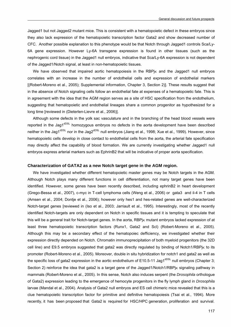
General discussion and future prospects
117
Jagged1 but not Jagged2 mutant mice. This is consistent with a hematopoietic defect in these embryos since
they also lack expression of the hematopoietic transcription factor Gata2 and show decreased number of
CFC. Another possible explanation to this phenotype would be that Notch through Jagged1 controls Sca/Ly-
6A gene expression. However Ly-6A transgene expression is found in other tissues (such as the
nephrogenic cord tissue) in the Jagged1 null embryos, indicative that Sca/Ly-6A expression is not dependent
of the Jagged1/Notch signal, at least in non-hematopoietic tissues.
We have observed that impaired aortic hematopoiesis in the RBPjκ and the Jagged1 null embryos
correlates with an increase in the number of endothelial cells and expression of endothelial markers
[(Robert-Moreno et al., 2005); Supplemental information, Chapter 3, Section 2)]. These results suggest that
in the absence of Notch signaling cells follow an endothelial fate at expenses of a hematopoietic fate. This is
in agreement with the idea that the AGM region serves as a site of HSC specification from the endothelium,
suggesting that hematopoietic and endothelial lineages share a common progenitor as hypothesized for a
long time [reviewed in (Dieterlen-Lievre et al., 2006)].
Although some defects in the yolk sac vasculature and in the branching of the head blood vessels were
reported in the Jag1dDSL homozygous embryos no defects in the aorta development have been described
neither in the Jag1dDSL nor in the Jag2dDSL null embryos (Jiang et al., 1998; Xue et al., 1999). However, since
hematopoietic cells develop in close contact to endothelial cells from the aorta, the arterial fate specification
may directly affect the capability of blood formation. We are currently investigating whether Jagged1 null
embryos express arterial markers such as EphrinB2 that will be indicative of proper aorta specification.
Characterization of GATA2 as a new Notch target gene in the AGM region. We have investigated whether different hematopoietic master genes may be Notch targets in the AGM.
Although Notch plays many different functions in cell differentiation, not many target genes have been
identified. However, some genes have been recently described, including ephrinB2 in heart development
(Grego-Bessa et al., 2007), c-myc in T-cell lymphoma cells (Weng et al., 2006) or gata3 and il-4 in T cells
(Amsen et al., 2004; Dontje et al., 2006); however only hes1 and hes-related genes are well-characterized
Notch-target genes [reviewed in (Iso et al., 2003; Jarriault et al., 1995). Interestingly, most of the recently
identified Notch-targets are only dependent on Notch in specific tissues and it is tempting to speculate that
this will be a general trait for Notch-target genes. In the aorta, RBPjκ mutant embryos lacked expression of at
least three hematopoietic transcription factors (Runx1, Gata2 and Scl) (Robert-Moreno et al., 2005).
Although this may be a secondary effect of the hematopoiec defficiency, we investigated whether their
expression directly depended on Notch. Chromatin immunoprecipitation of both myeloid progenitors (the 32D
cell line) and E9.5 embryos suggested that gata2 was directly regulated by binding of Notch1/RBPjκ to its
promoter (Robert-Moreno et al., 2005). Moreover, double in situ hybridization for notch1 and gata2 as well as
the specific loss of gata2 expression in the aortic endothelium of E10.5-11 Jag1dDSL null embryos (Chapter 3;
Section 2) reinforce the idea that gata2 is a target gene of the Jagged1/Notch1/RBPjκ signaling pathway in
mammals (Robert-Moreno et al., 2005). In this sense, Notch also induces serpent (the Drosophila orthologue
of Gata2) expression leading to the emergence of hemocyte progenitors in the fly lymph gland in Drosophila
larvae (Mandal et al., 2004). Analysis of Gata2 null embryos and ES cell chimeric mice revealed that this is a
clue hematopoietic transcription factor for primitive and definitive hematopoiesis (Tsai et al., 1994). More
recently, it has been proposed that Gata2 is required for HSC/HPC generation, proliferation and survival.
Chapter 4 _ _
118
Gata2 haploinsufficient embryos display reduced generation of HSCs from the AGM region (Ling et al.,
2004), an observation that agrees with our results from the Jagged1 null embryos. Moreover, the bone
marrow stem cell compartment of Gata2+/- mice shows a higher apoptotic ratio due to downregulation of the
antiapoptotic gene bcl-XL (Ling et al., 2004; Rodrigues et al., 2005) and Gata2-/- multipotential progenitors
proliferate poorly and display massive cell death further indicating that Gata2 is required for proliferation and
survival of early progenitors (Tsai and Orkin, 1997). Thus we propose that Jagged1/Notch1/RBPjκ-induced
activation of gata2 is required for generation, proliferation and/or survival of HSC/HPC from the hemogenic
endothelium of the AGM region. However, Jagged1 mutant embryos show reduced but not complete lack of
hematopoiesis suggesting that HSC specification may occur in the absence of Jagged1 and Gata2.
The hematopoietic transcription factor Runx1/Aml1 is also required for the generation of HSCs from the
aortic endothelium since Runx1-/- embryos completely lack definitive hematopoiesis (North et al., 1999;
Okuda et al., 1996). Runx1 has been recently described to act downstream of Notch in the establishment of
the hematopoietic stem cell fate in the zebrafish embryo (Burns et al., 2005). Consistently, in Drosophila,
hemocytogenesis occurs through Notch-induced activation of lozenge, the fly orthologue of Runx1 (Lebestky
et al., 2003). In addition, ectopic expression of runx1 (but not gata2 or scl) in the AGM-derived cells from
Notch1 null embryos partially rescues the hematopoietic defect in vitro (Nakagawa et al., 2006). Although
there is compiling evidence that Notch-Runx1 is also required for the initiation of the hematopoietic program
in the AGM region, we did not detect the runx1 gene in the chromatin precipitated with anti-Notch1 antibody
from E9.5 embryos (Robert-Moreno et al., 2005). Moreover, we have observed runx1 expression in 50% of
the Jagged1 null embryos. One possible explanation is that activation of Notch through different ligands
specifically activates runx1 or gata2 in specific populations of HSC and at different time points. However,
since runx1 infected Notch1 null P-sp cells are unable to repopulate the hematopoiesis of busulfan
compromised newborn mice (Nakagawa et al., 2006) it seems that both Runx1 and Gata2 transcription
factors act synergistically to determine or expand the pool of HSCs from the AGM region.
We have also investigated whether scl/tal-1 was a Notch-target. Although we were not able to detect
Notch1 binding to the scl promoter in our ChIP assays, scl was upregulated in the 32D cell line
overexpressing the intracellular domain of Notch (Robert-Moreno et al., 2005). Gata2 upregulates scl in the
BL-CFC population (Lugus et al., 2007) and scl is also transcriptionally increased after a brief pulse of
Notch1IC in the zebrafish embryo (Burns et al., 2005). These evidences together with our results suggest
that Scl could be acting downstream of Gata2.
We have also described expression of the classical Notch target genes hes1, hrt1 and hrt2 in the mid-
gestation AGM aorta [(Robert-Moreno et al., 2005) and Chapter 3, Section 2)]. Both Hes and Hrt are basic
helix-loop-helix proteins (bHLH) and they are reported to act as transcriptional repressors, usually leading to
an inhibition of differentiation [reviewed in (Iso et al., 2003)]. Some hrt1 and hrt2 expression was found in
E10.5 hematopoietic aortic clusters (data not shown) but the fact that Hrt1/Hrt2 double mutants show severe
vascular remodelling defects and dorsal aorta lacks EphrinB2 expression and display the vein marker EphB4
(Fischer et al., 2004) suggests that Hrt proteins may be related to arterial specification rather than
hematopoiesis.
On the other hand, we show hes1 expression not only in the endothelium lining the dorsal aorta but also
in the E10.5 aortic hematopoietic clusters [(Robert-Moreno et al., 2005) and Chapter 3, Section 2] which
suggest a role for this Notch target gene in this system. There is some evidence for a role of Hes1 inhibiting
General discussion and future prospects
119
the differentiation and maintaining the stemness of HSCs (Kunisato et al., 2003), thus one possibility is that
Jagged1/Notch1/RBPjκ would activate gata2 and hes1 expression in a subset of HSCs with high
repopulating ability in the AGM region (Figure1). Gata2 may regulate proliferation of HSCs, in agreement
with the fact that Jagged1 mutant embryos contain a reduced number of hematopoietic progenitors, whereas
Hes1 may be involved in the maintenance of the stem cell state.
Pleiotropy and context specificity of Notch in hematopoiesis It is widely demonstrated that Notch signaling regulates many processes during embryonic and adult
hematopoiesis. However, the high complexity of the Notch pathway itself, with a wide variety of ligands,
receptors and intra and extracellular modulators leads to a huge diversity of responses depending on the
tissue or cell context. For example, Notch activation inhibits differentiation in the majority of tissues [reviewed
in (Artavanis-Tsakonas et al., 1999)] however, in keratinocytes Notch activation promotes terminal
differentiation (Rangarajan et al., 2001). The mechanisms leading to Notch specificity may include 1)
different Notch receptors (Cheng et al., 2007), 2) accesibility of Notch-presenting cell to different Notch
ligands due to specific expression patterns (Robert-Moreno et al., 2005) or 3) ligand modifications through
fringe glycosyltransferases (Hicks et al., 2000; Moloney et al., 2000), that would induce different gene
expression programs. The role of Notch in the hematopoietic system cannot be explained without considering the idea of
context specificity. Notch activation effects are different in each hematopoietic lineage. During embryonic
development, the generation of primitive and definitive hematopoiesis seems to have completely different
Notch requirements. From the study of Notch1 and RBPjκ null mutants, others and we have concluded that
Notch signaling is required for definitive hematopoiesis but dispensable for the primitive hematopoiesis in the
yolk sac (Burns et al., 2005; Kumano et al., 2003; Robert-Moreno et al., 2005).
However, in the adult it is not so clear whether Notch is required for HSC self-renewal/maintenance.
Whereas notch1IC retrovirally infected in bone marrow cells, expands the number of repopulating HSCs
(Stier et al., 2002) and a dominant negative form of RBPjκ promotes their differentiation (Duncan et al.,
2005), adult mice carrying an inducible inactivation of Notch1 only display lymphoid defects (Radtke et al.,

Chapter 4 _ _
120
1999). Similarly, Jagged1 (Mancini et al., 2005) and RBPjκ (Han et al., 2002) are dispensable for HSC self-
renewal and repopulation ability in the adult bone marrow in constrast with our results in AGM
hematopoiesis. These results indicate that Notch may be specifically required for the generation of HSCs
during embryonic life.
On the other hand, the effect of Notch in the regulation of different cell-fate decisions within the lymphoid
lineage has extensively been demonstrated. Conditional inactivation of Notch1 (Radtke et al., 1999) or
RBPjκ (Han et al., 2002) in bone marrow progenitors lead to a depletion of T cells because the common
lymphoid progenitors commit to the B-cell fate. Moreover, a requirement of Notch signaling for the generation
of αβ T cells at expenses of γδ T cells (Tanigaki et al., 2004) and CD8+ cells at expenses of CD4+ cells has
been also described (Robey et al., 1996).
We have recently described that Notch/RBPjκ signaling induces apoptosis in the erythroid lineage of the
yolk sac most likely by downregulating the expression of molecules from the Epo/EpoR survival erythroid
pathway (Robert-Moreno et al., 2007). Binding of EPO to its receptor induces activation of Jak2 tyrosine
kinase and the downstream effectors PI3-kinase/AKT, STAT5-bcl-XL and ERK/MAPK leading to erythroid
progenitor expansion, terminal erythroid maturation and apoptosis protection [reviewed in (Testa, 2004)].
Another important effector of EPO signaling is GATA1 that leads to bcl-XL overexpression (Gregory et al.,
1999). However we did not detect GATA1 overexpression in the RBPjκ null yolk sacs although we cannot
exclude that this regulation could be at the post-translational level since we found upregulation of bcl-XL.
Moreover, it was previously published that Notch induces apoptosis in the human erythroleukemic cell line
K562 through Hes1 repression of GATA1 leading to downregulation of bcl-XL (Ishiko et al., 2005).
Another possibility is that the p53 tumor suppressor may be inducing apoptosis in primitive erythroid cells.
The Notch target genes hes1 and hey1 were found to transcriptionally repress the p53 inhibitor hdm2 in a
genome-wide screening (Huang et al., 2004). In agreement with this, Notch1-induced apoptosis in early
neural progenitor cells was suppressed in a p53-/- background (Yang et al., 2004). Moreover, conditional
deletion of mdm2 and mdm4 in EpoR+ erythroid cells revealed that Mdm2 is crucial for inhibiting p53-induced
apoptosis of yolk sac erythrocytes (Maetens et al., 2007). Although we did not find significant differences in
the p53 or mdm2 levels and of p53-dependent gene transcription (bax and noxa (albeit puma) in RBPjκ null
yolk sacs compared to wild-types, p53 phosphorylation should be assayed to further discard that Notch1
exerts its proapoptotic function through p53 [reviewed in (Chipuk and Green, 2006)].
Notch induction of apoptosis has previously been described in monocytes (Ohishi et al., 2000) and B-cell
lymphomas (Zweidler-McKay et al., 2005), and opposite effects has been found in T cells (Jehn et al., 1999)
indicating, once again, that Notch function is lineage and context-dependent. However, some of the reported
observations come from cell lines and cultured systems and should be cautiously considered. However, our
results have been generated from RBPjκ-deficient erythrocytes directly obtained from the yolk sac. The fact
that this is an in vivo system, strongly suggest that Notch may induce apoptosis in this lineage and contribute
to the homeostasis of erythroid cells.
In summary, Notch signaling is crucial not only in the generation/expansion of HSCs in the embryo, but
also in differentiation and programmed cell death in many of the committed hematopoietic lineages derived
from these HSCs. As a consequence, the fine tuned homeostasis of the hematopoietic system is reached.

CHAPTER 5: CONCLUSIONS


Conclusions
123
CONCLUSIONS In this thesis we have achieved the following conclusions:
1. Loss of the nuclear Notch pathway effector RBPjκ leads to a complete abrogation of intra-embryonic
hematopoiesis, concomitantly with a lack of the hematopoietic transcription factors Gata2, Runx1 and Scl
expression in the aortic endothelium.
2. Notch1 through RBPjκ directly binds to the Gata2 promoter and regulates its expression in the 32D cell
line and in the mid-gestation embryo.
3. Loss of Jagged1 results in impaired expression of Gata2, reduced hematopoietic progenitor generation
and Sca-1+ cells in the AGM region.
4. Inhibition of Notch activation in wild-type aortas by γ-secretase inhibitors diminishes the number of HSCs
from the AGM, thus resembling the phenotype described for the Notch, RBPjκ and Jagged1dDSL mutants.
5. Notch signaling is required to generate and/or expand a pool of high repopulating HSCs from the AGM
region.
6. Lack of intra-embryonic AGM hematopoiesis correlates with an increase of the endothelial layer in the
aorta of the RBPjκ-/- and Jagged1dDSL embryos, suggesting that a common progenitor for the
hematopoietic and endothelial lineages (or hemangioblast) does exist in the AGM aorta.
7. Yolk sac hematopoietic cells develop normally in RBPjκ deficient embryos with and increased number of
erythropoietic cells.
8. Notch signaling regulates erythroid homeostasis in the yolk sac by inducing apoptosis.
9. The apoptotic protection in the erythroid lineage of RBPjκ null yolk sacs is due to the upregulation of
Epo/Epo receptor pathway members leading to the overexpression of the antiapoptotic genes bcl-2 and
bcl-XL.

CHAPTER 6: RESUM DE LA TESI
“Paper de Notch i RBPjκ en
l’hemopoesi embrionària”

Resum de la tesi
127
6.1 INTRODUCCIÓ GENERAL 6.1.1 EL SISTEMA HEMOPOÈTIC 6.1.1.1 Introducció al sistema hemopoètic El sistema hemopoètic va aparèixer al llarg de l’evolució amb la finalitat d’assegurar l’arribada de
nutrients i la protecció en contra de perills externs en organismes multicel.lulars. La sang està composada
d’una gran varietat de tipus cel.lulars madurs amb una vida mitja limitada (dos dies pels neutròfils, trenta
dies pels eritròcits) i per tant les cèl.lules sanguínies necessiten constantment ser formades a partir d’un
grup de cèl.lules mare hemopoètiques (hematopoietic stem cells o HSCs en anglès). Aquest procès és
conegut amb el nom d’hemopoesi (revisat a Godin i Cumano, 2002). Els diferents tipus cel.lulars sanguinis
compleixen diferents funcions. Així, els eritròcits estan especialitzats en l’intercanvi gasós entre els diferents
teixits (proveïnt d’oxigen i eliminant el diòxid de carboni), mentre que les plaquetes són responsables de
processos de coagulació sanguínia en ferides. Finalment, altres tipus cel.lulars com macròfags, neutròfils,
eosinòfils, basòfils i cèl.lules natural killer pertanyen al sistema immunitari innat, mentre que limfòcits B i T i
limfòcits T citotòxics són responsables de la resposta immune adaptativa en front de patògens (revisat a
Godin i Cumano, 2002).
En l’adult l’organ hemopoètic principal és el moll de l’òs (Orkin, 2000) tot i que l’hemopoesi també té lloc
en la melsa i timus (responsables de la maduració final de cèl.lules B i T, respectivament) i en els nòduls
limfàtics. Tot i això, durant el desenvolupament embrionari, l’òrgan principal hemopoètic va canviant d’un a
altre òrgan d’una forma dinàmica en temps i en espai, com a continuació veurem.
Durant molt de temps es va pensar que la jerarquia en el sistema hemopoètic consistia en una cascada
de decisions binàries resumides en el model Akashi-Kondo-Weissman (Chapter1, Figura 1A) en el qual un
nombre limitat de HSC a la base d’aquesta jerarquia hemopoètica tindrien l’habilitat de proliferar i
diferenciar-se cap al progenitor comú limfoide (CLP en anglès) o el progenitor comú mieloide (CMP) (Akashi,
2000; Kondo, 1997). Aquests progenitors donarien lloc a progenitors més determinats a cada llinatge en
concret i finalment a cèl.lules sanguínies madures. No obstant, actualment es creu que això no seria tan
senzill i que progenitors hemopoètics ja determinats a un llinatge en concret podrien transdiferenciar-se cap
a altres llinatges hemopoètics, suggerint un arbre jeràrquic hemopoètic més complicat (Chapter1, Figura 1B)
(Adolfsson, 2005).
Definició de cèl.lula mare hemopoètica (HSC) Les cèl.lules mare poden distingir-se entre cèl.lules mare embrionàries o cèl.lules mare adultes o
somàtiques. Les cèl.lules mare embrionàries (ESC en anglès) deriven d’embrions i són totipotents, és a dir,
tenen la capacitat de generar tots els tipus cel.lulars i teixits presents en un animal adult. En canvi, les
cèl.lules mare adultes, estan localitzades en determinats òrgans del cos i tenen la funció d’anar generant
tipus cel.lulars específics de cada teixit (revisat en Graf, 2002).
Per tant, les cèl.lules mare hemopoètiques són cèl.lules mare adultes que donen lloc a tots els tipus
cel.lulars sanguinis en l’organisme (Orkin, 2002). Tot i que en l’adult es localitzen en el moll de l’òs, es creu
que entre dia 8.5 i dia 13 del desenvolupament embrionari en ratolí es generen entre 500 i 1000 HSCs

Chapter 6 _
128
principalment en l’artèria aorta rodejada per la gònada i el mesonefros (una regió anomenada AGM). Les
HSCs representen una població cel.lular pluripotent i quiescent amb la capacitat tant d’autoregenerar-se
(proliferar) com de repoblar tot el sistema hemopoètic quan es transplanten en ratolins adults irradiats. Seria
en el moll de l’òs i concretament en els nínxols de cèl.lules mare, on les HSCs rebrien senyals
microambientals que farien que proliferessin (per divisió simètrica o assimètrica: Chapter1, Figura 2) o es
diferenciessin cap a determinats llinatges sanguinis (Cheshier, 1999).
Actualment, la tècnica més utilitzada per a separar HSCs és per citometria de fluxe, basat en que les
HSCs expressen determinats antígens de superfície cel.lular (Chapter1, Figura3). Així, en ratolí, les HSCs
adultes expressen Sca-1, nivells elevats de c-Kit i són negatives pels marcadors específics de llinatge (Lin-).
Aquesta població c-Kit+Sca-1+Lin- o KSL està enriquida en cèl.lules mare repobladores a llarg termini (LTR-
HSCs) i a curt termini (STR-HSCs) tot i que també conté progenitors multipotents sense habilitat
repobladora (Osawa, 1996). Altres marcadors de superfície que s’utilitzen per aïllar HSCs són Thy, Flt3,
CD34 i CD38 (Zhao, 2000). En canvi, les HSCs murines embrionàries expressen c-Kit, CD41 i AA4.1
(Bertrand, 2005), nivells baixos de Sca-1 (de Bruijn, 2002) a més de marcadors de tipus endotelial com
CD31/PECAM (Baumann, 2004) i VE-cadherina (Kim, 2005).
Regulació del sistema hemopoètic Tant l’interacció d’una cèl.lula amb d’altres cèl.lules, com l’interacció també amb el seu entorn regulen el
desenvolupament de les cèl.lules hemopoètiques, ja sigui per interaccions directes cèl.lula-cèl.lula com per
factors secretats com són les citoquines o els factors de creixement. En conjunt, tots aquests senyals que
rep una cèl.lula determinada caracteritzen el nínxol de les cèl.lules mare hemopoètiques.
Com s’ha dit, el moll dels principals ossos llargs és l’òrgan hemopoètic més important durant la vida
adulta. Les cèl.lules sanguínies són retingudes en aquest òrgan fins que adquireixen l’estat de maduració
adequat i aleshores són alliberades al torrent sanguini. Tant les HSCs com els progenitors hemopoètics
estan rodejats per l’estroma del moll de l’òs format per condròcits, cèl.lules endotelials, fibroblastes i
osteoblastes a més de la matriu extracel.lular que produeixen (revisat a Dazzi, 2006). Tots aquests elements
formen el nínxol de les cèl.lules mare hemopoètiques i ofereixen un microambient adequat per a que les
HSCs proliferin o bé es diferencïin en la seva progènie. Entre els lligands i els seus corresponents receptors
que medien la interacció entre les HSCs i el seu nínxol s’inclou les vies de senyalització a través de Notch
(revisat en Li i Li, 2006), Stem Cell Factor (SCF)/c-Kit (revisat a Linnenkin, 1999), Wnt (Reya, 2003), basic
Fibroblast Growth Factor o Hedgehog (revisat a Yin i Li, 2006). Per exemple, el receptor Notch es trobaria
expressat en HSCs mentre que el seu lligand Jagged1 s’expressaria en osteoblastes i cèl.lules estromals
del moll de l’òs, suggerint que Notch activat induïria la proliferació de HSCs i progenitors hemopoètics
(Varnum-Finney, 2000).
Moltes són les citoquines i factors de creixement responsables de regular la supervivència cel.lular, la
proliferació i la diferenciació de les HSCs, mitjançant la interacció amb els seus receptors específics
(resumits a la Taula1, Chapter 1). Entre ells, s’inclouria l’interleukina 1 (IL-1) que activa cèl.lules T; IL-2, que
promouria la proliferació de cèl.lules B i T activades per antigen; IL-4, IL-5, IL-6 i IL-7 estimularien la
proliferació i diferenciació de limfòcits B; l’Interferó-gamma (IFNγ) que activaria macròfags; i IL-3, GM-CSF,
G-CSF o Flt3 que estimularien el llinatge mieloide. Pel que fa a les citoquines que regulen l’hemopoesi en

Resum de la tesi
129
l’embrió, tals com SCF, bFGF, Vascular Endothelial Growth Factor (VEGF) o IL-3 s’explicaran més
endavant.
Molts són els factors de transcripció que s’han identificat per la seva habilitat de controlar programes
genètics específics per cada llinatge hemopoètic i alguns d’ells es troben resumits a la Taula 2, Chapter 1.
Així per exemple Pu.1 s’ha vist que està implicat específicament en la diferenciació mieloide i limfoide
(McKercher, 1996), mentre que Ikaros es troba expressat tant en HSCs com en progenitors limfoides (Klug,
1998). Pel que fa a C/EBPα, se sap que està implicat en la diferenciació de granulòcits ja que ratolins
mutants per aquesta proteïna careixen totalment de neutròfils i eosinòfils (Zhang, 1997) i es creu que també
regularia negativament la proliferació de les HSCs (Zhang, 2004). Els factors de transcripció E2A i EBF, en
canvi, són necessaris per la limfopoesi de cèl.lules B (Bain, 1994), mentre que Pax5 induïria la diferenciació
cap a llinatge limfoide de cèl.lula B a expenses de cèl.lula T, a través de la inhibició de Notch (Souabni,
2002). Finalment, Notch seria el principal inductor de la diferenciació cap a limfòcits T a través de l’activació
del gen diana GATA3 (Dontje, 2006), mentre que Ikaros, Pu.1 i Id-2 actuarien en la generació tant de
cèl.lules natural killer com de cèl.lules dendrítiques (revisat a Laiosa, 2006).
Recentment s’ha suggerit que RNAs petits i no-codificants podrien estar regulant molts processos
cel.lulars tant en animals com en plantes. Aquest grup de reguladors, anomenat RNAs d’interferència (RNAi)
estaria format pels RNA d’interferència petits (siRNAs) i pels micro (mi)RNAs. En els darrers anys s’ha
descrit alguns exemples de microRNAs que regularien diferents processos hemopoètics. Així s’ha vist que
l’expressió ectòpica de miR-221 i miR-222 (que inhibirien específicament c-Kit) evitaria la proliferació i
induïria la diferenciació de progenitors humans de cordó umbilical CD34+ (Felli, 2005). En canvi, miR-223,
que seria activat per C/EBPα, induïria la diferenciació granulocítica a través de la repressió del factor de
transcripció NF1-A (Fazzi, 2005).
Finalment, es creu que la transdiferenciació (la diferenciació de HSCs en altres tipus cel.lulars no
hemopoètics tals com miòcits, neurones, hepatòcits o cèl.lules endotelials), com la reprogramació (la
diferenciació d’un progenitor hemopoètic d’un llinatge en cèl.lules madures d’un altre llinatge sanguini),
podrien regular l’hemopoesi, encara que aquesta hipòtesi genera encara força controvèrsia.
Metodologia per a l’estudi de HSCs i progenitors hemopoètics La classificació de les HSCs i els diferents progenitors hemopoètics es basa en l’habilitat de repoblar a
llarg termini el sistema hemopoètic de ratolins irradiats o químicament immunosuprimits (LTR-HSCs),
generar colònies macroscòpiques en la melsa de ratolins adults irradiats (STR-HSCs i/o CFU-S) o formar
colònies hemopoètiques in vitro (progenitors hemopoètics determinats a llinatge o cèl.lules formadores de
colònies-CFCs) (revisat a Dzierzak i Medvinsky, 1995).
Els assajos de repoblació multi-llinatge a curt i llarg termini permeten la detecció de LTR-HSCs, el
progenitor més indiferenciat i capaç de donar a lloc tot el sistema hemopoètic sencer (Müller, 1994).
Consisteix en injectar intravenosament les HSCs en ratolins en els que s’ha deplecionat el seu sistema
hemopoètic mitjançant irradiació. Un i quatre mesos després del transplantament, la contribució de les STR-
HSCs i LTR-HSCs respectivament del donant es detecta per la presència del marcador específic del donant
en la sang perifèrica del receptor (Müller, 1994). Una varietat consisteix en deplecionar l’hemopoesi del
receptor amb un agent quimioterapèutic anomenat busulfan i que s’usa en el cas que es vulgui transplantar
Chapter 6 _
130
en ratolins nou-nats per a detectar HSCs molt immadures i que no es poden detectar per transplantament
en adults irradiats.
Per a detectar progenitors multipotents es fa servir l’assaig de colònies en melsa (CFU-S assay) que
consisteix en injectar els progenitors en ratolins adults irradiats i al cap de 8-16 dies apareixen colònies
macroscòpiques a la melsa (Medvinsky, 1996) o l’assaig de cèl.lules formadores de colònies amb gran potential proliferatiu (HPP-CFC) que formen macrocolònies en agar en presència de les citoquines
adequades (Bertoncello, 1992).
El cultiu d’unitats formadores de colònies (CFU-C) permet la detecció de progenitors diferenciats
quan es cultiven en metilcel.lulosa i les citoquines adequades. Després de 5-14 dies es formen colònies de
cèl.lules madures que es poden distingir fàcilment al microscopi invertit. Entre els progenitors que es poden
reconèixer es troba el progenitor eritroide més indiferenciat (BFU-e), el progenitor eritroide més madur
(CFU-e), el progenitor de megacariòcits (CFU-Meg), el de macròfags (Mac-CFC), el de granulòcits (G-CFC),
el de macròfags i granulòcits (GM-CFC) i el progenitor de cèl.lula eritroide i mieloide (Mix-CFC). Alguns
d’aquests progenitors es troben a la Figura4 Chapter1. En canvi, per a detectar progenitors limfoides es
requereixen condicions més especials per la seva supervivència i diferenciació. Així per cultivar progenitors
de limfòcits B es requereix la línia cel.lular d’estroma S17 (Fluckiger, 1998) mentre que per a detectar
progenitors de limfòcits T es requereix el cultiu de timus fetal (revisat a Hare, 1999) o la línia cel.lular
d’estroma OP9 que sobreexpressa el lligand de Notch, Delta1 (Schmitt, 2002; Schmitt, 2004).
També són àmpliament utilitzades les cèl.lules mare embrionàries (ES) per la seva capacitat
d’autoregenerar-se i diferenciar-se en teixits adults. Les cèl.lules ES, en absència de la citoquina LIF
generen agregats de cèl.lules diferenciades anomenats embryoid bodies, que recapitulen l’hemopoesi
embrionària in vitro (Keller, 1993).
Finalment, s’ha establert moltes línies cel.lulars que s’utilitzen com a eina per a estudiar la regulació del
sistema hemopoètic. Entre elles, destaquen la lína cel.lular OP9 (provinent del moll d’òs de ratolins nou-nats
deficients per M-CSF) o la línia AGM-S3 provinent de la regió AGM d’embrions murins a dia 10.5 de
gestació, a més de les esmentades S17 i OP9-Delta1 utilitzades per a diferenciar progenitors de limfòcits B i
T respectivament. La línia cel.lular 32D cultivada en presència de IL3 es manté com a progenitors
indiferenciats mentre que amb G-CSF s’indueix la diferenciació granulocítica. Finalment la línia cel.lular
d’eritroleucèmia murina (MEL) són proeritroblastes provinents de cèl.lules de melsa transformades i que es
poden diferenciar in vitro en presència d’hexametilè bisacetamida (HMBA) (revisat a Marks, 1978).
6.1.1.2 Ontogènia del sistema hemopoètic en l’embrió de ratolí
Breu introducció al desenvolupament del sistema hemopoètic El sac vitel.lí (en anglès yolk sac; YS) extraembrionari és una estructura que embolcalla l’embrió i que a
dia 7 de desenvolupament (E7) forma unes estructures anomenades illes sanguínies (en anglès blood
islands; BI), responsables de la primera onada d’hemopoesi o hemopoesi primitiva (Silver, 1997). Durant
l’estadi de 6-8 parells de somites (E8-8.5), l’embrió murí sofreix el procès de rotació axial en el qual
adquireix la posició fetal característica i inmediatament després comença la segona onada d’hemopoesi
embrionària o hemopoesi definitiva. A E8.5 s’estableix la circulació entre embrió i sac vitel.lí a través de les
Resum de la tesi
131
artèries vitelines i per tant cèl.lules del sac es troben en l’embrió. A dia 9, el mesoderm intraembrionari de
l’esplancnopleura para-aòrtica (P-sp) dóna lloc a una única aorta fusionada rodejada de teixit gonadal i
mesonefros (AGM). És en aquesta regió on es generen les primeres HSCs entre dia E9 i E12 (Godin, 1995;
Medvinsky and Dzierzak, 1996), tot i que ja a dia E8.5, regions del mesoderm intraembrionari ja generen
cèl.lules hemopoètiques (Cumano, 1996). Aquestes HSCs i altres progenitors es generen de la part ventral
de l’aorta dorsal (Garcia-Porrero, 1995) tot i que també d’altres vasos majors com les artèries vitel.lines i
umbilicals (de Bruijn, 2000).
A dia 11, el fetge fetal comença a ser l’òrgan hemopoètic principal, sent col.lonitzat per les HSCs que
s’han generat prèviament en altres òrgans de l’embrió i iniciant tant la diferenciació com l’expansió de les
HSCs. La col.lonització del timus fetal (òrgan de diferenciació de cèl.lules T) i la melsa (de cèl.lules B) per
part de les HSCs ocorre a dia E12 (Godin, 1999). Cap a finals de la gestació (E15-16) tant el fetge com la
melsa deixen de ser els òrgans principals i és el moll de l’òs qui és col.lonitzat per les HSCs i acabarà sent
el principal òrgan hemopoètic en l’adult (Metcalf i Moore, 1971). A la Figura 6, Chapter 1 es pot veure una
il.lustració de tot aquest procés embrionari.
Hemopoesi primitiva El sac vitel.lí està format per una capa de cèl.lules de mesoderm extraembrionari íntimament associat a
una altra capa de cèl.lules d’endoderm visceral. Es creu que és aquest endoderm visceral la font de senyals
inductives per la generació de cèl.lules sanguínies (Belaousoff, 1999) i la red endotelial del sac (Palis,
1995). Les primeres cèl.lules sanguínies de l’embrió deriven del mesoderm dins de les illes sanguínies.
Aquestes estructures apareixen en l’embrió de ratolí a E7 com a masses de cèl.lules mesodermals que
acaben diferenciant-se en una capa de cèl.lules endotelials que rodeja la resta de cèl.lules, les quals perden
progressivament les seves unions intercel.lulars i es diferencien bàsicament en cèl.lules eritroides primitives
(Chapter1, Figura 7).
A partir de dia E7.5 el sac vitel.lí genera eritròcits primitius madurs caracteritzats per la presència de nucli
i l’expressió de globines embrionàries (ε i βH1) i que difereixen dels eritròcits definitius anucleats i que
expressen globines adultes (β-globin) (revisat a Palis i Segel, 1998). A més també genera macròfags i
megacariòcits lleugerament diferents que els presents en l’adult. Entre E7 i E9, el sac vitel.lí dóna lloc a
progenitors eritroides primitius (EryP) que es distingeixen en assajos de CFU-C perquè generen colònies
d’un centenar de cèl.lules eritroides nucleades. A partir de dia 9, es poden trobar progenitors hemopoètics
definitius (BFU-e, CFU-e, CFU-GM i CFU-Mix). No obstant, els eritròcits madurs definitius no es troben fins a
dia 12 que és quan el fetge fetal és l’òrgan hemopoètic principal.
Els progenitors multipotents apareixen en el YS a E8.5 detectats com a HPP-CFCs (Palis, 2001) i a dia
9.5 com a CFU-S (Medvinsky, 1993). Finalment, encara hi ha controvèrsia sobre si el sac vitel.lí té potencial
limfoide, ja que no s’ha trobat encara cap evidència de que en tingui (Yokota, 2006).
Hemopoesi definitiva L’hemopoesi definitiva s’origina en la regió intraembrionària de l’AGM (Medvinsky i Dzierzak, 1996).
Aquesta regió s’extén des dels primordis de pota anteriors als posteriors i prové de la capa de mesoderm
germinal que acaba originant l’aorta dorsal, les crestes genitals (que formaran les gònades) i el mesonefros
(teixit embrionari que formarà el ronyó). L’aorta dorsal conecta a dia 8.5 amb els vasos vitel.lins del sac
Chapter 6 _
132
embrionari i simultàneament amb l’artèria umbilical, que conecta l’aorta amb la placenta (Chapter 1, Figura
8).
El mesoderm de l’esplancnopleura para-aòrtica (teixit que dóna lloc a l’AGM) conté activitat progenitora
de limfòcits B i T a dia 8-9 (Godin, 1993) i activitat CFU-S a dia 9 (Medvinsky, 1996). No obstant, les HSCs
adultes (capaces de repoblar l’hemopoesi d’adults irradiats) no es troben fins a dia 10 (Müller, 1994). Tot i
que el sac vitel.lí i la P-sp a dia 9-10 no són capaços de repoblar l’hemopoesi d’adults irradiats, aquests
teixits sí que poden repoblar a llarg termini l’hemopoesi quan s’injecten al fetge de nou-nats deplecionats
químicament (Yoder, 1997). Aquests experiments suggereixen que el YS i la P-sp contenen pre-HSCs
immadures, que requereixen el microambient apropiat que els hi dóna el fetge dels nou-nats per a esdevenir
HSCs madures.
Recentment, s’ha proposat que una tercera font de HSCs en l’embrió podria ser la placenta (Ottersbach
and Dzierzak, 2005; Gekas, 2005). S’ha descrit activitat generadora de HSCs en la placenta a dia E10.5-11,
per tant al mateix temps que en l’AGM, però seria possible que aquestes HSCs vinguessin d’altres teixits i
que la placenta actués només com un lloc de maduració o d’expansió d’aquestes HSCs. No obstant, tots
aquests resultats reflecteixen que l’origen de les HSCs en l’embrió encara és una qüestió no resolta i
suggereix que les HSCs podrien ésser formades en algún d’aquests llocs i podrien migrar cap a altres
teixits, o que en canvi, es generen simultàniament en diferents òrgans que deriven del mesoderm. El que sí
és comú per a tots aquests llocs és que sembla que les HSCs es generarien en íntima associació amb
l’endoteli d’aquests òrgans (Chapter 1, Figura 9).
Relació directa entre els llinatges hemopoètic i endotelial A principis del segle passat, Sabin va observar que les cèl.lules hemopoètiques dins de les illes
sanguínies del sac vitel.lí es generaven properes en temps i espai amb les cèl.lules endotelials. Això va dur
a la idea que existiria un precursor mesodermal comú entre ambdós llinatges: l’hemangioblast. En la regió
de l’AGM s’ha descrit que grups de cèl.lules hemopoètiques sobresurten de la part ventral de l’aorta en
íntima associació amb o entre la capa endotelial (Garcia-Porrero, 1995; de Bruijn, 2002). Aquestes cèl.lules
expressarien marcadors hemopoètics com c-Kit, CD45 i factors de transcripció hemopoètics claus com
Runx1, c-myb, Gata2 i Scl (revisat a Godin i Cumano, 2002) però compartirien altres marcadors també
expressats en cèl.lules endotelials com PECAM1/CD31, el receptor d’angiopoietina Tie-2, CD34 i el
receptor-2 de VEGF (citoquina endotelial) o Flk-1 (Young, 1995; North, 1999; Hamaguchi, 1999; Hsu, 2000;
Lacaud, 2001; Baumann, 2004).
Recentment, l’estudi de ratolins transgènics que expressen la proteïna verda fluorescent (GFP) sota el
control del promotor Ly-6A/Sca-1, que és un marcador de HSC ben caracteritzat va revelar que hi havia una
població de cèl.lules GFP+ amb habilitat repobladora a llarg-termini i que residien en la capa de cèl.lules
endotelials de l’aorta, suggerint que les HSCs provenen de l’endoteli (de Bruijn, 2002). A més, se sap que
cèl.lules que expressen marcadors exclusivament endotelials poden originar cèl.lules hemopoètiques
(Eichmann, 1997). En aquest sentit, s’ha identificat una subpoblació de cèl.lules endotelials que expressen
PECAM1, VE-cadherina i Flk-1 entre les cèl.lules ES humanes amb propietats hemangioblàstiques (Wang,
2004) i també s’ha descrit una població mesodermal que expressa Brachyury i Flk-1, com el progenitor
comú de cèl.lula hemopoètica i endotelial en el YS de ratolí (Huber, 2004).

Resum de la tesi
133
Models proposats per la generació de HSCs de l’AGM S’ha proposat tres models diferents que explicarien la generació de HSCs de la regió de l’AGM:
1) Les HSCs es generarien de cèl.lules de la part ventral de l’aorta amb un fenotip endotelial i que es
transdiferenciarien a HSCs. Aquesta hipòtesi s’explicaria per la gran quantitat de marcadors de superfície
compartits entre ambdós tipus cel.lulars.
2) Les HSCs provindrien de diferents poblacions cel.lulars dins de l’endoteli aòrtic, ja sigui
hemangioblastes intraembrionaris o cèl.lules mesodermals menys diferenciades. A favor d’aquesta hipòtesi
hi hauria els estudis en models d’aus que suggereixen que l’aorta dorsal es formaria a partir de dues
poblacions mesodermals, una d’elles amb potencial hemògenic (Pardanaud, 1996).
3) Les HSCs es generarien en els parxes sub-aòrtics del mesènquima al voltant de la part ventral de
l’aorta i migrarien cap a l’endoteli de l’aorta, formant els grups de cèl.lules hemopoètics i extravasant cap al
torrent sanguini. En aquest sentit, s’ha trobat HSCs que expressen CD31 i CD41 en aquesta regió
(Bertrand, 2005).
Avui en dia, tots tres models segueixen sent vàlids ja que es requereix més investigació per desxifrar
com es generen les HSCs en l’embrió.
Reguladors de l’hemopoesi embrionària Tot i que fins fa poc no se’n sabia gaire cosa, actualment hi ha cada cop més evidències que
determinades vies de senyalització intervindrien en la regulació de la generació i manteniment de les
HSCs en l’embrió. En aquest sentit s’ha descrit que el lligand de la via de Wnt/β−catenin, Wnt3a indueix
l’autogeneració de HSCs in vitro (Willert, 2003). A més, la sobreexpressió d’una forma activada de β-
catenina en les HSCs incrementa la proliferació in vitro i la repoblació d’adults irradiats in vivo, incrementant
l’expressió de HoxB4 i Notch1 (Reya, 2003). A més, la via de Notch seria necessària per al manteniment de
la indiferenciació de les HSCs (Duncan, 2005), tot i que s’ha descrit recentment que l’activació de Wnt
conduïria a la diferenciació de les HSCs i no a la seva proliferació (Kirstetter, 2006).
D’altra banda, s’ha descrit que diferents membres de la família del TGFβ regularien tant positivament
com negativament el sistema hemopoètic, ja que els ratolins mutants per BMP4 i els mutants per TGFβ1,
careixen totalment de illes sanguínies i eritròcits en el sac vitel.lí (Winnier, 1995; Dickson, 1995). En canvi,
els ratolins mutants per Smad5 mostren un increment en el nombre de progenitors hemopoètics, suggerint
que Smad5 té un paper inhibidor en la determinació hemopoètica o en l’expansió dels progenitors (Liu,
2003).
Pel que fa a la via de senyalització de Hedgehog, s’ha descrit que els peixos zebra mutants per
Hedgehog o tractats amb un inhibidor de la via, anomenat ciclopamina careixen d’hemopoesi definitiva però
no primitiva (Gering and Patient, 2005). A més, Sonic hedgehog indueix la proliferació in vitro de progenitors
hemopoètics de cordó umbilical CD34+CD38+Lin- via BMP4 (Bhardwaj, 2001).
Finalment, la via de senyalització a través del receptor Notch regula molts processos del
desenvolupament embrionari i com que la funció de Notch en l’hemopoesi embrionària és l’objectiu
d’aquesta tesi, la seva funció es discutirà amb més detall, separadament.
Hi ha també una gran quantitat de factors de transcripció involucrats en la regulació del programa
hemopoètic en l’embrió. Els ratolins deficients per Runx1 moren a dia 12.5 i careixen totalment d’hemopoesi
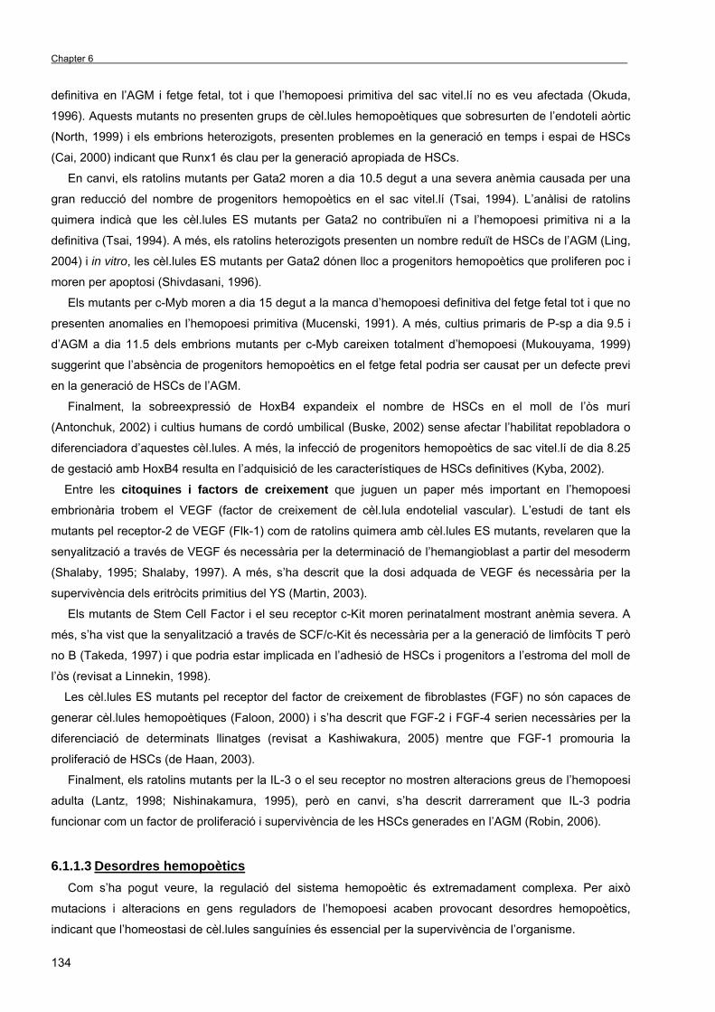
Chapter 6 _
134
definitiva en l’AGM i fetge fetal, tot i que l’hemopoesi primitiva del sac vitel.lí no es veu afectada (Okuda,
1996). Aquests mutants no presenten grups de cèl.lules hemopoètiques que sobresurten de l’endoteli aòrtic
(North, 1999) i els embrions heterozigots, presenten problemes en la generació en temps i espai de HSCs
(Cai, 2000) indicant que Runx1 és clau per la generació apropiada de HSCs.
En canvi, els ratolins mutants per Gata2 moren a dia 10.5 degut a una severa anèmia causada per una
gran reducció del nombre de progenitors hemopoètics en el sac vitel.lí (Tsai, 1994). L’anàlisi de ratolins
quimera indicà que les cèl.lules ES mutants per Gata2 no contribuïen ni a l’hemopoesi primitiva ni a la
definitiva (Tsai, 1994). A més, els ratolins heterozigots presenten un nombre reduït de HSCs de l’AGM (Ling,
2004) i in vitro, les cèl.lules ES mutants per Gata2 dónen lloc a progenitors hemopoètics que proliferen poc i
moren per apoptosi (Shivdasani, 1996).
Els mutants per c-Myb moren a dia 15 degut a la manca d’hemopoesi definitiva del fetge fetal tot i que no
presenten anomalies en l’hemopoesi primitiva (Mucenski, 1991). A més, cultius primaris de P-sp a dia 9.5 i
d’AGM a dia 11.5 dels embrions mutants per c-Myb careixen totalment d’hemopoesi (Mukouyama, 1999)
suggerint que l’absència de progenitors hemopoètics en el fetge fetal podria ser causat per un defecte previ
en la generació de HSCs de l’AGM.
Finalment, la sobreexpressió de HoxB4 expandeix el nombre de HSCs en el moll de l’òs murí
(Antonchuk, 2002) i cultius humans de cordó umbilical (Buske, 2002) sense afectar l’habilitat repobladora o
diferenciadora d’aquestes cèl.lules. A més, la infecció de progenitors hemopoètics de sac vitel.lí de dia 8.25
de gestació amb HoxB4 resulta en l’adquisició de les característiques de HSCs definitives (Kyba, 2002).
Entre les citoquines i factors de creixement que juguen un paper més important en l’hemopoesi
embrionària trobem el VEGF (factor de creixement de cèl.lula endotelial vascular). L’estudi de tant els
mutants pel receptor-2 de VEGF (Flk-1) com de ratolins quimera amb cèl.lules ES mutants, revelaren que la
senyalització a través de VEGF és necessària per la determinació de l’hemangioblast a partir del mesoderm
(Shalaby, 1995; Shalaby, 1997). A més, s’ha descrit que la dosi adquada de VEGF és necessària per la
supervivència dels eritròcits primitius del YS (Martin, 2003).
Els mutants de Stem Cell Factor i el seu receptor c-Kit moren perinatalment mostrant anèmia severa. A
més, s’ha vist que la senyalització a través de SCF/c-Kit és necessària per a la generació de limfòcits T però
no B (Takeda, 1997) i que podria estar implicada en l’adhesió de HSCs i progenitors a l’estroma del moll de
l’òs (revisat a Linnekin, 1998).
Les cèl.lules ES mutants pel receptor del factor de creixement de fibroblastes (FGF) no són capaces de
generar cèl.lules hemopoètiques (Faloon, 2000) i s’ha descrit que FGF-2 i FGF-4 serien necessàries per la
diferenciació de determinats llinatges (revisat a Kashiwakura, 2005) mentre que FGF-1 promouria la
proliferació de HSCs (de Haan, 2003).
Finalment, els ratolins mutants per la IL-3 o el seu receptor no mostren alteracions greus de l’hemopoesi
adulta (Lantz, 1998; Nishinakamura, 1995), però en canvi, s’ha descrit darrerament que IL-3 podria
funcionar com un factor de proliferació i supervivència de les HSCs generades en l’AGM (Robin, 2006).
6.1.1.3 Desordres hemopoètics Com s’ha pogut veure, la regulació del sistema hemopoètic és extremadament complexa. Per això
mutacions i alteracions en gens reguladors de l’hemopoesi acaben provocant desordres hemopoètics,
indicant que l’homeostasi de cèl.lules sanguínies és essencial per la supervivència de l’organisme.
Resum de la tesi
135
Els neoplasmes en cèl.lules hemopoètiques s’anomenen leucèmies. Sovint la transformació ocorre en
una única cèl.lula mare pluripotent, que comença a proliferar, expandir-se clonalment i evitar la mort per
apoptosi, tot i que pot ocórrer també en un progenitor hemopoètic no tan indiferenciat. Les leucèmies es
classifiquen segons l’estadi de diferenciació cel.lular i la severitat de la malaltia. Així podem trobar leucèmies
agudes o cròniques i segons el tipus cel.lular, limfoblàstiques o mieloides. Diferents translocacions
cromosòmiques que donen lloc a proteïnes de fusió han estat associades a la formació de leucèmies. Per
exemple, la translocació cromosòmica t(1;14)(p32;q11) que provoca la inserció del locus de Scl en els
elements reguladors del locus del receptor de cèl.lula T produeix leucèmia limfoblàstica aguda de cèl.lula T
(T-ALL) que afecta principalment a nens. Altres translocacions involucrades en leucèmies són la Tel-Runx1,
la Runx1-Eto i la Bcr-Abl (revisat a Izraeli, 2004; revisat a Leroy, 2005).
En canvi, els síndromes mielodisplàstics (MDS) són desordres de HSCs caracteritzats per alteracions
que resulten en anèmia, neutropènia i/o trombocitopènia. En molts casos, desemboquen en leucèmies
mieloides agudes (AML). S’ha descrit que tant mutacions en el factor de transcripció GATA1 com en el
receptor de SCF, c-Kit poden desenvolupar aquest tipus de desordres (revisat a Izraeli, 2004; revisat a
Linnekin, 1999).
6.1.2 ERITROPOESI 6.1.2.1 Diferenciació eritroide L’eritropoesi és un procès amb múltiples etapes que involucra la diferenciació des de les HSCs fins a
eritròcits madurs. En l’adult, l’eritropoesi té lloc en el moll de l’òs i implica la diferenciació des de les HSCs
en el progenitor comú mieloide (CMP), progenitor multipotent que genera el progenitor de granulòcits-
macròfags (GMP) i el progenitor de megacariòcits-eritròcits (MEP). En l’assaig de CFU-Culture, el CMP
genera colònies mixtes de cèl.lules eritroides i mieloides (Mix-CFC). Quan es determina cap al llinatge
eritroide, el progenitor CMP dóna lloc al progenitor BFU-e, que genera grans colònies de cèl.lules eritroides i
al progenitor eritroide més diferenciat CFU-e (revisat a Testa, 2004), el qual després de determinades
divisions mitòtiques, es diferencia en cèl.lules eritroides morfològicament diferenciables. Durant el procés de
diferenciació, les cèl.lules expressen gradualment el receptor d’eritropoietina esdevenint sensibles a aquesta
citoquina, que regula tant la proliferació com la supervivència de cèl.lules vermelles. Els últims estadis de
diferenciació eritroide estan composats per un compartiment proliferatiu-maduratiu format per
proeritroblastes, eritroblastes basofílics i eritroblastes policromatofílics i un altre compartiment maduratiu
però no proliferatiu format per eritroblastes ortocromàtics, reticulòcits i eritròcits madurs. Durant els últims
estadis de maduració, les cèl.lules eritroides es fan més petites, acumulen hemoglobina i incrementen la
densitat de la cromatina per excloure el nucli. Finalment, s’alliberen al torrent sanguini prenent part en
l’intercanvi gasós (revisat a Testa, 2004).
L’eritropoesi adulta està ben caracteritzada basada en la morfologia cel.lular, l’expressió de marcadors
de superfície cel.lular i el contingut d’hemoglobina (Chapter 1, Figura 10). No obstant, se sap molt poc del
desenvolupament eritroide primitiu. Els eritròcits primitius generats en les illes sanguínies del sac vitel.lí
entren en el torrent sanguini un cop s’estableix la circulació entre sac i embrió, en l’estadi d’eritroblastes
basofílics. Mentre circulen, els eritròcits acaben la seva maduració, caracteritzada per una acumulació
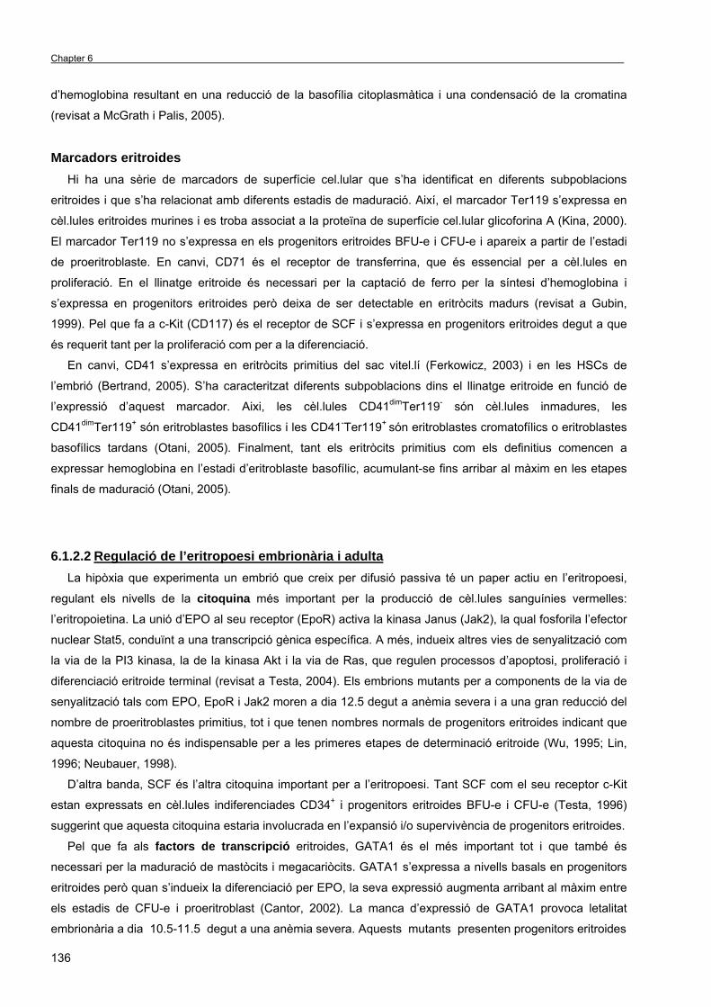
Chapter 6 _
136
d’hemoglobina resultant en una reducció de la basofília citoplasmàtica i una condensació de la cromatina
(revisat a McGrath i Palis, 2005).
Marcadors eritroides Hi ha una sèrie de marcadors de superfície cel.lular que s’ha identificat en diferents subpoblacions
eritroides i que s’ha relacionat amb diferents estadis de maduració. Així, el marcador Ter119 s’expressa en
cèl.lules eritroides murines i es troba associat a la proteïna de superfície cel.lular glicoforina A (Kina, 2000).
El marcador Ter119 no s’expressa en els progenitors eritroides BFU-e i CFU-e i apareix a partir de l’estadi
de proeritroblaste. En canvi, CD71 és el receptor de transferrina, que és essencial per a cèl.lules en
proliferació. En el llinatge eritroide és necessari per la captació de ferro per la síntesi d’hemoglobina i
s’expressa en progenitors eritroides però deixa de ser detectable en eritròcits madurs (revisat a Gubin,
1999). Pel que fa a c-Kit (CD117) és el receptor de SCF i s’expressa en progenitors eritroides degut a que
és requerit tant per la proliferació com per a la diferenciació.
En canvi, CD41 s’expressa en eritròcits primitius del sac vitel.lí (Ferkowicz, 2003) i en les HSCs de
l’embrió (Bertrand, 2005). S’ha caracteritzat diferents subpoblacions dins el llinatge eritroide en funció de
l’expressió d’aquest marcador. Aixi, les cèl.lules CD41dimTer119- són cèl.lules inmadures, les
CD41dimTer119+ són eritroblastes basofílics i les CD41-Ter119+ són eritroblastes cromatofílics o eritroblastes
basofílics tardans (Otani, 2005). Finalment, tant els eritròcits primitius com els definitius comencen a
expressar hemoglobina en l’estadi d’eritroblaste basofílic, acumulant-se fins arribar al màxim en les etapes
finals de maduració (Otani, 2005).
6.1.2.2 Regulació de l’eritropoesi embrionària i adulta La hipòxia que experimenta un embrió que creix per difusió passiva té un paper actiu en l’eritropoesi,
regulant els nivells de la citoquina més important per la producció de cèl.lules sanguínies vermelles:
l’eritropoietina. La unió d’EPO al seu receptor (EpoR) activa la kinasa Janus (Jak2), la qual fosforila l’efector
nuclear Stat5, conduïnt a una transcripció gènica específica. A més, indueix altres vies de senyalització com
la via de la PI3 kinasa, la de la kinasa Akt i la via de Ras, que regulen processos d’apoptosi, proliferació i
diferenciació eritroide terminal (revisat a Testa, 2004). Els embrions mutants per a components de la via de
senyalització tals com EPO, EpoR i Jak2 moren a dia 12.5 degut a anèmia severa i a una gran reducció del
nombre de proeritroblastes primitius, tot i que tenen nombres normals de progenitors eritroides indicant que
aquesta citoquina no és indispensable per a les primeres etapes de determinació eritroide (Wu, 1995; Lin,
1996; Neubauer, 1998).
D’altra banda, SCF és l’altra citoquina important per a l’eritropoesi. Tant SCF com el seu receptor c-Kit
estan expressats en cèl.lules indiferenciades CD34+ i progenitors eritroides BFU-e i CFU-e (Testa, 1996)
suggerint que aquesta citoquina estaria involucrada en l’expansió i/o supervivència de progenitors eritroides.
Pel que fa als factors de transcripció eritroides, GATA1 és el més important tot i que també és
necessari per la maduració de mastòcits i megacariòcits. GATA1 s’expressa a nivells basals en progenitors
eritroides però quan s’indueix la diferenciació per EPO, la seva expressió augmenta arribant al màxim entre
els estadis de CFU-e i proeritroblast (Cantor, 2002). La manca d’expressió de GATA1 provoca letalitat
embrionària a dia 10.5-11.5 degut a una anèmia severa. Aquests mutants presenten progenitors eritroides
Resum de la tesi
137
indicant que no és requerit per a la determinació a llinatge eritroide, però presenten un segrest de la
maduració en l’estadi de proeritroblast (Fujiwara, 1996) i moren per apoptosi (Weiss and Orkin, 1995). En
canvi, FOG-1 és un factor de transcripció que interacciona amb GATA1. Els embrions mutants per aquesta
proteïna presenten un fenotip similar als mutants de GATA1 però amb una total manca de megacariòcits
suggerint que FOG-1 també té funcions independents de GATA1 (Tsang, 1998).
Scl també té un paper en el llinatge eritroide. La sobreexpressió de Scl afavoreix la determinació a
aquest llinatge dels progenitors hemopoètics (Valtieri, 1998) mentre que la seva manca inhibeix la
proliferació de cèl.lules d’eritroleucèmia (Green, 1991).
Finalment, altres factors de transcripció involucrats en aquest llinatge són els de la família Krüppel-like o
KLF. KLF1/EKLF regula l’expressió d’un gran nombre de gens específics de la diferenciació eritroide, com el
de la β-globina (Hodge, 2005) mentre que els mutants de KLF6 tenen una manca d’expressió de GATA1 i
Scl, suggerint que aquest factor podria estar actuant per sobre dels altres dos (Matsumoto, 2006).
L’apoptosi també és un regulador molt important de l’homeostasi del llinatge eritroide. El promotor del
gen anti-apoptòtic bcl-xL conté llocs d’unió a GATA1 i la seva expressió és controlada per GATA1 en
cèl.lules mare embrionàries (Gregory, 1999). A més, també té llocs d’unió a Stat5, que es troba per sota de
la senyalització per EPO (Dolznig, 2002). En aquest sentit, els embrions deficients en Stat5 mostren anèmia
severa degut a una menor supervivència dels progenitors eritroides de fetge (Socolovsky, 1999), un fenotip
compartit pels mutants de Bcl-xL (Motoyama, 1995). A més, l’apoptosi en el llinatge eritroide també està
regulada per factors secretats o senyals mediades per lligand, actuant a través de la via d’apoptosi
extrínseca. En aquest sentit, l’activació de receptors de mort cel.lular com el receptor Fas o el receptor de
TNF-α provoquen l’apoptosi de progenitors eritroides (De Maria, 1999; Schneider, 1999; Jacobs-Helber,
2003).
6.1.2.3 Desordres eritropoètics Molts desordres del llinatge eritroide es deuen a la desregulació dels mecanismes apoptòtics que
controlen aquest compartiment. Per exemple, els pacients amb policitèmia vera (PV) presenten un quadre
clínic de síndrome mielodisplàstic amb una sobreproducció d’eritròcits, megacariòcits i granulòcits que
acaben desembocant en leucèmies agudes (Spivak, 2002), degudes a alteracions moleculars que resulten
en una resistència a l’apoptosi del llinatge eritroide (Zeuner, 2006).
En el cas dels pacients amb anèmia de Fanconi o anèmia associada a mieloma múltiple, la causa
principal és una major apoptosi espontània degut a una major sensitivitat a l’apoptosis mediada per FasL
(Silvestris, 2002).
Tot i això hi ha d’altres malalties del llinatge eritroide que no es deuen principalment a una desregulació
de la mort cel.lular per apoptosi. Les hemoglobinopaties (conegudes comuntment com a talassèmies) es
deuen a una expressió anormal de les globines. Els factors de transcripció GATA1 i EKLF controlen
l’expressió tant de la β-globina com de la proteïna estabilitzadora de la α-hemoglobina (AHSP). Per tant, els
ratolins EKLF-/- moren a dia 16 de gestació degut a un fenotip que s’assembla a la β-talassèmia (revisat a
Testa, 2004).

Chapter 6 _
138
6.1.3 LA VIA DE SENYALITZACIÓ DE NOTCH La senyalització a través del receptor de membrana Notch s’ha descrit que regula moltes de les
decisions de destí cel.lular, tant en el desenvolupament embrionari com en l’adult, mitjançant la
senyalització directa cèl.lula-cèl.lula (revisat a Lai, 2004). Els membres d’aquesta família s’ha trobat
conservats en organismes tan evolutivament divergents com les mosques i els humans i regulen processos
del desenvolupament tant variats com la neurogènesi, la miogènesi, l’hemopoesi o la segregació de somites
(revisat a Lewis, 1998).
6.1.3.1 Membres de la via de Notch i mecanisme Els membres de la família Notch inclouen bàsicament els receptors de membrana Notch, ells lligands
Delta i Serrate/Jagged i el factor de transcripció nuclear CSL (que significa CBF1/recombinant binding
protein J kappa (RBPjκ), Suppressor of Hairless Su[H], Lag-1). Tots els organismes metazous contenen un
o més ortòlegs d’aquestes proteïnes com es pot veure a la Taula 3, Chapter 1.
Els mamífers contenen quatre gens diferents que codifiquen per a quatre receptors de membrana
(Notch1-4). Es tracta d’heterodímers de transmembrana involucrats en transduir senyals extracel.lulars
específiques, cap al nucli en resposta a la unió del seu lligand. El domini extracel.lular conté una regió de
múltiples repeticions en tàndem del factor de creixement epidèrmic (EGF) involucrats en la unió al seu
lligand (Rebay, 1991) així com un domini anomenat LNR que manté l’estructura d’aquest heterodímer i evita
l’activació independent de lligand (Sánchez-Irizarry, 2004) En canvi, el domini intracel.lular de Notch
(NotchIC) conté el domini RAM i les repeticions de tipus ankirina (ANK) que es requereixen per la interacció
amb d’altres proteïnes efectores, senyals de localització nuclear, el domini de transactivació i el domini
PEST que regula l’estabilitat de la proteïna (revisat a Maillard, 2003) (Figura 13, Chapter 1).
L’activació de la senyal a través de Notch s’origina per la interacció del receptor amb un dels seus
lligands. També són proteïnes de transmembrana que contenen múltiples repeticions de tipus EGF i un
domini característic DSL, ambdós involucrats en la interacció amb el receptor. En Drosophila s’ha descrit
dos lligands diferents (Delta i Serrate), mentre que en vertebrats n’hi ha cinc (Jagged1 i 2, Delta1, Delta3 i
Delta4), tot i que un lligand addicional, Delta2 s’ha descrit en Xenopus.
Pel que fa al mecanisme d’activació, aquest s’inicia quan un dels lligands expressats en la cèl.lula
adjacent interacciona amb el receptor Notch (Kopan, 1996). Això provoca un primer processament
proteolític (anomenat S2, perquè el primer trencament, l’S1 es produeix en el complexe de Golgi per a crear
un receptor heterodimèric funcional a la superfície cel.lular) mediat per una metal.loproteasa de la família
ADAM, anomenada TACE (TNF-α converting enzyme). Aquest receptor truncat és aleshores substrat d’un
complexe multiproteic amb activitat γ-secretasa que trenca el receptor Notch pel domini transmembrana,
alliberant el domini intracel.lular Notch (NotchIC; revisat a Lai, 2004). El bloqueig de l’activitat γ-secretasa
amb inhibidors farmacològics com el DAPT o la inactivació genètica de membres del complexe γ-secretasa
inhibeix la senyalització a través de Notch (Zhang, 2000). Després del seu alliberament, el frament
intracel.lular de Notch transloca al nucli on s’uneix al seu efector, el factor de transcripció RBPjκ. En
absència d’activació de Notch, RBPjκ es troba unit a la seqüència d’unió dels seus promotors diana,
reprimint l’expressió a través del reclutament de corepressors i histona deacetilases (HDACs) (Kao, 1998).
Resum de la tesi
139
Un cop al nucli NotchIC s’uneix a RBPjκ desplaçant els corepressors i reclutant coactivadors activant-se
l’expressió gènica (Figura 13, Chapter 1).
Entre els gens diana de Notch es troben les famílies de factors de transcripció hes (hairy and Enhancer
of Split) i hrt (hes-related). Fins al moment, s’ha descrit 7 gens hes (hes1-7) en mamífers, tot i que només
hes1 (Jarriault, 1995), hes5 (Ohtsuka, 1999) i hes7 (Bessho, 2001) són activats per Notch. A més, hi ha dos
gens hrt (herp1/hrt2 i herp2/hrt1) que són gens diana de Notch (revisat a Iso, 2003). Hes i Hrt són factors de
transcripció del tipus bHLH que actúen com a repressors transcripcionals d’altres bHLH involucrats en la
diferenciació cel.lular de molts sistemes tals com neurogènesi, miogènesi, hemopoesi o diferenciació
intestinal (revisat a Ohishi, 2003).
6.1.3.2 Control de decisions de destí cel.lular Durant les primeres etapes del desenvolupament d’un determinat teixit, la majoria de cèl.lules tenen una
potencialitat equivalent. En aquesta població homogènia, les interaccions entre Notch i Jagged/Delta són
responsables de la generació de la diversitat cel.lular a través de l’activació de programes gènics específics
(revisat a Lewis, 1998). S’ha proposat dos mecanismes diferents per a explicar com Notch regula la
diversitat cel.lular:
El model d’inhibició lateral implica que en una població de cèl.lules equivalents que expressen nivells
baixos de receptor i lligand, la cèl.lula que primer produeix més lligand, activa el receptor Notch en la cèl.lula
veïna, la qual acaba disminuïnt l’expressió de lligand. Aquest mecanisme permet el manteniment i la
intensificació de les diferències d’expressió d’ambdós proteïnes, ja que la cèl.lula que expressa lligand no
rep senyals inhibitòries de les cèl.lules veïnes. Al final s’acaba aconseguint un mosaic cel.lular que es
coneix amb el nom de sal-i-pebre, que condueix a que la cèl.lula que expressa lligand es diferencïi cap a un
llinatge diferent al de les cèl.lules veïnes (Figura 14, Chapter 1). El clàssic exemple d’inhibició lateral ocorre
durant la decisió entre cèl.lula neural i epidèrmica a Drosophila (Parks, 1997). En mamífers, la via de Notch
reprimeix la neurògenesi i la miogènesi a través de repressors bHLH de la família Hes, els quals a la vegada
inhibeixen altres factors de transcripció del tipus bHLH com Mash1 o MyoD (revisat a Artavanis-Tsakonas,
1999).
El segon mecanisme d’actuació de Notch és el model d’inducció lateral. En aquest cas, l’activació de
Notch en una cèl.lula determinada promou l’expressió del lligand en la mateixa cèl.lula. Per tant, la cèl.lula
que expressa lligand activa Notch en la cèl.lula adjacent prevenint el patró de sal-i-pebre i induïnt les seves
decisions de destí cooperativament i formant marges definits d’expressió gènica (Figura 15, Chapter 1)
(revisat a Lewis, 1998). En Drosophila, l’exemple clàssic d’inducció lateral ocorre durant la formació de l’ala
(Couso, 1995). En vertebrats, el clàssic exemple és la generació de límits durant la somitogènesi, mediat a
través de l’activació oscil.latòria de hes1 i hes7 per part de Notch (Jouve, 2000).
Altres membres claus de la via de Notch La glicosilació del receptor de Notch a través de les glicosiltransferases Fringe (Radical, Lunatic i Manic
Fringe) té la funció de modular l’especificitat de lligand. Les proteïnes Fringe regulen doncs, l’activitat Notch i
contribueixen a la generació de diversitat cel.lular (revisat en Irvine, 1999).
En canvi, l’ubiqüitinació és requerida per a l’endocitosi del receptor Notch i els seus lligands. Neuralized
i Mind bomb pertanyen a les RING-type E3 ubiqüitin lligases, ubiqüitinen el lligand Delta i mutacions en
Chapter 6 _
140
aquestes proteïnes resulta en fenotips de pèrdua de funció de Notch (Itoh, 2003; Lai, 2001). Estudis recents
suggereixen que la ubiqüitinació de Delta incrementa l’afinititat d’unió a Notch. A més, l’endocitosi del lligand
Delta unit al domini extracel.lular de Notch facilita el processament proteolític S2 del receptor Notch (Nichols,
2007).
6.1.3.3 Funció de la via de senyalització de Notch en l’hemopoesi La via de Notch funciona en diferents estadis del desenvolupament hemopoètic i la seva activació pot ser
a través d’interaccions entre progenitors i cèl.lules de l’estroma però també entre cèl.lules hemopoètiques.
Expressió de receptors i lligands en el sistema hemopoètic Els precursors hemopoètics de moll de l’òs CD34+Lin- expressen el receptor Notch1 i Notch2, suggerint
que Notch estaria implicat en el desenvolupament temprà del sistema hemopoètic (Milner, 1994; Ohishi,
2000). A més, les cèl.lules de l’estroma del moll de l’òs expressen Jagged1 (Walker, 2001), així com Delta1 i
Delta4 (Karanu, 2001). Aquests mateixos lligands es troben expressats en cèl.lules d’epitel.li tímic, indicant
que Notch jugaria un paper important en el desenvolupament de cèl.lula T (Felli, 1999; Mohtashami, 2006).
En el llinatge mieloide, els monòcits expressen elevats nivells de Notch1 i Notch2 però els granulòcits no
(Ohishi, 2000). Finalment, els progenitors eritroides de moll de l’òs expressen Notch1 però l’expressió
disminueix en cèl.lules més madures com els normoblastes acidòfils (Ohishi, 2000; Walker, 2001).
Paper de Notch en l’autoregeneració de HSCs Actualment hi ha força evidències que indiquen que Notch té un paper crucial en l’autoregeneració de
HSCs. Duncan et al. demostraren que la via de Notch està activa en la subpoblació KSL del moll de l’òs i
s’inhibeix a mida que les cèl.lules diferencien. A més, la inhibició de Notch amb un dominant negatiu de
CBF1 condueix a una diferenciació accelerada de les HSCs in vitro i una depleció de l’activitat repobladora
in vivo, indicant que Notch és necessari pel manteniment de l’estat indiferenciat de les HSCs (Duncan,
2005).
La transducció del domini intracel.lular de Notch en progenitors hemopoètics murins c-Kit+Sca-1+Lin- duu
a la immortalització d’aquestes cèl.lules i a la repoblació quan es transplanten en ratolins adults irradiats
(Varnum-Finney, 2000). A més, Notch1IC expandeix el nombre de cèl.lules repobladores de moll de l’òs
(Stier, 2002), un resultat similar al que s’obté expressant Notch4IC en cèl.lules humanes Lin- de cordó
umbilical (Vercauteren, 2004). A més, l’adició de Jagged1 soluble a cultius ex vivo de cèl.lules CD34+CD38-
Lin- de cordó umbilical incrementa el nombre de cèl.lules mare hemopoètiques sense perdre l’habilitat
repobladora (Karanu, 2000). L’incubació amb una proteïna de fusió Delta1 tindria el mateix efecte (Varnum-
Finney, 2003). Molts d’aquests efectes són reproduïbles en cèl.lules CD34-KSL transduïdes amb Hes1,
suggerint que aquesta proteïna seria responsable dels efectes de l’activació de Notch1 en la proliferació de
les HSCs (Kunisato, 2003).
Regulació de decisions de destí cel.lular limfoide Notch regula vàries decisions de destí cel.lular en el llinatge limfoide. L’activació de Notch promou la
diferenciació cap al llinatge T a expenses del llinatge B. En aquest sentit, la deleció de RBPjκ en cèl.lules

Resum de la tesi
141
hemopoètiques resulta en un increment de la diferenciació de cèl.lula B i un bloqueig del desenvolupament
de cèl.lula T (Han, 2002). Contràriament, l’expressió de Notch1IC en progenitors bloqueja la limfopoiesi B i
condueix a la generació de limfòcits T CD4+CD8+ (Pui, 1999). A més, Notch regula la determinació de
cèl.lula αβ versus γδ dintre del llinatge TCR. L’activació de Notch afavoreix el destí αβ ja que la disminució
dels nivells de Notch1 resulta en un increment de cèl.lules γδ (Washburn, 1997), un resultat que també
s’obtingué amb la inactivació condicional de RBPjκ (Tanigaki, 2004). Finalment i en l’últim estadi de
diferenciació de limfòcits T, Notch també estaria involucrat en la presa de decisió entre cèl.lula CD4+ o
CD8+, ja que l’expressió de Notch1IC en timòcits afavoreix la generació de cèl.lules T CD8+ en detriment de
les cèl.lules T CD4+ (Robey, 1996).
Notch en la diferenciació mieloide La implicació de Notch en mielopoiesi s’ha estudiat tant en models in vivo com en línies cel.lulars in vitro.
En aquest sentit, la sobreactivació de Notch1 i Notch2 inhibeix la diferenciació de línies cel.lulars mieloides
de forma citoquina depenent (Milner, 1995; Bigas, 1998), segurament a través de l’expressió de Gata2
(Kumano, 2001). Més recentment, s’obtingué un resultat similar quan es va co-cultivar progenitors KSL amb
la línia cel.lular d’estroma OP9 que expressa Delta1 (de Pooter, 2006). Tot i això, la funció de Notch en el
llinatge mieloide no està tant clara, ja que hi ha resultats que suggereixen que l’activitat de Notch és
requerida només per la diferenciació de limfòcits T i B in vivo (Radtke, 1999).
Notch i apoptosi Molts estudis suggereixen que Notch també regula la mort cel.lular programada o apoptosi. En molts,
casos, l’activació de Notch inhibeix l’apoptosi, com s’ha descrit en cèl.lules T (Jehn, 1999), cèl.lules de
sarcoma de Kaposi (Curry, 2005) i cèl.lules de limfoma de Hodgkin (Jundt, 2002), tot i que degut a
l’especificat de Notch segons el context, hi ha molts exemples que Notch actúa també al contrari. Per
exemple, en línies cel.lulars de leucèmia limfoblàstica aguda de cèl.lules B, l’activació de Notch indueix
apoptosi, segurament a través de Hes1 (Zweidler-McKay, 2005), així com en monòcits de sang perifèrica
cultivats en presència de lligand Delta1 inmobilitzat i M-CSF (Ohishi, 2000).
Pel que fa a la funció de Notch en la regulació de l’apoptosi en el llinatge eritroide, encara ara hi ha força
controvèrsia. En la línia cel.lular d’eritroleucèmia humana K562, Notch inhibeix la diferenciació i indueix
apoptosi, a través de Hes1, el qual inhibeix l’activitat GATA1 i en últim lloc, l’expressió de bcl-XL (Ishiko,
2005). Contràriament, Notch1 preve l’apoptosi en la línia cel.lular d’eritroleucèmia murina (MEL) durant la
diferenciació induïda per HMBA (Shelly, 1999; Jang, 2004).
Implicació de Notch en l’ontogènia del sistema hemopoètic En els darrers anys, la funció de Notch en l’hemopoesi embrionària ha estat parcialment descrita. Part
del que se sap ha estat l’objectiu d’aquesta tesi i per tant es discutirà amb detall. Tot i això mentre estàvem
treballant-hi, van aparèixer d’altres treballs que inclourem en aquesta secció.
La primera evidència que la via de Notch actúa en la generació de HSCs durant el desenvolupament
embrionari aparegué dels treballs de Kumano et al. amb l’estudi dels ratolins mutants per Notch1. Cultivant
cèl.lules de P-sp sobre la línia cel.lular d’estroma OP9, varen trobar que l’hemopoesi definitiva està
totalment afectada en els embrions mutants de Notch1 degut a una manca de HSCs. Contràriament, el

Chapter 6 _
142
nombre de CFC en els sacs vitel.lins Notch1-/- era similar als embrions salvatges, suggerint que l’hemopoesi
primitiva estava almenys parcialment intacta. Tot i això, les HSCs tant del sac vitel.lí com de la P-sp no eren
capaces de reconstituir l’hemopoesi de ratolins nou-nats hematològicament compromesos, suggerint que la
manca d’hemopoesi definitiva era deguda a un problema de determinació de les HSCs des de l’endoteli
aòrtic (Kumano, 2003). En aquest sentit es va demostrar que en ratolins quimera amb cèl.lules mare
embrionàries mutants per Notch1, aquestes no contribueixen a l’hemopoesi definitiva (Hadland, 2004).
Contràriament, l’anàlisi dels embrions mutants per Notch2 indica que Notch2 no és requerit per la generació
de cèl.lules hemopoètiques (Kumano, 2003).
Finalment, estudis amb peix zebra també han ajudat a esclarir la funció de Notch en la formació de les
HSCs. Els mutants per la proteïna Mind bomb tenen una hemopoesi primitiva normal però careixen de
desenvolupament de HSCs, així com expressió de c-myb i runx1 en l’aorta. Contràriament, l’expressió
transient de Notch1 revelà una expansió del nombre de HSCs, depenent de l’expressió de Runx1 (Burns,
2005). Per tant Runx1 actuaria per sota de Notch en la generació de HSCs de l’AGM.
6.1.3.4 Malalties degudes a alteracions de la via de Notch S’ha descrit tres malalties congènites degudes a mutacions en membres de la família Notch. Així,
mutacions en el gen jagged1 són responsables de la síndrome Alagille, que resulta en una generació i
funció aberrant de diferent òrgans com són el cor, l’ull i el fetge (Li, 1997). En canvi, mutacions de Delta3
són responsables de la disostosi espondilocostal, una malaltia del desenvolupament caracteritzada per
alteracions de l’esquelet entre les quals la fusió de les costelles (Bulman, 2000). Finalment, mutacions en
les repeticions de tipus EGF de Notch3 resulta en la arteriopatia cerebral autosòmica dominant amb infarts
subcorticals i leucoencefalopatia (CADASIL), caracteritzada per migranyes i demència (Joutel, 1996). A
més, Notch també participa en processos tumorigènics en diferents teixits entre els que s’inclouen la
glàndula mamària, pell, cèrvix i pròstata (revisat a Lai, 2004), còlon (Fernández-Majada, 2007) o pàncreas
(Miyamoto, 2003).
Desordres hemopoètics El gen Notch1 es va identificar en humans com a responsable de leucèmies de limfòcits T degut a que es
veia involucrat en la translocació cromosòmica t(7;9)(q34;q34.3) que provocava una proteïna de fusió entre
Notch1 i el receptor de cèl.lula T (TCR-β) resultant en la activació constitutiva de Notch1 en aquestes
cèl.lules (Ellisen, 1991). No obstant, la majoria de leucèmies produïdes per Notch es deuen a mutacions
puntuals que afecten el domini d’heterodimerització (provocant l’activació aberrant de Notch) o en el domini
PEST (resultant en una major estabilitat de Notch1IC) (Weng, 2004). A més en els darrers anys s’ha descrit
que darrera d’algunes leucèmies de tipus T podria haver-hi una cooperació entre Notch i Ikaros. En aquest
sentit, ratolins homozigòtics per un al.lel hipomòrfic d’Ikaros, desenvolupen limfomes tímics per una major
activitat Notch i expressió de hes1 en timòcits (Dumortier, 2006).
6.1.3.5 Models animals Per tal d’estudiar la funció de Notch tant en el desenvolupament com en organismes adults, s’ha generat
organismes transgènics i mutants per a diferents membres de la família Notch, tant en Drosophila (mosca),
C. elegans (nemàtodes), Danio rerio (peix zebra) com Mus musculus (ratolí).

Resum de la tesi
143
La mutació de pèrdua de funció de Notch amb un fenotip més server s’obté de la deleció del gen rbpjκ, ja
que és l’únic efector de la via de senyalització de Notch (Oka, 1995). Els embrions RBPjκ-/- moren a dia 10.5
i presenten un fenotip complexe que inclou alteracions en la somitogènesi, defectes en la formació del tub
neural i creixement retardat (Figura 16, Chapter1). A més, la fusió de l’alantoides embrionari amb la placenta
materna no ocorre i per tant no es poden formar els vasos umbilicals. Tots aquestes característiques
indiquen que la senyalització a través de Notch és requerida per a la vasculogènesi, somitogènesi i
neurogènesi (Oka, 1995).
El fenotip dels embrions Notch1-/- és similar al dels mutants de RBPjκ tot i que la mort ocorre una mica
després, a dia 11.5. Els mutants de Notch1 presenten també retard del creixement, alteració de la
neurogènesi i la formació dels somites tot i que la fusió corioalantoidea sí que es produeix (Conlon, 1995).
Els mutants de Notch2 moren a 11.5 i presenten una apoptosi massiva en el neuroepiteli de les vesícules
òtiques i òptiques (Hamada, 1999). Pel que fa als mutants de Notch3 són viables i només mostren alguns
defectes en la vasculogènesi (Krebs, 2003). Finalment, els mutants de Notch4 són viables i fèrtils tot i que
aquest receptor coopera amb la mutació de Notch1 en la inducció de defectes vasculogènics (Krebs, 2000).
Els mutants de Jagged1 moren a dia 10. Aquests embrions poden formar els vasos primitius del sac
vitel.lí i de l’embrió però tenen defectes en la remodelació del plexe vascular per a formar vasos vitel.lins
grans, suggerint que l’angiogènesi però no la vasculogènesi està afectada (Xue, 1999). En canvi, la mutació
Jagged2 no és letal fins etapes properes al naixement, i aquests mutants presenten defectes en la
morfogènesi craniofacial i en la fusió palatal, defectes en la formació de les extremitats així com
desenvolupament tímic anormal (Jiang, 1998).
Finalment, el mutant de Mind bomb i els doble mutants per Presenilin1 i Presenilin2 (proteïnes del
complexe γ-secretasa) moren a dia 10.5 i 9.5 respectivament, amb un fenotip molt similar al dels mutants de
RBPjκ suggerint que aquestes proteïnes actúen per damunt de Notch i són necessàries per a la
senyalització (Koo, 2005; Donoviel, 1999).

Chapter 6 _
144
6.2 OBJECTIUS D’AQUESTA TESI Tot i que ja s’havia descrit prèviament que la via de Notch regula diferents decisions de destí cel.lular en
el sistema hemopoètic, la seva funció durant l’ontogènia de l’hemopoesi embrionària no es coneixia en l’inici
d’aquesta tesi. Per tant, mitjançant l’ús del model de ratolí mutant per RBPjκ, el nostre objectiu era estudiar
si la senyalització a través de Notch estava involucrada en la generació de l’hemopoesi primitiva i/o
definitiva en l’embrió murí.
Per tant, els objectius específics que ens vam proposar per aquest projecte foren:
* Estudiar si la senyalització a través de Notch/RBPjκ té una funció específica en l’ontogènia del sistema
hemopoètic primitiu en el sac vitel.lí murí des d’E7.5 a E9.5.
* Determinar si la senyalització a través de Notch/RBPjκ és necessària per a la generació de cèl.lules
mare hemopoètiques i altres progenitors definitius en la regió de la P-sp/AGM murina des d’E9.5 a E11.5.

Resum de la tesi
145
6.3 RESULTATS
CAPÍTOL 6.3.1: La funció de Notch depenent de RBPjκ regula Gata2 i és essencial
per la formació de cèl.lules hemopoètiques intraembrionàries (Aquest capítol va ser publicat a Development 132, 1117-1126, 2005). La regió intraembrionària que inclou l’aorta rodejada per gònada i mesonefros ha estat descrita com el
primer òrgan involucrat en la generació de progenitors hemopoètics (HP) i de cèl.lules mare hemopoètiques
(HSC) que més tard col.lonitzaran el moll de l’òs i mantindran l’hemopoesi en l’adult (Medvinsky, 1996; de
Bruijn, 2000; de Bruijn, 2002). Prèviament al nostre treball hi havia força evidències que la via de
senyalització de Notch regula diferents processos biològics en moltes línies cel.lulars hemopoètiques (Bigas,
1998; Karanu, 2000; Varnum-Finney, 2000; Kumano, 2001) tot i que hi havia poca informació de què
passava in vivo. Per tant, fent ús dels ratolins mutants per RBPjκ (Oka, 1995), el nostre objectiu era
determinar si la senyalització per Notch és necessària per la generació de HP/HSC en l’AGM.
Aquí mostrem que en embrions salvatges, alguns membres de la via de Notch entre els que s’inclouen
els receptors notch1 i notch4 i els lligands delta4, jagged1 i jagged2 es troben expressats en cèl.lules
endotelials de l’aorta de l’AGM a dia 9.5 i 10.5 de desenvolupament. A més, gens diana de Notch tals com
hes1, hrt1 i hrt2 també es troben expressats indicant que la via de Notch està activada en aquesta regió.
Aquest patró d’expressió de receptors Notch i lligands es troba alterat en els embrions mutants per
RBPjκ (als quals els hi falta l’efector nuclear comú, per tant manca la senyalització per sota dels quatre
receptors Notch) demostrant que la regulació de la seva expressió depèn de feed-back loops positius i
negatius.
També demostrem que els embrions RBPjκ-/- mostren una manca de potencial hemopoètic en la regió de
l’AGM amb una completa falta de progenitors hemopoètics (determinat per l’assaig de CFC) i de cèl.lules
hemopoètiques CD45+ després de 6 dies en cultiu. L’absència d’hemopoesi correlaciona amb una completa
manca d’expressió dels factors de transcripció hemopoètics claus com són Gata2, Runx1 i Scl i amb un
increment dels nombre de cèl.lules endotelials, suggerint la presència d’un progenitor comú (o
hemangioblast) pels llinatges hemopoètic i endotelial.
Finalment, per doble hibridació in situ i immunoprecipitació de cromatina, describim que Notch s’uneix
directament a través de RBPjκ al promotor de gata2, regulant la seva expressió no només a la línia cel.lular
32D però també en l’embrió. Finalment, describim quins lligands s’expressen en la cèl.lula adjacent a la que
expressa notch1 en l’endoteli aòrtic i suggerim que l’activació de l’expressió de gata2 induïda per Notch1 en
algunes de les cèl.lules de l’endoteli ventral de l’aorta de l’AGM i l’activació del programa hemopoètic en
aquesta cèl.lula podria ser induït pels lligands Jagged2 o Delta4.
Chapter 6 _
146
CAPÍTOL 6.3.2: El lligand de Notch, Jagged1 és requerit per l’hemopoesi
intraembrionària de l’AGM (Aquest capítol conté resultats encara no publicats)
Nosaltres hem descrit que l’expressió de gata2 mediada per Notch1/RBPjκ és necessària per
l’hemopoesi de l’AGM (Robert-Moreno, 2005). Aquests resultats concordaven amb el treball de Kumano et
al. analitzant el ratolí mutant per Notch1 (Kumano, 2003). Tot i això, no se’n sap gaire sobre quin lligand
específic és el que activa Notch i indueix l’hemopoesi de l’AGM. A més, degut als severs defectes vasculars
que mostren els mutants de Notch1 i RBPjκ (Oka, 1995; Krebs, 2000) i per tal d’evitar la possibilitat que la
manca d’hemopoesi intraembrionària és deguda a la manca d’una artèria prèviament especificada, l’estudi
de la funció de Notch en l’hemopoesi d’aortes salvatges era necessària.
En aquest capítol, describim que el potencial hemopoètic de l’aorta de l’AGM dels embrions mutants pel
lIigand Jagged1 però no dels mutants de Jagged2 es troba severament compromès (tot i que no totalment
afectat). Els embrions mutants per Jagged1 careixen totalment d’expressió de gata2, mostren un nombre
menor de progenitors hemopoètics i una reducció en el nombre de cèl.lules Sca-1+ (una població enriquida
per HSCs). A més i de forma similar als embrions mutants per RBPjκ, la manca d’hemopoesi
intraembrionària en els mutants de Jagged1 correlaciona amb un increment del nombre de cèl.lules
endotelials.
També mostrem que la inhibició de la senyalització per Notch amb l’inhibidor de l’activitat γ-secretasa en
aortes salvatges d’embrions de dia 11 de gestació, cultivades com a explants, redueix significativament el
nombre de progenitors hemopoètics (determinat amb l’assaig de CFCs) i de HSCs amb alta capacitat
repobladora (determinat per estudis de repoblació d’adults irradiats).
Degut a que també describim que notch1, jagged1 i hes1 estan expressats en els grups de cèl.lules
hemopoètiques que sobresurten de la part ventral de l’aorta de l’AGM, proposem que l’activació induïda per
Jagged1 condueix a l’expressió de gata2 i hes1 conduïnt a l’expansió del nombre de progenitors
hemopoètics i HSCs en l’endoteli aòrtic i a la vegada, a un manteniment de l’estat indiferenciat d’aquestes
cèl.lules.
Resum de la tesi
147
CAPÍTOL 6.3.3: La via de Notch regula positivament la mort cel.lular programada
durant la diferenciació eritroide (Aquest capítol va ser publicat a Leukemia, Maig 2007) Una sèrie d’estudis de mutagènesi gènica dirigida revelaren que l’hemopoesi en el sac embrionari i en la
regió intraembrionària de l’AGM estan governades per dos programes genètics diferents, ja que algunes
mutacions afecten tant a l’hemopoesi primitiva com la definitiva, mentre que d’altres només afecten a la
definitiva (revisat a Cumano i Godin, 2002).
En aquest article, mostrem que en contrast amb l’absolut requeriment de la senyalització a través de
Notch/RBPjκ per l’hemopoesi definitiva de l’AGM, l’eritropoesi primitiva ocorre normalment en els sacs
vitel.lins dels mutants per RBPjκ. Aquests embrions mutants generen tots els tipus diferents de progenitors
hemopoètics i el desenvolupament i maduració del llinatge hemopoètic no mostra cap defecte. No obstant,
el percentatge de cèl.lules eritroides Ter119+ és major en els sacs vitel.lins mutants per RBPjκ comparat
amb els embrions salvatges. Ni una major proliferació, ni diferències en la diferenciació eritroide poden
explicar aquest increment cel.lular.
En aquest treball describim que l’apoptosi es troba específicament reduïda en les cèl.lules eritroides dels
sacs vitel.lins mutants, indicant que la via de Notch promou la mort cel.lular programada en aquest llinatge.
Mitjançant RT-PCR quantitativa, demostrem que la pèrdua de RBPjκ correlaciona amb un increment de
l’expressió dels gens EPO i del receptor d’Epo (EpoR) així com dels gens anti-apoptòtics bcl-2 i bcl-XL,
concordant amb la major supervivència de la població Ter119+ en els mutants.
Finalment, mostrem que Notch indueix apoptosi en el llinatge eritroide no només durant el
desenvolupament embrionari però també en d’adult, ja que cèl.lules de moll de l’òs tractades amb els
inhibidors de l’activitat γ-secretasa DAPT i L685,458 mostren menor mort cel.lular per apoptosi. A més, la
inducció de l’apoptosi a través de Notch, mitjançant la incubació de moll d’òs amb cèl.lules que expressen
Jagged1, és revertit amb el tractament amb inhibidors de l’activitat γ-secretasa. En aquest sentit, cèl.lules
d’eritroleucèmia murina que sobreexpressen la forma activada de Notch1 presenten una major mort cel.lular
programada en condicions de diferenciació induïda per hexametilè-bisacetamida (HMBA).
Per tant, amb tota aquesta informació proposem que la senyalització a través de Notch no és essencial
per l’hemopoesi del sac embrionari però pot induïr específicament la mort cel.lular programada en el llinatge
eritroide no només durant la vida embrionària però també en teixits adults, regulant l’homeostasi d’aquest
compartiment hemopoètic.
Chapter 6 _
148
6.4 DISCUSIÓ GENERAL i PERSPECTIVES FUTURES Les cèl.lules mare hemopoètiques tenen una gran importància tant pels transplantaments clínics com per
la medicina regenerativa. Aquest fet ha motivat enèrgicament l’estudi dels mecanismes que controlen
l’autoregeneració de les HSCs i com el microambient regula el seu comportament. El nostre treball ha
contribuït a esclarir la funció de Notch en l’hemopoesi embrionària del sac vitel.lí així com la formació de
HSCs en l’AGM, un dels nínxols de generació i amplificació de HSCs de l’embrió millor caracteritzats. Tot i
això encara ara hi ha controvèrsia sobre el lloc precís on es formen les HSCs en l’embrió.
La història interminable: quin és l’origen de les HSCs? L’actual model diu que les primeres HSCs adultes capaces de nïar en el moll de l’òs d’adults irradiats es
troben en l’embrió murí a dia 10 (de Bruijn et al., 2002; de Bruijn et al., 2000) mentre que a dia 11 es
detecten en altres teixits tals com el fetge fetal, el sac vitel.lí, la placenta i el torrent sanguini (Gekas et al.,
2005; Kumaravelu et al., 2002; Medvinsky and Dzierzak, 1996; Muller et al., 1994; Ottersbach and Dzierzak,
2005). No obstant, la idea que el sac vitel.lí contribueix a l’hemopoesi de l’adult no s’ha descartat mai, tot i
que clàssicament s’ha cregut que la seva funció bàsica era la de generar eritròcits pel creixement de
l’embrió. De fet, el sac vitel.lí i el mesoderm de la P-Sp de dia 9 de gestació, no són capaços de repoblar
adults irradiats però sí que reconstitueixen l’hemopoesi de nou-nats (Yoder and Hiatt, 1997; Yoder et al.,
1997; Yoder et al., 1997). Aquestes evidències suggeririen la idea que a dia 9, tant el sac vitel.lí com la P-sp
contenen HSCs immadures que necessiten madurar per adquirir l’habilitat de nïar en el moll de l’òs adult.
Per tant, el model actual suggereix que l’AGM serveix com a l’òrgan principal de generació de HSCs adultes,
però que d’altres teixits com el sac vitel.lí i la placenta també contribueixen a l’hemopoesi adulta. No obstant, un treball recent el qual proposa que el sac embrionari és l’origen d’un percentatge elevat de
les HSCs de l’adult ha afegit encara més controvèrsia (Samokhvalov et al., 2007). No obstant, als anys 70,
experiments de quimeres entre embrions de pollastre i guatlla demostraven que era l’embrió qui contribuïa
al compartiment hemopoètic definitiu (Dieterlen-Lievre, 1975), mentre que més recentment s’ha demostrat
que l’aorta està formada per dues poblacions de cèl.lules mesodermals i només una d’elles amb potencial
hemopoètic (Pardanaud et al., 1996). A més, l’expressió de marcadors hemopòetics entre les cèl.lules de
l’endoteli aòrtic reforçarien encara més la idea que l’AGM és l’òrgan generador de HSCs (Bertrand et al.,
2005; de Bruijn et al., 2002; North et al., 1999; Robert-Moreno et al., 2005). Finalment un model alternatiu
suggereix que les HSCs s’originen en el mesènquima sota la part ventral de l’aorta dorsal, unes estructures
anomenades parxes subaòrtics (SAPs) i que expressen marcadors hemopoètics com gata2, gata3 i lmo2 i
que contenen HSCs CD45low/ckit+/AA4.1+/flk-1- capaces de reconstituir l’hemopoesi de ratolins
immunodeficients Rag2γc-/- (Bertrand et al., 2005; Manaia et al., 2000). Els nostres resultats no donarien
suport a aquesta possibilitat ja que no hem detectat per hibridació in situ cap tipus de cèl.lula hemopoètica
en el mesènquima sota la part ventral de l’aorta. Tots aquest models reflecteixen que les idees sobre
l’origen endotelial o mesenquimàtic de les HSCs són variades, però que la integració de tota la informació
que hi ha en l’actualitat resultaria en un millor coneixement de la generació de les HSCs.
L’objectiu principal del nostre treball era estudiar la funció que podia estar realitzant la via de Notch tant
en l’hemopoesi primitiva com en la definitva de l’embrió murí. Els nostres resultats de l’estudi dels embrions
mutants per RBPjκ (Robert-Moreno et al., 2005) i dels cultius d’explant d’aorta (Chapter 3, Section 2)
suggereixen que la via de Notch és necessària per la determinació de les HSC i altres progenitors hemopo-

Resum de la tesi
149
ètics de l’endoteli hemogènic de l’aorta de l’AGM. Per tant, proposem que l’activitat de Notch a l’aorta ventral
genera un patró d’expressió de sal-i-pebre que restringeix el potencial hemogènic a un nombre determinat
de HSCs/HP entre una majoria de cèl.lules aòrtiques endotelials, recordant el procès de la inhibició lateral
descrit en la neurogènesi de Drosophila (Parks et al., 1997). En aquest sentit, la hibridació in situ (WISH)
pels diferents lligands i receptors Notch en embrions temprans (Supplemental information; Chapter 3,
Section 2) recolsarien aquest model d’inhibició lateral ja que les cèl.lules que expressen lligand apareixen
abans en el desenvolupament (en l’estadi de 14 parells de somites) que les cèl.lules que expressen notch1
(cap a l’estadi de 20 parells de somites) i només en cèl.lules aïllades de la paret ventral de l’endoteli aòrtic.
Per tant, hi ha només un lloc on es generen HSCs en l’embrió o per contra és un procès redundant que
té lloc en diferents teixits al llarg del desenvolupament fetal? Els nostres resultats concordarien amb la idea
que l’hemopoesi primitiva i la definitiva són resultat de dos programes genètics ben diferenciats. Els
embrions mutants per RBPjκ careixen totalment d’hemopoesi intraembrionària (Robert-Moreno et al., 2005)
mentre que l’hemopoesi primitiva del sac vitel.lí té lloc de forma normal (Robert-Moreno et al., 2007). Aquest
mateix fenotip va ser descrit en el cas d’embrions mutants per altres molècules de la via de Notch, com per
exemple els ratolins mutants per Notch1 (Kumano et al., 2003) o els mutants de Mindbomb en el peix zebra
(Burns et al., 2005). Tot i que no hem estudiat si els sacs vitel.lins serien capaços de generar HSCs, els
embrions mutants per Notch1 no en generen (ni al sac ni a la P-sp) (Kumano et al., 2003) indicant que
Notch és requerit per la generació/determinació de totes les HSCs definitives.
La senyalització a través de Notch regula la generació i/o expansió de HSCs en la regió de l’AGM Molts estudis conclouen que la via de Notch regula decisions de destí final durant el desenvolupament de
molts sistemes (revisat a Lai, 2004). Un d’ells és la decisió entre artèria i vena. En aquest sentit Notch
indueix l’expressió de gridlock (l’ortòleg en el peix zebra de hrt1) en els vasos sanguinis per a promoure
l’especificació a artèria (Zhong et al., 2001) mentre que la inhibició de Notch pel factor de transcripció
COUP-TFII resulta en l’especificació de vena (You et al., 2005). Degut a que l’hemopoesi de l’AGM ocorre
només en les principals artèries (de Bruijn et al., 2002; de Bruijn et al., 2000; Medvinsky and Dzierzak, 1996)
i no en venes, la manca d’hemopoesi intraembrionària en els mutants de Notch1 (Kumano et al., 2003) i
RBPjκ (Robert-Moreno et al., 2005) podria ser deguda al defecte d’especificació d’una aorta. Per aquesta
raó, vam fer ús dels cultius d’explant d’aorta/AGM (Medvinsky and Dzierzak, 1996) per investigar la funció
de Notch en la generació de HSCs. De fet, els estudis de repoblació que es van fer amb aortes salvatges de
dia 11 de gestació incubades amb l’inhibidor de l’activitat γ-secretasa DAPT revelaren un paper directe de
Notch en la generació i/o expansió de HSCs a l’embrió i que és independent de l’especificació d’aorta
(Chapter 3, Section 2). En aquests experiments, vam trobar una disminució del nombre de ratolins irradiats
que varen ser repoblats per les aortes que havien estat tractades amb DAPT. Aquests resultats es podrien
explicar de tres maneres diferents: 1) La inhibició de Notch podria estar afectant la generació de noves
HSCs o 2) podria estar disminuïnt la proliferació de les HSCs pre-existents, ja que aquest cultiu permet
l’expansió d’aquestes cèl.lules mare (Medvinsky and Dzierzak, 1996), o ja que l’activitat Notch normalment
correlaciona amb el manteniment d’un estat indiferenciat en molts sistemes (revisat a Lai, 2004) 3) podria
ser que la inhibició de Notch estigués induïnt la diferenciació de les HSCs. Tot i això, aquesta última
Chapter 6 _
150
possibilitat no seria consistent amb els nostres resultats, ja que vam trobar un nombre menor de progenitors
hemopoètics en el cas de les aortes tractades amb l’inhibidor de γ-secretasa (Chapter 3, Section 2).
Recentment, s’ha proposat que el moll de l’òs adult conté un nombre determinat de HSCs diferents, cada
una amb la seva pròpia cinètica de repoblació i comportament proliferatiu (Sieburg et al., 2006). Els nostres
resultats però indicaren que les aortes tractades amb DAPT mostraven la mateixa eficiència repobladora (en
el cas de les HSCs amb una capacitat repobladora menor al 10%) que les aortes sense tractar, però en
canvi, el percentatge de ratolins repoblats amb HSCs amb una capacitat repobladora major al 10% era 4
vegades menor en el cas de les aortes tractades amb el DAPT. Per tant suggeririen que Notch és necessari
per amplificar una hipotètica subpoblació de HSCs amb gran capacitat repobladora (Chapter 3, Section 2).
Qui activa el receptor Notch? Vàrem descriure que notch1 i notch4, juntament amb jagged1, jagged2 i delta4 estaven expressats en
l’endoteli aòrtic a dia 9.5-10.5 (Robert-Moreno et al., 2005). Els embrions mutants per Notch4 són viables,
mentre que els doble mutants per Notch1/Notch4 presenten un defecte vasculogènic més sever que els
mutants per Notch1 (Conlon et al., 1995; Krebs et al., 2000) indicant una funció vascular comú per Notch1 i
Notch4. No obstant, l’expressió de gens de la família de Notch i el gen diana hes1 en els grups de cèl.lules
hemopoètiques de l’endoteli aòrtic (Chapter 3, Section 2) suggereix que la via de Notch també té una funció
en l’hemopoesi. Per tant, podria ser que dues senyals de Notch diferents coexistissin en l’endoteli aòrtic en
aquest estadi, el primer per establir la identitat artèria/vena i el segon per generar o amplificar el
compartiment de HSCs.
Notch1 però no Notch4 és essencial per la remodelació angiogènica dels vasos sanguinis, tot i que hi ha
certa redundància entre ambdós receptors (Conlon et al., 1995; Krebs et al., 2000). D’altra banda, Notch1 és
necessari per l’hemopoesi de la P-sp/AGM (Kumano et al., 2003) i a més l’expressió de notch4 (però no la
de notch1) es troba disminuïda en els grups de cèl.lules hemopoètiques aòrtiques a dia 10.5 (Chapter 3,
Section 2). A més vàrem trobar que Notch1 (a través de RBPjκ) s’associa al promotor del factor de
transcripció Gata2 i per doble hibridació in situ vàrem descriure que tots dos gens són coexpressats en les
mateixes cèl.lules de l’endoteli a dia 10.5 (Robert-Moreno et al., 2005). Totes aquestes evidències reforcen
la idea de que seria el receptor Notch1 el que estaria activant el programa hemopoètic en l’endoteli de
l’aorta. Per tant seria interessant conèixer com s’aconsegueix l’especificitat i quin grup de genes s’activa en
cada cas. Una possibilitat podria ser que el receptor Notch1 fóra activat per un lligand diferent per a
especificar el programa vasculogènic o l’hemopoètic. Ja que els embrions mutants per Delta4 moren a dia
9.5 amb uns defectes vasculogènics severs i una manca d’especificació del destí arterial (Duarte et al.,
2004) podríem suggerir que l’activació de Notch pel receptor Delta4 seria la combinació que especificaria el
desenvolupament vascular. Tot i això Notch4 podria compensar parcialment la deficiència de Notch1 en la
vasculogènesi. Els estudis que vam realitzat per doble hibridació in situ en aortes a dia 10.5 van revelar que les cèl.lules
que expressaven jagged2 es trobaven majoritàriament adjacents a les que eren positives per
notch1/jagged1 (Robert-Moreno et al., 2005). Encara que aquests resultats suggerien que el lligand
Jagged2 era el millor candidat a activar Notch1, per demostrar-ho, vam estudiar si els embrions mutants per
Jagged1 com per Jagged2 presentaven defectes en l’hemopoesi intraembrionària in vivo. De fet, quan
vàrem estudiar el potencial hemopoètic d’ambdós mutants (ja sigui per CFC o comptant el nombre de

Resum de la tesi
151
cèl.lules Sca-1-GFP+ cells), vàrem trobar que els mutants per Jagged1 però no per Jagged2 presentaven un
menor nombre de cèl.lules hemopoètiques d’ambdós tipus, indicant que Jagged1 tenia una funció important
en aquest sistema (Chapter 3, Section 2). Per tant, tot i que els primers resultats apuntaven a que Jagged2
podria ser el responsable d’activar Notch, no hem trobat cap defecte hemopoètic en els embrions mutants
per Jagged2. Una possible explicació seria que Jagged2 podria tenir una funció hemopoètica menor i que la
seva deficiència fos compensada per Jagged1. Per a comprovar aquesta possiblitat actualment estem
estudiant si els dobles mutants per Jagged1 i Jagged2 tenen un fenotip hemopoètic més sever comparat
amb els mutants de Jagged1. A més, la disminució en el nombre de cèl.lules hemopoètiques que vàrem
observar en els mutants de Jagged1 podria ser el resultat d’un defecte en l’amplificació o autoregeneració
de les HSCs o de progenitors menys indiferenciats. Per tal de poder abordar aquesta qüestió tenim pensat
fer experiments de repoblació de ratolins adults irradiats amb aortes d’embrions mutants per Jagged1.
En aquest sentit, hem fet ús dels ratolins Ly-6A/Sca-1 GFP (de Bruijn et al., 2002) per tal d’identificar les
HSCs que emergeixen de l’aorta en els diferents embrions mutants pels lligands de Notch. Tot i que aquest
és un excel.lent sistema per a aquest tipus d’estudis s’hauria de tenir en compte que Sca-1 (Stem cell
antigen-1) s’utilitza com a marcador de HSCs (de Bruijn et al., 2002; Miles et al., 1997) tot i que també
s’expressa en progenitors hemopoètics, endoteli vascular i ronyó. Per tant, tot i que està ben caracteritzat
que la fracció GFP+ conté l’activitat HSC (de Bruijn et al., 2002) cal saber que també hi ha cèl.lules
endotelials no hemopoètiques que expressen GFP. Mitjançant el creuament dels mutants de Jagged1 i
Jagged2 amb els ratolins transgènics per Ly-6A-GFP, vàrem detectar una gran disminució del nombre de
cèl.lules GFP+ a l’aorta dels mutants de Jagged1 però no de Jagged2. Això seria consistent amb un defecte
hemopoètic en aquests embrions ja que recordem, no tenen expressió del factor de transcripció gata2 i
presenten menor nombre de progenitors (CFCs). Una altra possible explicació a aquest fenotip seria que
Notch a través de Jagged1 podria estar regulant l’expressió gènica de ly-6A/sca. Tot i això el fet que
s’observi expressió del transgen en d’altres teixits no-hemopoètics com en teixit nefrogènic, fa pensar que
l’expressió de Ly-6A/Sca no depèn de Jagged1/Notch.
També hem observat que el defecte en l’hemopoesi aòrtica en els mutants de RBPjκ i de Jagged1
correlaciona amb un increment del nombre de cèl.lules endotelials i de l’expressió de marcadors endotelials
[(Robert-Moreno et al., 2005); Supplemental information; Chapter 3, Section 2)]. Aquests resultats
suggereixen que en absència de senyalització per Notch les cèl.lules segueixen el destí endotelial a
expenses del destí hemopoètic. Això doncs, estaria d’acord amb la idea que la regió de l’AGM serveix com
un lloc d’especificació de HSCs des de l’endoteli, suggerint que els llinatges hemopoètics i endotelials
comparteixen un progenitor comú com s’havia ja hipotetizat fa molt de temps (revisat a Dieterlen-Lievre,
2006).
Tot i que s’ha descrit alguns defectes en la vasculatura del sac vitel.lí i dels vasos del cap en els
embrions mutants per Jagged1, no s’ha descrit cap defecte en el desenvolupament de l’aorta, ni en els
mutants de Jagged1 ni en els de Jagged2 (Jiang et al., 1998; Xue et al., 1999). Tot i això, ja que les cèl.lules
hemopoètiques es desenvolupen en contacte amb les cèl.lules endotelials de l’aorta, l’especificació del destí
arterial podria estar afectant la capacitat de generació de cèl.lules sanguínies. Per tant, actualment estem
investigant si els mutants de Jagged1 expressen marcadors arterials tals com ephrinB2, que seria indicatiu
de que s’ha especificat correctament l’aorta.
Chapter 6 _
152
Caracterització de gata2 com a gen diana de Notch en la regió de l’AGM Hem investigat també si diferent gens claus per l’hemopoesi podien ser gens diana de Notch en l’AGM.
Tot i que Notch regula diverses funcions de la diferenciació cel.lular, fins al moment no s’ha descrit molts
gens diana. Tot i això alguns gens s’han descrit, com per exemple ephrinB2 en el desenvolupament del cor
(Grego-Bessa et al., 2007), c-myc en cèl.lules de limfoma (Weng et al., 2006) o gata3 i il-4 en cèl.lules T
(Amsen et al., 2004; Dontje et al., 2006), tot i que només hes1 i els hrts s’han caracteritzat extensivament
[revisat a (Iso et al., 2003; Jarriault et al., 1995). És interessant que la majoria dels gens diana de Notch que
s’ha descrit recentment ho són només en determinats teixits. A l’aorta, els embrions mutants per RBPjκ
careixen totalment l’expressió d’almenys tres dels factors de transcripció claus: Runx1, Gata2 i Scl (Robert-
Moreno et al., 2005). Tot i que això podria ser degut a un efecte secundari de la manca d’hemopoesi, vàrem
investigar si la seva expressió depenia directament de Notch. Mitjançant la inmunoprecipitació de cromatina
tant en progenitors mieloides (línia cel.lular 32D) com en embrions de dia 9.5, vàrem trobar que el gen gata2
estava directament regulat per la unió de Notch1/RBPjκ al seu promotor (Robert-Moreno et al., 2005). A
més, mitjançant la doble hibridació in situ per notch1 i gata2, així com la manca d’expressió de gata2 en
l’endoteli aòrtic dels embrions mutants de Jagged1 a dia 10.5-11 (Chapter 3, Section 2) reforcen la idea que
gata2 és un gen diana de la via de senyalització formada per Jagged1/Notch1/RBPjκ en mamífers (Robert-
Moreno et al., 2005). En aquest sentit, Notch també indueix l’expressió de serpent (l’ortòleg a Drosophila de
gata2) conduïnt a l’aparició dels progenitors hemopoètics en la glàndula limfàtica de la larva de mosca
(Mandal et al., 2004). De l’anàlisi dels embrions mutants per Gata2 i dels embrions quimera amb cèl.lules
mare Gata2-/- se n’obtingué que aquest és un factor de transcripció clau per l’hemopoesi primitiva i definitiva
(Tsai et al., 1994). Recentment, s’ha proposat que Gata2 és necessari per la generació, proliferació i
supervivència de les HSCs i altres progenitors hemopoètics. Els embrions heterozigots per gata2 presenten
un menor nombre de HSCs generades a l’AGM (Ling et al., 2004), una observació que aniria en la mateixa
direcció que els nostres resultats amb els mutants de Jagged1. A més, el compartiment de cèl.lules mare del
moll de l’ós dels ratolins Gata2+/- presenta major apoptosi degut a la downregulació de l’expressió del gen
anti-apoptòtic bcl-XL (Ling et al., 2004; Rodrigues et al., 2005) i els progenitors multipotencials mutants per
Gata2 proliferen menys i mostren una mort massiva per apoptosi indicant que Gata2 és necessari tant per la
proliferació com per la supervivència de progenitors indiferenciats (Tsai and Orkin, 1997). Per tant,
proposem que l’activació de l’expressió de gata2 induïda per Jagged1/Notch1/RBPjκ és necessària per la
generació, proliferació i/o supervivència de les HSCs i altres progenitors de l’endoteli hemogènic de l’AGM.
El fet que els mutants de Jagged1 presentin un menor nombre però no una total manca d’hemopoesi
suggeriria però que l’especificació de les HSCs podria estar ocorreguent en absència de Jagged1 i de
Gata2.
El factor de transcripció hemopoètic Runx1/Aml1 també és necessari per la generació de HSCs de
l’endoteli aòrtic ja que els embrions Runx1-/- careixen completament d’hemopoesi definitiva (North et al.,
1999; Okuda et al., 1996). S’ha descrit recentment que Runx1 actuaria per sota de Notch en l’establiment
del destí hemopoètic en l’embrió de peix zebra (Burns et al., 2005). De la mateixa forma, l’hemocitogènesi
de Drosophila ocorre a través de l’activació induïda per Notch de lozenge, l’ortòleg de runx1 a la mosca
(Lebestky et al., 2003). A més, l’expressió ectòpica de runx1 (però no de gata2 o scl) en cèl.lules derivades
de l’AGM d’embrions mutants per Notch1 rescataria parcialment el defecte hemopoètic in vitro (Nakagawa et
al., 2006). Per tant, tot i que hi ha evidències que Notch-Runx1 també és indispensable per la iniciació del

Resum de la tesi
153
programa hemopoètic en l’AGM, no vàrem detectar el gen runx1 en la cromatina precipitada amb l’anticòs
anti-Notch1 dels embrions a dia 9.5 (Robert-Moreno et al., 2005). A més, vàrem trobar expressió de runx1
en el 50% dels embrions mutants per Jagged1. Una possible explicació a aquests resultats seria que
l’activació de Notch a través de diferents lligands podria estar activant específicament runx1 o gata2 en
diferents poblacions de HSC i/o a diferents estadis. No obstant, ja que les cèl.lules de la P-sp dels embrions
deficients en Notch1 infectades amb runx1 no són capaces de repoblar l’hemopoesi de nou-nats (Nakagawa
et al., 2006) semblaria que tant Runx1 com Gata2 estarien actuant sinergísticament per a determinar i/o
expandir les HSCs de l’AGM.
També vàrem investigar si scl/tal-1 podria ser un gen diana de Notch. Tot i que no vam detectar Notch1
unit al promotor de scl en els nostres ChIPs, l’expressió de scl estava incrementada en les cèl.lules 32D que
sobreexpressen el domini intracel.lular de Notch (Robert-Moreno et al., 2005). Està descrit que Gata2
augmenta els nivells de scl en la població BL-CFC (Lugus et al., 2007) i que scl també està
transcripcionalment incrementat després d’un breu pols de NotchIC en l’embrió de peix zebra (Burns et al.,
2005). Aquestes evidències juntament amb els nostres resultats suggeririen que Scl podria estar actuant per
sota de Gata2.
També hem descrit l’expressió dels clàssics gens diana de Notch hes1, hrt1 i hrt2 a l’aorta de l’AGM
(Robert-Moreno et al., 2005) i Chapter 3, Section 2). Tant Hes com els Hrts són proteïnes bHLH descrites
com a repressors transcripcionals, normalment conduïnt a la inhibició de la diferenciació [revisat a (Iso et al.,
2003)]. Tot i que vàrem detectar certa expressió de hrt1 i hrt2 en els grups de cèl.lules hemopoètiques de
l’aorta a dia 10.5 (dada no mostrada), el fet que els dobles mutants per Hrt1 i Hrt2 mostrin defectes severs
en la remodelació vascular i que l’aorta dorsal careixi de l’expressió de ephrinB2 però expressi el marcador
de vena ephB4 (Fischer et al., 2004) fa pensar que les proteïnes Hrt estarien més relacionades amb
l’especificació arterial i no en l’hemopoesi.
D’altra banda, hem ensenyat expressió de hes1 no només en l’endoteli aòrtic però també en els grups de
cèl.lules hemopoètiques de l’endoteli a dia 10.5 de gestació [(Robert-Moreno et al., 2005) i Chapter 3,
Section 2] suggerint que aquest gen diana de Notch podria estar actuant en aquest sistema. Hi ha certes
evidències d’un possible paper de Hes1 en la inhibició de la diferenciació i el manteniment de l’estat
indiferenciat de les HSCs (Kunisato et al., 2003) i per tant seria possible que Jagged1/Notch1/RBPjκ
estigués activant l’expressió tant de gata2 com de hes1 en una subpoblació de HSCs amb gran capacitat
repobladora en l’AGM (Figura1, Chapter 5). Gata2 podria estar regulant la proliferació de les HSCs (en
concordància amb el fet que els mutants de Jagged1 contenen un menor nombre de progenitors) mentre
que Hes1 podria estar involucrat en el manteniment de l’estat indiferenciat.
Especificitat de Notch depenent de contexte en l’hemopoesi La via de Notch regula molts processos del desenvolupament tant en l’etapa embrionària com en l’adult.
Tot i això la gran complexitat de la via de Notch, amb una gran varietat de lligands, receptors i moduladors
intra i extracel.lulars comporta una gran varietat de respostes depenent del teixit o del contexte cel.lular. Per
exemple, l’activació de Notch inhibiria la diferenciació en la majoria de teixits [revisat a (Artavanis-Tsakonas
et al., 1999)] mentre que en queratinòcits, l’activació de Notch promouria la diferenciació terminal
(Rangarajan et al., 2001). Els mecanismes que condueixen a l’especificitat de Notch podrien ser 1) diferents
receptors de Notch (Cheng et al., 2007), 2) l’accessibilitat de la cèl.lula que expressa Notch a diferents
Chapter 6 _
154
lligands degut a patrons d’expressió específics (Robert-Moreno et al., 2005) o 3) modificacions dels lligands
a través de les glicosiltransferases Fringe (Hicks et al., 2000; Moloney et al., 2000), que conduïrien a
l’expressió de diferents programes gènics.
Per tant, la funció de Notch en el sistema hemopoètic no es podria entendre sense considerar la idea de
l’especificitat segons el contexte. L’activació de Notch duu a efectes diferents segons el llinatge hemopoètic.
Durant el desenvolupament embrionari, la generació de l’hemopoesi primitiva i definitiva sembla que té
diferents requeriments de Notch. De l’estudi dels mutants Notch1 i RBPjκ, d’altra gent i nosaltres mateixos
hem proposat que la senyalització a través de Notch és requerida per l’hemopoesi definitiva però
dispensable per l’hemopoesi primitiva del sac vitel.lí (Burns et al., 2005; Kumano et al., 2003; Robert-
Moreno et al., 2005).
Tot i això, en l’adult no està tan clar que Notch sigui indispensable pel manteniment i/o autoregeneració
de les HSCs. Mentre que la infecció retroviral de notch1IC en les cèl.lules de moll de l’òs expandeix el
nombre de HSCs repobladores (Stier et al., 2002) i en canvi, la infecció d’un dominant negatiu de RBPjκ
promou la seva diferenciació (Duncan et al., 2005), la inactivació induïble de notch1 en ratolins adults
només provoca defectes en el llinatge limfoide (Radtke et al., 1999). De la mateixa forma, en l’adult, sembla
ser que Jagged1 (Mancini et al., 2005) i RBPjκ (Han et al., 2002) són dispensables per l’autoregeneració de
HSCs i l’habilitat repobladora d’aquestes cèl.lules del moll de l’òs en contrast amb els nostres resultats de
l’hemopoesi de l’AGM. Aquests resultats serien indicatius que Notch seria específicament requerit per la
generació de HSCs només durant la vida embrionària.
D’altra banda, l’efecte de Notch en la regulació de diferents decisions de destí cel.lular dins del llinatge
limfoide ha estat extensament demostrat. Així, la inactivació condicional de Notch1 (Radtke et al., 1999) o
RBPjκ (Han et al., 2002) en progenitors de moll d’òs provoca una depleció de limfòcits T perquè el
progenitor limfoide comú es diferencia cap al destí de cèl.lula B. A més, també s’ha descrit la necessitat de
Notch per a la generació de cèl.lules T αβ a expenses de les cèl.lules T γδ (Tanigaki et al., 2004) i de les
cèl.lules T CD8+ a expenses de les CD4+ (Robey et al., 1996).
Nosaltres hem descrit recentment que la senyalització a través de Notch/RBPjκ indueix l’apoptosi
específicament en el llinatge eritroide del sac vitel.lí degut a la disminució de l’expressió de membres de la
via de supervivència eritroide de Epo/EpoR (Robert-Moreno et al., 2007). La unió de l’EPO al seu receptor
indueix l’activació de la tyrosin-kinasa Jak2 i dels efectors PI3-kinasa/Akt, STAT5-bcl-XL i ERK/MAPK que
provoca la proliferació dels progenitors eritroides, la maduració eritroide i una protecció de l’apoptosi [revisat
a (Testa, 2004)]. Un altre important efector de la via de EPO és GATA1, que controla l’expressió de bcl-XL i
per tant indueix supervivència (Gregory et al., 1999). En el nostre cas, no vam observar una sobreexpressió
de GATA1 en els sacs mutants per RBPjκ tot i que no podem excloure que aquesta regulació pogués ser a
nivell post-transcripcional ja que sí que vam observar una sobreexpressió de bcl-XL. A més, prèviament
s’havia descrit que Notch indueix l’apoptosi en la línia d’eritroleucèmia humana K562, mitjançant la repressió
de GATA1 per Hes1, conduïnt a una downregulació de l’expressió de bcl-XL (Ishiko et al., 2005).
Una altra possibilitat seria que el supressor tumoral p53 estigués induïnt apoptosi en les cèl.lules
eritroides primitives. Es va descriure que els gens diana de Notch hes1 i hey1 eren els candidats a reprimir
transcripcionalment l’expressió de l’inhibidor de p53, hdm2 (Huang et al., 2004). En concordància amb això,
l’apoptosi induïda per Notch1 en progenitors neurals és suprimida en un background mutant per p53 (Yang
et al., 2004). A més, la deleció condicional de mdm2 i mdm4 en cèl.lules eritroides EpoR+ revelà que Mdm2
Resum de la tesi
155
és crucial per inhibir l’apoptosi induïda per p53 en eritròcits de sac vitel.lí (Maetens et al., 2007). Per tant, tot
i que no vàrem trobar diferències significatives en els nivells transcripcionals de p53 o mdm2, ni en els
nivells dels gens depenents de p53 (bax i noxa, apart de puma) en els sacs vitel.lins mutants per RBPjκ
comparat amb els salvatges, s’hauria d’estudiar la fosforilació de p53 per a descartar completament que
Notch1 exerceix la seva funció pro-apoptòtica a través de p53 [revisat a (Chipuk and Green, 2006)].
La funció de Notch induïnt apoptosi s’havia descrit prèviament en monòcits (Ohishi et al., 2000) i en
limfoma de cèl.lules B (Zweidler-McKay et al., 2005) i els efectes oposats s’havien descrit en cèl.lules T
(Jehn et al., 1999) indicant altra vegada que la funció de Notch és llinatge i contexte depenent. Tot i això
algunes de les evidències que s’han descrit provenen de l’estudi de línies cel.lulars i de sistemes in vitro i
per tant s’haurien de considerar amb cautela. En canvi, els nostres resultats han estat generats a partir
d’eritròcits directament obtinguts de sacs vitel.lins mutants per RBPjκ. El fet que haguem obtingut els
resultats en un sistema in vivo, indica que Notch estaria induïnt l’apoptosi en aquest llinatge i per tant estaria
contribuïnt a l’homeostasi de les cèl.lules eritroides.
En resum, la senyalització a través de Notch és crucial no només en la generació/expansió de HSCs en
l’embrió, però també en la diferenciació i en la mort cel.lular programada d’alguns llinatges hemopoètics
derivats d’aquestes HSCs. Com a conseqüència de tota aquesta regulació, s’aconseguiria l’homeostasi
adequada del sistema hemopoètic.
Chapter 6 _
156
6.5 CONCLUSIONS En aquesta tesi hem arribat a les següents conclusions:
1. La manca de l’efector nuclear de la via de Notch, RBPjκ, comporta una completa inhibició de l’hemopoesi
intraembrionària conjuntament amb una pèrdua total d’expressió dels factors de transcripció hemopoètics
Gata2, Runx1 i Scl en l’endoteli aòrtic.
2. Notch1 a través de RBPjκ s’uneix directament al promotor de gata2 i regula la seva expressió tant en la
línia cel.lular 32D com en l’embrió de ratolí.
3. La manca de Jagged1 resulta en una pèrdua d’expressió de gata2, una reducció de la generació de
progenitors hemopoètics i un menor nombre de cèl.lules Sca-1+ en la regió de l’AGM.
4. La inhibició de l’activació de Notch mitjançant inhibidors de l’activitat γ-secretasa en aortes salvatges,
disminueix el nombre de HSC en l’AGM, fenotip semblant al descrit pels mutants de Notch, RBPjκ i
Jagged1dDSL.
5. La senyalització a través de Notch és necessària per a la generació i/o expansió d’una subpoblació de
HSCs amb gran potencial repoblador de la regió de l’AGM.
6. La manca d’hemopoesi intraembrionària de l’AGM correlaciona amb un increment de la capa endotelial
de l’aorta en els embrions mutants per RBPjκ i Jagged1, suggerint que en l’aorta de l’AGM hi ha un
progenitor comú (hemangioblast) dels llinatges hemopoètic i endotelial.
7. L’hemopoesi del sac vitel.lí es desenvolupa de forma normal en els embrions mutants per RBPjκ tot i que
aquests mostren un increment en el nombre de cèl.lules eritroides.
8. La senyalització a través de Notch regula l’homeostasi eritroide en el sac vitel.lí mitjançant la inducció de
l’apoptosi.
9. La protecció davant de l’apoptosi en el llinatge eritroide dels sacs embrionaris mutants per RBPjκ és
deguda a la sobreexpressió de gens de la via de Epo/receptor d’Epo, que condueix a una sobreexpressió
dels gens anti-apoptòtics bcl-2 i bcl-XL.

CHAPTER 7: ABOUT THE AUTHOR


About the author
159
PERSONAL DATA Name: Alexandre Robert i Moreno
Date/Place of birth: 1st February 1978 / Lleida, Spain
EDUCATION 1996-2001: Faculty of Biology, Universitat Autònoma de Barcelona, Spain. Specialised in “Genethics and Cellular Biology”. 2000-2001: Undergraduate student in the Human Genethics Department. Universitat Autònoma de Barcelona, Spain. Supervised by Dr. Francesc Xavier Barquinero. Research project “Chromosomal aberrations in irradiation-exposed peripheral blood”. July 2001 Trainee student in the Molecular Oncology Department from the Institut D’Investigació - 2002: Biomèdica de Bellvitge (IDIBELL, Barcelona), under the supervision of Dr. Anna Bigas Salvans. Research project “Cross-talk between Notch an NF-κB pathways” 2002-to date: PhD student in the Molecular Oncology Department from the Institut D’Investigació Biomèdica de Bellvitge (IDIBELL, Barcelona), under the supervision of Dr. Anna Bigas Salvans. Research project “Role of Notch/RBPjκ signaling pathway in embryonic hematopoiesis” FELLOWSHIPS
2002-2005: CIRIT predoctoral fellowship (2002-SI00791) from the Agència de Gestió d’Ajuts Universitaris i de Recerca (AGAUR) of the catalan government. April 2005: Fellowship for “young researchers” for attendance to EMBO Workshop in Rome, Italy. July 2005: CIRIT fellowship (BE 00215) for a 3 month stay in Prof. Dr. Elaine Dzierzak’s lab in Erasmus MC, Rotterdam, The Netherlands. PUBLICATIONS * Robert-Moreno A, Espinosa Ll, Sanchez MJ, de la Pompa J, Bigas A. “The Notch Pathway Positively Regulates Programmed Cell Death during Erythroid Differentiation”. Leukemia, May 2007 * Fernandez-Majada V, Aguilera C, Villanueva A, Vilardell F, Robert-Moreno A, Aytes A, Real FX, Capella G, Mayo MV, Espinosa L, Bigas A. “Nuclear IKK activity leads to dysregulated notch-dependent gene expression in colorectal cancer”. Proc Natl Acad Sci USA Jan 2; 104 (1): 276-81. Epub 2006 Dec 26 * Robert-Moreno A, Espinosa Ll, De la Pompa JL, Bigas A. “RBPjk-dependent Notch function regulates GATA-2 and is essential for the formation of intraembryonic hematopoietic cells”. Development 132(5): 1117-26, 2005 * Espinosa L, Ingles-Esteve J, Robert-Moreno A, Bigas A. “IkappaBalpha and p65 regulate the cytoplasmic shuttling of nuclear corepressors: cross-talk between Notch an NFkappaB pathways”. Mol. Biol. Cell, 2003
Chapter 7 _
160
PARTICIPATION IN INTERNATIONAL MEETINGS
* Robert-Moreno A and Bigas A. “Altered Hematopoiesis in RBPjκ null embryos”. European Journal of Biochemistry, Vol 270, supl1, Brussels 2003 * EMBO Workshop: “The Notch signaling pathway in development and cancer”. Rome, Italy (April, 2005). Participation: oral communication. * Robert-Moreno A and Bigas A. “Notch signaling in embryonic hematopoiesis”. 12th congress du Club Hématopoïèse et Oncogenèse, France. October 2005. * 1st Developmental Meeting (Bioregion). November, 2005. Barcelona, Spain. Participation: oral communication. * Robert-Moreno A, Ruiz C, Espinosa L, Bigas A. “Notch functions in the embryonic Development of hematopoietic stem cells by regulating GATA2 expression”. EMBO workshop: “The role and control of GATA factors in tissue development and disease” Capri, Italy, April 2007. * Molecular Hematopoiesis workshop. 12th congress of the European Hematology Association. Viena, June 2007.

CHAPTER 8:
REFERENCES


References
163
References • Adolfsson, J., Mansson, R., Buza-Vidas, N., Hultquist, A., Liuba, K., Jensen, C. T., Bryder, D., Yang, L., Borge, O. J., Thoren, L. A. et al.
(2005). Identification of Flt3+ lympho-myeloid stem cells lacking erythro-megakaryocytic potential a revised road map for adult blood lineage commitment. Cell 121, 295-306.
• Aguilera, C., Hoya-Arias R., Haegeman, G., Espinosa, L., Bigas, A. (2004). Recruitment of IkBalpha to the hes1 promoter is associated with transcriptional repression. Proc Natl Acad Sci U S A in press.
• Akashi, K., Traver, D., Miyamoto, T. and Weissman, I. L. (2000). A clonogenic common myeloid progenitor that gives rise to all myeloid lineages. Nature 404, 193-7.
• Amsen, D., Blander, J. M., Lee, G. R., Tanigaki, K., Honjo, T. and Flavell, R. A. (2004). Instruction of distinct CD4 T helper cell fates by different notch ligands on antigen-presenting cells. Cell 117, 515-26.
• Antonchuk, J., Sauvageau, G. and Humphries, R. K. (2002). HOXB4-induced expansion of adult hematopoietic stem cells ex vivo. Cell 109, 39-45. • Arnold, S. J., Stappert, J., Bauer, A., Kispert, A., Herrmann, B. G. and Kemler, R. (2000). Brachyury is a target gene of the Wnt/beta-catenin
signaling pathway. Mech Dev 91, 249-58. • Artavanis-Tsakonas, S., Rand, M. D. and Lake, R. J. (1999). Notch signaling: cell fate control and signal integration in development. Science 284,
770-6. • Artavanis-Tsakonas, S. and Simpson, P. (1991). Choosing a cell fate: a view from the notch locus. Trends Genet 7, 403-408. • Austin, T. W., Solar, G. P., Ziegler, F. C., Liem, L. and Matthews, W. (1997). A role for the Wnt gene family in hematopoiesis: expansion of
multilineage progenitor cells. Blood 89, 3624-35. • Bain, G., Maandag, E. C., Izon, D. J., Amsen, D., Kruisbeek, A. M., Weintraub, B. C., Krop, I., Schlissel, M. S., Feeney, A. J., van Roon, M. et
al. (1994). E2A proteins are required for proper B cell development and initiation of immunoglobulin gene rearrangements. Cell 79, 885-92. • Baron, M. (2001). Induction of embryonic hematopoietic and endothelial stem/progenitor cells by hedgehog-mediated signals. Differentiation 68, 175-
85. • Barrantes, I. B., Elia, A. J., Wunsch, K., De Angelis, M. H., Mak, T. W., Rossant, J., Conlon, R. A., Gossler, A. and de la Pompa, J. L. (1999).
Interaction between Notch signalling and Lunatic fringe during somite boundary formation in the mouse. Curr Biol 9, 470-80. • Bartunek, P., Kralova, J., Blendinger, G., Dvorak, M. and Zenke, M. (2003). GATA-1 and c-myb crosstalk during red blood cell differentiation
through GATA-1 binding sites in the c-myb promoter. Oncogene 22, 1927-35. • Baumann, C. I., Bailey, A. S., Li, W., Ferkowicz, M. J., Yoder, M. C. and Fleming, W. H. (2004). PECAM-1 is expressed on hematopoietic stem
cells throughout ontogeny and identifies a population of erythroid progenitors. Blood 104, 1010-6. • Belaoussoff, M., Farrington, S. M. and Baron, M. H. (1998). Hematopoietic induction and respecification of A-P identity by visceral endoderm
signaling in the mouse embryo. Development 125, 5009-18. • Berg, C. P., Engels, I. H., Rothbart, A., Lauber, K., Renz, A., Schlosser, S. F., Schulze-Osthoff, K. and Wesselborg, S. (2001). Human mature
red blood cells express caspase-3 and caspase-8, but are devoid of mitochondrial regulators of apoptosis. Cell Death Differ 8, 1197-206. • Bertoncello, I. (1992). Status of high proliferative potential colony-forming cells in the hematopoietic stem cell hierarchy. Curr Top Microbiol Immunol
177, 83-94. • Bertrand, J. Y., Giroux, S., Golub, R., Klaine, M., Jalil, A., Boucontet, L., Godin, I. and Cumano, A. (2005). Characterization of purified
intraembryonic hematopoietic stem cells as a tool to define their site of origin. Proc Natl Acad Sci U S A 102, 134-9. • Bessho, Y., Miyoshi, G., Sakata, R. and Kageyama, R. (2001). Hes7: a bHLH-type repressor gene regulated by Notch and expressed in the
presomitic mesoderm. Genes Cells 6, 175-85. • Bhardwaj, G., Murdoch, B., Wu, D., Baker, D. P., Williams, K. P., Chadwick, K., Ling, L. E., Karanu, F. N. and Bhatia, M. (2001). Sonic
hedgehog induces the proliferation of primitive human hematopoietic cells via BMP regulation. Nat Immunol 2, 172-80. • Bielinska, M., Narita, N., Heikinheimo, M., Porter, S. B. and Wilson, D. B. (1996). Erythropoiesis and vasculogenesis in embryoid bodies lacking
visceral yolk sac endoderm. Blood 88, 3720-30. • Bigas, A., Martin, D. I. and Milner, L. A. (1998). Notch1 and Notch2 inhibit myeloid differentiation in response to different cytokines. Mol.Cell Biol 18,
2324-2333. • Bray, S. J. (2006). Notch signalling: a simple pathway becomes complex. Nat Rev Mol Cell Biol 7, 678-89. • Broudy, V. C. (1997). Stem cell factor and hematopoiesis. Blood 90, 1345-64. • Bryder, D., Ramsfjell, V., Dybedal, I., Theilgaard-Monch, K., Hogerkorp, C. M., Adolfsson, J., Borge, O. J. and Jacobsen, S. E. (2001). Self-
renewal of multipotent long-term repopulating hematopoietic stem cells is negatively regulated by Fas and tumor necrosis factor receptor activation. J Exp Med 194, 941-52.
• Bulman, M. P., Kusumi, K., Frayling, T. M., McKeown, C., Garrett, C., Lander, E. S., Krumlauf, R., Hattersley, A. T., Ellard, S. and Turnpenny, P. D. (2000). Mutations in the human delta homologue, DLL3, cause axial skeletal defects in spondylocostal dysostosis. Nat Genet 24, 438-41.
• Burns, C. E., Traver, D., Mayhall, E., Shepard, J. L. and Zon, L. I. (2005). Hematopoietic stem cell fate is established by the Notch-Runx pathway. Genes Dev 19, 2331-42.
• Buske, C., Feuring-Buske, M., Abramovich, C., Spiekermann, K., Eaves, C. J., Coulombel, L., Sauvageau, G., Hogge, D. E. and Humphries, R. K. (2002). Deregulated expression of HOXB4 enhances the primitive growth activity of human hematopoietic cells. Blood 100, 862-8.
• Byrd, N., Becker, S., Maye, P., Narasimhaiah, R., St-Jacques, B., Zhang, X., McMahon, J., McMahon, A. and Grabel, L. (2002). Hedgehog is required for murine yolk sac angiogenesis. Development 129, 361-72.
• Cai, Z., de Bruijn, M., Ma, X., Dortland, B., Luteijn, T., Downing, R. J. and Dzierzak, E. (2000). Haploinsufficiency of AML1 affects the temporal and spatial generation of hematopoietic stem cells in the mouse embryo. Immunity 13, 423-31.
• Cantor, A. B. and Orkin, S. H. (2002). Transcriptional regulation of erythropoiesis: an affair involving multiple partners. Oncogene 21, 3368-76. • Catenacci, D. V. and Schiller, G. J. (2005). Myelodysplasic syndromes: a comprehensive review. Blood Rev 19, 301-19. • Ciau-Uitz, A., Walmsley, M. and Patient, R. (2000). Distinct origins of adult and embryonic blood in Xenopus. Cell 102, 787-96. • Conlon, R. A., Reaume, A. G. and Rossant, J. (1995). Notch1 is required for the coordinate segmentation of somites. Development 121, 1533-45. • Couso, J. P., Knust, E. and Martinez Arias, A. (1995). Serrate and wingless cooperate to induce vestigial gene expression and wing formation in
Drosophila. Curr Biol 5, 1437-48. • Cudennec, C. A., Thiery, J. P. and Le Douarin, N. M. (1981). In vitro induction of adult erythropoiesis in early mouse yolk sac. Proc Natl Acad Sci U
S A 78, 2412-6. • Cumano, A., Dieterlen-Lievre, F. and Godin, I. (1996). Lymphoid potential, probed before circulation in mouse, is restricted to caudal
intraembryonic splanchnopleura. Cell 86, 907-16. • Cumano, A., Ferraz, J. C., Klaine, M., Di Santo, J. P. and Godin, I. (2001). Intraembryonic, but not yolk sac hematopoietic precursors, isolated
before circulation, provide long-term multilineage reconstitution. Immunity 15, 477-85. • Curry, C. L., Reed, L. L., Golde, T. E., Miele, L., Nickoloff, B. J. and Foreman, K. E. (2005). Gamma secretase inhibitor blocks Notch activation
and induces apoptosis in Kaposi's sarcoma tumor cells. Oncogene 24, 6333-44. • Chen, C. Z. and Lodish, H. F. (2005). MicroRNAs as regulators of mammalian hematopoiesis. Semin Immunol 17, 155-65. • Cheng, H. T., Kim, M., Valerius, M. T., Surendran, K., Schuster-Gossler, K., Gossler, A., McMahon, A. P. and Kopan, R. (2007). Notch2, but not
Notch1, is required for proximal fate acquisition in the mammalian nephron. Development 134, 801-11. • Cheshier, S. H., Morrison, S. J., Liao, X. and Weissman, I. L. (1999). In vivo proliferation and cell cycle kinetics of long-term self-renewing
hematopoietic stem cells. Proc Natl Acad Sci U S A 96, 3120-5. • Chipuk, J. E. and Green, D. R. (2006). Dissecting p53-dependent apoptosis. Cell Death Differ 13, 994-1002. • Chitnis, A. B. (1995). The role of Notch in lateral inhibition and cell fate specification. Mol.Cell Neurosci., 311-321.
Chapter 8 _
164
• Cho, N. K., Keyes, L., Johnson, E., Heller, J., Ryner, L., Karim, F. and Krasnow, M. A. (2002). Developmental control of blood cell migration by
the Drosophila VEGF pathway. Cell 108, 865-76. • Choi, K., Kennedy, M., Kazarov, A., Papadimitriou, J. C. and Keller, G. (1998). A common precursor for hematopoietic and endothelial cells.
Development 125, 725-32. • Dale, J. K., Maroto, M., Dequeant, M. L., Malapert, P., McGrew, M. and Pourquie, O. (2003). Periodic notch inhibition by lunatic fringe underlies
the chick segmentation clock. Nature 421, 275-8. • Dazzi, F., Ramasamy, R., Glennie, S., Jones, S. P. and Roberts, I. (2006). The role of mesenchymal stem cells in haemopoiesis. Blood Rev 20,
161-71. • de Bruijn, M. F., Ma, X., Robin, C., Ottersbach, K., Sanchez, M. J. and Dzierzak, E. (2002). Hematopoietic stem cells localize to the endothelial
cell layer in the midgestation mouse aorta. Immunity 16, 673-83. • de Bruijn, M. F., Speck, N. A., Peeters, M. C. and Dzierzak, E. (2000). Definitive hematopoietic stem cells first develop within the major arterial
regions of the mouse embryo. Embo J 19, 2465-74. • de Celis, J. F., Mari-Beffa, M. and Garcia-Bellido, A. (1991). Cell-autonomous role of Notch, an epidermal growth factor homologue, in sensory
organ differentiation in Drosophila. Proc Natl Acad Sci U S A 88, 632-6. • de Groot, R. P., Raaijmakers, J. A., Lammers, J. W. and Koenderman, L. (2000). STAT5-Dependent CyclinD1 and Bcl-xL expression in Bcr-Abl-
transformed cells. Mol Cell Biol Res Commun 3, 299-305. • de Guzman, C. G., Warren, A. J., Zhang, Z., Gartland, L., Erickson, P., Drabkin, H., Hiebert, S. W. and Klug, C. A. (2002). Hematopoietic stem
cell expansion and distinct myeloid developmental abnormalities in a murine model of the AML1-ETO translocation. Mol Cell Biol 22, 5506-17. • de Haan, G., Weersing, E., Dontje, B., van Os, R., Bystrykh, L. V., Vellenga, E. and Miller, G. (2003). In vitro generation of long-term repopulating
hematopoietic stem cells by fibroblast growth factor-1. Dev Cell 4, 241-51. • de la Pompa, J. L., Wakeham, A., Correia, K. M., Samper, E., Brown, S., Aguilera, R. J., Nakano, T., Honjo, T., Mak, T. W., Rossant, J. et al.
(1997). Conservation of the Notch signalling pathway in mammalian neurogenesis. Development 124, 1139-1148. • De Maria, R., Testa, U., Luchetti, L., Zeuner, A., Stassi, G., Pelosi, E., Riccioni, R., Felli, N., Samoggia, P. and Peschle, C. (1999). Apoptotic
role of Fas/Fas ligand system in the regulation of erythropoiesis. Blood 93, 796-803. • De Maria, R., Zeuner, A., Eramo, A., Domenichelli, C., Bonci, D., Grignani, F., Srinivasula, S. M., Alnemri, E. S., Testa, U. and Peschle, C.
(1999). Negative regulation of erythropoiesis by caspase-mediated cleavage of GATA-1. Nature 401, 489-93. • de Pooter, R. F., Schmitt, T. M., de la Pompa, J. L., Fujiwara, Y., Orkin, S. H. and Zuniga-Pflucker, J. C. (2006). Notch signaling requires GATA-
2 to inhibit myelopoiesis from embryonic stem cells and primary hemopoietic progenitors. J Immunol 176, 5267-75. • Dickson, M. C., Martin, J. S., Cousins, F. M., Kulkarni, A. B., Karlsson, S. and Akhurst, R. J. (1995). Defective haematopoiesis and
vasculogenesis in transforming growth factor-beta 1 knock out mice. Development 121, 1845-54. • Dieterlen-Lievre, F. (1975). On the origin of haemopoietic stem cells in the avian embryo: an experimental approach. J Embryol Exp Morphol 33,
607-19. • Dieterlen-Lievre, F., Pouget, C., Bollerot, K. and Jaffredo, T. (2006). Are intra-aortic hemopoietic cells derived from endothelial cells during
ontogeny? Trends Cardiovasc Med 16, 128-39. • Dolznig, H., Grebien, F., Deiner, E. M., Stangl, K., Kolbus, A., Habermann, B., Kerenyi, M. A., Kieslinger, M., Moriggl, R., Beug, H. et al.
(2006). Erythroid progenitor renewal versus differentiation: genetic evidence for cell autonomous, essential functions of EpoR, Stat5 and the GR. Oncogene 25, 2890-900.
• Domenga, V., Fardoux, P., Lacombe, P., Monet, M., Maciazek, J., Krebs, L. T., Klonjkowski, B., Berrou, E., Mericskay, M., Li, Z. et al. (2004). Notch3 is required for arterial identity and maturation of vascular smooth muscle cells. Genes Dev 18, 2730-5.
• Donoviel, D. B., Hadjantonakis, A. K., Ikeda, M., Zheng, H., Hyslop, P. S. and Bernstein, A. (1999). Mice lacking both presenilin genes exhibit early embryonic patterning defects. Genes Dev 13, 2801-10.
• Dontje, W., Schotte, R., Cupedo, T., Nagasawa, M., Scheeren, F., Gimeno, R., Spits, H. and Blom, B. (2006). Delta-like1-induced Notch1 signaling regulates the human plasmacytoid dendritic cell versus T-cell lineage decision through control of GATA-3 and Spi-B. Blood 107, 2446-52.
• Duarte, A., Hirashima, M., Benedito, R., Trindade, A., Diniz, P., Bekman, E., Costa, L., Henrique, D. and Rossant, J. (2004). Dosage-sensitive requirement for mouse Dll4 in artery development. Genes Dev 18, 2474-8.
• Dumortier, A., Jeannet, R., Kirstetter, P., Kleinmann, E., Sellars, M., dos Santos, N. R., Thibault, C., Barths, J., Ghysdael, J., Punt, J. A. et al. (2006). Notch activation is an early and critical event during T-Cell leukemogenesis in Ikaros-deficient mice. Mol Cell Biol 26, 209-20.
• Duncan, A. W., Rattis, F. M., DiMascio, L. N., Congdon, K. L., Pazianos, G., Zhao, C., Yoon, K., Cook, J. M., Willert, K., Gaiano, N. et al. (2005). Integration of Notch and Wnt signaling in hematopoietic stem cell maintenance. Nat Immunol 6, 314-22.
• Dyer, M. A., Farrington, S. M., Mohn, D., Munday, J. R. and Baron, M. H. (2001). Indian hedgehog activates hematopoiesis and vasculogenesis and can respecify prospective neurectodermal cell fate in the mouse embryo. Development 128, 1717-30.
• Dzierzak, E. and Medvinsky, A. (1995). Mouse embryonic hematopoiesis. Trends Genet 11, 359-66. • Eichmann, A., Corbel, C., Nataf, V., Vaigot, P., Breant, C. and Le Douarin, N. M. (1997). Ligand-dependent development of the endothelial and
hemopoietic lineages from embryonic mesodermal cells expressing vascular endothelial growth factor receptor 2. Proc Natl Acad Sci USA 94, 5141-6. • Elefanty, A. G., Begley, C. G., Hartley, L., Papaevangeliou, B. and Robb, L. (1999). SCL expression in the mouse embryo detected with a
targeted lacZ reporter gene demonstrates its localization to hematopoietic, vascular, and neural tissues. Blood 94, 3754-63. • Ellisen, L. W., Bird, J., West, D. C., Soreng, A. L., Reynolds, T. C., Smith, S. D. and Sklar, J. (1991). TAN-1, the human homolog of the
Drosophila notch gene, is broken by chromosomal translocations in T lymphoblastic neoplasms. Cell 66, 649-61. • Ema, H. and Nakauchi, H. (2000). Expansion of hematopoietic stem cells in the developing liver of a mouse embryo. Blood 95, 2284-8. • Emambokus, N., Vegiopoulos, A., Harman, B., Jenkinson, E., Anderson, G. and Frampton, J. (2003). Progression through key stages of
haemopoiesis is dependent on distinct threshold levels of c-Myb. Embo J 22, 4478-88. • Faloon, P., Arentson, E., Kazarov, A., Deng, C. X., Porcher, C., Orkin, S. and Choi, K. (2000). Basic fibroblast growth factor positively regulates
hematopoietic development. Development 127, 1931-41. • Fazi, F., Rosa, A., Fatica, A., Gelmetti, V., De Marchis, M. L., Nervi, C. and Bozzoni, I. (2005). A minicircuitry comprised of microRNA-223 and
transcription factors NFI-A and C/EBPalpha regulates human granulopoiesis. Cell 123, 819-31. • Felli, M. P., Maroder, M., Mitsiadis, T. A., Campese, A. F., Bellavia, D., Vacca, A., Mann, R. S., Frati, L., Lendahl, U., Gulino, A. et al. (1999).
Expression pattern of notch1, 2 and 3 and Jagged1 and 2 in lymphoid and stromal thymus components: distinct ligand-receptor interactions in intrathymic T cell development. Int Immunol 11, 1017-25.
• Felli, N., Fontana, L., Pelosi, E., Botta, R., Bonci, D., Facchiano, F., Liuzzi, F., Lulli, V., Morsilli, O., Santoro, S. et al. (2005). MicroRNAs 221 and 222 inhibit normal erythropoiesis and erythroleukemic cell growth via kit receptor down-modulation. Proc Natl Acad Sci U S A 102, 18081-6.
• Ferkowicz, M. J., Starr, M., Xie, X., Li, W., Johnson, S. A., Shelley, W. C., Morrison, P. R. and Yoder, M. C. (2003). CD41 expression defines the onset of primitive and definitive hematopoiesis in the murine embryo. Development 130, 4393-403.
• Fernandez-Majada, V., Aguilera, C., Villanueva, A., Vilardell, F., Robert-Moreno, A., Aytes, A., Real, F. X., Capella, G., Mayo, M. W., Espinosa, L. et al. (2007). Nuclear IKK activity leads to dysregulated notch-dependent gene expression in colorectal cancer. Proc Natl Acad Sci U S A 104, 276-81.
• Fischer, A., Schumacher, N., Maier, M., Sendtner, M. and Gessler, M. (2004). The Notch target genes Hey1 and Hey2 are required for embryonic vascular development. Genes Dev 18, 901-11.
• Fluckiger, A. C., Sanz, E., Garcia-Lloret, M., Su, T., Hao, Q. L., Kato, R., Quan, S., de la Hera, A., Crooks, G. M., Witte, O. N. et al. (1998). In vitro reconstitution of human B-cell ontogeny: from CD34(+) multipotent progenitors to Ig-secreting cells. Blood 92, 4509-20.
• Fortini, M. E., Rebay, I., Caron, L. A. and Artavanis-Tsakonas, S. (1993). An activated notch receptor blocks cell-fate commitment in the developing drosophila eye. Nature 365, 555-557.
• French, S. W., Hoyer, K. K., Shen, R. R. and Teitell, M. A. (2002). Transdifferentiation and nuclear reprogramming in hematopoietic development and neoplasia. Immunol Rev 187, 22-39.
• Fujiwara, Y., Browne, C. P., Cunniff, K., Goff, S. C. and Orkin, S. H. (1996). Arrested development of embryonic red cell precursors in mouse embryos lacking transcription factor GATA-1. Proc Natl Acad Sci U S A 93, 12355-8.
References
165
• Ganguli, G., Back, J., Sengupta, S. and Wasylyk, B. (2002). The p53 tumour suppressor inhibits glucocorticoid-induced proliferation of erythroid
progenitors. EMBO Rep 3, 569-74. • Garcia-Porrero, J. A., Godin, I. E. and Dieterlen-Lievre, F. (1995). Potential intraembryonic hemogenic sites at pre-liver stages in the mouse. Anat
Embryol (Berl) 192, 425-35. • Gekas, C., Dieterlen-Lievre, F., Orkin, S. H. and Mikkola, H. K. (2005). The placenta is a niche for hematopoietic stem cells. Dev Cell 8, 365-75. • Gering, M. and Patient, R. (2005). Hedgehog signaling is required for adult blood stem cell formation in zebrafish embryos. Dev Cell 8, 389-400. • Godin, I. and Cumano, A. (2002). The hare and the tortoise: an embryonic haematopoietic race. Nat Rev Immunol 2, 593-604. • Godin, I., Dieterlen-Lievre, F. and Cumano, A. (1995). Emergence of multipotent hemopoietic cells in the yolk sac and paraaortic splanchnopleura
in mouse embryos, beginning at 8.5 days postcoitus. Proc Natl Acad Sci U S A 92, 773-7. • Godin, I., Garcia-Porrero, J. A., Dieterlen-Lievre, F. and Cumano, A. (1999). Stem cell emergence and hemopoietic activity are incompatible in
mouse intraembryonic sites. J Exp Med 190, 43-52. • Godin, I. E., Garcia-Porrero, J. A., Coutinho, A., Dieterlen-Lievre, F. and Marcos, M. A. (1993). Para-aortic splanchnopleura from early mouse
embryos contains B1a cell progenitors. Nature 364, 67-70. • Graf, T. (2002). Differentiation plasticity of hematopoietic cells. Blood 99, 3089-101. • Green, A. R., DeLuca, E. and Begley, C. G. (1991). Antisense SCL suppresses self-renewal and enhances spontaneous erythroid differentiation of
the human leukaemic cell line K562. Embo J 10, 4153-8. • Grego-Bessa, J., Luna-Zurita, L., del Monte, G., Bolos, V., Melgar, P., Arandilla, A., Garratt, A. N., Zang, H., Mukouyama, Y. S., Chen, H. et al.
(2007). Notch signaling is essential for ventricular chamber development. Dev Cell 12, 415-29. • Gregory, T., Yu, C., Ma, A., Orkin, S. H., Blobel, G. A. and Weiss, M. J. (1999). GATA-1 and erythropoietin cooperate to promote erythroid cell
survival by regulating bcl-xL expression. Blood 94, 87-96. • Hadland, B. K., Huppert, S. S., Kanungo, J., Xue, Y., Jiang, R., Gridley, T., Conlon, R. A., Cheng, A. M., Kopan, R. and Longmore, G. D.
(2004). A requirement for Notch1 distinguishes 2 phases of definitive hematopoiesis during development. Blood 104, 3097-105. • Hamada, Y., Kadokawa, Y., Okabe, M., Ikawa, M., Coleman, J. R. and Tsujimoto, Y. (1999). Mutation in ankyrin repeats of the mouse Notch2
gene induces early embryonic lethality. Development 126, 3415-24. • Hamaguchi, I., Huang, X. L., Takakura, N., Tada, J., Yamaguchi, Y., Kodama, H. and Suda, T. (1999). In vitro hematopoietic and endothelial cell
development from cells expressing TEK receptor in murine aorta-gonad-mesonephros region. Blood 93, 1549-56. • Han, H., Tanigaki, K., Yamamoto, N., Kuroda, K., Yoshimoto, M., Nakahata, T., Ikuta, K. and Honjo, T. (2002). Inducible gene knockout of
transcription factor recombination signal binding protein-J reveals its essential role in T versus B lineage decision. Int Immunol 14, 637-45. • Hare, K. J., Jenkinson, E. J. and Anderson, G. (1999). In vitro models of T cell development. Semin Immunol 11, 3-12. • Heitzler, P., Bourouis, M., Ruel, L., Carteret, C. and Simpson, P. (1996). Genes of the Enhancer of split and achaete-scute complexes are
required for a regulatory loop between Notch and Delta during lateral signalling in Drosophila. Development, 161-171. • Hengartner, M. O. (2000). The biochemistry of apoptosis. Nature 407, 770-6. • Hicks, C., Johnston, S. H., diSibio, G., Collazo, A., Vogt, T. F. and Weinmaster, G. (2000). Fringe differentially modulates Jagged1 and Delta1
signalling through Notch1 and Notch2. Nat Cell Biol 2, 515-20. • Hirai, H., Samokhvalov, I. M., Fujimoto, T., Nishikawa, S., Imanishi, J. and Nishikawa, S. (2005). Involvement of Runx1 in the downregulation of
fetal liver kinase-1 expression during transition of endothelial cells to hematopoietic cells. Blood 106, 1948-55. • Hodge, D., Coghill, E., Keys, J., Maguire, T., Hartmann, B., McDowall, A., Weiss, M., Grimmond, S. and Perkins, A. (2006). A global role for
EKLF in definitive and primitive erythropoiesis. Blood 107, 3359-70. • Hsu, H. C., Ema, H., Osawa, M., Nakamura, Y., Suda, T. and Nakauchi, H. (2000). Hematopoietic stem cells express Tie-2 receptor in the murine
fetal liver. Blood 96, 3757-62. • Huang, Q., Raya, A., DeJesus, P., Chao, S. H., Quon, K. C., Caldwell, J. S., Chanda, S. K., Izpisua-Belmonte, J. C. and Schultz, P. G. (2004).
Identification of p53 regulators by genome-wide functional analysis. Proc Natl Acad Sci U S A 101, 3456-61. • Huber, T. L., Kouskoff, V., Fehling, H. J., Palis, J. and Keller, G. (2004). Haemangioblast commitment is initiated in the primitive streak of the
mouse embryo. Nature 432, 625-30. • Huppert, S. S., Le, A., Schroeter, E. H., Mumm, J. S., Saxena, M. T., Milner, L. A. and Kopan, R. (2000). Embryonic lethality in mice homozygous
for a processing-deficient allele of Notch1. Nature 405, 966-70. • Irvine, K. D. (1999). Fringe, Notch, and making developmental boundaries. Curr Opin Genet Dev 9, 434-41. • Ishiko, E., Matsumura, I., Ezoe, S., Gale, K., Ishiko, J., Satoh, Y., Tanaka, H., Shibayama, H., Mizuki, M., Era, T. et al. (2005). Notch signals
inhibit the development of erythroid/megakaryocytic cells by suppressing GATA-1 activity through the induction of HES1. J Biol Chem 280, 4929-39. • Iso, T., Kedes, L. and Hamamori, Y. (2003). HES and HERP families: multiple effectors of the Notch signaling pathway. J Cell Physiol 194, 237-55. • Itoh, M., Kim, C. H., Palardy, G., Oda, T., Jiang, Y. J., Maust, D., Yeo, S. Y., Lorick, K., Wright, G. J., Ariza-McNaughton, L. et al. (2003). Mind
bomb is a ubiquitin ligase that is essential for efficient activation of Notch signaling by Delta. Dev Cell 4, 67-82. • Izraeli, S. (2004). Leukaemia -- a developmental perspective. Br J Haematol 126, 3-10. • Jacobs-Helber, S. M., Roh, K. H., Bailey, D., Dessypris, E. N., Ryan, J. J., Chen, J., Wickrema, A., Barber, D. L., Dent, P. and Sawyer, S. T.
(2003). Tumor necrosis factor-alpha expressed constitutively in erythroid cells or induced by erythropoietin has negative and stimulatory roles in normal erythropoiesis and erythroleukemia. Blood 101, 524-31.
• Jang, M. S., Miao, H., Carlesso, N., Shelly, L., Zlobin, A., Darack, N., Qin, J. Z., Nickoloff, B. J. and Miele, L. (2004). Notch-1 regulates cell death independently of differentiation in murine erythroleukemia cells through multiple apoptosis and cell cycle pathways. J Cell Physiol 199, 418-33.
• Jarriault, S., Brou, C., Logeat, F., Schroeter, E. H., Kopan, R. and Israel, A. (1995). Signalling downstream of activated mammalian Notch. Nature 377, 355-8.
• Jehn, B. M., Bielke, W., Pear, W. S. and Osborne, B. A. (1999). Cutting edge: protective effects of notch-1 on TCR-induced apoptosis. J Immunol 162, 635-8.
• Jiang, R., Lan, Y., Chapman, H. D., Shawber, C., Norton, C. R., Serreze, D. V., Weinmaster, G. and Gridley, T. (1998). Defects in limb, craniofacial, and thymic development in Jagged2 mutant mice. Genes Dev 12, 1046-57.
• Joutel, A., Corpechot, C., Ducros, A., Vahedi, K., Chabriat, H., Mouton, P., Alamowitch, S., Domenga, V., Cecillion, M., Marechal, E. et al. (1996). Notch3 mutations in CADASIL, a hereditary adult-onset condition causing stroke and dementia. Nature 383, 707-10.
• Jouve, C., Palmeirim, I., Henrique, D., Beckers, J., Gossler, A., Ish-Horowicz, D. and Pourquie, O. (2000). Notch signalling is required for cyclic expression of the hairy-like gene HES1 in the presomitic mesoderm. Development 127, 1421-9.
• Juarez, J. and Bendall, L. (2004). SDF-1 and CXCR4 in normal and malignant hematopoiesis. Histol Histopathol 19, 299-309. • Jundt, F., Anagnostopoulos, I., Forster, R., Mathas, S., Stein, H. and Dorken, B. (2002). Activated Notch1 signaling promotes tumor cell
proliferation and survival in Hodgkin and anaplastic large cell lymphoma. Blood 99, 3398-403. • Kao, H. Y., Ordentlich, P., Koyano-Nakagawa, N., Tang, Z., Downes, M., Kintner, C. R., Evans, R. M. and Kadesch, T. (1998). A histone
deacetylase corepressor complex regulates the Notch signal transduction pathway. Genes Dev 12, 2269-77. • Karanu, F. N., Murdoch, B., Gallacher, L., Wu, D. M., Koremoto, M., Sakano, S. and Bhatia, M. (2000). The notch ligand jagged-1 represents a
novel growth factor of human hematopoietic stem cells. J Exp Med 192, 1365-72. • Karanu, F. N., Murdoch, B., Miyabayashi, T., Ohno, M., Koremoto, M., Gallacher, L., Wu, D., Itoh, A., Sakano, S. and Bhatia, M. (2001). Human
homologues of Delta-1 and Delta-4 function as mitogenic regulators of primitive human hematopoietic cells. Blood 97, 1960-7. • Kashiwakura, I. and Takahashi, T. A. (2005). Fibroblast growth factor and ex vivo expansion of hematopoietic progenitor cells. Leuk Lymphoma 46,
329-33. • Kawamata, S., Du, C., Li, K. and Lavau, C. (2002). Overexpression of the Notch target genes Hes in vivo induces lymphoid and myeloid alterations.
Oncogene 21, 3855-63. • Keller, G., Kennedy, M., Papayannopoulou, T. and Wiles, M. V. (1993). Hematopoietic commitment during embryonic stem cell differentiation in
culture. Mol Cell Biol 13, 473-86. • Khan, N. I. and Bendall, L. J. (2006). Role of WNT signaling in normal and malignant hematopoiesis. Histol Histopathol 21, 761-74. • Kim, I., Yilmaz, O. H. and Morrison, S. J. (2005). CD144 (VE-cadherin) is transiently expressed by fetal liver hematopoietic stem cells. Blood 106,
903-5.

Chapter 8 _
166
• Kimura, T., Sonoda, Y., Iwai, N., Satoh, M., Yamaguchi-Tsukio, M., Izui, T., Suda, M., Sasaki, K. and Nakano, T. (2000). Proliferation and cell
death of embryonic primitive erythrocytes. Exp Hematol 28, 635-41. • Kina, T., Ikuta, K., Takayama, E., Wada, K., Majumdar, A. S., Weissman, I. L. and Katsura, Y. (2000). The monoclonal antibody TER-119
recognizes a molecule associated with glycophorin A and specifically marks the late stages of murine erythroid lineage. Br J Haematol 109, 280-7. • Kirstetter, P., Anderson, K., Porse, B. T., Jacobsen, S. E. and Nerlov, C. (2006). Activation of the canonical Wnt pathway leads to loss of
hematopoietic stem cell repopulation and multilineage differentiation block. Nat Immunol 7, 1048-56. • Klug, C. A., Morrison, S. J., Masek, M., Hahm, K., Smale, S. T. and Weissman, I. L. (1998). Hematopoietic stem cells and lymphoid progenitors
express different Ikaros isoforms, and Ikaros is localized to heterochromatin in immature lymphocytes. Proc Natl Acad Sci U S A 95, 657-62. • Koibuchi, N., Taniyama, Y., Nagao, K., Ogihara, T., Kaneda, Y. and Morishita, R. (2006). The effect of VEGF on blood vessels and blood cells
during Xenopus development. Biochem Biophys Res Commun 344, 339-45. • Kondo, M., Weissman, I. L. and Akashi, K. (1997). Identification of clonogenic common lymphoid progenitors in mouse bone marrow. Cell 91, 661-
72. • Koo, B. K., Lim, H. S., Song, R., Yoon, M. J., Yoon, K. J., Moon, J. S., Kim, Y. W., Kwon, M. C., Yoo, K. W., Kong, M. P. et al. (2005). Mind
bomb 1 is essential for generating functional Notch ligands to activate Notch. Development 132, 3459-70. • Kopan, R., Schroeter, E. H., Weintraub, H. and Nye, J. S. (1996). Signal transduction by activated mNotch: importance of proteolytic processing
and its regulation by the extracellular domain. Proc Natl Acad Sci U S A 93, 1683-8. • Krebs, L. T., Shutter, J. R., Tanigaki, K., Honjo, T., Stark, K. L. and Gridley, T. (2004). Haploinsufficient lethality and formation of arteriovenous
malformations in Notch pathway mutants. Genes Dev 18, 2469-73. • Krebs, L. T., Xue, Y., Norton, C. R., Shutter, J. R., Maguire, M., Sundberg, J. P., Gallahan, D., Closson, V., Kitajewski, J., Callahan, R. et al.
(2000). Notch signaling is essential for vascular morphogenesis in mice. Genes Dev 14, 1343-52. • Krebs, L. T., Xue, Y., Norton, C. R., Sundberg, J. P., Beatus, P., Lendahl, U., Joutel, A. and Gridley, T. (2003). Characterization of Notch3-
deficient mice: normal embryonic development and absence of genetic interactions with a Notch1 mutation. Genesis 37, 139-43. • Kumano, K., Chiba, S., Kunisato, A., Sata, M., Saito, T., Nakagami-Yamaguchi, E., Yamaguchi, T., Masuda, S., Shimizu, K., Takahashi, T. et
al. (2003). Notch1 but not Notch2 is essential for generating hematopoietic stem cells from endothelial cells. Immunity 18, 699-711. • Kumano, K., Chiba, S., Shimizu, K., Yamagata, T., Hosoya, N., Saito, T., Takahashi, T., Hamada, Y. and Hirai, H. (2001). Notch1 inhibits
differentiation of hematopoietic cells by sustaining GATA-2 expression. Blood 98, 3283-9. • Kumaravelu, P., Hook, L., Morrison, A. M., Ure, J., Zhao, S., Zuyev, S., Ansell, J. and Medvinsky, A. (2002). Quantitative developmental
anatomy of definitive haematopoietic stem cells/long-term repopulating units (HSC/RUs): role of the aorta-gonad-mesonephros (AGM) region and the yolk sac in colonisation of the mouse embryonic liver. Development 129, 4891-9.
• Kunisato, A., Chiba, S., Nakagami-Yamaguchi, E., Kumano, K., Saito, T., Masuda, S., Yamaguchi, T., Osawa, M., Kageyama, R., Nakauchi, H. et al. (2003). HES-1 preserves purified hematopoietic stem cells ex vivo and accumulates side population cells in vivo. Blood 101, 1777-83.
• Kurz, H. and Christ, B. (1998). Embryonic CNS macrophages and microglia do not stem from circulating, but from extravascular precursors. Glia 22, 98-102.
• Kyba, M., Perlingeiro, R. C. and Daley, G. Q. (2002). HoxB4 confers definitive lymphoid-myeloid engraftment potential on embryonic stem cell and yolk sac hematopoietic progenitors. Cell 109, 29-37.
• Lacaud, G., Gore, L., Kennedy, M., Kouskoff, V., Kingsley, P., Hogan, C., Carlsson, L., Speck, N., Palis, J. and Keller, G. (2002). Runx1 is essential for hematopoietic commitment at the hemangioblast stage of development in vitro. Blood 100, 458-66.
• Lai, E. C. (2004). Notch signaling: control of cell communication and cell fate. Development 131, 965-73. • Lai, E. C., Deblandre, G. A., Kintner, C. and Rubin, G. M. (2001). Drosophila neuralized is a ubiquitin ligase that promotes the internalization and
degradation of delta. Dev Cell 1, 783-94. • Laiosa, C. V., Stadtfeld, M. and Graf, T. (2006). Determinants of lymphoid-myeloid lineage diversification. Annu Rev Immunol 24, 705-38. • Lantz, C. S., Boesiger, J., Song, C. H., Mach, N., Kobayashi, T., Mulligan, R. C., Nawa, Y., Dranoff, G. and Galli, S. J. (1998). Role for
interleukin-3 in mast-cell and basophil development and in immunity to parasites. Nature 392, 90-3. • Larsson, J. and Karlsson, S. (2005). The role of Smad signaling in hematopoiesis. Oncogene 24, 5676-92. • Lebestky, T., Jung, S. H. and Banerjee, U. (2003). A Serrate-expressing signaling center controls Drosophila hematopoiesis. Genes Dev 17, 348-
53. • Leroy, H., Roumier, C., Huyghe, P., Biggio, V., Fenaux, P. and Preudhomme, C. (2005). CEBPA point mutations in hematological malignancies.
Leukemia 19, 329-34. • Lewis, J. (1998). Notch signalling and the control of cell fate choices in vertebrates. Semin Cell Dev Biol 9, 583-9. • Li, L., Krantz, I. D., Deng, Y., Genin, A., Banta, A. B., Collins, C. C., Qi, M., Trask, B. J., Kuo, W. L., Cochran, J. et al. (1997). Alagille syndrome
is caused by mutations in human Jagged1, which encodes a ligand for Notch1. Nat Genet 16, 243-51. • Li, Z. and Li, L. (2006). Understanding hematopoietic stem-cell microenvironments. Trends Biochem Sci 31, 589-95. • Lin, C. S., Lim, S. K., D'Agati, V. and Costantini, F. (1996). Differential effects of an erythropoietin receptor gene disruption on primitive and
definitive erythropoiesis. Genes Dev 10, 154-64. • Ling, K. W. and Dzierzak, E. (2002). Ontogeny and genetics of the hemato/lymphopoietic system. Curr Opin Immunol 14, 186-91. • Ling, K. W., Ottersbach, K., van Hamburg, J. P., Oziemlak, A., Tsai, F. Y., Orkin, S. H., Ploemacher, R., Hendriks, R. W. and Dzierzak, E.
(2004). GATA-2 plays two functionally distinct roles during the ontogeny of hematopoietic stem cells. J Exp Med 200, 871-82. • Linnekin, D. (1999). Early signaling pathways activated by c-Kit in hematopoietic cells. Int J Biochem Cell Biol 31, 1053-74. • Liu, B., Sun, Y., Jiang, F., Zhang, S., Wu, Y., Lan, Y., Yang, X. and Mao, N. (2003). Disruption of Smad5 gene leads to enhanced proliferation of
high-proliferative potential precursors during embryonic hematopoiesis. Blood 101, 124-33. • Lotem, J. and Sachs, L. (2002). Cytokine control of developmental programs in normal hematopoiesis and leukemia. Oncogene 21, 3284-94. • Lozzio, B. B., Lozzio, C. B., Bamberger, E. G. and Feliu, A. S. (1981). A multipotential leukemia cell line (K-562) of human origin. Proc Soc Exp
Biol Med 166, 546-50. • Lugus, J. J., Chung, Y. S., Mills, J. C., Kim, S. I., Grass, J., Kyba, M., Doherty, J. M., Bresnick, E. H. and Choi, K. (2007). GATA2 functions at
multiple steps in hemangioblast development and differentiation. Development 134, 393-405. • Ma, X., Robin, C., Ottersbach, K. and Dzierzak, E. (2002). The Ly-6A (Sca-1) GFP transgene is expressed in all adult mouse hematopoietic stem
cells. Stem Cells 20, 514-21. • Maciejewski, J., Selleri, C., Anderson, S. and Young, N. S. (1995). Fas antigen expression on CD34+ human marrow cells is induced by interferon
gamma and tumor necrosis factor alpha and potentiates cytokine-mediated hematopoietic suppression in vitro. Blood 85, 3183-90. • Maetens, M., Doumont, G., Clercq, S. D., Francoz, S., Froment, P., Bellefroid, E., Klingmuller, U., Lozano, G. and Marine, J. C. (2007). Distinct
roles of Mdm2 and Mdm4 in red cell production. Blood 109, 2630-3. • Maillard, I., Adler, S. H. and Pear, W. S. (2003). Notch and the immune system. Immunity 19, 781-91. • Manaia, A., Lemarchandel, V., Klaine, M., Max-Audit, I., Romeo, P., Dieterlen-Lievre, F. and Godin, I. (2000). Lmo2 and GATA-3 associated
expression in intraembryonic hemogenic sites. Development 127, 643-53. • Mancini, S. J., Mantei, N., Dumortier, A., Suter, U., MacDonald, H. R. and Radtke, F. (2005). Jagged1-dependent Notch signaling is dispensable
for hematopoietic stem cell self-renewal and differentiation. Blood 105, 2340-2. • Mandal, L., Banerjee, U. and Hartenstein, V. (2004). Evidence for a fruit fly hemangioblast and similarities between lymph-gland hematopoiesis in
fruit fly and mammal aorta-gonadal-mesonephros mesoderm. Nat Genet 36, 1019-23. • Marks, P. A. and Rifkind, R. A. (1988). Hexamethylene bisacetamide-induced differentiation of transformed cells: molecular and cellular effects and
therapeutic application. Int J Cell Cloning 6, 230-40. • Martin, R., Lahlil, R., Damert, A., Miquerol, L., Nagy, A., Keller, G. and Hoang, T. (2004). SCL interacts with VEGF to suppress apoptosis at the
onset of hematopoiesis. Development 131, 693-702. • Matsumoto, N., Kubo, A., Liu, H., Akita, K., Laub, F., Ramirez, F., Keller, G. and Friedman, S. L. (2006). Developmental regulation of yolk sac
hematopoiesis by Kruppel-like factor 6. Blood 107, 1357-65.

References
167
• Matsuoka, S., Tsuji, K., Hisakawa, H., Xu, M., Ebihara, Y., Ishii, T., Sugiyama, D., Manabe, A., Tanaka, R., Ikeda, Y. et al. (2001). Generation of
definitive hematopoietic stem cells from murine early yolk sac and paraaortic splanchnopleures by aorta-gonad-mesonephros region-derived stromal cells. Blood 98, 6-12.
• McGrath, K. E. and Palis, J. (2005). Hematopoiesis in the yolk sac: more than meets the eye. Exp Hematol 33, 1021-8. • McKercher, S. R., Torbett, B. E., Anderson, K. L., Henkel, G. W., Vestal, D. J., Baribault, H., Klemsz, M., Feeney, A. J., Wu, G. E., Paige, C. J.
et al. (1996). Targeted disruption of the PU.1 gene results in multiple hematopoietic abnormalities. Embo J 15, 5647-58. • Medvinsky, A. and Dzierzak, E. (1996). Definitive hematopoiesis is autonomously initiated by the AGM region. Cell 86, 897-906. • Medvinsky, A. L., Samoylina, N. L., Muller, A. M. and Dzierzak, E. A. (1993). An early pre-liver intraembryonic source of CFU-S in the developing
mouse. Nature 364, 64-7. • Metcalf, D., Moore, A. S. and Shortman, K. (1971). Adherence column and buoyant density separation of bone marrow stem cells and more
differentiated cells. J Cell Physiol 78, 441-50. • Mikkola, H. K., Gekas, C., Orkin, S. H. and Dieterlen-Lievre, F. (2005). Placenta as a site for hematopoietic stem cell development. Exp Hematol
33, 1048-54. • Mikkola, H. K., Klintman, J., Yang, H., Hock, H., Schlaeger, T. M., Fujiwara, Y. and Orkin, S. H. (2003). Haematopoietic stem cells retain long-
term repopulating activity and multipotency in the absence of stem-cell leukaemia SCL/tal-1 gene. Nature 421, 547-51. • Miles, C., Sanchez, M. J., Sinclair, A. and Dzierzak, E. (1997). Expression of the Ly-6E.1 (Sca-1) transgene in adult hematopoietic stem cells and
the developing mouse embryo. Development 124, 537-47. • Milner, L. A. and Bigas, A. (1999). Notch as a mediator of cell fate determination in hematopoiesis: evidence and speculation. Blood 93, 2431-48. • Milner, L. A., Bigas, A., Kopan, R., Brashem-Stein, C., Bernstein, I. D. and Martin, D. I. (1996). Inhibition of granulocytic differentiation by
mNotch1. Proc.Natl.Acad.Sci.U.S.A. 93, 13014-13019. • Milner, L. A., Kopan, R., Martin, D. I. and Bernstein, I. D. (1994). A human homologue of the Drosophila developmental gene, Notch, is expressed
in CD34+ hematopoietic precursors. Blood 83, 2057-62. • Minegishi, N., Morita, S., Minegishi, M., Tsuchiya, S., Konno, T., Hayashi, N. and Yamamoto, M. (1997). Expression of GATA transcription
factors in myelogenous and lymphoblastic leukemia cells. Int J Hematol 65, 239-49. • Minegishi, N., Ohta, J., Yamagiwa, H., Suzuki, N., Kawauchi, S., Zhou, Y., Takahashi, S., Hayashi, N., Engel, J. D. and Yamamoto, M. (1999).
The mouse GATA-2 gene is expressed in the para-aortic splanchnopleura and aorta-gonads and mesonephros region. Blood 93, 4196-207. • Miyamoto, Y., Maitra, A., Ghosh, B., Zechner, U., Argani, P., Iacobuzio-Donahue, C. A., Sriuranpong, V., Iso, T., Meszoely, I. M., Wolfe, M. S.
et al. (2003). Notch mediates TGF alpha-induced changes in epithelial differentiation during pancreatic tumorigenesis. Cancer Cell 3, 565-76. • Mohtashami, M. and Zuniga-Pflucker, J. C. (2006). Three-dimensional architecture of the thymus is required to maintain delta-like expression
necessary for inducing T cell development. J Immunol 176, 730-4. • Moloney, D. J., Panin, V. M., Johnston, S. H., Chen, J., Shao, L., Wilson, R., Wang, Y., Stanley, P., Irvine, K. D., Haltiwanger, R. S. et al.
(2000). Fringe is a glycosyltransferase that modifies Notch. Nature 406, 369-75. • Moore, M. A. (2005). Converging pathways in leukemogenesis and stem cell self-renewal. Exp Hematol 33, 719-37. • Motoyama, N., Wang, F., Roth, K. A., Sawa, H., Nakayama, K., Nakayama, K., Negishi, I., Senju, S., Zhang, Q., Fujii, S. et al. (1995). Massive
cell death of immature hematopoietic cells and neurons in Bcl-x-deficient mice. Science 267, 1506-10. • Mucenski, M. L., McLain, K., Kier, A. B., Swerdlow, S. H., Schreiner, C. M., Miller, T. A., Pietryga, D. W., Scott, W. J., Jr. and Potter, S. S.
(1991). A functional c-myb gene is required for normal murine fetal hepatic hematopoiesis. Cell 65, 677-89. • Mukouyama, Y., Chiba, N., Mucenski, M. L., Satake, M., Miyajima, A., Hara, T. and Watanabe, T. (1999). Hematopoietic cells in cultures of the
murine embryonic aorta-gonad-mesonephros region are induced by c-Myb. Curr Biol 9, 833-6. • Mulcahy, L. (2001). The erythropoietin receptor. Semin Oncol 28, 19-23. • Muller, A. M., Medvinsky, A., Strouboulis, J., Grosveld, F. and Dzierzak, E. (1994). Development of hematopoietic stem cell activity in the mouse
embryo. Immunity 1, 291-301. • Nakagawa, M., Ichikawa, M., Kumano, K., Goyama, S., Kawazu, M., Asai, T., Ogawa, S., Kurokawa, M. and Chiba, S. (2006). AML1/Runx1
rescues Notch1-null mutation-induced deficiency of para-aortic splanchnopleural hematopoiesis. Blood 108, 3329-34. • Nakagawa, O., McFadden, D. G., Nakagawa, M., Yanagisawa, H., Hu, T., Srivastava, D. and Olson, E. N. (2000). Members of the HRT family of
basic helix-loop-helix proteins act as transcriptional repressors downstream of notch signaling. Proc Natl Acad Sci U S A 97, 13655-60. • Neubauer, H., Cumano, A., Muller, M., Wu, H., Huffstadt, U. and Pfeffer, K. (1998). Jak2 deficiency defines an essential developmental checkpoint
in definitive hematopoiesis. Cell 93, 397-409. • Nichogiannopoulou, A., Trevisan, M., Neben, S., Friedrich, C. and Georgopoulos, K. (1999). Defects in hemopoietic stem cell activity in Ikaros
mutant mice. J Exp Med 190, 1201-14. • Nichols, J. T., Miyamoto, A., Olsen, S. L., D'Souza, B., Yao, C. and Weinmaster, G. (2007). DSL ligand endocytosis physically dissociates Notch1
heterodimers before activating proteolysis can occur. J Cell Biol 176, 445-58. • Nichols, K. E., Crispino, J. D., Poncz, M., White, J. G., Orkin, S. H., Maris, J. M. and Weiss, M. J. (2000). Familial dyserythropoietic anaemia and
thrombocytopenia due to an inherited mutation in GATA1. Nat Genet 24, 266-70. • Nishinakamura, R., Miyajima, A., Mee, P. J., Tybulewicz, V. L. and Murray, R. (1996). Hematopoiesis in mice lacking the entire granulocyte-
macrophage colony-stimulating factor/interleukin-3/interleukin-5 functions. Blood 88, 2458-64. • North, T., Gu, T. L., Stacy, T., Wang, Q., Howard, L., Binder, M., Marin-Padilla, M. and Speck, N. A. (1999). Cbfa2 is required for the formation of
intra-aortic hematopoietic clusters. Development 126, 2563-75. • North, T. E., de Bruijn, M. F., Stacy, T., Talebian, L., Lind, E., Robin, C., Binder, M., Dzierzak, E. and Speck, N. A. (2002). Runx1 expression
marks long-term repopulating hematopoietic stem cells in the midgestation mouse embryo. Immunity 16, 661-72. • Nutt, S. L., Urbanek, P., Rolink, A. and Busslinger, M. (1997). Essential functions of Pax5 (BSAP) in pro-B cell development: difference between
fetal and adult B lymphopoiesis and reduced V-to-DJ recombination at the IgH locus. Genes Dev 11, 476-91. • Ohishi, K., Katayama, N., Shiku, H., Varnum-Finney, B. and Bernstein, I. D. (2003). Notch signalling in hematopoiesis. Semin Cell Dev Biol 14,
143-50. • Ohishi, K., Varnum-Finney, B., Flowers, D., Anasetti, C., Myerson, D. and Bernstein, I. D. (2000). Monocytes express high amounts of Notch and
undergo cytokine specific apoptosis following interaction with the Notch ligand, Delta-1. Blood 95, 2847-54. • Ohneda, K. and Yamamoto, M. (2002). Roles of hematopoietic transcription factors GATA-1 and GATA-2 in the development of red blood cell
lineage. Acta Haematol 108, 237-45. • Ohtsuka, T., Ishibashi, M., Gradwohl, G., Nakanishi, S., Guillemot, F. and Kageyama, R. (1999). Hes1 and Hes5 as notch effectors in
mammalian neuronal differentiation. Embo J 18, 2196-207. • Oka, C., Nakano, T., Wakeham, A., de la Pompa, J. L., Mori, C., Sakai, T., Okazaki, S., Kawaichi, M., Shiota, K., Mak, T. W. et al. (1995).
Disruption of the mouse RBP-J kappa gene results in early embryonic death. Development 121, 3291-301. • Okada, H., Watanabe, T., Niki, M., Takano, H., Chiba, N., Yanai, N., Tani, K., Hibino, H., Asano, S., Mucenski, M. L. et al. (1998). AML1(-/-)
embryos do not express certain hematopoiesis-related gene transcripts including those of the PU.1 gene. Oncogene 17, 2287-93. • Okuda, T., van Deursen, J., Hiebert, S. W., Grosveld, G. and Downing, J. R. (1996). AML1, the target of multiple chromosomal translocations in
human leukemia, is essential for normal fetal liver hematopoiesis. Cell 84, 321-30. • Olsen, A. L., Stachura, D. L. and Weiss, M. J. (2006). Designer blood: creating hematopoietic lineages from embryonic stem cells. Blood 107, 1265-
75. • Orkin, S. H. (2000). Diversification of haematopoietic stem cells to specific lineages. Nat Rev Genet 1, 57-64. • Osawa, M., Hanada, K., Hamada, H. and Nakauchi, H. (1996). Long-term lymphohematopoietic reconstitution by a single CD34-low/negative
hematopoietic stem cell. Science 273, 242-5. • Otani, T., Inoue, T., Tsuji-Takayama, K., Ijiri, Y., Nakamura, S., Motoda, R. and Orita, K. (2005). Progenitor analysis of primitive erythropoiesis
generated from in vitro culture of embryonic stem cells. Exp Hematol 33, 632-40. • Ottersbach, K. and Dzierzak, E. (2005). The murine placenta contains hematopoietic stem cells within the vascular labyrinth region. Dev Cell 8, 377-
87. • Palis, J., McGrath, K. E. and Kingsley, P. D. (1995). Initiation of hematopoiesis and vasculogenesis in murine yolk sac explants. Blood 86, 156-63.

Chapter 8 _
168
• Palis, J., Robertson, S., Kennedy, M., Wall, C. and Keller, G. (1999). Development of erythroid and myeloid progenitors in the yolk sac and embryo
proper of the mouse. Development 126, 5073-84. • Palis, J. and Segel, G. B. (1998). Developmental biology of erythropoiesis. Blood Rev 12, 106-14. • Palis, J. and Yoder, M. C. (2001). Yolk-sac hematopoiesis: the first blood cells of mouse and man. Exp Hematol 29, 927-36. • Panin, V. M., Papayannopoulos, V., Wilson, R. and Irvine, K. D. (1997). Fringe modulates Notch-ligand interactions. Nature 387, 908-912. • Pardanaud, L., Luton, D., Prigent, M., Bourcheix, L. M., Catala, M. and Dieterlen-Lievre, F. (1996). Two distinct endothelial lineages in ontogeny,
one of them related to hemopoiesis. Development 122, 1363-71. • Parks, A. L., Huppert, S. S. and Muskavitch, M. A. (1997). The dynamics of neurogenic signalling underlying bristle development in Drosophila
melanogaster. Mech Dev 63, 61-74. • Patient, R. K. and McGhee, J. D. (2002). The GATA family (vertebrates and invertebrates). Curr Opin Genet Dev 12, 416-22. • Peller, S., Frenkel, J., Lapidot, T., Kahn, J., Rahimi-Levene, N., Yona, R., Nissim, L., Goldfinger, N., Sherman, D. J. and Rotter, V. (2003). The
onset of p53-dependent apoptosis plays a role in terminal differentiation of human normoblasts. Oncogene 22, 4648-55. • Porcher, C., Swat, W., Rockwell, K., Fujiwara, Y., Alt, F. W. and Orkin, S. H. (1996). The T cell leukemia oncoprotein SCL/tal-1 is essential for
development of all hematopoietic lineages. Cell, 47-57. • Pouget, C., Gautier, R., Teillet, M. A. and Jaffredo, T. (2006). Somite-derived cells replace ventral aortic hemangioblasts and provide aortic smooth
muscle cells of the trunk. Development 133, 1013-22. • Pui, J. C., Allman, D., Xu, L., DeRocco, S., Karnell, F. G., Bakkour, S., Lee, J. Y., Kadesch, T., Hardy, R. R., Aster, J. C. et al. (1999). Notch1
expression in early lymphopoiesis influences B versus T lineage determination. Immunity 11, 299-308. • Radtke, F. and Raj, K. (2003). The role of Notch in tumorigenesis: oncogene or tumour suppressor? Nat Rev Cancer 3, 756-67. • Radtke, F., Wilson, A., Stark, G., Bauer, M., van Meerwijk, J., MacDonald, H. R. and Aguet, M. (1999). Deficient T cell fate specification in mice
with an induced inactivation of Notch1. Immunity 10, 547-58. • Rangarajan, A., Talora, C., Okuyama, R., Nicolas, M., Mammucari, C., Oh, H., Aster, J. C., Krishna, S., Metzger, D., Chambon, P. et al. (2001).
Notch signaling is a direct determinant of keratinocyte growth arrest and entry into differentiation. Embo J 20, 3427-36. • Rebay, I., Fleming, R. J., Fehon, R. G., Cherbas, L., Cherbas, P. and Artavanis-Tsakonas, S. (1991). Specific EGF repeats of Notch mediate
interactions with Delta and Serrate: implications for Notch as a multifunctional receptor. Cell 67, 687-99. • Reya, T., Duncan, A. W., Ailles, L., Domen, J., Scherer, D. C., Willert, K., Hintz, L., Nusse, R. and Weissman, I. L. (2003). A role for Wnt
signalling in self-renewal of haematopoietic stem cells. Nature 423, 409-14. • Robb, L., Elwood, N. J., Elefanty, A. G., Kontgen, F., Li, R., Barnett, L. D. and Begley, C. G. (1996). The scl gene product is required for the
generation of all hematopoietic lineages in the adult mouse. Embo J 15, 4123-9. • Robb, L., Lyons, I., Li, R., Hartley, L., Kontgen, F., Harvey, R. P., Metcalf, D. and Begley, C. G. (1995). Absence of yolk sac hematopoiesis from
mice with a targeted disruption of the scl gene. Proc Natl Acad Sci U S A 92, 7075-9. • Robert-Moreno, A., Espinosa, L., de la Pompa, J. L. and Bigas, A. (2005). RBPjkappa-dependent Notch function regulates Gata2 and is essential
for the formation of intra-embryonic hematopoietic cells. Development 132, 1117-26. • Robert-Moreno, A., Espinosa, L., Sanchez, M. J., de la Pompa, J. L. and Bigas, A. (2007). The notch pathway positively regulates programmed
cell death during erythroid differentiation. Leukemia. • Robey, E., Chang, D., Itano, A., Cado, D., Alexander, H., Lans, D., Weinmaster, G. and Salmon, P. (1996). An activated form of Notch influences
the choice between CD4 and CD8 T cell lineages. Cell 87, 483-92. • Robin, C., Ottersbach, K., Durand, C., Peeters, M., Vanes, L., Tybulewicz, V. and Dzierzak, E. (2006). An unexpected role for IL-3 in the
embryonic development of hematopoietic stem cells. Dev Cell 11, 171-80. • Rodrigues, N. P., Janzen, V., Forkert, R., Dombkowski, D. M., Boyd, A. S., Orkin, S. H., Enver, T., Vyas, P. and Scadden, D. T. (2005).
Haploinsufficiency of GATA-2 perturbs adult hematopoietic stem-cell homeostasis. Blood 106, 477-84. • Romer, S., Saunders, U., Jack, H. M. and Jehn, B. M. (2003). Notch1 enhances B-cell receptor-induced apoptosis in mature activated B cells
without affecting cell cycle progression and surface IgM expression. Cell Death Differ 10, 833-44. • Sabin, F. R. (2002). Preliminary note on the differentiation of angioblasts and the method by which they produce blood-vessels, blood-plasma and
red blood-cells as seen in the living chick. 1917. J Hematother Stem Cell Res 11, 5-7. • Samokhvalov, I. M., Samokhvalova, N. I. and Nishikawa, S. (2007). Cell tracing shows the contribution of the yolk sac to adult haematopoiesis.
Nature 446, 1056-61. • Sanchez-Irizarry, C., Carpenter, A. C., Weng, A. P., Pear, W. S., Aster, J. C. and Blacklow, S. C. (2004). Notch subunit heterodimerization and
prevention of ligand-independent proteolytic activation depend, respectively, on a novel domain and the LNR repeats. Mol Cell Biol 24, 9265-73. • Sanchez, M. J., Holmes, A., Miles, C. and Dzierzak, E. (1996). Characterization of the first definitive hematopoietic stem cells in the AGM and liver
of the mouse embryo. Immunity 5, 513-25. • Schmitt, T. M., de Pooter, R. F., Gronski, M. A., Cho, S. K., Ohashi, P. S. and Zuniga-Pflucker, J. C. (2004). Induction of T cell development and
establishment of T cell competence from embryonic stem cells differentiated in vitro. Nat Immunol 5, 410-7. • Schmitt, T. M. and Zuniga-Pflucker, J. C. (2002). Induction of T cell development from hematopoietic progenitor cells by delta-like-1 in vitro.
Immunity 17, 749-56. • Schneider, E., Moreau, G., Arnould, A., Vasseur, F., Khodabaccus, N., Dy, M. and Ezine, S. (1999). Increased fetal and extramedullary
hematopoiesis in Fas-deficient C57BL/6-lpr/lpr mice. Blood 94, 2613-21. • Shalaby, F., Ho, J., Stanford, W. L., Fischer, K. D., Schuh, A. C., Schwartz, L., Bernstein, A. and Rossant, J. (1997). A requirement for Flk1 in
primitive and definitive hematopoiesis and vasculogenesis. Cell 89, 981-90. • Shalaby, F., Rossant, J., Yamaguchi, T. P., Gertsenstein, M., Wu, X. F., Breitman, M. L. and Schuh, A. C. (1995). Failure of blood-island
formation and vasculogenesis in Flk-1-deficient mice. Nature 376, 62-6. • Shelly, L. L., Fuchs, C. and Miele, L. (1999). Notch-1 inhibits apoptosis in murine erythroleukemia cells and is necessary for differentiation induced
by hybrid polar compounds. J Cell Biochem 73, 164-75. • Shimizu, K., Chiba, S., Saito, T., Kumano, K. and Hirai, H. (2000). Physical interaction of Delta1, Jagged1, and Jagged2 with Notch1 and Notch3
receptors. Biochem Biophys Res Commun 276, 385-9. • Shimizu, R., Kuroha, T., Ohneda, O., Pan, X., Ohneda, K., Takahashi, S., Philipsen, S. and Yamamoto, M. (2004). Leukemogenesis caused by
incapacitated GATA-1 function. Mol Cell Biol 24, 10814-25. • Shintani, N., Kohgo, Y., Kato, J., Kondo, H., Fujikawa, K., Miyazaki, E. and Niitsu, Y. (1994). Expression and extracellular release of transferrin
receptors during peripheral erythroid progenitor cell differentiation in liquid culture. Blood 83, 1209-15. • Shivdasani, R. A. (2006). MicroRNAs: regulators of gene expression and cell differentiation. Blood 108, 3646-53. • Shivdasani, R. A., Mayer, E. L. and Orkin, S. H. (1995). Absence of blood formation in mice lacking the T-cell leukaemia oncoprotein tal-1/SCL.
Nature 373, 432-4. • Sieburg, H. B., Cho, R. H., Dykstra, B., Uchida, N., Eaves, C. J. and Muller-Sieburg, C. E. (2006). The hematopoietic stem compartment consists
of a limited number of discrete stem cell subsets. Blood 107, 2311-6. • Silva, M., Grillot, D., Benito, A., Richard, C., Nunez, G. and Fernandez-Luna, J. L. (1996). Erythropoietin can promote erythroid progenitor survival
by repressing apoptosis through Bcl-XL and Bcl-2. Blood 88, 1576-82. • Silver, L. and Palis, J. (1997). Initiation of murine embryonic erythropoiesis: a spatial analysis. Blood 89, 1154-64. • Silvestris, F., Cafforio, P., Tucci, M. and Dammacco, F. (2002). Negative regulation of erythroblast maturation by Fas-L(+)/TRAIL(+) highly
malignant plasma cells: a major pathogenetic mechanism of anemia in multiple myeloma. Blood 99, 1305-13. • Socolovsky, M., Fallon, A. E., Wang, S., Brugnara, C. and Lodish, H. F. (1999). Fetal anemia and apoptosis of red cell progenitors in Stat5a-/-5b-
/- mice: a direct role for Stat5 in Bcl-X(L) induction. Cell 98, 181-91. • Souabni, A., Cobaleda, C., Schebesta, M. and Busslinger, M. (2002). Pax5 promotes B lymphopoiesis and blocks T cell development by
repressing Notch1. Immunity 17, 781-93. • Spivak, J. L. (2002). Polycythemia vera: myths, mechanisms, and management. Blood 100, 4272-90. • Staal, F. J. and Clevers, H. C. (2005). WNT signalling and haematopoiesis: a WNT-WNT situation. Nat Rev Immunol 5, 21-30.

References
169
• Stier, S., Cheng, T., Dombkowski, D., Carlesso, N. and Scadden, D. T. (2002). Notch1 activation increases hematopoietic stem cell self-renewal in
vivo and favors lymphoid over myeloid lineage outcome. Blood 99, 2369-78. • Suwabe, N., Takahashi, S., Nakano, T. and Yamamoto, M. (1998). GATA-1 regulates growth and differentiation of definitive erythroid lineage cells
during in vitro ES cell differentiation. Blood 92, 4108-18. • Takahashi, S., Onodera, K., Motohashi, H., Suwabe, N., Hayashi, N., Yanai, N., Nabesima, Y. and Yamamoto, M. (1997). Arrest in primitive
erythroid cell development caused by promoter-specific disruption of the GATA-1 gene. J Biol Chem 272, 12611-5. • Takeda, S., Shimizu, T. and Rodewald, H. R. (1997). Interactions between c-kit and stem cell factor are not required for B-cell development in vivo.
Blood 89, 518-25. • Tanigaki, K., Tsuji, M., Yamamoto, N., Han, H., Tsukada, J., Inoue, H., Kubo, M. and Honjo, T. (2004). Regulation of alphabeta/gammadelta T
cell lineage commitment and peripheral T cell responses by Notch/RBP-J signaling. Immunity 20, 611-22. • Tavian, M., Coulombel, L., Luton, D., Clemente, H. S., Dieterlen-Lievre, F. and Peault, B. (1996). Aorta-associated CD34+ hematopoietic cells in
the early human embryo. Blood 87, 67-72. • Testa, U. (2004). Apoptotic mechanisms in the control of erythropoiesis. Leukemia 18, 1176-99. • Testa, U., Fossati, C., Samoggia, P., Masciulli, R., Mariani, G., Hassan, H. J., Sposi, N. M., Guerriero, R., Rosato, V., Gabbianelli, M. et al.
(1996). Expression of growth factor receptors in unilineage differentiation culture of purified hematopoietic progenitors. Blood 88, 3391-406. • Thompson, M. A., Ransom, D. G., Pratt, S. J., MacLennan, H., Kieran, M. W., Detrich, H. W., 3rd, Vail, B., Huber, T. L., Paw, B., Brownlie, A. J.
et al. (1998). The cloche and spadetail genes differentially affect hematopoiesis and vasculogenesis. Dev Biol 197, 248-69. • Timmerman, L. A., Grego-Bessa, J., Raya, A., Bertran, E., Perez-Pomares, J. M., Diez, J., Aranda, S., Palomo, S., McCormick, F., Izpisua-
Belmonte, J. C. et al. (2004). Notch promotes epithelial-mesenchymal transition during cardiac development and oncogenic transformation. Genes Dev 18, 99-115.
• Ting, C. N., Olson, M. C., Barton, K. P. and Leiden, J. M. (1996). Transcription factor GATA-3 is required for development of the T-cell lineage. Nature 384, 474-8.
• Tsai, F. Y., Keller, G., Kuo, F. C., Weiss, M., Chen, J., Rosenblatt, M., Alt, F. W. and Orkin, S. H. (1994). An early haematopoietic defect in mice lacking the transcription factor GATA-2. Nature 371, 221-6.
• Tsai, F. Y. and Orkin, S. H. (1997). Transcription factor GATA-2 is required for proliferation/survival of early hematopoietic cells and mast cell formation, but not for erythroid and myeloid terminal differentiation. Blood 89, 3636-43.
• Tsang, A. P., Fujiwara, Y., Hom, D. B. and Orkin, S. H. (1998). Failure of megakaryopoiesis and arrested erythropoiesis in mice lacking the GATA-1 transcriptional cofactor FOG. Genes Dev 12, 1176-88.
• Turpen, J. B., Kelley, C. M., Mead, P. E. and Zon, L. I. (1997). Bipotential primitive-definitive hematopoietic progenitors in the vertebrate embryo. Immunity 7, 325-34.
• Urbanek, P., Wang, Z. Q., Fetka, I., Wagner, E. F. and Busslinger, M. (1994). Complete block of early B cell differentiation and altered patterning of the posterior midbrain in mice lacking Pax5/BSAP. Cell 79, 901-12.
• Valtieri, M., Tocci, A., Gabbianelli, M., Luchetti, L., Masella, B., Vitelli, L., Botta, R., Testa, U., Condorelli, G. L. and Peschle, C. (1998). Enforced TAL-1 expression stimulates primitive, erythroid and megakaryocytic progenitors but blocks the granulopoietic differentiation program. Cancer Res 58, 562-9.
• Varnum-Finney, B., Brashem-Stein, C. and Bernstein, I. D. (2003). Combined effects of Notch signaling and cytokines induce a multiple log increase in precursors with lymphoid and myeloid reconstituting ability. Blood 101, 1784-9.
• Varnum-Finney, B., Xu, L., Brashem-Stein, C., Nourigat, C., Flowers, D., Bakkour, S., Pear, W. S. and Bernstein, I. D. (2000). Pluripotent, cytokine-dependent, hematopoietic stem cells are immortalized by constitutive Notch1 signaling. Nat Med 6, 1278-81.
• Vercauteren, S. M. and Sutherland, H. J. (2004). Constitutively active Notch4 promotes early human hematopoietic progenitor cell maintenance while inhibiting differentiation and causes lymphoid abnormalities in vivo. Blood 104, 2315-22.
• Vivanco, I. and Sawyers, C. L. (2002). The phosphatidylinositol 3-Kinase AKT pathway in human cancer. Nat Rev Cancer 2, 489-501. • Walker, L., Carlson, A., Tan-Pertel, H. T., Weinmaster, G. and Gasson, J. (2001). The notch receptor and its ligands are selectively expressed
during hematopoietic development in the mouse. Stem Cells 19, 543-52. • Wang, G. L. and Semenza, G. L. (1995). Purification and characterization of hypoxia-inducible factor 1. J Biol Chem 270, 1230-7. • Wang, L., Li, L., Shojaei, F., Levac, K., Cerdan, C., Menendez, P., Martin, T., Rouleau, A. and Bhatia, M. (2004). Endothelial and hematopoietic
cell fate of human embryonic stem cells originates from primitive endothelium with hemangioblastic properties. Immunity 21, 31-41. • Wang, Q., Stacy, T., Binder, M., Marin-Padilla, M., Sharpe, A. H. and Speck, N. A. (1996). Disruption of the Cbfa2 gene causes necrosis and
hemorrhaging in the central nervous system and blocks definitive hematopoiesis. Proc Natl Acad Sci U S A 93, 3444-9. • Washburn, T., Schweighoffer, E., Gridley, T., Chang, D., Fowlkes, B. J., Cado, D. and Robey, E. (1997). Notch activity influences the alphabeta
versus gammadelta T cell lineage decision. Cell 88, 833-43. • Weiss, M. J. and Orkin, S. H. (1995). Transcription factor GATA-1 permits survival and maturation of erythroid precursors by preventing apoptosis.
Proc Natl Acad Sci U S A 92, 9623-7. • Weng, A. P., Ferrando, A. A., Lee, W., Morris, J. P. t., Silverman, L. B., Sanchez-Irizarry, C., Blacklow, S. C., Look, A. T. and Aster, J. C.
(2004). Activating mutations of NOTCH1 in human T cell acute lymphoblastic leukemia. Science 306, 269-71. • Weng, A. P., Millholland, J. M., Yashiro-Ohtani, Y., Arcangeli, M. L., Lau, A., Wai, C., Del Bianco, C., Rodriguez, C. G., Sai, H., Tobias, J. et al.
(2006). c-Myc is an important direct target of Notch1 in T-cell acute lymphoblastic leukemia/lymphoma. Genes Dev 20, 2096-109. • Willert, K., Brown, J. D., Danenberg, E., Duncan, A. W., Weissman, I. L., Reya, T., Yates, J. R., 3rd and Nusse, R. (2003). Wnt proteins are lipid-
modified and can act as stem cell growth factors. Nature 423, 448-52. • Winnier, G., Blessing, M., Labosky, P. A. and Hogan, B. L. (1995). Bone morphogenetic protein-4 is required for mesoderm formation and
patterning in the mouse. Genes Dev 9, 2105-16. • Wu, H., Liu, X., Jaenisch, R. and Lodish, H. F. (1995). Generation of committed erythroid BFU-E and CFU-E progenitors does not require
erythropoietin or the erythropoietin receptor. Cell 83, 59-67. • Xu, M. J., Matsuoka, S., Yang, F. C., Ebihara, Y., Manabe, A., Tanaka, R., Eguchi, M., Asano, S., Nakahata, T. and Tsuji, K. (2001). Evidence for
the presence of murine primitive megakaryocytopoiesis in the early yolk sac. Blood 97, 2016-22. • Xue, Y., Gao, X., Lindsell, C. E., Norton, C. R., Chang, B., Hicks, C., Gendron-Maguire, M., Rand, E. B., Weinmaster, G. and Gridley, T. (1999).
Embryonic lethality and vascular defects in mice lacking the Notch ligand Jagged1. Hum Mol Genet 8, 723-30. • Yang, X., Klein, R., Tian, X., Cheng, H. T., Kopan, R. and Shen, J. (2004). Notch activation induces apoptosis in neural progenitor cells through a
p53-dependent pathway. Dev Biol 269, 81-94. • Yin, T. and Li, L. (2006). The stem cell niches in bone. J Clin Invest 116, 1195-201. • Yoder, M. C. and Hiatt, K. (1997). Engraftment of embryonic hematopoietic cells in conditioned newborn recipients. Blood 89, 2176-83. • Yoder, M. C., Hiatt, K., Dutt, P., Mukherjee, P., Bodine, D. M. and Orlic, D. (1997). Characterization of definitive lymphohematopoietic stem cells in
the day 9 murine yolk sac. Immunity 7, 335-44. • Yoder, M. C., Hiatt, K. and Mukherjee, P. (1997). In vivo repopulating hematopoietic stem cells are present in the murine yolk sac at day 9.0
postcoitus. Proc Natl Acad Sci U S A 94, 6776-80. • Yokota, T., Huang, J., Tavian, M., Nagai, Y., Hirose, J., Zuniga-Pflucker, J. C., Peault, B. and Kincade, P. W. (2006). Tracing the first waves of
lymphopoiesis in mice. Development 133, 2041-51. • You, L. R., Lin, F. J., Lee, C. T., DeMayo, F. J., Tsai, M. J. and Tsai, S. Y. (2005). Suppression of Notch signalling by the COUP-TFII transcription
factor regulates vein identity. Nature 435, 98-104. • Young, P. E., Baumhueter, S. and Lasky, L. A. (1995). The sialomucin CD34 is expressed on hematopoietic cells and blood vessels during murine
development. Blood 85, 96-105. • Zamai, L., Secchiero, P., Pierpaoli, S., Bassini, A., Papa, S., Alnemri, E. S., Guidotti, L., Vitale, M. and Zauli, G. (2000). TNF-related apoptosis-
inducing ligand (TRAIL) as a negative regulator of normal human erythropoiesis. Blood 95, 3716-24. • Zeuner, A., Eramo, A., Testa, U., Felli, N., Pelosi, E., Mariani, G., Srinivasula, S. M., Alnemri, E. S., Condorelli, G., Peschle, C. et al. (2003).
Control of erythroid cell production via caspase-mediated cleavage of transcription factor SCL/Tal-1. Cell Death Differ 10, 905-13.

Chapter 8 _
170
• Zeuner, A., Pedini, F., Signore, M., Ruscio, G., Messina, C., Tafuri, A., Girelli, G., Peschle, C. and De Maria, R. (2006). Increased death receptor
resistance and FLIPshort expression in polycythemia vera erythroid precursor cells. Blood 107, 3495-502. • Zhang, D. E., Zhang, P., Wang, N. D., Hetherington, C. J., Darlington, G. J. and Tenen, D. G. (1997). Absence of granulocyte colony-stimulating
factor signaling and neutrophil development in CCAAT enhancer binding protein alpha-deficient mice. Proc Natl Acad Sci U S A 94, 569-74. • Zhang, P., Behre, G., Pan, J., Iwama, A., Wara-Aswapati, N., Radomska, H. S., Auron, P. E., Tenen, D. G. and Sun, Z. (1999). Negative cross-
talk between hematopoietic regulators: GATA proteins repress PU.1. Proc Natl Acad Sci U S A 96, 8705-10. • Zhang, P., Iwasaki-Arai, J., Iwasaki, H., Fenyus, M. L., Dayaram, T., Owens, B. M., Shigematsu, H., Levantini, E., Huettner, C. S., Lekstrom-
Himes, J. A. et al. (2004). Enhancement of hematopoietic stem cell repopulating capacity and self-renewal in the absence of the transcription factor C/EBP alpha. Immunity 21, 853-63.
• Zhang, Z., Nadeau, P., Song, W., Donoviel, D., Yuan, M., Bernstein, A. and Yankner, B. A. (2000). Presenilins are required for gamma-secretase cleavage of beta-APP and transmembrane cleavage of Notch-1. Nat Cell Biol 2, 463-5.
• Zhao, Y., Lin, Y., Zhan, Y., Yang, G., Louie, J., Harrison, D. E. and Anderson, W. F. (2000). Murine hematopoietic stem cell characterization and its regulation in BM transplantation. Blood 96, 3016-22.
• Zhong, T. P., Childs, S., Leu, J. P. and Fishman, M. C. (2001). Gridlock signalling pathway fashions the first embryonic artery. Nature 414, 216-20. • Zweidler-McKay, P. A., He, Y., Xu, L., Rodriguez, C. G., Karnell, F. G., Carpenter, A. C., Aster, J. C., Allman, D. and Pear, W. S. (2005). Notch
signaling is a potent inducer of growth arrest and apoptosis in a wide range of B-cell malignancies. Blood 106, 3898-906.

CHAPTER 9: PUBLICATIONS

173
174
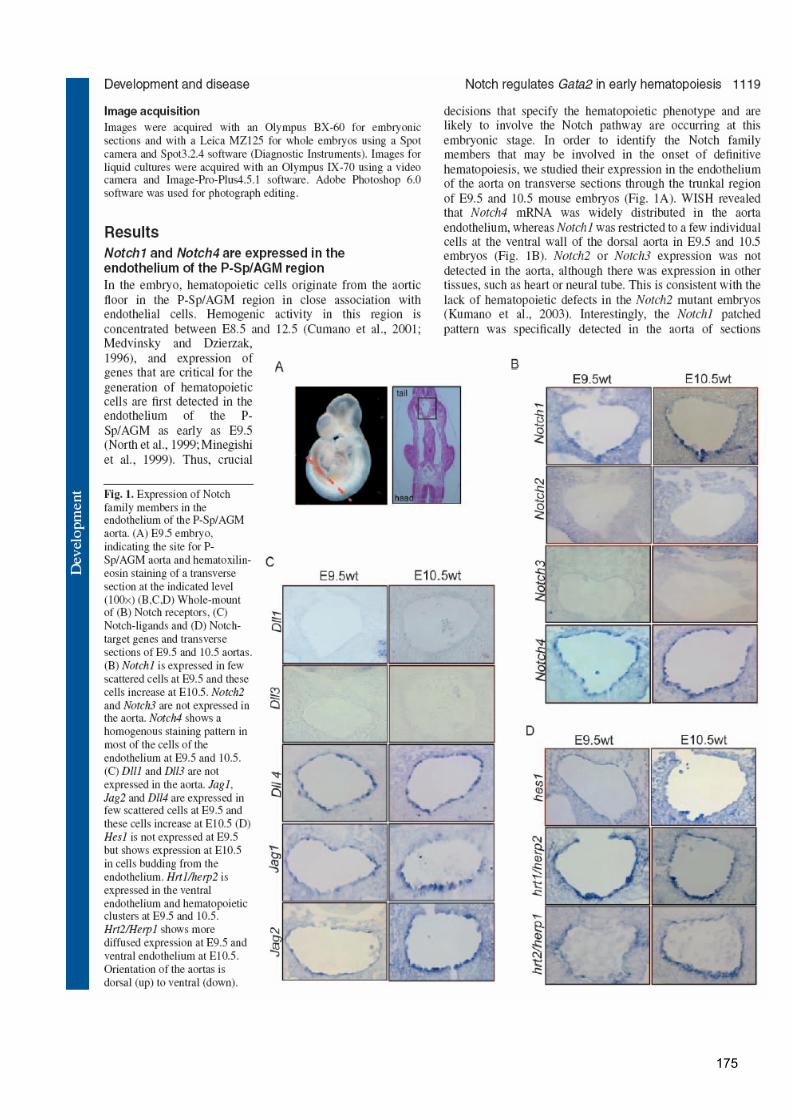
175

176

177

178

179

180
181
182


185
186
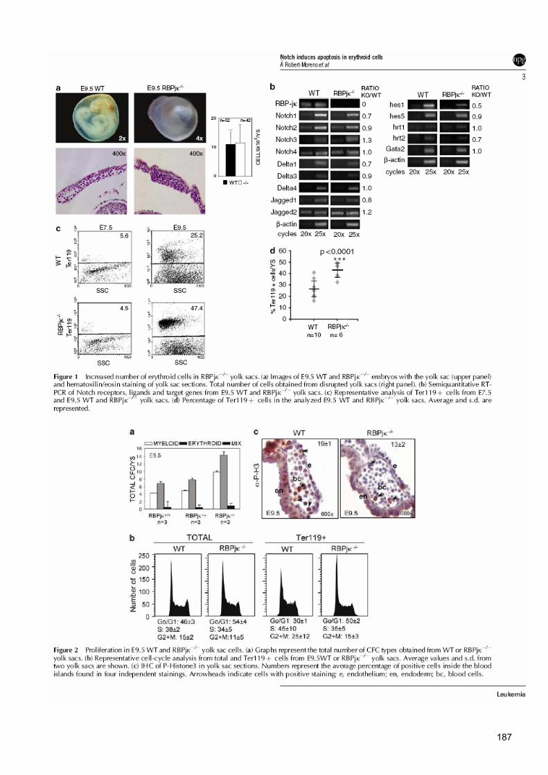
187
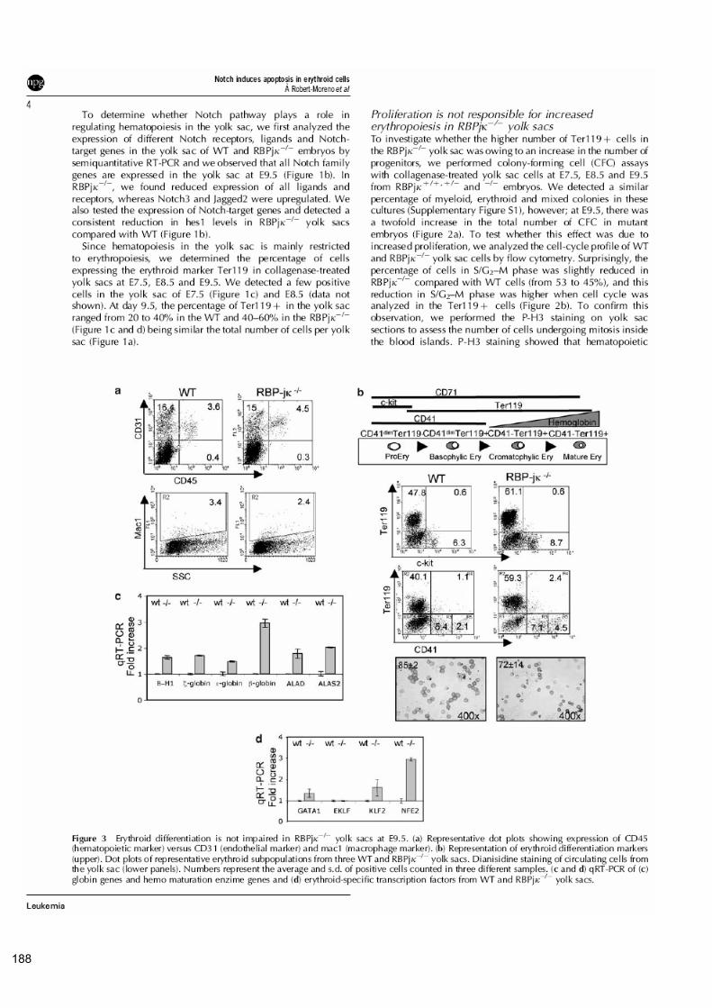
188

189

190

191
192

193
SUPPLEMENTARY INFORMATION
Supplementary Figure S1: Analysis of myeloid and erythroid progenitors from collagenase-treated yolk sacs. (a) Total CFC/YS at
E7.5 and E8.5. (b) Percentage of CFC at E9.5
Supplementary Figure S2: Ter119+ cells from the yolk sac express CD71. (a) Ter119+ cells from WT and mutant yolk sacs. X-
mean of SSC is shown for both samples. (b) Percentage of gated Ter119+ cells expressing CD71. Numbers represent the average
percentage and standard deviation of 3 different yolk sacs.
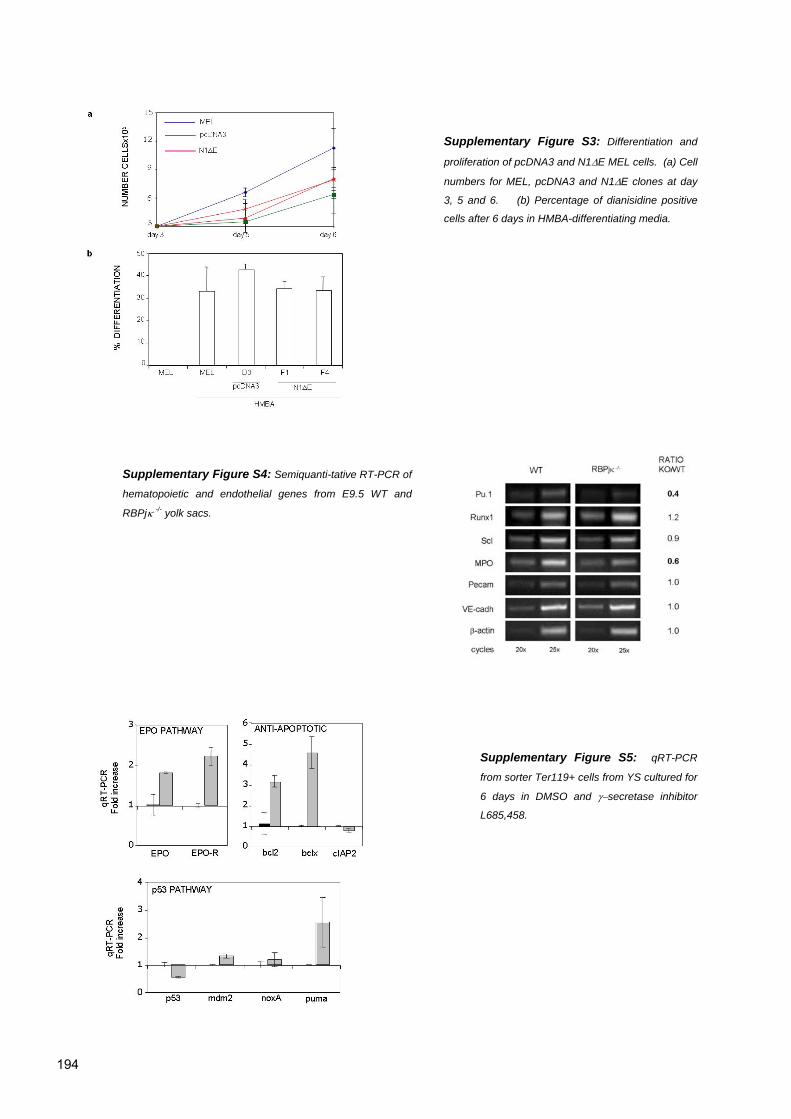
194
Supplementary Figure S3: Differentiation and
proliferation of pcDNA3 and N1∆E MEL cells. (a) Cell
numbers for MEL, pcDNA3 and N1∆E clones at day
3, 5 and 6. (b) Percentage of dianisidine positive
cells after 6 days in HMBA-differentiating media.
Supplementary Figure S4: Semiquanti-tative RT-PCR of
hematopoietic and endothelial genes from E9.5 WT and
RBPjκ -/- yolk sacs.
Supplementary Figure S5: qRT-PCR
from sorter Ter119+ cells from YS cultured for
6 days in DMSO and γ−secretase inhibitor
L685,458.

