role of inositol trisphosphate receptors in autophagy in dt40 cells
TRANSCRIPT

Role of Inositol Trisphosphate Receptors in Autophagy inDT40 Cells□S
Received for publication, February 14, 2010 Published, JBC Papers in Press, March 22, 2010, DOI 10.1074/jbc.M110.114207
M. Tariq Khan and Suresh K. Joseph1
From the Department of Pathology and Cell Biology, Thomas Jefferson University School of Medicine,Philadelphia, Pennsylvania 19107
Previous studies have shown that small interfering RNAknockdown and pharmacological inhibition of inositol 1,4,5-trisphosphate receptors (IP3Rs) stimulate autophagy. We haveinvestigated autophagy in chicken DT40 cell lines containingtargeted deletions of all three IP3R isoforms (triple knock-out(TKO) cells). Using gel shifts of microtubule-associated protein1 light chain 3 as a marker of autophagy, we find that TKO cellshave enhanced basal autophagic flux even under nutrient-re-plete conditions. Stable DT40 cell lines derived from TKO cellscontaining the functionally inactive D2550A IP3R mutant didnot suppress autophagy in the samemanner as wild-type recep-tors. This suggests that the channel function of the receptor isimportant in its regulatory role in autophagy. There were nomarked differences in the phosphorylation state of AMP-acti-vated protein kinase, Akt, or mammalian target of rapamycinbetween wild-type and TKO cells. The amount of immunopre-cipitated complexes of Bcl-2-Beclin-1 and Beclin-1-Vps34 werealso not different between the two cell lines. The major differ-ence notedwas a substantially decreasedmTORC1 kinase activ-ity in TKO cells based on decreased phosphorylation of S6kinase and 4E-BP1. The discharge of intracellular stores withthapsigargin stimulated mTORC1 activity (measured as S6kinase phosphorylation) to a greater extent in wild-type than inTKO cells. We suggest that basal autophagic flux may be nega-tively regulated by IP3R-dependent Ca2� signals acting tomain-tain an elevated mTORC1 activity in wild-type cells and thatCa2� regulation of this enzyme is defective in TKO cells. Theprotective effect of a higher autophagic flux in cells lackingIP3Rsmayplay a role in thedelayed apoptotic responseobservedin these cells.
It is well established that Ca2� has an important regulatoryrole in controlling apoptosis (1–3). Inositol 1,4,5-trisphosphatereceptors (IP3R)2 participate in this pathway at several levels.First, they provide a conduit for the transfer of Ca2� betweenthe ER and the mitochondria to sensitize the mechanism that
facilitates the release of cytochrome c from the mitochondria(4). Second, IP3Rs interact with, and are regulated by, severalproteins that modify apoptotic pathways, including the anti-apoptotic proteins Bcl-2/Bcl-XL (5, 6), cytochrome c (7, 8), andAkt kinase (9–11). Finally, with certain apoptotic stimuli (e.g.staurosporine), IP3Rs support apoptosis independently of thechannel function of the receptor via a mechanism that may belinked to a direct role of IP3Rs in activating Ca2� entry mecha-nisms across the plasma membrane (12).Macroautophagy is a proteolytic process in which cytoplas-
mic constituents (including organelles) are sequestered withindouble-membraned vesicles (autophagosomes) that ultimatelyfusewith lysosomes leading to the degradation of their contents(13). A major physiological regulator of this process is nutrientsupply, although the process is also regulated by various hor-mones and can be dysregulated under pathological conditions(14). The complicated steps involved in autophagosome forma-tion and lysosome fusion involve multiple proteins and regula-tion by many different inputs, including the activities of themTOR pathway and class III phosphatidylinositol 3-kinase.There have been several reports suggesting that Ca2� regulatesthis pathway. Hoyer-Hansen et al. (15) showed that agents thatelevated Ca2� in MCF-7 cells increased the formation of auto-phagosomes and that this was blocked by treatment with theintracellular Ca2� chelator BAPTA-AM. However, others havereported that blocking Ca2� elevations (e.g. with L-type Ca2�
channel antagonists) can enhance autophagy suggesting thatCa2�has an inhibitory effect on autophagy (16). In addition, thedepletion of intracellular stores with thapsigargin has beenreported to have both a stimulatory (15, 17) and inhibitory (16,18, 19) effect on autophagy. Manipulations designed to changethe levels of IP3 in cells (e.g. addition of myo-inositol or Li�)alter the rate of starvation-induced autophagy (17). A specificrole for IP3Rs in the autophagic process was suggested by thefinding that small interfering RNA knockdown or pharmaco-logical blockade of the IP3R with xestospongin B led to anenhancement of autophagy (17). These data suggest that IP3Rsnegatively modulate autophagy. However, it is unclear if thisinvolves the channel function of the IP3R, because some effec-tive agents, such as xestospongin B, had no detectable effects oncytosolic or ER luminal Ca2�(17). Disruption of the single IP3Rgene in Dictyostelium discoideum impairs an autophagic deathpathway (20). The specific autophagic signaling pathway(s)modulated by IP3Rs remains to be identified.DT40 chicken B-cell lines containing targeted deletion of all
three IP3R isoforms (TKO) show a markedly delayed cell deathresponse to various apoptotic stimuli (6, 12, 21).We considered
□S The on-line version of this article (available at http://www.jbc.org) containssupplemental Fig. S1.
1 To whom correspondence should be addressed: Dept. of Pathology and CellBiology, Thomas Jefferson University, Rm. 230A JAH, 1020 Locust St., Phil-adelphia, PA 19107. Tel.: 215-503-1221; Fax: 215-923-6813; E-mail:[email protected].
2 The abbreviations used are: IP3R, inositol 1,4,5-trisphosphate receptor;mTOR, mammalian target of rapamycin; BAPTA-AM, 1,2-bis(2-aminophe-noxy)ethane-N,N,N�,N�-tetraacetic acid tetrakis(acetoxymethyl ester); ER,endoplasmic reticulum; Ab, antibody; STS, staurosporine; TKO, tripleknock-out; WT, wild type; AMPK, AMP kinase.
THE JOURNAL OF BIOLOGICAL CHEMISTRY VOL. 285, NO. 22, pp. 16912–16920, May 28, 2010© 2010 by The American Society for Biochemistry and Molecular Biology, Inc. Printed in the U.S.A.
16912 JOURNAL OF BIOLOGICAL CHEMISTRY VOLUME 285 • NUMBER 22 • MAY 28, 2010
by guest on April 10, 2019
http://ww
w.jbc.org/
Dow
nloaded from

the possibility that adaptive changes in autophagy may haveoccurred in these cells thereby providing a useful experimentalsystem to investigate the role of IP3Rs in autophagy. In thisstudy, we show that TKO cells have a markedly enhanced rateof autophagy compared with wild-type cells, even under nutri-ent-replete conditions. The suppression of autophagy requiredthe Ca2� channel function of the IP3R and was not observed incell lines transfected with the pore-inactivating D2550Amutant. Several key factors that regulate autophagy were com-pared in wild-type and TKO cell lines and were not found to besignificantly different. These included the activity of AMP andAkt kinase. The differences in basal autophagy could also not beaccounted for by altered levels of Beclin-1-Vps34 complexes.Instead, our experiments suggest that altered activity of themTORC1 complex may be one potential mechanism by whichIP3R-mediated Ca2� fluxes could regulate the autophagicpathway.
MATERIALS AND METHODS
Reagents—RPMI 1640 culture media and G418 sulfate(Geneticin) were obtained from Cellgro-Mediatech (Herndon,VA). Staurosporine, rapamycin, bafilomycin, and okadaic acidwere purchased from Sigma. Protogel-stabilized acrylamidesolution was from National Diagnostics (Atlanta, GA). Nitro-cellulose membrane (0.45 �m) was from Bio-Rad. Polyvinyli-dene difluoride membrane (Immobilon-P, 0.45 �m) was pur-chased fromMillipore (Bedford, MA).Antibodies—The following antibodies were obtained from
Cell Signaling (MA): LC3B rabbit polyclonal Ab; phospho-mTOR (Ser-2448) rabbit polyclonal Ab; phospho-mTOR (Ser-2481) rabbit polyclonal Ab; total mTOR rabbit polyclonal Ab;phospho-4E-BP1 (Thr-37/46) (236B4) rabbit monoclonal Ab;phospho-Akt (Ser-473) (587F11) mouse monoclonal Ab; totalAkt (5G3)mousemonoclonalAb; phospho-p70 S6 kinase (Thr-389) (108D2) rabbit monoclonal Ab; total p70 S6 kinase rabbitpolyclonal Ab; Beclin-1 rabbit polyclonal Ab; phospho-AMPK� (Thr-172) rabbit polyclonalAb; and total AMPK� rab-bit polyclonal Ab. Mouse anti-Bcl-2 monoclonal Ab was pur-chased from BD Transduction Laboratories. The rabbitpolyclonal calnexin Ab has been described previously (22).DT40 Cell Culture and Incubation Conditions—Wild-type
(WT) DT40 cells and IP3R triple knock-out (TKO) cells werethe kind gifts of Dr. T. Kurosaki (Kansai Medical University,Moriguchi, Japan). Stable DT40 cells expressing the rat type IIP3R were a gift from Dr. Kevin Foskett (University of Pennsyl-vania). DT40 cells were grown in RPMI 1640 media supple-mented with 10% fetal bovine serum, 1% chicken serum, 100units/ml penicillin, 100 units/ml streptomycin and maintainedat 37 °C in 5% CO2 atmosphere. The stable cell line containingthe inactivating D2550A pore mutation was prepared asdescribed previously (12).DT40 cellswere seeded at a density of104 cells/ml and were used for experiments after 24 h. Therewere no significant differences in the growth rate of the indi-vidual cell lines (data not shown).Measurement of Autophagy—The DT40 cells were treated
with staurosporine or rapamycin and then centrifuged at1,000 � g for 10 min. The cells were washed once in ice-coldphosphate-buffered saline and then lysed in amedium contain-
ing 1% Triton X-100 and 50 mM Tris/HCl, pH 7.8, 150 mM
NaCl, 2 mM sodium orthovanadate, 10 mM sodium pyrophos-phate, 20 mM NaF, 5 nM okadaic acid, and a 1� dilution of acomplete protease inhibitor mixture (Roche Diagnostics). Thelysates were cleared by centrifugation at 10,000 � g. Proteinsamples (25 �g) were run on 15% polyacrylamide gels andtransferred on polyvinylidene difluoridemembranes. Immuno-blotting was done using the LC3B antibody. Only the lowerband of the LC3 doublet (LC3-II) was quantitated because vari-able immunodetection and efficiency of transfer of the upperband (LC3-I) has been observed (23). Electron microscopy ofDT40 cells was carried out as described by Csordas et al. (24).
RESULTS
Autophagy was measured by immunoblotting for the micro-tubule-associated light chain 3 (LC3) protein. The covalentmodification of the cytosolic LC3-I formwith phosphatidyleth-anolamine generates the LC3-II form, which migrates morerapidly on SDS-PAGE (25). The specific association of LC3-IIwith autophagosomes has led to this protein being widely usedas a marker of autophagy (23). Fig. 1A shows a comparison ofLC3-II levels detected by immunoblotting in lysates from wild-type and TKODT40 cells grown under nutrient-replete condi-tions. The data show that basal steady-state levels of LC3-II arelow in wild-type cells but are �4-fold elevated in the TKO cells(Fig. 1B). The induction of apoptosis with staurosporine (STS)over a 6-h period enhanced the levels of LC3-II in wild-typecells and decreased the elevated levels of LC3-II in TKO cells.To determine whether the Ca2� channel function of IP3Rs wasimportant in mediating its inhibitory effects on autophagy, weutilized a stable DT40 cell line expressing the “pore-dead”D2550Amutant of the rat type I IP3R (26). The expression levelof type I IP3R in the mutant has previously been shown to becomparable with wild-type cells (12). The pore-dead cell linealso showed elevated levels of LC3-II that were decreased bySTS treatment as observed with the TKO cells (Fig. 1,A and B).The wild-type DT40 cells used as a control contain all threechicken IP3R isoforms. Levels of LC3-II in pore-dead cellswere also elevated when compared with a stable DT40 cell lineexpressing the wild-type rat type I IP3R (Fig. 1C). We concludethat the channel function of the IP3R is required for suppressingautophagy.It has been pointed out that measurements of the steady-
state levels of LC3-II may not reflect autophagic flux becausethe levels are also determined by the degradation of LC3-IIupon fusion of autophagosomes with lysosomes (27, 28). Auto-phagosome/lysosome fusion can be prevented by bafilomycin(29). A 6-h incubation with 10 nM bafilomycin caused a largeenhancement in LC3-II levels such that no differences betweenWT and TKO cells could be distinguished, although the inhib-itory effect of STS on LC3-II levels in the TKO cells was stillobserved (Fig. 1B). Shorter periods of treatment with bafilomy-cin, which produced submaximal elevations of LC3-II (e.g. after1 h), still revealed differences between wild-type and TKO cells(Fig. 1D). The basic findings using LC3 immunoblotting wereconfirmed by analysis of electron microscope images of DT40cells that showed an enhanced number of debris-containingvacuoles in TKO cells when compared with either wild-type or
IP3Rs and Autophagy
MAY 28, 2010 • VOLUME 285 • NUMBER 22 JOURNAL OF BIOLOGICAL CHEMISTRY 16913
by guest on April 10, 2019
http://ww
w.jbc.org/
Dow
nloaded from

TKO cells stably expressing the rat type I IP3R (supple-mental Fig. S1). These data in DT40 cells are in agreement withprevious findings in other cell types that knockdown or inhibi-tion of IP3Rs enhances autophagic flux (17).
Fig. 2 depicts the canonical pathway of autophagy togetherwith known sites that could potentially be regulated by Ca2�
based on information in the literature. Because IP3Rs have anestablished role in supplying Ca2� to the mitochondria, whichis known to stimulatemitochondrial oxidativemetabolism, it ispossible that IP3R-deficientDT40 cellsmight have an increasedAMP/ATP ratio. This, in turn, could enhance the activity ofAMPK that would stimulate autophagy through the intermedi-ary steps shown in Fig. 2. Calmodulin-dependent kinasekinase-� is an upstream activator of AMPK and has also beenproposed to be a site where cytosolic Ca2� elevations couldstimulate autophagy (15). We therefore compared the activa-tion state of AMPK in wild-type, TKO, and pore-dead DT40cell lines by measuring phosphorylation of the protein at Thr-172 using a phospho-specific Ab. This rabbit polyclonal Ab
recognized a doublet of bands in thechicken cells (Fig. 3) of which onlythe lower band was recognized bythe pan-�-subunit AMPK Ab at theappropriate molecular mass of �63kDa. This AMPK band was not sig-nificantly different between wild-type, TKO, and pore-dead cells. STSdid not affect the phosphorylationstate of the AMPK band but sub-stantially decreased the phosphor-ylation of the upper unknown bandin all three cell lines.Activated Akt kinase inhibits
autophagy (Fig. 2) (30). Analysis ofthe phosphorylation state of Aktkinase using phospho-specific Ser-473 Ab indicates a modest increasein activation of Akt in TKO andpore-dead cells. However, thesechanges are in the opposite direc-tion to account for the stimulatedautophagy in these cell lines. STSdecreased Ser-437 phosphorylationin TKO and pore-dead cell lines butnot in wild-type cells. Thesechanges also do not correlate withthe different effects of STS on auto-phagy in these cell lines. The effectsof Akt and AMPK on autophagy aremediated through a pathway thatinvolves TSC and Rheb proteinsthat regulate the activity of themTORC1 complex (Fig. 2). Highlevels of mTOR kinase suppressautophagy bymechanisms that havenot been fully characterized butinclude the phosphorylation ofAtg1/Ulk kinases that are involved
in the induction of autophagosomes (31). We examined thefollowing two phosphorylation sites onmTOR: Ser-2448 (a sitefor phosphorylation by several kinases, including Akt (32)) andSer-2481 (an autophosphorylation site (33)). The basal phos-phorylation state of neither site was different in the three dif-ferent DT40 cell lines (Fig. 3). However, mutation of serines2448 or 2481 does not alter mTOR kinase activity (32), which isconventionallymeasured bymonitoring the phosphorylation ofits substrates. One such substrate is p70 S6 kinase, which isspecifically phosphorylated bymTORonThr-389 (34). Fig. 4,Aand B, shows that there are substantially lower levels of phos-phorylated S6 kinase in the TKO and pore-dead cells. We con-firmed this finding by examining another mTOR substrate4E-BP1 using anAb directed at Ser-65 (35). As with S6 kinase, alower phosphorylation of 4E-BP1 was observed in TKO andpore-dead cells (Fig. 4A). The phosphorylation state of S6kinase could be restored by stable transfection of the rat type IIP3R into TKO cells and remained elevated in a DT40 cell linecontaining only the endogenous chicken type I isoform
FIGURE 1. Lack of functional IP3Rs in DT40 cells is associated with higher levels of basal autophagy. A, WT,IP3R TKO, and the D2550A pore-inactive (pore-dead) DT40 cells lines were incubated in nutrient-repletemedium with serum in the presence or absence of staurosporine (1 �M) or bafilomycin A (10 nM) for 6 h. Aftertreatment, the cells were lysed and processed for immunoblotting with LC3 Ab as described under “Materialsand Methods.” The same samples were processed on 5% SDS-PAGE to monitor the levels of calnexin used as aloading control. B, levels of the LC3-II band were quantitated densitometrically and normalized to the calnexinlevels. The data shown are the mean � S.E. of 3–5 independent experiments. C, DT40 cell line stably expressingthe wild-type rat type I IP3R (type I) and the functionally inactive D2550A mutant in the rat type I background(pore-dead) were directly compared for basal levels of LC3. D, WT, TKO, and pore-dead cell lines were incubatedwith 10 nM bafilomycin (Baf) A for the indicated times and processed for LC3 immunoblotting as describedabove.
IP3Rs and Autophagy
16914 JOURNAL OF BIOLOGICAL CHEMISTRY VOLUME 285 • NUMBER 22 • MAY 28, 2010
by guest on April 10, 2019
http://ww
w.jbc.org/
Dow
nloaded from

(DKO-1) (Fig. 4C).We suggest that a lowermTORkinase activ-ity in the IP3R-deficient cell lines could account for their higherbasal autophagy.We examined the effects of rapamycin, an inhibitor ofmTOR
(36), on the levels of LC3-II in wild-type and TKO cells (Fig. 5).In both cell lines, 100 nM rapamycin completely inhibitedmTOR activity within 2 h as judged by inhibition of phosphor-ylation of Thr-389 of S6 kinase (Fig. 5A). Rapamycin produceda smaller and slower decrease in Ser-2448 phosphorylation ofmTOR itself, consistent with this site being a relatively insensi-tive indicator of mTOR kinase activity. Rapamycin elevated thelevels of LC3-II in wild-type cells with a peak increase at 4 h,which averaged almost 80% of the elevated levels observed inuntreatedTKOcells (Fig. 5B). Rapamycin addition toTKOcellscaused only a small further increase in LC3-II levels. The resultswith rapamycin are consistent with the hypothesis that differ-ences in the activity of themTORkinase pathway are an impor-tant determinant in setting the different levels of basal autoph-agy observed in wild-type and TKO cells.Another potential site of autophagy regulation by IP3Rs has
been proposed to involve Beclin-1 (37), which stimulates theactivity of the class III phosphatidylinositol 3-kinase Vps34 (31,38). This enzyme has been directly implicated in the formationof autophagosomes from ER membranes (39) and also posi-tively modulates mTOR activity (38). Bcl-2, which inhibitsautophagy by sequestering Beclin-1, can also bind to IP3Rs (40).Thus, the presence or absence of IP3Rs could indirectly regulatethe amount of Beclin-1-Vps34 complex. This was examined in
the experiments shown in Fig. 6. The total levels of Bcl-2 orBeclin-1 were not different in wild-type or TKO cells in thepresence or absence of STS (Fig. 6A). Detecting the coimmu-noprecipitation of Beclin-1 by Bcl-2 Ab was complicated by theclose proximity of the Beclin-1 signal to the heavy chain of IgG.Using a method that minimizes this problem, we found no dif-ferences in Bcl-2-Beclin-1 complexes between wild-type andTKOcells (Fig. 6B). The proportion of Beclin-1 complexedwithVps34, determined by immunoprecipitation with Beclin-1 Ab,was also not different in the two cell lines. There was also noevidence that IP3Rs were directly or indirectly complexed withVps34 (Fig. 6C).A mechanism that would be consistent with our experimen-
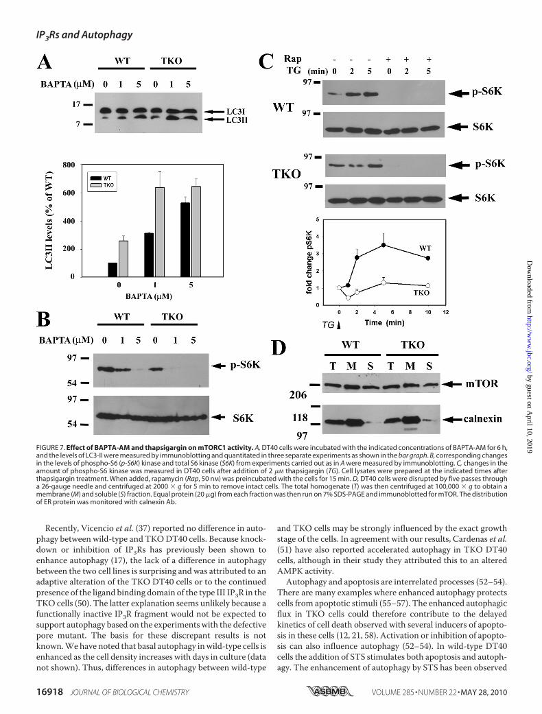
tal observations is that mTORC1 activity in intact cells isdependent on Ca2� signaling. In unstimulated DT40 cells, theloading of BAPTA-AM is sufficient to enhance LC3-II levels inboth wild-type and TKO cells, which is consistent with restingintracellular Ca2� exerting a tonic negative effect on autophagy(Fig. 7A). Loading with BAPTA-AM also decreased the basalphosphorylation state of S6 kinase in both wild-type and TKOcells (Fig. 7B) confirming observations made in other cell types(41, 42). In wild-type DT40 cells, the addition of thapsigargin tomobilize intracellular Ca2� stores caused a robust and rapidincrease in phospho-S6 kinase levels (�3-fold in 2 min), and
FIGURE 2. Possible sites of regulation of the canonical pathway of auto-phagy by IP3 receptors and Ca2�. The middle box shows several of themolecular complexes that participate in autophagy. The pathways by whichphysiological factors such as nutrient availability and growth factors (indi-cated in red) modulate autophagy are shown schematically. Possible sites atwhich IP3Rs or elevated cytosolic Ca2� could also modulate this pathway areindicated in blue. The specific mechanisms are as follows: 1, transfer of Ca2�
from the ER to the mitochondria mediated by IP3Rs could contribute to themaintenance of an elevated “energy state” (high ATP/AMP ratio); 2, Ca2� act-ing through calmodulin-dependent kinase kinase-� (CaMKK-�) can activateAMPK (15); 3, Vps34 has also been suggested to be a CaM-dependent enzyme(46); 4, Bcl-2, by binding IP3Rs, can influence the amount bound to the Beclin-1-Vps34 complex (37) ; and 5, activation of calpains has been proposed toinhibit autophagy (16). The only target of calpains in the autophagic pathwaythat has been described is Atg5 (63). Abbreviations used: FIP200, focal adhe-sion kinase-interacting protein; Ulk, Unc51-like kinase; TSC, tuberous sclerosiscomplex protein; Rheb, Ras homology enriched in brain protein; S6K-1, p70ribosomal protein S6 kinase-1; 4E-BP1, eukaryotic translation initiation factor4E-binding protein 1.
FIGURE 3. Autophagy is independent of the phosphorylation state ofAMPK, Akt, or mTOR. WT, IP3R TKO, and the D2550A pore-inactive (pore-dead) DT40 cells lines were incubated in nutrient-replete medium with serumin the presence or absence of staurosporine (1 �M) for 6 h. After treatment, thecells were lysed in a buffer containing protein phosphatase inhibitors andprocessed for immunoblotting with the indicated phospho-specific Abs asdescribed under “Materials and Methods.” The data shown is representativeof three experiments. * indicates an unknown band.
IP3Rs and Autophagy
MAY 28, 2010 • VOLUME 285 • NUMBER 22 JOURNAL OF BIOLOGICAL CHEMISTRY 16915
by guest on April 10, 2019
http://ww
w.jbc.org/
Dow
nloaded from

levels were entirely suppressed by pretreatment with rapamy-cin (Fig. 7C). By contrast, thapsigargin did not cause significantchanges in pS6K levels in TKO cells over a 10-min period (Fig.7C). Because the discharge of intracellular Ca2� stores medi-ated by thapsigargin is of similar kinetics andmagnitude in bothcell lines (21), it must be concluded that themTORC1 complexhas a diminished Ca2� sensitivity in the TKO cells. The possi-bility that the localization of mTOR protein may be an impor-
tant factor in these experiments was explored in Fig. 7D. Thedata show that a large fraction of mTOR was localized in themicrosomal membranes, consistent with previous reports thatER localization signals are found in the HEAT domains ofmTOR (43). However, localization was not significantly differ-ent in WT or TKO cells (Fig. 7D). In addition, localization wasnot altered by treatment with thapsigargin or A23187 and noevidence for co-immunoprecipitation of mTOR with type IIP3R was obtained (data not shown).
DISCUSSION
In this study, we have documented an enhanced autophagicflux inDT40 cell lines inwhich IP3Rs are absent or are nonfunc-tional. We hypothesize that the normal responses of wild-typecells tomany environmental cues, including growth factors andnutrients, may require periodic elevations of cytosolic Ca2�
that would be severely impaired in cell lines lacking functionalIP3Rs. Thus, the TKO/pore-dead cells may exhibit behaviorthat is characteristic of nutrient insufficiency. In agreementwith this, the autophagic response of TKO cells to removal ofamino acids from the culture medium was diminished whencompared with wild-type cells (data not shown). The depen-
FIGURE 4. Higher autophagic flux in IP3R TKO and pore-dead cell lines isassociated with a reduced activity of mTOR. A, WT, IP3R TKO, and theD2550A pore-inactive (pore-dead) DT40 cells lines were incubated in nutrient-replete medium with serum in the presence or absence of staurosporine (1�M) for 6 h. After treatment, the cells were lysed in a buffer containing proteinphosphatase inhibitors and processed for immunoblotting with the indi-cated Abs against p70 phospho-S6 kinase (S6K) and 4E-BP1, two key sub-strates of the mTORC1 kinase complex. The data shown are representative ofthree experiments. B, data from three experiments were quantitated withdensitometry and are shown as the means � S.E. with the wild-type levels setto 100%. C, amount of phospho- and total S6 kinase was measured in lysatesfrom wild-type and TKO DT40 cells as well as cells expressing the chicken(DKO-1) or rat type 1 IP3Rs.
FIGURE 5. Effect of mTORC1 inhibition with rapamycin on autophagy inIP3R wild-type and TKO cells. A, wild-type and TKO DT40 cells were incu-bated with 100 nM rapamycin for the indicated times. Cell-free lysates wereimmunoblotted for LC3, phosphorylated mTOR, and S6 kinase and totalmTOR, and S6 kinase. The immunoblots shown are representative of threeexperiments. B, is the quantitation of the levels of LC3-II at the 2-h time pointwith the levels of the untreated wild-type cells used as the reference control(100%).
IP3Rs and Autophagy
16916 JOURNAL OF BIOLOGICAL CHEMISTRY VOLUME 285 • NUMBER 22 • MAY 28, 2010
by guest on April 10, 2019
http://ww
w.jbc.org/
Dow
nloaded from

dence of autophagy on IP3Rs is not a unique characteristic ofDT40 cells because it has been observed in other cell types inwhich type I or type III IP3R isoforms have been knocked downby small interfering RNA methods or when channel functionwas inhibited by xestospongin B (17).There are several potential sites at which the IP3R protein (or
the Ca2� it translocates) could regulate the canonical autoph-agic pathway (Fig. 2). The energy status of the cell, sensed byAMPK, is a prime regulator of autophagy. The shuttling ofCa2�
from the ER to themitochondria is a key function of IP3Rs. Theabsence of IP3Rs could potentially modify the AMP/ATP ratioof the cell and secondarily influence AMPK activity. In anexperimental model where chronic elevation of Ca2� stimu-lates autophagy, it has been suggested that Ca2�/calmodulin-dependent kinase kinase-�, an upstream regulator of AMPKphosphorylation, may be involved (15). However, we did notobserve any differences in the phosphorylation state of AMPKin wild-type and TKODT40 cells. Growth factors such as insu-lin that inhibit autophagy act through Akt kinase. Again, weobserved no differences in the phosphorylation state of Akt.The anti-apoptotic protein Bcl-2 inhibits autophagy when spe-cifically targeted to ERmembranes (17). The inhibition of auto-
phagy is thought to result from the binding of Bcl-2 to Beclin-1,which is a regulator of Vps34, a class III phosphatidylinositol3-kinase. Because IP3Rs also bind Bcl-2, it has been suggestedthat the effects of IP3R knockdown or IP3R inhibition with xes-tospongin B may be related to an altered availability of Bcl-2(17, 37). However, we observed no differences in beclin-1-Vps34 complexes in wild-type and TKO cells. In addition, theabsence of IP3Rs should make more Bcl-2 available for inhibit-ing autophagy, whereas a stimulation of autophagy is what isexperimentally observed.The main difference between wild-type and TKO cells that
could account for the differences in autophagic flux is thereduced basal activity of themTORC1 complex in TKOcells, asmeasured by a reduced phosphorylation of its substrates S6kinase and 4E-BP1. There is evidence in the literature to suggestthat mTORC1 activity in intact cells is regulated by Ca2�. Sev-eral studies have reported that enzyme activity is enhanced bythapsigargin or Ca2�-mobilizing stimuli and is inhibited byBAPTA-AM loading of cells (41, 42, 44, 45). The absence ofIP3Rs in TKO cells would be expected to diminish Ca2� signalsthat act to maintain mTORC1 activity. However, the availabil-ity of Ca2� is not the only factor that is altered because theresponse to Ca2� mobilized by thapsigargin is also lost in TKOcells (Fig. 7). This suggests that Ca2� regulation of themTORC1 complex is somehow different in TKO cells. Unfor-tunately, the mechanism(s) by which Ca2� regulates thisenzyme are not well characterized. One possibility is that thecomponents of the mTORC1 complex are assembled or local-ized differently in the IP3R-deficient cells. Although we notedno differences in the membrane localization of the mTOR pro-tein in TKO cells, it is possible that the other proteins associ-ated with the mTORC1 complex may be targeted differently(e.g. Raptor, mLST8, and Rheb). mTORC1 activity is positivelyregulated byVps34, which has been proposed to be activated byCa2�/CaM (46, 47). Therefore, an alternative possibility is thata decreased activity of mTORC1 in TKO cells is secondary to adecreased activity of the pool of Vps34 involved in regulatingautophagy. Amore direct role for Vps34 in autophagy has beensuggested by recent studies indicating thatVps34 is recruited tospecific ER structures that form a platform for the assembly ofautophagosomes (39). Thus, it is possible that the localizedrelease of Ca2� from IP3Rs could directly impede some earlyER-dependent step(s) in autophagosome biogenesis.Previous studies that have examined a role for IP3 or Ca2� in
autophagy have concluded that the regulatory effects are inde-pendent of mTOR activation. This was found for lithium(which induces autophagy by a mechanism involving depletionof IP3 levels (48)), for xestospongin-B (37), and for pharmaco-logical compounds that induce autophagy by a mechanisminvolving inhibition of calpain activity (16). The status of themTOR pathway was not examined in studies where autophagywas induced by small interfering RNA knockdown of IP3Rs(17). Infection by Toxoplasma gondii stimulates autophagy inthe host cell by a mechanism involving Ca2� elevation withoutan associated change in mTOR signaling (49). The presence ofmultiple regulatory mechanisms is not surprising in view of thecomplexity of the autophagic pathway and the differences incell types and stimuli.
FIGURE 6. Complex formation of the autophagy regulator Beclin-1 withBcl-2 and Vps34 in IP3R wild-type and TKO cells. A, levels of Bcl-2 andBeclin-1 in wild-type (WT) and TKO DT40 cell lysates (30 �g) were measuredby immunoblotting. B, WT and TKO DT40 cell lysates were immunoprecipi-tated (IP) with a monoclonal Bcl-2 Ab, and the immunoprecipitates wereprobed by immunoblotting for the presence of Beclin-1. Immunoprecipita-tion and immunoblotting were carried out according to the instructions forthe TrueBlot kit (eBiosciences, San Diego), which minimizes detection of theIgG band that runs just below the Beclin-1 band. A sample containing bufferalone and Bcl-2 mAb was used to verify the effectiveness of the method.C, Abs to IP3R and Beclin-1 were used to immunoprecipitate 0.5 mg of WT andTKO DT40 cell lysate. The immunoprecipitates were probed by immunoblot-ting for the presence of Vps34. All data are representative of 2 or 3experiments.
IP3Rs and Autophagy
MAY 28, 2010 • VOLUME 285 • NUMBER 22 JOURNAL OF BIOLOGICAL CHEMISTRY 16917
by guest on April 10, 2019
http://ww
w.jbc.org/
Dow
nloaded from
Recently, Vicencio et al. (37) reported no difference in auto-phagy between wild-type and TKODT40 cells. Because knock-down or inhibition of IP3Rs has previously been shown toenhance autophagy (17), the lack of a difference in autophagybetween the two cell lines is surprising and was attributed to anadaptive alteration of the TKO DT40 cells or to the continuedpresence of the ligand binding domain of the type III IP3R in theTKO cells (50). The latter explanation seems unlikely because afunctionally inactive IP3R fragment would not be expected tosupport autophagy based on the experiments with the defectivepore mutant. The basis for these discrepant results is notknown.Wehave noted that basal autophagy inwild-type cells isenhanced as the cell density increases with days in culture (datanot shown). Thus, differences in autophagy between wild-type
and TKO cells may be strongly influenced by the exact growthstage of the cells. In agreement with our results, Cardenas et al.(51) have also reported accelerated autophagy in TKO DT40cells, although in their study they attributed this to an alteredAMPK activity.Autophagy and apoptosis are interrelated processes (52–54).
There are many examples where enhanced autophagy protectscells from apoptotic stimuli (55–57). The enhanced autophagicflux in TKO cells could therefore contribute to the delayedkinetics of cell death observed with several inducers of apopto-sis in these cells (12, 21, 58). Activation or inhibition of apopto-sis can also influence autophagy (52–54). In wild-type DT40cells the addition of STS stimulates both apoptosis and autoph-agy. The enhancement of autophagy by STS has been observed
FIGURE 7. Effect of BAPTA-AM and thapsigargin on mTORC1 activity. A, DT40 cells were incubated with the indicated concentrations of BAPTA-AM for 6 h,and the levels of LC3-II were measured by immunoblotting and quantitated in three separate experiments as shown in the bar graph. B, corresponding changesin the levels of phospho-S6 (p-S6K) kinase and total S6 kinase (S6K) from experiments carried out as in A were measured by immunoblotting. C, changes in theamount of phospho-S6 kinase was measured in DT40 cells after addition of 2 �M thapsigargin (TG). Cell lysates were prepared at the indicated times afterthapsigargin treatment. When added, rapamycin (Rap, 50 nM) was preincubated with the cells for 15 min. D, DT40 cells were disrupted by five passes througha 26-gauge needle and centrifuged at 2000 � g for 5 min to remove intact cells. The total homogenate (T) was then centrifuged at 100,000 � g to obtain amembrane (M) and soluble (S) fraction. Equal protein (20 �g) from each fraction was then run on 7% SDS-PAGE and immunoblotted for mTOR. The distributionof ER protein was monitored with calnexin Ab.
IP3Rs and Autophagy
16918 JOURNAL OF BIOLOGICAL CHEMISTRY VOLUME 285 • NUMBER 22 • MAY 28, 2010
by guest on April 10, 2019
http://ww
w.jbc.org/
Dow
nloaded from

in various cell lines, but the underlying mechanism is unknown(59). Presumably, the action of STS as a protein kinase inhibitorcould lead to a decreased phosphorylation of key proteins in theautophagic pathway that are regulated by phosphorylation, e.g.the complex of the mammalian homologues of Atg1 (Ulk1/2)/Atg13/FIP200 (49), Bcl-2 (60), or Beclin-1 (61). In addition, theactivation of pro-apoptotic proteins, such as Bad, could dimin-ish the Bcl-2-Bclxl complexed to Beclin-1 (62). In contrast tothe stimulation of autophagy seen in wild-type cells, the addi-tion of STS inhibited the elevated levels of autophagy seen inTKO cells. This suggests that regulation by STS is complex, andits stimulatory or inhibitory mode of actionmust be influencedby the availability of Ca2� signaling pathways. Ca2� elevationhas been suggested to either stimulate (15) or inhibit autophagy(16). Our results are more consistent with Ca2� having aninhibitory role. In addition to the calmodulin- and calpain-de-pendent mechanisms mentioned previously, the possible rolesof Ca2�-dependent protein kinases or protein phosphataseshave not been investigated. A more complete understanding ofthe role of Ca2�will require further studies to assess the relativeimportance of potential Ca2� sensors and their sites of action inthe autophagic pathway.
REFERENCES1. Orrenius, S., Zhivotovsky, B., and Nicotera, P. (2003) Nat. Rev. Mol. Cell
Biol. 4, 552–5652. Joseph, S. K., and Hajnoczky, G. (2007) Apoptosis 12, 951–9683. Pinton, P., Giorgi, C., Siviero, R., Zecchini, E., and Rizzuto, R. (2008) On-
cogene 27, 6407–64184. Szalai, G., Krishnamurthy, R., and Hajnoczky, G. (1999) EMBO J. 18,
6349–63615. Rong, Y. P., Aromolaran, A. S., Bultynck, G., Zhong, F., Li, X., McColl, K.,
Matsuyama, S., Herlitze, S., Roderick, H. L., Bootman, M. D., Mignery,G. A., Parys, J. B., De Smedt, H., andDistelhorst, C.W. (2008)Mol. Cell 31,255–265
6. Li, C., Wang, X., Vais, H., Thompson, C. B., Foskett, J. K., and White, C.(2007) Proc. Natl. Acad. Sci. U.S.A. 104, 12565–12570
7. Boehning, D., Patterson, R. L., Sedaghat, L., Glebova, N. O., Kurosaki, T.,and Snyder, S. H. (2003) Nat. Cell Biol. 5, 1051–1061
8. Boehning, D., van Rossum, D. B., Patterson, R. L., and Snyder, S. H. (2005)Proc. Natl. Acad. Sci. U.S.A. 102, 1466–1471
9. Khan,M.T.,Wagner, L., 2nd, Yule, D. I., Bhanumathy, C., and Joseph, S. K.(2006) J. Biol. Chem. 281, 3731–3737
10. Szado, T., Vanderheyden, V., Parys, J. B., De Smedt, H., Rietdorf, K.,Kotelevets, L., Chastre, E., Khan, F., Landegren, U., Soderberg, O., Boot-man, M. D., and Roderick, H. L. (2008) Proc. Natl. Acad. Sci. U.S.A. 105,2427–2432
11. Marchi, S., Rimessi, A., Giorgi, C., Baldini, C., Ferroni, L., Rizzuto, R., andPinton, P. (2008) Biochem. Biophys. Res. Commun. 375, 501–505
12. Khan, M. T., Bhanumathy, C. D., Schug, Z. T., and Joseph, S. K. (2007)J. Biol. Chem. 282, 32983–32990
13. Eskelinen, E. L. (2008) Int. Rev. Cell Mol. Biol. 266, 207–24714. Mizushima, N., and Klionsky, D. J. (2007) Annu. Rev. Nutr. 27, 19–4015. Høyer-Hansen, M., Bastholm, L., Szyniarowski, P., Campanella, M., Sz-
abadkai, G., Farkas, T., Bianchi, K., Fehrenbacher,N., Elling, F., Rizzuto, R.,Mathiasen, I. S., and Jaattela, M. (2007)Mol. Cell 25, 193–205
16. Williams, A., Sarkar, S., Cuddon, P., Ttofi, E. K., Saiki, S., Siddiqi, F. H.,Jahreiss, L., Fleming, A., Pask, D., Goldsmith, P., O’Kane, C. J., Floto, R. A.,and Rubinsztein, D. C. (2008) Nat. Chem. Biol. 4, 295–305
17. Criollo, A., Maiuri, M. C., Tasdemir, E., Vitale, I., Fiebig, A. A., Andrews,D., Molgo, J., Díaz, J., Lavandero, S., Harper, F., Pierron, G., di Stefano, D.,Rizzuto, R., Szabadkai, G., and Kroemer, G. (2007) Cell Death Differ. 14,1029–1039
18. Gordon, P. B., Holen, I., Fosse, M., Røtnes, J. S., and Seglen, P. O. (1993)
J. Biol. Chem. 268, 26107–2611219. Brady, N. R., Hamacher-Brady, A., Yuan, H., and Gottlieb, R. A. (2007)
FEBS J. 274, 3184–319720. Lam, D., Kosta, A., Luciani, M. F., and Golstein, P. (2008) Mol. Biol. Cell
19, 691–70021. Sugawara, H., Kurosaki, M., Takata, M., and Kurosaki, T. (1997) EMBO J.
16, 3078–308822. Joseph, S. K., Boehning, D., Bokkala, S., Watkins, R., andWidjaja, J. (1999)
Biochem. J. 342, 153–16123. Mizushima, N., and Yoshimori, T. (2007) Autophagy 3, 542–54524. Csordas, G., Renken, C., Varnai, P., Walter, L., Weaver, D., Buttle, K. F.,
Balla, T., Mannella, C. A., and Hajnoczky, G. (2006) J. Cell Biol. 174,915–921
25. Kabeya, Y., Mizushima, N., Yamamoto, A., Oshitani-Okamoto, S., Oh-sumi, Y., and Yoshimori, T. (2004) J. Cell Sci. 117, 2805–2812
26. Boehning, D., and Joseph, S. K. (2000) J. Biol. Chem. 275, 21492–2149927. Tanida, I., Minematsu-Ikeguchi, N., Ueno, T., and Kominami, E. (2005)
Autophagy 1, 84–9128. Klionsky, D. J., Abeliovich, H., Agostinis, P., Agrawal, D. K., Aliev, G.,
Askew, D. S., Baba, M., Baehrecke, E. H., Bahr, B. A., Ballabio, A., Bamber,B. A., Bassham, D. C., Bergamini, E., Bi, X., Biard-Piechaczyk, M., Blum,J. S., Bredesen, D. E., Brodsky, J. L., Brumell, J. H., Brunk, U. T., Bursch,W.,Camougrand, N., Cebollero, E., Cecconi, F., Chen, Y., Chin, L. S., Choi, A.,Chu, C. T., Chung, J., Clarke, P. G., Clark, R. S., Clarke, S. G., Clave, C.,Cleveland, J. L., Codogno, P., Colombo, M. I., Coto-Montes, A., Cregg,J.M., Cuervo, A.M., Debnath, J., Demarchi, F., Dennis, P. B., Dennis, P. A.,Deretic, V., Devenish, R. J., Di Sano, F., Dice, J. F., Difiglia, M., Dinesh-Kumar, S., Distelhorst, C.W., Djavaheri-Mergny,M., Dorsey, F. C., Droge,W., Dron, M., Dunn, W. A., Jr., Duszenko, M., Eissa, N. T., Elazar, Z.,Esclatine, A., Eskelinen, E. L., Fesus, L., Finley, K. D., Fuentes, J. M., Fueyo,J., Fujisaki, K., Galliot, B., Gao, F. B., Gewirtz, D.A., Gibson, S. B., Gohla, A.,Goldberg, A. L., Gonzalez, R., Gonzalez-Estevez, C., Gorski, S., Gottlieb,R. A., Haussinger, D., He, Y.W.,Heidenreich, K., Hill, J. A., Høyer-Hansen,M., Hu, X., Huang,W. P., Iwasaki, A., Jaattela,M., Jackson,W. T., Jiang, X.,Jin, S., Johansen, T., Jung, J. U., Kadowaki,M., Kang, C., Kelekar, A., Kessel,D. H., Kiel, J. A., Kim, H. P., Kimchi, A., Kinsella, T. J., Kiselyov, K., Kita-moto, K., Knecht, E., Komatsu,M., Kominami, E., Kondo, S., Kovacs, A. L.,Kroemer, G., Kuan, C. Y., Kumar, R., Kundu, M., Landry, J., Laporte, M.,Le,W., Lei, H. Y., Lenardo,M. J., Levine, B., Lieberman, A., Lim, K. L., Lin,F. C., Liou, W., Liu, L. F., Lopez-Berestein, G., Lopez-Otín, C., Lu, B.,Macleod, K. F., Malorni, W., Martinet, W., Matsuoka, K., Mautner, J.,Meijer, A. J., Melendez, A., Michels, P., Miotto, G., Mistiaen, W. P., Mi-zushima, N., Mograbi, B., Monastyrska, I., Moore, M. N., Moreira, P. I.,Moriyasu, Y., Motyl, T., Munz, C., Murphy, L. O., Naqvi, N. I., Neufeld,T. P., Nishino, I., Nixon, R. A., Noda, T., Nurnberg, B., Ogawa, M., Olein-ick, N. L., Olsen, L. J., Ozpolat, B., Paglin, S., Palmer, G. E., Papassideri, I.,Parkes, M., Perlmutter, D. H., Perry, G., Piacentini, M., Pinkas-Kramarski,R., Prescott, M., Proikas-Cezanne, T., Raben, N., Rami, A., Reggiori, F.,Rohrer, B., Rubinsztein, D. C., Ryan, K. M., Sadoshima, J., Sakagami, H.,Sakai, Y., Sandri, M., Sasakawa, C., Sass, M., Schneider, C., Seglen, P. O.,Seleverstov, O., Settleman, J., Shacka, J. J., Shapiro, I. M., Sibirny, A., Silva-Zacarin, E. C., Simon, H. U., Simone, C., Simonsen, A., Smith, M. A.,Spanel-Borowski, K., Srinivas, V., Steeves, M., Stenmark, H., Stromhaug,P. E., Subauste, C. S., Sugimoto, S., Sulzer, D., Suzuki, T., Swanson, M. S.,Tabas, I., Takeshita, F., Talbot, N. J., Talloczy, Z., Tanaka, K., Tanaka, K.,Tanida, I., Taylor, G. S., Taylor, J. P., Terman, A., Tettamanti, G., Thomp-son, C. B., Thumm, M., Tolkovsky, A. M., Tooze, S. A., Truant, R., Tu-manovska, L. V., Uchiyama, Y., Ueno, T., Uzcategui, N. L., van der Klei, I.,Vaquero, E. C., Vellai, T., Vogel, M. W., Wang, H. G., Webster, P., Wiley,J. W., Xi, Z., Xiao, G., Yahalom, J., Yang, J. M., Yap, G., Yin, X. M., Yoshi-mori, T., Yu, L., Yue, Z., Yuzaki, M., Zabirnyk, O., Zheng, X., Zhu, X., andDeter, R. L. (2008) Autophagy 4, 151–175
29. Yamamoto, A., Tagawa, Y., Yoshimori, T., Moriyama, Y., Masaki, R., andTashiro, Y. (1998) Cell Struct. Funct. 23, 33–42
30. Degtyarev, M., De Maziere, A., Orr, C., Lin, J., Lee, B. B., Tien, J. Y., Prior,W.W., vanDijk, S.,Wu, H., Gray, D. C., Davis, D. P., Stern, H.M.,Murray,L. J., Hoeflich, K. P., Klumperman, J., Friedman, L. S., and Lin, K. (2008)J. Cell Biol. 183, 101–116
IP3Rs and Autophagy
MAY 28, 2010 • VOLUME 285 • NUMBER 22 JOURNAL OF BIOLOGICAL CHEMISTRY 16919
by guest on April 10, 2019
http://ww
w.jbc.org/
Dow
nloaded from

31. Pattingre, S., Espert, L., Biard-Piechaczyk, M., and Codogno, P. (2008)Biochimie 90, 313–323
32. Sekuliæ, A., Hudson, C. C., Homme, J. L., Yin, P., Otterness, D. M., Kar-nitz, L. M., and Abraham, R. T. (2000) Cancer Res. 60, 3504–3513
33. Peterson, R. T., Beal, P. A., Comb, M. J., and Schreiber, S. L. (2000) J. Biol.Chem. 275, 7416–7423
34. Kim, D. H., Sarbassov, D. D., Ali, S. M., King, J. E., Latek, R. R., Erdjument-Bromage, H., Tempst, P., and Sabatini, D. M. (2002) Cell 110, 163–175
35. Gingras, A. C., Gygi, S. P., Raught, B., Polakiewicz, R. D., Abraham, R. T.,Hoekstra, M. F., Aebersold, R., and Sonenberg, N. (1999) Genes Dev. 13,1422–1437
36. Corradetti, M. N., and Guan, K. L. (2006) Oncogene 25, 6347–636037. Vicencio, J. M., Ortiz, C., Criollo, A., Jones, A. W., Kepp, O., Galluzzi, L.,
Joza, N., Vitale, I., Morselli, E., Tailler, M., Castedo, M., Maiuri, M. C.,Molgo, J., Szabadkai, G., Lavandero, S., and Kroemer, G. (2009)Cell DeathDiffer. 16, 1006–1017
38. Backer, J. M. (2008) Biochem. J. 410, 1–1739. Axe, E. L., Walker, S. A., Manifava, M., Chandra, P., Roderick, H. L.,
Habermann, A., Griffiths, G., and Ktistakis, N. T. (2008) J. Cell Biol. 182,685–701
40. Rong, Y. P., Barr, P., Yee, V. C., and Distelhorst, C. W. (2009) Biochim.Biophys. Acta 1793, 971–978
41. Graves, L. M., He, Y., Lambert, J., Hunter, D., Li, X., and Earp, H. S. (1997)J. Biol. Chem. 272, 1920–1928
42. Sarbassov, D. D., and Sabatini, D. M. (2005) J. Biol. Chem. 280,39505–39509
43. Liu, X., and Zheng, X. F. (2007)Mol. Biol. Cell 18, 1073–108244. Altamirano, F., Oyarce, C., Silva, P., Toyos, M., Wilson, C., Lavandero, S.,
Uhlen, P., and Estrada, M. (2009) J. Endocrinol. 202, 299–30745. Markova, B., Albers, C., Breitenbuecher, F., Melo, J. V., Brummendorf,
T. H., Heidel, F., Lipka, D., Duyster, J., Huber, C., and Fischer, T. (2010)Oncogene 29, 739–751
46. Gulati, P., Gaspers, L. D., Dann, S. G., Joaquin, M., Nobukuni, T., Natt, F.,Kozma, S. C., Thomas, A. P., and Thomas, G. (2008) Cell Metab. 7,
456–46547. Yan, Y., Flinn, R. J.,Wu,H., Schnur, R. S., andBacker, J.M. (2009)Biochem.
J. 417, 747–75548. Sarkar, S., Floto, R. A., Berger, Z., Imarisio, S., Cordenier, A., Pasco, M.,
Cook, L. J., and Rubinsztein, D. C. (2005) J. Cell Biol. 170, 1101–111149. Wang, Y., Weiss, L. M., and Orlofsky, A. (2009) J. Biol. Chem. 284,
1694–170150. Guillemette, J., Caron, A. Z., Regimbald-Dumas, Y., Arguin, G., Mignery,
G. A., Boulay, G., and Guillemette, G. (2005) Cell Calcium 37, 97–10451. Cardenas, C., Cheung, K. H., Yang, J., Vais, H., and Foskett, J. K. (2008)
J. Gen. Physiol. 132, 24a52. Maiuri, M. C., Zalckvar, E., Kimchi, A., and Kroemer, G. (2007) Nat. Rev.
Mol. Cell Biol. 8, 741–75253. Ferraro, E., and Cecconi, F. (2007) Arch. Biochem. Biophys. 462, 210–21954. Thorburn, A. (2008) Apoptosis 13, 1–955. Yang, C., Kaushal, V., Shah, S. V., and Kaushal, G. P. (2008) Am. J. Physiol.
Renal Physiol. 294, F777–F78756. Wu, Y. T., Tan, H. L., Huang, Q., Kim, Y. S., Pan, N., Ong,W. Y., Liu, Z. G.,
Ong, C. N., and Shen, H. M. (2008) Autophagy 4, 457–46657. Abedin, M. J., Wang, D., McDonnell, M. A., Lehmann, U., and Kelekar, A.
(2007) Cell Death Differ. 14, 500–51058. Assefa, Z., Bultynck, G., Szlufcik, K., Nadif, Kasri, N., Vermassen, E., Goris,
J., Missiaen, L., Callewaert, G., Parys, J. B., and De Smedt, H. (2004) J. Biol.Chem. 279, 43227–43236
59. Ciechomska, I. A., Goemans, C. G., and Tolkovsky, A. M. (2008)MethodsMol. Biol. 445, 175–193
60. Pattingre, S., Bauvy, C., Carpentier, S., Levade, T., Levine, B., and Co-dogno, P. (2009) J. Biol. Chem. 284, 2719–2728
61. Zalckvar, E., Berissi, H., Eisenstein, M., and Kimchi, A. (2009) Autophagy5, 720–722
62. Levine, B., Sinha, S., and Kroemer, G. (2008) Autophagy 4, 600–60663. Yousefi, S., Perozzo, R., Schmid, I., Ziemiecki, A., Schaffner, T., Scapozza,
L., Brunner, T., and Simon, H. U. (2006) Nat. Cell Biol. 8, 1124–1132
IP3Rs and Autophagy
16920 JOURNAL OF BIOLOGICAL CHEMISTRY VOLUME 285 • NUMBER 22 • MAY 28, 2010
by guest on April 10, 2019
http://ww
w.jbc.org/
Dow
nloaded from

M. Tariq Khan and Suresh K. JosephRole of Inositol Trisphosphate Receptors in Autophagy in DT40 Cells
doi: 10.1074/jbc.M110.114207 originally published online March 22, 20102010, 285:16912-16920.J. Biol. Chem.
10.1074/jbc.M110.114207Access the most updated version of this article at doi:
Alerts:
When a correction for this article is posted•
When this article is cited•
to choose from all of JBC's e-mail alertsClick here
Supplemental material:
http://www.jbc.org/content/suppl/2010/03/22/M110.114207.DC1
http://www.jbc.org/content/285/22/16912.full.html#ref-list-1
This article cites 63 references, 28 of which can be accessed free at
by guest on April 10, 2019
http://ww
w.jbc.org/
Dow
nloaded from