role of commensal microbiota in neonatal calf gut development · the dynamics of microbial...
TRANSCRIPT

Role of Commensal Microbiota in Neonatal Calf Gut Development
by
Nilusha Malmuthuge
A thesis submitted in partial fulfillment of the requirements for the degree of
Doctor of Philosophy
In
Animal Science
Agricultural, Food and Nutritional Science
University of Alberta
© Nilusha Malmuthuge, 2016
ii
Abstract
Evidence is accumulating regarding the potential long-term impact of the early
gut microbiota on host health. However, our current understanding of the early
microbiome of cattle, a domestic livestock species that plays an important role in meeting
the increasing demand for high quality protein, is very limited. This thesis investigated
the dynamics of microbial colonization within different gut regions (rumen and small
intestine) and the impact of the microbiota on the calf gut development from birth to six
weeks of life (neonatal period). Study 1 used next-generation sequencing to assess gut
microbiota colonization at birth. The calf gut was colonized with an active, dense and
diverse bacterial community during the birthing process. However, the small intestinal
microbiota composition had diverged significantly from the maternal microbiota (birth
canal, rectum). A disparity between the newborn calf gut and maternal microbiota was
apparent in the composition of Bifidobacterium. B. longum subsp. infantis dominated the
calf small intestine, but only B. pseudolongum and B. longum were detected in the
maternal communities. Study 2 revealed that the abundance of Bifidobacterium was
significantly increased (P < 0.01) in the small intestine epimural community within six
hours postpartum, as shown by comparison of calves fed heat-treated colostrum versus
calves fed either fresh colostrum or no colostrum. Feeding heat-treated colostrum also
decreased Escherichia coli colonization in the small intestine. These results suggest that
feeding heat-treated colostrum enhances the establishment of beneficial microbiota and
prevents colonization by potential pathogens. Study 3 revealed that the small intestinal
microbiome of individual calves could be clustered into separate groups based on the
abundance of specific bacterial taxa and microbial functions. Taxonomy-based clusters

iii
were differentiated by either a high level of Lactobacillus or Bacteroides, whereas
function-based clusters were differentiated by either a high abundance of protein
metabolism-related functions or sulfur metabolism-related functions. Integration of the
ileal microbiome and transcriptome revealed that expression of chemokines, which
activate Th1 responses, tended to be higher in the Lactobacillus-dominant calves
compared to the Bacteroides-dominant calves. This result suggests that unique bacterial
communities within the calf small intestine may be linked to the intestinal immune
functions. Study 4 revealed substantial differences in the taxonomic and functional
composition of the rumen microbiome when comparing one-week-old calves with three
and six-week-old calves. Moreover, the observed changes in the rumen microbiome
coincided with significant differences in rumen papillae development and production of
volatile fatty acids (VFAs). Network analyses revealed that 3,595 protein coding genes
(26.3% of transcriptome) and 169 miRNAs (46.4% of microRNAome) were associated
with calf age, concentration of VFAs and development of rumen epithelium (papillae
length and width). A three-way interaction among zinc finger protein genes, miRNAs
targeting those genes and bacteria suggested a potential role of bacteria-driven
transcriptional regulation via miRNAs during early rumen development. In summary, this
thesis generated fundamental knowledge regarding bovine gut colonization during birth
and the following neonatal period and provided evidence that compositional differences
in early gut microbiota may play a significant role in rumen and intestinal development.

iv
Preface
This thesis is an original work by Nilusha Malmuthuge. The research project, of which
this thesis is a part, received research ethics approval from the Livestock Care Committee
of the University of Alberta (AUP00001012).
A version of chapter 1 of this thesis has been published as N. Malmuthuge, P.J. Griebel
and L.L. Guan (2015) “Gut microbiome and its potential role in the development and
functions of newborn calf gastrointestinal tract”, Frontiers in Veterinary Science, 2:36,
doi: 10.3389/fvets.2015.00036. I was responsible for manuscript writing; P.J. Griebel
(supervisory committee member) and L.L. Guan (supervisor) contributed to manuscript
writing, editing and concept formation.
Chapter 3 of this thesis has been published as N. Malmuthuge, Y. Chen, G. Liang, L.A.
Goonewardene, and L.L. Guan (2015) “Heat-treated colostrum feeding promotes
beneficial bacteria colonization in the small intestine of neonatal calves”, Journal of
Dairy Science, 98:8044-8053, doi: 10.3168/jds.2015-9607. I was responsible for
conducting experiments, data analysis and manuscript writing; Y. Chen performed
quantitative real-time PCR; G. Liang involved in animal experiment, L.A. Goonewardene
assisted with statistical analysis, L.L. Guan (supervisor) designed experiment, contributed
to data analysis and interpretation and finalized the manuscript.

v
Dedication
To my mom,
For letting me dream and encouraging me to work
hard till dreams are realized…

vi
Acknowledgments
This is a combined effort of many great people from all parts of my life. First of all, I
would like to give my heartiest gratefulness to my supervisor Dr. Leluo Guan for her
excellent guidance and exceptional mentoring that she provided all this time. Thank you
for being a role model for my career, a great inspirer for my life and for being there
always for me, whether it is a matter of science or life. You have taught me how to enjoy
my life, what it means by hard work and how I can truly be a passionate researcher. Many
thanks again for guiding me towards greatness.
I have also received a great deal of support from my supervisory committee members,
Drs. Philip Griebel and Paul Stothard. Thank you for your time, directions and valuable
input towards my research project. Special thank to my examination committee, Drs.
Monika Keelan and David Yanez-Ruiz.
I am really grateful to my past and present lab mates (Guanxiang Liang, Mi Zhou,
Yanhong Chen, Bibaswan Ghoshal, Fuyong Li, Ou Wang, Rebecca Kong, Yang Song,
Xu Sun, Emma Hernandez-Sanabria, Soledad Urrutia, Josue Romao) for their friendship
and time. Without your support and sleepless nights at the DRTC while waiting for our
calves to be born, and hours long sampling, this research would not be a success.
I must thank my loving parents for bringing me up with the courage and determination as
well as molding me into who I am today. My loving husband, Chamila, has been very

vii
understanding, while giving me the supportive shoulder. Thank you very much for loving
your always-busy wife.
I would also like to thank Alberta Livestock and Meat Agency and National Science
Engineering Research Council for funding this project and Alberta Innovates Doctoral
Graduate Student Scholarship for funding my PhD studies.
viii
Table of Contents
Chapter 1. Literature Review .............................................................................................. 1
1.0 Introduction ............................................................................................................... 1
1.1 Calf rearing and management ................................................................................... 2
1.1.1 Colostrum management and passive transfer of immunity ................................ 3
1.2 Gut microbial colonization in non-ruminant mammals ............................................ 6
1.2.1 Neonatal gut microbial composition in non-ruminants ..................................... 8
1.2.2 Factors influencing neonatal gut colonization ................................................. 11
1.3 Gut microbiota in ruminants ................................................................................... 15
1.3.1 Rumen colonization in pre-ruminants .............................................................. 16
1.3.2 Intestinal tract colonization in pre-ruminants .................................................. 22
1.3.3 Factors influencing pre-ruminant rumen/gut microbiota composition ............ 27
1.4 Gut microbiota and neonatal health ........................................................................ 29
1.4.1 Impact of microbiota on gut and mucosal immune system development ........ 29
1.4.2 Commensal microbiota and enteric infections in young ruminants ................. 34
1.5 Methods to study gut microbiota ............................................................................ 37
1.6 Knowledge gap ....................................................................................................... 40
1.7 Hypothesis and objectives ....................................................................................... 41
1.8 References ............................................................................................................... 43
1.9 Tables and Figures .................................................................................................. 62
Chapter 2. Assessment of the initial gut microbiota at birth revealed the establishment of
region-specific microbiomes in neonatal ruminants ......................................................... 65
2.1 Introduction ............................................................................................................. 66

ix
2.2 Materials and Methods ............................................................................................ 67
2.2.1 Animal experiments and sampling ................................................................... 67
2.2.2 Nucleic acid isolation ....................................................................................... 68
2.2.3 Diversity and phylogenetic analyses ................................................................ 69
2.2.4 Co-occurrence analysis of identified bacterial families ................................... 70
2.2.5 Quantification of bacterial density ................................................................... 70
2.2.6 Taxonomic identification of the ileal Bifidobacterium population .................. 71
2.2.7 Statistical analysis ............................................................................................ 71
2.3 Results ..................................................................................................................... 72
2.3.1 Active and dense bacterial population at birth ................................................. 72
2.3.2 Taxonomic assessment of the gut microbiota at birth ..................................... 73
2.3.3 Firmicutes and Bacteroidetes families co-occur at birth ................................. 76
2.3.4 Comparison of maternal, environmental and calf body habitat microbiota ..... 76
2.4 Discussion ............................................................................................................... 78
2.5 Conclusion .............................................................................................................. 85
2.6 References ............................................................................................................... 86
2.7 Tables and Figures .................................................................................................. 93
Chapter 3. Feeding heat-treated colostrum promotes beneficial bacteria colonization in
the small intestine of neonatal calves .............................................................................. 106
3.1 Introduction ........................................................................................................... 107
3.2 Materials and methods .......................................................................................... 108
3.2.1 Colostrum preparation and animal experiments ............................................ 108
3.2.2 Intestinal sample collection ............................................................................ 110

x
3.2.3 Intestinal sample collection from newborn calves ......................................... 110
3.2.4 DNA extraction from tissue and digesta samples .......................................... 111
3.2.4 Quantification of total bacteria, Lactobacillus and Bifidobacterium in calf
small intestine ......................................................................................................... 111
3.2.5 Statistical analysis .......................................................................................... 112
3.3 Results ................................................................................................................... 113
3.3.1 Impact of colostrum feeding on the colonization of newborn calf gut within the
first 12 hours of life ................................................................................................. 113
3.3.2 Impact of colostrum feeding on the bacterial colonization process ............... 114
3.3.3 Effect of feeding heat-treated colostrum on small intestinal bacteria within the
first 12 hours of life ................................................................................................. 115
3.4 Discussion ............................................................................................................. 116
2.5 References ............................................................................................................. 119
3.6 Tables and Figures ................................................................................................ 125
Chapter 4. Taxonomic and functional composition of the small intestinal microbiome in
newborn calves provides a novel framework to analyze the evolution of pioneer species
......................................................................................................................................... 131
4.1 Introduction ........................................................................................................... 132
4.2 Materials and methods .......................................................................................... 134
4.2.1 Animal experiments and sampling ................................................................. 134
4.2.2 Analysis of the small intestinal microbiome .................................................. 135
4.2.3 Estimation of small intestinal bacterial densities using quantitative real-time
PCR ......................................................................................................................... 136

xi
4.2.4 Data analysis .................................................................................................. 137
4.3 Results ................................................................................................................... 138
4.3.1 Postnatal changes in the taxonomic composition of small intestinal microbial
communities ............................................................................................................ 138
4.3.2 Diversity and richness of calf small intestinal microbiota ............................. 140
4.3.4 Small intestinal metagenome of pre-weaned calves ...................................... 141
4.3.5 Small intestinal microbiome variation among individual animals ................ 143
4.3.6 Linking identified ileal bacterial taxonomic-based clusters to the ileal
transcriptome ........................................................................................................... 146
4.4 Discussion ............................................................................................................. 147
4.5 Conclusion ............................................................................................................ 154
4.6 References ............................................................................................................. 155
4.7 Tables and Figures ................................................................................................ 162
Chapter 5. A three-way systematic regulation of rumen development through microbial
metagenomes and host transcriptome-microRNAomes during early life ....................... 184
5.1 Introduction ........................................................................................................... 185
5.2 Materials and Methods .......................................................................................... 187
5.2.1 Animal experiments and sampling ................................................................. 187
5.2.2 Analysis of content-associated rumen microbiome ....................................... 188
5.2.3 Estimation of bacterial/archaeal density using quantitative real-time PCR ... 189
5.2.4 Measurement of rumen papillae and volatile fatty acids ............................... 190
5.2.5 Transcriptome profiling and integration with rumen microbiome and calf
phenotypic traits ...................................................................................................... 190

xii
5.2.6 Statistical analyses ......................................................................................... 192
5.3 Results ................................................................................................................... 192
5.3.1 Active and functional microbiota establishes at birth .................................... 192
5.3.2 Rumen microbiome undergoes rapid changes during early life .................... 193
5.3.4 Active archaea population is established in neonatal calves from the first week
of life ....................................................................................................................... 195
5.3.5 Rumen epithelium development and volatile fatty acid profile in pre-weaned
calves ....................................................................................................................... 196
5.3.6 Microbiome-transcriptome interactions may influence rumen epithelial
development and tissue metabolism ....................................................................... 197
5.3.7 microRNAome coordinates microbiome-host transcriptome crosstalk ......... 199
5.4 Discussion ............................................................................................................. 200
5.5 Conclusions ........................................................................................................... 206
5.6 References ............................................................................................................. 207
5.7 Tables and Figures ................................................................................................ 214
Chapter 6. General Discussion ........................................................................................ 239
6.1 Significance of the study ....................................................................................... 239
6.2 Understanding gut microbial composition at birth ............................................... 241
6.3 Understanding the effect of colostrum feeding practices on gut microbiota ........ 243
6.4 Understanding postnatal changes in small intestinal microbiome ........................ 244
6.5 Understanding the role of microbiota in rumen development in neonatal calves . 246
6.6 Implications and future directions ........................................................................ 249
6.7 References ............................................................................................................. 252

xiii
Bibliography ................................................................................................................... 259

xiv
List of Tables
Table 1.1 Factors influencing pre-weaned calf rumen/gut microbiota ............................. 62
Table 2.1 Bacterial diversity and richness in calf small intestine at birth ........................ 93
Table 2.2 Maternal, calf body habitats and birth environment bacteria ........................... 94
Table 2.3 Comparison of calf, dam and birth environment bacterial communities .......... 95
Table 3.1 Bacterial primers used to estimate the copy number of 16S rRNA gene in the
calf small intestine .................................................................................................. 125
Table 3.2 Impact of heat-treated colostrum feeding on small intestinal bacterial densities
within the first 12 hours .......................................................................................... 126
Table 4.1 Prevalence of bacterial phyla and genera observed in the small intestinal
digesta communities ................................................................................................ 162
Table 4.2 Small intestinal bacterial diversity and richness ............................................. 163
Table 4.3 Bacterial density in the small intestinal communities of pre-weaned calves . 164
Table 4.4 Differentially abundant bacterial genera of function-based clusters identified in
the calf gut .............................................................................................................. 165
Table 5.1 Primers used in qPCR to estimate bacterial/archaeal densities……………...214
Table 5.2 Postnatal changes in active rumen bacteria, rumen morphology and metabolites
in pre-weaned calves ............................................................................................... 215
Table 5.3 Differentially abundant functions of the rumen microbiome ......................... 216
Table 5.4 Relative abundance of glycoside hydrolases and archaeal-specific glycolysis
enzymes in calf rumen ............................................................................................ 219
Table 5.5 Association between rumen papillae development, rumen VFAs, and
cellulolytic bacteria ................................................................................................. 221

xv
List of Figures
Figure 2.1 Number of publication entries in Medline (PubMed) trend* from 1995 – 2013.
................................................................................................................................... 63
Figure 1.2. Colonization of neonatal calf rumen/gut, immediately postpartum and within
the first 12 weeks of life. ........................................................................................... 64
Figure 2.1 Estimation of small intestinal bacteria at birth using DNA (16S rRNA gene
copy/g of fresh sample) and RNA (16S rRNA copy/g of fresh sample) extracted
from small intestinal tissue and content samples. PJ – proximal jejunum, DJ – distal
jejunum, IL – ileum. .................................................................................................. 96
Figure 2.2 Ileal (tissue and content together) bifidobacterial composition at birth obtained
through sequencing of 16S clone libraries. ............................................................... 97
Figure 2.3 Bacterial composition in epimural and luminal communities at phylum level
(454 sequening of V1-V3 region of 16S rRNA gene).. ............................................ 98
Figure 2.4 Co-occurrence network of bacterial families detected from calf small intestine.
................................................................................................................................. 100
Figure 2.5 Bacterial compositions in maternal (vagina/birth canal, rectum), body habitat
(mouth, nose, skin) and birth environment (pen floor) communities at phylum level..
................................................................................................................................. 101
Figure 2.6 Comparison of calf gut, calf body habitats, maternal and birth environment
bacterial communities. ............................................................................................ 102
Figure 2.7 PCR-detection of Bifidobacterium species in maternal communities (birth
canal and rectum). ................................................................................................... 104

xvi
Figure 2.8 Comparison of bacterial compositions (phylum level) among maternal
communities (birth canal/vagina and rectum) and calf small intestinal communities.
................................................................................................................................. 105
Figure 3.1 Effect of colostrum feeding on small intestinal total bacterial density of
neonatal calves ........................................................................................................ 127
Figure 3.2. Impact of colostrum feeding on beneficial bacterial establishment in calf
small intestine ......................................................................................................... 128
Figure 3.3 Impact of colostrum feeding on the colonization of E. coli in the calf small
intestine. .................................................................................................................. 129
Figure 3.4 Bacterial colonization within the first 12 hours of life when calves fed with
differing colostrum feeding methods. ..................................................................... 130
Figure 4.1 Composition of small intestinal microbiomes obtained through whole genome
sequencing of digesta communities. ....................................................................... 167
Figure 4.2 Individual variations in the relative abundance of four main bacterial phyla
detected in small intestinal digesta communities. ................................................... 168
Figure 4.3 Bacterial genera with mean relative abundance displaying increasing (A) and
decreasing (B) treands with increasing calf age. .................................................... 169
Figure 4.4 Phylum-level composition of tissue-attached bacteria generated through 454
sequencing of V1-V3 region of 16S rRNA gene. ................................................... 170
Figure 4.5 Small intestinal microbial functions. ............................................................. 171
Figure 4.6 Coefficient of variations (ΔCV) of the relative abundance of the subsytems
and bacterial genera (present in > 50% of the samples). ........................................ 178

xvii
Figure 4.7 Clustering of microbial profiles based on taxonomic and functional
composition. ............................................................................................................ 179
Figure 5.1 Establishment of rumen microbiome from birth up to the first six weeks of life
and the development of rumen papillae. ................................................................. 222
Figure 5.2 Associations among transcriptome networks (gene modules), calf phenotypic
traits (concentration of VFAs, papillae length and width, calf age) and bacterial
composition (taxonomy – genus level). .................................................................. 229
Figure 5.3 Level 2 microbial functions associated with host protein coding genes
expression. .............................................................................................................. 232
Figure 5.4 Association between rumen miRNA profile and rumen microbiota. ............ 235
Figure 5.5 Proposed host-microbial interactions and their regulatory mechanisms in the
developing rumen. ................................................................................................... 238

xviii
List of Abbreviations
CLDN – claudin
CV – coefficient of variance
ERK – extracellular signal-related kinase
FC – fresh colostrum
FPT – failure of passive transfer
GH – glycoside hydrolases
GIT – gastrointestinal tract
HC – heat-treated colostrum
HDAC – histone deacetylase
IgG – immunoglobulin G
MALT – mucosa-associated lymphoid tissue
MAPK – Mitogen-activated protein kinase
miRNA – microRNA
mRNA – messenger RNA
NC – no colostrum
NCD – neonatal calf diarrhea
NGS – next generation sequencing
OS – oligosaccharides
PCR – polymerase chain reaction
PP – Peyer’s patches
PPARG – peroxisome proliferator-activated receptor gamma
qPCR – quantitative real-time PCR

xix
SLC – solute carrier family
SRB – sulfur reducing bacteria
Th1 – T helper 1 cell
Th2 – T helper 2 cell
TLR – toll-like receptors
TPC – total plate count
VFA – volatile fatty acids
ZNFs – zinc finger proteins

1
Chapter 1. Literature Review
1.0 Introduction
Calf management has a major impact on the economic sustainability of dairy cattle
operations. However, the North American dairy industry faces a great challenge due to
high morbidity and mortality in pre-weaned calves (USDA, 2010). Pre-weaned calf
mortality in the United States ranges from 6.5 – 9.1% (USDA, 2010), whereas in Canada
it ranges from 7.8 – 11% (OMAFRA, 2011). The major causes of neonatal calf deaths are
diarrhea and respiratory diseases that require antibiotic treatments (USDA, 2010). In the
United States, 23.9% of pre-weaned heifers are reported to have had diarrhea and 74.5%
of them have been treated with antibiotics (USDA, 2010). Likewise, 12.4% of pre-
weaned heifers have suffered from respiratory diseases and 66.7% of them have been
treated with antibiotics (USDA, 2010). The growing human population combined with
limited resources creates an increasing demand for highly productive, yet healthy animals
to meet the increasing demand for milk. In this context, strategies to decrease/prevent
infectious diseases and deaths in neonatal calves and the replacement herds are
fundamental to ensure a viable industry.
Since neonatal calf diarrhea (NCD) is the major cause of calf death (Lorenz et al.,
2011; Uetake, 2013), the dairy industry is mainly focused on feeding strategies that
minimize NCD incidence in young calves. These recommended practices focus primarily
on passive transfer of maternal immunity, which is defined as adequate transfer of
maternal/colostral immunoglobulin (Ig) to calves within the first 24 hours of life
(Godden, 2008). Failure of passive transfer (FPT) of maternal immunity (less than 10 mg

2
IgG/ml of serum within 24 hours after birth) is one reason for the high incidence of NCD
(Lorenz et al., 2011). Thus, timed feeding (within the first hour after birth) of good
quality colostrum (IgG > 50 mg/ml of colostrum) is critical in calf management (Godden,
2008). Although passive transfer of immunity is a key management practice in calf
rearing due to the absence of placental transfer of immunoglobulin in ruminants (Godden,
2008), the establishment of a healthy microbiota is also pivotal for the development of the
mammalian immune system and health (Hooper, 2004). Human and mouse studies have
clearly described the importance of gut microbiota for the development of mucosal and
systemic lymphoid tissues (Guarner, 2006). There is also a link between early gut
microbiota and susceptibility to enteric infections, metabolic disorders and allergies in
children and adults (Arrieta et al., 2014; Subramanian et al., 2015). Knowledge regarding
gut microbiome of cattle is limited and has not been a research priority until recently.
Understanding of gut microbiota establishment, compositional changes associated with
varying calf management practices, and the role of early gut microbiota on gut
development is essential to develop effective strategies to minimize enteric infections and
neonatal calf deaths. Moreover, this knowledge may be used to develop multidisciplinary
approaches, such as microbial manipulation tools and techniques, to further enhance calf
health and development.
1.1 Calf rearing and management
The Canadian dairy industry ranks third in the agriculture sector with 6.07 billion
CAD total net farm receipt (Canadian Dairy Information Centre). Alberta has the third
highest number of dairy farms in Canada, following Québec and Ontario (Canadian Dairy
3
Information Centre). Canadian research on dairy calf rearing, welfare and diseases is
mainly conducted in Québec and Ontario and is very limited in Alberta. One survey
performed in the early 1990s reported high calf mortality (15.6%) in Alberta dairy farms
within the first week of life (Spicer et al., 1994); however, the present status of calf
rearing is not known. A recent study looking at colostrum management practices in
central Alberta found 14.1% and 29.4% colostrum samples with < 50 mg IgG/mL, when
measured using a colostrometer and radial immunodiffusion assays, respectively (Doepel
and Bartier, 2014). A survey conducted in Québec dairy farms revealed that none of the
dairy farms checked colostrum quality or checked passive transfer of immunity (Vasseur
et al., 2010). These studies suggest that the Canadian dairy industry requires more
effective transfer of research knowledge to improve calf management practices that can
increase calf health, growth and welfare. Besides affecting pre-weaned calf health, poor
calf management practices may also lead to poor post-weaning performance in heifers,
affecting parameters such as age at first calving (Waltner-Toews et al., 1986).
1.1.1 Colostrum management and passive transfer of immunity
Ruminants are born agammaglobulinemic due to restricted placental transfer of
immunoglobulins (Godden, 2008). Therefore, feeding good quality colostrum soon after
the birth is an important management practice in ruminants. Newborn calves absorb
colostral immunoglobulin through the small intestinal epithelium; therefore, colostrum
quantity and quality is crucial for passive transfer of maternal immunoglobulin
successfully. Good quality colostrum contains > 50 mg IgG/mL and is capable of
increasing the calf serum IgG levels to > 10 mg/mL within the first 24 hours of life

4
(Godden, 2008). FPT occurs when the calf serum IgG level is < 10 mg/mL or serum total
protein level is < 5.2 g/dL within 24 hours of birth (Doepel and Bartier, 2014). Although
FPT has been associated with increased calf mortality (Godden, 2008), the North
American dairy industry is still struggling to achieve these basic recommendations for
colostrum management (Vasseur et al., 2010; Morril et al., 2012). In the US, 30% of
farms feed low quality colostrum (< 50 mg IgG/mL) (Morril et al., 2012), whereas in
Canada 15.6% of farms rely on dams to feed colostrum (Vasseur et al., 2010). Besides the
availability of adequate immunoglobulin, the presence of pathogenic microbiota is
another factor to be considered in colostrum management (Morril et al., 2012). Industrial
recommendations for bacterial counts in colostrum are a total plate count (TPC) < 105
CFU/mL and coliforms < 103 CFU/mL (Morril et al., 2012). However, nearly 43% the
US dairy farms had TPC > 105 CFU/mL (Morril et al., 2012). Moreover, 16.9% of the
farms with a higher TPC than the recommendation had TPC > 106 CFU/mL (Morril et al.,
2012).
Maximum IgG absorption occurs immediately after the birth and decreases
gradually with decreasing intestinal permeability (Quigley, 2002). Thus, feeding good
quality colostrum as soon as postpartum is important for a successful transfer of passive
immunity (Godden, 2008). A poor colostrum management practice results in FPT
(Godden, 2008), which may increase the calf susceptibility to diseases. The prevalence of
FPT is extensive in central Alberta, reaching levels of 44.2% and 27.8% based on total
serum protein measurement (< 5.2 g/dL) and serum IgG measurements (< 10 mg/mL),
respectively (Doepel and Bartier, 2014). FPT also increases calf mortality (Godden,
2008); thus, it is important to maintain records on colostrum quality and passive transfer

5
of immunity within the first 24 hours after the birth to optimize calf management. Record
keeping on colostrum immunoglobulin levels can be used to identify cows with lower
IgG in colostrum, and to prevent those dams from directly feeding their calves. Even if
calves are allowed to suckle dams, measuring serum IgG within the first 24 hours can be
used to monitor that passive transfer of immunity was successful.
Other than the maternal colostrum, the newborn calves may also be fed colostrum
replacers (Priestely et al., 2013). Feeding colostrum replacers should be done when there
is a lack of available colostrum or when colostrum quality is poor (Priestely et al., 2013).
Unlike the maternal colostrum that can vary greatly in IgG concentration (< 1 to 200 mg
IgG/mL) (Morril et al., 2012), colostrum replacers ensure homogeneity of the available
IgG. Moreover, good hygienic practices during preparation of colostrum replacers can
decrease the level of pathogenic bacterial contamination (Priestely et al., 2013). However,
Priestely and colleagues (2013) have also shown that successful passive transfer of
maternal IgG is higher with good quality maternal colostrum (91.8%) than with
colostrum replacers (colostrum-derived colostrum replacer – 49%, plasma-derived
colostrum 28.6%). Feeding maternal colostrum improves calf body weight gain and
decreases calf morbidity and mortality than colostrum replacer (Priestely et al., 2013).
This further implies the importance of feeding an adequate amount of good quality
maternal colostrum soon after the birth. Although colostrum management is one of the
key factors to prevent NCD (Meganck et al., 2014), researches are concentrating
primarily on understanding the effect of immunoglobulin in calf health. Besides
immunoglobulin, colostrum contains other bioactive compounds, such as growth factors
(insulin-like growth factors), antimicrobial compounds (lactoferrin), and oligosaccharides

6
(Gopal and Gill, 2000; Tripathi and Vashishtha, 2006) that may play important roles in
gut development and colonization, and pathogen control. These other bioactive
components, enriched in the maternal colostrum, may also contribute to the low mortality
observed in calves fed fresh maternal colostrum versus colostrum replacer (Preistely et
al., 2013). However, research has largely ignored the potential impact of these other
bioactive compounds on calf health. The impact of human milk oligosaccharides on gut
colonization and development has been widely studied (Musilova et al., 2014), but no
similar information is available for bovine colostrum/milk. Moreover, how these
bioactive components may be involved in controlling enteric infections in pre-weaned
calves is of interest. Such knowledge may better reveal the overall importance of
colostrum in preventing NCD and improving calf gut health.
1.2 Gut microbial colonization in non-ruminant mammals1
The in utero sterile mammalian gastrointestinal tract (GIT) is rapidly colonized by an
array of microbiota during and after birth. This process of colonization has been
described as a co-evolution due to the two-way interaction between host and microbes
(van den Abbeele et al., 2011). Host (luminal pH, food retention time in the gut, immune
defense mechanisms), microbial factors (adhesion, survival mechanisms under oxygen
gradient, mechanisms to obtain nutrients from the host), and external factors, such as
maternal microbiota, delivery mode, diet, and antibiotic treatment during early life all
combine to influence gut colonization (Fanaro et al., 2003; Penders et al., 2006;
1 Gut microbial colonization in mammals section is a part of a paper published: Malmuthuge, N., Griebel, P.J., Guan,
L.L. (2015) Gut microbiome and its potential role in the development and functions of newborn calf gastrointestinal
tract”, Frontiers in Veterinary Science, 2:36, doi: 10.3389/fvets.2015.00036.

7
Adlerberth and Wold, 2009). Although the initial colonizers are aerobic or facultative
anaerobic, a rapid establishment of obligate anaerobes (Bifidobacterium and
Bacteroides), which play crucial role in host health, occurs in the GIT (Fanaro et al.,
2003; Conroy et al., 2009; Jost et al., 2012). Bifidobacterium and Bacteroides are two of
the main gut bacteria present in the majority of human infants (Penders et al., 2006) that
have a beneficial impact on mucosal immune system. The presence of Bacteroides in the
gut plays a vital role in the development of immunological tolerance to commensal
microbiota (Mazmanian et al., 2008), while the composition of Bifidobacterium in the
infant gut is linked to a reduced incidence of allergy (Sjogren et al., 2009). Therefore,
neonatal gut colonization is a crucial period for the developing gut and naïve immune
system (Fouhy et al., 2012; Hansen et al., 2012) and may have long-term health effects
(Conroy et al., 2009). The composition of the gut microbiota following birth is
subsequently shape by various factors such as diet, age, phylogeny, host genetics, gut
morphology, and geography (Ley et al., 2008; Yatsunenko et al., 2012; Org et al., 2015),
leading to a unique microbiome within each individual.
The central dogma of postnatal GIT microbial colonization was recently
challenged by studies reporting the presence of microbiota in the first meconium prior to
feeding (Jimenez et al., 2008), in placenta (Aagaard et al., 2014) and amniotic fluid
(Jimenez et al., 2005). The meconium of infants that were not fed has been shown to
contain Enterococcus and Staphylococcus (Jimenez et al., 2008), whereas the umbilical
cord blood contains Enterococcus, Staphylococcus, Streptococcus and Propionibacterium
(Jimenez et al., 2005). The same group also reported maternal transfer of Enterococcus
faecium into amniotic fluid and meconium in mice (Jimenez et al., 2005; Jimenez et al.,

8
2008). Escherichia coli and a few other bacterial species found in the oral cavity, such as
Prevotella tannerae and Neissaria species have been reported as the dominant microbiota
present in human placenta (Aagaard et al., 2014). Although it was suggested that
translocation from gut to bloodstream was the source of in utero bacteria observed in
these studies (Funkhouser and Bordenstein, 2013), this still requires experimental
validation. Moreover, the lack of details on maintaining tissue sterility during the
collection of placenta, duration between amniotic membrane rupturing and collection of
the first meconium, and DNA-based identification of microbiota are questions that must
be fully addressed before accepting the concept of prenatal microbial colonization of the
fetal gut.
1.2.1 Neonatal gut microbial composition in non-ruminants
Neonatal gut microbiota is different from that of adult; however, the gut
microbiome stabilizes around three years of life in humans, after introducing solid food
(Yatsunenko et al., 2012). Neonatal gut bacteria composition is mainly described using
meconium samples obtained within 24 hours after the birth (Hansen et al., 2015).
Following the initial acquisition, the neonatal gut is colonized with a less diverse and
highly variable microbiome that contains aerobes and facultative anaerobes (Arrieta et al.,
2014). These initial colonizers (Staphylococcus, Streptococcus, Enterococcus,
Enterobacteria) utilize available oxygen to permit the establishment of autochthonous
anaerobes in the gut (Fanaro et al., 2003; Jost et al., 2012). However, a recent study using
fluorescent in-situ hybridization revealed the presence of anaerobes (Bifidobacterium and
Bacteroides-Prevotella) in meconium of naturally born, healthy full-term human infants

9
(Hansen et al., 2015). They further reported that bacteria could not be detected in the first
meconium of all the infants tested (only 66% of samples were positive for bacteria),
while the abundance of the observed bacterial families differed markedly among
individuals (Hansen et al., 2015). However, another study using next-generation
sequencing of the 16S rRNA gene reported that microbial composition of the newborn
meconium was more similar to the mother’s birth canal microbiota (Dominguez-Bello et
al., 2010). For example, when mothers had a high level of Lactobacillus in the birth
canal, these bacteria dominated the meconium microbiota of vaginally delivered infants
within 24 hours of birth. Prevotella-dominated mothers had infants with a high
abundance of Prevotella in their meconium (Domingeuz-Bello et al., 2010). In contrast to
other studies, Domingeuz-Bello et al., (2010) could not detect Bifidobacterium in the
newborn meconium. This disparity in results may be explained by the primers (27F and
338R) used by Dominguez-Bello and colleagues (2010) for microbial profiling. These
primers are not efficient in capturing Bifidobacterium species, which may have a few
mismatches between the 27F primer and the 16S rRNA gene sequence of genus
Bifidobacterium (Kim et al., 2013). Bifidobacterium rapidly becomes the predominant
bacterium identified in feces from one-week-old infants that are breast-fed and two-
month-old infants fed formula (Fanaro et al., 2003). A recent study also revealed a higher
abundance of Bifidobacterium and Bacteroides in infants during the early neonatal period
(four to six-day-old infants) when using next-generation sequencing of fecal bacteria
(Jost et al., 2012). Moreover, the dominance of Bifidobacterium in human infants has
been reported to occur regardless of geographical location (Yatsunenko et al., 2012).

10
The presence of Bifidobacterium, a probiotic bacterial genus, in human infants has
been well studied due to their known roles in the health of the host (Matsuki et al., 1999;
Makino et al., 2011; Turroni et al., 2012; Makino et al., 2013). These studies reported
vertical transmission of Bifidobacterium species from mothers to vaginally delivered
infants (Makino et al., 2011; Makino et al., 2013). Strains from B. adolescentis, B.
bifidum, B. catenulatum, B. longum subsp. longum and B. pseudocatenulatum transmitted
from mothers to infants (Makino et al., 2013). However, these studies did not discuss the
colonization of B. longum subsp. infantis, which is the main Bifidobacterium species
observed in infant feces (Underwood et al., 2015). B. longum subsp. infantis successfully
utilizes human milk oligosaccharides and intestinal mucin glycans, which enhance their
growth in breast milk fed infants (Underwood et al., 2015). Colonization by
Lactobacillus, another probiotic bacterial genus, is less frequent (Adlerberth and Wold,
2009) and usually less abundant (Jost et al., 2012) in human infants. Despite studies
suggesting a vertical transmission of birth canal bacteria to infants (Dominguez-Bello et
al., 2010), a study comparing Lactobacillus species among infant gut, birth canal and
breast milk suggested that Lactobacillus species observed in the infant gut were more
similar to those in milk than the birth canal (Martin et al., 2007). Composition of
Lactobacillus in the infant gut varies significantly among individuals (Martin et al.,
2007); however, their colonization during the early life has been associated with a
reduced risk for allergy (Johansson et al., 2011).

11
1.2.2 Factors influencing neonatal gut colonization
Infant gut is first colonized by aerobes/facultative anaerobes to create an
appropriate environment for obligate anaerobes; however, various factors (host, microbial
and environmental) influence the succession and relative abundance of these obligate
anaerobes. The available nutrients in the intestine, the ability of microbiota to utilize
these available nutrients, and microbial adhesins are major factors during the acquisition
and colonization phase (Van den Abbeele et al., 2011). For example, the mucin glycans
present in the mucus layer, which provides binding sites and energy sources for
microbiota, are one of the mechanisms mediating host selection of gut microbiota (Juge,
2012). Coincidentally, the presence of mucus binding proteins in microbes mediates
microbial adhesion to the host, determining microbial fitness for that particular
environment (Juge, 2012). These host and microbial factors ultimately regulate gut
colonization with a host-specific microbiota and this process may be influenced by other
interventions. Thus, environmental factors that can fine-tune gut colonization are of
major interest among researchers. These factors include gestational age (full-term versus
pre-term), delivery mode (vaginally delivered versus C-section), feeding method (breast-
fed versus formula-fed), and early antibiotic treatments (Rodriguez et al., 2015).
Pre-term infants are challenged by various complications during pregnancy,
exposing them to in utero infections, long-term hospitalization, delayed first feeding,
prolonged rupture of amniotic membranes and antibiotics treatments (Groer et al., 2014;
Unger et al., 2015). Thus, colonization process and composition of gut microbiota in pre-
term infants is markedly different from that of full-term infants (Groer et al., 2014). Pre-
term infants are colonized with hospital microbiota (Staphylococcus epidermidis,
12
Klebsiella pneumoniae, Bacteroides fragilis and E. coli), due to the prolonged stay in the
neonatal intensive care units (Brooks et al., 2014). Although there are numerous studies
analyzing pre-term gut microbiota, composition reported was heterogeneous among
studies due to compounding factors, such as gestation period, antibiotic treatments, milk
feeding (breast milk, formula, donor human milk) and enteric disorders (necrotic
enterocolitis) (Unger et al., 2015). Regardless a high abundance of Enterobacteriaceae in
the pre-term gut was reported in all studies (Unger et al., 2015). Moreover, bacterial
diversity in the pre-term gut was considerably less than the full-term infant gut (Unger et
al., 2015). In contrast to the higher abundance of Actinobacteria in full-term infants
(Turroni et al., 2012), a higher abundance of species from phylum Proteobacteria was
observed in pre-term infant fecal samples even with human milk feeding (Gomez et al.,
2016). The absence of Bifidobacterium colonization in human milk-fed pre-term infants
suggests that maternal exposure is required for the early establishment of these beneficial
bacteria, which can then increase with milk feeding.
Composition of infant gut microbiota on the first day of life and gut colonization
process varies between vaginally delivered and C-section delivered neonates (Fanaro et
al., 2003). Gut microbial composition of vaginally delivered infants closely represents
that of the mothers’ birth canal, whereas the gut microbiota C-section delivered infants
was more similar to skin flora (Dominguez-Bello et al., 2010) and flora from the hospital
environment (Fanaro et al., 2003). A less diverse bacterial community with delayed
colonization by Bacteroidetes has been reported in C-section delivered infants when
compared to naturally born infants (Jakobsson et al., 2014). This observation suggests
that limited exposure to mothers alters the early microbial composition and succession in

13
newborns. The prevalence of Actinobacteria and Bifidobacterium in infant gut was the
highest around three months of age and then decreased gradually, regardless of the
delivery mode or feeding method (Jakobsson et al., 2014). Furthermore, Firmicutes
dominated the fecal microbiota of vaginally delivered and C-section delivered infants
within the first week of life, which then remained predominant throughout the first two
years (Jakobsson et al., 2014).
Microbiota observed during the first day of life does not persist in the gut; instead
breast-fed infants acquire a Bifidobacterium-predominant microbiome within the first
week of life (Fanaro et al., 2003; Jost el al., 2012). The abundance of Bacteroides in the
gut increases gradually with breast-feeding; however, there are some infants with high
levels of Bacteroides even within the first week of life (Jost et al., 2012). Colonization by
other bacterial groups, such as Clostridium and Lactobacillus was also observed in
breast-fed infants within the first week of life, but with a lower prevalence than
Bifidobacterium, Bacteroides, and Staphylococcus (Jost et al., 2012). When infants are
fed with formula, a higher prevalence of Firmicutes and Proteobacteria has been
observed relative to Actinobacteria (Harmsen et al., 2000; Penders et al., 2006;
Adlerberth and Wold, 2009). However, the prevalence of Bifidobacterium and
Bacteroides increased gradually and within the first month of life these microbiota
became predominant (Harmsen et al., 2000; Fanaro et al., 2003). It is not clear, however,
whether breast-feeding/formula-feeding shapes gut colonization differently in vaginally
delivered and C-section delivered infants, since these factors are compounded in most of
the studies. Formula milk has now been supplemented with prebiotics (oligosaccharides)
to increase its similarity to breast milk and feeding prebiotic-supplemented formula has

14
been successful in achieving microbiota similar to the breast-fed infants (Rinne et al.,
2005). Although there are variations in the gut microbial composition and succession
with the type of feeding during the early infancy, such differences can no longer be
detected in infants following the introduction of solid food (Arrieta et al., 2014). The
predominant anaerobe populations in infant gut (Bifidobacterium and Bacteroides) are
also influenced by the oral administration of antibiotics during early life, but not with
mothers’ antibiotic usage (Penders et al., 2006). Gut microbiota of antibiotic-treated
infants contained a higher proportion of Proteobacteria and a lower proportion of
Actinobacteria compared to those not receiving antibiotics (Foughy et al., 2012).
Moreover, this Proteobacteria-dominant microbiome was observed in infants for a
prolonged period following antibiotic treatment (eight weeks after treatments), although
the populations of Bifidobacterium and Lactobacillus progressively recovered (Foughy et
al., 2012). This suggests that antibiotic treatment may shift the infant gut microbial
composition, creating relatively long lasting dysbiosis.
Neonatal gut microbial composition and the factors influencing gut colonization
have been studied primarily in humans, but there is a paucity of such knowledge in other
animal species. Based on human studies it is evident that early microbiota play a crucial
role in host health and growth (Backhed et al., 2015). Thus, it is essential to study how
the early microbiome of livestock species influences growth, health and production.

15
1.3 Gut microbiota in ruminants2
Gut microbes of ruminants, mainly the rumen microbiota, provide 70% of their daily
energy requirement via the fermentation of undigestible dietary substrates (Yeoman and
White, 2014). Therefore, studies in ruminant gut microbiota have focused mainly on the
rumen to understand how this microbiome impacts meat and milk production. Rumen
microbiota consists of bacteria, archaea, protozoa and fungi involved in the fermentation
of complex carbohydrates and its composition is influenced by a number of factors. For
example, distinct microbial populations have been identified for the particle-attached,
fluid-associated, and tissue-attached fractions of the rumen (Cho et al., 2006). Rumen
microbial composition can also vary significantly depending on the ruminant species,
diet, host age, season and geographic region (Tajima et al, 2001). Bacteria dominate the
rumen microbiota and contribute mainly to the production of volatile fatty acids (VFAs)
and microbial protein (Kim et al., 2011). Despite numerous human and mouse studies
reporting the importance of early gut microbiota on host health, there are few attempts to
understand the role of early gut/rumen colonization on GIT development or host health in
neonatal ruminants. Furthermore, rumen/gut development and establishment of the
microbiota have always been studied as separate aspects of ruminant biology and there
have been few attempts to understand possible interactions between these two events.
Although research focused on understanding gut colonization of mammals has increased
dramatically over the last decade (Figure 1.1A & 1.1B), there are still very few studies
focused on domestic livestock species, especially ruminants (Figure 1.1C & 1.1D).
2 Gut microbiota in ruminants section is a part of a paper published: Malmuthuge, N., Griebel, P.J., Guan, L.L. (2015)
Gut microbiome and its potential role in the development and functions of newborn calf gastrointestinal tract”,
Frontiers in Veterinary Science, 2:36, doi: 10.3389/fvets.2015.00036.

16
Information is extremely limited on ruminant gut colonization, especially when focusing
on the role of the microbiota in early development of the GIT during the pre-ruminant
period.
1.3.1 Rumen colonization in pre-ruminants
Colonization of pre-ruminant rumen was first studied using light microscopy and
Gram-staining to visualize bacteria in the late 1940s (Pounden and Hibbs, 1948; Pounden
and Hibbs, 1949). In the 1980s, Gerard Fonty started to investigate the establishment of
the rumen microbial community in lambs by using culture-dependent approaches and was
the first to report age-dependent changes in the appearance of different microbial
populations (Fonty et al., 1987). Anaerobic bacteria dominate in the rumen of neonatal
ruminants by the second day of life (109 CFU/mL of rumen fluid) and the density of
cellulolytic bacteria stabilized (107 CFU/mL of rumen fluid) within the first week of life
(Fonty et al., 1987). This study revealed that the dominant bacterial species in the
neonatal lamb rumen was different from those species colonizing the adult rumen. When
the establishment of other rumen microbiota was investigated, their appearance was
delayed until after bacteria were established (Fonty et al., 1987). Anaerobic fungi and
methanogens appear in the neonatal rumen between 8-10 days postpartum (Fonty et al.,
1987), while protozoa appear only after 15 days postpartum (Fonty et al., 1988).
Furthermore, comparison of conventionalized lambs with conventionally reared lambs
suggested that the establishment of protozoa required a well-established bacterial
population (Fonty et al., 1988).

17
The early rumen microbiota consists of bacterial species from Propionibacterium,
Clostridium, Peptostreptococcus and Bifidobacterium genera, while Ruminococcus
species dominated the cellulolytic bacterial population (Fonty et al., 1987). Restricted
exposure of lambs to their dams or other animals also delayed the establishment of
cellulolytic bacteria, when compared to lambs reared in close contact with their dams
during the first few weeks of life (Fonty et al., 1989). This observation revealed the
important role of early environmental exposure for the establishment of a host-specific
microbiota. Fonty and colleagues have also extended their studies to explore the
establishment of tissue-attached (epimural) bacteria in the ovine rumen (Rieu et al.,
1990). Similar to the fluid-associated community, the complexity of the epimural
community and homogeneity among individuals increased with increasing age (Rieu et
al., 1990). A recent study revealed, however, that the rumen epimural bacterial
community in pre-weaned calves differs significantly from the content-associated
community (Malmuthuge et al., 2014). This observation suggested that host-microbial
interactions might play an important role in defining these two distinct microbial
communities.
The rumen microbiota has a significant impact on pre-ruminant management,
especially during the weaning process, which depends on rumen development and the
ability of the microbiome to ferment complex carbohydrates (Heinrichs, 2005). The
presence of VFAs (acetate, propionate, butyrate) in the rumen plays an important role in
rumen development, especially the development of rumen papillae (Lane and Jesse,
1997). Fermentation of undigestible dietary substrates by rumen microbiota is the major
source of VFAs in ruminants (Kim et al., 2011; Yeoman and White, 2014) and it is

18
generally believed that feeding a solid diet accelerates this process in pre-ruminants
(Heinrichs, 2005). Although the establishment of rumen microbiota has long been studied
and their importance in the rumen development has been suggested, the mechanisms by
which bacteria influence rumen development remain poorly defined. Moreover, culture-
based studies can only identify around 10% of the total rumen microbiota, leaving the
majority of the microbiome undefined (Weimer, 2015).
Recently, enhanced molecular-based technologies, such as next-generation
sequencing (NGS), provide an excellent platform to identify both culturable and non-
culturable microbes, as well as characterizing their potential functions (McCann et al.,
2014). It is now possible to generate a comprehensive profile of both microbial diversity
and functions and explore potential associations between the microbiome and early
rumen development. Using NGS, a comparison of the rumen bacteria and metagenome in
two-week-old and six-week-old calves, fed a milk replacer diet, revealed a taxonomically
and functionally diverse rumen microbiome in pre-ruminant calves with significant age-
dependent changes (Li et al., 2012). This study revealed that Bacteroidetes, followed by
Firmicutes and Proteobacteria colonized in the rumen content of pre-weaned calves,
which displayed age-dependent variations in their relative abundance. For example, the
abundance of Bacteroidetes increased from 45.7% at two weeks to 74.8% at six weeks of
age, despite calves receiving the same diet over time. Such age-related differences were
more prominent at the bacterial genera level, where the predominant Prevotella (33.1%)
at two weeks was replaced by Bacteroides (71.4%) at six weeks.
Since the study by Li and colleagues (2012), there have been further studies
analyzing changes in the composition of the rumen microbial community from birth to

19
weaning. Rumen fluid or content was used as a proxy for the rumen microbiome and 16S
rRNA amplicon-based sequencing approaches were used to identify and quantify bacteria
(Jami et al., 2013; Malmuthuge et al., 2014; Rey et al., 2014; De Barbieri et al., 2015).
These studies revealed marked heterogeneity in the rumen bacterial composition of
individual animals immediately postpartum, but greater similarity in bacterial
composition was observed with increasing age (Li et al., 2012; Jami et al., 2013; Rey et
al., 2014; De Barbieri et al., 2015). There were, however, a number of discrepancies in
terms of rumen bacterial composition when comparing among studies. For example, Jami
and colleagues (2013) reported a higher abundance of Streptococcus belonging to the
phylum Firmicutes in one to three-day-old calves. In contrast, Rey and colleagues (2014)
reported a higher abundance of Proteobacteria in two-day-old calves. Furthermore, both
Jami et al., (2013) and Rey et al. (2014) reported a higher abundance of Bacteroides in
rumen fluid at two weeks of life, while Li and colleagues (2012) observed a greater
abundance of Prevotella in rumen content. Targeting different variable regions of 16S
rRNA gene (V1/V2 versus V3/V4) for amplicon-based sequencing and differences in the
environment in which these calves were raised may have influenced the apparent
bacterial composition of rumen fluid.
A study comparing content-associated versus epimural bacterial populations in
three-week-old calves revealed that bacterial phylotypes belonging to Bacteroidetes
(43.8%) and β-Proteobacteria (25.1%) dominated the epimural community. In contrast,
phylotypes from Bacteroidetes (54.8%) and Firmicutes (29.6%) dominated the rumen
content-associated community (Malmuthuge et al., 2014). Using 16S rRNA amplicon-
based sequencing, temporal changes in the epimural bacterial community have also been

20
reported in goat kids during the first 10 weeks of life (Jiao et al., 2015). The predominant
Proteobacteria (> 85%) during the first week of life were gradually replaced by an
increasing abundance of Bacteroidetes (~10%) and Firmicutes (> 15%) (Jiao et al.,
2015). Similar to previous culture-based approaches, these recent studies have confirmed
that dynamic changes occur in the rumen bacterial community during early life, with
significant differences between the epimural and content-associated communities in the
pre-weaned rumen.
Associated with the age-dependent changes in rumen microbial composition
(Figure 1.2), there are also changes in the activity of the rumen microbiota. These
functional changes occur in the absence of dietary changes during the first six weeks of
life (Li et al., 2012). Currently, this is the only study using a metagenomics approach to
assess the metabolic potential of pre-ruminant rumen microbiome. Li and colleagues
(2012) revealed that ATP-binding cassette family transporters are more abundant at two
weeks than six weeks of age but TonB-dependent receptors are more abundant at six
weeks. Glycoside hydrolases (GH2, GH3, GH42, GH92), which breakdown complex
carbohydrates, were also detected in the pre-ruminant rumen, even when the diet did not
contain complex carbohydrates. These observations suggest that early rumen microbiota
has the capacity to ferment dietary fiber prior to being exposed to this material.
Moreover, a recent study investigating the activity of the early rumen microbiota revealed
that VFA production and xylanase and amylase, enzymes that breakdown complex
carbohydrates, were active within two days postpartum (Rey et al., 2012). The observed
glycoside hydrolase activity, in conjunction with VFA production, reveals establishment
of a metabolically active adult-like microbiome in the neonatal rumen prior to exposure

21
to appropriate dietary substrates. Thus, the establishment of metabolically active
microbiome may occur along with the transfer of microbiota from the dam to newborn
calf and the colonization of a species-specific microbiome.
Diet is one of the main factors that influences the composition of gut microbiota
and may also play an important role in the observed temporal changes of the rumen
microbiota in neonatal calves (Jami et al., 2013; Rey et al., 2014). The rumen content of
three-week-old calves fed milk replacer, supplemented with a calf starter ration (20%
crude protein, 3% crude fat, 5.7% crude fiber), contained a similar abundance of
Prevotella (15.1%) and Bacteroides (15.8%) (Malmuthuge et al., 2014). Calves that
received milk replacer only, however, displayed a shift in the predominant rumen
content-associated bacteria from Prevotella to Bacteroides (Li et al., 2012) within the
first six weeks of life. Thus, the observed similar abundance of these two bacterial genera
in three-week-old calves fed milk supplemented with calf starter suggests that the age-
dependent shift in the dominant bacteria may have been triggered by the dietary
supplement that contained fiber. In general, it is believed that introduction of solid diet
plays a key role in promoting the establishment of rumen microbiota, as milk bypasses
the rumen to enter the abomasum (Heinrichs, 2005). Moreover, pre-weaning diet and
feeding methods have been reported to have more pronounced and long-lasting impacts
on rumen microbial composition (Abecia et al., 2013; Abecia et al., 2014a; Da Barbieri et
al., 2015). Altering feeding practices during the pre-weaning period were reported to
significantly alter methanogen composition after weaning (Abecia et al., 2013), as well as
the density of bacteria and protozoa in pre-weaned lambs (Abecia et al., 2014a).
Therefore, managing pre-weaning feeding may be as importance as managing feeding

22
during the weaning period in terms of microbiota establishment as well as development
of the microbial fermentation capacity of the rumen.
Currently, characterization of the rumen microbiota is based primarily on the
sequencing of DNA, which represents both active and dead microbiota. Therefore, the
use of RNA-based metatranscriptome approaches may provide a better understanding of
the biological activity of the early rumen microbiome. Understanding the activity of the
rumen microbiota may help designing multidisciplinary approaches to engineer the early
rumen microbiome with the objective of promoting both rumen development and
function that better supports the critical transition that occurs when ruminants are
weaned.
1.3.2 Intestinal tract colonization in pre-ruminants
Early studies on bacterial colonization of the pre-ruminant intestine focused
primarily on pathogenic E. coli in calves and described the pathogenesis of neonatal
diarrhea (Chanter et al., 1984; Hall et al., 1985; Moxley and Francis, 1986; Janke et al,
1989). Microscopic imaging revealed that pathogenic E. coli preferably attached to and
effaced the mucosal epithelium in the ileum and large intestine but not the duodenum and
jejunum of neonatal calves (Moxley and Francis, 1986). Feeding of probiotic strains
isolated from the intestine of calves reduced enteric colonization of pathogenic E. coli
O157:H7 in pre-weaned calves (Zhao et al., 1998). Furthermore, the administration of
Bifidobacterium and Lactobacillus to newborn calves during the first week of life
increased weight gain and the feed conversion ratio, while decreasing diarrhea incidences
(Abe et al., 1995). These effects were most pronounced in pre-weaned calves than
23
weaned calves (Abe et al., 1995), suggesting the probiotic supplements are more effective
when the gut microbiota is being established and less effective when the microbiome has
stabilized.
Supplementation of Lactobacillus in young calves was also reported to increase
the total serum IgG concentration (Al-Saiady, 2010), providing evidence of a host-
microbial interaction that may influence calf health. More recently, supplementation of
newborn calves with prebiotics (galactooligosaccharides) was associated with an
increased abundance of Lactobacillus and Bifidobacterium in the colon of two-week-old
calves (Marquez, 2014). However, this effect was less pronounced in four-week-old
calves (Marquez, 2014), suggesting that as with probiotics it may be easier to manipulate
the microbiome during the early colonization period (Abe et al., 1995). In an attempt to
reduce antibiotic usage during the pre-weaning period, studies continue to investigate the
impact of probiotics and prebiotics on calf growth and health (Uyeno et al., 2015). The
full impact of these approaches on gut microbial colonization and composition
throughout the pre-ruminant period has yet to be understood and studies are lacking on
how altering the gut microbiota may impact mucosal immune defenses in the GIT.
In 1965, Williams Smith used culture-dependent approaches for the first time to
study bacterial colonization in the pre-ruminant GIT, beginning immediately postpartum.
He reported colonization by E. coli and Streptococcus in all gut regions (stomach, small
intestine and cecum) of calves within eight hours after birth, while Lactobacillus
colonization was only observed one-day after birth. Lactobacillus then predominated
throughout all regions of the GIT tested within the first week (Smith, 1965). Bacteroides
were observed only in the cecum and feces after the second day of life (Smith, 1965). The
24
colonization of Clostridium perfringens, previously known as C. welchii, was also
observed in the cecum within eight hours after birth; however, it was not detected in other
gut regions until 18 hours after birth (Smith, 1965). Similar to the colonization observed
in human (Jost et al., 2012), this study also suggested that the newborn calf GIT was first
colonized by facultative anaerobes, which then created the anaerobic conditions required
for colonization by obligate anaerobic gut microbiota, such as Bifidobacterium and
Bacteroides.
Subsequent studies have revealed a higher abundance of Bifidobacterium and
Lactobacillus in fecal samples and throughout the GIT of newborn calves (Rada et al.,
2006; Vlkova et al., 2006). A higher abundance of Bifidobacterium in three- to seven-
day-old calves was also associated with a lower abundance of E. coli (Rada et al., 2006).
More recently, culture-independent approaches have been employed to better understand
the diversity and abundance of bacteria throughout the neonatal ruminant GIT (Uyeno et
al., 2010; Oikonomou et al., 2013). RNA-based, sequence-specific rRNA cleavage
analysis of bacteria throughout the first 12 weeks postpartum revealed a higher
abundance of the Bacteroides-Prevotella and Clostridium coccoides-Eubacterium rectale
groups in the feces of dairy calves (Uyeno et al, 2010). Faecalibacterium was one of the
most abundant bacteria in one-week-old calves (21.7%), but then declined with
increasing calf age (Uyeno et al., 2010). Ruminococcus flavefaciens and Fibrobacter,
fibrolytic bacteria, were only observed after five weeks postpartum, while Streptococcus
and Lactococcus could not be detected after the fifth week (Uyeno et al., 2010). These
studies confirmed there were significant age-dependent changes in the composition of the

25
GIT microbiome and revealed substantial differences between the rumen and lower GIT
microbiome.
Regional variations in bacterial phylotypes richness, diversity, density and
composition throughout the GIT of newborn calves have described, using both culture-
dependent and independent approaches (Vlkova et al., 2006; Collado and Sanz, 2007;
Malmuthuge et al., 2012b, Malmuthuge et al., 2014). When bacterial populations
throughout the GIT of 20-week-old calves were analyzed, Bifidobacterium and
Lactobacillus displayed a greater survival of stomach passage than coliforms and E. coli
(Vlkova et al., 2006). The density of these beneficial bacteria was high throughout all
GIT regions (rumen, abomasum, duodenum, jejunum, cecum and colon) of the 20-week-
old calves (Vlkova et al., 2006). Using culture-independent approaches, higher bacterial
phylotype richness was observed in the rumen and large intestinal regions than the small
intestinal regions of lambs and calves (Collado and Sanz, 2007; Malmuthuge et al.,
2012b, Malmuthuge et al., 2014). Collado and Sanz (2007) reported, however, a similar
bacterial richness throughout the GIT, when using a culture-dependent approach. This
observation is consistent with there being many more unculturable bacterial species in the
rumen and large intestine than the small intestine. A longer retention time, higher
availability of nutrients, and reduced scrutiny by the host mucosal immune system have
all been suggested to contribute to the increase bacterial diversity and density in the
rumen and large intestine of mammals (Van den Abbeele et al., 2011).
When bacterial composition throughout the GIT is explored, the rumen and large
intestinal regions consist primarily of Bacteroidetes and Firmicutes, while > 95% of the
bacteria in the small intestine contents are composed of Firmicutes (Malmuthuge et al.,

26
2014). In contrast, the mucosa-associated bacterial community in the small intestine is
composed of primarily Bacteroidetes, Firmicutes and Proteobacteria, represented by 17
genera that are unique to this region of the GIT (Malmuthuge et al., 2014). The presence
of bacteria unique to the small intestine (Malmuthuge et al., 2014) suggests that fecal
sample-based studies do not reveal the true GIT microbiome and may not reveal
important regional host-microbial interactions, which was also suggested by a recent
study in human infants (Romano-Keeler et al., 2014).
There is increasing evidence that mucosa-attached microbiota are significantly
different from those associated with ingesta and present in the intestinal lumen. Collado
and Sanz (2007) first studied mucosa-attached bacteria and reported that Bifidobacterium
and Lactobacillus were predominant throughout the GIT (rumen, duodenum, colon) of
calves (9 - 11 months) and lambs (6 - 9 months). They did not, however, compare
mucosa-associated versus intestinal content communities. Studies by Malmuthuge and
colleagues (2012b) compared mucosa-attached and content-associated bacterial
communities throughout the GIT of calves and reported that at three weeks of life distinct
mucosa-attached bacterial phylotypes had been established. Furthermore, bacterial
richness in mucosa-attached communities, especially in the ileum, was higher than the
content-associated community (Malmuthuge et al., 2012b). These distinct and richer
mucosa-attached bacterial communities were subsequently confirmed by using
pyrosequencing of 16S rRNA gene amplicons (Malmuthuge et al., 2014). Although the
majority of mucosa-attached bacteria could not be assigned at a genus level, the use of a
NGS approach provided a greater understanding of region- (rumen, small intestine, large

27
intestine) and sample type- (content, mucosa) specific bacteria throughout the GIT of pre-
weaned calves (Malmuthuge et al., 2014).
Based on the previously cited studies, it is clear that the composition, diversity
and richness of rumen and intestinal microbiota in pre-weaned ruminants can vary
depending on factors such as age, diet, feeding method, feed additives, sampling location
(content, mucosa, feces) and gut region (rumen, large intestine, small intestine) (Table
1.1, Figure 1.2). Furthermore, variation in microbial composition among individual
animals is higher in young than adult ruminants (Jami et al., 2013). The high variation in
bacterial diversity and density (Malmuthuge et al., 2012b; Jami et al., 2013) among
individual ruminants during early life also suggests that the gut microbiome may be more
easily changed at this time of life than in adults. This may explain why probiotics and
prebiotics have been reported to have a much greater effect in young animals than older
calves (Abe et al., 1995; Marquez, 2014). Of particular interest are the recent studies
conducted by Abecia and colleagues (2014b), which revealed long lasting consequences
when dietary interventions were used to manipulate the rumen microbiota in young
calves. Thus, a much greater understanding of early gut microbial colonization and the
factors influencing establishment of microbiota may provide the basis for rational
strategies to manipulate the gut microbiome and improve the growth and health of
ruminants throughout the entire production cycle.
1.3.3 Factors influencing pre-ruminant rumen/gut microbiota composition
Factors influencing the early GIT microbial composition are well described in
human; however, this area is not clearly understood in livestock research. Especially,

28
there is a lack of knowledge on the early gut microbial composition and the factors
shaping the early colonization process in pre-ruminants. However, based on industrial
practices it is known that there is a variation in the early feeding management (Vasseur et
al., 2010; Morril et al., 2012), use of antibiotics to treat NCD and respiratory diseases
(USDA, 2010), exposure to dam (Vasseur, et al., 2010), and calf pen-housing system
(group pen-housing versus individual pen-housing) (Pereira et al., 2014). In Canada,
15.6% of farms tested relying on dams to provide colostrum to newborn (Vasseur et al.,
2010), exposing newborn calves to dams while influencing the first feeding time after
birth. Based on human studies it is clear that the exposure to moms and the first feeding
impact on early microbial colonization (Unger et al., 2015). Importance of the early
exposure for the gut colonization has also reported in piglets via comparing indoor-,
outdoor- and isolator-reared piglets (Mulder et al., 2011). Moreover, the colonization of
cellulolytic bacteria in pre-ruminant rumen was delayed in the absence of dam or other
lambs (Fonty et al., 1987). Thus, the exposure to dams by allowing calves to suck as well
as group pen-housing systems may change microbial colonization and composition from
that of bottle-fed calves and individual pen-housing systems. Besides, the high morbidity
levels in calves observed in North American dairy industry and the heavy use of
antibiotics to treat these diseases (USDA, 2010) also suggest a possible variation in the
gut colonization of antibiotic-treated calves compared to calves that do not receive
treatments. Furthermore, Oikonomou et al., (2013) have also suggested an association of
bacterial diversity variations with NCD and pneumonia in calves. The same study
reported the importance of early microbial composition on growth and health, thus, it is

29
important to study these factors in detail to optimize calf gut colonization that may
improve growth and health.
1.4 Gut microbiota and neonatal health
While it is apparent that early microbial composition can be varied depending on
various environmental factors, it is crucial to know the influence of the early microbiota
on host health and how these compositional changes influence the host susceptibility to
diseases. Dysbiosis in the early gut microbiome has been shown to mainly associate with
the development of allergies, eczema and necrotize enterocolitis in human infants
(Francino 2014). The influence of early gut microbiota on mucosal immune system
development has been considered as the main reason behind the occurrence of the above-
mentioned diseases with gut microbial dysbiosis (Francino, 2014). Furthermore, a recent
study reported that early microbiota might influence calf growth and susceptibility to
enteric infections (Oikonomou et al., 2013), suggesting their roles in livestock
production. Restricted exposure of piglets to sows or to the environment has also delayed
the colonization of Lactobacillus, leading to distorted immune responses (Mulder et al.,
2011).
1.4.1 Impact of microbiota on gut and mucosal immune system development3
Gut microbiota are essential for the development and differentiation of the
intestinal mucosal epithelium, as well as the mucosal immune system (Sommer and
3 Impact of microbiota on gut and mucosal immune system development section is a part of a paper published:
Malmuthuge, N., Griebel, P.J., Guan, L.L. (2015) Gut microbiome and its potential role in the development and
functions of newborn calf gastrointestinal tract”, Frontiers in Veterinary Science, 2:36, doi: 10.3389/fvets.2015.00036.

30
Backhed, 2013). Most of our knowledge regarding host-microbial interactions in the GIT
has been obtained from a variety of mouse models. Comparisons between gnotobiotic and
conventionally reared mice revealed decreased development of the intestinal epithelium
and the mucosal immune system in the absence of gut microbiota. Thickness of the
mucus barrier is reduced in germfree mice, but administration of microbe-derived
lipopolysaccharides and peptidoglycans to the colonic mucosal surface stimulated mucus
production and within 40 minutes restored the thickness of the mucus layer to that of
conventional mice (Petersson et al., 2011). This observation supports the conclusion that
the gut microbiota is essential for the secretion of intestinal mucus, an important physical
barrier throughout the GIT. In addition, the generation rate of epithelial cells in germfree
mice is lower than that of the conventionally raised mice (Nowacki, 1993), revealing the
importance of gut microbiota for maintaining intestinal epithelial cells proliferation and
ensuring recovery of the mucosal barrier following injuries.
The presence of gut microbiota in mice is also necessary for the development of
secondary lymphoid structure such as Peyer’s patches (PPs), mesenteric lymph nodes and
isolated lymphoid follicles (Sommer and Backhed, 2013). The establishment of host-
specific microbiota, especially bacterial species belong to phylum Firmicutes, is essential
for the development of a variety of intestinal immune cells (Chung et al., 2012). For
example, when human microbiota colonized the mouse intestine there were low numbers
of CD4+ and CD8+ T cells, and fewer proliferating T cells and dendritic cells when
compared to mice colonized with mouse microbiota (Chung, et al., 2012). Interestingly,
the immune cell profile of human microbiota colonized mice was similar to that of
germfree mice (Chung et al., 2012), suggesting the presence of a host-specific microbiota

31
is fundamental for mucosal immune system development. Thus, host-microbial
interactions in the developing gut of newborn animals must be studied within relevant
host species to accurately understand the role of early microbiota on gut development.
In ruminants, development of mucosa-associated lymphoid tissues (MALT) in the
GIT begins in utero and there is active proliferation of B cells in lymphoid follicles of the
PP in the complete absence of the gut microbiota (Griebel and Hein, 1996; Yasuda et al.
2004). Furthermore, oral delivery of antigens in utero has confirmed that these MALTs
are fully functional and can generate specific immune responses with the production of
secretory IgA (Gerdts et al. 2000). In the absence of an in utero infection, however, the
appearance of IgG+ and IgA+ cells in PPs is delayed until after birth (Yasuda et al.,
2004). Since immunoglobulin class switching occurs in the germinal centers of PPs
(Sommer and Backhed, 2013), this suggests that full development of germinal centers
requires exposure to the gut microbiota. However, information regarding the role of the
gut microbiota in the early postnatal development of MALT in ruminants is scarce. There
is a single report that preventing exposure of the ileal PPs to gut microbiota results in
premature involution of lymphoid follicles in the PPs of newborn lambs (Reynolds and
Morris, 1984). However, restoration of the gut microbiota at four weeks after birth
reversed lymphoid follicle involution in the ileal PPs. Thus, the gut microbiota appears to
provide critical signals that maintain the production of the pre-immune B cell repertoire.
It remains to be determined whether specific microbial species may influence the
selection of this immunoglobulin repertoire or if this interaction is restricted to an
interaction with innate immune receptors.

32
The host uses pattern recognition receptors, such as toll-like receptors (TLRs) to
recognize the commensal microbiota and maintain intestinal homeostasis (Rakoff-
Nahoum et al., 2004). Activation of TLR signaling by intestinal tissue invading
pathogens generally stimulates inflammatory responses. In contrast, commensal
microbiota activation of TLR signaling promotes the production of interleukin 6 and
tumor necrosis factor that protect intestinal epithelial cells against injuries (Rakoff-
Nahoum et al., 2004). Therefore, commensal microbial recognition by mucosal TLRs is
crucial for the maintenance of intestinal homeostasis and protection of the gut from
injuries. The expression of TLRs in the blood of infants (Teran et al., 2011) was
downregulated with increasing age, while memory T cells, such as CD4+ and CD8+,
increased in number (Teran et al., 2011). These changes are consistent with a decrease in
innate immune responses that is balanced by an increase in adaptive immune responses
with increasing age. Downregulation of innate immune responses with increasing age has
been suggested as one mechanism by which the host avoids unnecessary inflammatory
responses to commensal microbiota (Teran et al., 2011). Similar results have been
reported when analyzing the intestinal immune system of calves (Fries et al., 2011;
Malmuthuge et al., 2012a). The expression of mucosal TLR genes was downregulated in
weaned calves when compared to pre-weaned calves (Malmuthuge et al., 2012a). In
contrast, total leukocytes including, CD3+, CD4+ and CD8+ T cells, increased in the
jejunal and ileal mucosa of calves with increasing age (Fries et al., 2011). Moreover, a
negative correlation was observed between the expression of mucosal TLRs and mucosa-
attached bacteria, suggesting a possible link between the gut microbiota and the observed
age-related changes in the mucosal immune responses (Malmuthuge et al., 2012a).

33
However, the mechanism by which gut microbiome colonization affects this shift of
mucosal and systemic immune responses from innate to adaptive remains to be defined.
There is, however, emerging evidence that microbial colonization is associated with
substantial changes in the transcriptome of the bovine intestine during the first week of
life (Liang et al. 2014). Transcriptome changes occurred at the level of miRNA and
significant correlations were identified between the gut bacteria and these transcriptome
changes.
Experiments with the mouse model have clearly demonstrated the importance of
gut microbiota in the development of innate and adaptive components of the mucosal
immune system as well as development and maintenance of the intestinal epithelial
barrier. Increased susceptibility to enteric infections in gnotobiotic and antibiotic-treated
mice may also be due to the underdeveloped mucosal immune system and epithelial
barrier (Sommer and Backhed, 2013). The immunologically naïve neonatal GIT and the
colonizing microbiota undergo a rapid co-evolution during early life and these
interactions may be crucial in determining the susceptibility of the neonate to enteric
infections. Pre-weaned ruminants are highly susceptible to a variety of viral and bacterial
enteric infection within the first few weeks of life (Uetake, 2013). Therefore, a thorough
understanding of early gut microbiota and its role in regulating and directing early
development of the mucosal immune system is essential to improving the health of young
calves and reducing susceptibility to enteric infections.

34
1.4.2 Commensal microbiota and enteric infections in young ruminants4
NCD is the major cause of deaths in pre-weaned calves and accounts for more
than 50% of calf deaths in the dairy industry (Uetake, 2013). A recent study reported an
association between early gut microbial composition and calf health (neonatal diarrhea,
pneumonia) and growth (weight gain) (Oikonomou et al., 2013). Bacterial diversity was
lower in calves with pneumonia and neonatal diarrhea when compared to healthy calves
(Oikonomou et al., 2013), suggesting a possible link between gut microbiota and host
health. The authors speculate that antibiotic treatment may have been one factor
influencing the gut microbiome in pneumonic calves. Furthermore, colonization by
enteric pathogens may be responsible for the observed dysbiosis in gut microbiota during
neonatal diarrhea (Oikonomou et al., 2013). Increased fecal bacteria diversity was also
associated with increased weight gain in healthy calves, while a high abundance of
Faecalibacterium during the first week of life was associated with a lower incidence of
diarrhea in calves after the fourth week of life (Oikonomou et al., 2013). Thus, it is
difficult to determine if changes in the fecal microbiome were a consequence of prior
disease and associated therapeutic interventions or if colonization of the GIT by specific
commensal bacteria had a beneficial effect in terms of disease resistance.
Uyeno and colleagues (2010) also reported a high abundance of Faecalibacterium
in the feces of one-week-old calves and their abundance was higher in the large intestine
compared to the small intestine of three-week-old calves (Malmuthuge et al., 2014).
Faecalibacterium prausnitzii, one of the main butyrate producers in the large intestine,
4 Commensal microbiome and enteric infections in young ruminants section is a part of a paper published:
Malmuthuge, N., Griebel, P.J., Guan, L.L. (2015) Gut microbiome and its potential role in the development and
functions of newborn calf gastrointestinal tract”, Frontiers in Veterinary Science, 2:36, doi: 10.3389/fvets.2015.00036.

35
displayed a negative association with calf diarrhea incidences (Oikonomou et al., 2013),
suggesting the high prevalence of this species during early life may decrease
susceptibility to enteric infections. F. prausnitzii also plays a pivotal role in maintaining
intestinal homeostasis by promoting anti-inflammatory responses and has been shown to
decrease in prevalence in patients with inflammatory bowel disease (Sokol et al., 2009).
Inflammatory bowel disease was also associated with a reduced prevalence of
Bifidobacterium (Sokol et al., 2009), suggesting that these two bacterial groups may have
important roles in maintaining intestinal homeostasis and preventing enteric infections.
Thus, it will be important to further explore the potential role of such beneficial bacteria
in early gut development and their capacity to promote host health.
Poor management of colostrum feeding in newborn calves is one of the main
triggers of NCD. Although the importance of timed feeding of high quality colostrum for
passive transfer of immunity has been well studied (Godden et al., 2012), the influence of
colostrum on gut microbial establishment and susceptibility to enteric infection in young
ruminants is not clearly understood. One of the industrial recommended good
management practices is to feed calves with heat-treated colostrum that increases passive
transfer of immunity (Gelsinger et al., 2015). Moreover, heat treatment (60°C, 60 min)
decreases the density of total bacteria including pathogens present in colostrum, one of
the reasons behind decreased neonatal diarrhea in calves (Godden et al., 2012).
Nonetheless, the impact of feeding heat-treated colostrum on the establishment of gut
microbiota is yet to be discovered, especially how this may influence the Bifidobacterium
dominant calf GIT microbiome (Rada et al., 2006). Establishing a bacterial population
dominated by beneficial bacteria may suppress colonization of enteropathogens (Fukuda

36
et al., 2011) immediately postpartum and provide protection against enteric infections in
young ruminants with a naïve immune system. Further investigations are also necessary
to understand how a Bifidobacterium-dominated early gut microbiome may influence
host performances (weight gain, resistance to enteric infections) within the first few
weeks and identify the mechanisms by which the commensal microbiome alter enteric
health and general physiology. This knowledge is exceptionally important in designing
multidisciplinary approaches, such as microbial manipulation tools and techniques, to
prevent enteric infections, to increase calf health, to decrease neonatal calf deaths and
finally to improve cattle production.
Manipulation of gut microbiota either by feeding microbes, probiotics and
prebiotics has been widely studied in livestock animals as a strategy to improve
production and health through altered rumen fermentation and preventing pathogen
colonization (Uyeno et al., 2015; Weimer, 2015). Direct fed microbials have been shown
to decrease rumen acidosis in cattle, increase milk production in cows and decrease fecal
shedding of E. coli in calves (Krehbiel et al., 2003). These direct fed microbials may
prevent enteropathogen colonization of the gut by either competing for nutrients and
space in the gut environment, or producing antimicrobial substances (Krehbiel et al.,
2003). Megasphaera elsdenii modifies ruminal fermentation and decreases ruminal
acidosis by utilizing lactic acid produced in the rumen (Krehbiel et al., 2003). However,
most of these outcomes are limited to a relatively short interval following feeding
(Weimer, 2015) or are effective only in pre-weaned calves (Abe et al., 1995), suggesting
that these manipulations are either temporary or need to be instituted within a defined
developmental period. Moreover, it is essential to know how the autochthones gut

37
microbial population responds to these dietary manipulations and how their
compositional changes influence overall gut metabolic and immune functions. It may also
be important to determine if developing probiotics or direct fed microbials, based on
Faecalibacterium and Bifidobacterium that have already been linked to calf health,
provides a more effective or long-lasting effect. The establishment of host-specific
bacteria is crucial for the development of mucosal immune system, especially for the
differentiation and proliferation of T cell populations (Chung et al., 2012). Thus, there
would be substantial value in isolating and testing bacteria within the same host species
that might provide the basis for the developing microbial manipulation techniques.
1.5 Methods to study gut microbiota
Composition of gut microbiota and their functions have long been studied using
culture-dependent techniques. Staining techniques, microscopy and liquid or agar culture
media were used to identify species, whereas pH-based reactions and fermentation
capacity (different sugars, protein, lipids) were used to study functions of culturable
bacteria (Lagier et al., 2015). However, it is known that only less than 10% of gut
microbiota can be studied using the available anaerobic culture methods (Yeoman and
White, 2014). Thus, the culture-independent sequencing techniques, which allowed rapid
identification of bacteria largely based on 16S rRNA gene sequence (Tringe and
Hugenholtz, 2008; Lagier et al 2015), were soon adopted in the gut microbial research.
16S rRNA gene is the most conserved molecule generated from 30S rRNA precursor
molecule (Rajendhran and Gunasekaran, 2011), which contains conserved and variable
regions. The hypervariable regions (69-99 (V1) 137-242 (V2), 433-497 (V3), 576-682

38
(V4), 822-879 (V5), 986-1043 (V6), 1117-1173 (V7), 1243-1294 (V8) and 1435-1465
(V9)) in 16S rRNA gene are used in differentiating bacterial species; however, different
hypervariable regions are efficient in distinguishing different bacterial groups
(Chakravorty et al., 2007). For example, the analysis of pathogenic bacterial 16S rRNA
gene sequences revealed that V2 region could distinguish most of pathogens, except
species from Enterobacteriaceae family (Chakravorty et al., 2007). Although 16S rRNA
gene is widely used, there are few limitations with 16S rRNA gene-based bacterial
identification. The bacterial species with higher variations in their genome can still be
highly similar in their 16S rRNA gene sequences, making it difficult to differentiate such
species via 16S rRNA gene sequencing (Rajendhran and Gunasekaran, 2011). Moreover,
if there is a horizontal gene transfer within 16S rRNA gene, leading to mosaicism, it can
also influence the taxonomic classification of bacteria (Rajendhran and Gunasekaran,
2011). It has also been suggested that sequences of multiple copies of 16S rRNA may not
be identical within a species (Rajendhran and Gunasekaran, 2011). Therefore, few other
marker genes with a single gene copy in the bacterial genome were used for taxonomic
classification of bacteria, such as RNA polymerase beta subunit-encoding gene (rpoB),
DNA gyrase (gyrB), GroEL chaperonin, and heat shock protein (dnaK) (Rajendhran and
Gunasekaran, 2011). For example, the use of rpoB is more powerful in discriminating
Enterobacteriaceae family than 16S rRNA gene (Mollet et al., 1997). However, 16S
rRNA gene is still the main marker gene used in most of the microbial studies conducted
up to date.
The first generation of sequencing was mainly based on Sanger sequencing,
which used a termination-based chemistry (Shendure and Ji, 2008). Desired region of 16S

39
rRNA gene is first amplified using either universal bacterial primers or specific primers
and then cloned using plasmid, before sequencing (Chakravorty et al., 2007; Tringe and
Hugenholtz, 2008). The main limitation of Sanger sequencing is low throughput, which is
overcome by the introduction of NGS approaches (Shendure and Ji, 2008). NGS
approaches generate high throughput data via sequencing large number of samples pooled
together, after the addition of identifiers/barcodes (Tringe and Hugenholtz, 2008). Use of
NGS approaches not only allowed sequencing large number of samples at once, but also
drastically decreased the cost of sequencing (Tringe and Hugenholtz, 2008). The two
main NGS approaches used by the gut microbiologists are 454 sequencing and Illumina
sequencing (Arrieta et al., 2014).
Other than 16S amplicon-based sequencing, NGS can be used to sequence
genomic DNA and messenger RNA to study metagenome and metatranscriptome of a gut
microbiome, respectively (collective microbial gene content – Yeoman and White, 2014).
These two approaches allow studying the functions of microbial communities, while
generating taxonomic composition data. Metagenomics approaches provide information
on microbial gene composition/functional abundance, whereas metatranscriptomics
approaches provide details on the activity of microbial community in terms of gene
expression (Hugenholtz and Tyson, 2008). Metagenomics-based study of gut microbiome
revealed a highly diverse human gut microbiome that contained over 1000 bacterial
species and 3.3 million non-redundant microbial genes (Qin et al., 2010). Moreover, one
of these studies broadly categorized human population into three groups, known as
enterotypes (Bacteroides – enterotype 1, Prevotella - enterotype 2, Ruminococcus -
enterotype 3), based on the gut bacterial composition (Arumugam et al., 2011). These

40
different enterotypes were enriched with distinctive microbial functions; for example,
enterotype 1 microbial functions were enriched with carbohydrate (galactosidases) and
protein (proteases) metabolism (Arumugam et al., 2011). This study further revealed that
low abundant bacterial taxa might contribute largely to the microbial functions within an
ecosystem, which might also represent their survival strategies in a complex environment.
This further suggests the importance of low abundance taxa in terms of functions within a
microbiome, which is generally overlooked by 16S amplicon-based approaches.
Although metagenomics approaches provide in-depth understanding of gut mcirobiome
functionality, metatranscriptomics approaches suggest that metagenome can misinterpret
the activity of gut microbiota (Fransoza et al., 2014). A comparison between metagenome
and metatranscriptome of human gut revealed that 22% of microbial gene families are
over or under expressed, when comparing to their abundance (Fransoza, et al., 2014).
This indicates that highly abundant microbial genes may not be the highly active genes or
vice versa in a microbiome. Therefore, combination of 16S amplicon-based approaches
along with omics approaches can improve our understanding on gut microbiome as well
as its associations with host.
1.6 Knowledge gap
Although there is an increasing interest in studying the calf gut microbiota, these
studies still focus primarily on either the rumen (Li et al., 2012; Jami et al., 2013; Rey et
al., 2014) or fecal microbiota (Uyeno et al., 2010; Oikonomou et al., 2013). Composition
of gut microbiota (Malmuthuge et al., 2012b; Malmuthuge et al., 2014; Romano-Keeler
et al., 2014) and host-microbial interactions (Sommer et al., 2015) can be gut region- or

41
sample type- (mucosa versus digesta) specific, thus studying fecal microbiota may fail to
fully explain the role of microbiota on gut health. Therefore, this thesis focuses on the
establishment of early microbiota in the calf gut (small intestine and rumen), management
practices-associated changes in the colonization process and host-microbial interactions
during the early development of calf gut using region-specific (small intestine, rumen)
samples.
1.7 Hypothesis and objectives
The hypothesis for the current research project was that region-specific
microbiota establishes in the neonatal calf gut during birth and varying calf management
practices can shape microbial composition during the succession process. The present
study is the first step in understanding the role of early microbiota in calf gut
development and health. This knowledge may then be used to identify microbial markers
to define healthier calves as well as to design feeding strategies that optimize gut
colonization to ensure gut health during early life.
The specific objectives of this research were: 1) to analyze calf small intestinal
microbiota composition at birth and to investigate possible associations among the calf
intestinal, maternal and birth environment microbiota (Chapter 2); 2) to elucidate short-
term (within the first 12 hours) effects of colostrum feeding immediately after birth on
microbiota colonization (Chapter 3); 3) to analyze postnatal evolution of the gut
microbiome (taxonomy and functions) in the calf small intestine within the first six weeks
of life (Chapter 4); and 4) to analyze pre-ruminant rumen colonization and to identify

42
potential host-microbial interactions that influence the development of rumen (Chapter
5).

43
1.8 References
Aagaard, K., Ma, J., Antony, K.M., Ganu, R., Petrosino, J., Versalovic, J. (2014). The
placenta harbors a unique microbiome. Sci. Transl. Med. 6, 237ra65. doi:
10.1126/scitranslmed.3008599.
Abe, F., Ishibashi, N., Shimamura, S. (1995). Effect of administration of bifidobacteria
and lactic acid bacteria to newborn calves and piglets. J. Dairy Sci. 73, 2838-
2846.
Abecia, L., Martin-Garcia, A.I., Martinez-Fernandez, G., Newbold, C.J., Yanez-Ruiz,
D.R. (2013) Nutritional intervention in early life to manipulate rumen microbial
colonization and methane output by kid goats postweaning. J. Anim. Sci. 91,
4832-4840. doi: 10.2527/jas.2012-6142.
Abecia, L., Waddams, K.E., Martinez-Fernandez, G., Martin-Garcia, A.I., Ramos-
Morales, E., Newbold, C.J., Yanez-Ruiz, D.R. (2014a). An antimethanogenic
nutritional intervention in early life of ruminants modifies ruminal colonization by
archaea. Archaea. 2014, Article ID 841463. doi:
http://dx.doi.org/10.1155/2014/841463.
Abecia, L., Ramos-Morales, E., Martinez-Fernandez, G., Arco, A., Martin-Garcia, A.I.,
Newbold, C.J. et al., (2014b) Feeding management in early life influences
microbial colonization and fermentation in the rumen of newborn goat kids.
Animal Production Science 54, 1449-1454. http://dx.doi.org/10.1071/AN14337.
Adlerberth, I. and Wold, A.E. (2009). Establishment of the gut microbiota in Western
infants. Acta Paediatr. 98, 229-238. doi: 10.1111/j.1651-2227.2008.01060.

44
Al-Saidy, M.Y. (2010). Effect of probiotic bacteria on immunoglobulin G concentration
and other blood components of newborn calves. Journal of Animal and veterinary
Advances. 9, 604-609. doi:10.3923/java.2010.604.609.
Arrieta, M.C., Stiemsma, L.T., Amenyogbe, N., Brown, E.M., Finlay, B. (2014) The
intestinal microbiome in early life: health and disease. Front. Immunol. 5, 427.
doi: 10.3389/fimmu.2014.00427.
Arumugam, M., Raes, J., Pelletier, E., Le Paslier, D., Yamada, T., Mende, D.R. et al.,
(2011). Enterotypes of the human gut microbiome. Nature. 473, 174-180. doi:
10.1038/nature09944.
Backhed, F., Roswall, J., Peng, Y., Feng, Q., Jia, H., Kovatcheva-Datchary, P., et al.,
(2015). Dynamics and stabilization of the human gut microbiome during the first
year of life. Cell Host Microbe.17, 852. doi: 10.1016/j.chom.2015.05.012.
Brooks, B., Firek, B.A., Miller, C.S., Sharon, I., Thomas, B.C., Baker, R., et al., (2014).
Microbes in the neonatal intensive care unit resemble those found in the gut of
premature infants. Microbiome, 2, 1. 10.1186/2049-2618-2-1.
Chakravorty, S., Helb, D., Burday, M., Connell, N., Alland, D. (2007). A detailed
analysis of 16S ribosomal RNA gene segments for the diagnosis of pathogenic
bacteria. J. Microbiol. Methods. 69, 330-339.
Chanter, N., Hall, G.A. Bland, A.P., Hayle, A.J., Parsons, K.R. (1984). Dysentery in
calves caused by an atypical strain of Escherichia coli (S102-9). Vet. Microbiol.
12, 241- 253. doi:10.1016/0378-1135(86)90053-2.

45
Cho, S.J., Kim, H., Yun, H.D., Cho, K.M., Shin, E.C., Lim, W.J. et al., (2006). 16S
rDNA analysis of bacterial diversity in three fractions of cow rumen. J. Microbiol.
16, 92-101.
Chung, H., Pamp, S.J., Hill, J.A., Surana, N.K., Edelman, S.M., Troy, E.B., et al., (2012).
Gut immune maturation depends on colonization with a host-specific microbiota.
Cell. 149, 1578-1593. doi: 10.1016/j.cell.2012.04.037.
Collado, M.C. and Sanz, Y. (2007). Quantification of mucosa-adhered microbiota of
lambs and calves by the use of culture methods and fluorescent in situ
hybridization coupled with flow cytometry techniques. Vet. Microbiol. 121, 299-
306.
Conroy, M.E., Shi, H.N., Walker, W.A. (2009). The long-term health effects of neonatal
microbial flora. Curr. Opin. Allergy Clin. Immunol. 9, 197-201. doi:
10.1097/ACI.0b013e32832b3f1d.
De Barbieri, I., Hegarty, R.S., Silveira, C., Gulino, L.M., Oddy, V.H., Gilbert, R.A. et al.,
(2015). Programming rumen bacterial communities in newborn Merino lambs.
Small Ruminant Research. 129, 48-59. doi:10.1016/j.smallrumres.2015.05.015.
Dominguez-Bello, M.G., Costello, E.K., Contreras, M., Magris, M., Hidalgo, G., Fierer,
N., et al., (2010). Delivery mode shapes the acquisition and structure of the initial
microbiota across multiple body habitats in newborns. Proc. Natl. Acad. Sci. U S
A. 107, 11971-11975. doi: 10.1073/pnas.1002601107.
Doepel, L., Bartier, A. (2014). Colostrum management and factors related to poor calf
immunity. WCDS Advances in Dairy Technology. 26, 137- 149.

46
Fanaro, S., Chierici, R., Guerrini, O., Vigi, V. (2003). Intestinal microflora in early
infancy: Composition and development. Acta Paediatr. Suppl. 91, 48-55.
Fonty, G., Gouet, P., Jounay, J.P., Senaud, J. (1987). Establishment of the microflora and
anaerobic fungi in the rumen of lambs. Microbiology. 133, 1835-1843, doi:
10.1099/00221287-133-7-1835.
Fonty, G., Senaud, J., Jouany, J.P., Gouet, P. (1988). Establishment of ciliate protozoa in
the rumen of conventional and conventionalized lambs: influence of diet and
management conditions. Can. J. Microbiol. 34, 235-241.
Fonty, G., Gouet, P., Nebout, J.M. (1989). Development of the cellulolytic microflora in
the rumen of lambs transferred into sterile isolators a few days after birth. Can. J.
Microbiol. 35, 416-422.
Foughy, F., Guinane, C.M., Hussey, S., Wall, R., Ryan, C.A., Dempsey, E.M. et al.,
(2012). High-throughput sequencing reveals the incomplete, short-term recovery
of infant gut microbiota following parenteral antibiotic treatment with ampicillin
and gentamicin. Antimicrob. Agents Chemother. 56, 5811-5820. doi:
10.1128/AAC.00789-12.
Francino, M.P. (2014). Early development of the gut microbiota and immune health.
Pathogens. 3, 769-790; doi:10.3390/pathogens3030769.
Franzosa, E.A., Morgan, X.C., Segata, N., Waldron, L., Reyes, J., Earl, A.M., (2014).
Relating the metatranscriptome and metagenome of the human gut. Proc. Natl.
Acad. Sci. U S A. 111, E2329-E2338. doi: 10.1073/pnas.1319284111.

47
Fries, P., Popwych, Y.I., Guan, L.L., Griebel, P.J. (2011). Age-related changes in the
distribution and frequency of myeloid and T cell populations in the small intestine
of calves. Cell. Immunol. 271, 428-437. doi:10.1016/j.cellimm.2011.08.012.
Fukuda, S., Toh, H., Hase, K., Oshima, K., Nakanishi, Y., Yoshimura, K., et al., (2011).
Bifidobacteria can protect from enteropathogenic infection through production of
acetate. Nature. 469, 543-547. doi: 10.1038/nature09646.
Funkhouser, L.J., Bordenstein, S.R. (2013). Mom knows best: the universality of
maternal microbial transmission. PLoS Biol. 11, e1001631. doi:
10.1371/journal.pbio.1001631.
Gelsinger, S.L., Jones, C.M., Heinrichs, A.J. (2015). Effect of colostrum heat treatment
and bacterial population on immunoglobulin G absorption and health of neonatal
calves. J. Dairy Sci. 98, 4640-4645. doi: 10.3168/jds.2014-8790.
Gerdts, V., Babiuk, L.A., van Drunen Littel-van den Hurk, S., Griebel, P.J. (2000) Fetal
immunization by a DNA vaccine delivered orally into the amniotic fluid. Nat.
Medicine. 6, 929-932.
Godden, S. (2008) Colostrum management for dairy calves. Vet. Clin. North Am. Food.
Anim. Pract. 24,19-39. doi: 10.1016/j.cvfa.2007.10.005.
Godden, S.M., Smolenski, D.J., Donahue, M., Oakes, J.M., Bey, R., Wells, S. et al.,
(2012). Heat-treated colostrum and reduced morbidity in preweaned dairy calves:
Results of a randomized trial and examination of mechanisms of effectiveness. J.
Dairy Sci. 95, 4029-4040. doi: 10.3168/jds.2011-5275.

48
Gomez, M. Moles, L., Melgar, A., Ureta, N., Bustos, G., Fernandez, L., et al., (2016).
Early gut colonization of preterm infants: effect of enteral feeding tubes. J.
Pediatr. Gastroenterol. Nutr. 62, 893-900. doi: 10.1097/MPG.0000000000001104.
Gopal, P.K., Gill, H.S. (2000). Oligosaccharides and glycoconjugates in bovine milk and
colostrum. Br. J. Nutr. 84, S69- S74.
Groer, M.W., Luciano, A.A., Dishaw, L.J., Ashmeade, T.L., Miller, E., Gilbert, J.A.
(2014). Development of the preterm infant gut microbiome: a research priority.
Microbiome 2, 38. doi: 10.1186/2049-2618-2-38.
Griebel, P.J., Hein, W.R. (1996). Expanding the role of Peyer’s patches in B-cell
ontogeny. Immunol. Today. 17, 30-38.
Guarner, F. (2006) Enteric flora in health and disease. Digestion. 73, 5-12.
Hall G.A., Reynold, D.J., Chanter, N., Morgan, J.H., Parsons, K.R., Debney, T.A., et al.,
(1985). Dysentery caused by Escherichia coli (S102-9) in calves: natural and
experimental disease. Vet. Pathol. 22, 156-163. doi:
10.1177/030098588502200210.
Hansen, C.H.F., Nielsen, D.S., Kverka, M., Zakostelska, Z., Klimesova, K., Hudcovic, T.
et al., (2012). Patterns of early gut colonization shape future immune responses of
the host. PLoS One. 7, e34043. doi:10.1371/journal.pone.0034043.
Hansen, R., Scott, K.P., Khan, S., Martin, J.C., Berry, S.H., Stevenson, M., et al., (2015).
First-pass meconium samples from healthy term vaginally delivered neonates: an
analysis of the microbiota. PLoS One. 10, e0133320. doi:
10.1371/journal.pone.0133320.

49
Harmsen, H.J., Wildeboer-Veloo, A.C., Raangs, G.C., Wagendorp, A.A., Klijn, N.,
Bindels, J.G., et al., (2000). Analysis of intestinal flora development in breast-fed
and formula-fed infants by using molecular identification and detection methods.
J. Pediatr. Gastroenterol. Nutr. 30, 61-67.
Hart, A.L., Lammers, K., Brigidi, P., Vitali, B., Rizzello, F., Gionchetti, P., et al., (2004).
Modulation of human dendritic cell phenotype and function by probiotic bacteria.
Gut. 53, 1602-1609.
Heinrichs, J. (2005). Rumen development in the dairy calf. Advances in Dairy
Technology. 17, 179-187.
Hooper, L.V. (2004) Bacterial contributions to mammalian gut development. Trends
Microbiol. 12, 129-134.
Jakobsson, H.E., Abrahamsson, T.R., Jenmalm, M.C., Harris, K., Quince, C., Jernberg,
C., et al., (2014). Decreased gut microbiota diversity, delayed Bacteroidetes
colonisation and reduced Th1 responses in infants delivered by caesarean section.
Gut. 63, 559-566. doi: 10.1136/gutjnl-2012-303249.
Jami, E., Israel, A., Kotser, A., Mizrahi, I. (2013). Exploring the bovine rumen bacterial
community from birth to adulthood. ISME J. 7, 1069-1079. doi:
10.1038/ismej.2013.2.
Janke, B.H., Francis, D.H., Collins, J.E., Libal, M.C., Zeman, D.H., Johnson, D.D.
(1989). Attaching and effacing Escherichia coli infections in calves, pigs, lambs,
and dogs. J. Vet. Diagn. Invest. 1, 6-11.

50
Jiao, J., Huang, J., Zhou, C., Tan, Z. (2015). Taxonomic identification of ruminal
epithelial bacterial diversity during rumen development in goats. Appl. Environ.
Microbiol. 81, 3502–3509. doi:10.1128/AEM.00203-15.
Jimenez, E., Fernandez, L., Marin, M.L., Martin, R., Odriozola, J.M., Nueno-Palop, C., et
al., (2005). Isolation of commensal bacteria from umbilical cord blood of healthy
neonates born by cesarean section. Curr. Microbiol. 51, 270-274.
Jimenez, E., Marin, M.L., Martin, R., Odriozola, J.M., Olivares, M., Xaus, J., et al.
(2008). Is meconium from healthy newborns actually sterile? Res. Microbiol. 159,
187-193. doi: 10.1016/j.resmic.2007.12.007.
Johansson, M.A., Sjogren, Y.M., Persson, J.O., Nilsson, C., Sverremark-Ekstrom, E.
(2011). Early colonization with a group of Lactobacilli decreases the risk for
allergy at five years of age despite allergic heredity. PLoS One. 6, e23031. doi:
10.1371/journal.pone.0023031.
Jost, T., Lacroix, C., Braegger, C.P., Chassard, C. (2012). New insights in gut microbiota
establishment in healthy breast fed neonates. PLoS One, 7, e44595. doi:
10.1371/journal.pone.0044595.
Juge, N. (2012). Microbial adhesins to gastrointestinal mucus. Trends Microbiol. 20, 30-
39.
Kim, M., Morrison, M., Yu, Z. (2011). Status of the phylogenetic diversity census of
ruminal microbiomes. FEMS Microbiol. Ecol. 76, 49-63. doi:
http://dx.doi.org/10.1111/j.1574-6941.2010.01029.
Kim, S-W., Suda, W. Kim, S., Oshima, K., Fukuda, S., Ohno, H., et al., (2013).
Robustness of gut microbiota of healthy adults in response to probiotic

51
intervention revealed by high-throughput pyrosequencing. DNA Res. 20, 241–
253. doi:10.1093/dnares/dst006
Krehbiel, C.R., Rust, S.R., Zhang, G., Gilliland, S.E. (2003). Bacterial direct-fed
microbials in ruminant diets: Performance response and mode of action. J. Anim.
Sci. 81, E120-E132.
Kryzer, A.A., Godden, S.M., Schell, R. (2015). Heat-treated (in single aliquot or batch)
colostrum outperforms non-heat-treated colostrum in terms of quality and transfer
of immunoglobulin G in neonatal Jersey calves. J. Dairy Sci. 98, 1870-1877. doi:
10.3168/jds.2014-8387.
Lagier, J-C., Hugon, P., Khelaifia, S., Fournier, P-E., Scola, B.L., Raoult, D. (2015). The
rebirth of culture in microbiology through the example of culturomics to study
human gut microbiota. Clin. Microbiol. Rev. 28, 237-264. doi:
10.1128/CMR.00014-14
Lane, B.A. and Jesse, B.W. (1997). Effect of volatile fatty acid infusion on development
of the rumen epithelium in neonatal sheep. J. Dairy Sci. 80, 740-746.
Ley, R.E., Lozupone, C.A., Hamady, M., Knight, R., Gordon, J.I. (2008). Worlds within
worlds: evolution of the vertebrate gut microbiota. Nat. Rev. Microbiol. 6, 776-
788.
Li, R.W., Connor, E.E., Baldwin, R.L., Sparks, M.L. (2012). Characterization of the
rumen microbiota of pre-ruminant calves using metagenomic tools. Environ.
Microbiol. 14, 129-139. doi: 10.1111/j.1462-2920.2011.02543.
Liang, G., Malmuthuge, N., Bao, H., McFadden, T., Stothard, P., Griebel, P.J., et al.,
(2014). Potential regulatory role of microRNAs in the development of bovine

52
gastrointestinal tract during early life. PLoS One. 9, e92592. doi:
10.1371/journal.pone.0092592.
Lorenz, I., Fagan, J., More, S.J. (2011). Calf health from birth to weaning. II.
Management of diarrhoea in pre-weaned calves. Ir. Vet. J. 64, 9. doi:
10.1186/2046-0481-64-9.
Makino, H., Kushiro, A., Ishikawa, E., Muylaert, D., Kubota, H., Sakai, T., et al., (2011).
Transmission of intestinal Bifidobacterium longum subsp. longum strains from
mother to infant, determined by multilocus sequencing typing and amplified
fragment length polymorphism. Appl. Environ. Microbiol. 77, 6788-6793. doi:
10.1128/AEM.05346-11.
Makino, H., Kushiro, A., Ishikawa, E., Kubota, H., Gawad, A., Sakai, T., et al., (2013).
Mother-to-infant transmission of intestinal bifidobacterial strains has an impact on
the early development of vaginally delivered infant's microbiota. PLoS One. 8,
e78331. doi: 10.1371/journal.pone.0078331.
Malmuthuge, N., Fries, P., Griebel, P.J., Guan, L.L. (2012a). Regional and age dependent
changes in gene expression of toll-like receptors and key antimicrobial defence
molecules throughout the gastrointestinal tract of dairy calves. Vet. Immunol.
Immunopathol. 146, 18-26. doi:10.1016/j.vetimm.2012.01.010.
Malmuthuge, N., Li, M., Chen, Y., Fries, P., Griebel, P.J., Baurhoo, B. et al., (2012b).
Distinct commensal bacteria associated with ingesta and mucosal epithelium in
the gastrointestinal tracts of calves and chickens. FEMS Microbiol. Ecol. 79, 337-
347. doi: 10.1111/j.1574-6941.2011.01220.

53
Malmuthuge, N., Griebel, P.J, Guan, L.L. (2014). Taxonomic identification of
commensal bacteria associated with the mucosa and digesta throughout the
gastrointestinal tracts of pre-weaned calves. Appl. Environ. Microbiol. 80, 2012-
2028. doi: 10.1128/AEM.03864-13.
Martin, R., Heilig, G.H., Zoetendal, E.G., Smidt, H., Rodriguez, J.M. (2007). Diversity of
the Lactobacillus group in breast milk and vagina of healthy women and potential
role in the colonization of the infant gut. J. Appl. Microbiol. 103, 2638-2644.
Marquez, C.J. (2014). Calf intestinal health: Assessment and dietary interventions for its
improvement. PhD Thesis. University of Illinois. Urbana-Champaign, USA.
Matsuki, T., Watanabe, K., Tanaka, R., Fukuda, M., Oyaizu, H. (1999). Distribution of
bifidobacterial species in human intestinal microflora examined with 16S rRNA-
gene-targeted species-specific primers. Appl. Environ. Microbiol. 65, 4506-4512.
Mazmanian, S.K., Round, J.L., Kasper, D.L. (2008). A microbial symbiosis factor
prevents intestinal inflammatory disease. Nature. 453, 620-625. doi:
10.1038/nature07008.
McCann, J.C., Wickersham, T.A., Loor, J.J. (2014). High-throughput methods redefine
the rumen microbiome and its relationship with nutrition and metabolism.
Bioinformatics and Biology Insights. 8, 109-125. doi: 10.4137/BBI.S15389.
Meganck, V., Hoflack, G., Opsomer, G. (2014). Advances in prevention and therapy of
neonatal dairy calf diarrhoea: a systematical review with emphasis on colostrum
management and fluid therapy. Acta. Vet. Scand. 56, 75. doi: 10.1186/s13028-
014-0075-x.

54
Mollet, C., Drancourt, M., Raoult, D. (1997). rpoB sequence analysis as a novel basis for
bacterial identification. Mol. Microbiol. 26, 1005-1011.
Morrill, K.M., Conrad, E., Lago, A., Campbell, J., Quigley, J., Tyler, H. (2012).
Nationwide evaluation of quality and composition of colostrum on dairy farms in
the United States. J. Dairy Sci. 95, 3997-4005. doi: 10.3168/jds.2011-5174.
Moxley, R.A. and Francis, D.H. (1986). Natural and experimental infection with an
attaching and effacing strain of Escherichia coli in calves. Infect. Immunol. 53,
336-349.
Mulder, I., Schmidt, B., Lewis, M., Delday, M., Stokes, C.R., Bailey, M., et al., (2011).
Restricting microbial exposure in early life negates the immune benefits
associated with gut colonization in environments of high microbial diversity.
PLoS One 6, e28279. doi: 10.1371/journal.pone.0028279.
Musilova, S., Rada, V., Vlkova, E., Bunesova, V. (2014). Beneficial effects of human
milk oligosaccharides on gut microbiota. Benef. Microbes. 5, 273-283. doi:
10.3920/BM2013.0080.
Nowacki, M.R. (1993). Cell proliferation in colonic crypts of germ-free and conventional
mice - preliminary report. Folia Histochem. Cytobiol. 31, 77–81.
Oikonomou, G., Teixeira, A.G., Foditsch, C., Bichalho, M.L., Machado, V.S., Bicalho,
R.C. (2013). Fecal microbial diversity in pre-weaned dairy calves as described by
pyrosequencing of metagenomic 16S rDNA. Associations of Faecalibacterium
species with health and growth. PLOS One. 8, e63157. doi:
10.1371/journal.pone.0063157.

55
OMFARA (2011). Optimizing calf survival at birth. Ontario Ministry of Agriculture,
Food and Rural Affairs. Last accessed 2016 June 14.
http://www.omafra.gov.on.ca/english/livestock/dairy/facts/optbirth.htm.
Org, E., Parks, B.W., Joo, J.W.J., Emert, B., Schwartzman, W., Kang, E.Y., et al., (2015).
Genetic and environmental control of host-gut microbiota interactions. Genome
Res. 25, 1558–1569. doi: 10.1101/gr.194118.115
Penders, J., Thijs, C., Vink, C., Stelma, F.F., Snijders, B., Kummeling, S. et al., (2006).
Factors influencing the composition of the intestinal microbiota in early infancy.
Pediatrics. 118, 511-521.
Pereira, R.V., Siler, J.D., Ng, J.C., Davis, M.A., Warnick, L.D. (2014). Effect of
preweaned dairy calf housing system on antimicrobial resistance in commensal
Escherichia coli. J. Dairy Sci. 97, 7633-7643. doi: 10.3168/jds.2014-8588.
Petersson, J., Schreiber, O., Hansson, G.C., Gendler, S.J., Velcich, A., Lundberg, J.O., et
al., (2011). Importance and regulation of the colonic mucus barrier in a mouse
model of colitis. Am. J. Physiol. Gastrointest. Liver Physiol. 300, G327-333. doi:
10.1152/ajpgi.00422.2010.
Pounden, W.D. and Hibbs, J.W. (1948). The influence of ration and rumen inoculation on
the establishment of certain microorganisms in the rumens of young calves. J.
Dairy Sci. 31, 1041-1050.
Pounden, W.D. and Hibbs, J.W. (1949). The influence of pasture and rumen inoculation
on the establishment of certain microorganisms in the rumen of young dairy
calves. J. Dairy Sci. 32, 1025-1032. doi: http://dx.doi.org/10.3168/jds.S0022-
0302(49)92157-8.

56
Priestley, D., Bittar, J.H., Ibarbia, L., Risco, C.A,, Galvao, K.N. (2013). Effect of feeding
maternal colostrum or plasma-derived or colostrum-derived colostrum replacer on
passive transfer of immunity, health, and performance of preweaning heifer
calves. J. Dairy Sci. 96, 3247-3256. doi: 10.3168/jds.2012-6339.
Qin, J., Li, R., Raes, J., Arumugam, M., Burgdorf, K.S., Manichanh, C., et al., (2010). A
human gut microbial gene catalogue established by metagenomic sequencing.
Nature. 464, 59-65. doi:10.1038/nature08821.
Quigley, J.D. (2002). Passive immunity in newborn calves. Advances in Dairy
Technology. 14, 273-292.
Rada, V., Vlkova, E., Nevoral, J., Trojanov, I. (2006). Comparison of bacterial flora and
enzymatic activity in faeces of infants and calves. FEMS Microbiol. Lett. 258, 25-
28. doi:10.1111/j.1574-6968.2006.00207.
Rajendhran, J. and Gunasekaran, P. (2011). Microbial phylogeny and diversity: small
subunit ribosomal RNA sequence analysis and beyond. Microbiol. Res. 166, 99-
110. doi: 10.1016/j.micres.2010.02.003.
Rakoff-Nahoum, S., Paglino, J., Eslami-Varzaneh, F., Edberg, S., Medzhitov, R. (2004).
Recognition of commensal microflora by toll-like receptors is required for
intestinal homeostasis. Cell. 118, 229-241.
Rey, M., Enjalbert, F., Monteils, V. (2012). Establishment of ruminal enzymatic activities
and fermentation capacity in dairy calves from birth through weaning. J. Dairy
Sci. 95, 1500-1512. http://dx.doi.org/ 10.3168/jds.2011-4902.
57
Rey, M., Enjalbert, F., Combes, S., Cauquil, L., Bouchez, O., Monteils, V. (2014).
Establishment of ruminal bacterial community in dairy calves from birth to
weaning is sequential. J. Appl. Microbiol. 116, 245-257. doi: 10.1111/jam.12405.
Reynolds, J.D., Morris, B. (1984). The effect of antigen on the development of Peyer's
patches in sheep. Eur. J. Immunol. 14, 1-6.
Rieu, F., Fonty, G., Gaillard, B., Gouet, P. (1990). Electron microscopy study of the
bacteria adherent to the rumen wall in young conventional lambs. Can. J.
Microbiol. 36, 140-144.
Rinne, M.M., Gueimonde, M., Kalliomaki, M., Hoppu, U., Salminen, S.J., Isolauri, E., et
al., (2005). Similar bifidogenic effects of prebiotic-supplemented partially
hydrolyzed infant formula and breastfeeding on infant gut microbiota. FEMS
Immunol. Med. Microbiol. 43, 59-65.
Rodriguez, J.M., Murphy, K., Stanton, C., Ross, R.P., Kober, O.I., Juge, N., et al., (2015).
The composition of the gut microbiota throughout life, with an emphasis on early
life. Microb. Ecol. Health Dis. 26, 26050. 10.3402/mehd.v26.26050.
Romano-Keeler, J., Moore, D.J., Wang, C., Brucker, R.M., Fonnesbeck, C., Slaughter,
J.C. et al., (2014). Early life establishment of site-specific microbial communities
in the gut. Gut Microbes. 5, 192-201. doi: 10.4161/gmic.28442.
Shendure, J. and Ji, H. (2008). Next-generation DNA sequencing. Nat. Biotechnol. 26,
1135 – 1145. doi:10.1038/nbt1486
Sjogren, Y.M., Jenmalm, M.C., Bottcher, M.F., Bjorksten, B., Sverremark-Ekstrom, E.
(2009). Altered early infant gut microbiota in children developing allergy up to 5

58
years of age. Clin. Exp. Allergy. 39, 518-526. doi: 10.1111/j.1365-
2222.2008.03156.
Smith, H.W. (1965). The development of the flora of the alimentary tract in young
animals. The Journal of Pathology and Bacteriology. 90, 495-513. doi:
10.1002/path.1700900218.
Sokol, H., Seksik, P., Furet, J.P., Fermisse, O., Nion-Larmurier, I., Beaugerie, L. et al.,
(2009). Low counts of Faecalibacterium prausnitzii in colitis microbiota.
Inflammatory Bowel Diseases. 15, 1183-1189. doi: 10.1002/ibd.20903.
Sommer, F. and Backhed, F. (2013). The gut microbiota - masters of host development
and physiology. Nat. Rev. Microbiol. 11, 227-238. doi: 10.1038/nrmicro2974.
Sommer, F., Nookaew, I., Sommer, N., Fogelstrand, P., Backhed, F. (2015). Site-specific
programming of the host epithelial transcriptome by the gut microbiota. Genome
Biol. 16, 62. doi: 10.1186/s13059-015-0614-4.
Spicer, H.M., Goonewardene, L.A., McNeil, A.O., Slack, W.L. (1994). Alberta dairy
farm survey response. J. Dairy Sci. 77, 3460-3472.
Subramanian, S., Blanton, L.V., Frese, S.A., Charbonneau, M., Mills, D.A., Gordon, J.I.
(2015). Cultivating healthy growth and nutrition through the gut microbiota. Cell.
161, 36-48.
Tajima, K., Aminov, R.I., Nagamine, T., Matsui, H., Nakamura, M., Benno, Y. (2001).
Diet-dependent shifts in the bacterial population of the rumen revealed with real-
time PCR. Appl. Enivorn. Microbiol. 66, 2766-2774. doi:
10.1128/AEM.67.6.2766-2774.2001.

59
Teran, R., Mitre, E., Vaca, M., Erazo, S., Oviedo, G., Hubner, M.P. et al., (2011).
Immune system development during early childhood in tropical Latin America:
evidence for the age-dependent down regulation of the innate immune response.
Clin. Immunol. 138, 299-310.
Tringe, S.G. and Hugenholtz, P. (2008). A renaissance for the pioneering 16S rRNA
gene. Curr. Opin. Microbiol. 11, 442-446. doi:10.1016/j.mib.2008.09.011
Tripathi, V., and Vashishtha, B. (2006). Bioactive compounds of colostrum and its
application. Food Reviews International. 22, 225-244. doi:
10.1080/87559120600694606
Turroni, F., Peano, C., Pass, D.A., Foroni, E., Severgnini, M., Claesson, M.J., et al.,
(2012). Diversity of bifidobacteria within the infant gut microbiota. PLoS One. 7,
e36957. doi: 10.1371/journal.pone.0036957.
Uetake, K. (2013). Newborn calf welfare: A review focusing on mortality rates. Anim.
Sci. J. 84,101-105. doi: 10.1111/asj.12019.
Underwood, M.A., German, J.B., Lebrilla, C.B., Mills, D.A. (2015). Bifidobacterium
longum subspecies infantis: champion colonizer of the infant gut. Pediatr Res. 77,
229-235. doi: 10.1038/pr.2014.156.
Unger, S., Stintzi, A., Shah, P., Mack, D., O'Connor, D.L. (2015). Gut microbiota of the
very-low-birth-weight infant. Pediatr Res. 77, 205-213. doi: 10.1038/pr.2014.162.
USDA (2010). Dairy 2007, Heifer calf health and management practices on US dairy
operations. pp 122-131. USDA:APHIS:VS, CEAH. Fort Collins, CO.
60
Uyeno, Y., Sekiguchi, Y., Kamagata, Y. (2010). rRNA-based analysis to monitor
succession of fecal bacterial communities of Holstein calves. Lett. Appl.
Microbiol. 51, 570-577. doi: 10.1111/j.1472-765X.2010.02937.
Uyeno, Y., Shigemori, S., Shimosato, T. (2015). Effect of probiotics/prebiotics on cattle
health and productivity. Microbes. Environ. 30, 126-132. doi:
10.1264/jsme2.ME14176.
Van den Abbeele, P., Van de Wiele, T., Verstraete, W., Possemiers, S. (2011). The host
selects mucosal and luminal associations of coevolved gut microorganisms: a
novel concept. FEMS Microbiol. Rev. 35, 681-704. doi: 10.1111/j.1574-
6976.2011.00270.
Vasseur, E., Borderas, F., Cue, R.I., Lefebvre, D., Pellerin, D., Rushen, J., et al., (2010).
A survey of dairy calf management practices in Canada that affect animal welfare.
J. Dairy Sci. 93, 1307-1315. doi: 10.3168/jds.2009-2429.
Vlkova, E., Nevoral, J., Rada, V. (2006). Distribution of bifidobacteria in the
gastrointestinal tract of calves. Folia Microbiol. 51, 325-328.
Waltner-Toews D., Martin, S.W., Meek, A.H. (1986). The effect of early calfhood health
status on survivorship and age at first calving. Can. J. Vet. Res. 50, 314-317.
Weimer, P.J. (2015). Redundancy, resilience, and host specificity of the ruminal
microbiota: implications for engineering improved ruminal fermentations. Front.
Microbiol. 6, 296. doi: http://dx.doi.org/10.3389/fmicb.2015.00296.
Yasuda, M., Fujino, M., Nasu, T., Murakami, T. (2004). Histological studies on the
ontogeny of bovine gut-associated lymphoid tissue: appearance of T cells and

61
development of IgG+ and IgA+ cells in lymphoid follicles. Dev. Comp. Immunol.
28, 357-369.
Yatsunenko, T., Rey, F.E., Manary, M.J., Trehan, I., Dominguez-Bello, M.G., Contreras,
M., et al (2012). Human gut microbiome viewed across age and geography.
Nature. 486, 222-227. doi: 10.1038/nature11053.
Yeoman, C.J., White, B.A. (2014). Gastrointestinal tract microbiota and probiotics in
production animals. Ann. Rev. Anim. Biosci. 2, 469-486. doi: 10.1146/annurev-
animal-022513-114149.
Zhao, T., Doyle, M.P., Harmon, B.G., Brown, C.A., Mueller, P.O.E., Parks, A.H. (1998).
Reduction of carriage of enterohemorrhagic Escherichia coli O157:H7 in cattle by
inoculation with probiotic bacteria. J. Clin. Microbiol. 36, 641-647.

62
1.9 Tables and Figures
Table 1.1 Factors influencing pre-weaned calf rumen/gut microbiota.
Factor Study
Age Fonty et al., 1987, Uyeno et al., 2010, Li et al., 2012, Malmuthuge et al., 2012, Jami et
al., 2013, Oikonomou et al., 2013, Rey et al., 2014, Jiao et al., 2015
Diet (colostrum, calf starter) Uyeno et al., 2010, Abecia et al., 2013, Malmuthuge et al., 2013, Abecia et al, 2014a,
Rey et al., 2014, De Barbieri et al., 2015
Feeding method (suckling, bottle feeding) Abecia et al., 2014b
Probiotics, prebiotics Abe et al., 1995, Marquez, 2014
Exposure to dam Fonty et al., 1989, Abecia et al., 2014b
Sample site Smith, 1965, Collado and Sanz, 2007, Malmuthuge et al., 2012, Malmuthuge et al., 2014
Sample type (fluid, content, mucosa) Reiu et al., 1990, Malmuthuge et al., 2012, Malmuthuge et al., 2014
Host (individuality) Jami et al., 2013
Infections Oikonomou et al., 2013

63
Figure 1.1 Number of publication entries in Medline (PubMed) trend* from 1995 – 2013.
(A) Publication entries searched with query “gut colonization” (B) Publication entries
searched with query “gut colonization and human” (C) Publication entries searched with
query “gut colonization and ruminant” (D) Publication entries searched with query
“rumen colonization.” * Medline Trend, URL:http://dan.corlan.net/medline-trend.html.

64
Figure 1.2. Colonization of neonatal calf rumen/gut, immediately postpartum and within
the first 12 weeks of life.

65
Chapter 2. Assessment of the initial gut microbiota at birth revealed the
establishment of region-specific microbiomes in neonatal ruminants
Abstract
Initial microbial colonization plays an important role in neonatal gut health.
However, studies examining the gut microbial composition at birth are challenging due to
limited access for accurate sampling. The present study characterized the jejunal and ileal
bacterial composition (epimural and luminal) of neonatal calves within five minutes after
birth and compared the calf microbiota with the maternal (birth canal and rectum) and
birth environment microbiota. RNA-based quantification along with 454 pyrosequencing
revealed the colonization the newborn calf small intestine by active, dense (1.1 – 9.4E08
16S rRNA copy/g of sample) and diverse bacterial populations. Pseudomonadaceae and
Propionibacteriaceae dominated the epimural communities, whereas
Propionibacteriaceae, Prevotellaceae, Ruminococcaceae and Lachnospiraceae
dominated the luminal communities. The calf gut bacterial composition at birth was
significantly different from the birth canal and rectal bacteria, especially for
Bifidobacterium species. Bifidobacterium longum subsp. infantis dominated the calf gut,
but only B. pseudolongum and B. longum were detected in the birth canal and rectum.
The bacterial composition of the calf body habitat was similar to that in the birth
environment, which was again divergent from the gut microbiota. This study suggested
the establishment of a small intestinal-specific microbiota during birth that deviated
substantially from the maternal microbiota.
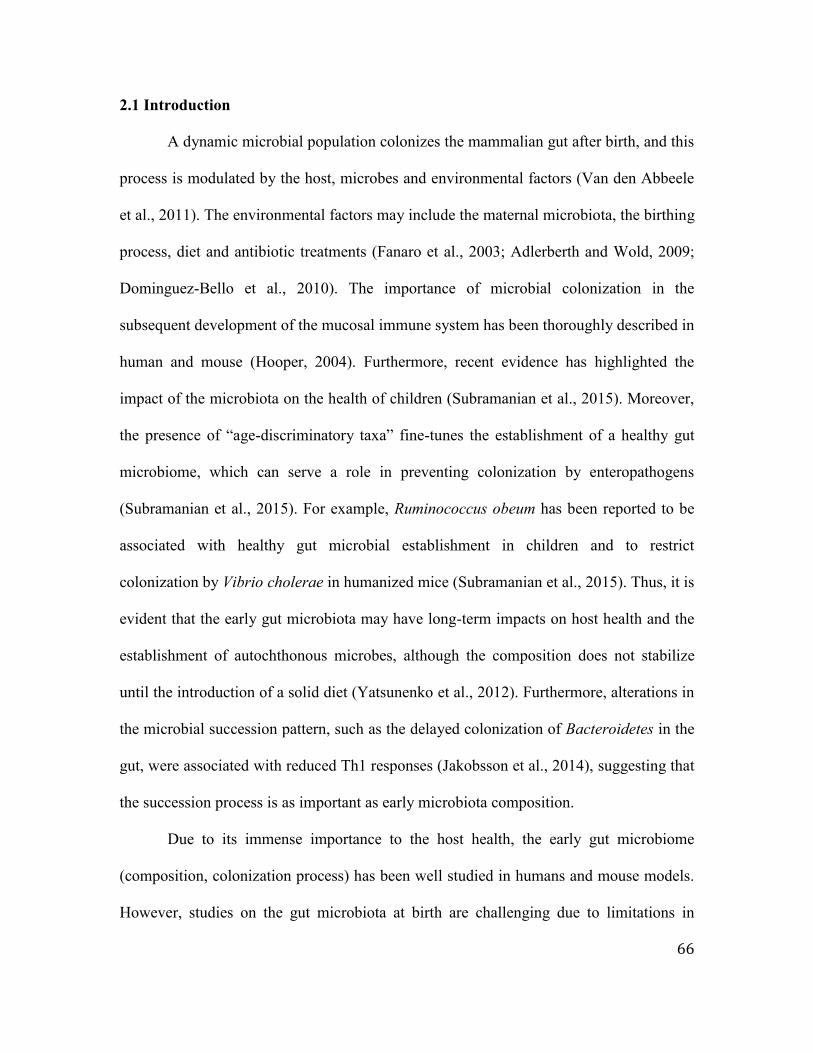
66
2.1 Introduction
A dynamic microbial population colonizes the mammalian gut after birth, and this
process is modulated by the host, microbes and environmental factors (Van den Abbeele
et al., 2011). The environmental factors may include the maternal microbiota, the birthing
process, diet and antibiotic treatments (Fanaro et al., 2003; Adlerberth and Wold, 2009;
Dominguez-Bello et al., 2010). The importance of microbial colonization in the
subsequent development of the mucosal immune system has been thoroughly described in
human and mouse (Hooper, 2004). Furthermore, recent evidence has highlighted the
impact of the microbiota on the health of children (Subramanian et al., 2015). Moreover,
the presence of “age-discriminatory taxa” fine-tunes the establishment of a healthy gut
microbiome, which can serve a role in preventing colonization by enteropathogens
(Subramanian et al., 2015). For example, Ruminococcus obeum has been reported to be
associated with healthy gut microbial establishment in children and to restrict
colonization by Vibrio cholerae in humanized mice (Subramanian et al., 2015). Thus, it is
evident that the early gut microbiota may have long-term impacts on host health and the
establishment of autochthonous microbes, although the composition does not stabilize
until the introduction of a solid diet (Yatsunenko et al., 2012). Furthermore, alterations in
the microbial succession pattern, such as the delayed colonization of Bacteroidetes in the
gut, were associated with reduced Th1 responses (Jakobsson et al., 2014), suggesting that
the succession process is as important as early microbiota composition.
Due to its immense importance to the host health, the early gut microbiome
(composition, colonization process) has been well studied in humans and mouse models.
However, studies on the gut microbiota at birth are challenging due to limitations in

67
sampling, and the first pass meconium is often used as a proxy. The collection time of the
first pass meconium can vary from a few hours to a day after the birth of human infants
(Jimenez et al., 2008; Hansen et al., 2015), and this sample may not be representative of
the gut microbiome at birth. In addition, establishment of gut region-specific microbiota
may regulate intestinal epithelium development in a region-specific manner (Sommer et
al., 2015), suggesting the importance of understanding the local establishment of
microbiota.
To date, our understanding of the initial colonizers in the gut, especially the small
intestine of ruminants, is very limited. Gut health during early life is an emerging
research area that aims to reduce the impact of infectious diseases and antibiotics usage in
the cattle industry. Therefore, this study investigated small intestinal bacterial
composition at birth and compared it with the calf body habitat, maternal environment
(birth canal and rectum) and birthing environment (calving pen floor). This analysis
provided information regarding initial microbial composition and the source of the
inoculum, which could provide a basis for the future improvement of gut health in young
ruminants.
2.2 Materials and Methods
2.2.1 Animal experiments and sampling
All experimental protocols were approved by the Livestock Care Committee of
the University of Alberta (AUP00001012) and were conducted following the guidelines
of the Canadian Council on Animal Care. All calves and dams were obtained from the
Dairy Research and Technology Center (DRTC), University of Alberta (Edmonton, AB).

68
Dams with male fetuses were transferred into calving pens a week before their predicted
due dates and were closely monitored through cameras. The newborn calves (n = 6) were
removed from dams soon after birth using clean plastic containers to prevent the calves
from contacting the floor, immediately transferred to a surgery room, and humanely
euthanized within five minutes. Small intestinal tissue and contents samples were
collected as closed segments at pre-determined regions within 30 minutes after
euthanization. All samples were snap frozen in liquid nitrogen and stored at -80°C.
Briefly, the esophagus and rectum were first ligated to occlude the lumen and prevent
environmental contamination of the intestine. The proximal jejunum was defined as 100
cm distal to the pylorus sphincter, the distal jejunum was defined as 30 cm proximal to
the collateral branch of the cranial mesenteric artery and the ileum was defined as 30 cm
proximal to the ileo-cecal junction. Ten centimeter-long intestinal segments were
collected in the middle of each segment aligned with the above-mentioned measurements.
The rectal and birth canal swab samples from respective dams were collected
using BD Screw Cap Single SWUBE™ Applicators (polyester) (Becton, Dickinson and
Company, NJ, USA) immediately following calving. A similar approach was used to
collect calf body habitat swabs (nose, mouth, skin) before euthanasia. Litter
materials/shavings from the calving pens were also collected immediately following
calving.
2.2.2 Nucleic acid isolation
Total genomic DNA was extracted from the small intestinal tissue and contents
samples using the repeated bead-beating plus column method (Yu and Morrison, 2004).

69
Total DNA from the swab samples and litter material was extracted using the GeneJET
Genomic DNA Purification Kit (ThermoFisher Scientific, MA, USA), following the
manufacturer’s instructions for Gram-positive bacteria genomic DNA purification. DNA
quality and quantity were measured using an ND 1000 spectrophotometer (NanoDrop
Technologies, DE, USA). The intestinal epithelium was scraped along with its contents to
extract total RNA for the estimation of active/live bacteria using quantitative real-time
PCR. Total RNA was extracted from the intestinal epithelial tissue and contents together
using a mirVana™ miRNA isolation kit (Ambion, Carlsbad, CA), following the
manufacturer’s instructions.
2.2.3 Diversity and phylogenetic analyses5
The diluted total DNA (25 ng/µl) from all samples was used to amplify the V1-V3
region using universal bacterial primers (9F - 5’-
ACACTGACGACATGGTTCTACAGAGTTTGATCMTGGCTCAG-3’ and 519R - 5’-
TACGGTAGCCAGAGACTTGGTCTCCGCGGCKGCTGGCAC-3’) with added tags.
PCR products (~500 bp) were purified using a 1% agarose gel and the QIAEX II gel
extraction kit (QIAGEN Science, MD, USA) following the manufacturer’s instructions.
Barcodes were added to the purified PCR products, and they were subjected to 454
sequencing using the Roche GS-FLX System with titanium chemistry at Genome
Quebec, McGill University (Quebec, Canada). Phylogenetic assignments and the analysis
of bacterial diversity (α- and β-diversity) were performed using the Quantitative Insights
Into Microbial Ecology (QIIME) software version 1.8.0 (Caporaso et al., 2010). Briefly,
5Generation of PCR amplicons for 454 sequencing was done by the laboratory technician Yanhong Chen

70
the raw sequences were first filtered through a quality control pipeline to retain sequences
longer than 200 bp and bases with a Phred quality score > 25. Then, the chimeric
sequences were filtered out using usearch61 within the QIIME platform, and the
remaining high quality sequences were used for taxonomic assignments using the
Greengenes database 13_8 (2013 July release). Raw sequences were deposited in the
NCBI Sequence Read Archive (SRA) under the accession number SRP072997.
2.2.4 Co-occurrence analysis of identified bacterial families
A co-occurrence analysis was performed using the relative abundance of bacterial
families and Spearman rank correlation analysis to explore the interactions among initial
bacteria colonizing the calf small intestine. A positive correlation (ρ > 0.5, P < 0.05)
between two families was considered a co-occurrence incidence, and the distance was
calculated using the Spearman rank correlation value (distance =1 – ρ). The distance
values were used to generate co-occurrence networks using Cytoscape 3.3.0 (Shannon et
al., 2003).
2.2.5 Quantification of bacterial density6
Diluted DNA and RNA were used to estimate total bacteria, Lactobacillus and
Bifidobacterium densities using SYBR green chemistry (Fast SYBR® Green Master Mix,
Applied Biosystems) with a StepOnePlus real-time PCR system (Applied Biosystems)
employing universal bacterial primers and genus-specific primers (Liang et al., 2014).
The standard curves for total bacteria, Lactobacillus and Bifidobacterium were generated
6 qPCR was performed by laboratory technician Yanhong Chen

71
using purified 16S rRNA genes of Butyrivibrio hungateii, Lactobacillus acidophilus
ATCC4356 and Bifidobacterium longum, respectively. Bacterial density (DNA and
RNA-based) was then calculated using the equation described by Li et al., (2009).
2.2.6 Taxonomic identification of the ileal Bifidobacterium population7
The extracted total DNA was used to amplify 16S rRNA genes with primers
targeting the genus Bifidobacterium (BifF- 5’-CTCCTGGAAACGGGTGG-3’ and BifR-
5’-GGTGTTCTTCCCGATATCTACA-3’) (Matsuki et al., 2002). 16S clone libraries
were prepared using the purified PCR products (Malmuthuge et al., 2012) to further
explore the Bifidobacterium composition in the calf ileum at birth. Briefly, the purified
PCR products from intestinal tissue and content samples of individual calves were first
pooled and cloned into a TOP10 vector (TOPOTA cloning kit; Invitrogen, Carlsbad, CA,
USA) using chemical transformation. Plasmid DNA extraction, sequencing and
taxonomic assignment were performed according to the method described by
Malmuthuge et al., 2012. Sequence data were deposited in the NCBI database under the
accession numbers KF828998 – KF829902.
2.2.7 Statistical analysis
Effects of sample type (tissue versus content) and small intestinal region were
evaluated using group_significance.py function and Kruskal-wallis non-parametric
ANOVA with P value corrected by the Benjamini-Hochberg FDR procedure for multiple
comparisons within QIIME. Significant comparisons were declared at FDR < 0.05. Beta
7 Preparation of clone libraries and sequencing was performed by laboratory technician Yanhong Chen

72
diversity was computed using beta_diversity.py and the weighted UniFrac distance
matrices were used to perform analysis of similarity (ANOSIM) using
compare_category.py function to declare bacterial communities (intestinal, body habitat,
maternal and birth environmental communities) different from each other (ANOSIM R
0.5-1, P < 0.05).
Bacterial population data (total bacteria density, density/proportion of
Bifidobacterium, density/proportion of Lactobacillus) were analyzed using SAS version
9.4 (SAS Inc., Cary, NC) and analysis of variance. A repeated-measures experimental
design was used with small intestinal region as the repeated measurement and individual
animal as the experimental unit. Compound symmetry covariance structure was selected
as the best fit by the lowest Bayesian information criteria (BIC) and the following
statistical model was fitted to test the effect of small intestinal region and sample type on
bacterial populations: Yijk = μ + Ri + Sj + TS(ij) + eijk, where Y = bacterial
density/proportion (total bacteria, Lactobacillus, Bifidobacterium); μ = mean; R = small
intestinal region; S = sample type (tissue, content); and e = residual error. Differences in
least square means were declared at P < 0.05 using the PDIFF option in SAS when
applicable.
2.3 Results
2.3.1 Active and dense bacterial population at birth
DNA and RNA-based estimations revealed dense and active bacterial colonization
in the calf small intestine at birth (Figure 2.1). Live/active bacterial densities detected
using RNA-based approach were lower than the densities detected using DNA-based

73
approach that estimates dead and active bacteria (Figure 2.1). Although the bacterial
densities (total, Bifidobacterium, Lactobacillus) were not different among intestinal
regions, the proportion of Bifidobacterium was higher in the ileum than in the proximal
and distal jejunum (Figure 2.1). When the composition of Bifidobacterium in the ileum
was further explored, more than half of Bifidobacterium (69.8%) could not be identified
at the species level. However, B. longum subsp. infantis (23.9%) accounted for the
majority of the identified species in the calf ileum at birth. The other Bifidobacterium
species identified in the calf small intestine at birth were B. pseudolongum, B.
pseudolongum subsp. pseudolongum and B. boum (Figure 2.2).
2.3.2 Taxonomic assessment of the gut microbiota at birth
The small intestine epimural (tissue-attached) and luminal (content-associated)
communities of the calves at birth were already colonized with a diverse bacterial
population comprised of 12 phyla (Figure 2.3A). Proteobacteria (40.4±4.9%), followed
by Firmicutes (23.2±3.0%), Actinobacteria (20.5±3.7%) and Bacteroidetes (10.7±1.8%)
dominated the calf small intestine, regardless of the sample type (epimura versus lumen).
In total, 89 bacterial families and 122 genera were identified from all of the small
intestinal regions. Among the detected families, Pseudomonadaceae,
Propionibacteriaceae and Ruminococcaceae dominated the calf gut at birth, while
Pseudomonas, Propionibacterium, Prevotella and Bacillus were predominant at the
genus level of the taxonomic hierarchy (Figure 2.3B). However, the abundance of these
detected bacterial genera varied greatly among individuals. For example, the abundance

74
of Pseudomonas ranged from 0.3% to 83.8%, Propionibacterium ranged from 0 to 86.4%
and Prevotella ranged from 0 to 20.8% in six newborn calves.
The luminal bacteria were dominated by Firmicutes (28.6±4.2%), followed by
Proteobacteria (27.4±5.3%), Actinobacteria (20.2±5.1%) and Bacteroidetes
(15.9±2.8%), but their abundances were numerically different among the small intestinal
regions. For example, Firmicutes dominated the ileal lumen (36.7±7.4%), while
Actinobacteria dominated the distal jejunal lumen (37.3±11.3%). Although Firmicutes
was abundant in all of the luminal communities, the majority of them were only identified
at either the bacterial order or the family levels of the taxonomic hierarchy. For example,
most of the Firmicutes were only assigned to order Clostridiales (6.8±2.0%) and families
Ruminococcaceae (3.5±1.1%) and Lachnospiraceae (4.9±1.9%). The abundance of the
dominant bacterial genera was also numerically different among the three small intestinal
regions. The abundance of Prevotella was higher in the lumen of the ileum (14.2±1.4%)
than the jejunal communities (proximal jejunum - 6.9±3.5%; distal jejunum - 5.0±1.7%),
whereas the abundance of Pseudomonas was lower in the lumen of the ileum (1.4±0.7%)
than the jejunal communities (proximal jejunum - 15.8±13.5%; distal jejunum -
9.9±4.3%).
The epimural bacteria were dominated by Proteobacteria (53.4±7.2%),
Firmicutes (17.8±3.9%), Actinobacteria (20.8±5.6%) and Bacteroidetes (5.6±1.7%);
however, Actinobacteria was the second most predominant phylum in the jejunal
epimural communities (proximal jejunum - 34.0±14.9%; distal jejunum - 20.8±4.2%).
Similar to the luminal bacteria, the abundance of the observed dominant bacterial genera
was numerically different among the three small intestinal regions. Pseudomonas was the

75
most abundant genus in the epimura of the distal jejunum (32.4±12.3%) and ileum
(39.2±12.9%); however, Propionibacterium (30.6±15.8%) dominated the proximal
jejunum. Moreover, the abundance of Bacillus was higher in the distal jejunum
(11.9±7.3%) than the proximal jejunum (2.5±1.9%) and ileum (1.8±1.7%).
When the bacterial composition was compared between the lumen and epimural
communities, abundances of detected bacterial phylotypes were not statistically different.
A higher prevalence of Bacteroidetes was observed in the small intestinal lumen
(proximal jejunum - 11.7±5.0%; distal jejunum - 9.4±3.4%; ileum - 26.6±2.7%), than the
corresponding epimural communities (proximal jejunum - 0.6±0.2%; distal jejunum -
6.6±2.6%; ileum - 9.5±3.8%). At the genus level, Propionibacterium (17.8±5.0%),
Pseudomonas (9.0±4.7%), Prevotella (8.7±1.6%), Ralstonia (3.1±1.7%), Ruminococcus
(2.8±1.2%), Lactobacillus (1.8±0.6%), Bacillus (1.6±1.2%), Corynebacterium
(1.3±1.0%), Succinivibrio (1.1±0.6%) and Butyrivibrio (1.0±0.6%) were abundant (> 1%
relative abundance) in the luminal communities. Additionally, Pseudomonas
(33.8±7.6%), Propionibacterium (19.6±5.6%), Bacillus (5.4±2.7%), Prevotella
(3.6±1.4%), Ralstonia (2.3±0.9%), Lysinibacillus (3.5±1.3%), Akkermansia (1.7±1.7%)
and Burkholderia (1.5±0.9%) were also predominant in the epimura. Among the
observed genera, Burkholderia and Akkermansia were not identified in any of the luminal
communities, while Coprococcus, Succinivibrio, Staphylococcus and Clostridium were
absent in the epimural communities.
In addition to the composition, bacterial richness (number of families/genera) was
different among the small intestinal regions, between the epimural and luminal
communities and among individual calves (Table 2.1). The number of identified families

76
and genera in the ileal lumen was numerically higher than the jejunal communities (Table
2.1). Similarly, a significantly higher bacterial diversity was observed in the ileum
(phylogenetic distance, observed species, Shannon index) when comparing two jejunal
regions, regardless of the gut environment (epimura versus lumen) (Table 2.1). However,
the bacterial diversity and richness were lower in the small intestinal epimural
communities at birth compared to the luminal communities (Table 2.1).
2.3.3 Firmicutes and Bacteroidetes families co-occur at birth
When the co-occurrence of bacteria at birth was explored, families from
Firmicutes and Bacteroidetes phyla were mainly found as the hubs of the co-occurrence
network. The main Firmicutes families detected in the network hubs were
Lachnospiraceae (14 positive correlations), Clostridiaceae (8 positive correlations),
Ruminococcaceae (5 positive correlations) and an unidentified family belonging to the
order Clostridiales (13 positive correlations). Prevotellaceae (10 positive correlations)
and Bacteroidaceae belonging to the order Bacteroidetes were also among the main hubs
identified in the co-occurrence network. The only Proteobacteria family observed among
the main hubs was Succinivibrionaceae, with 10 positive correlations identified from all
three small intestinal regions (Figure 2.4).
2.3.4 Comparison of maternal, environmental and calf body habitat microbiota
The microbial compositions of the maternal communities (rectum and birth canal
of dams), the calving pen floor/birth environment, and the calf body habitats (mouth,
nose, skin) were explored to understand the impact of exposure to the dam and

77
environment on early gut bacterial composition. Ten phyla were identified from the rectal
and birth canal communities of dams, and the dominant phylum was Firmicutes (Figure
2.5). In total, there were 44 bacterial families identified from the birth canal environment.
However, the relative abundances of eight dominant families (Streptococcaceae,
Aerococcaceae, Ruminococcaceae, Clostridiaceae, Peptostreptococcaceae,
Lachnospiraceae, Bacteroidaceae, and Corynebacteriaceae) accounted for 77.2% of total
detected bacterial families. There were seven dominant families (Ruminococcaceae,
Clostridiaceae, Peptostreptococcaceae, Lachnospiraceae, Bacteroidaceae,
Veillonellaceae and Streptococcaceae) in the rectum that accounted for 59.2% of the 44
detected bacterial families. The main bacterial genera identified from the maternal
communities were Streptococcus, Facklamia, Clostridium and Ruminococcus (Table 2.2).
The calf body habitat (10 phyla) and the birth environment (9 phyla) bacteria mainly
consisted of Firmicutes and Proteobacteria at the phylum level (Supplementary Figure
2.5). The main families identified from these communities were Aerococcaceae,
Staphylococcaceae and Xanthomonadaceae, whereas the main genera observed were
Jeotgalicoccus and Facklamia (Table 2.2).
The operational taxonomic units (OTUs) generated from the environmental, calf
body habitat and maternal communities were compared with the calf small intestinal
communities using weighted UniFrac distance matrices (Figure 2.6) and analysis of
similarity (ANOSIM) (Table 2.3) in QIIME. The bacterial communities of the calf small
intestine at birth were significantly different from those of the maternal, calf body habitat
and birth environments (Table 2.3, Figure 2.6A, 2.6B, 2.6C). Although Firmicutes was
one of the main phyla in all of these communities, its composition and abundance were

78
different at lower levels of the taxonomical hierarchy. For example, Ruminococcus was
mainly found in the calf gut, whereas Streptococcus, Facklamia, and Clostridium
dominated the maternal communities (Table 2.2). Although the calf body habitat
communities were different from maternal communities, they were not different from
those in the birth environment (Table 2.3, Figure 2.6D). The microbiota observed in the
calf body habitat at birth were similar to the birth environment (calving pen floor)
microbiota, which were dominated by facultative anaerobes (Jeotgalicoccus, Facklamia)
and aerobes (Corynebacterium). Additionally, the birth environment could not be
differentiated from the rectal microbiota (ANOSIM R = 0.0741, P = 0.50) but was
different from that of the birth canal (ANOSIM R = 0.4012, P < 0.01).
2.4 Discussion
The early microbiome, which plays a crucial role in the development of the
mucosal immune system, subsequent microbial succession and host health, has been well
studied in human and mouse models; however, knowledge in ruminants is very limited.
This is the first attempt to report the colonization of the small intestinal epimural and
luminal communities with an active bacterial population soon after birth and to evaluate
their link to the maternal, birth environment and calf body habitat microbiota. Use of
next-generation sequencing along with RNA-based quantification of bacteria residing the
small intestinal tissue and content samples revealed that newborn calf gut colonized with
a dynamic microbial population, which mainly consisted of Proteobacteria. This
observation suggests that gut microbial establishment may have started during the
birthing process. The process of calving (from rupturing of amniotic membranes and

79
leaking of amniotic fluid till delivering the calf) extended nearly around an hour to three
hours, which may allow dense and diverse microbial colonization in calf gut. The
bacterial composition of the calf gut at birth differed from that of three-week-old calves
(Malmuthuge et al., 2014) as well as the fecal microbiota of one-week-old calves
(Oikonomou et al., 2013). Firmicutes (> 97%) dominated the jejunal and ileal digesta-
associated bacteria of three-week-old calves fed milk and calf starter (Malmuthuge et al.,
2014). Bacteroidetes were predominant in the jejunal (23.6%) and ileal (33.7%) tissue-
attached communities at three weeks (Malmuthuge et al., 2014). Studies on fecal bacterial
communities have also reported a higher abundance of Firmicutes in one-week-old calves
(Uyeno et al., 2010; Oikonomou et al., 2013). However, the present study revealed a lack
(< 10%) of these obligate anaerobes in the calf gut at birth. Thus, these studies indicate
that obligate anaerobes belonging to Firmicutes and Bacteroidetes, which are dominant in
the gut microbiomes (Ley et al., 2008), establish gradually and become the predominant
bacteria with increasing calf age. In addition to the observed differences among the fecal
or small intestinal bacteria of older calves, the calf gut microbiota also differed from the
meconium bacterial composition of human infants. The gut microbiota of human infants
is dominated by Lactobacillus, Bifidobacterium, Enterobacteriaceae, and
Enterococcaceae during the first 24 hours (Dominguez-Bello et al., 2010; Hansen et al.,
2015). In contrast, the present study revealed a higher abundance of Propionibacterium,
Pseudomonas, Prevotella, Bacillus and Ralstonia in the calf small intestine at birth.
However, it is not clear whether these differences represent species specificity or
variations in sampling time and location. Actinobacteria dominates the three- to four-
month-old infant fecal microbiota (Turroni et al., 2012; Azad et al., 2013), whereas

80
Firmicutes dominates the calf fecal microbiota within the first seven weeks of life
(Oikonomou et al., 2013). Thus, these observed differences at birth might indicate
establishment of a species-specific microbiome from birth.
In addition to the differences in the abundance of detected bacteria, the diversity
of bacterial communities at birth also differed from that of three-week- and six-month-old
calves (Malmuthuge et al., 2012). In contrast to the higher diversity and richness
observed in the small intestine epimural communities compared to the respective luminal
communities in older calves (Malmuthuge et al., 2012; Malmuthuge et al., 2014), this
study revealed a less diverse bacterial community in the small intestinal epimura at birth.
The low bacterial diversity in the epimura might be attributable to the higher abundance
of Pseudomonas, an aerobic bacterial genus, observed in all epimural communities when
compared to the luminal communities. Pseudomonas as a transient species may play a
role in creating an anaerobic environment for the autochthonous gut bacteria through
tissue oxygen scavenging, which is similar to the roles of Enterococcus and
Streptococcus observed in the infant gut (Fanaro et al., 2003; Jost et al., 2012). Despite
the higher abundance, Psuedomonadaceae (only 2 positive correlations) was not among
the main hubs in the co-occurrence networks, which may also be due to its role as a
transient bacterium. Although the roles of aerobes and facultative anaerobes during gut
colonization have been defined, the role of anaerobic bacteria present at birth on
subsequent microbial colonization is not yet understood. Prevotellaceae,
Ruminococcaceae and Lachnospiraceae families, the main hubs of the co-occurrence
network, were also predominant in the calf small intestine at birth. These anaerobic
bacterial families belonging to Firmicutes and Bacteroidetes, the two main phyla found in

81
the small intestinal contents and tissue communities of pre-weaned calves (Malmuthuge
et al., 2014), may be the inoculum for obligate-anaerobic gut microbiota. The
composition and abundance of Firmicutes and Bacteroidetes have been shown to be
influenced by intestinal inflammation (Hansen et al., 2012; Dillon et al., 2014), indicating
their responses towards changes in the mucosal immune system. Therefore, the roles of
these co-occurring families in the gut colonization process and their interactions with the
host need to be investigated further to understand the establishment of region-specific
microbiota in depth.
DNA- and RNA-based estimations revealed a higher abundance of
Bifidobacterium in the calf small intestinal communities, especially in the epimura. This
beneficial bacterium has been reported to be one of the main bacteria to first colonize the
neonatal calf gut (three to seven-day-old calves) and remains predominant until six to
nine months of age (Rada et al., 2006; Vlkova et al., 2006). Although our pyrosequencing
primers failed to detect Bifidobacterium species, 16S clone libraries revealed that the
majority of Bifidobacterium in the calf ileum belonged to B. longum infantis. The
presence of conserved solute binding proteins in Bifidobacterium facilitates their binding
to mucin-glycans present on the intestinal epithelium (LoCascio et al., 2010). This may
also enhance the density of Bifidobacterium in the calf gut (Rada et al., 2006; Vlkova et
al., 2006; Uyeno et al., 2010) and also their preferable rapid colonization of the intestinal
tissue (Malmuthuge et al., 2015). Moreover, the highly conserved milk oligosaccharide
utilization genes in B. longum infantis enable the effective utilization of sialylated
oligosaccharides (LoCascio et al., 2010), which are the major oligosaccharide in the
bovine colostrum and milk (ten Bruggencate et al., 2014). The present study revealed an

82
increase in the abundance of B. longum infantis (50.2%) in the ileum of one-week-old
calves compared to the abundance at birth. These calves were fed with colostrum during
the first three days of life, followed by exclusive milk feeding until sample collection.
Sialylated oligosaccharides present in the bovine colostrum and milk (ten Bruggencate et
al., 2014) may have boosted the population of B. longum infantis in the ileum within the
first week of life. However, the abundance of B. longum infantis later decreased to 0.4%
in three-week-old calves and could not be detected in six-week-old calves, when calves
were supplemented with calf starter. Alternatively, the proportion of B. pseudolongum
subsp. globosum, a bovine-specific Bifidobacterium (Balleste and Blanch, 2011), was
increased in three- and six-week-old calves. Human and mouse studies have reported that
Bifidobacterium species play a vital role in the development of the mucosal immune
system and also prevent pathogen colonization (Hart et al., 2004; Hidalgo-Cantabrana et
al., 2014). Thus, future studies are necessary to understand the roles of Bifidobacterium in
calf gut development and health to prevent diarrhea, which is the major cause of neonatal
calf death.
Human studies have suggested the vertical transmission of Bifidobacterium
species from mother to infant (Makino et al., 2013). DNA-based estimation in the present
study revealed the presence of Bifidobacterium in the birth canal (16.8±13.2%) and the
rectum (13.9±5.8%) of dams, which could be the inoculum for the newborn calf gut.
However, only B. longum and B. pseudolongum (PCR amplification using species-
specific primers; Figure 2.7) were detected in the maternal communities, not B. longum
subsp. infantis. A past study also reported that B. longum subsp. infantis was not
detectable in the fecal samples of adult humans or other animal species (Lamendella et

83
al., 2008). Moreover, when the vertical transmission of Bifidobacterium species was
explored, B. longum subsp. infantis was not listed among the species transmitted from
mother to infant (Makino et al., 2013). The reason for these observations may be the low
abundance of B. longum subsp. infantis in the adult gut (Lamendella et al., 2008), which
is below the detection limit of PCR. Nonetheless, I speculated that B. longum subsp.
infantis transmitted from the dam might become predominant in the calf gut at birth and
during early life due its ability to adapt well in the neonatal gut environment, which is
enriched with mucin-glycans and milk oligosaccharides.
In humans, the fecal microbiota of vaginally delivered infants highly resembles
the maternal microbiota (Dominguez-Bello et al., 2010; Backhed et al., 2015). In contrast
to humans, our data revealed the establishment of a notably different bacterial community
in the calf small intestine at birth compared to the maternal bacteria (birth canal and
rectum) in naturally born calves (Figure 2.8). Although bacterial genera detected in the
maternal communities (birth canal and rectum), such as Prevotella, Ruminococcus,
Clostridium, Streptococcus, Butyrivibrio and Staphylococcus were present in the calf
small intestine, relative abundances were different among these communities. Similar to
Bifidobacterium, these bacterial genera may also be acquired from maternal microbiota;
yet, their colonization may depend on the available nutrition and adhesion mechanisms
leading to a unique microbial community in newborn calf gut. Jami and colleagues
(2013) reported that Streptococcus predominated the rumen fluid of one-day-old calves,
which was also the predominant genus observed in the birth canal of dams used in the
present study. This suggests that newborn calf rumen fluid at birth may be more similar
to maternal communities than the small intestine. The rumen (content and tissue together)

84
of the same calves used in the present study were colonized with bacterial genera, such as
Veillonella (23.3±14.1%), Prevotella (23.0±4.0%), Bacteroides (21.6±5.6%), and
Eubacterium (7.0±0.7%) (Malmuthuge et al., unpublished data). Whereas the small
intestine content-associated bacteria were colonized with Propionibacterium,
Psuedomonas, Prevotella, Ruminococcus, Corynebacterium, Ralstonia and Bacillus at
birth. Our previous studies (Malmuthuge et al., 2012; Malmuthuge et al., 2014) and a
recent study in humans (Romano-Keeler et al., 2014) revealed region-specific bacterial
composition in the gut that could not be detected in the large intestinal regions or in the
feces. Thus, the observed variations between the small intestinal and ruminal bacterial
composition within a few minutes after birth also suggest that the gut region-specific
bacteria are established from the birth.
The small intestinal bacterial community was also lacked a core microbiome (no
single bacterial genus was observed in all of the samples). The absence of a core
microbiome among individual calves implies that the impact of the host on the
establishment of the small intestinal microbiota may be stronger than that of the
environment. The ruminant small intestine, the major site of the mucosal immune system,
contains Peyer’s patches (PP) that develop prenatally (Griebel et al., 1996; Yasuda et al.,
2004). The microbiota colonizing the small intestine largely undergoes comprehensive
scrutiny by the host immune system (Hansen et al., 2012; Van den Abbeele et al., 2011).
This may result in a higher impact of the host on the small intestinal microbiota than
environmental factors. The observed differences between the luminal and epimural
communities also suggest a segregation of microbial colonization in these communities
from birth. Although the activity levels of gut-associated lymphoid tissues or secretary

85
immunoglobulin in calves at birth are not clearly understood, the establishment of a
unique microbial population suggests that calves may have an active immune system that
can influence the microbiota colonization process.
2.5 Conclusion
The present study revealed the colonization of the calf small intestine with a
dense, diverse and active microbiota at birth. However, the small intestinal bacterial
composition within a few minutes after birth was different from that of the maternal
microbiota (birth canal and rectum). This suggests that small intestine-specific bacterial
establishment begins during the birthing process and may be influenced by mucosal
immune components. Although aerobic bacteria (Pseudomonadaceae) were the most
abundant, a high abundance of anaerobic bacterial families (Prevotellaceae,
Ruminococcaceae and Lachnospiraceae) was also observed at birth, which were
generally present in the adult gut. These anaerobic families were the hubs of the co-
occurrence networks; however, their roles in the succession of region-specific microbiota
have yet to be elucidated. Although a high prevalence of Bifidobacterium in the maternal
and calf small intestinal microbiomes was observed, the composition was significantly
different between the calves and dams, in that B. longum infantis dominated the ileum
within the first week of life. The favorable conditions (mucin-glycans, milk
oligosaccharides) for B. longum infantis in the newborn gut may have enhanced their
population immediately postnatal. The microbial colonization process in the small
intestine must be carefully moderated during early life, as newborn gut permeability is
high at birth to absorb colostrum components. Such meticulous modulation prevents

86
unnecessary inflammatory responses directed at the establishing commensal microbiome
and will not exert stress on the naïve immune system. Thus, studies to explore variations
in microbial colonization immediately postnatal with varying management practices
(different colostrum/milk feeding methods) may be valuable to understand the role of the
initial microbiota in subsequent succession and gut development. This will also reveal a
means to manipulate the microbial colonization process by modifying early management
practices for dairy calves.
2.6 References
Adlerberth, I., Wold, A.E. (2009). Establishment of the gut microbiota in western infants.
Acta. Paediatr. 98, 229-238. doi: 10.1111/j.1651-2227.2008.01060.
Azad, M.B., Konya, T., Maughan, H., Guttman, D.S., Field, C.J., Chari, R.S., et al.,
(2013). Gut microbiota of healthy Canadian infants: profiles by mode of delivery
and infant diet at 4 months. CMAJ. 185, 385-394. doi: 10.1503/cmaj.121189.
Backhed, F., Roswall, J., Peng, Y., Feng, Q., Jia, H., Kovatcheva-Datchary, P., et al.,
(2015). Dynamics and stabilization of the human gut microbiome during the first
year of life. Cell Host Microbes.17, 852. doi: 10.1016/j.chom.2015.05.012.
Balleste, E., Blanch, A.R. (2011). Bifidobacterium diversity and the development of new
microbial source tracking indicators. Appl. Environ. Microbiol. 77, 3518-3525.
Benjamini, Y., Hochberg, y. (1995). Controlling the false discovery rate: A practical and
powerful approach to multiple testing. Journal of the Royal Statistical Society.
Series B (Methodological). 57, 289-300.

87
Caporaso, J.G., Kuczynski, J., Stombaugh, J., Bittinger, K., Bushman, F.D., Costello,
E.K., et al., (2010). QIIME allows analysis of high-throughput community
sequencing data. Nat. Methods. 7, 335-336. doi: 10.1038/nmeth.f.303.
Dillon, S.M., Lee, E.J., Kotter, C.V., Austin, G.L., Dong, Z., Hecht, D.K., et al., (2014).
An altered intestinal mucosal microbiome in HIV-1 infection is associated with
mucosal and systemic immune activation and endotoxemia. Mucosal Immunol. 7,
983-994. doi: 10.1038/mi.2013.116.
Dominguez-Bello, M.G., Costello, E.K., Contreras, M., Magris, M., Hidalgo, G., Fierer,
N., et al., (2010). Delivery mode shapes the acquisition and structure of the initial
microbiota across multiple body habitats in newborns. Proc. Natl. Acad. Sci. U S
A. 107, 11971-11975. doi: 10.1073/pnas.1002601107.
Fanaro, S., Chierici, R., Guerrini, O., Vigi, V. (2003). Intestinal microflora in early
infancy: Composition and development. Acta. Paediatr. Suppl. 91, 48-55.
Griebel, P.J., Kugelberg, B., Ferrari, G. (1996). Two distinct pathways of B cell
development in Peyer’s patches. Dev. Immunol. 4, 263-277.
Hansen, C.H.F., Nielsen, D.S., Kverka, M., Zakostelska, Z., Klimesova, K., Hudcovic, T.,
et al., (2012). Patterns of early gut colonization shape future immune responses of
the host. PLoS One. 7, e34043. doi:10.1371/journal.pone.0034043.
Hansen, R., Scott, K.P., Khan, S., Martin, J.C., Berry, S.H., Stevenson, M., et al., (2015).
First-pass meconium samples from healthy term vaginally-delivered neonates: an
analysis of the microbiota. PLoS One. 10, e0133320. doi:
10.1371/journal.pone.0133320.

88
Hart, A.L., Lammers, K., Brigidi. P., Vitali. B., Rizzello, F., Gionchetti, P., et al., (2004).
Modulation of human dendritic cell phenotype and function by probiotic bacteria.
Gut. 53, 1602-1609.
Hidalgo-Cantabrana, C., Sanchez, B., Milani, C., Ventura, M., Margolles, A., Ruas-
Madiedo, P. (2014). Genomic overview and biological functions of
exopolysaccharide biosynthesis in Bifidobacterium spp. Appl. Environ. Microbiol.
80, 9-18. doi: 10.1128/AEM.02977-13.
Jakobsson, H.E., Abrahamsson, T.R., Jenmalm, M.C., Harris, K., Quince, C., Jernberg,
C., et al., (2014). Decreased gut microbiota diversity, delayed Bacteroidetes
colonisation and reduced Th1 responses in infants delivered by caesarean section.
Gut. 63, 559-566. doi: 10.1136/gutjnl-2012-303249.
Jami, E., Israel, A., Kotser, A., Mizrahi, I. (2013). Exploring the bovine rumen bacterial
community from birth to adulthood. ISME J. 7, 1069-1079. doi:
10.1038/ismej.2013.2.
Jimenez, E., Marin, M.L., Martin, R., Odriozola, J.M., Olivares, M., Xaus, J., et al.
(2008). Is meconium from healthy newborns actually sterile? Res. Microbiol. 159,
187-193. doi: 10.1016/j.resmic.2007.12.007.
Jost, T., Lacroix, C., Braegger, C.P., Chassard, C. (2012). New insights in gut microbiota
establishment in healthy breast fed neonates. PLoS One. 7, e44595. doi:
10.1371/journal.pone.0044595.
Lamendella, R., Domingo, J.W.S., Kelty, C., Oerther, D.B. (2008). Bifidobacteria in
feces and environmental waters. Appl. Environ. Microbiol. 74, 575-584. doi:
10.1128/AEM.01221-07.

89
Ley, R.E., Lozupone, C.A., Hamady, M., Knight, R., Gordon, J.I. (2008). Worlds within
worlds: evolution of the vertebrate gut microbiota. Nat. Rev. Microbiol. 6, 776-
788. doi: 10.1038/nrmicro1978.
Li, M., Penner, G.B., Hernandez-Sanabria, E., Oba, M., Guan, L.L. (2009). Effects of
sampling location and time, and host animal on assessment of bacterial diversity
and fermentation parameters in the bovine rumen. J. Appl. Microbiol. 107, 1924-
1934.
Liang, G., Malmuthuge, N., Bao, H., McFadden, T., Stothard, P., Griebel, P.J., et al.,
(2014). Potential regulatory role of microRNAs in the development of bovine
gastrointestinal tract during early life. PLoS One. 9, e92592. doi:
10.1371/journal.pone.0092592.
LoCascio, R.G., Ninonuveo, M.R., Freeman, S.L., Sela, D.A., Grimm, R., Lebrilla, C.B.,
et al., (2007). Glycoprofiling of bifidobacterial consumption of human milk
oligosaccharides demonstrates strain specific, preferential consumption of small
chain glycans secreted in early human lactation. J. Agric. Food Chem. 55, 8914-
8919.
Makino, H., Kushiro, A., Ishikawa, E., Kubota, H., Gawad, A., Sakai, T., et al., (2013).
Mother-to-infant transmission of intestinal bifidobacterial strains has an impact on
the early development of vaginally delivered infant's microbiota. PLoS One. 8,
e78331. doi: 10.1371/journal.pone.0078331.
Malmuthuge, N., Li, M., Chen, Y., Fries, P., Griebel, P.J., Baurhoo, B., et al., (2012).
Distinct commensal bacteria associated with ingesta and mucosal epithelium in

90
the gastrointestinal tracts of calves and chickens. FEMS Microbiol. Ecol. 79, 337-
347. doi: 10.1111/j.1574-6941.2011.01220.
Malmuthuge, N., Griebel, P.J., Guan, L.L. (2014). Taxonomic identification of
commensal bacteria associated with the mucosa and digesta throughout the
gastrointestinal tracts of pre-weaned calves. Appl. Environ. Microbiol. 80, 2012-
2028. doi: 10.1128/AEM.03864-13.
Malmuthuge, N., Chen, Y., Liang, G., Goonewardene, L.A., Guan, L.L. (2015). Heat-
treated colostrum feeding promotes beneficial bacteria colonization in the small
intestine of neonatal calves. J. Dairy Sci. 98, 8044-8053. doi: 10.3168/jds.2015-
9607.
Matsuki, T., Watanabe, K., Fujimoto, J., Miyamoto, Y., Takada, T., Matsumoto, K., et
al., (2002). Development of 16S rRNA-gene-targeted group specific primers for
the detection and identification of predominant bacteria in human feces. Appl.
Environ. Microbiol. 68, 5445-5451.
Oikonomou, G., Teixeira, A.G., Foditsch, C., Bichalho, M.L., Machado, V.S., Bicalho,
R.C. (2013). Fecal microbial diversity in pre-weaned dairy calves as described by
pyrosequencing of metagenomic 16S rDNA. Associations of Faecalibacterium
species with health and growth. PLOS One. 8, e63157. doi:
10.1371/journal.pone.0063157.
Rada, V., Vlkova, E., Nevoral, J., Trojanov, I. (2006). Comparison of bacterial flora and
enzymatic activity in faeces of infants and calves. FEMS Microbiol. Lett. 258, 25-
28. doi:10.1111/j.1574-6968.2006.00207.

91
Romano-Keeler, J., Moore, D.J., Wang, C., Brucker, R.M., Fonnesbeck, C., Slaughter,
J.C., et al., (2014). Early life establishment of site-specific microbial communities
in the gut. Gut Microbes. 5, 192-201. doi: 10.4161/gmic.28442.
Shannon, P., Markiel, A., Ozier, O., Baliga, N.S., Wang, J.T., Ramage, D., et al., (2003).
Cytoscape: a software environment for integrated models of biomolecular
interaction networks. Genome Res. 13, 2498-2504.
Sommer, F., Nookaew, I., Sommer, N., Fogelstrand, P., Backhed, F. (2015). Site-specific
programming of the host epithelial transcriptome by the gut microbiota. Genome
Biol. 16, 62. doi: 10.1186/s13059-015-0614-4.
Subramanian, S., Blanton, L.V., Frese, S.A., Charbonneau, M., Mills, D.A., Gordon, J.I.
(2015). Cultivating healthy growth and nutrition through the gut microbiota. Cell.
161, 36-48. doi: http://dx.doi.org/10.1016/j.cell.2015.03.013.
ten Bruggencate, S.J., Bovee-Oudenhoven, I.M., Feitsma, A.L., van Hoffen, E.,
Schoterman, M.H. (2014). Functional role and mechanisms of sialyllactose and
other sialylated milk oligosaccharides. Nutr. Rev. 72, 377-389. doi:
10.1111/nure.12106.
Turroni, F., Peano, C., Pass, D.A., Foroni, E., Severgnini, M., Claesson, M.J., et al.,
Diversity of bifidobacteria within the infant gut microbiota. PLoS One. 7, e36957.
doi: 10.1371/journal.pone.0036957.
Uyeno, Y., Sekiguchi, Y., Kamagata, Y. (2010). rRNA-based analysis to monitor
succession of fecal bacterial communities of Holstein calves. Lett. Appl.
Microbiol. 51, 570-577. doi: 10.1111/j.1472-765X.2010.02937.

92
Van den Abbeele, P., van de Wiele, T., Verstraete, W., Possemiers, S. (2011). The host
selects mucosal and luminal associations of coevolved gut microorganisms: a
novel concept. FEMS Microbiol. Rev. 35, 681-704. doi: 10.1111/j.1574-
6976.2011.00270.
Vlkova, E., Nevoral, J., Rada, V. (2006). Distribution of bifidobacteria in the
gastrointestinal tract of calves. Folia Microbiol. 51, 325-328.
Yasuda, M., Fujino, M., Nasu, T., Murakami, T. (2004). Histological studies on the
ontogeny of bovine gut-associated lymphoid tissue: appearance of T cells and
development of IgG+ and IgA+ cells in lymphoid follicles. Dev. Comp. Immunol.
28, 357-369.
Yatsunenko, T., Rey, F.E., Manary, M.J., Trehan, I., Dominguez-Bello, M.G., Contreras,
M., et al (2012). Human gut microbiome viewed across age and geography.
Nature. 486, 222-227. doi: 10.1038/nature11053.
Yu, Z., Morrison, M. (2004). Improved extraction of PCR-quality community DNA from
digesta and fecal samples. Biotechniques. 36, 808-812.

93
2.7 Tables and Figures
Table 2.1 Bacterial diversity and richness in calf small intestine at birth.
All regions Proximal jejunum Distal jejunum Ileum
Tissue Content Tissue Content Tissue Content Tissue Content
OTUs (97%
similarity) 4233±508 5271±827 4564±1223 6840±1539 3455±587 2813±274 4679±782 6160±1636
Assigned genera 20±2 33±4 12±1 12±2 9±1 11±1 9±1 20±4
Unassigned
sequences genus
level (%)
15.7±3.2 33.6±4.1 14.3±4.8 30.1±6.9 16.0±7.5 27.5±8.8 17.6±5.0 43.2±4.2
Assigned families 19±1 33±3 12±1 14±2 9±1 13±2 10±1 22±3
Unassigned
sequences family
level (%)
1.0±0.5 7.1±2.0 0.1±0.05 2.6±1.8 1.5±1.3 4.1±2.6 1.4±0.5 14.5±3.6
Good’s coverage 0.98±0.002 0.96±0.004 0.97±0.004 0.97±0.01 0.98±0.002 0.97±0.002 0.97±0.003 0.94±0.01
Shannon index* 3.4±0.1 4.3±0.2 3.2±0.5 3.8±0.6 3.4±0.2 3.7±0.5 3.8±0.1 5.6±0.6
Chao 1* 80.8±6.4 125.4±10.7 79.4±9.3 97.6±29.4 77.6±7.7 103.2±8.0 82.2±8.7 175.3±43.2
Observed species* 47.1±2.2 81.2±8.8 46.3±20.0 58.7±3.9 44.8±2.1 61.8±5.9 50.2±4.1 123.3±30.7
Phylogenetic
distance* 4.7±0.3 9.3±0.8 4.7±0.4 7.4±2.1 4.5±0.3 6.9±1.0 5.0±0.3 13.5±2.8
*Phylogenetic distance, Observed species, Shannon index – Ileum > Proximal jejunum, Distal jejunum at P < 0.05
*Phylogenetic distance, Observed species, Shannon index, Chao1 – Content-associated community > tissue-associated
community at P < 0.01

94
Table 2.2 Maternal, calf body habitats and birth environment bacteria.
Birth canal Rectum Mouth Nose Skin Floor
OTUs (97%
similarity) 5533±1450 4655±2331 4587±1176 7548±1654 4042±294 3308±1342
Assigned
genera 21±3 26±4 33±8 31±4 29±6 31±9
Unassigned
sequences
genus level
(%)
44.3±9.6 72.5±3.5 35.1±8.2 47.6±10.3 24.7±9.3 44.1±9.6
Assigned
families 19±3 29±4 28±5 27±5 29±4 28±8
Unassigned
sequences
family level
(%)
17.9±4.9 32.6±4.0 4.8±2.8 7.7±4.9 10.4±5.6 14.3±8.3
Main
families (%)
Streptococcaceae
(25.5±13.2)
Aerococcaceae
(16.7±13.1)
Ruminococcaceae
(11.4±4.6)
Clostridiaceae
(9.6±3.4)
Ruminococcaceae
(24.6±1.1)
Clostridiaceae
(13.3±2.7)
Peptostreptococcaceae
(6.6±2.6)
Lachnospiraceae
(6.4±0.6)
Staphylococcaceae
(17.9±10.6)
Aerococcaceae
(16.2±8.5)
Xanthomonadaceae
(11.7±6.4)
Caulobacteraceae
(10.6±6.5)
Xanthomonadaceae
(14.3±14.2)
Staphylococcaceae
(12.3±11.1)
Aerococcaceae
(8.4±7.3)
Aerococcaceae
(32.6±10.5)
Staphylococcaceae
(12.5±12.3)
Methylobacteriaceae
(11.8±4.8)
Ruminococcaceae
(16.9±13.9)
Aerococcaceae
(16.2±10.8)
Staphylococcaceae
(13.6±13.0)
Xanthomonadaceae
(7.9±7.3)
Main genera
(%)
Streptococcus
(22.5±13.2)
Facklamia
(11.6±10.6)
Corynebacterium
(2.9±1.2)
Ruminococcus
(2.4±0.7)
Clostridium
(5.3±0.4)
Ruminococcus
(4.2±3.2)
5-7N15
(3.7±2.1)
Dorea
(2.2±0.3)
Jeotgalicoccus
(17.8±10.6)
Facklamia
(9.6±5.6)
Streptococcus
(3.8±3.7)
Brevundimonas
(2.8±1.7)
Jeotgalicoccus
(12.2±11.2)
Facklamia
(4.2±4.1)
Corynebacterium
(3.9±3.0)
Bacillus
(2.9±2.3)
Facklamia
(19.8±9.1)
Methylobacterium
(12.3±12.2)
Jeotgalicoccus
(10.7±4.4)
Aerococcus
(6.5±5.9)
Facklamia
(12.1±7.6)
Jeotgalicoccus
(11.8±11.3)
Stenotrophomonas
(5.3±5.0)
Corynebacterium
(3.5±3.1)

95
Table 2.3 Comparison of calf, dam and birth environment bacterial communities.
Comparison ANOSIM R P-value
Calf gut vs. Dam 0.8893 < 0.01
Calf gut vs. Birth canal 0.9049 < 0.01
Calf gut vs. Rectum 0.9130 < 0.01
Calf gut content vs. Birth canal 0.7187 < 0.01
Calf gut content vs. Rectum 0.6788 < 0.01
Calf gut tissue vs. Birth canal 0.7574 < 0.01
Calf gut tissue vs. Rectum 0.6133 < 0.01
Calf gut vs. Body habitats 0.7090 < 0.01
Calf gut vs. Floor 0.7942 < 0.01
Content vs. Tissue 0.2652 < 0.01
Body habitats vs. Floor 0.0595 0.29
Body habitats vs. Dam 0.6557 < 0.01
Floor vs. Dam 0.0984 0.24
Floor vs. Rectum 0.0741 0.50
Floor vs. Birth canal 0.4012 0.04

96
Figure 2.1 Estimation of small intestinal bacteria at birth using DNA (16S rRNA gene
copy/g of fresh sample) and RNA (16S rRNA copy/g of fresh sample) extracted from
small intestinal tissue and content samples. PJ – proximal jejunum, DJ – distal jejunum,
IL – ileum.
(A) Density of total bacteria (B) Density of Bifidobacterium (C) Proportion of
Bifidobacterium [(density of Bifidobacterium / density of total bacteria)*100]. a,b
represents mean proportions that are different among gut regions at P < 0.05 (D) Density
of Lactobacillus (E) Proportion of Lactobacillus [(density of Lactobacillus / density of
total bacteria)*100]

97
Figure 2.2 Ileal (tissue and content together) bifidobacterial composition at birth obtained
through sequencing of 16S clone libraries.
98
Figure 2.3A Bacterial composition in epimural and luminal communities at phylum level
(454 sequencing of V1-V3 region of 16S rRNA gene). Relative abundances of main four
phyla in all three small intestinal regions are presented together and each data point
represents an individual calf.

99
Figure 2.3B Relative abundance of predominant (relative abundance > 1% in at least one
intestinal sample) bacterial families and genera.
(a) Predominant bacterial families in luminal communities (b) Predominant bacterial
genera in luminal communities (c) Predominant bacterial families in epimural
communities (d) Predominant bacterial genera in epimural communities. Bars represent
mean relative abundance of bacterial families and genera.

100
Figure 2.4 Co-occurrence network of bacterial families detected from calf small intestine.
Positive correlations (ρ > 0.5, P < 0.05) among bacterial families are defined as co-
occurred families and distance between two families are calculated using Spearman
correlation co-efficient (distance = 1 – ρ). Lower distance represents higher correlation
and vice versa. All the co-occurrence incidences in all three-gut regions were plotted in
one network using Cytoscape software platform. Black arrows represent hubs of the co-
occurrence network.

101
Figure 2.5 Bacterial compositions in maternal (vagina/birth canal, rectum), body habitat
(mouth, nose, skin) and birth environment (pen floor) communities at phylum level.
Mean relative abundances of detected bacterial phyla are presented in bars.

102
Figure 2.6 Comparison of calf gut, calf body habitats, maternal and birth environment
bacterial communities.
OTU (97% level) profiles generated from each samples are compared using beta-diversity
function within QIIME platform to generate distance matrices. Weighted UniFrac

103
distance matrices are used to perform principle co-ordinate analysis (PCoA) and analysis
of similarity (ANOSIM). R represents ANOSIM R that ranges from 0 to1, 0 representing
microbial communities that are not separated from each other and 1 representing
microbial communities that are separated from each other. P value represent significance
level of ANOSIM R value.

104
Figure 2.7 PCR-detection of Bifidobacterium species in maternal communities (birth
canal and rectum).
Dam1, dam2 and dam3 represent individual IDs of dams. PCR primers target groEL
chaperone gene of the Bifidobacterium genome (Junick and Blaut, Appl. Environ.
Microbiol. 2012, 78: 2613–2622).

105
Figure 2.8 Comparison of bacterial compositions (phylum level) among maternal
communities (birth canal/vagina and rectum) and calf small intestinal communities.
PJC – proximal jejunum content, PJT – proximal jejunum tissue, DJC – distal jejunum
content, DJT – distal jejunum tissue, ILC – ileum content, ILT – ileum tissue.

106
Chapter 3. Feeding heat-treated colostrum promotes beneficial bacteria colonization
in the small intestine of neonatal calves8
Abstract
The present study investigated the impact of feeding heat-treated colostrum on
bacterial colonization in the small intestine of neonatal calves, within the first 12 hours of
life. Newborn Holstein bull calves (n = 32) were assigned to three treatment groups and
fed either fresh colostrum (FC, n = 12) or heat-treated colostrum (60C, 60 min.; HC, n =
12) soon after birth, while the control (NC, n = 8) group did not receive colostrum or
water. Small intestinal tissues and contents were collected from proximal jejunum, distal
jejunum and ileum at 6 and 12 hours after birth, following euthanasia. Quantitative real-
time PCR was used to explore the colonization of total bacteria, Lactobacillus,
Bifidobacterium and E. coli. Feeding colostrum soon after birth increased the
colonization of total bacteria in calf gut within the first 12 hours compared to NC. In
contrast, the prevalence of Lactobacillus was lower in HC and FC, comparing to NC.
Remarkable changes in the prevalence of small intestinal tissue-attached Bifidobacterium
were observed with feeding HC, but not in contents. The prevalence of Bifidobacterium
was 3.2-fold and 5.2-fold higher in HC than FC and NC, respectively, at 6 hours.
Although feeding FC did not enhance the prevalence of tissue-attached Bifidobacterium
at 6 hours compared to NC, it displayed a gradual increase over the time that was higher
than NC, but similar to HC at 12 hours. Moreover, the colonization of E. coli was
8 A version of this chapter has been published as, Malmuthuge, N., Chen, Y., Liang, G., Goonewardene, L.A., Guan,
L.L. (2015). Heat-treated colostrum feeding promotes beneficial bacteria colonization in the small intestine of neonatal
calves. J. Dairy Sci. 98, 8044-8053. doi: 10.3168/jds.2015-9607.

107
drastically reduced in HC calves compared to FC and NC. Thus, the present study
suggests that feeding HC enhances the colonization of Bifidobacterium, but lessens E.
coli, in the calf small intestine immediately postpartum compared to FC and NC. The
increased colonization of beneficial bacteria along with the decreased colonization of
potential pathogens in calf gut may also diminish the neonatal calf diarrhea, when they
fed heat-treated colostrum soon after birth.
3.1 Introduction
Colostrum management and feeding is crucial for passive immune transfer in calf
management. Calves are immune-deficient at birth, as there is no placental transferring of
immunoglobulin into the fetuses in cattle (Godden, 2008). Thus, calves are solely
depending on the absorption of immunoglobulin present in colostrum until the
development of their own immune system (Godden, 2008). Feeding high quality
colostrum (immunoglobulin G (IgG) > 50 mg/mL) soon after birth plays a vital role in
the passive transfer of immunity (Jaster, 2005, Chigerwe et al. 2008), which decreases
calf mortality and morbidity and increases calf weaning weight and body weight gain
(Priestely et al., 2013). Despite the industrial recommendations, the North American
dairy industry is still having concerns regarding the colostrum management (Vasseur et
al., 2010; Morril et al., 2012). Feeding of calves with contaminated (high bacterial count),
and low quality colostrum (< 50 IgG mg/mL) (Morril et al., 2012) as well as low
surveillance of calf birth at night and relying on dams to feed colostrum (Vasseur et al.,
2010) are some of the major concerns observed in the current North American dairy
industry.

108
Feeding heat-treated colostrum is one of the management practices introduced
recently to the dairy industry, aiming to decrease bacterial contaminations and increase
passive immune transfer (Donahue et al., 2012; Godden et al., 2012; Teixeira et al., 2013;
Gelsinger et al., 2014). Heat treatments successfully (60C, 60 min.) decrease total
bacterial count including pathogenic bacteria while maintaining concentration of IgG
(Donahue et al., 2012). Additionally, feeding heat-treated colostrum increases serum
colostrum concentration (Godden et al., 2012; Teixeira et al., 2013) and reduces the risk
for illnesses and treatment for scours in dairy farms, when comparing to fresh colostrum
(Godden et al., 2012). These observations suggest a decrease in disease transmission
among the calves in dairy herds, when they receive heat-treated colostrum soon after
birth. Although the decreased total bacteria including pathogens in colostrum, is one of
the possibilities of reduced enteric infections in calves fed heat-treated colostrum
(Godden et al., 2012), the impact of heat-treated colostrum on gut colonization is not well
studied. The present study hypothesized that feeding heat-treated colostrum influences
bacterial colonization in the calf intestine and facilitates the colonization of beneficial
bacteria. Here, we investigated the impact of colostrum feeding (heat-treated and fresh)
soon after birth on the colonization of total bacteria, Lactobacillus, Bifidobacterium and
E. coli in the calf small intestine within the first 12 hours of life.
3.2 Materials and methods
3.2.1 Colostrum preparation and animal experiments
Prior to the animal experiment, first-milking colostrum containing ≥ 50 IgG
mg/mL was collected from cows raised at the Dairy Research and Technology Center

109
(DRTC), University of Alberta, Edmonton, Canada and immediately laid flat on wire
racks and frozen at -20C. Once ~ 48 L of colostrum was collected, all the samples were
thawed slowly for 24 hours in 4C cold room and mixed thoroughly to obtain the pool of
colostrum that will be using during the entire animal experiment. Half of the colostrum
(24 L) was pasteurized (60 minutes at 60C) using commercial a batch pasteurizer DT
10G (Dairy Tech Inc., Greeley, CO, USA). Colostrum was held at 60C for 60 minutes
apart from the time (~30 minutes) taken to reach 60C, followed by rapid cooling. The
heat-treated colostrum and the remaining half of fresh colostrum were aliquoted into 1-
liter plastic freezer bags and stored at -20C.
Animal experiment was conducted at the DRTC, University of Alberta, following
the guidelines of the Canadian Council on Animal Care. The Livestock Care Committee
of the University of Alberta approved all the protocols (AUP00001012) prior to
beginning the experiment. Near parturition, Holstein cows predicted to have bull calves
were transferred into individual maternity pens and monitored closely with video
cameras. Calves were removed from the dams soon after birth, before exposing to the
dams, and transferred into individual calf units with fresh wood shavings followed by
navel dipping in 7% (v/v) iodine solution. Frozen colostrum (2 L/calf) was thawed to 37 -
38°C using a water bath and bottle-fed to calves within an hour after birth. Calves (n =
32) were randomly allocated into three treatment groups; fresh colostrum-fed calves (FC,
n = 12), heat-treated colostrum-fed calves (HC, n = 12), and control calves that did not
receive either colostrum or water during the experimental period (NC, n = 8).

110
3.2.2 Intestinal sample collection
Intestinal samples from all calves were collected at 6 hours (HC, n = 6; FC, n = 6;
NC, n = 4) and 12 hours (HC, n = 6; FC, n = 6; NC, n = 4) after birth. All calves were
euthanized following captive bolt gun stunning and small intestinal tissues and contents
(proximal jejunum, distal jejunum, ileum) were collected together as closed gut sections
within 30 minutes after euthanization. The esophagus and rectum were first ligated to
occlude the lumen and prevent environmental contamination of the intestine. Then, 10 cm
long closed intestinal segments were collected in the middle of pre-defined gut regions.
Ileum was defined as 30 cm proximal to the ileo-cecal junction, distal jejunum was
defined as 30 cm proximal to the collateral branch of the cranial mesenteric artery and
proximal jejunum was defined as 100 cm distal to the pylorus sphincter. All samples were
snap-frozen and transferred into a -80°C freezer until further processing.
3.2.3 Intestinal sample collection from newborn calves
Dams predicted to have bull calves were transferred into calving pens three days
before the predicted due date and monitored via remote video cameras. Newborn calves
(n = 6) were separated from dams soon after birth to make sure there were no interactions
between calves and dams. Then, the calves were transferred into the surgical room at the
DRTC and humanely euthanized immediately following captive bolt gun stunning
(within five minutes after birth). The collection of small intestinal segments from the
newborn calves was completed within 30 minutes after the calf birth, similar to the 6- and
12-hour calves used in the colostrum feeding trial.
111
3.2.4 DNA extraction from tissue and digesta samples9
A portion of frozen intestinal section was thawed on ice and separated content
from tissue. Then, genomic DNA from tissues and contents was extracted separately
using the repeated bead-beating plus column (RBB+C) method (Yu and Morrison, 2004).
Briefly, content (0.5g) and tissue (0.5g) samples were subjected to physical disruption
with a cell lysis buffer containing 4% SDS using the BioSpec Mini Beads beater 8
(BioSpec, OK, USA) at 4800 rpm for three minutes. Then, the tubes containing lysed
cells were incubated at 70ºC for 15 minutes and separated the supernatant. The bead-
beating and incubation steps were repeated once more. Any remaining impurities and
SDS from supernatants were removed with 10M-ammonium acetate and DNA was
precipitated using isopropanol. The genomic DNA was then further purified using the
QIAmp fast DNA stool mini kit (QIAmp fast DNA stool mini kit, QIAGEN Inc. CA,
USA). The DNA quantity was measured using an ND 1000 spectrophotometer
(NanoDrop Technologies, Wilmington) and stored at -20ºC.
3.2.4 Quantification of total bacteria, Lactobacillus and Bifidobacterium in calf small
intestine
Quantitative real-time PCR (qPCR) was performed to estimate densities of total
bacteria, Lactobacillus, Bifidobacterium and E. coli using SYBR green chemistry (Fast
SYBR® Green Master Mix, Applied Biosystems) with a StepOnePlus real-time PCR
system (Applied Biosystems, Foster City, CA, USA) and bacterial primers (Table 3.1).
Standard curves for total bacteria, Lactobacillus, Bifidobacterium and E. coli were
9 DNA extraction and qPCR was performed by laboratory technician Yanhong Chen

112
generated using purified 16S rRNA genes of Butyrivibrio hungateii, Lactobacillus
acidophilus ATCC4356, Bifidobacterium longum and Escherichia coli K12, respectively.
The copy number of 16S rRNA gene/g of fresh tissue or content was then calculated
using the equation described by Li et al., (2009). The prevalence of Lactobacillus,
Bifidobacterium and E. coli was calculated by dividing the copy number of 16S rRNA
gene of each genus/species by the copy number of total bacteria.
3.2.5 Statistical analysis
All bacterial density data were analyzed using MIXED procedure in SAS (SAS
9.4, SAS Inc., Cary, NC) with small intestinal region as the repeated measurement and
animal as the experimental unit. Compound symmetry covariance structure was selected
as the best fit by the Bayesian information criteria (BIC). The following statistical model
was fitted to test the effect of colostrum treatment, time point, gut region and sample type
on bacterial density: Yijklm = μ + Ci + Tj + Rk + Sl + CT(ij) + CR(ik) + CS(il) + TR(jk) + TS(jl)
+ RS(kl) + CTR(ijk) + CTS(ijl) + CRS(ikl) + TRS(jkl) + eijklm, where Y = bacterial density
(total bacteria, Lactobacillus, Bifidobacterium, E. coli); μ = mean; C = colostrum
treatment; T = time point; R = small intestinal region; S = sample type (tissue, content);
and e = residual error. A similar model was fitted when comparing newborn calves
against 6 and 12-hour-old calves under each diet, after removing colostrum treatment
effect from the model (Yijkl = μ + Ti + Rj + Sk + TR(ij) + TS(ik) + RS(jk) + TRS(ijk) + eijkl).
Differences in LSM were declared at P < 0.05 using the PDIFF option in SAS when
applicable.

113
3.3 Results
3.3.1 Impact of colostrum feeding on the colonization of newborn calf gut within the
first 12 hours of life
Feeding of colostrum (heat-treated or fresh) accelerated bacterial colonization in
the calf small intestine, compared to the calves that did not receive colostrum soon after
birth. Density of total bacteria was significantly higher (P < 0.01) in the small intestine of
HC (9.77±3.44×10⁹ 16S copy/g of sample) and FC (1.37±0.73×10¹⁰ 16S copy/g of
sample) calves, when comparing to NC calves (5.26±2.17×10⁸ 16S copy/g of sample),
regardless of time point and small intestinal region (Figure 3.1A). Besides the effect of
colostrum treatment, effect of type by time interaction on total bacterial density was also
significant (P < 0.01). The content-associated total bacterial density was higher at 12
hours comparing to that of 6 hours as well as tissue-attached bacteria at 12 hours (Figure
3.1B). When the prevalence of beneficial bacteria in the calf small intestine was explored,
the prevalence of Lactobacillus was higher (P < 0.01) in NC calves compared to the
colostrum fed calves (Figure 3.2A). In contrast, the prevalence of Bifidobacterium was
higher (P < 0.01) in tissue-attached community of HC (28.6±10.3%) calves than FC
(8.9±2.5%) and NC (5.5±1.5%) calves at 6 hours (Figure 3.2B). Moreover, the
prevalence of Bifidobacterium was higher (P < 0.01) in proximal jejunum (19.0±8.0%) of
the calves, when comparing to distal jejunum (11.1±4.5%) and ileum (10.4±3.7%),
regardless of colostrum treatment, time point and sample type. In contrast, feeding
colostrum significantly decreased the colonization of E. coli in the calf small intestine
within the first 12 hours of life (Figure 3.3). The prevalence of E. coli was higher in the
tissue and content-associated small intestinal communities of NC calves (tissue –

114
0.17±0.03%, content – 0.16±0.01%) compared to FC (tissue – 0.019±0.005%, content –
0.069±0.061%) and HC (tissue – 0.001±0.0004%, content – 0.004±0.002%).
3.3.2 Impact of colostrum feeding on the bacterial colonization process
Bacterial densities of 6- and 12-hour-old calves were also compared with the
newborn calves obtained within 5 minutes after birth to explore the changes in the initial
bacterial densities within the first 12 hours of life and how this process influence by
feeding colostrum (Figure 3.4). Total bacterial density observed at birth (1.21±0.56×10⁹
16S copy/g of sample) was significantly increased (P < 0.01) within the first 12 hours of
life, when the calves were fed heat-treated colostrum (1.25±0.50×10¹⁰ 16S copy/g of
sample) or fresh colostrum (2.03±1.13×10⁹ 16S copy/g of sample) soon after birth.
Although the prevalence of Lactobacillus was lower (P = 0.01) in HC calves at 6
(0.008±0.002%) and 12 (0.006±0.003%) hours after birth compared to the newborn
calves (0.046±0.024%), there were no such changes (P = 0.12) observed in FC calves
within the first 12 hours (6hr-0.018±0.010%; 12hr-0.010±0.006%). The prevalence of
Bifidobacterium in the small intestinal tissue-attached community was significantly (P =
0.03) lower at 6 hours in FC (8.9±2.5%) calves compared to newborn (31.2±9.0%);
however, there were no such differences (P = 0.51) observed in HC calves (28.6±10.3%).
When the colonization of E. coli within the first 12 hours of life was compared, there
were no differences (P = 0.32) observed between the newborn and FC calves. However,
the HC group had lower (P < 0.01) colonization of E. coli in the small intestinal tissue-
associated communities at 6 (0.0004±0.0002%) and 12 hours (0.0008±0.0003%) after
birth compared to the newborn calves (0.054±0.001%). There were no differences
115
observed when comparing total bacteria, Lactobacillus and Bifidobacterium of NC calves
with the newborn calves. However, the prevalence of E. coli in the small intestinal tissue
of NC increased (P < 0.01) at 12 hours (0.13±0.02%) compared to the newborn calves.
3.3.3 Effect of feeding heat-treated colostrum on small intestinal bacteria within the
first 12 hours of life
Feeding heat-treated colostrum to calves soon after birth did not significantly
influence (P = 0.22) the total bacteria density, when comparing to fresh colostrum;
however, there was a reduction in the number of bacteria colonized in the small intestinal
regions (Table 3.2). When the prevalence of Lactobacillus was compared, it was 4 times
higher (P < 0.01) in the small intestinal content-associated community (0.023±0.015%)
of FC calves compared to the tissue community (0.005±0.002%), regardless of the time
point. In general, feeding heat-treated colostrum decreased Lactobacillus prevalence in
the small intestine of calves compared to fresh colostrum (Table 3.2). Feeding heat-
treated colostrum, however, had remarkable impact on the prevalence of Bifidobacterium
in the small intestine tissue-attached community. The prevalence of Bifidobacterium was
3.2-fold higher in HC calves than FC calves, at 6 hours after birth (P < 0.01). In contrast,
feeding fresh colostrum gradually increased the colonization of Bifidobacterium in the
small intestine and there was no effect of colostrum treatment on the prevalence of
Bifidobacterium at 12 hours after birth (Figure 3.2B). When the colonization of E. coli in
the calf small intestine was compared between two colostrum-feeding methods, FC
calves had higher (P < 0.01) density of E. coli, but not proportions compared to HC
calves (Table 3.2).

116
3.4 Discussion
Neonatal diarrhea is responsible for ~ 50% of the calf deaths occur in dairy
industry (Uetake, 2013). A good colostrum management is one of the most important
preventive measures of neonatal calf diarrhea, because colostrum transfers passive
immunity to the newborn calves. Besides, the bioactive compounds, such as
oligosaccharides (OS), present in bovine colostrum also inhibit the adherence of
pathogens to the intestinal epithelial cells and prevent infections (Maldonado-Gomez et
al., 2015). Feeding heat-treated colostrum to dairy calves is one of the industrial
suggested good practices to increase passive transfer of immunity (Godden et al., 2012;
Teixeira et al., 2013; Gelsinger et al., 2014) and to minimize calf mortality and morbidity.
Heat treatments (60C, 60 min) decrease total bacterial count and eliminate pathogens,
such as Mycobacterium avium subsp paratuberculosis present in colostrum, while
maintaining IgG concentration (Godden et al., 2006; Johnson et al., 2007). Although the
effect of feeding heat-treated colostrum on passive immune transfer has been widely
studied, there is a very limited knowledge regarding its impact on gut microbial
colonization. Gut microbiota plays a key role in host health and susceptibility to diseases
(Round et al, 2009). Besides, the importance of early gut microbiota on immune system
development has been well described in humans (Russell et al., 2013; Jakobsson et al.,
2014).
The present study revealed that feeding colostrum (either fresh or heat-treated)
facilitated the gut microbial colonization, allowing the bacterial numbers to reach 1010
16S rRNA gene/g in fresh samples within the first 12 hours of life. In contrast, the calves
that did not receive colostrum soon after birth had very low number of total bacteria,
117
which was similar to that of newborn calves, suggesting a slower bacterial colonization in
the absence of colostrum. A lower total bacterial density in mouse pups has been shown
to associate with differed mucosal and systemic immune responses compared to the pups
with normal bacterial densities (Lamouse-Smith et al., 2011). Here, the authors speculate
that the timed feeding of colostrum is important for the establishment of stable gut
microbiome that plays a crucial role in the development of naïve newborn calf immune
system. Therefore, besides the efficient and effective absorption of immunoglobulin, the
timed feeding of colostrum also plays a vital role in calf health via promoting gut
microbial colonization.
The calf small intestine was already colonized with a higher level of
Bifidobacterium within five minutes after birth. However, only the heat-treated colostrum
was capable of maintaining the higher level of Bifidobacterium throughout the first 12
hours of life. Bifidobacterium is widely studied due to its use as a probiotics, and it can
modulate host immune responses and immune cell phenotypes in vitro (Hart et al., 2004).
Moreover, Bifidobacterium protects host against enteropathogenic infections via the
production of acetate (Fukuda et al., 2011). The elevated Bifidobacterium colonization,
especially in the tissue-attached community immediately postpartum may play an
important role in the development of calf immune system and gut barrier. Besides the
improved IgG absorption (Godden et al., 2012), increased Bifidobacterium population
may be another reason for the reduction of enteric infections in the newborn calves fed
heat-treated colostrum. Feeding heat-treated colostrum soon after birth was also
associated with the reduced colonization of E. coli, within the first 12 hours of life. The
prevalence of E. coli in the calf small intestine was 58-fold and 15-fold higher in NC and

118
FC calves, respectively compared to HC calves. E. coli is one of the most commonly
associated pathogens in neonatal calf diarrhea (Foster and Smith, 2009). Thus, the
reduced potential pathogenic bacteria and increased beneficial bacteria colonization in the
small intestine can be considered as one of the advantages of feeding heat-treated
colostrum to calves soon after birth.
Estimation of total bacteria in colostrum using qPCR revealed that detectable
bacteria were only present in fresh colostrum (1.8×10³ 16S rRNA gene copy/ml of
colostrum), but not in the heat-treated colostrum (not detected) used in the present study.
Besides, 70.6% (1.31×10³ 16S rRNA gene copy/ml of colostrum) of these detectable
bacteria were consisted of E. coli, and neither Lactobacillus nor Bifidobacterium was
detected in fresh colostrum samples used in the present study. Thus, the increased
colonization of Bifidobacterium observed in HC calves was not due to the introduction of
this bacterium via colostrum, rather this increase might have been promoted by the
bioactive components. OS are one of the main energy sources for Bifidobacterium and B.
longum subsp. infantis grows robustly on human milk oligosaccharides, due to their
ability to breakdown sialylated OS (LoCascio et al., 2007; LoCascio et al., 2010; Sela et
al., 2011). Moreover, sialylated OS promotes adhesion of Bifidobacterium to epithelial
cells in vitro (Kavanaugh et al., 2013). Sialylated OS, the main oligosaccharides present
in bovine colostrum (ten Bruggencate et al., 2014), is mainly bound to milk proteins
(Nesser et al., 1991). Moreover, the availability of sialylated OS in heat-treated bovine
milk is higher than fresh milk (Nesser et al., 1991). Thus, the authors propose that the
increased availability of sialylated OS in heat-treated colostrum retains the higher level of
Bifidobacterium observed in the newborn small intestine, when they received heat-treated

119
colostrum soon after birth.
In conclusion, feeding heat-treated colostrum soon after birth enhanced the
colonization of Bifidobacterium and reduced the colonization of E. coli in the calf small
intestine, immediately postpartum. This may be one of the reasons for observed lower
prevalence of enteric infections in calves fed heat-treated colostrum compared to calves
fed fresh colostrum (Godden et al., 2012). However, the present study only explored the
microbial colonization immediately postpartum (with the first 12 hours), therefore, the
long-term effects of feeding heat-treated colostrum on the gut microbial colonization and
succession is not clearly defined. Moreover, the impact of enhanced colonization of
Bifidobacterium on calf performances (body weight gain, resistance to enteric infections,
weaning weight) needs to be studied in detail. Lastly, to the authors’ knowledge this is
the first study to understand the influence of heat-treated colostrum on calf gut
colonization.
2.5 References
Chigerwe, M., Tyler, J.W., Schultz, L.G., Middleton, J.R., Steevens, B.J., Spain, J.N.
(2008). Effect of colostrum administration by use of oroesophageal intubation on
serum IgG concentrations in Holstein bull calves. Am. J. Vet. Res. 69, 1158–
1163. doi: 10.2460/ajvr.69.9.1158.
Delroisse, J-M., Boulvin, A-L., Parmentier, I., Dauphin, R.D., Vandenbol, M., Portetelle,
D. (2008). Quantification of Bifidobacterium spp. and Lactobacillus spp. in rat
fecal samples by real-time PCR. Microbiol. Res. 163, 663-670.
Donahue, M., Godden, S.M., Bey, R., Wells, S., Oakes, J.M., Sreevatsan, S., et al.,

120
(2012). Heat treatment of colostrum on commercial dairy farms decreases
colostrum microbial counts while maintaining colostrum immunoglobulin G
concentrations. J. Dairy Sci. 95, 2697–2702. doi: 10.3168/jds.2011-5220.
Fukuda, S., Toh, H., Hase, K., Oshima, K., Nakanishi, Y., Yoshimura, K., et al., (2011).
Bifidobacteria can protect from enteropathogenic infection through production of
acetate. Nature. 469, 543-547. doi: 10.1038/nature09646.
Gelsinger, S.L., Jones, C.M., Heinrichs, A.J. (2015). Effect of colostrum heat treatment
and bacterial population on immunoglobulin G absorption and health of neonatal
calves. J. Dairy Sci. 98, 4640-4645. doi: 10.3168/jds.2014-8790.
Godden, S. (2008) Colostrum management for dairy calves. Vet. Clin. North Am. Food.
Anim. Pract. 24, 19-39. doi: 10.1016/j.cvfa.2007.10.005.
Godden, S.M., Smolenski, D.J., Donahue, M., Oakes, J.M., Bey, R., Wells, S. et al.,
(2012). Heat-treated colostrum and reduced morbidity in preweaned dairy calves:
Results of a randomized trial and examination of mechanisms of effectiveness. J.
Dairy Sci. 95, 4029-4040. doi: 10.3168/jds.2011-5275.
Godden, S., McMartin, S., Feirtag, J., Stabel, J.R., Bey, R., Goyal, S., et al., (2006). Heat
treatment of bovine colostrum II: Effects of heating duration on pathogen viability
and immunoglobulin G. J. Dairy Sci. 89, 3476–3483.
Hart, A.L., Lammers, K., Brigidi, P., Vitali, B., Rizzello, F., Gionchetti, P., et al., (2004).
Modulation of human dendritic cell phenotype and function by probiotic bacteria.
Gut. 53:1602-1609.
Jakobsson, H.E., Abrahamsson, T.R., Jenmalm, M.C., Harris, K., Quince, C., Jernberg,
C., et al., (2014). Decreased gut microbiota diversity, delayed Bacteroidetes

121
colonisation and reduced Th1 responses in infants delivered by caesarean section.
Gut. 63, 559-566. doi: 10.1136/gutjnl-2012-303249.
Jaster, E.H. (2005). Evaluation of quality, quantity, and timing of colostrum feeding on
immunoglobulin G1 absorption in Jersey calves. J. Dairy Sci. 88, 296-302.
Johnson, J.L., Godden, S.M., Molitor, T., Ames, T., Hagman, D. (2007). Effects of
feeding heat-treated colostrum on passive transfer of immune and nutritional
parameters in neonatal dairy calves. J. Dairy Sci. 90, 5189–5198
Kavanaugh, D.W., O’Callaghan, J., Butto, L.F., Slattery, H., Lane, J. Clyne, M., et al.,
(2013). Exposure of Bifidobacterium longum subsp. infantis to milk
oligosaccharides increases adhesion to epithelial cells and induces a substantial
transcriptional response. PLoS One 8, e67224. doi:
10.1371/journal.pone.0067224.
Lamouse-Smith, E.S., Tzeng, A., Starnbach, M.N. (2011). The intestinal flora is required
to support antibody responses to systemic immunization in infants and germfree
mice. PLoS One 6, e27662. doi:10.1371/journal.pone.0027662.
Li, M., Penner, G.B., Hernandez-Sanabria, E., Oba, M., Guan, L.L. (2009). Effects of
sampling location and time, and host animal on assessment of bacterial diversity
and fermentation parameters in the bovine rumen. J. Appl. Microbiol. 107, 1924-
1934. doi: 10.1111/j.1365-2672.2009.04376.
Liang, G., Malmuthuge, N., McFadden, T.B., Bao, H., Griebel, P.J., Stothard, P., et al.,
(2014). Potential regulatory role of microRNAs in the development of bovine
gastrointestinal tract during early life. PLoS One. 9, e92592.
doi:10.1371/journal.pone.0092592.

122
LoCascio, R.G., Ninonuveo, M.R., Freeman, S.L., Sela, D.A., Grimm, R., Lebrilla, C.B.,
et al., (2007). Glycoprofiling of bifidobacterial consumption of human milk
oligosaccharides demonstrates strain specific, preferential consumption of small
chain glycans secreted in early human lactation. J. Agric. Food Chem. 55, 8914–
8919
LoCascio, R.G., Desai, P., Sela, D.A., Weimer, B., Mills, D.A. (2010). Broad
conservation of milk utilization genes in Bifidobacterium longum subsp. infantis
as revealed by comparative genomic hybridization. Appl. Environ. Microbiol. 76,
7373-7381. doi: 10.1128/AEM.00675-10.
Maldonado-Gomez, M.X., Lee, H., Barile, D., Lu, M. Hutkins, R.W. (2015). Adherence
inhibition of enteric pathogens to epithelial cells by bovine colostrum fractions.
Int. Dairy J. 40, 24-32. doi: 10.1016/j.idairyj.2014.08.014.
Morrill, K.M., Conrad, E., Lago, A., Campbell, J., Quigley, J., Tyler, H. (2012).
Nationwide evaluation of quality and composition of colostrum on dairy farms in
the United States. J. Dairy Sci. 95, 3997-4005. doi: 10.3168/jds.2011-5174.
Nesser, J-R., Golliard, M., Vedovo, S.D. (1991). Quantitative determination of complex
carbohydrates in bovine milk and milk-based infant formulas. J. Dairy Sci. 74,
2860-2871.
Priestley, D., Bittar, J.H., Ibarbia, L., Risco, C.A., Galvao, K.N. (2013). Effect of feeding
maternal colostrum or plasma-derived or colostrum-derived colostrum replacer on
passive transfer of immunity, health, and performance of preweaning heifer
calves. J. Dairy Sci. 96, 3247–3256. doi: 10.3168/jds.2012-6339.

123
Round, J.L., Mazmanian, S.K. (2009). The gut microbiota shapes intestinal immune
responses during health and disease. Nat. Rev. Immunol. 9, 313-323.
doi: 10.1038/nri2515.
Russell, S.L., Gold, M.J., Willing, B.P., Thorson, L., McNagny, K.M., Finlay, B.B.
(2013). Perinatal antibiotic treatment affects murine microbiota, immune
responses and allergic asthma. Gut Microbes. 4, 158-164. doi:
10.4161/gmic.23567.
Sabat, G., Rose, P., Hickey, W.J., Harkin, J.M. (2000). Selective and sensitive method for
PCR amplification of Escherichia coli 16S rRNA genes in soil. Appl. Environ.
Microbiol. 66, 844-849.
Sela, D.A., Li, Y., Lerno, L., Wu, S., Marcobal, A.M., German, J.B., ET AL., (2011). An
infant-associated bacterial commensal utilizes breast milk sialyloligosaccharides.
J. Biol. Chem. 286, 11909-11918. doi: 10.1074/jbc.M110.193359.
Stevenson, D.M., Weimer, P.J. (2007). Dominance of Prevotella and low abundance of
classical ruminal bacterial species in the bovine rumen revealed by relative
quantification real-time PCR. Appl. Microbiol. Biotechnol. 75, 165–174.
Teixeira, A.V.G., Bicalho, M.L.S., Machado, V.S., Oikonomou, G., Kacar, C., Foditsch,
C., et al., (2013). Heat and ultraviolet light treatment of colostrum and hospital
milk: Effects on colostrum and hospital milk characteristics and calf health and
growth parameters. Vet. J. 197, 175-181. doi: 10.1016/j.tvjl.2013.03.032.
ten Bruggencate, S.J., Bovee-Oudenhoven, I.M, Feitsma, A.L., van Hoffen, E.,
Schoterman, M.H. (2014). Functional role and mechanisms of sialyllactose and
other sialylated milk oligosaccharides. Nutr. Rev. 72, 377-389. doi:

124
10.1111/nure.12106.
Uetake K. (2013). Newborn calf welfare: a review focusing on mortality rates. Anim. Sci.
J. 84, 101-105. doi: 10.1111/asj.12019.
Vasseur, E., Borderas, F., Cue, R.I., Lefebvre, D., Pellerin, D., Rushen, J., et al., (2010).
A survey of dairy calf management practices in Canada that affect animal welfare.
J. Dairy Sci. 93, 1307-1315. doi: 10.3168/jds.2009-2429.
Yu Z., Morrison, M. (2004). Improved extraction of PCR-quality community DNA from
digesta and fecal samples. BioTechniques. 36, 808-812.

125
3.6 Tables and Figures
Table 3.1 Bacterial primers used to estimate the copy number of 16S rRNA gene in the calf small intestine
Bacterial group Primers Product
size
Annealing
temperature
Reference
Total bacteria F: 5’-actcctacgggaggcag-3’
R: 5’-gactaccagggtatctaatcc-3’
467bp 62C Stevenson & Weimer, 2007
Lactobacillus F: 5’-gaggcagcagtagggaatcttc-3’
R: 5’-ggccagttactacctctatccttcttc-3’
120bp 62C Delroisse et al., 2008
Bifidobacterium F: 5’-tacaccggaatagctcctgg-3’
R: 5’-cgtcaagctgataggacgc-3’
115bp 64C Liang et al., 2014
E. coli F: 5’-ggaagaagcttgcttctttgctgac-3’
R: 5’-agcccggggatttcacatctgactta-3’
544bp 62C Sabat et al., 2000

126
Table 3.2 Impact of heat-treated colostrum feeding on small intestinal bacterial densities within the first 12 hours
Colostrum treatment Time point Small intestinal region Sample type
FC HC 6hr 12hr PJ DJ IL Tissue Content
Total bacteria1 1.3±0.3×10¹⁰ 8.4±1.4×10⁹ 6.3±0.7×10⁹a 1.5±0.4×10¹⁰b 5.8±1.1×10⁹ 1.3±0.4×10¹⁰ 1.7±0.5×10¹⁰ 8.2±2.4×10⁹a 1.5±0.8×10¹⁰b
P - value 0.22 < 0.01 0.14 0.03
Lactobacillus1 4.4±2.6×10⁵ 3.7±1.6×10⁵ 3.6±1.6×10⁵ 4.5±2.6×10⁵ 3.5±1.9×10⁵ 3.7±1.8×10⁵ 4.7±2.6×10⁵ 3.3±1.3×10⁵ 5.1±3.2×10⁵
P - value 0.56 0.44 0.61 0.25
Bifidobacterium1 6.0±2.1×10⁸ 9.4±3.2×10⁸ 8.3±2.9×10⁸ 7.2±2.4×10⁸ 6.3±2.2×10⁸ 8.4±3.0×10⁸ 8.5±2.9×10⁸ 1.2±0.4×10⁹a 3.7±1.6×10⁸b
P - value 0.16 0.65 0.26 < 0.01
E. coli1 7.9±3.4×10⁵ 7.8±4.3×10⁴ 1.3±0.5×10⁵ 7.4±3.3×10⁵ 1.5±0.6×10⁵ 5.9±2.7×10⁵ 5.6±2.4×10⁵ 2.6±0.9×10⁵ 5.7±2.8×10⁵
P - value < 0.01 < 0.01 0.18 0.07
Lactobacillus2 0.01±0.003 0.007±0.001 0.01±0.003 0.008±0.002 0.02±0.005 0.01±0.002 0.01±0.002 0.005±0.002a 0.016±0.010b
P - value 0.28 0.20 0.12 < 0.01
Bifidobacterium2 14.3±2.0 16.2±2.0 15.0±1.8 15.5±2.2 19.7±3.4 12.7±2.2 12.6±2.4 21.2±8.1a 7.8±2.3b
P - value 0.79 0.95 0.07 < 0.01
E. coli2 0.022±0.02 0.001±0.0008 0.004±0.002 0.019±0.015 0.004±0.002 0.03±0.02 0.004±0.001 0.005±0.001 0.017±0.014
P - value 0.17 0.28 0.37 0.35
1 copy number of 16S rRNA gene/g of fresh sample; 2 prevalence of Lactobacillus, Bifidobacterium and E. coli as a % of total
bacteria
a,b means with different superscript within a raw are significantly different at P < 0.05 (mean ± SEM)
FC – fresh colostrum, HC – heat-treated colostrum, PJ – proximal jejunum, DJ – distal jejunum, IL - ileum

127
Figure 3.1 Effect of colostrum feeding on small intestinal total bacterial density of
neonatal calves
(A) Total bacterial density in the calf small intestine within the first 12 hours of life with
differing colostrum feeding methods (B) Total bacterial density in tissue and content-
associated communities within the first 12 hours postpartum (NC- no colostrum; FC –
fresh colostrum; HC – heat-treated colostrum). (Bars represent mean ± SEM; a,b – means
with different letters are different at P < 0.05; ** - mean densities of total bacteria within
content- or tissue-associated communities are different between 6 and 12 hours at P <
0.05)

128
Figure 3.2. Impact of colostrum feeding on beneficial bacterial establishment in calf
small intestine
(A) Prevalence of Lactobacillus in the calf small intestine content and tissue-associated
communities with differing colostrum feeding methods (B) Prevalence of
Bifidobacterium in the small intestinal tissue-attached community within the first 12
hours of life with differing colostrum feeding methods. (Bars represent mean ± SEM; a,b;
x, y – means with different letters are different at P < 0.05)

129
Figure 3.3 Impact of colostrum feeding on the colonization of E. coli in the calf small
intestine.
a,b – mean E. coli prevalence in the small intestinal contents is different among the
colostrum feeding methods at P < 0.05, x,y – mean E. coli prevalence is different among
the colostrum feeding methods at P < 0.05, ** - mean E. coli prevalence within NC is
different between 6 and 12 hours after birth at P < 0.05. (Bars represent mean ± SEM)

130
Figure 3.4 Bacterial colonization within the first 12 hours of life when calves fed with
differing colostrum feeding methods.
(A) Total bacterial density in the calf small intestine (B) The prevalence of Lactobacillus
in the calf small intestine (C) The prevalence of Bifidobacterium in the calf small
intestine (D) The prevalence of E. coli in the calf small intestine (data present as mean ±
SEM, a,b – means with different letters are different at P < 0.05)

131
Chapter 4. Taxonomic and functional composition of the small intestinal
microbiome in newborn calves provides a novel framework to analyze the evolution
of pioneer species
Abstract
This study analyzed the taxonomic and functional composition of the small
intestinal microbiome of newborn calves to understand the dynamics of microbial
establishment and to elucidate potential functional differences in the microbiomes of
individual animals. Despite highly individualized microbial communities in each animal,
I identified two distinct taxonomic-based clusters of microbial profiles that comprised of
either a high level of Lactobacillus or Bacteroides. Furthermore, Bacteroides, Prevotella,
Roseburia, Ruminococcus, and Veillonella were of low abundance (≤ 0.1%) or were
absent in the ileum of Lactobacillus-dominant calves. Microbiome profiles also revealed
two distinct function-based clusters that related to either high protein metabolism or
sulfur metabolism. The bacterial composition of these two function-based clusters
suggests an antagonistic relationship between sulfur reducing bacteria and
Bifidobacterium, which compete for the sulfide amino acid cysteine. The present study
identified novel microbial markers that can be used to broadly categorize the microbiome
in newborn calves, despite highly individualized early microbiomes. These distinct
taxonomic and functional clusters may provide a novel framework to further analyze
interactions between the intestinal microbiome and the immune function, health, and
growth of newborn calves.

132
4.1 Introduction
Microbial functions associated with the human microbiome include three broad
categories: housekeeping processes universal to microbes, biosynthesis processes
conserved across all body sites, and body site-specific pathways (Shafquat et al., 2014).
The gut microbiome consists of millions of non-redundant genes that are necessary for
microbial survival in the gastrointestinal tract environment, and the majority of these
microbial genes (~99%) are of bacterial origin (Qin et al., 2010). The biosynthetic
processes of the gut microbiota are now being used to explain the mechanisms underlying
host-microbial interactions in humans (Turnbaugh et al., 2009). For example, the
Firmicutes: Bacteroidetes ratio is high in obese individuals, and the high-energy
harvesting capacity of Firmicutes relative to Bacteroidetes has been implicated as a
causal factor contributing to the obese phenotype in humans (Turnbaugh et al., 2009).
Thus, it is important to understand both the microbial functions and the taxonomic
composition when elucidating the mechanisms underlying host-microbial interactions.
Metagenome (genetic content of a microbial community) sequencing has been
widely used to explore human microbiomes (The Human Microbiome Project
Consortium, 2012). Metagenomic analyses provide information on gene composition and
abundance, which can then be used to predict microbial functions and to profile
taxonomic composition (Petrosino et al., 2009). Few metagenomic studies have focused
specifically on cattle, an economically important livestock species. Furthermore, these
existing studies have focused primarily on functional characterizations of the
microbiomes in adult cattle (Denman and McSweeney, 2015), including a recent study of
antibiotic resistance genes present in cattle feces (Chambers et al., 2015).

133
Metagenomic-based studies in newborn calves have been limited to a single
publication reporting the presence of glycoside hydrolases in the rumen microbiota of
two-week-old calves prior to the ingestion of a solid diet (Li et al., 2012). Links between
early gut microbial composition and calf health and growth were first suggested in a
study characterizing the fecal microbiota of pre-weaned calves (Oikonomou et al., 2013).
This type of study fails, however, to reveal whether changes in the microbiome are a
cause or the result of enteric infections. The small intestine of newborn dairy calves
harbors region-specific microbiota (Malmuthuge et al., 2014), and a recent study in mice
demonstrated region-specific regulation of the intestinal epithelium by gut microbes
(Sommer et al., 2015). There is no information on microbial functions in the intestinal
tracts of newborn calves, a developmental period characterized by diverse pioneer species
(Malmuthuge et al. 2014). Neonatal dairy calves are highly susceptible to infection by a
variety of enteric pathogens that target the mucosal epithelium in the small intestine (Cho
et al., 2014). Therefore, the taxonomic diversity and functions of the early intestinal
microbiome must be characterized before the mechanisms by which the microbiome may
alter mucosal immune and barrier function can be elucidated.
In this study, whole genome sequencing of the metagenome was used to
characterize the taxonomic and functional composition of the microbiome in the small
intestine of newborn calves. The present study generated a 296.6 Gb dataset
(26,090,126±1,062,327 reads) via metagenomic sequencing of small intestinal digesta-
associated communities collected from one-, three- and six-week-old healthy calves. In
addition, we sequenced the V1-V3 region of the 16S rRNA genes from mucosa-attached
bacteria (0.21 Gb dataset; 22,176±1,171 reads) to better define colonization by epimural

134
bacteria. The results from these analyses provide the first characterization of functions
associated with the intestinal microbiome and provide a novel framework for
investigating the functional consequences of microbial dysbiosis during the neonatal
period.
4.2 Materials and methods
4.2.1 Animal experiments and sampling
All experimental protocols were approved by the Livestock Care Committee of
the University of Alberta (AUP00001012) and were conducted following the guidelines
of the Canadian Council on Animal Care. Holstein bull calves (n = 18) were obtained
from the Dairy Research and Technology Center (DRTC), University of Alberta
(Edmonton, AB), and reared under the DRTC standard management practices. Calves
received 4 L of colostrum/day during the first three days postpartum and 4 L of whole
milk/day from the fourth day onwards. Calves had ad libitum access to calf starter (29.5%
crude protein, 4.0% crude fat, 9.0% crude fiber, Wetaskwin Co-Op Country Junction,
Wetaskwin, Alberta) from the second to the sixth week postpartum. Calves used in the
present study had no record of respiratory or enteric diseases.
Mucosal tissue and digesta samples (proximal jejunum, distal jejunum and ileum)
were collected from calves at week one (1W; n = 6), week three (3W; n = 6) and week six
(6W; n=6) after birth, snap-frozen and stored in a -80°C freezer. Sampling sites were
consistent among all animals. Ileal samples were collected 30 cm proximal to the ileo-
cecal junction, distal jejunal samples were collected 30 cm proximal to the collateral
branch of the cranial mesenteric artery and proximal jejunal samples were collected 100

135
cm distal to the pylorus sphincter. Ten-centimeter intestinal segments were collected
from each site in the middle of each segment aligned with the above-mentioned
measurements.
4.2.2 Analysis of the small intestinal microbiome10
Total DNA was extracted from tissue and digesta samples (n = 5/age group) using
the repeated bead-beating plus column (RBB+C) method (Yu and Morrison, 2004). DNA
libraries were prepared for whole-genome sequencing using the Truseq DNA PCR-free
library preparation kit (Illumina, CA, USA) following the manufacturer’s instructions.
The genomic DNA was first normalized with a resuspension buffer to a final volume of
55 µL at 20 ng/µL, and 50 µL of the solution was transferred into a Covaris microTUBE
(Covaris Inc., Massachusetts, USA) for fragmentation using a Covaris S2 focused-
ultrasonicator (Covaris Inc., MA, USA). Then, the cleaned up fragmented DNA was
subjected to end repair and size selection, followed by the adenylation of the 3’ ends and
ligation of the adaptor index. Each metagenomic library was then quantified using a
Qubit 2.0 Fluorometer (ThermoFisher Scientific, MA, USA) and was subjected to whole
genome sequencing using an Illumina Hiseq 2000 at Génome Québec (Montréal,
Canada).
Data were analyzed using the MG-RAST metagenomic analysis server, version
3.3.9 (Meyer et al., 2008) to explore the functional and taxonomical composition of the
small intestinal digesta-associated microbiome. The subsystems approach (set of related
functions that implement a biological process or structural complex; Overbeek et al.,
10 Laboratory technicians Xu Sun and Yanhong Chen assisted with library preparation and qPCR

136
2005) in the SEED hierarchy was used to assign microbial functions to the small
intestinal microbiomes. The taxonomic assignments were performed using the M5 non-
redundant protein database (M5NR) within MG-RAST. All metagenome sequence data
were deposited in MG-RAST and are publicly accessible at
http://metagenomics.anl.gov/linkin.cgi?project=6020.
DNA extracted from tissue samples (n = 3/age group) was amplified with the 27F
and 338R primers containing pyrotags (Malmuthuge et al., 2014), and purified PCR
products (~400 bp) were subjected to 454 sequencing (Roche GS-FLX Titanium) at
Génome Québec (Montréal, Canada). Data were analyzed using the Quantitative Insight
into Microbial Ecology (QIIME) tool kit (Caparaso et al., 2010), following the removal
of low quality (Phred score < 25, length < 100 bp) and chimeric sequences. All sequences
were deposited at the NCBI Sequence Read Archive (SRR1697258 – SRR1697304).
4.2.3 Estimation of small intestinal bacterial densities using quantitative real-time
PCR
Total bacteria, Lactobacillus and Bifidobacterium densities were estimated using
SYBR green chemistry (Fast SYBR® Green Master Mix, Applied Biosystems) with a
StepOnePlus real-time PCR system (Applied Biosystems, Foster City, CA, USA).
Quantitative real-time PCR was performed using universal bacterial primers (U2F - 5’-
ACTCCTACGGGAGGCAG-3’, U2R - 5’-GACTACCAGGGTATCTAATCC-3’;
Stevenson and Weimer, 2007) for total bacteria as well as genus-specific primers for
Lactobacillus (LacF - 5’-GAGGCAGCAGTAGGGAATCTTC-3’, LacR - 5’-
GGCCAGTTACTACCTCTATCCTTCTTC-3’; Delroisse et al., 2008) and

137
Bifidobacterium (BifF - 5’-TACACCGGAATAGCTCCTGG-3’, BifR - 5’-
CGTCAAGCTGATAGGACGC-3’; Liang et al., 2014). Standard curves for total
bacteria, Lactobacillus and Bifidobacterium were generated using purified 16S rRNA
genes of Butyrivibrio hungateii, Lactobacillus acidophilus ATCC4356 and
Bifidobacterium longum, respectively. The copy number of 16S rRNA gene/g of fresh
tissue or digesta was then calculated using the equation described by Li et al., (2009).
4.2.4 Data analysis
Taxonomic and functional abundances of digesta-associated communities
obtained from the MG-RAST platform were subjected to pairwise comparisons (1W
versus 3W; 1W versus 6W; 3W versus 6W) using metastats (White et al., 2009) to
explore the postnatal changes in microbial taxonomy and functions. Regional effects on
the gut microbiota were evaluated using pairwise comparisons between gut regions
within an age group (proximal jejunum versus distal jejunum; proximal jejunum versus
ileum, distal jejunum versus ileum). Multiple test correction was performed according to
the method described in Benjamini and Hochberg (1995) and the significant comparisons
were declared at FDR < 0.05. All bacterial density data were analyzed using the MIXED
procedure in SAS (SAS 9.4, SAS Inc., Cary, NC). Repeated measurement experimental
design (experimental unit – animal; repeated measure – gut region) with a compound
symmetry covariance structure was selected as the best fit according to the Bayesian
information criteria (BIC), and the following statistical model was fitted to evaluate
postnatal changes in bacterial density. Yijkl= μ + Ai + Rj + Sk + (AR)ij + (AS)ik + (RS)jk +
(ARS)ijk+ eijkl, where Y = bacterial density/proportion (total bacteria, Lactobacillus,

138
Bifidobacterium); μ = mean; A = calf age; R = small intestinal region; S = sample type
(tissue, content); and e = residual error.
4.3 Results
4.3.1 Postnatal changes in the taxonomic composition of small intestinal microbial
communities
4.3.1.1 Digesta-associated microbiota
The small intestinal digesta microbiome of newborn calves was dominated by
bacteria (Figure 4.1) that consisted of 21 phyla, 46 families and 510 genera. Four
bacterial phyla accounted for nearly 93% of all identified bacteria; however, there was
substantial variation in phyla abundance among individual calves (Firmicutes - 11-80%;
Bacteroidetes - 0.5-75%; Proteobacteria - 0-35%; and Actinobacteria - 1-85%) (Figure
4.2). These four main phyla were observed in all intestinal regions of all calves (n = 15),
with the exception of Proteobacteria, which was absent from the proximal jejunum of
one 6W calf. Individual animal variation in bacterial composition was highest within the
lower taxonomic hierarchy, and no single bacterial family or genus was detected in the
intestinal samples collected from all animals. The most frequently detected families (> 40
out of 45 samples) were Clostridiaceae, Eubacteriaceae, Lachnospiraceae, Bacillaceae,
Bacteroidaceae, Bifidobacteriaceae, Lactobacillaceae, Streptococcaceae and
Veillonellaceae.
Among the bacterial genera detected in newborn calf intestinal samples,
Clostridium and Eubacterium were present in all jejunal samples, whereas Prevotella and
Bacteroides were observed in all ileal samples. Bifidobacterium, Prevotella,

139
Lactobacillus, Clostridium, Bacteroides, Streptococcus, and Eubacterium were present in
all calves sampled, although they were not detected in all intestinal regions surveyed. For
example, Eubacterium was not detected in the ileum sample collected from one of the
1W calves. Moreover, the relative abundance of the bacteria detected differed when
comparing among the three age groups (Table 4.1) but not when comparing the three
small intestinal regions. These differences were most apparent when 1W calves were
compared to either 3W or 6W calves, but not when 3W and 6W calves were compared
(Table 4.1). There were increasing and decreasing trends in the abundance of individual
bacterial genera as calves aged (Figure 4.3A, 1B). For example, the mean abundance of
Bifidobacterium and Ruminococcus increased with calf age (Figure 4.3A), whereas the
abundance of Bacteroides and Lactobacillus decreased with calf age (Figure 4.3B).
4.3.1.2 Tissue-attached microbiota
Sequencing of the V1-V3 region of 16S rRNA genes revealed that the small
intestine epimural bacterial community was comprised of 6 phyla, 38 families and 104
genera. Similar to the digesta-associated bacteria, the relative abundance of phyla varied
greatly among individual calves (Actinobacteria - 0-93.3%, Firmicutes - 0-94.4%,
Proteobacteria - 0-99.9% and Bacteroidetes - 0-35.3%) (Figure 4.4). No single bacterial
family was identified in all epimural samples; however, Lachnospiraceae,
Clostridiaceae, Prevotellaceae, Coriobacteriaceae and Ruminococcaceae were observed
in at least one intestinal region of all calves sampled. Similarly, no single bacterial genus
was observed in all tissue samples anlayzed. The predominant epimural bacteria observed
were Propionibacterium (7.3±4.0%), Prevotella (7.0±2.9%), and Lactobacillus

140
(4.8±2.0%) in 1W samples, Propionibacterium (12.2±8.9%) and Prevotella (6.6±3.3%)
in 3W samples, and Bifidobacterium (8.5±4.2%), Sharpea (4.6±3.8%) and Prevotella
(3.0±1.4%) in 6W samples. Prevotella was detected in at least one intestinal region of all
calves sampled. The mean abundances of Sharpea (1W – 0%, 3W – 0.4±0.2%, 6W –
4.6±4.2%) and Faecalibacterium (1W – 0.2±0.2%, 3W – 0.4±0.2%, 6W – 0.7±0.6%)
increased with age, while the abundances of Prevotella (1W – 6.8±2.3%, 3W –
6.7±3.5%, 6W – 3.3±2.0%), Bifidobacterium (1W – 6.0±4.8%, 3W – 3.4±1.2%, 6W –
2.3±2.1%) and Succiniclasticum (1W – 2.4±2.3%, 3W – 0.4±0.2%, 6W – 0.2±0.2%)
decreased with increasing calf age. Candidatus Arthromitus (segmented filamentous
bacteria) was observed in the ileum of only one calf at 1W but with a high abundance
(40.8%).
4.3.2 Diversity and richness of calf small intestinal microbiota
The digesta-associated gut microbiome increased in diversity with age and dietary
changes, but this increase was not significantly different when comparing among age
groups (Table 4.2). Although the total number of bacterial genera detected was not
different among age groups, there was a significantly (P = 0.02) lower number of
predominant genera (relative abundance > 1% in at least one sample) in the ileal digesta
of 3W and 6W calves than in the age-matched jejunal communities (Table 4.2). When
bacterial diversity and richness (number of detected genera) were compared for tissue-
attached bacteria, they were not significantly different among either intestinal regions or
age groups (Table 4.2).
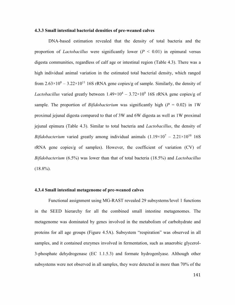
141
4.3.3 Small intestinal bacterial densities of pre-weaned calves
DNA-based estimation revealed that the density of total bacteria and the
proportion of Lactobacillus were significantly lower (P < 0.01) in epimural versus
digesta communities, regardless of calf age or intestinal region (Table 4.3). There was a
high individual animal variation in the estimated total bacterial density, which ranged
from 2.63×10⁸ – 3.22×10¹¹ 16S rRNA gene copies/g of sample. Similarly, the density of
Lactobacillus varied greatly between 1.49×10⁴ – 3.72×10⁹ 16S rRNA gene copies/g of
sample. The proportion of Bifidobacterium was significantly high (P = 0.02) in 1W
proximal jejunal digesta compared to that of 3W and 6W digesta as well as 1W proximal
jejunal epimura (Table 4.3). Similar to total bacteria and Lactobacillus, the density of
Bifidobacterium varied greatly among individual animals (1.19×10⁷ – 2.21×10¹⁰ 16S
rRNA gene copies/g of samples). However, the coefficient of variation (CV) of
Bifidobacterium (6.5%) was lower than that of total bacteria (18.5%) and Lactobacillus
(18.8%).
4.3.4 Small intestinal metagenome of pre-weaned calves
Functional assignment using MG-RAST revealed 29 subsystems/level 1 functions
in the SEED hierarchy for all the combined small intestine metagenomes. The
metagenome was dominated by genes involved in the metabolism of carbohydrate and
proteins for all age groups (Figure 4.5A). Subsystem “respiration” was observed in all
samples, and it contained enzymes involved in fermentation, such as anaerobic glycerol-
3-phosphate dehydrogenase (EC 1.1.5.3) and formate hydrogenlyase. Although other
subsystems were not observed in all samples, they were detected in more than 70% of the

142
samples. Small intestine metagenomes (functional abundance) did not differ significantly
by age group or gut region sampled (Figure 4.5B). Thus, for further analysis of postnatal
changes in microbial functions, we grouped all of the sampled intestinal regions. The top
subsystems identified from the collective small intestine metagenomes were
“carbohydrate metabolism” (range = 0-13.7%; mean±sem = 10.5±0.6%), “protein
metabolism” (range = 0-19.5%; mean±sem = 9.7±0.6%), “amino acids and derivatives”
(range = 0-12.6%; mean±sem = 6.7±0.4%), “phages, prophages, transposable elements,
plasmids” (range = 0-95.4%; mean±sem = 9.3±3.0%), “DNA metabolism” (range = 0-
9.6%; mean = 4.8±0.3%), “RNA metabolism” (range = 0-13.7%; mean±sem =
4.8±0.3%), and “cofactors, vitamins, prosthetic groups, pigments” (range = 0-8.1%;
mean±sem = 4.3±0.3%).
When the collective intestinal metagenomes were compared among different age
groups, the subsystems could be categorized into three patterns depending on age-
dependent trends in their abundances (Figure 4.5C, 4.5D, 4.5E). Essential functions for
all microbiota, such as “protein metabolism”, “carbohydrate metabolism”, and “DNA and
RNA metabolism” as well as biosynthesis processes (“cofactors, vitamins, prosthetic
groups, pigments”), were among the functions that increased in abundance from the first
to the third week and then remained stable from the third to the sixth week (Figure 4.5C).
In contrast, the abundance of “phages, prophages, transposable elements, plasmids”,
“membrane transport”, “virulence, disease and defense”, “iron acquisition”, “respiration”
and “sulfur and potassium metabolism” decreased from the first to the third week and
then remained stable from the third to the sixth week (Figure 4.5D). However, the
abundances of “motility and chemotaxis”, “cell wall and capsule”, “regulation and cell

143
signaling”, “secondary metabolism”, “phosphorus metabolism” and “stress response”
remained stable throughout the first six weeks of life (Figure 4.5E).
In total, 185 level 2 functions in the SEED hierarchy were detected within the
small intestinal communities (proximal jejunum – 179, distal jejunum – 144, ileum –
183). However, there was no single level 2 function observed in all of the samples
analyzed. We identified 16 and 5 level 2 functions present in all distal jejunum and ileum
samples, respectively, but no level 2 functions were conserved among all of the proximal
jejunum samples. Microbial functions related to “protein biosynthesis”, “folate and
pterines”, “DNA repair”, “lysine, threonine, methionine, cysteine”, and “RNA processing
and modification” were detected in all of the distal jejunum and ileal samples. Among the
detected level 2 functions, the abundances of “DNA repair”, “di and oligosaccharides”,
“carbohydrates”, “transcription”, “alanine, serine and glycine”, as well as “pyridoxine
(vitamin B6)” increased numerically with age (Figure 4.5F). In contrast, the abundances
of “electron donating reactions”, “protein biosynthesis”, “cell division and cell cycle”,
“central carbohydrate metabolism”, “lysine, threonine, methionine and cysteine”, and
“ABC transporters” decreased numerically with age (Figure 4.5G).
4.3.5 Small intestinal microbiome variation among individual animals
The composition of small intestinal microbiomes exhibited substantial variation
among individual animals. Therefore, the taxonomic and functional compositions of
digesta-associated microbiomes were further analyzed together as a whole, regardless of
the calf age and intestinal region, to understand the individual variations. The CV within
an individual animal was compared to that of the relevant age group to define the level of

144
individuality. The results of this analysis revealed a lower intra-individual variation
(variation among three intestinal regions) than the inter-individual variation (variation
among individuals within an age group) for the digesta-associated bacterial taxonomic
abundance of all age groups (Figure 4.6). Therefore, the bacterial genera (27) present in
at least half of the digesta samples (≥ 22 out of 45 samples) were used to perform a
Spearman rank correlation analysis to understand the similarities/dissimilarities among
samples. This analysis revealed that the majority of individual bacterial profiles (27 out
of 45 samples) were not correlated with each other based on their taxonomic
composition, while the rest of the samples separated into two clusters (Figure 4.7A). One
cluster had a higher abundance of Lactobacillus [ileal profiles of 4 calves: 1W – calf5
(72.4%), 3W – calf7 (86.4%) and calf8 (85.8%), 6W – calf12 (56.4%); jejunal profiles of
six calves: proximal jejunum: 1W – calf2 (40.4%), calf3 (23.0%), and calf5 (36.2%), 6W
– calf12 (28.0%); distal jejunum; 1W – calf1 (38.7%), calf3 (14.9%), and calf5 (58.1%),
3W – calf8 (14.9%), 6W – calf12 (31.2%)], when compared to the other small intestinal
samples. Another cluster contained a higher abundance of Bacteroides compared to other
samples (0.01 – 5.5%) and included all three small intestinal regions of one 1W calf
(calf4 - proximal jejunum – 23.7%; distal jejunum – 41.7%; ileum – 56.3%), the distal
jejunum of one 1W calf (calf2 – 22.2%) and one 6W calf (calf11 – 16.4%), and the ileum
of two 1W calves (calf2 – 45.8%; calf3 – 33.9%) as well as one 3W calf (calf6 – 17.9%).
Furthermore, Bacteroides, Prevotella, Roseburia, Ruminococcus, and Veillonella were
either less prevalent (≤ 0.1%) or absent in the four-ileal bacterial profiles of
Lactobacillus-dominant calves compared to the ileum of Bacteroides-dominant calves.

145
A comparison of the CVs of microbial functions revealed a lower inter-individual
variation in the abundance of functions compared to that of taxonomic abundance (Figure
4.6). Moreover, the individual variations of subsystems containing essential functions
(protein and carbohydrate metabolism, RNA and DNA metabolism) were lower than
other identified subsystems. Functions related to “phages, prophages, transposable
elements, plasmids” had the highest CV when compared to all other functions, especially
during the first week of life. Spearman rank correlation analysis further confirmed a
higher correlation (R2 ≥ 0.9, P < 0.01) among small intestine functional composition (34
out of 45 samples) than taxonomic composition, regardless of calf age or gut region
(Figure 4.7B). Within the highly correlated samples, one cluster contained samples from
3W and 6W calves (cluster 1), whereas the other cluster contained samples from 1W and
3W calves (cluster 2). Comparison of these two clusters revealed that functions related to
“protein metabolism” (cluster 1- 12.3±0.5%, cluster 2 - 9.0±0.5%; P < 0.01), “amino
acids and derivatives” (cluster 1- 7.9±0.4%, cluster 2 - 6.4±0.6%; P = 0.06) and
“nucleosides and nucleotides” (cluster 1- 5.8±0.3%; cluster 2 - 3.8±0.3%; P < 0.01) were
significantly enriched in cluster 1 compared to cluster 2. In contrast, functions related to
“cell wall and capsule” (cluster 1- 3.1±0.3%; cluster 2 - 4.4±0.3%; P = 0.01) and “sulfur
metabolism” (cluster 1- 0.3±0.05%; cluster 2 - 0.8±0.2%; P = 0.02) were enriched in
cluster 2 when compared to cluster 1. Comparison of taxonomic composition revealed
differences in the abundances of bacterial genera detected in these two function-based
clusters (Figure 4.7B; Table 4.4). The abundances of the predominant genera
Bifidobacterium, Bacillus, Streptococcus, Lactococcus, and Corynebacterium were
relatively higher in cluster 1, while the abundance of sulfur reducing bacteria

146
(Desulfarculus, Dethiosulfovibrio, Desulfatibacillum, Desulfuromonas,
Desulfurispirillum, Desulfotalea) was relatively higher in cluster 2. Furthermore, there
were 19 and 16 additional other genera that were relatively enriched in cluster 1 and
cluster 2, respectively (Table 4.4).
4.3.6 Linking identified ileal bacterial taxonomic-based clusters to the ileal
transcriptome
Ileal transcriptome data generated from the same calves (Liang et al., In Press)
were used to explore whether the variations identified in microbial composition may be
associated with the expression of protein coding genes of the host. Although there were
no significantly different protein-coding genes between the two groups, expressions of
the pro-inflammatory chemokines CXCL9, CXCL10, CXCL11 and CCL2 were
numerically higher (> 2-fold change) in Lactobacillus-dominant calves than in
Bacteroides-dominant calves (Figure 4.7C). These genes exhibited an enrichment in
functions related to “leukocytes and lymphocytes chemotaxis” as well as
“cytokine/chemokine-mediated signaling pathway” (Figure 4.7D). In contrast to
Lactobacillus-dominant calves, the Bacteroides-dominant calves had a numerically
higher (> 2-fold change) expression of genes involved in the mitogen-activated protein
kinase (MAPK) pathway (PLCE1, CCL22 and EGFR) (Figure 4.7C). These genes
revealed an enrichment of processes related to “cell adhesion”, “response to stimulus”
and “regulation of MAPK cascades” (Figure 4.7E).

147
4.4 Discussion
Although functions of the human gut microbiome are being studied extensively
through metagenomics approaches, such knowledge is very limited for ruminants. The
present study profiled the small intestine metagenomes of healthy newborn calves to
better understand the temporal dynamics of compositional and functional variation
associated with the establishment of the gut microbiome. Small intestinal microbiome
was mainly colonized by bacteria; however, the metagenomic sequencing revealed the
presence of archaea, fungi, protozoa and virus in pre-weaned calf gut. Genus
Methanobrevibacter dominated small intestine archaeal population, whereas family
Microviridae dominated virus (phage) population of pre-weaned calves. These
observations are in agreement with human studies reporting a higher abundance of
Methanobrevibacter (Lozupone et al., 2012) and Microviridae (Minot et al., 2013) in
fecal microbiomes. Presence of fungi phyla Ascomycota and Basidiomycota, the most
abundant fungi identified in the present study, have also reported in a metagenomic-based
study conducted using fecal samples of healthy human (Hoffmann et al., 2013). The main
protozoa observed in the pre-weaned calf small intestine was genus Cryptosporidium. C.
parvum is the main parasitic protozoa that contributes to neonatal calf diarrhea; however,
calves may harbor Cryptospiridium even at the absence of clinical signs or symptoms (De
Waele et al., 2013). The present study explored the bacterial population of pre-weaned
calves in greater depth.
The increased diversity and richness in small intestinal bacterial communities
with increasing age suggests a progressive establishment of a complex microbiome
during the first six weeks of life. Furthermore, total bacterial densities throughout the
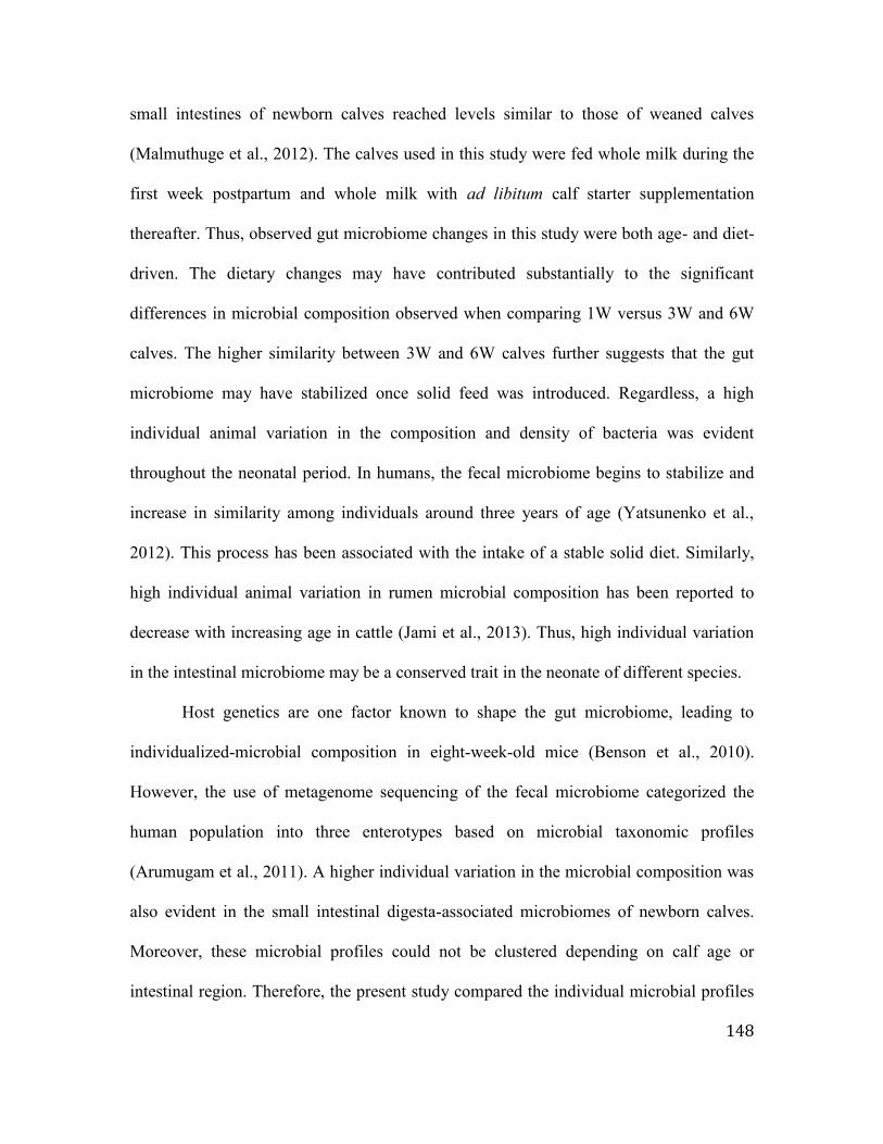
148
small intestines of newborn calves reached levels similar to those of weaned calves
(Malmuthuge et al., 2012). The calves used in this study were fed whole milk during the
first week postpartum and whole milk with ad libitum calf starter supplementation
thereafter. Thus, observed gut microbiome changes in this study were both age- and diet-
driven. The dietary changes may have contributed substantially to the significant
differences in microbial composition observed when comparing 1W versus 3W and 6W
calves. The higher similarity between 3W and 6W calves further suggests that the gut
microbiome may have stabilized once solid feed was introduced. Regardless, a high
individual animal variation in the composition and density of bacteria was evident
throughout the neonatal period. In humans, the fecal microbiome begins to stabilize and
increase in similarity among individuals around three years of age (Yatsunenko et al.,
2012). This process has been associated with the intake of a stable solid diet. Similarly,
high individual animal variation in rumen microbial composition has been reported to
decrease with increasing age in cattle (Jami et al., 2013). Thus, high individual variation
in the intestinal microbiome may be a conserved trait in the neonate of different species.
Host genetics are one factor known to shape the gut microbiome, leading to
individualized-microbial composition in eight-week-old mice (Benson et al., 2010).
However, the use of metagenome sequencing of the fecal microbiome categorized the
human population into three enterotypes based on microbial taxonomic profiles
(Arumugam et al., 2011). A higher individual variation in the microbial composition was
also evident in the small intestinal digesta-associated microbiomes of newborn calves.
Moreover, these microbial profiles could not be clustered depending on calf age or
intestinal region. Therefore, the present study compared the individual microbial profiles

149
generated from three small intestinal regions of 1W, 3W and 6W calves together to
understand individuality of the newborn calf intestinal microbiota in depth. Despite the
small number of samples, the clustering of samples based on microbial taxonomic
composition revealed individual calves with either high Lactobacillus or high
Bacteroides in the ileum, regardless of calf age. Comparison of the intestinal
transcriptomes of these calves suggested a potential link between the individualized gut
microbiome and the host immune system.
Lactobacillus-dominant calves exhibited higher expression of CXCL9, CXCL10,
and CXCL11 in the ileal tissue than Bacteroides-dominant calves. These pro-
inflammatory chemokines have been shown to be upregulated in dendritic cells and
human intestinal epithelial cells following exposure to Lactobacillus acidophilus (Weiss
et al., 2010; O’Flaherty and Klaenhammer, 2012). The upregulation of pro-inflammatory
genes in intestinal epithelial cells quickly returned to baseline levels one-hour after
exposure to L. acidophilus (O’Flaherty and Klaenhammer, 2012). These observations
suggest a potential priming of immune responses by Lactobacillus during the first
encounter. The priming effect of early Lactobacillus exposure may prepare the host for
subsequent anti-inflammatory regulatory responses, which may be a crucial process in
neonatal calves with a naïve mucosal immune system. In contrast to these studies,
infectious models (colitis and pneumovirus infection in mouse) have demonstrated that
Lactobacillus exerts an anti-inflammatory effect through the downregulation of CXCL10
and CXCL11 (Gabryszewski et al., 2011; von Schillde et al., 2012). Therefore, these
studies further suggest that the beneficial effects of Lactobacillus may vary depending on

150
the context, with a priming of immune responses upon initial encounter but a reduction of
inflammatory responses during infections.
MAPK cascade-related genes are involved in the regulation of T-cell
differentiation (Rincon et al., 2001) and increased expression of these genes in
Bacteroides-dominant calves may be consistent with an increased capacity for T-cell-
dependent responses. Although our transcriptome data from calves with either a higher
abundance of Lactobacillus or Bacteroides in the ileum revealed differential activation of
the mucosal immune system, further investigations are required to determine how these
changes in mucosal immune function may alter the host responses to enteric infections.
The expression of enriched-genes in each cluster was not statistically different. Therefore,
validation of these enriched-genes in a larger population and/or using challenged models
with phenotypic measurements will be required to determine if Lactobacillus and
Bacteroides define enterotypes in the calf small intestine that are associated with specific
immune functions and health outcomes.
Although taxonomic profiles of the microbiome in individual calves were
markedly different, such differences were not directly reflected in functional profiles.
This may be due to the observed higher abundance of universal microbial functions that
are important for microbial survival, such as “protein biosynthesis”, “RNA processing
and modification”, “DNA replication”, “central carbohydrate metabolism”, and “cell
cycle”. Nevertheless, two distinct clusters based on functional abundance were observed
within the small intestinal microbiome. Cluster 1 was enriched with functions related to
microbial protein metabolism, which has been suggested to play an important role in host
health through the regulation of amino acid homeostasis in the gut (Neis et al., 2015).

151
Moreover, the bacteria of calves in cluster 1 had a higher abundance (69-fold) of
Bifidobacterium than calves in cluster 2. A recent study has shown that Bifidobacterium
utilize milk protein effectively and require cysteine, a sulfide amino acid, for growth
(Ferrario et al., 2015). Sulfur reducing bacteria (SRB), which were mainly found in
cluster 2, also utilize cysteine as their energy source (Carbonero et al., 2012). Thus, the
observed low abundance or absence of Bifidobacterium in cluster 2 calves may be due to
the lack of cysteine for their growth. Dissimilatory sulfur reduction and cysteine
degradation by SRB produce H2S that can have detrimental effects on intestinal epithelial
cells (Carbonero et al., 2012). An accumulation of H2S in the colon inhibits the activity of
acyl-CoA dehydrogenase, which catalyzes the oxidation of butyrate in colonocytes and
compromises epithelial barrier permeability (Babidge et al., 1998). Bifidobacterium has
been reported to promote intestinal epithelial barrier functions (Ulluwishewa et al., 2011;
Hsieh et al., 2015), while SRB have been reported to have a negative impact on the
epithelial barrier (Babidge et al., 1998). Although these observed differences in bacterial
composition might be consistent with potential differences in epithelial barrier functions
between cluster 1 and cluster 2, transcriptome data (Liang et al., In Press) revealed no
differences in the expression of tight junction genes (< 2-fold change in the expression of
genes belonging to the claudin family, occludin, zonula occludens and junctional
adhesion molecules) when comparing animals in cluster1 and cluster2. Intestinal barrier
integrity loss can be measured using changes in the expression of tight junction proteins,
epithelial cell damage or the translocation of bacterial products (Grootjans et al., 2010).
Therefore, assessing barrier function by using these alternative indicators may provide

152
more sensitive measurement of how composition and functional variations in the gut
microbiota impact epithelial barrier function.
The small intestine bacterial taxonomic composition observed through sequencing
of the digesta-associated metagenome in the present study was similar to previous studies
reporting a higher abundance of Firmicutes in the small intestine (Malmuthuge et al.,
2014) and the fecal microbiome (Oikonomou et al., 2013) of young calves. However,
there were significant differences within the lower taxonomic hierarchy that could be
attributable to differences in the microbial profiling tools (whole genome sequencing
versus 16S amplicon sequencing), calf age, and sample type (fecal versus regional
intestine). Sequencing the V1-V3 region of 16S rRNA genes revealed a higher abundance
of Lactobacillus in the ileum and Sharpea in the proximal jejunum at three weeks of life
(Malmuthuge et al. 2014). However, the use of metagenomic profiling in the present
study revealed a higher abundance of Prevotella (17.9±12.8%) in the ileum and
Streptococcus (12.0±6.1%) in the proximal jejunum of three-week-old calves. Moreover,
an analysis of postnatal changes in the fecal microbiome reported a decrease in
Bifidobacterium abundance with increasing calf age (Uyeno et al., 2010; Oikonomou et
al., 2013), but the present study revealed that abundance of Bifidobacterium in the small
intestine increased with increasing calf age. As the present study used different animals at
each time point to analyze the patterns in postnatal changes, caution is required when
comparing these results with previous studies that sampled the same animals repeatedly
overtime. In contrast to digesta communities, the epimural communities differed from
those reported in a previous study, even at a higher level of the taxonomic hierarchy
(Malmuthuge et al., 2014). In contrast to our previous study (Malmuthuge et al., 2014),

153
the present study revealed a higher abundance of Actinobacteria (41.2±15.1%), followed
by Firmicutes (26.8±9.6%), Proteobacteria (20.1±15.5%) and Bacteroidetes (11.8±6.1%)
in the ileal tissues of three-week-old calves, and a higher abundance of Proteobacteria
(66.5±23.2%), followed by Firmicutes (14.0±8.1%), Actinobacteria (11.0±10.5%) and
Bacteroidetes (8.0±4.2%) in the jejunal tissues. The use of animals from different herds
and feeding calves different starter rations may explain some of the observed differences
between these two studies. Additionally, the small intestinal epimural bacterial
community is closely scrutinized by the host mucosal immune system (Hansen et al.,
2012), which may also contribute to taxonomic variation in the microbial communities
among different animals.
Despite the extensive use of preventative health management strategies, including
vaccination of pregnant cows and management of colostrum feeding (Smith, 2015), a
high rate of diarrhea-related neonatal deaths remains a major health problem in the cattle
industry (USDA, 2010). Neonatal calf diarrhea is often treated with antibiotics, but the
unrestricted use of antibiotics in the livestock industry is a controversial issue (Smith,
2015), especially given that antibiotic treatment of calf diarrhea has a low efficacy (Berge
et al., 2009). One reason for the use of antibiotics as a treatment for calf diarrhea is to
prevent pathogenic E. coli colonization of the small intestine (Smith, 2015). However,
with increasing restrictions on the prophylactic use of antibiotics (Smith, 2015), it is
important to explore other microbial manipulation techniques that can limit or prevent
pathogen colonization of the calf small intestine. Feeding probiotics has been shown to
reduce the frequency of pathogenic E. coli in the calf gastrointestinal tract (Zhao et al.,
1998), suggesting that promoting the colonization of beneficial bacteria may exclude

154
pathogens. Limited knowledge of the calf microbiome, especially in the small intestine, is
a barrier to developing such strategies to prevent or treat diarrhea. Thus, the present study
is a first step in understanding the dynamics of commensal microbiota establishment and
the function of the microbiome in the small intestine of newborn calves.
4.5 Conclusion
The present study analyzed the taxonomic and functional evolution of the
microbiome in the small intestine of newborn calves during the first six weeks of life.
During this developmental period, age- and diet-related increases in microbial diversity,
richness and density were observed; however, the taxonomic composition varied widely
among individual animals. The comparison of individual animal taxonomic profiles of
the ileal digesta classified calves as either Lactobacillus-dominant or Bacteroides-
dominant calves based on the relative abundance of the detected bacterial genera. A
comparison of functional profiles also classified calves into two distinct clusters,
revealing an antagonism between SRB and Bifidobacterium. This is the first study to
generate a detailed analysis of the taxonomical and functional development of the small
intestinal microbiome in healthy pre-weaned calves. This analysis provides a novel
framework to further develop strategies to manipulate the microbiome and improve calf
health.

155
4.6 References
Arumugam, M., Raes, J., Pelletier, E., Le Paslier, D., Yamada, T., Mende, D.R. et al.,
(2011). Enterotypes of the human gut microbiome. Nature. 473, 174-180. doi:
10.1038/nature09944.
Babidge, W., Millard, S., Roediger, W. (1998). Sulfides impair short chain fatty acid
beta-oxidation at acyl-CoA dehydrogenase level in colonocytes: implications for
ulcerative colitis. Mol. Cell Biochem. 181, 117-124.
Benjamini, Y., Hochberg, y. (1995). Controlling the false discovery rate: A practical and
powerful approach to multiple testing. Journal of the Royal Statistical Society.
Series B (Methodological). 57, 289-300.
Benson, A.K., Kelly, S.A., Legge, R., Ma, F., Low, S.J., Kim, J., et al., (2010).
Individuality in gut microbiota composition is a complex polygenic trait shaped
by multiple environmental and host genetic factors. Proc. Natl. Acad. Sci. U S A.
107, 18933-18938. doi: 10.1073/pnas.1007028107.
Berge, A.C.B., Moore, D.A., Besser, T.E., Sischo, W.M. (2009). Targeting therapy to
minimize antimicrobial use in preweaned calves: Effects on health, growth, and
treatment costs. J. Dairy Sci. 92, 4707-4714. doi: 10.3168/jds.2009-2199.
Caporaso, J.G., Kuczynski, J., Stombaugh, J., Bittinger, K., Bushman, F.D., Costello,
E.K., et al., (2010). QIIME allows analysis of high-throughput community
sequencing data. Nat. Methods. 7:335-336. doi: 10.1038/nmeth.f.303.
Carbonero, F., Benefiel, A.C., Alizadeh-Ghamsari, A.H., Gaskins, H.R. (2012). Microbial
pathways in colonic sulfur metabolism and links with health and disease. Front.
Physiol. 3, 448. doi: 10.3389/fphys.2012.00448.

156
Chambers, L., Yang, Y., Littier, H., Ray, P., Zhang, T., Pruden, A. et al., (2015).
Metagenomic analysis of antibiotic resistance genes in dairy cow feces following
therapeutic administration of third generation cephalosporin. PLoS One. 10,
e0133764. doi: 10.1371/journal.pone.0133764.
Delroisse, J-M., Boulvin, A-L., Parmentier, I., Dauphin, R.D., Vandenbol, M., Portetelle,
D. (2008). Quantification of Bifidobacterium spp. and Lactobacillus spp. in rat
fecal samples by real-time PCR. Microbiol. Res. 163, 663-670.
Denman, S.E., McSweeney, C.S. (2015). The early impact of genomics and
metagenomics on ruminal microbiology. Annu. Rev. Anim. Biosci. 3, 447-465.
doi: 10.1146/annurev-animal-022114-110705.
De Waele, V., Van den Broeck, F., Huyse, T., McGrath, G., Higgins, I., Speybroeck, N.,
et al., (2013). Panmictic structure of the Cryptosporidium parvum population in
Irish calves: influence of prevalence and host movement. Appl. Environ.
Microbiol. 79, 2534-2541. doi: 10.1128/AEM.03613-12.
Ferrario, C., Duranti, S., Milani, C., Mancabelli, L., Lugli, GA., Turroni, F., et al., (2015).
Exploring amino acid auxotrophy in Bifidobacterium bifidum PRL2010. Front.
Microbiol. 6, 1331. doi: 10.3389/fmicb.2015.01331.
Gabryszewski, S.J., Bachar, O., Dyer, K.D., Percopo, C.M., Killoran, K.E.,
Domachowske, J.B., et al., (2011). Lactobacillus-mediated priming of the
respiratory mucosa protects against lethal pneumovirus infection. J. Immunol.
186, 1151-1161. doi: 10.4049/jimmunol.1001751.

157
Grootjans, J., Thuijls, G., Verdam, F., Derikx, J.P.M., Lenaerts, K., and Buurman, W.A.
(2010). Non-invasive assessment of barrier integrity and function of the human
gut. World J. Gastrointest. Surg. 2: 61-69. doi: 10.4240/wjgs.v2.i3.61.
Hansen, C.H.F., Nielsen, D.S., Kverka, M., Zakostelska, Z., Klimesova, K., Hudcovic, T.,
et al., (2012). Patterns of early gut colonization shape future immune responses of
the host. PLoS One. 7, e34043. doi:10.1371/journal.pone.0034043.
Hoffmann, C., Dollive, S., Grunberg, S., Chen, J., Li, H., Wu, G.D., et al., (2013).
Archaea and fungi of the human gut microbiome: correlations with diet and
bacterial residents. PLoS One. 17, 8:e66019. doi: 10.1371/journal.pone.0066019.
Hsieh, C.Y., Osaka, T., Moriyama, E., Date, Y., Kikuchi, J., Tsuneda, S. (2015).
Strengthening of the intestinal epithelial tight junction by Bifidobacterium
bifidum. Physiol. Rep. 3, e12327. doi: 10.14814/phy2.12327.
Jami, E., Israel, A., Kotser, A., Mizrahi, I. (2013). Exploring the bovine rumen bacterial
community from birth to adulthood. ISME J. 7, 1069-1079. doi:
10.1038/ismej.2013.2.
Li, M., Penner, G.B., Hernandez-Sanabria, E., Oba, M., Guan, L.L. (2009). Effects of
sampling location and time, and host animal on assessment of bacterial diversity
and fermentation parameters in the bovine rumen. J. Appl. Microbiol. 107:1924-
1934. doi: 10.1111/j.1365-2672.2009.04376.x.
Li, R.W., Connor, E.E., Baldwin, R.L., Sparks, M.L. (2012). Characterization of the
rumen microbiota of pre-ruminant calves using metagenomic tools. Environ.
Microbiol. 14, 129-139. doi: 10.1111/j.1462-2920.2011.02543.

158
Liang, G., Malmuthuge, N., Bao, H., McFadden, T., Stothard, P., Griebel, P.J., et al.,
(2014) Potential regulatory role of microRNAs in the development of bovine
gastrointestinal tract during early life. PLoS One. 9, e92592. doi:
10.1371/journal.pone.0092592.
Lozupone, C.A., Stombaugh, J.I., Gordon, J.I., Jansson, J.K., Knight, R. (2012).
Diversity, stability and resilience of the human gut microbiota. Nature. 489, 220-
230. doi: 10.1038/nature11550.
Malmuthuge, N., Griebel, P.J, Guan, L.L. (2014). Taxonomic identification of
commensal bacteria associated with the mucosa and digesta throughout the
gastrointestinal tracts of pre-weaned calves. Appl. Environ. Microbiol. 80, 2012-
2028. doi: 10.1128/AEM.03864-13.
Malmuthuge, N., Li, M., Chen, Y., Fries, P., Griebel, P.J., Baurhoo, B. et al., (2012).
Distinct commensal bacteria associated with ingesta and mucosal epithelium in
the gastrointestinal tracts of calves and chickens. FEMS Microbiol. Ecol. 79, 337-
347. doi: 10.1111/j.1574-6941.2011.01220.
Meyer, F., Paarmann, D., D'Souza, M., Olson, R., Glass, E.M., Kubal, M., et al., (2008).
The metagenomics RAST server – a public resource for the automatic
phylogenetic and functional analysis of metagenomes. BMC Bioinformatics. 9,
386. doi: 10.1186/1471-2105-9-386.
Minot, S., Bryson, A., Chehoud, C., Wu, G.D., Lewis, J.D., Bushman, F.D. (2013). Rapid
evolution of the human gut virome. Proc. Natl. Acad. Sci. U S A. 110, 12450-
12455. doi: 10.1073/pnas.1300833110.

159
Neis, E.P., Dejong, C.H., Rensen, S.S. (2015). The role of microbial amino acid
metabolism in host metabolism. Nutrients. 7, 2930-2946. doi:
10.3390/nu7042930.
O'Flaherty, S., Klaenhammer, T.R. (2012). Influence of exposure time on gene expression
by human intestinal epithelial cells exposed to Lactobacillus acidophilus. Appl.
Environ. Microbiol. 78, 5028-5032. doi: 10.1128/AEM.00504-12.
Oikonomou, G., Teixeira, A.G., Foditsch, C., Bichalho, M.L., Machado, V.S., Bicalho,
R.C. (2013). Fecal microbial diversity in pre-weaned dairy calves as described by
pyrosequencing of metagenomic 16S rDNA. Associations of Faecalibacterium
species with health and growth. PLOS One. 8, e63157. doi:
10.1371/journal.pone.0063157.
Petrosino, J.F., Highlander, S., Luna, R.A., Gibbs, R.A., Versalovic, J. (2009).
Metagenomic pyrosequencing and microbial identification. Clin. Chem. 55, 856-
866. doi: 10.1373/clinchem.2008.107565.
Qin, J., Li, R., Raes, J., Arumugam, M., Burgdorf, K.S., Manichanh, C., et al., (2010). A
human gut microbial gene catalogue established by metagenomic sequencing.
Nature. 464, 59-65. doi: 10.1038/nature08821.
Rincon, M., Flavell, R.A., Davis, R.J. (2001). Signal transduction by MAP kinases in T
lymphocytes. Oncogene. 20, 2490-2497.
Shafquat, A., Joice, R., Simmons, S.L., Huttenhower, C. (2014). Functional and
phylogenetic assembly of microbial communities in the human microbiome.
Trends Microbiol. 22, 261-266. doi: 10.1016/j.tim.2014.01.011.

160
Smith, G. (2015). Antimicrobial decision making for enteric diseases of cattle. Vet. Clin.
North Am. Food Anim. Pract. 31, 47-60. doi: 10.1016/j.cvfa.2014.11.004.
Sommer, F., Nookaew, I., Sommer, N., Fogelstrand, P., Backhed, F. (2015). Site-specific
programming of the host epithelial transcriptome by the gut microbiota. Genome
Biol. 16, 62. doi: 10.1186/s13059-015-0614-4.
Stevenson, D.M., Weimer, P.J. (2007). Dominance of Prevotella and low abundance of
classical ruminal bacterial species in the bovine rumen revealed by relative
quantification real-time PCR. Appl. Microbiol. Biotechnol. 75, 165–174.
The Human Microbiome Project Consortium. (2012). A framework for human
microbiome research. Nature. 486, 215–221. doi:10.1038/nature11209.
Turnbaugh, P.J., Hamady, M., Yatsunenko, T., Cantarel, B.L., Duncan, A., Ley, R.E., et
al., (2009). A core gut microbiome in obese and lean twins. Nature. 457, 480-484.
doi: 10.1038/nature07540.
Ulluwishewa, D., Anderson, R.C., McNabb, W.C., Moughan, P.J., Wells, J.M., Roy, N.C.
(2011). Regulation of tight junction permeability by intestinal bacteria and dietary
components. J. Nutr. 141, 769-776. doi: 10.3945/jn.110.135657.
USDA (2010). Dairy 2007, Heifer calf health and management practices on US dairy
operations. pp 122-131. USDA:APHIS:VS, CEAH. Fort Collins, CO.
Uyeno, Y., Shigemori, S., Shimosato, T. (2015). Effect of probiotics/prebiotics on cattle
health and productivity. Microbes. Environ. 30, 126-132. doi:
10.1264/jsme2.ME14176.
von Schillde, M.A., Hormannsperger, G., Weiher, M., Alpert, C.A., Hahne, H., Bauerl,
C., et al., (2012). Lactocepin secreted by Lactobacillus exerts anti-inflammatory

161
effects by selectively degrading proinflammatory chemokines. Cell Host Microbe.
11, 387-396. doi: 10.1016/j.chom.2012.02.006.
Weiss, G., Rasmussen, S., Nielsen Fink, L., Jarmer, H., Nohr Nielsen, B., Frokiaer, H.
(2010). Bifidobacterium bifidum actively changes the gene expression profile
induced by Lactobacillus acidophilus in murine dendritic cells. PLoS One. 5,
e11065. doi: 10.1371/journal.pone.0011065.
White, J.R., Nagarajan, N., Pop, M. Statistical methods for detecting differentially
abundant features in clinical metagenomic samples. PLoS Comput. Biol. 5,
e1000352. doi: 10.1371/journal.pcbi.1000352.
Yatsunenko, T., Rey, F.E., Manary, M.J., Trehan, I., Dominguez-Bello, M.G., Contreras,
M., et al., (2012). Human gut microbiome viewed across age and geography.
Nature. 486, 222-227. doi: 10.1038/nature11053.
Yu Z, Morrison M. (2004). Improved extraction of PCR-quality community DNA from
digesta and fecal samples. Biotechniques. 36, 808-812.
Zhao, T., Doyle, M.P., Harmon, B.G., Brown, C.A., Mueller, P.O.E., Parks, A.H. (1998).
Reduction of carriage of enterohemorrhagic Escherichia coli O157:H7 in cattle by
inoculation with probiotic bacteria. J. Clin. Microbiol. 36, 641-647.

162
4.7 Tables and Figures
Table 4.1 Prevalence of bacterial phyla and genera observed in the small intestinal digesta communities
Proximal jejunum Distal jejunum Ileum Significant comparisons
1W 3W 6W SEM 1W 3W 6W SEM 1W 3W 6W SEM Proximal jejunum Distal jejunum Ileum
Phylum1
Actinobacteria 6.1 22.7 32.1 5.2 2.3 18.7 10.1 0.5 2.7 2.8 28.8 5.5 1W vs. 3W, 6W 1W vs. 3W, 6W
Bacteroidetes 18.4 14.3 25.0 3.9 26.7 5.8 9.8 2.0 35.4 7.3 9.2 3.8
1W vs. 3W, 6W 1W vs. 3W, 6W
Firmicutes 53.9 50.6 31.8 3.6 43.2 48.0 59.5 4.0 47.4 63.8 36.7 9.6
Proteobacteria 19.4 7.6 6.9 1.1 26.6 20.6 12.7 2.2 11.2 23.3 16.6 3.8 1W vs. 3W, 6W 3W vs. 6W
Genus1
Bifidobacterium 4.3 6.0 23.5 5.2 1.1 8.1 3.6 0.2 1.4 1.1 2.2 2.4
1W vs. 3W
Prevotella 7.9 8.0 20.7 6.1 10.6 2.3 3.8 0.5 5.4 61.7 6.2 0.2
1W vs. 3W, 6W
Lactobacillus 23.6 3.9 7.4 2.6 24.9 5.4 9.3 2.4 17.1 44.4 11.5 2.5 1W vs. 3W, 6W 1W vs. 3W, 6W
Clostridium 5.4 7.1 3.6 2.5 3.7 2.8 9.5 0.3 9.8 2.7 6.6 1.7
1W vs. 3W
Ruminococcus 0.7 3.6 3.4 1.3 0.6 1.0 1.9 0.0 1.2 0.7 1.8 0.0 1W vs. 3W, 6W
Bacteroides 9.0 4.3 3.3 1.8 15.1 2.6 4.9 1.2 27.9 4.7 2.2 0.1
1W vs. 3W, 6W
Streptococcus 3.0 5.7 2.9 2.0 1.6 13.4 7.2 0.8 1.0 4.8 2.3 0.3
1W vs. 3W, 6W
Eubacterium 4.0 4.0 1.7 0.2 2.1 2.1 4.0 0.1 2.0 2.7 0.9 0.0
1 Mean relative abundance as a % of total bacterial phyla or genera observed. 1W- 1-week-old calves, 3W – 3-week-old calves,
6W – 6-week-old calves

163
Table 4.2 Small intestinal bacterial diversity and richness
Proximal jejunum Distal jejunum Ileum
1W 3W 6W 1W 3W 6W 1W 3W 6W
Digesta-associated bacteria (whole genome sequencing)
Shannon index 1.8±0.8 1.9±1.6 2.2±1.6 1.6±1.0 2.1±1.5 2.2±1.4 1.7±1.2 2.1±1.7 2.0±1.5
# of genera 100±21 66±16 98±21 96±23 96±12 109±12 79±16 78±24 89±19
Predominant genera1 15±2 16±1 13±4 11±2 21±1 17±2 13±3 8±3 9±3
Tissue-attached bacteria (16S amplicon sequencing of V1-V3)
Shannon index 4.2±0.4 4.8±0.7 4.7±0.5 4.2±0.7 3.3±0.6 4.1±0.5 3.7±0.6 3.8±0.9 4.0±0.4
# of genera 23±8 36±7 32±3 23±7 19±9 30±7 22±6 28±9 25±6
Predominant genera1 11±2 14±4 8±2 13±4 8±4 8±3 11±3 7±3 11±4 1 relative abundance > 1% in at least one sample. 1W – 1-week-old calves, 3W – 3-week-old calves, 6W – 6-week-old calves.
Data presented as mean±SEM.

164
Table 4.3 Bacterial densities in the small intestinal communities of pre-weaned calves
Gut region Sample type Age of calves
1W 3W 6W
Total bacteria (16S rRNA gene copy/g of fresh sample)1
Proximal
jejunum
Tissue 1.1±0.4×10⁹ 1.3±0.3×10⁹ 3.5±0.9×10⁹
Digesta 1.6±1.2×10¹⁰ 2.0±0.3×10¹⁰ 8.5±6.1×10¹⁰
Distal jejunum Tissue 1.1±0.3×10⁹ 0.8±0.2×10⁹ 2.5±0.9×10⁹
Digesta 2.6±2.1×10¹⁰ 9.4±6.6×10⁹ 2.0±1.1×10¹⁰
Ileum Tissue 1.0±0.2×10⁹ 1.0±0.2×10⁹ 3.2±1.6×10⁹
Digesta 1.5±0.8×10¹⁰ 3.7±2.2×10⁹ 9.0±7.4×10⁹
Lactobacillus (%)2
Proximal
jejunum
Tissue 0.02±0.01 0.001±0.0001 0.012±0.01
Digesta 3.3±1.7 9.0±8.3 0.80±0.79
Distal jejunum Tissue 0.03±0.02 0.003±0.001 0.001±0.0001
Digesta 11.1±6.8 6.1±4.1 0.08±0.07
Ileum Tissue 0.01±0.0001 0.001±0.0001 0.001±0.0001
Digesta 6.7±6.1 2.1±1.4 0.56±0.55
Bifidobacterium (%)3
Proximal
jejunum
Tissue 5.6±1.9 12.6±0.9 18.7±12.5
Digesta 25.7±6.4 9.9±6.5 6.2±2.4
Distal jejunum Tissue 3.9±0.7 9.0±2.2 7.5±2.2
Digesta 7.8±2.7 17.5±4.5 11.4±3.4
Ileum Tissue 5.0±0.9 6.0±0.5 2.6±0.4
Digesta 11.4±3.6 9.6±4.3 14.4±3.3 1 total bacterial density differed between tissue and digesta, regardless of age and gut
region (P < 0.01)
2 prevalence of Lactobacillus differed between tissue and digesta, regardless of age and
gut region (P < 0.01)
3 prevalence of Bifidobacterium was high; in 1W proximal jejunum digesta than 3W and
6W; in 1W proximal jejunum digesta than 1W proximal jejunum tissue; in 1W ileum
digesta than 1W ileum tissue (P = 0.02)
1W – 1-week-old calves, 3W – 3-week-old calves, 6W – 6-week-old calves.

165
Table 4.4 Differentially abundant bacterial genera of function-based clusters identified in
the calf gut
Genus Cluster1 Cluster2 P – value1
Aquifex 0.009±0.0 0.10±0.0 2.56456E-10
Alicycliphilus 0 0.18±0.002 6.16124E-10
Desulfarculus 0.003±0.0 0.08±0.0 4.72058E-09
Asticcacaulis 0.003±0.0 0.16±0.001 1.51907E-06
Lysinibacillus 0.033±0.0 0.04±0.0 4.36666E-05
Dethiosulfovibrio 0.053±0.0 0.30±0.01 9.53096E-05
Erwinia 0.001±0.0 0.14±0.0 0.0002
Desulfatibacillum 0 0.74±0.05 0.0001
Alkaliphilus 0 0.05±0.0 0.0001
Eggerthella 0 0.05±0.0 0.0001
Leeuwenhoekiella 0 0.60±0.02 0.0001
Klebsiella 0.22±0.001 0 0.0001
Mycobacterium 0.17±0.0 0.007±0.0 0.0001
Campylobacter 0.39±0.002 0.02±0.0 0.0001
Streptococcus 6.8±1.3 0.21±0.003 0.0001
Bacillus 1.1±0.02 0.08±0.0 0.0001
Bifidobacterium 12.6±5.7 0.18±0.002 0.0001
Allochromatium 0.018±0.0 0.06±0.0 0.001
Desulfuromonas 0.033±0.0 0.05±0.0 0.002
Halanaerobium 0.15±0.001 0 0.003
Lactococcus 3.5±1.0 0.01±0.0 0.003
Acinetobacter 0.34±0.004 0.02±0.0 0.003
Desulfurispirillum 0.02±0.0 0.09±0.001 0.003
Rickettsia 0.04±0.0 0 0.005
Mycoplasma 0.47±0.005 0.01±0.0 0.005
Corynebacterium 1.48±0.10 0.04±0.0 0.006
Leuconostoc 0.58±0.01 0.01±0.0 0.007
Proteus 0.04±0.0 0.002±0.0 0.01
Anabaena 0.01±0.0 0.001±0.0 0.01
Desulfotalea 0.006±0.0 0.05±0.0 0.01
Erysipelothrix 0.008±0.0 0.002±0.0 0.01
Ammonifex 0.06±0.0 0.07±0.0 0.01
Rothia 0.06±0.0 0.04±0.0 0.01
Megasphaera 0.16±0.002 0 0.01
Pedobacter 0.04±0.0 0.02±0.0 0.02
Vibrio 0.26±0.001 0.03±0.0 0.02
Pelodictyon 0.02±0.0 0.05±0.0 0.02
Comamonas 0.23±0.002 6.1±0.8 0.02
Brucella 0.13±0.0 6.8±1.5 0.02
Alicyclobacillus 0.12±0.0 5.4±1.3 0.03

166
Francisella 0.07±0.0 2.07±0.2 0.03
Myxococcus 0.14±0.002 0 0.03
Geobacillus 0.29±0.002 0.021±0.0 0.04
Exiguobacterium 0.12±0.0 1.504±0.06 0.04
Haemophilus 0.22±0.001 0.026±0.0 0.04
Aeromonas 0.17±0.002 0.014±0.0 0.05
Data presented as mean±SEM.
1Mulitple testing correction is performed using Benjamini and Hochberg, 1995

167
Figure 4.1 Composition of small intestinal microbiomes obtained through whole genome
sequencing of digesta communities.
Each data series present mean relative abundance (%) of small intestinal microbiota per
age category. 1 – 1-week-old calves, 3 – 3-week-old calves, 6 – 6-week-old calves.

168
Figure 4.2 Individual variations in the relative abundance of four main bacterial phyla
detected in small intestinal digesta communities.
Each data point represents an individual calf and bar represents mean relative abundance
of a small intestinal region or age group. Upper panel presents all three age groups within
a small intestinal region (15 data point/ phylum) and lower panel presents all three small
intestinal regions within a age category.

169
Figure 4.3 Bacterial genera with mean relative abundance displaying increasing (A) and
decreasing (B) trends with increasing calf age.
1W – 1-week-old calves, 3W – 3-week-old calves, 6W – 6-week-old calves.

170
Figure 4.4 Phylum-level composition of tissue-attached bacteria generated through 454
sequencing of V1-V3 region of 16S rRNA gene.
Numerical values represent individual calf IDs. 1W – 1-week-old calves, 3W – 3-week-
old calves, 6W – 6-week-old calves. Each data series represent relative abundance of
detected bacterial phyla.

171
Figure 4.5 Small intestinal microbial functions.
(A) Abundance of the subsystems (level 1 functions of the SEED hierarchy) detected in
the calf small intestine digesta-associated microbial communities. Numerical values
represent individual calf IDs. 1W – 1-week-old calves, 3W – 3-week-old calves, 6W – 6-
week-old calves. Each data series represent relative abundance of detected subsystems.

172
Figure 4.5B PCoA plot for functional composition.
PC1 and PC2 values of the individual microbial profiles obtain within MG-RAST
platform using operational taxonomic units (OTUs) identified at gene level (level 4
functions of the SEED hierarchy) and plot is generated using R package (plot function).

173
Figure 4.5C Subsystems with mean relative abundance displaying increasing and
stabilizing trends with increasing calf age.
1W – 1-week-old calves, 3W – 3-week-old calves, 6W – 6-week-old calves.

174
Figure 4.5D Subsystems with mean relative abundance displaying decreasing and
stabilizing trends with increasing calf age.
1W – 1-week-old calves, 3W – 3-week-old calves, 6W – 6-week-old calves.

175
Figure 4.5E Subsystems with stable mean relative abundance throughout the neonatal
period (1-week to 6-week).
1W – 1-week-old calves, 3W – 3-week-old calves, 6W – 6-week-old calves.

176
Figure 4.5F Level 2 functions with mean relative abundance displaying increasing trends
with increasing calf age.
1W – 1-week-old calves, 3W – 3-week-old calves, 6W – 6-week-old calves.

177
Figure 4.5G Level 2 functions with mean relative abundance displaying decreasing trends
with increasing calf age.
1W – 1-week-old calves, 3W – 3-week-old calves, 6W – 6-week-old calves.

178
Figure 4.6 Coefficient of variations (ΔCV) of the relative abundance of the subsystems
and bacterial genera (present in > 50% of the samples).
Axis represent ΔCV value of each genera/subsystem and ΔCV = CVpopulation – CVindividual
animal. Positive ΔCV represents higher inter-individual and lower intra-individual variation
within an age group. Negative ΔCV represents lower inter-individual and higher intra-
individual variation within an age group. Numerical values represent individual calf IDs.

179
Figure 4.7 Clustering of microbial profiles based on taxonomic/functional similarities.
Figure 4.7A Clustering of calves based on microbial taxonomic composition. Clustering
is based on the Spearman rank correlation coefficient between two samples generated
based on the relative abundance of 27 bacterial genera identified in at least 50% of
samples. Each raw and column represents an individual microbial profile generated from
small intestinal samples (45 microbial profiles).

180
Figure 4.7 B Clustering of calves based on microbial functional abundance.
Clustering is based on the Spearman rank correlation coefficient between two samples
generated based on the relative abundance of identified Subsystems. Abundances of
bacterial genera between two clusters are compared using metastats and data presented as
mean±SEM. Each raw and column represent an individual microbial profile generated
from small intestinal samples (45 microbial profiles).

181
Figure 4.7C Genes enriched in the ileum of Lactobacillus-dominant and Bacteroides-
dominant calves.
Fold change is calculated by dividing expression of genes (mean CPM) in Lactobacillus-
dominant calves by expression of genes (mean CPM) in Bacteroides-dominant calves and
vice versa. CXCL9 – chemokine ligand 9, CXCL10 – chemokine ligand 10, CXCL11 –
chemokine ligand 11, PLCE1 - Phospholipase C Epsilon 1, CCL22 - C-C Motif
Chemokine Ligand 22, EGFR - Epidermal Growth Factor Receptor.

182
Figure 4.7D Functions of the genes highly expressed in the ileum of the Lactobacillus-
dominant calves.
Functional enrichment is performed using GO enrichment analysis in Gene Ontology
Consortium. P – value - significance for the enrichment in the dataset of the listed GO
identifier. A P – value close to zero (a higher –log (P - value) means that the group of
genes associated with the particular GO term is more significant and is less likely the
observed annotation of the particular GO term to a group of genes occurs by chance.

183
Figure 4.7E Functions of the genes highly expressed in the ileum of the Bacteroides-
dominant calves.
Functional enrichment is performed using GO enrichment analysis in Gene Ontology
Consortium. P – value - significance for the enrichment in the dataset of the listed GO
identifier. A P – value close to zero (a higher –log (P - value) means that the group of
genes associated with the particular GO term is more significant and is less likely the
observed annotation of the particular GO term to a group of genes occurs by chance.
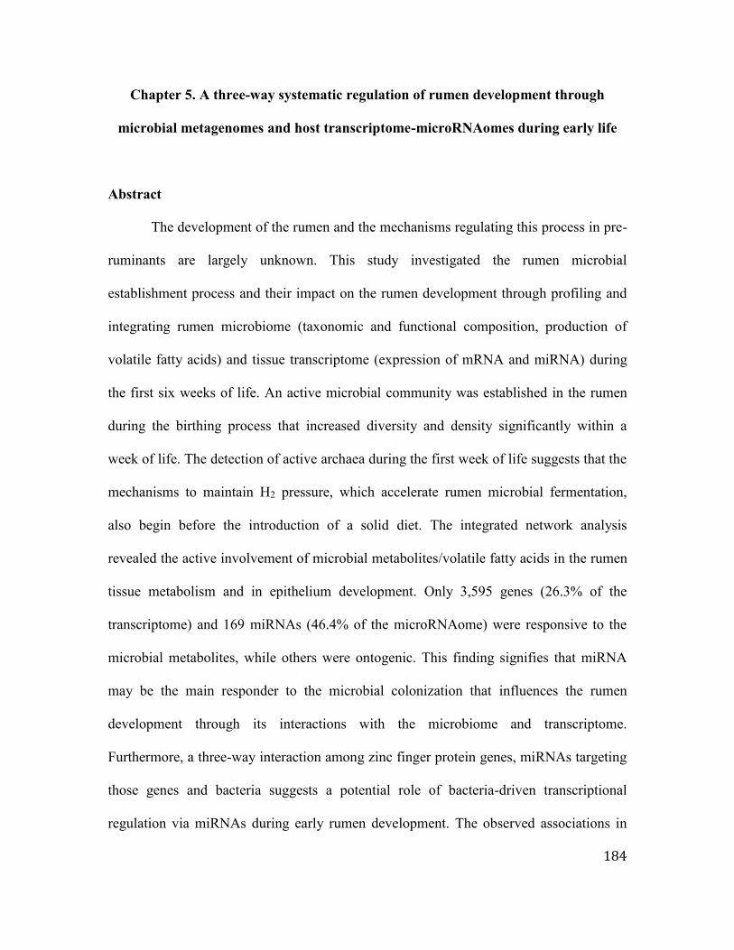
184
Chapter 5. A three-way systematic regulation of rumen development through
microbial metagenomes and host transcriptome-microRNAomes during early life
Abstract
The development of the rumen and the mechanisms regulating this process in pre-
ruminants are largely unknown. This study investigated the rumen microbial
establishment process and their impact on the rumen development through profiling and
integrating rumen microbiome (taxonomic and functional composition, production of
volatile fatty acids) and tissue transcriptome (expression of mRNA and miRNA) during
the first six weeks of life. An active microbial community was established in the rumen
during the birthing process that increased diversity and density significantly within a
week of life. The detection of active archaea during the first week of life suggests that the
mechanisms to maintain H2 pressure, which accelerate rumen microbial fermentation,
also begin before the introduction of a solid diet. The integrated network analysis
revealed the active involvement of microbial metabolites/volatile fatty acids in the rumen
tissue metabolism and in epithelium development. Only 3,595 genes (26.3% of the
transcriptome) and 169 miRNAs (46.4% of the microRNAome) were responsive to the
microbial metabolites, while others were ontogenic. This finding signifies that miRNA
may be the main responder to the microbial colonization that influences the rumen
development through its interactions with the microbiome and transcriptome.
Furthermore, a three-way interaction among zinc finger protein genes, miRNAs targeting
those genes and bacteria suggests a potential role of bacteria-driven transcriptional
regulation via miRNAs during early rumen development. The observed associations in

185
the present study suggests that miRNAs may coordinate the host-microbial interactions
during early rumen development in neonatal calves and this phenomenon may also be
applicable to early gut development in other mammalian species.
5.1 Introduction
A dynamic microbial population rapidly colonizes the in utero sterile mammalian
gut during and after birth. The impact of microbiota on gut development has mainly been
studied using germfree animal models, reporting structural and functional (mucosal
immune system) changes in the small intestine in their absence (Sommer and Backhed,
2013). A recent study revealed that the colonization of gut region-specific microbiota,
which cannot be detected via fecal sampling, regulates the gut development in a region-
specific manner through its interactions with the host transcriptome (Sommer et al.,
2015). Furthermore, the altered composition of the early microbiota has been linked to
the development of chronic metabolic disorders (obesity) (Subramanian et al., 2015),
revealing long-lasting impacts of the early microbiota on the host health.
The rumen is the unique organ of ruminants that converts low-quality forage into
high-quality animal protein through fermentation. Rumen fermentation is a complex
process conducted by the symbiotic microbiota, which produces 70% of the ruminant’s
daily energy in the form of volatile fatty acids (VFAs) (Yeoman and White, 2014).
Ruminants are considered functionally monogastric animals before weaning (Heinrichs,
2005), and the development and function of the rumen have only been studied during
weaning itself. The proper development of the rumen facilitates a smooth weaning
transition from pre-ruminant to ruminant (Heinrichs, 2005), which is influenced by the

186
calf’s diet (Gorka et al., 2011; Khan et al., 2011), feeding methods (Khan et al., 2007)
and the microbial colonization (Li et al., 2011). Recently, an increasing number of studies
have explored the molecular mechanisms underlying rumen development during the
weaning transition (Connor et al., 2013; Naeem et al., 2014) as well as the rumen
microbiota in pre-ruminants (Li et al., 2012; Jami et al., 2013; Malmuthuge et al., 2014;
Rey et al., 2014). Rumen microbial colonization begins as early as the first day of life
(Jami et al., 2013), and the pre-weaning diet alters its composition and the production of
VFAs (Abecia et al., 2014a), suggesting the importance and the potential of pre-weaning
feeding strategies to manipulate the early rumen microbiota to alter rumen development.
Manipulation of the rumen microbiota is one of the potential approaches to enhancing
rumen fermentation (Eisler et al., 2014). However, the current understanding of the
establishment of the rumen microbiome and its importance for rumen development
remains very limited, which is a barrier to achieving such improvement.
Our previous studies revealed the establishment of rumen-specific bacteria
(Malmuthuge et al., 2014) as well as the presence of a rumen-specific microRNA
(miRNA) profile associated with the bacterial densities (Liang et al., 2014) of pre-weaned
calves. Thus, this study hypothesized that the early microbiome is actively involved in
rumen development through its interaction with the host transcriptome and employed
next-generation sequencing of the rumen metagenomes and tissue transcriptomes with an
integrated bioinformatics approach to explore host-microbial interactions in the pre-
ruminant rumen. A detailed understanding of early rumen development (functions,
morphology, colonization) may provide a means to manipulate its functions in the future

187
to improve the productivity and health of ruminants and to meet global food production
demands.
5.2 Materials and Methods
5.2.1 Animal experiments and sampling
All the experimental protocols were approved by the Livestock Care Committee
of the University of Alberta (AUP00001012) and were conducted following the
guidelines of the Canadian Council on Animal Care. Holstein bull calves at day 0 (n = 6,
within five minutes after birth), week 1 (1W, n = 6), week 3 (3W, n = 6) and week 6 (6W,
n = 6) were obtained from the Dairy Research and Technology Center, University of
Alberta (Edmonton, AB). Dams with male fetuses were transferred into calving pens a
week before the predicted due dates and closely monitored by camera. Newborn calves (n
= 6) were removed from the dams soon after birth, transferred to a surgery room
immediately, and humanely euthanized within few minutes. The whole rumen of each of
these newborn calves was collected as a closed section to avoid environmental
contamination. The remaining calves (n = 18) used in the study were also removed from
the dams soon after birth and fed with 2 L of colostrum within one hour. Calves were fed
with 4 L of colostrum/day during the first three days postpartum, followed by 4 L of
whole milk/day from the fourth day onward throughout the experimental period. From
the second week onward, the calves were supplemented with 23% accelerated calf starter
(23.0% crude protein, 4.0% crude fat, 9.0% crude fiber, Wetaskiwin Co-op. Association,
Wetaskiwin, Alberta, Canada) ad libitum along with 4 L of milk/day. Rumen samples
(tissue and content separately) were collected from the pre-weaned calves at week 1,

188
week 3 and week 6 within 30 minutes after euthanization. Tissue and content samples
were collected at the bottom of ventral sac and the site of sampling kept constant for all
the animals.
5.2.2 Analysis of content-associated rumen microbiome11
Total DNA was extracted from the rumen content sample using the repeated bead-
beating plus column method (Yu and Morrison, 2004). However, DNA extraction was
performed for tissue and contents together for day 0 calves. DNA libraries were prepared
for whole-genome sequencing using the Truseq DNA PCR-free library preparation kit
(Illumina, CA, USA) following the manufacturer’s instructions. Briefly, the genomic
DNA was first normalized with a resuspension buffer to a final volume of 55 µL at 20
ng/µL. Then, 50 µL of the buffer containing genomic DNA was transferred into a
Covaris microTUBE (Covaris Inc., MA, USA) for fragmentation using a Covaris S2
focused-ultrasonicator (Covaris Inc., MA, USA). The cleaned up fragmented DNA was
then subjected to end repair and size selection, followed by the adenylation of the 3’ ends
and ligation of the adaptor index. Each metagenomic library was quantified using a Qubit
2.0 Fluorometer (ThermoFisher Scientific, MA, USA), and sequencing was performed at
Génome Québec (Montréal, Canada) using the HiSeq 2000 system (Illumina, CA, USA).
The demultiplexed (CASAVA version 1.8, Illumina) 100 bp paired-end reads
(82.9 Gb) were uploaded into the MG-RAST metagenomic analysis server, version 3.3.9,
and paired-ends were joined for each sample before submitting for processing (Meyer et
al., 2008). Artificial replicates, host (bovine) DNA and low-quality (Phred score < 25)
11 Laboratory technicians Xu Sun and Yanhong Chen assisted with library preparation and qPCR

189
sequences were removed from the raw data, and the remaining good-quality sequences
were used to assign the taxonomy and functions. The data were deposited in MG-RAST
and are publicly accessible at http://metagenomics.anl.gov/linkin.cgi?project=6020. The
taxonomic abundance was analyzed using the best-hit classification method and the
M5NR annotation source within the MG-RAST platform. The functional abundance of
the rumen microbiome was analyzed using the hierarchical classification and the
subsystems annotation source in the SEED hierarchy. A maximum cut-off e-value of 1e-
10, maximum identity of 70% and maximum alignment length of 50 were used as data
selection criteria for both the taxonomy and function abundance analyses. The taxonomic
and functional abundances were then subjected to pairwise comparisons (0-day versus 1-
week; 1-week versus 3-week; 1-week versus 6-week; 3-week versus 6-week) using
metastats (White et al., 2009) to explore the rumen microbiome changes throughout calf
growth. Multiple test correction was performed using Benjamini and Hochberg (1995)
and significant comparisons were declared at FDR < 0.05.
5.2.3 Estimation of bacterial/archaeal density using quantitative real-time PCR
DNA- and RNA-based quantitative real-time PCR (qPCR) was performed to
estimate the bacterial (total bacteria, Ruminococcus flavefaciens, R. albus, Eubacterium
ruminantium, Prevotella ruminicola) and total archaea density using SYBR green
chemistry (Fast SYBR® Green Master Mix, Applied Biosystems) with the StepOnePlus
real-time PCR system (Applied Biosystems, Foster City, CA, USA) and group-specific
primers (Table 5.1). The bacterial densities were calculated using the equation described
by Li et al., (2009).

190
5.2.4 Measurement of rumen papillae and volatile fatty acids
Rumen tissue sections (~1 cm2) adjacent to the sample collected for RNA and
DNA extraction were collected into cassettes and then fixed in 10% formalin. After 24
hours of fixing in formalin, the cassettes were stored in 70% ethanol until further
processing. The rumen tissue samples were embedded in paraffin blocks, and 4-5 μm
sections were stained with hematoxylin and eosin at Li Ka Shing Centre for Health
Research Innovation (Edmonton, AB, Canada). The height and width of the rumen
papillae (20 papillae/calf) were measured using the Axiovision software (Zeiss,
Oberkochen, Germany).
Concentration of ruminal VFAs was quantified using a Varian 430-gas
chromatograph (Varian, Walnut creek, CA) with a Stabilax® -DA column (Restek Corp.,
Bellefonte, PA). The concentrations of acetate, propionate, butyrate, isobutyrate, valerate,
and isovalerate were calculated according to the method described in Guan et al., (2008).
5.2.5 Transcriptome profiling and integration with rumen microbiome and calf
phenotypic traits12
5.2.5.1 Transcriptome profiling
Total RNA was extracted from the rumen tissue samples using the mirVana™
miRNA isolation kit (Ambion, CA, USA), and 100 bp paired-end RNA libraries were
prepared using the TrueSeq RNA Sample Preparation Kit v2 (Illumina, CA, USA). The
total RNA was sequenced at Génome Québec (Montréal, Canada) using the HiSeq 2000
12 Guanxiang Liang (former PhD student worked on the same research) performed laboratory works and data
generation

191
system (Illumina, CA, USA). Demultiplexed reads (CASAVA version 1.8, Illumina) were
aligned to the bovine genome (UMD 3.1) using Tophat 2.0.10 with the default parameters
(Kim et al., 2013), and gene expression levels were calculated by normalizing the reads
number to counts per million reads (CPM) using the following equation: CPM = (reads
number of a gene / total mapped reads number per library) × 1,000,000. Sequencing data
were deposited in the publicly available NCBI GEO database
(http://www.ncbi.nlm.nih.gov/geo/) and are accessible through GEO series accession
number GSE65017 (http://www.ncbi.nlm.nih.gov/geo/query/acc.cgi?acc=GSE65017).
5.2.5.2 Network analysis
The interactions among the protein coding genes, miRNAs (Liang et al., 2014)
and metagenomes were explored through network analysis and correlation analysis.
Weighted gene co-expression network analysis (WGCNA) (Langfelder and Horvath,
2008) was performed to understand the link between the host transcriptome/miRNAome
(profiles generated from same calves) and the calf phenotypic traits (calf age,
concentration of acetate, propionate, butyrate, valerate, isobutyrate, isovalerate and total
VFAs, papillae length and width). The associations between the host transcriptome and
the rumen bacteria were explored using the genes co-expressed in the M10 module of the
mRNA network, the miRNAs co-expressed in the R7 module of the miRNA network and
the relative abundance of the identified rumen bacterial genera. The associations between
the host transcriptome and the microbial functions were explored using the microbial
level 2 functions in the SEED hierarchy and GO terms enriched under “host carbohydrate
metabolism” (GO: 0005975, 20 genes), “tight junction protein genes” (GO: 0005923, 14

192
genes), “membrane transportation” (GO: 0008643, 14 genes) and “epithelial
development” (GO: 0060429, 34 genes).
5.2.6 Statistical analyses
The DNA- and RNA-based bacterial/archaeal density, concentration of VFAs, and
papillae length and width were analyzed using mixed procedure in SAS (SAS 9.4, SAS
Inc., Cary, NC) and one-way analysis of variance. The following statistical model was
fitted to test the effect of calf age on bacterial/archaeal densities, papillae length and
width as well as the concentration and molar proportion of VFAs: Yij = μ + Ai + eij,
where Y = bacterial/archaeal density (total bacteria, R. flavefaciens, R. albus, E.
ruminantium P. ruminicola, total archaea), VFA concentration/molar proportion, papillae
length or width; μ = mean; A = calf age; and e = residual error. Correlations among the
concentration of VFAs, bacterial densities and papillae length and width were identified
using PROC CORR in SAS. Differences in LSM were declared at P < 0.05 using the
PDIFF option in SAS when applicable.
5.3 Results
5.3.1 Active and functional microbiota establishes at birth
We used a metagenomics-based approach together with DNA- and RNA-based
quantification of microbiota to explore the calf rumen colonization from birth up to six
weeks of life. Use of metagenomic-based sequencing revealed that rumen was mainly
colonized with bacteria (99.9±0.5%) at birth (Figure 5.1A), which was dense
(9.1±3.1×10⁸ 16S rRNA gene copy/g) (Figure 5.1B), diverse (83 genera), and active

193
(1.9±0.4×10⁸ 16S rRNA copy/g) (Figure 5.1B). Veillonella, followed by Prevotella,
Bacteroides, Eubacterium, Streptococcus, Acidaminococcus, Clostridium,
Bifidobacterium and Ruminococcus, were predominant in the calf rumen at birth. The
abundance of the other identified 72 genera accounted for only 11.3% of the rumen
bacteria. No archaea and protozoa were detected in the calf rumen at birth, while fungi
and virus together accounted for 0.1% of total identified rumen microbiota (Figure 5.1A).
Metagenomics profiling of the rumen microbiome revealed 27 level 1 (subsystems) and
116 level 2 functions in the SEED hierarchy along with 543 microbial genes at birth. The
predominant subsystems identified in the calf rumen were “respiration” and “protein
metabolism”, whereas “folate and pterines” (11.2±2.3%) and “electron donating
(9.1±0.5%) and accepting” (5.3±0.6%) functions were prevalent among the level 2
functions. The predominant microbial genes identified at birth were “decarboxylase”
(8.6±7.7%) and “NADH dehydrogenase” (4.7±4.3%).
5.3.2 Rumen microbiome undergoes rapid changes during early life
Relative abundance of other detected microbial groups in rumen increased in pre-
weaned calves, when comparing to newborn calves; however, bacteria remained as the
predominant microbial group in calf rumen (Figure 5.1A). The bacterial density in the
calf rumen increased 438-fold (RNA-based) and 7829-fold (DNA-based) within the first
week of life (Figure 5.1B). These bacteria belonged to 14 different phyla, dominated by
Firmicutes, Bacteroidetes, Proteobacteria and Actinobacteria (Figure 5.1C). A total of
167 genera were identified, with 9.3±2.2% unassigned sequences, 63 of which were
predominant bacterial genera (abundance > 1% in at least one sample). Among the

194
detected genera, Prevotella, Bifidobacterium, Corynebacterium, Streptococcus,
Lactobacillus, Clostridium, Staphylococcus, Bacillus, Campylobacter, Pseudomonas,
Yersinia, Neisseria, Campylobacter, Haemophilus, Burkholderia, Vibrio and Brucella
were present in all the pre-weaned calves. The prevalence of the identified bacterial
genera varied with the calf age, with substantial differences observed when comparing
week 1 (1W) against weeks 3 (3W) and 6 (6W). For example, the abundance of
Prevotella was higher in 1W than in 3W and 6W; however, the density of active P.
ruminicola increased numerically with calf age (Table 5.2). A higher prevalence of
Ruminococcus was observed from the first week of life, with active R. flavefaciens and R.
albus increasing over time (Table 5.2). The prevalence of Eubacterium and Roseburia
also increased over time, with the introduction of solid feed. For example, the abundance
of Eubacterium and Roseburia increased by 12- and 86-fold, respectively, from 1W to
6W. However, there were no significant temporal changes in the active E. ruminantium
density (Table 5.1).
In total, 28 level 1 and 168 level 2 functions in the SEED hierarchy were observed
in the pre-weaned calf (1W to 6W) rumen microbiomes, dominated by the “protein and
carbohydrate metabolism” subsystems (Figure 5.1D). “Protein metabolism” mainly
consisted of functions related to “protein biosynthesis”, while “carbohydrate metabolism”
comprised functions related to “central carbohydrate metabolism” at level 2 of the SEED
hierarchy. The differentially abundant microbial functions were mainly identified when
comparing 1W calves against 3W and 6W calves (Table 5.3). In total, 3,443 microbial
genes were identified from all the pre-weaned calves, but with a high inter-individual
variation. The majority of differentially abundant genes were observed between 1W and
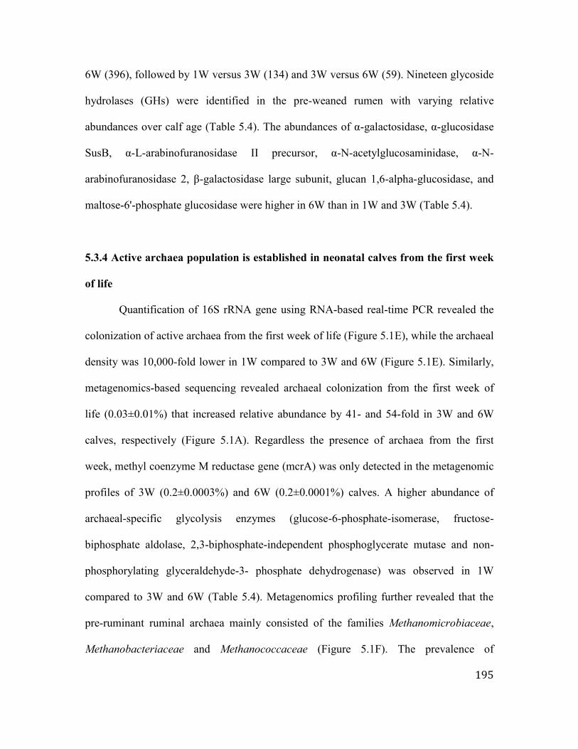
195
6W (396), followed by 1W versus 3W (134) and 3W versus 6W (59). Nineteen glycoside
hydrolases (GHs) were identified in the pre-weaned rumen with varying relative
abundances over calf age (Table 5.4). The abundances of α-galactosidase, α-glucosidase
SusB, α-L-arabinofuranosidase II precursor, α-N-acetylglucosaminidase, α-N-
arabinofuranosidase 2, β-galactosidase large subunit, glucan 1,6-alpha-glucosidase, and
maltose-6'-phosphate glucosidase were higher in 6W than in 1W and 3W (Table 5.4).
5.3.4 Active archaea population is established in neonatal calves from the first week
of life
Quantification of 16S rRNA gene using RNA-based real-time PCR revealed the
colonization of active archaea from the first week of life (Figure 5.1E), while the archaeal
density was 10,000-fold lower in 1W compared to 3W and 6W (Figure 5.1E). Similarly,
metagenomics-based sequencing revealed archaeal colonization from the first week of
life (0.03±0.01%) that increased relative abundance by 41- and 54-fold in 3W and 6W
calves, respectively (Figure 5.1A). Regardless the presence of archaea from the first
week, methyl coenzyme M reductase gene (mcrA) was only detected in the metagenomic
profiles of 3W (0.2±0.0003%) and 6W (0.2±0.0001%) calves. A higher abundance of
archaeal-specific glycolysis enzymes (glucose-6-phosphate-isomerase, fructose-
biphosphate aldolase, 2,3-biphosphate-independent phosphoglycerate mutase and non-
phosphorylating glyceraldehyde-3- phosphate dehydrogenase) was observed in 1W
compared to 3W and 6W (Table 5.4). Metagenomics profiling further revealed that the
pre-ruminant ruminal archaea mainly consisted of the families Methanomicrobiaceae,
Methanobacteriaceae and Methanococcaceae (Figure 5.1F). The prevalence of

196
Methanobacteriaceae in the metagenomic profiles was higher (P = 0.01) in 3W
(39.0±9.8%) and 6W (36.1±14.3%) than in 1W (9.6±6.0%). Although no single genus
was present in all the calves, Methanobrevibacter, Methanothermobacter,
Methanobacterium, and Methanoplanus were observed in 60% of six-week-old calves.
5.3.5 Rumen epithelium development and volatile fatty acid profile in pre-weaned
calves
The rumen epithelium at birth displayed a unique structure compared to pre-
weaned calves (Figure 5.1G). There were no separated protruding papillae or stratified
squamous epithelium in the calf rumen soon after birth; however, developing papillae
were noticeable (Figure 5.1G). The rumen epithelium of newborn calves consisted of a
large number of nucleated squamous cells with a thickness of 279.9±7.6 µm that
developed into 678.1±41.1 µm length papillae within six weeks. The increase in the
length and width of the rumen papillae was significantly different among the three age
groups (Table 5.2, Figure 5.1G).
The concentrations of total VFA, acetate, butyrate, propionate, valerate,
isobutyrate and isovalerate increased with increasing age and dietary changes (Table 5.2).
However, only the molar proportions of acetate and valerate displayed age-related
variations, while the molar proportion of butyrate ranged from 13-16% of total VFA
during the first six weeks of life (Table 5.2). In addition, the concentration of VFAs was
positively correlated with R. flavefaciens density and the rumen papillae development
(Table 5.5).

197
5.3.6 Microbiome-transcriptome interactions may influence rumen epithelial
development and tissue metabolism
We investigated the associations between the rumen development and the
microbiome by examining the rumen tissue transcriptome in association with the papillae
measurements, the concentrations of VFAs and the microbial composition and functions.
In total, the calf rumen tissue consisted of 13,676±399 genes (CPM > 1). Weighted gene
co-expression network analysis (WGCNA) clustered the common genes (11,772)
detected among all calves into 29 gene modules (M1-M29) that displayed varying
associations with the phenotypic measurements (Figure 5.2A). The expression of genes in
the M2 module (2,313 genes) and M18 module (212 genes) was negatively correlated and
the expression of genes in the M10 module (1,070 genes) positively correlated with the
calf phenotypic traits and the microbial metabolites (Figure 5.2A). The genes co-
expressed in the M2 module were related to “transcription”, “splicing”,
“ribonucleoprotein complex biogenesis”, and “RNA metabolic process”. The genes in the
M18 module were enriched with functions related to “chromatin organization”, “histone
modification” and “transcription”. There were nine genes involved in “chromatin
organization”, such as histone genes (H1F0, H1FX), and histone deacetylase coding
genes (HDAC3). M10, the largest gene module (22.5% of all the sequencing reads), was
related to “tissue metabolism”, and the largest proportion of these genes (38 genes, 7.65%
of total reads) related to “respiratory electron transport chain”. They consisted of
“mitochondrial respiratory chain complex proteins”, such as “cytochrome c oxidase
subunits” (COX1, COX3, and COII), “NADH dehydrogenase subunits” (ND2, ND5),

198
“succinate dehydrogenase subunits”, “ubiquinol-cytochrome c reductase subunits” and
“ATP synthase subunits”.
The use of the M10 module to understand the role of bacteria in rumen
development revealed six bacterial clusters based on their associations with the
expression of genes (Figure 5.2B). A cluster (cluster 1) consisting of Prevotella,
Bacteroides, Ruminococcus, Klebsiella and Propionibacterium was positively correlated
with 49 genes involved in “ion binding”, “regulation of cell cycle, catalytic activity,
molecular functions” and “transcription regulatory activity” (Figure 5.2B, 5.2C). The
majority of “ion binding” genes (8/13) were related to zinc finger proteins (ZNFs) (LIM
and calponin homology domain 1, ZNF238, ZNF445, ZNF397, bromodomain adjacent to
zinc finger domain 1B, ADAM metallopeptidase with thrombospondin type 1 motif 10,
deltex 1 E3 ubiquitin ligase, ash2 (absent, small, or homeotic)-like). Another cluster
(cluster 6) containing genera mainly from Firmicutes and Proteobacteria was negatively
correlated with the same sets of genes (Figure 5.2B, 5.2C).
Among the level 2 microbial functions, “microbial carbohydrate metabolism” was
mainly linked to the rumen tissue transcriptome. The genes associated with these
microbial functions included 19 of 34 genes related to “rumen epithelium development”
(Figure 5.3A), genes involved in “rumen tissue carbohydrate metabolism” (Figure 5.3B)
and “membrane transportation” (solute carrier family 35 and monocarboxylate
transporters - SLC16A3/MCT3, SLC16A9/MCT9, SLC16A11/MCT11,
SLC16A13/MCT13) (Figure 5.3C), and 8 of 14 “tight junction protein genes” (TJs)
(Figure 5.3D). Some of these correlated host genes were co-expressed in the M10
module, such as FUCA1, GANC, and GALC from “rumen tissue carbohydrate

199
metabolism” (Figure 5.3B); SLC35A3 from “membrane transportation” (Figure 5.3C);
CLDN23 from TJs (Figure 5.3D); and PPARG, GSTK1, SULT1B1, and GJA1 from
“rumen epithelial development” (Figure 5.3A).
5.3.7 microRNAome coordinates microbiome-host transcriptome crosstalk
To identify the potential regulatory mechanisms of host-microbial interactions,
microRNAome data (364±17 miRNAs) generated using the same animals in a previous
study (Liang et al., 2014) were analyzed using WGCNA. The rumen microRNAome was
clustered into nine modules (R1-R9) based on the expression of miRNAs. The R7 module
(129 miRNAs) was negatively correlated with the calf phenotypic traits and the microbial
metabolites, except for isovalerate (Figure 5.4A). The target prediction analyses
(TargetScan and mirBase) revealed 3,847 genes that were targeted by the R7 module,
3,710 of which (~96%) were identified in the calf rumen tissue transcriptome. Moreover,
258 of the detected 3710 genes were co-expressed in the M10 gene module. The R7
member miR-375, which was downregulated with calf age (Figure 5.4B), was associated
with “rumen epithelial morphogenesis” and “blood vessel development”. The R8 module
(40 miRNAs) was also negatively correlated with the calf phenotypic traits, propionate
and isobutyrate (Figure 5.4A). The miRNAs co-expressed in the R8 module had 2,751
target genes, and 2,649 (~96%) of these genes were observed in the calf rumen tissue
transcriptome. Functional analysis revealed that miRNAs co-expressed in the R8 module
were involved in “protein localization and transportation” and “cell motility”. However,
only R7 miRNAs had their targets co-expressed in the M10 gene module.

200
The roles of miRNAs in regulating host-microbial interactions were further
evaluated by exploring the relationships among the expression of R7 miRNAs and M10
genes and the relative abundance of bacterial genera. Nearly 37% (55/147) of the M10
genes associated with bacterial cluster 1 and cluster 6 (Figure 5.2D, 5.2E) were targeted
by 28 miRNAs co-expressed in R7 (Figure 5.4D). Among these miRNAs, bta-miR-2904,
bta-miR-199b, bta-miR-541, bta-miR-574 and bta-miR-423-5p were associated with a
bacterial cluster comprising Prevotella, Bacteroides, Ruminococcus, Propionibacterium,
Klebsiella (cluster 1 from Figure 5.2D) and Megasphaera (Figure 5.4E). Furthermore,
these five miRNAs targeted 65 different genes related to ZNFs.
5.4 Discussion
The microbiota that rapidly colonizes the in utero sterile mammalian gut during
and after birth constantly interacts with the host to maintain the mammal’s metabolism
and health. The early gut microbiome has been suggested to have long-term impacts on
human health (Subramanian et al., 2015). Despite the accumulating knowledge on the
diversity of the rumen microbiome during early life (Fonty et al., 1987; Li et al., 2012;
Jami et al., 2013; Malmuthuge et al., 2014; Rey et al., 2014), the importance of rumen
colonization for tissue development and the regulatory mechanisms of host-microbial
interactions in pre-ruminants are largely unknown.
This study, using metagenomic and RNA-based approaches, is the first to reveal
the establishment of a dynamic, dense and active microbiome in the pre-ruminant rumen
at birth that undergoes rapid changes during the first six weeks of life. The gut microbiota
has been widely studied in mammalian species using DNA-based approaches; however, it

201
is evident that such evaluation may over-estimate both the organisms and their activities.
The RNA-based quantification used in this study revealed the colonization of active
bacteria within a few minutes of birth, indicating that the process might have started
during the birthing process, which extended from an hour to three hours. Exploring the
dam birth canal (Streptococcus - 23.3±13.3%, Ruminococcaceae - 12.6±4.6%) and rectal
bacteria (Ruminococcaceae - 18.9±1.8%) following birth (data not shown) suggested that
the vaginal/fecal bacteria of dams were the main inoculum of the calf rumen bacteria at
birth. Our findings also confirmed previous studies claiming the establishment of
fibrolytic bacteria within the first week of life (Fonty et al., 1987), a higher prevalence of
Prevotella (Li et al., 2012; Rey et al., 2014) and the presence of GHs in the absence of
proper substrates (Li et al., 2012). We revealed colonization with active R. flavefaciens,
R. albus, E. ruminantium and P. ruminicola, the classical rumen bacteria that degrade
plant polysaccharides (cellulose, hemicellulose, xylan and glycan) (Koike and Kobayashi,
2009; Purushe et al., 2010), from the first week of life, when calves were fed solely with
milk. The increasing density of these species coincided with elevated concentrations of
VFAs as well as increased papillae length and width in 3W and 6W calves fed with
starter and milk. This finding suggests that the introduction of a solid diet stimulates the
rapid growth of the rumen papillae by influencing the rumen microbial composition and
functions. Traditionally, solid feed is considered the major driver of rumen development,
which stimulates microbial fermentation (Heinrichs, 2005; Connor et al., 2013).
However, the appearance of cellulolytic bacteria (Fonty et al., 1987) and the activity of
xylanase and amylase (Rey et al., 2012) can be detected from the second day of life.
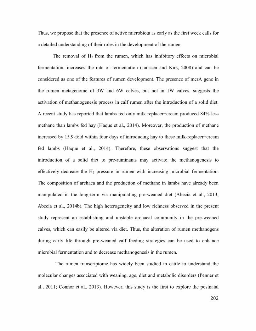
202
Thus, we propose that the presence of active microbiota as early as the first week calls for
a detailed understanding of their roles in the development of the rumen.
The removal of H2 from the rumen, which has inhibitory effects on microbial
fermentation, increases the rate of fermentation (Janssen and Kirs, 2008) and can be
considered as one of the features of rumen development. The presence of mcrA gene in
the rumen metagenome of 3W and 6W calves, but not in 1W calves, suggests the
activation of methanogenesis process in calf rumen after the introduction of a solid diet.
A recent study has reported that lambs fed only milk replacer+cream produced 84% less
methane than lambs fed hay (Haque et al., 2014). Moreover, the production of methane
increased by 15.9-fold within four days of introducing hay to these milk-replacer+cream
fed lambs (Haque et al., 2014). Therefore, these observations suggest that the
introduction of a solid diet to pre-ruminants may activate the methanogenesis to
effectively decrease the H2 pressure in rumen with increasing microbial fermentation.
The composition of archaea and the production of methane in lambs have already been
manipulated in the long-term via manipulating pre-weaned diet (Abecia et al., 2013;
Abecia et al., 2014b). The high heterogeneity and low richness observed in the present
study represent an establishing and unstable archaeal community in the pre-weaned
calves, which can easily be altered via diet. Thus, the alteration of rumen methanogens
during early life through pre-weaned calf feeding strategies can be used to enhance
microbial fermentation and to decrease methanogenesis in the rumen.
The rumen transcriptome has widely been studied in cattle to understand the
molecular changes associated with weaning, age, diet and metabolic disorders (Penner et
al., 2011; Connor et al., 2013). However, this study is the first to explore the postnatal

203
changes in the rumen transcriptome and the molecular mechanisms behind the host-
microbial interactions during the rumen development process. Integrated analysis of the
host transcriptome and the microbiome revealed potential molecular mechanisms behind
the early rumen development, which could be divided into microbial-driven and
ontogenic mechanisms (Figure 5.5). Only three gene modules (3,595 genes) and two
miRNA modules (169 miRNAs) were associated with the concentration of VFAs and the
development of papillae, indicating that only a portion of the rumen transcriptome was
microbial-driven, while the majority was ontogenic (Figure 5.5). These ontogenic
miRNA and gene modules revealed three mRNA-miRNA pairs (miR-25 and fatty acid
binding protein 7 (FABP7); miR-30 and integrin-linked kinase (ILK); and miR29a and
platelet-derived growth factor α polypeptide (PDGFa)) involved in the rumen
development (Figure 5). FABP7 is involved in “fatty acid uptake, transport and
metabolism” (Liu et al., 2010); ILK-mediated signal transduction in “cytoskeletal
organization” (Longhurst and Jennings, 1998); and PDGFa involved in intestinal villus
morphogenesis (Betsholtz et al., 2001). The ontogenic control of the calf rumen
development has been suggested previously (Baldwin et al., 2004); however, this study
mainly focuses on the microbial-driven molecular mechanisms, as they are the black box
of rumen development.
The identified genes in M10 and the predicted target genes of R7 provide a
common ground to identify host-microbial interactions and their potential regulatory
mechanisms in the developing rumen (Figure 5.5). Approximately 22% of the M10 genes
(235/1,070) were similar to the differentially expressed genes identified in a previous
study examining the rumen epithelial gene expression changes when calves were weaned

204
from milk replacer (42 days) to hay/grain (56 – 72 days) (Connor et al., 2013). These 235
common genes were differentially expressed in the rumen epithelial transcriptome when
calves were weaned from a milk replacer-based diet (42 days) to a hay/grain-based diet
(56 – 72 days), but not with calf age while they received milk replacer from day 14 – 42
(Connor et al., 2013). In this study, 87 of these 235 genes were differentially expressed
when 1W was compared against 3W and 6W, after the introduction of solid diet. The
strong positive correlations between these genes and the microbial metabolites (VFAs)
suggest that they may be responsive to diet-driven changes in the microbial metabolites
and may facilitate the early rumen development. Connor et al., (2013) also identified
peroxisome proliferator-activated receptor-α (PPARA) as an important molecular
mechanism of the rumen epithelial development during the weaning process. Although
PPARA was expressed in all the pre-weaned calves in this study, it did not display a
temporal expression pattern with calf age. However, the expression of PPARG, which co-
expressed in the M10 module and was correlated with “microbial carbohydrate
metabolism”, was upregulated with the calf age. Similarly to adult cattle (Bionaz et al.,
2013), the expression of PPARG in the calf rumen tissue was higher than the expression
of PPARA. PPARG is widely studied in ruminants, and its expression level in the rumen
is second only to its expression in the bovine adipose tissue (Bionaz et al., 2013). It
induces epithelial cell proliferation in the colon (Su et al., 2007), upregulates the barrier
functions within nasal epithelial cells (Ogasawara et al., 2010) and is one of the
regulators of intestinal inflammation (Annese et al., 2012) stimulated via butyrate
(Kinoshita et al., 2002). Butyrate has been shown to upregulate PPARG epigenetically via
the inhibition of HDAC (Paparo et al., 2014). The observed negative correlations of

205
HDAC3 (M18 module) with the rumen papillae length and width and the butyrate
concentration further reinforces the positive impact of butyrate on early rumen
development through the modulation of host transcriptome. Therefore, I speculate that in
addition to influencing cell apoptosis (Mentschel et al., 2001), butyrate may also be
involved in rumen development via the activation of PPARG. The observed positive
associations between the expression of PPARG and the concentration of VFAs as well as
the functions related to “ microbial carbohydrate metabolism” suggest its involvement in
the overall rumen tissue development in response to microbial fermentation.
ZNFs are transcriptional factors that regulate a wide array of functions, including
“recognition of DNA”, “packaging of RNA”, “activation of transcription”, “protein
folding and assembly”, and “regulation of apoptosis” (Laity et al., 2001). The absorption
of zinc, a major component of ZNFs, also plays an important role in the early rumen
papillae development and keratinization in goat kids (Cernik et al., 2013). The present
study revealed that five R7 miRNAs and eight M10 genes related to ZNFs were
correlated with the abundance of the same bacterial genera (Prevotella, Bacteroides,
Propionibacterium, Ruminococcus), suggesting that the early microbiota may influence
the rumen development through zinc absorption, and this interaction may be regulated via
miRNAs (Figure 5.5). The supplementation of cattle diets with zinc has long been studied
to understand its impact on milk production and calf health (Spears, 1996); however, its
role in the early rumen development and the microbial modulation of this process are yet
to be understood.
Direct (abundance of bacteria) and indirect (microbial metabolites) associations
between the expression of miRNAs and the early microbiota were evident in this study. A

206
higher proportion of miRNAs (169/364 or 46.4% of microRNAome) than of protein
coding genes (3,595/13,676 or 26.3% of transcriptome) was associated with the
concentration of VFAs, further corroborating our previous findings and speculations on
interactions between miRNAs and microbes (Liang et al., 2014). A VFA-associated
miRNA from R7, miR-375, inhibits the alveolar epithelial cell differentiation via the
Wnt/β-catenin pathway, which participates in “tissue differentiation” and
“organogenesis” in rats (Wang et al., 2013). The temporal downregulation of miR-375
and its negative associations with the concentration of VFAs and the development of
papillae indicate one of the miRNA regulatory mechanisms that can be initiated by
microbial metabolites. Thus, the M10 and R7 modules are indeed biologically important
during rumen development and may serve as potential candidates to explore the host-
microbial interactions and their regulatory mechanisms (Figure 5.5).
5.5 Conclusions
This study demonstrated that rumen colonization began during the birthing
process and the pre-ruminant rumen microbiota was active and ready to ferment a solid
diet even from the first week of life. The VFAs produced by the early microbiome were
associated with the rumen tissue metabolism and the development of the epithelium via
interacting with the host transcriptome and microRNAome (Figure 5.5). I therefore
propose that early feeding management has a similar importance to the weaning period
and may enhance the rumen development and facilitate the weaning transition. This study
urges to understand host-microbial interactions in the developing rumen and lower gut in
depth that may allow developing multidisciplinary approaches to improve the production

207
and health of livestock ruminants through early-life manipulations of rumen and/or gut
microbiota.
5.6 References
Abecia, L., Martin-Garcia, A.I., Martinez-Fernandez, G., Newbold, C.J., Yanez-Ruiz,
D.R. (2013) Nutritional intervention in early life to manipulate rumen microbial
colonization and methane output by kid goats postweaning. J. Anim. Sci. 91,
4832-4840. doi: 10.2527/jas.2012-6142.
Abecia, L., Waddams, K.E., Martinez-Fernandez, G., Martin-Garcia, A.I., Ramos-
Morales, E., Newbold, C.J., Yanez-Ruiz, D.R. (2014a). An antimethanogenic
nutritional intervention in early life of ruminants modifies ruminal colonization by
archaea. Archaea. 2014, Article ID 841463. doi:
http://dx.doi.org/10.1155/2014/841463.
Abecia. L., Ramos-Morales, E., Martinez-Fernandez, G., Arco, A., Martin-Garcia, A.I.,
Newbold, C.J. et al., (2014b) Feeding management in early life influences
microbial colonization and fermentation in the rumen of newborn goat kids.
Animal Production Science. 54, 1449-1454. http://dx.doi.org/10.1071/AN14337.
Annese, V., Rogai, F., Settesoldi, A., Bagnoli, S. (2012). PPARγ in inflammatory bowel
disease. PPAR Res. 2012, Article ID 620839.
http,//dx.doi.org/10.1155/2012/620839.
Baldwin, R.L., McLeod, K.R., Klotz, J.L., Heitmann, R.N. (2004). Rumen development,
intestinal growth and hepatic metabolism in the pre- and postweaning ruminant. J.
Dairy Sci. 87, E55-E65.

208
Benjamini, Y., Hochberg, y. (1995). Controlling the false discovery rate: A practical and
powerful approach to multiple testing. Journal of the Royal Statistical Society.
Series B (Methodological). 57, 289-300.
Betsholtz, C., Karlson, L., Lindhal, P. (2001). Developmental roles of platelet-derived
growth factors. BioEssays. 23,494-507.
Bionaz, M., Chen, S., Khan, M.J., Loor, J.J. (2013). Functional role of PPARs in
ruminants, Potential targets for fine-tuning metabolism during growth and
lactation. PPAR Res. 2013, Article ID 684159. doi:
http,//dx.doi.org/10.1155/2013/684159.
Cernik, J., Pavlata, L., Misurova, L., Jokverova, O., Lunacek, J., Halouzka, R. (2013).
Effect of peroral supplementation of zinc on the ruminal mucosa of goat kids - a
morphometric study. Acta. Vet. Brno. 82, 399-403. doi:
http://dx.doi.org/10.2754/avb201382040399.
Connor, E.E., Baldwin, R.L., Li, C., Li, R.W., Chung, H. (2013). Gene expression in
bovine rumen epithelium during weaning identifies molecular regulators of rumen
development and growth. Funct. Integr. Genomics. 13, 133-142. doi:
10.1007/s10142-012-0308-x.
Fonty, G., Gouet, P., Jouany, J.P., Senaud, J. (1987). Establishment of the microflora and
aerobic fungi in the rumen of lambs. J. General Microbiol. 133, 1835-1843.
Gorka, P., Kowalski, Z.M., Pietrzak, P., Kotunia, A., Jagusiak, W., Zabielski, R. (2011).
Is rumen development in newborn calves affected by different liquid feeds and
small intestine development? J. Dairy Sci. 94, 3002-3013. doi: 10.3168/jds.2010-
3499.

209
Heinrichs, A.J. (2005). Rumen development in the dairy calf. Adv. Dairy Tech. 17, 179-
187.
Jami, E., Israel, A., Kotser, A., Mizrahi, I. (2013). Exploring the bovine rumen bacterial
community from birth to adulthood. ISME J. 7, 1069-1079. doi:
10.1038/ismej.2013.2.
Khan, M.A., Lee, H.J., Lee, W.S., Kim, H.S., Ki, K.S., Hur, T.Y., et al., (2007).
Structural growth, rumen development and metabolic and immune responses of
Holstein male calves fed milk through step-down and conventional methods. J.
Dairy Sci. 90:3376-3387.
Khan, M.A., Weary, D.M., von Keyserlingk, M.A.G. (2011). Hay intake improves
performances and rumen development of calves fed higher quantities of milk. J.
Dairy Sci. 94, 3547-3553. doi: 10.3168/jds.2010-3871.
Kim, D., Pertea, G., Trapnell, C., Pimentel, H., Kelley, R., Salzberg, S.L. (2013).
TopHat2: Accurate alignment of transcriptomes in the presence of insertions, deletions
and gene fusions. Genome Biol. 14, R36. doi: 10.1186/gb-2013-14-4-r36.
Kinoshita, M., Suzuki, Y., Saito, Y. (2002). Butyrate reduces colonic paracellular
permeability by enhancing PPARgamma activation. Biochem. Biophys. Res.
Commun. 293, 827-831.
Koike, S., Kobayashi, Y. (2009). Fibrolytic rumen bacteria: Their ecology and functions.
Asian-Aust. J. Anim. Sci. 22, 131-138. doi:
http://dx.doi.org/10.5713/ajas.2009.r.01.
Laity, J.H., Lee, B.M., Wright, P.E. (2001). Zinc finger protein, new insight into
structural and functional diversity. Curr. Opin. Struc. Biol. 11, 39-46.

210
Langfelder, P., Horvath, S. (2008). WGCNA, An R package for weighted correlation
network analysis. BMC Bioinf. 9, 559. doi,10.1186/1471-2105-9-559.
Li, R.W., Sparks, M.E., Connor, E.E. (2011). Dynamics of the rumen microbiota. In
Metagenomics and its applications in agriculture, biomedicine and environmental
studies R.W. Li, ed, (Nova Science Publishers, New York, USA) pp.135-164.
Li, R.W., Connor, E.E., Baldwin, R.L., Sparks, M.L. (2012). Characterization of the
rumen microbiota of pre-ruminant calves using metagenomic tools. Environ.
Microbiol. 14, 129-139. doi: 10.1111/j.1462-2920.2011.02543.
Li, M., Penner, G.B., Hernandez-Sanabria, E., Oba, M., Guan, L.L. (2009). Effects of
sampling location and time, and host animal on assessment of bacterial diversity and
fermentation parameters in the bovine rumen. J. Appl. Microbiol. 107, 1924-1934. doi:
10.1111/j.1365-2672.2009.04376.x.
Liang, G., Malmuthuge, N., McFadden, T.B., Bao, H., Griebel, P.J., Stothard, P., et al.,
(2014). Potential regulatory role of microRNAs in the development of bovine
gastrointestinal tract during early life. PLoS One. 9, e92592.
doi,10.1371/journal.pone.0092592.
Liu, R.Z., Mita, R., Beauliu, M., Gao, Z., Godbout, R. (2010). Fatty acid binding proteins
in brain development and disease. Int. J. Dev. Biol. 54, 1229-1239. doi:
10.1387/ijdb.092976rl.
Longhurst, C.M., Jennings, L.K. (1998). Integrin-mediated cellular transduction. Cell
Mol. Life Sci. 54, 514-526.
Malmuthuge, N., Griebel, P.J, Guan, L.L. (2014). Taxonomic identification of
commensal bacteria associated with the mucosa and digesta throughout the

211
gastrointestinal tracts of pre-weaned calves. Appl. Environ. Microbiol. 80, 2012-
2028. doi: 10.1128/AEM.03864-13.
Mentschel, J., Leiser, R., Mulling, C., Pfarrer, C., Claus, R. (2001). Butyric acid
stimulates rumen mucosa development in the calf mainly by a reduction of
apoptosis. Arch. Anim. Nutr. 55, 85-102.
Meyer, F., Paarmann, D., D'Souza, M., Olson, R., Glass, E.M., Kubal, M., et al., (2008).
The metagenomics RAST server- a public resource for the automatic phylogenetic
and functional analysis of metagenomes. BMC Bioinf. 9, 386. doi,10.1186/1471-
2105-9-386. doi: 10.1186/1471-2105-9-386.
Ogasawara, N., Kojima, T., Go, M., Ohkuni, T., Koizumi, J., Kamekura, R., et al.,
(2010). PPARgamma agonists upregulate the barrier function of tight junctions
via a PKC pathway in human nasal epithelial cells. Pharmacol Res. 61, 489-498.
doi: 10.1016/j.phrs.2010.03.002.
Paparo, L., di Costanzo, M., di Scala, C., Cosenza, L., Leone, L., Nocerino, R., et al.,
(2014). The influence of early life nutrition on epigentic regulatory mechanisms
of the immune system. Nutrition. 6, 4706-4719. doi: 10.3390/nu6114706.
Penner, G.B., Steele, M.A., Aschenbach, J.R., McBride, B.W. (2011). Ruminant nutrition
symposium, Molecular adaptation of ruminal epithelia to highly fermentable diets.
J. Anim. Sci. 89, 1108-1119. doi: 10.2527/jas.2010-3378.
Purushe, J., Fouts, D.E., Morrison, M., White, B.A., Mackie, R.I. (2010). Comparative
genome analysis of Prevotella ruminicola and Prevotella bruantii, Insights into
their environmental niche. Microb. Ecol. 60, 721-729. doi: 10.1007/s00248-010-
9692-8.

212
Rey, M., Enjalbert, F., Monteils, V. (2012). Establishment of ruminal enzymatic activities
and fermentation capacity in dairy calves from birth through weaning. J. Dairy
Sci. 95, 1500-1512. http://dx.doi.org/ 10.3168/jds.2011-4902.
Rey, M., Enjalbert, F., Combes, S., Cauquil, L., Bouchez, O., Monteils, V. (2014).
Establishment of ruminal bacterial community in dairy calves from birth to
weaning is sequential. J. Appl. Microbiol. 116, 245-257. doi: 10.1111/jam.12405.
Sommer, F. and Backhed, F. (2013). The gut microbiota - masters of host development
and physiology. Nat. Rev. Microbiol. 11, 227-238. doi: 10.1038/nrmicro2974.
Sommer, F., Nookaew, I., Sommer, N., Fogelstrand, P., Backhed, F. (2015). Site-specific
programming of the host epithelial transcriptome by the gut microbiota. Genome
Biol. 16, 62. doi: 10.1186/s13059-015-0614-4.
Spears, J.W. (1996). Organic trace minerals in ruminant nutrition. Animal Feed Science
Technology. 58, 151-163.
Stevenson, D.M., Weimer, P.J. (2007). Dominance of Prevotella and low abundance of
classical ruminal bacterial species in the bovine rumen revealed by relative
quantification real-time PCR. Appl. Microbiol. Biotechnol. 75, 165-174.
Su, W., Bush, C.R., Necela, B.M., Calcagno, S.R., Murray, N.R., Fields, A.P., et al.,
(2007). Differential expression, distribution, and function of PPAR-gamma in the
proximal and distal colon. Physiol. Genomics. 30, 342-353.
Subramanian, S., Blanton, L.V., Frese, S.A., Charbonneau, M., Mills, D.A., Gordon, J.I.
(2015). Cultivating healthy growth and nutrition through the gut microbiota. Cell.
161, 36-48. doi:10.1016/j.cell.2015.03.013.

213
Wang, Y., Huang, C., Chintagari, N.R., Bhaskaran, M., Weng, T., Guo, Y., et al., (2013).
miR375 regulates rat alveolar epithelial cell trans-differentiation by inhibiting
Wnt/β catenin pathway. Nucleic Acids Res. 41, 3833-3844. doi:
10.1093/nar/gks1460.
White, J.R., Nagarajan, N., Pop, M. (2009). Statistical methods for detecting
differentially abundant features in clinical metagenomic samples. PLoS Comput. Biol.
5, e1000352. doi: 10.1371/journal.pcbi.1000352.
Yeoman, C.J., White, B.A. (2014). Gastrointestinal tract microbiota and probiotics in
production animals. Annu. Rev. Anim. Biosci. 2, 469-486. doi: 10.1146/annurev-
animal-022513-114149.
Yu, Z., Morrison, M. (2004). Improved extraction of PCR-quality community DNA from
digesta and fecal samples. BioTechniques. 36, 808-812.
Zhou, M., Hernandez-Sanabria, E., Guan, L.L. (2010). Characterization of variation in
rumen methanogenic communities under different dietary and host feed efficiency
conditions, as determined by PCR-denaturing gradient gel electrophoresis analysis.
Appl. Microbiol. Ecol. 76, 3776-3786. doi: 10.1128/AEM.00010-10.

214
5.7 Tables and Figures
Table 5.1 Primers used in qPCR to estimate bacterial/archaeal densities
Forward primer (5’-3’) Reverse primer (5’-3’) Source
Total bacteria actcctacgggaggcag gactaccagggtatctaatcc Stevenson & Weimer, 2007
E. ruminantium ctcccgagactgaggaagcttg gtccatctcacaccaccgga Stevenson & Weimer, 2007
P. ruminicola gaaagtcggattaatgctctatgttg catcctatagcggtaaacctttgg Stevenson & Weimer, 2007
R. albus gttaacagagggaagcaaagca tgcagcctacaatccgaactaa Stevenson & Weimer, 2007
R. flavefaciens tggcggacgggtgagtaa ttaccatccgtttccagaagct Stevenson & Weimer, 2007
Total archaea ccggagatggaacctgagac cggtcttgcccagctcttattc Zhou et al., 2010

215
Table 5.2 Postnatal changes in active rumen bacteria, rumen morphology and metabolites
in pre-weaned calves
Calf age P- value
1-week 3-week 6-week
Active rumen bacteria
R. flavefaciens1 3.8±1.9E08a 1.6±0.4E09b 1.3±0.5E09b 0.03
R. albus 1.4±0.5E05 6.3±6.2E06 2.2±1.7E07 0.34
P. ruminicola 6.4±2.7E04 4.4±3.0E05 7.6±7.4E07 0.37
E. ruminantium 7.8±3.8E05 1.5±0.6E06 1.6±0.6E06 0.48
Rumen papillae
Length (μm) 317.8±7.6a 413.0±13.6b 678.1±41.1c < 0.01
Width (μm) 155.5±2.7a 224.0±6.3b 275.8±9.0c < 0.01
Rumen fermentation parameters
Acetate2 21.1±2.2a 37.0±2.8b 50.3±3.8c < 0.01
Propionate 10.6±2.0a 25.8±4.3b 36.3±1.8c < 0.01
Butyrate 5.6±1.8a 11.8±2.7b 17.8±2.2b < 0.01
Isobutyrate 0.3±0.04a 0.8±0.1b 1.3±0.1c < 0.01
Valerate 1.0±0.4a 3.7±0.7b 5.1±0.8b < 0.01
Isovalerate 0.3±0.07a 0.8±0.1a 1.3±0.3b < 0.01
Total 39.6±6.2a 80.5±9.0b 113.9±6.9c < 0.01
Acetate3 55.3±2.7a 47.0±2.4b 44.2±1.9b 0.01
Propionate 26.7±1.4 31.3±2.9 32.1±1.4 NS
Butyrate 13.0±2.0 14.4±2.5 15.4±1.4 NS
Isobutyrate 0.8±0.1 1.0±0.1 1.2±0.1 NS
Valerate 2.1±0.5a 4.6±0.6b 4.5±0.6b 0.01
Isovalerate 0.8±0.2 1.0±0.2 1.2±0.2 NS a, b, c – means with different superscript within a raw are significantly different at P <
0.05.
1Density of active rumen bacteria (16S rRNA copy/ g of rumen content).
2VFA concentration (mM/ml of rumen fluid).
3Molar proportion of VFA (%).

216
Table 5.3 Differentially abundant functions of the rumen microbiome
Functions 0D 1W 3W 6W Significant comparisons2
Subsystems1
Amino acids and derivatives 5.6 11 8.3 7.9 1W vs. 3W, 6W
Carbohydrates 9.4 12.4 14.5 11 3W vs. 6W
Cofactors, vitamins, prosthetic groups, pigments 5.8 5.4 4.7 4.8 1W vs. 3W
Dormancy and sporulation 0.02 0.2 0.3 0.4 0D vs. 1W, 3W, 6W
Metabolism of Aromatic Compounds 0.1 0.5 0.7 0.5 0D vs. 1W
Fatty acids, lipids, isoprenoids 0.6 1.7 1.1 2.1 3W vs. 6W
Motility and chemotaxis 0 0.3 1.1 0.3 0D vs. 1W, 1W vs. 3W, 3W vs. 6W
Nitrogen metabolism 1.1 1.6 0.6 1.4 0D vs. 3W
Nucleosides and Nucleotides 1.6 3.8 3.8 4.4 0D vs. 1W, 3W, 6W
Phages, prophages, transposable elements, plasmids 1.1 4.4 7 6 0D vs. 1W, 3W, 6W
Potassium metabolism 0.08 0.2 0.01 0.05 1W vs. 3W
Virulence, disease and defense 1.2 3 2.2 2.9 0D vs. 1W
Respiration 20.5 3.2 4.5 2.8 0D vs. 1W, 3W, 6W
Level 2 functions in SEED hierarchy1
Adhesion 0 0.05 0.04 0.02 0D vs. 1W; 1W vs. 6W
ATP synthases 0.2 0.5 1.6 0.8 1W vs. 3W
Biologically active compounds in metazoan cell
defence and differentiation 0.1 0.1 0 0.5 1W vs. 3W
Biosynthesis of galactoglycans and related
lipopolysacharides 0 0.03
4.00E-
05 0.03 0D vs. 1W
Biotin 0.2 0.2 0 0.06 1W vs. 3W
Branched-chain amino acids 0.2 2.3 2.9 1.8 0D vs. 1W
Carotenoid biosynthesis 0.01 0 0.003 0 0D vs. 1W

217
Cell Wall and Capsule 1.6 1.3 0.6 0.6 1W vs. 3W; 1W vs. 6W
Cell wall of Mycobacteria 0 0.1 0 0.001 0D vs. 1W; 1W vs. 3W
Central carbohydrate metabolism 2.8 6.4 10.5 9.2 1W vs. 6W
Choline bitartrate degradation, putative 0 0.1 0.2 0.2 0D vs. 1W
Coenzyme A 0.04 0.3 0 0.3 0D vs. 1W; 1W vs. 3W
Coenzyme F420 0 0.001 0.06 0.03 1W vs. 6W
Cofactors, Vitamins, Prosthetic Groups, Pigments 0.05 0.4 0.4 0.2 0D vs. 1W
Cold shock 0 0 0.01 0 1W vs. 3W
DNA recombination 0.01 0.1 0.1 0.2 0D vs. 1W
Dormancy and Sporulation 0.01 0.2 0.2 0.3 0D vs. 1W
Fatty acids 0.1 0.4 0.4 2.5 0D vs. 1W
Fatty Acids, Lipids, and Isoprenoids 0 0.1 0.2 0.1 0D vs. 1W
Flagellar motility in Prokaryota 0 0.3 1.3 0.4 0D vs. 1W; 1W vs. 3W; 3w vs. 6W
Glycoside hydrolases 0.01 0.1 0 0.03 0D vs. 1W
Heat shock 0.2 0.5 1 1.2 1W vs. 6W
Histidine Metabolism 0.1 0.5 0.3 0.2 0D vs. 1W
Lysine, threonine, methionine, and cysteine 1.9 3.1 2.3 1.9 1W vs. 6W
Metabolism of Aromatic Compounds 0 0.03 0.01 0.02 0D vs. 1W
Methylamine utilization 0.1 0.7 0.1 0.5 1W vs. 3W
Motility and Chemotaxis 0 0.03 0.3 0.1 1W vs. 3W
Nucleosides and Nucleotides 0.03 0.6 0.5 0.2 0D vs. 1W; 1W vs. 6W; 3W vs. 6W
Organic acids 0.8 1 0.7 0.9 1W vs. 3W; 3W vs. 6W
Osmotic stress 0 0.06 0.01 0.03 0D vs. 1W; 1W vs. 3W
Pathogenicity islands 0.2 0.3 0.5 0.8 1W vs. 6W
Peripheral pathways for catabolism of aromatic
compounds 0.01 0.3 0.001 0.3 0D vs. 1W
Periplasmic Stress 0.03 0.04 0.01 0.01 1W vs. 6W
Phages, Prophages 0.2 0.7 2.2 3.9 1W vs. 3W
Phospholipids 0.3 0.7 0.22 0.5 1W vs. 3W

218
Plant-Prokaryote DOE project 11.7 7.1 6.4 4.6 1W vs. 6W
Potassium metabolism 0.05 0.2 0.01 0.07 1W vs. 3W
Programmed Cell Death and Toxin-antitoxin Systems 0.01 0.05 0.01 0 1W vs. 3W; 1W vs. 6W
Proteasome related clusters 0.4 0 0.04 0.02 1W vs. 3W
Protein biosynthesis 0.01 0 0.03 0.05 0D vs. 1W
Protein folding 0.05 0.6 1 1.9 1W vs. 6W
Protein secretion system, Type II 0 0.034 0.08 0.03 0D vs. 1W
Proteolytic pathway 0.2 0 0 0 0D vs. 1W
Putrescine/GABA utilization cluster-temporal 0 0.05 0.004 0.008 1W vs. 3W
Pyridoxine 0.1 0.5 0.3 0.4 0D vs. 1W
Pyrimidines 0.5 1.2 0.7 1.8 0D vs. 1W; 1W vs. 3W; 3w vs. 6W
Pyruvate kinase associated cluster 0.03 0 0 0 0D vs. 1W
Quinone cofactors 0.1 0.3 0.1 0.03 0D vs. 1W; 1W vs. 3W; 1W vs. 6W;
3W vs. 6W
Resistance to antibiotics and toxic compounds 0.7 2.4 1.4 1.9 0D vs. 1W
Reverse electron transport 0.03 0.04 0.3 0.3 1W vs. 6W
Riboflavin, FMN, FAD 0.04 0.3 0.2 0.2 0D vs. 1W
Ribosome-related cluster 0.1 0.2 0.7 0.4 1W vs. 3W
Selenoproteins 0 0.1 0.5 0.4 0D vs. 1W
Transcription 0.5 1.6 1.4 2.2 0D vs. 1W
Translation 0.1 0.2 0.1 0.5 1W vs. 6W; 3W vs. 6W
Two related proteases 0 0.3 0.3 0.4 0D vs. 1W
1relative abundance as % of total detected subsystems or level 2 functions in the SEED hierarchy.
0D – at birth/newborn, 1W – week-old calves, 3W – 3-week-old calves, 6W – 6-week-old calves.

219
Table 5.4 Relative abundance of glycoside hydrolases and archaeal-specific glycolysis enzymes in calf rumen
Enzyme 1-week 3-week 6-week GH family*
Glycosides Hydrolases*
6-phospho-beta-glucosidase ascB (EC 3.2.1.86) 10.0±5.0 10.0±5.0 0.2±0.001 1, 4
Alpha-amylase (Neopullulanase) SusA (EC 3.2.1.135) 21.5±19.3 22.8±19.3 5.0±1.2 13
Alpha-galactosidase (EC 3.2.1.22) 5.0±1.2 5.0±1.2 11.7±6.9
4, 21, 31, 36, 57, 97,
110
Alpha-glucosidase (EC 3.2.1.20) 3.6±0.7 4.0±0.7 0 4, 13, 31, 63, 97, 122
Alpha-glucosidase SusB (EC 3.2.1.20) 12.3±5.5 12.0±5.5 36.1±17.0 4, 13, 31, 63, 97, 122
Alpha-L-arabinofuranosidase II precursor (EC
3.2.1.55) 0 0 2.2±0.2 2, 3, 10, 43, 54, 62
Alpha-mannosidase (EC 3.2.1.24) 7.7±3.0 8.0±2.9 0 31, 38, 92
Alpha-N-acetylglucosaminidase (EC 3.2.1.50) 0 0 1.8±0.2 89
Alpha-N-arabinofuranosidase 2 (EC 3.2.1.55) 2.1±0.2 2.1±0.2 11.8±7.0 2, 3, 10, 43, 54, 62
Beta-galactosidase large subunit (EC 3.2.1.23) 0.2±0.001 0.02±0.001 11.1±0.06 1, 2, 3, 35, 49, 50, 52
Endo-beta-1,3-1,4 glucanase (Licheninase) (EC
3.2.1.73) 10.0±5.0 10.0±5.0 0
5, 7, 8, 9, 11, 12, 16,
17, 26
Glucan 1,6-alpha-glucosidase (EC 3.2.1.70) 0 0 17.4±10.6 13, 15
Glycogen debranching enzyme (EC 3.2.1.-) 14.6±5.5 14.6±5.5 0
1-5, 8-10, 13, 16-20,
26, 28, 30, 32, 33, 35,
38, 42-44, 48, 51, 55
Maltose-6'-phosphate glucosidase (EC 3.2.1.122) 1.4±0.1 1.4±0.1 8.8±1.5 4
Periplasmic beta-glucosidase (EC 3.2.1.21) 0 0 3.4±0.6 1, 3, 5, 9, 30, 116
Polygalacturonase (EC 3.2.1.15) 10.6±0.05 1.1±0.06 0 28
Spore cortex-lytic enzyme, N-acetylglucosaminidase
SleL (EC 3.2.1.-) 0.2±0.001 0.15±0.001 0 2, 3, 10, 43, 54, 62
Trehalase (EC 3.2.1.28) 8.9±2.8 8.9±2.8 0.3±0.005 13, 15, 37, 65
Trehalose-6-phosphate hydrolase (EC 3.2.1.93) 1.5±0.1 1.5±0.1 0 13

220
Archaeal-specific glycolysis enzymes
Glucose-6-phosphate isomerase, archaeal (EC 5.3.1.9) 0.00012±0.00 0 0
Fructose-bisphosphate aldolase, archaeal class I (EC
4.1.2.13) 0.01±0.00 0 0
2,3-bisphosphoglycerate-independent
phosphoglycerate mutase, archaeal type (EC 5.4.2.1) 0.2±0.00 0.006±0.00 0.1±0.00
Non-phosphorylating glyceraldehyde-3-phosphate
dehydrogenase (NADP) (EC 1.2.1.9) 0.03±0.00 0.0008±0.00 0.0003±0.00
* http://www.cazy.org/Glycoside-Hydrolases.html. Data presented as mean±SEM.

221
Table 5.5 Association between rumen papillae development, concentration of VFAs, and cellulolytic bacteria
Acetate Propionate Butyrate Valerate Isobutyrate Isovalerate
Papillae length1 0.7*** 0.6*** 0.7*** 0.6** 0.7*** 0.6***
Papillae width 0.8*** 0.8*** 0.7*** 0.7*** 0.8*** 0.7***
R. flavefaciens 0.6*** 0.4 0.6*** 0.4* 0.4* 0.3 1 Pearson correlation coefficient.
* P < 0.1; ** P < 0.05; *** P < 0.01.

222
Figure 5.1 Establishment of rumen microbiome from birth up to the first six weeks of life
and the development of rumen papillae.
(A) Composition of rumen microbiome in calves from birth (at birth) up to six weeks of
life. Each data series represent mean relative abundance of microbial group as a % of
total detected microbiota.

223
Figure 5.1B Estimated total bacterial density (DNA-based (16S rRNA gene copy/g of
sample) and RNA-based (16S rRNA copy/ g of sample) in calf rumen during the first six
weeks of life (P = 0.02).
Bars represent mean bacterial densities and error bars represent SEM. a,b – represents
mean RNA-based bacterial densities different at P < 0.05. x,y – represents mean DNA-
based bacterial densities different at P < 0.05.

224
Figure 5.1C Composition of rumen content-associated bacteria (mean relative abundance)
at phylum level.
Each data series represent mean relative abundance of bacterial phylum as a % of total
detected bacteria.
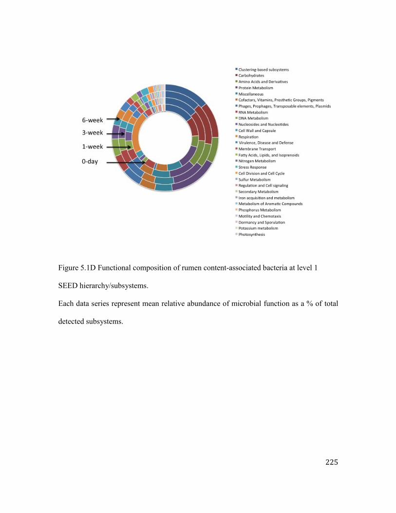
225
Figure 5.1D Functional composition of rumen content-associated bacteria at level 1
SEED hierarchy/subsystems.
Each data series represent mean relative abundance of microbial function as a % of total
detected subsystems.

226
Figure 5.1E Estimated total archaea density using DNA-based (16S rRNA gene copy/g of
sample) and RNA-based (16S rRNA copy/ g of sample) quantifications.
Bars represent mean bacterial densities and error bars represent SEM.

227
Figure 5.1F Rumen content-associated archaeal composition at family level.
Each data series represent mean relative abundance of archaeal family as a % of total
detected archaea.
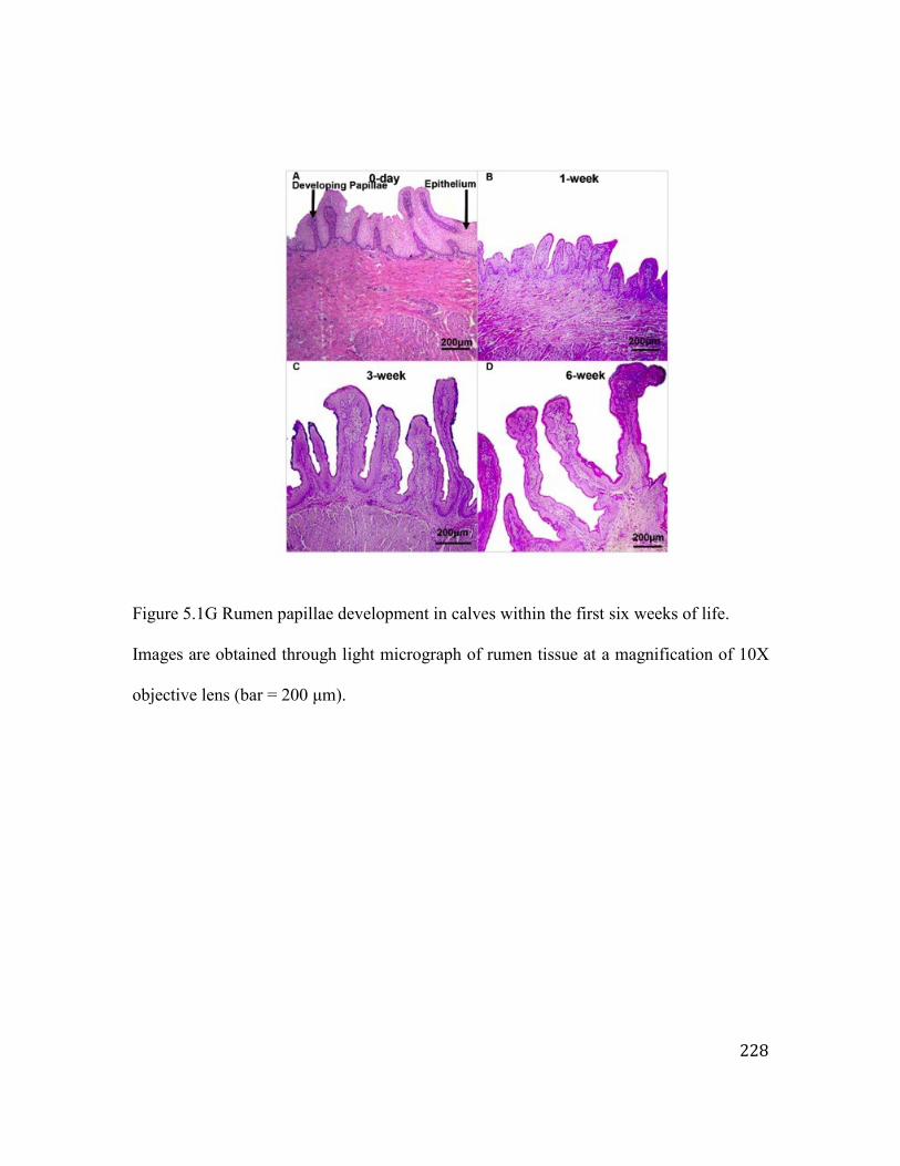
228
Figure 5.1G Rumen papillae development in calves within the first six weeks of life.
Images are obtained through light micrograph of rumen tissue at a magnification of 10X
objective lens (bar = 200 μm).

229
Figure 5.2 Associations among transcriptome networks (gene modules), calf phenotypic
traits (concentration of VFAs, papillae length and width, calf age) and bacterial
composition (taxonomy – genus level).
Figure 5.2A Relationship between gene modules (M1 – M29) and calf phenotypic traits.
Gene modules obtained using weighted gene co-expression network analysis and
eigengene/PC1 value of each gene module is correlated with the calf phenotypic traits.

230
Figure 5.2B Association between the genes co-expressed in the M10 module and content-
associated bacterial genera relative abundance.
Heatmap is generated using Pearson correlation value between the expression of a gene
and the relative abundance of a bacterial genus. Blue represents positive correlations,
whereas yellow represents negative correlations. Numerical values represents the
identified bacterial clusters based on their associations with the expression of genes.

231
Figure 5.2C Bacterial clusters associated with ion binding related genes co-expressed in
the M10 module.
Cluster 1 (Bacteroides, Ruminococcus, Propionibacterium, Klebsiella, Prevotella)
positively correlates to the expression of the ion binding related genes (P < 0.05, r ≥ 0.5).
Cluster 6 (Pectobacterium, Bordetella, Mycobacterium, Bartonella, Brachyspira,
Ralstonia, Actinobacillus, Leptospira, Tannerella, Leuconostoc, Escherichia,
Selenomonas, Francisella, Gallibacterium) negatively correlates to the expression of the
ion binding related genes (P < 0.05, r ≤ -0.5).

232
Figure 5.3 Level 2 microbial functions associated with host protein coding genes
expression.
Figure 5.3A Level 2 microbial functions associated with (P < 0.01, r2 ≥ 0.98) host genes
involved in rumen epithelial tissue development (GO: 0060429, 34 genes). Lower panel
represents genes co-expressed in M10 gene module. PPARG – peroxisome proliferator
activated receptor gamma; SULT1B1 – sulfotranferase family 1B member 1; GSTK1 –
glutathione S-transferase kappa 1; GJA1 – gap junction protein alpha 1. 0D – at birth, 1W
– 1-week-old calves, 3W – 3-week-old calves, 6W – 6-week-old claves.
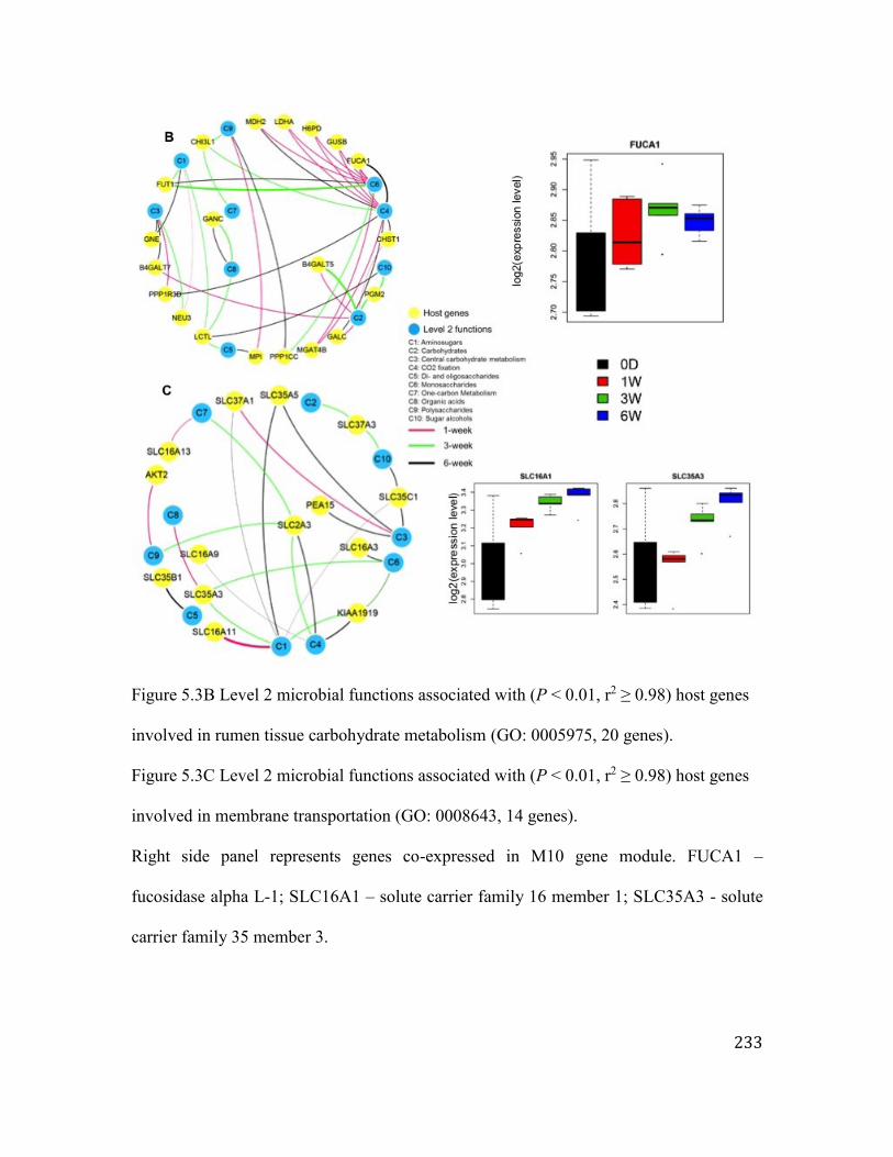
233
Figure 5.3B Level 2 microbial functions associated with (P < 0.01, r2 ≥ 0.98) host genes
involved in rumen tissue carbohydrate metabolism (GO: 0005975, 20 genes).
Figure 5.3C Level 2 microbial functions associated with (P < 0.01, r2 ≥ 0.98) host genes
involved in membrane transportation (GO: 0008643, 14 genes).
Right side panel represents genes co-expressed in M10 gene module. FUCA1 –
fucosidase alpha L-1; SLC16A1 – solute carrier family 16 member 1; SLC35A3 - solute
carrier family 35 member 3.

234
Figure 5.3D Level 2 microbial functions associated with (P < 0.01, r2 ≥ 0.98) host genes
involved in tight junction protein genes (TJs) (GO: 0005923, 14 genes).
Upper network represents TJs correlated with microbial carbohydrate metabolism. Lower
network represents TJs correlated with other microbial functions. Left side panel
represents genes co-expressed in M10 gene module. CLDN23 – claudin 23.

235
Figure 5.4 Association between rumen miRNA profile (expression of miRNA) and rumen
microbiota (bacterial genera, concentration of VFAs).
Figure 5.4A Relationship between miRNA modules (R1-R9) and calf phenotypic traits.
miRNA modules are generated using WGCNA and eigengene/PC1 values of each
modules is correlated with calf phenotypic traits. Numerical values within a square
represent Pearson correlation (upper value) and P value (lower value). Color bar
represents Pearson correlation from -1 to 1.

236
Figure 5.4B Temporal changes in the expression (CPM) of miR-375 in calf rumen (day 0
- 605.1±40.3; week 1 - 171.5±15.6; week 3 - 10.9±3.8; week 6 - 2.9±1.2; P < 0.01).
FC (fold change) is the expression ratio between two adjacent age groups.

237
Figure 5.4C Association between rumen bacterial taxonomy and miRNAs co-expressed
in R7 miRNA module.
Heatmap is generated using Pearson correlation value between the expression of a
miRNA and the relative abundance of a bacterial genus. Blue represents positive
correlations, whereas yellow represents negative correlations. A bacterial cluster consists
of Klebsiella, Megasphaera, Prevotella, Propionibacterium, Bacteroides and
Ruminococcus displayed strong correlation with the expression of bta-miR-574, bta-miR-
423-5p, bta-miR-5441, bta-miR-199b and bta-miR-2904 (-0.9 > R > 0.9, P < 0.05).

238
Figure 5.5 Proposed host-microbial interactions and their regulatory mechanisms in the
developing rumen.
Early rumen microbiota alters rumen development via direct and indirect (miRNAs)
interactions with the rumen tissue transcriptome. Microbial-derived VFAs are associated
with genes involved in ruminal tissue metabolism (M10 gene module), non-coding RNA
processing (M2 gene module) and epigenetic modifications (M18 gene module) as well
as miRNAs regulating epithelial morphogenesis (R7 miRNA module). miRNAs regulate
the host transcriptome either in response to microbial metabolites/ rumen microbiota or
directly during the early rumen development.

239
Chapter 6. General Discussion
6.1 Significance of the study
Evidence is accumulating regarding the immense importance of early gut
microbiota on the postnatal development and adult health of humans (Arrieta et al.,
2014). These findings suggest that a “healthy gut microbiome” may prevent a wide
variety of potential gut microbiota-associated diseases (Subramanian et al., 2015).
Microbial composition has long been linked with obesity (metabolic disorder) in adults
(Turnbaugh et al., 2009), and the early microbial establishment process has now been
identified as a potential factor that leads to obesity later in life (Dogra et al., 2015).
Children with a high abundance of Streptococcus, an initial colonizer of the infant gut
(Fanaro et al., 2003), at six months of life have a greater tendency toward adiposity than
other infants (Dogra et al., 2015). Moreover, the risk of adiposity is higher in cesarean-
delivered infants, who have a different initial colonization from breast-fed infants
(Mesquita et al., 2013). Although colonization by host-specific gut microbiota is widely
studied in humans and mice, such knowledge is limited for ruminants.
The findings in this thesis add new information regarding the pre-ruminant gut
microbiome. Further, this thesis generates fundamental knowledge regarding the
microbial establishment process in the rumen and small intestine, which may be crucial
for cattle gut health. Recent studies have demonstrated the existence of region-specific
microbiota throughout the gastrointestinal tract (Malmuthuge et al., 2014) that cannot be
reflected by fecal microbiome profiling (Romano-Keeler et al., 2014). The region-
specific microbiota modulate the intestinal transcriptome in a region-specific manner

240
(Sommer et al., 2015). Hence, studying host-microbial interactions demands the use of
regional sampling relevant to the host phenotype. Thus, this study has advantages over
studies of early microbial colonization in human infants, which are based mainly on the
analysis of fecal microbiota. In contrast to human infants, the calf small intestine is
observed to be colonized with a unique microbial population that differs from the
maternal and environmental microbiota at birth (chapter 2). Moreover, the feeding of
heat-treated colostrum within an hour after birth facilitated rapid colonization of the small
intestine by Bifidobacterium within the first six hours of life (chapter 3). Thus, these
findings suggest that good management practices are critical for optimal gut microbiota
establishment during early life. The observed postnatal variations in the small intestinal
microbiota revealed the establishment of an individualized microbiome in each animal;
however, the calves could be categorized based on the relative abundances of microbial
taxonomy and functions (chapter 4). In contrast to the small intestinal microbiota, the pre-
ruminant rumen microbiota consisted of bacterial genera found primarily in the maternal
(birth canal and rectum) environments (chapter 5). However, the relative abundance of
phylotypes was different between the calf rumen and maternal communities (birth canal
and rectum). The postnatal changes in the rumen microbial taxonomy and functions
revealed involvement in rumen tissue metabolism and epithelial development. Thus, this
thesis addressed a major knowledge gap on the pre-ruminant gut microbiome by
revealing important details on gut microbial acquisition and succession in newborn dairy
calves. This knowledge provides a basis for future studies and the development of
microbial manipulation tools and techniques to improve cattle production and health.

241
6.2 Understanding gut microbial composition at birth
The in utero sterility of the gastrointestinal tract is generally accepted, despite
reports that microbiota are present in the umbilical cord blood of mice (Jimenez et al.,
2005) and the human placenta (Aagaard et al., 2014). Thus, colonization by gut
microbiota is believed to begin during and after the birthing process. The gut microbial
composition at birth has, however, been studied primarily using first-pass meconium in
human infants. However, the first passing of meconium can vary between 0 hours and
one day after birth (Hansen et al., 2015). Therefore, the meconium may not be
representative of the gut microbiota at birth. Moreover, evidence is accumulating
regarding site-specific microbial colonization throughout the gastrointestinal tract of
mammals (Malmuthuge et al., 2014; Romano-Keeler et al., 2014; Sommer et al., 2015).
Thus, studying the gut microbiota at birth is challenging, and existing knowledge based
on fecal samples may be of limited relevance. This thesis (chapter 2) characterized the
small intestinal microbiome using luminal and epimural samples collected from calves
soon (less than five minutes) after birth. These calves were separated from their dams
immediately and were not allowed to interact with the dam or calving pen environment.
Hence, the samples used in this research are representative of microbiota colonization
during the birthing process.
In contrast to human studies reporting similarities between the mother and infant
gut microbiomes at birth using fecal samples (Dominguez-Bello et al., 2010; Backhead et
al., 2015), this research (chapter 2) revealed that the calf small intestine was colonized by
a unique microbial population that differed from the maternal microbiota (birth canal and
rectal communities). The maternal microbial communities contained primarily bacterial

242
families such as Streptococcaceae, Ruminococcaceae, Clostridiaceae, Lachnospiraceae,
Peptostreptococcaceae and Bacteroidaceae. However, the small intestinal communities
of newborn calves consisted predominantly of Pseudomonadaceae, Propionibacteriaceae
and Ruminococcaceae. Clostridiaceae, Bacteroidaceae and Lachnospiraceae families
were also observed in the calf gut at birth, but their abundance was less than those
observed in the maternal communities.
Understanding of Bifidobacterium as potential probiotic bacteria for use to
prevent neonatal calf diarrhea (Abe et al., 1995) is limited. However, Bifidobacterium has
been reported to be predominant throughout the calf gut (Rada et al., 2006; Vlkova et al.,
2006). Therefore, in addition to the profiling of intestinal microbiota using 454
sequencing, this research sequenced 16S clone libraries to analyze the composition of
Bifidobacterium in the calf ileum at birth. Although the majority of Bifidobacterium
could not be identified at the species level, B. longum subsp. infantis dominated the calf
ileum at birth. When the Bifidobacterium species in the maternal microbiomes were
compared to the calf microbiomes using species-specific primers, the identified species
differed between the dams and calves. The maternal communities were positive for B.
pseudolongum and B. longum but not B. longum subsp. infantis. An absence of B. longum
subsp. infantis in the adult gut has also been reported in other mammals, including
humans (Lamendella et al., 2008), possibly due to a population density below the
detection limit of PCR. I speculate that this strictly anaerobic bacterium may be vertically
transmitted from dam to calf, and the favorable growth conditions in the calf intestine
(mucin-glycans) may preferentially enhance their growth relative to the other detected
Bifidobacterium species. Thus, the developmental differences between the newborn calf

243
and the maternal gastrointestinal tract may contribute to the observed microbial
composition variations at birth. This difference may be very important when microbial
manipulation techniques and tools are implemented to alter early colonization.
6.3 Understanding the effect of colostrum feeding practices on gut microbiota
Colostrum management is one of the most important aspects of early calf
management (Godden, 2008). Bovine colostrum is rich in immunoglobulin G (IgG)
(Korhonen et al., 2000), which transfers passive immunity to the newborn calves
(Godden, 2008). Previous research on colostrum feeding has focused primarily on the
successful transfer of maternal immunity. The findings of this thesis (chapter 3) revealed
that colostrum might also be important for gut microbial colonization. The feeding of
colostrum (fresh or heat-treated) soon after birth (within the first hour of life) increased
the bacterial population density in the calf small intestine within 12 hours of birth,
compared to calves not receiving colostrum. This suggests that the timed feeding of
colostrum is also crucial for the colonization of microbiota. Heat-treated colostrum
feeding has recently been introduced as a good management practice by the industry to
improve the successful transfer of IgG from dam to calf (Godden et al., 2012; Gelsinger
et al., 2014). Heat-treated colostrum has been shown to decrease neonatal calf diarrhea
(Godden et al., 2012); however, the mechanism of this reduction is not clear. The feeding
of heat-treated colostrum enhanced colonization by Bifidobacterium within six hours
after birth compared to the feeding of fresh colostrum. Moreover, it decreased
colonization by potential pathogens (Escherichia coli) in the small intestine during the
first 12 hours post-partum. The increased Bifidobacterium population in calves fed heat-

244
treated colostrum might have prevented colonization by potential enteropathogens. This
study only evaluated the effect of colostrum feeding on bacterial densities. Therefore,
future studies are necessary to understand the impact of colostrum on microbial
composition. Moreover, future studies may provide in-depth understanding of the
mechanism(s) mediating changes in the epimural Bifidobacterium population under
different regimens of colostrum feeding.
6.4 Understanding postnatal changes in small intestinal microbiome
The small intestinal microbiota, which is different from the large intestinal
(Malmuthuge et al., 2014; Sommer et al., 2015) and fecal microbiomes (Romano-Keeler
et al., 2014), maintains close interactions with the host mucosal immune system and
regulates the epithelial transcriptome in a site-specific manner (Sommer et al., 2015).
However, our current understanding of the calf small intestinal microbiota is very limited.
Profiling the small intestinal microbial composition of newborn calves in this thesis
revealed the establishment of a microbial composition that was unique to each animal
during the first six weeks of life. Despite the substantial variation observed in taxonomic
composition, some of the newborn calves could be grouped into two types based on the
abundance and composition of the bacterial genera. One group was dominated by
Lactobacillus (Lactobacillus-dominant calves) and the other by Bacteroides
(Bacteroides-dominant calves). The ileal transcriptome of the Lactobacillus-dominant
calves was enriched with pro-inflammatory chemokines (CXCL9, CXCL10, CXCL11)
compared to Bacteroides-dominant calves. These chemokines are known to stimulate Th1
responses in epithelial cell lines upon exposure to L. acidophilus (Weiss et al., 2010;

245
O’Flaherty and Klaenhammer, 2012). L. acidophilus downregulates the expression of
CXCL10 and CXCL11 in infectious mouse models (Gabryszewski et al., 2011; von
Schillde et al., 2012). Thus, these findings may suggest that during microbial
establishment, Lactobacillus can prime the mucosal immune responses and prepare the
mucosal immune system to better respond to pathogenic infections during the first six
weeks of life. This process may be a crucial step in mucosal immune system development
and may play a role in preventing enteric infections in neonatal calves. Thus, in future
work, comparing healthy and diseased calves and linking them to host phenotypes
(weight gain, fecal scores, fecal shedding of enteropathogens) may help to determine
whether Lactobacillus can serve as a microbial marker/enterotype.
The Bacteroides-dominant calves displayed enrichment in MAPK cascades,
especially the ERK1/ERK2 (extracellular signal-related kinases) pathway. The
ERK1/ERK2 pathway has been associated with increased Th2 cell differentiation
(Yamashita et al., 1999), which is associated with allergic reactions and the recognition
of extracellular pathogens (Wu and Wu, 2012). A study on the human gut metagenome
has also suggested that Bacteroides species predominate in the gut metagenome of low-
gene-count individuals (Le Chatelier et al., 2013). The microbial functions of these low-
gene-count individuals were biased toward handling oxidative stress, compared to high-
gene-count individuals dominated by Bifidobacterium, Lactobacillus, and
Faecalibacterium (Le Chatelier et al., 2013). For example, the dissimilatory nitrate
reduction-related microbial functions were higher in low-gene-count individuals than in
high-gene-count individuals. In this study, Bacteroides-dominant calves had a higher
abundance of functions related to “phages, prophages”, “nitrogen metabolism”, and “iron

246
acquisition” than Lactobacillus-dominant calves. Furthermore, a lower number of
microbial functions (level 2 functions in the SEED hierarchy) were detected in the ileum
(114±2) of Bacteroides-dominant calves than Lactobacillus-dominant calves (129±4).
Therefore, future studies are necessary to understand the reasons behind the enrichment
of virulence-associated microbial functions in Bacteroides-dominant gut communities
and whether Bacteroides occur with other inflammation-associated microbiota.
The clusters based on the microbial functions revealed an antagonism between
Bifidobacterium and SRB colonization in the calf small intestine. This antagonistic
relationship between Bifidobacterium and SRB suggested potential differences in the
intestinal barrier functions. Bifidobacterium and SRB compete for cysteine (Carbonero et
al., 2012; Ferrario et al., 2015), and the absence of cysteine inhibits the growth of
Bifidobacterium (Ferrario et al., 2015). Bifidobacterium has been shown to strengthen the
epithelial barrier (Ulluwishewa et al., 2011; Hsieh et al., 2015). In contrast, the use of
cysteine by SRB produces H2S, which interrupts the butyrate utilization pathway in
epithelial cells (Carbonero et al., 2012), resulting in disruption of the epithelial barrier.
However, the transcriptome data did not reveal a difference in the expression of tight
junction genes between the two function-based clusters. Thus, future research is
necessary to validate our speculation on the changes in epithelial barrier functions by
measuring the barrier integrity.
6.5 Understanding the role of microbiota in rumen development in neonatal calves
Recently, several studies analyzed the pre-ruminant rumen microbial composition
and the postnatal colonization process in the rumen (Li et al., 2012; Jami et al., 2013;

247
Abecia et al., 2014; Rey et al., 2014). Dietary manipulations of the early rumen
microbiota have also been shown to influence post-weaning rumen functions (Abecia et
al., 2013), suggesting a long-term effect by changing the pre-ruminant rumen microbiota.
The adult rumen microbiota is highly resistant to manipulation and quickly resumes its
original status following any manipulation (Weimer, 2015). In contrast, the unstable and
evolving pre-ruminant rumen microbiota may be more affected by dietary interventions.
Thus, it has been suggested that the pre-weaning period may offer a better window of
opportunity for microbial manipulations to improve rumen fermentation (Yanez-Ruiz et
al., 2015). However, our understanding of the host-microbial interactions in the pre-
ruminant rumen remains limited. The integrated analysis of microbial metagenomes with
host transcriptomes (mRNA and miRNA profiles) in this thesis (chapter 5) revealed that
early rumen microbiota were involved in rumen tissue metabolism and papillae
development through interactions with the transcriptome and microRNAome. Sommer et
al., (2015) reported that only 10% of the small intestinal transcriptome was regulated by
its microbiota in mice. This thesis also revealed that a minority of host genes (26.3%) was
modulated by the rumen microbiota, and the majority of transcriptomic changes were
ontogenic. However, the proportion of microbiota-driven miRNAs (46.4%) was higher
than the proportion of microbiota-driven mRNAs, suggesting that miRNAs may be more
responsive to the early microbiota. This research is the first to reveal the three-way
interactions among the microbiome, transcriptome and microRNAome of neonatal calves.
One of the identified three-way interactions suggested bacteria-driven transcriptional
regulation (zinc finger protein related functions) via miRNAs during the early rumen
development. Zinc plays an important role in the rumen papillae development and

248
keratinization in goat kids (Cernik et al., 2013). Therefore, the role of the early
microbiome in modulating rumen development through zinc absorption needs to be
further investigated. In addition to these three-way interactions, I also identified microbial
metabolites (VFAs) associated with the expression of host protein coding genes. The
increasing concentration of butyrate with increasing calf age was associated with an
upregulation of PPARG, which modulates epithelial barrier functions (Ogasawara et al.,
2010) and inflammation (Annese et al., 2012). Therefore, this observation suggests that
the early microbiome may also play a role in the development of the rumen epithelial
barrier in newborn calves. Epithelial barrier functions are important in pre-weaned calves
during the weaning transition, as the intake of concentrate diet can affect the rumen
epithelium through decreased ruminal pH (Aschenbach et al., 2000). A higher individual
animal variation in rumen microbial composition has been reported during early life
compared to the variation reported for adults (Jami et al., 2013). However, this thesis
used different individuals to understand the microbial regulation of the rumen
development during the first six weeks of life. Thus, the repeated sampling of the rumen
microbiome and epithelium throughout the pre-weaning period of calves may provide a
better understanding of the interactions between the host and the rumen microbiota.
Moreover, it is well known that solid feed intake is crucial for the development of the
rumen epithelium (Heinrichs, 2005). In this study, calf feed intake was not monitored,
which limits the analysis of the role of this potentially important factor. The high
individual animal variation in the microbial profiles of the 3W and 6W calves may be due
in part to the variation in calf starter intake.

249
6.6 Implications and future directions
The findings of this thesis provide new knowledge regarding calf gut microbial
establishment from birth to six weeks of life. The negative impact of Bifidobacterium on
the colonization of potentially pathogenic E. coli suggests the use of these bacteria as a
probiotic in newborn calves to prevent pathogen colonization. The use of regional
samples revealed that the establishment of region-specific microbiota began during birth.
This information may be crucial for developing effective microbial manipulation
techniques to improve rumen fermentation and/or gut health later in life. The microbial
markers identified in chapter 4, which are associated with the differential expression of
genes related to mucosal immunity, may provide a new framework for the identification
of calves resistant to neonatal calf diarrhea or other enteric infections. However, these
taxonomic and function-based groupings require further validation in larger populations
and by comparing infected and healthy calves. Our study highlights the importance of the
early rumen microbiome for rumen development, which may then facilitate the transition
from pre-ruminant to ruminant. The current observations indicate that further research is
required to understand the role of VFA production in the pre-ruminant rumen in terms of
calf nutrition and gut development. Such understanding will support the design of new
calf management strategies to improve the weaning transition. Furthermore, I used RNA-
based quantitative real-time PCR, a novel approach to quantifying alive/active
microbiota, instead of the widely used DNA-based quantification. Our RNA-based
density estimations revealed that past studies may have over-estimated microbial
densities through DNA-based approaches that amplified both dead and alive microbiota.
Therefore, our approach adds a new dimension to existing microbial quantification

250
methods that can be used in future work to provide more accurate information on
microbial populations. In addition to fundamental knowledge on the calf gut microbiome,
the information generated by our study suggests a new appreciation for the importance of
colostrum feeding in newborn calves, which is of direct relevance to the industry. Our
study on the effects of colostrum feeding suggested that the feeding of heat-treated
colostrum, a suggested good management practice to improve the passive transfer of
immunity (Godden et al., 2012), also has a major impact on early microbial colonization.
The probiotic effect of colostrum requires further investigation to identify potential long-
term benefits.
Comparing the newborn gut microbiome (rumen and small intestine) to the
microbiome of one-week-old calves, as well as the significant changes in bacterial
densities observed within the first 12 hours of life, suggests that the first week of life is a
very dynamic time for microbial establishment. There are significant differences in the
microRNAome (Liang et al., 2014) and transcriptome (Liang et al., In Press) when
comparing newborn and one-week-old calves. Therefore, future studies to understand the
host-microbial interactions during this critical period of life may provide a greater
understanding of the role of early colonizers in gut development. This study used 16S
amplicon sequencing and metagenomic approaches to explore the gut microbiome;
however, these approaches may not provide a full understanding of the postnatal
dynamics of alive/active microbiota and the functionality of the rumen microbiome.
Thus, a metatranscriptomics-based approach would be a more appropriate tool to study
the gut microbiome in future studies. Finally, as addressed throughout this chapter, future
studies using challenged and/or infectious disease models may be useful for validating

251
the newly identified microbial markers and monitoring the effects of microbial
manipulation techniques intended to improve ruminant production and health.

252
6.7 References
Aagaard, K., Ma, J., Antony, K.M., Ganu, R., Petrosino, J., Versalovic, J. (2014). The
placenta harbors a unique microbiome. Sci. Transl. Med. 6, 237ra65. doi:
10.1126/scitranslmed.3008599.
Abe, F., Ishibashi, N., Shimamura, S. (1995). Effect of administration of bifidobacteria
and lactic acid bacteria to newborn calves and piglets. J. Dairy Sci. 73, 2838-
2846.
Abecia, L., Martin-Garcia, A.I., Martinez-Fernandez, G., Newbold, C.J., Yanez-Ruiz,
D.R. (2013) Nutritional intervention in early life to manipulate rumen microbial
colonization and methane output by kid goats postweaning. J. Anim. Sci. 91,
4832-4840. doi: 10.2527/jas.2012-6142.
Abecia. L., Ramos-Morales, E., Martinez-Fernandez, G., Arco, A., Martin-Garcia, A.I.,
Newbold, C.J. et al., (2014) Feeding management in early life influences
microbial colonization and fermentation in the rumen of newborn goat kids.
Animal Production Science. 54, 1449-1454. http://dx.doi.org/10.1071/AN14337.
Annese, V., Rogai, F., Settesoldi, A., Bagnoli, S. (2012). PPARγ in inflammatory bowel
disease. PPAR Res. 2012, Article ID 620839.
http//dx.doi.org/10.1155/2012/620839.
Arrieta, M.C., Stiemsma, L.T., Amenyogbe, N., Brown, E.M., Finlay, B. (2014) The
intestinal microbiome in early life: health and disease. Front. Immunol. 5, 427.
doi: 10.3389/fimmu.2014.00427.
Aschenbach, J. R., Gabel, G. (2000). Effect and absorption of histamine in sheep rumen:
significance of acidotic epithelial damage. J. Anim. Sci. 78, 464-470.

253
Backhed, F., Roswall, J., Peng, Y., Feng, Q., Jia, H., Kovatcheva-Datchary, P., et al.,
(2015). Dynamics and stabilization of the human gut microbiome during the first
year of life. Cell Host Microbe.17, 852. doi: 10.1016/j.chom.2015.05.012.
Carbonero, F., Benefiel, A.C., Alizadeh-Ghamsari, A.H., and Gaskins, H.R. (2012)
Microbial pathways in colonic sulfur metabolism and links with health and
disease Front Physiol. 3:448. doi: 10.3389/fphys.2012.00448.
Cernik, J., Pavlata, L., Misurova, L., Jokverova, O., Lunacek, J., Halouzka, R. (2013).
Effect of peroral supplementation of zinc on the ruminal mucosa of goat kids - a
morphometric study. Acta. Vet. Brno. 82, 399-403.
Chung, H., Pamp, S.J., Hill, J.A., Surana, N.K., Edelman, S.M., Troy, E.B., et al., (2012).
Gut immune maturation depends on colonization with a host-specific microbiota.
Cell. 149, 1578-1593. doi: 10.1016/j.cell.2012.04.037.
Dogra, S., Sakwinska, O., Soh, S-E., Ngom-Bru, C., Bruck, W.M., Berger, B., et al.,
(2014). Dynamics of infant gut microbiota are influenced by delivery mode and
gestational duration and are associated with subsequent adiposity. mBio. 6,
e02419-14doi: 10.1128/mBio.02419-14.
Dominguez-Bello, M.G., Costello, E.K., Contreras, M., Magris, M., Hidalgo, G., Fierer,
N., et al., (2010). Delivery mode shapes the acquisition and structure of the initial
microbiota across multiple body habitats in newborns. Proc Natl Acad Sci U S A.
107, 11971-11975. doi: 10.1073/pnas.1002601107.
Fanaro, S., Chierici, R., Guerrini, O., Vigi, V. (2003). Intestinal microflora in early
infancy: Composition and development. Acta Paediatr. Suppl. 91, 48-55.

254
Ferrario, C., Duranti, S., Milani, C., Mancabelli, L., Lugli, GA., Turroni, F., et al., (2015)
Exploring amino acid auxotrophy in Bifidobacterium bifidum PRL2010. Front
Microbiol 6:1331. doi: 10.3389/fmicb.2015.01331.
Fukuda, S., Toh, H., Hase, K., Oshima, K., Nakanishi, Y., Yoshimura, K., et al., (2011).
Bifidobacteria can protect from enteropathogenic infection through production of
acetate. Nature. 469, 543-547. doi: 10.1038/nature09646.
Gelsinger, S.L., Jones, C.M., Heinrichs, A.J. (2015). Effect of colostrum heat treatment
and bacterial population on immunoglobulin G absorption and health of neonatal
calves. J Dairy Sci. 98, 4640-4645. doi: 10.3168/jds.2014-8790.
Godden, S. (2008) Colostrum management for dairy calves. Vet. Clin. North Am. Food.
Anim. Pract. 24,19-39. doi: 10.1016/j.cvfa.2007.10.005.
Godden, S.M., Smolenski, D.J., Donahue, M., Oakes, J.M., Bey, R., Wells, S. et al.,
(2012). Heat-treated colostrum and reduced morbidity in preweaned dairy calves:
Results of a randomized trial and examination of mechanisms of effectiveness. J.
Dairy Sci. 95, 4029-4040. doi: 10.3168/jds.2011-5275.
Hansen, R., Scott, K.P., Khan, S., Martin, J.C., Berry, S.H., Stevenson, M., et al., (2015).
First-pass meconium samples from healthy term vaginally-delivered neonates: an
analysis of the microbiota. PLoS One. 10, e0133320. doi:
10.1371/journal.pone.0133320.
Heinrichs, J. (2005). Rumen development in the dairy calf. Advances in Dairy
Technology. 17, 179-187.

255
Hsieh, C.Y., Osaka, T., Moriyama, E., Date, Y., Kikuchi, J., and Tsuneda, S. (2015)
Strengthening of the intestinal epithelial tight junction by Bifidobacterium
bifidum. Physiol Rep 3:e12327. doi: 10.14814/phy2.12327.
Jami, E., Israel, A., Kotser, A., Mizrahi, I. (2013). Exploring the bovine rumen bacterial
community from birth to adulthood. ISME J. 7, 1069-1079. doi:
10.1038/ismej.2013.2.
Jimenez, E., Fernandez, L., Marin, M.L., Martin, R., Odriozola, J.M., Nueno-Palop, C., et
al., (2005). Isolation of commensal bacteria from umbilical cord blood of healthy
neonates born by cesarean section. Curr Microbiol. 51, 270-274.
Korhonen, H., Marnila, P., Gill, H.S. (2000). Milk immunoglobulins and complement
factors. B. J. Nutr. 84, 75-80. doi: http://dx.doi.org/10.1017/S0007114500002282.
Lamendella, R., Domingo, J.W.S., Kelty, C., Oerther, D.B. (2008). Bifidobacteria in
feces and environmental waters. Appl. Environ. Microbiol. 74, 575-584. doi:
10.1128/AEM.01221-07.
Le Chatelier, E., Nielsen, T., Qin, J., Prifti, E., Hildebrand, F., Falony, G., et al., (2013).
Richness of human gut microbiome correlates with metabolic markers. Nature.
500, 541-546. doi: 10.1038/nature12506.
Li, R.W., Connor, E.E., Baldwin, R.L., Sparks, M.L. (2012). Characterization of the
rumen microbiota of pre-ruminant calves using metagenomic tools. Environ.
Microbiol. 14, 129-139. doi: 10.1111/j.1462-2920.2011.02543.
Liang, G., Malmuthuge, N., Bao, H., McFadden, T., Stothard, P., Griebel, P.J., et al.,
(2014) Potential regulatory role of microRNAs in the development of bovine

256
gastrointestinal tract during early life. PLoS One. 9, e92592. doi:
10.1371/journal.pone.0092592.
Malmuthuge, N., Griebel, P.J, Guan, L.L. (2014). Taxonomic identification of
commensal bacteria associated with the mucosa and digesta throughout the
gastrointestinal tracts of pre-weaned calves. Appl. Environ. Microbiol. 80, 2012-
2028. doi: 10.1128/AEM.03864-13.
Mazmanian, S.K., Liu, C.H., Tzianabos, A.O., Kasper, D.L. (2005). An
immunomodulatory molecule of symbiotic bacteria directs maturation of the host
immune system. Cell. 122, 107-118.
Mesquita, D.N., Barbieri, M.A., Goldani, H.A.S., Cardoso, V.C., Goldani, M.Z., Kac, G.
et al., (2013). Cesarean section is associated with increased peripheral and central
adiposity in young adulthood: cohort study. PLoS One. 8, e66827. doi:
10.1371/journal.pone.0066827.
O'Flaherty, S., and Klaenhammer, T.R. (2012). Influence of exposure time on gene
expression by human intestinal epithelial cells exposed to Lactobacillus
acidophilus. Appl Environ Microbiol 78:5028-5032. doi: 10.1128/AEM.00504-
12.
Ogasawara, N., Kojima, T., Go, M., Ohkuni, T., Koizumi, J., Kamekura R, et al., (2010).
PPARgamma agonists upregulate the barrier function of tight junctions via a PKC
pathway in human nasal epithelial cells. Pharmacol Res. 61, 489-498.
Rey, M., Enjalbert, F., Combes, S., Cauquil, L., Bouchez, O., Monteils, V. (2014).
Establishment of ruminal bacterial community in dairy calves from birth to
weaning is sequential. J. Appl. Microbiol. 116, 245-257. doi: 10.1111/jam.12405.

257
Romano-Keeler, J., Moore, D.J., Wang, C., Brucker, R.M., Fonnesbeck, C., Slaughter,
J.C. et al., (2014). Early life establishment of site-specific microbial communities
in the gut. Gut Microbes. 5, 192-201. doi: 10.4161/gmic.28442.
Sommer, F., Nookaew, I., Sommer, N., Fogelstrand, P., Backhed, F. (2015). Site-specific
programming of the host epithelial transcriptome by the gut microbiota. Genome
Biol. 16, 62. doi: 10.1186/s13059-015-0614-4.
Subramanian, S., Blanton, L.V., Frese, S.A., Charbonneau, M., Mills, D.A., Gordon, J.I.
(2015). Cultivating healthy growth and nutrition through the gut microbiota. Cell.
161, 36-48.
ten Bruggencate, S.J., Bovee-Oudenhoven, I.M., Feitsma, A.L., van Hoffen, E.,
Schoterman, M.H. (2014). Functional role and mechanisms of sialyllactose and
other sialylated milk oligosaccharides. Nutr. Rev. 72, 377-389. doi:
10.1111/nure.12106.
Turnbaugh, P.J., Hamady, M., Yatsunenko, T., Cantarel, B.L., Duncan, A., Ley, R.E., et
al., (2009). A core gut microbiome in obese and lean twins. Nature 457:480-484.
doi: 10.1038/nature07540.
Ulluwishewa, D., Anderson, R.C., McNabb, W.C., Moughan, P.J., Wells, J.M., and Roy,
N.C. (2011). Regulation of tight junction permeability by intestinal bacteria and
dietary components. J Nutr 141:769-776. doi: 10.3945/jn.110.135657.
Underwood, M.A., German, J.B., Lebrilla, C.B., Mills, D.A. (2015). Bifidobacterium
longum subspecies infantis: champion colonizer of the infant gut. Pediatr Res. 77,
229-235. doi: 10.1038/pr.2014.156.

258
von Schillde, M.A., Hormannsperger, G., Weiher, M., Alpert, C.A., Hahne, H., Bauerl,
C., et al., (2012). Lactocepin secreted by Lactobacillus exerts anti-inflammatory
effects by selectively degrading proinflammatory chemokines. Cell Host Microbe
11:387-396. doi: 10.1016/j.chom.2012.02.006.
Weimer, P.J. (2015). Redundancy, resilience, and host specificity of the ruminal
microbiota: implications for engineering improved ruminal fermentations. Front.
Microbiol. 6, 296. doi: http://dx.doi.org/10.3389/fmicb.2015.00296.
Weiss, G., Rasmussen, S., Nielsen Fink, L., Jarmer, H., Nohr Nielsen, B., and Frokiaer,
H. (2010). Bifidobacterium bifidum actively changes the gene expression profile
induced by Lactobacillus acidophilus in murine dendritic cells. PLoS One
5:e11065. doi: 10.1371/journal.pone.0011065.
Wu, H-J., Wu, E. (2012). The role of gut microbiota in immune homeostasis and
autoimmunity. Gut Microbes. 3, 4-14. doi: 10.4161/gmic.19320.
Yanez-Ruiz, D.R., Abecia, L., Newbold, C.J. (2015). Manipulating rumen microbiome
and fermentation through interventions during early life: a review Front
Microbiol. 6, 1133. doi: 10.3389/fmicb.2015.01133.

259
Bibliography
Aagaard, K., Ma, J., Antony, K.M., Ganu, R., Petrosino, J., Versalovic, J. (2014). The
placenta harbors a unique microbiome. Sci. Transl. Med. 6, 237ra65. doi:
10.1126/scitranslmed.3008599.
Abe, F., Ishibashi, N., Shimamura, S. (1995). Effect of administration of bifidobacteria
and lactic acid bacteria to newborn calves and piglets. J. Dairy Sci. 73, 2838-
2846.
Abecia, L., Martin-Garcia, A.I., Martinez-Fernandez, G., Newbold, C.J., Yanez-Ruiz,
D.R. (2013) Nutritional intervention in early life to manipulate rumen microbial
colonization and methane output by kid goats postweaning. J. Anim. Sci. 91,
4832-4840. doi: 10.2527/jas.2012-6142.
Abecia, L., Waddams, K.E., Martinez-Fernandez, G., Martin-Garcia, A.I., Ramos-
Morales, E., Newbold, C.J., Yanez-Ruiz, D.R. (2014a). An antimethanogenic
nutritional intervention in early life of ruminants modifies ruminal colonization by
archaea. Archaea. 2014, Article ID 841463. doi:
http://dx.doi.org/10.1155/2014/841463.
Abecia. L., Ramos-Morales, E., Martinez-Fernandez, G., Arco, A., Martin-Garcia, A.I.,
Newbold, C.J. et al., (2014b) Feeding management in early life influences
microbial colonization and fermentation in the rumen of newborn goat kids.
Animal Production Science 54, 1449-1454. http://dx.doi.org/10.1071/AN14337.
Adlerberth, I., Wold, A.E. (2009). Establishment of the gut microbiota in western infants.
Acta. Paediatr. 98, 229-238. doi: 10.1111/j.1651-2227.2008.01060.
260
Al-Saidy, M.Y. (2010). Effect of probiotic bacteria on immunoglobulin G concentration
and other blood components of newborn calves. Journal of Animal and Veterinary
Advances. 9, 604-609. doi:10.3923/java.2010.604.609.
Annese, V., Rogai, F., Settesoldi, A., Bagnoli, S. (2012). PPARγ in inflammatory bowel
disease. PPAR Res. 2012, Article ID 620839.
http//dx.doi.org/10.1155/2012/620839.
Arrieta, M.C., Stiemsma, L.T., Amenyogbe, N., Brown, E.M., Finlay, B. (2014) The
intestinal microbiome in early life: health and disease. Front. Immunol. 5, 427.
doi: 10.3389/fimmu.2014.00427.
Arumugam, M., Raes, J., Pelletier, E., Le Paslier, D., Yamada, T., Mende, D.R. et al.,
(2011). Enterotypes of the human gut microbiome. Nature. 473, 174-180. doi:
10.1038/nature09944.
Aschenbach, J. R., Gabel, G. (2000). Effect and absorption of histamine in sheep rumen:
significance of acidotic epithelial damage. J. Anim. Sci. 78, 464-470.
Azad, M.B., Konya, T., Maughan, H., Guttman, D.S., Field, C.J., Chari, R.S., et al.,
(2013). Gut microbiota of healthy Canadian infants: profiles by mode of delivery
and infant diet at 4 months. CMAJ. 185, 385-394. doi: 10.1503/cmaj.121189.
Babidge, W., Millard, S., Roediger, W. (1998). Sulfides impair short chain fatty acid
beta-oxidation at acyl-CoA dehydrogenase level in colonocytes: implications for
ulcerative colitis. Mol. Cell Biochem. 181, 117-124.
Backhed, F., Roswall, J., Peng, Y., Feng, Q., Jia, H., Kovatcheva-Datchary, P., et al.,
(2015). Dynamics and stabilization of the human gut microbiome during the first
year of life. Cell Host Microbe.17, 852. doi: 10.1016/j.chom.2015.05.012.

261
Baldwin, R.L., McLeod, K.R., Klotz, J.L., Heitmann, R.N. (2004). Rumen development,
intestinal growth and hepatic metabolism in the pre- and postweaning ruminant. J.
Dairy Sci. 87, E55-E65.
Balleste, E., Blanch, A.R. (2011). Bifidobacterium diversity and the development of new
microbial source tracking indicators. Appl. Environ. Microbiol. 77, 3518-3525.
Benson, A.K., Kelly, S.A., Legge, R., Ma, F., Low, S.J., Kim, J., et al., (2010).
Individuality in gut microbiota composition is a complex polygenic trait shaped
by multiple environmental and host genetic factors. Proc. Natl. Acad. Sci. U S A.
107, 18933-18938. doi: 10.1073/pnas.1007028107.
Benjamini, Y., Hochberg, y. (1995). Controlling the false discovery rate: A practical and
powerful approach to multiple testing. Journal of the Royal Statistical Society.
Series B (Methodological). 57, 289-300.
Berge, A.C.B., Moore, D.A., Besser, T.E., Sischo, W.M. (2009). Targeting therapy to
minimize antimicrobial use in preweaned calves: Effects on health, growth, and
treatment costs. J. Dairy Sci. 92, 4707-4714. doi:10.3168/jds.2009-2199.
Betsholtz, C., Karlson, L., Lindhal, P. (2001). Developmental roles of platelet-derived
growth factors. BioEssays. 23,494-507.
Bionaz, M., Chen, S., Khan, M.J., Loor, J.J. (2013). Functional role of PPARs in
ruminants, Potential targets for fine-tuning metabolism during growth and
lactation. PPAR Res 2013, Article ID 684159.
http,//dx.doi.org/10.1155/2013/684159.

262
Brooks, B., Firek, B.A., Miller, C.S., Sharon, I., Thomas, B.C., Baker, R., et al., (2014).
Microbes in the neonatal intensive care unit resemble those found in the gut of
premature infants. Microbiome. 2, 1. 10.1186/2049-2618-2-1.
Caporaso, J.G., Kuczynski, J., Stombaugh, J., Bittinger, K., Bushman, F.D., Costello,
E.K., et al., (2010). QIIME allows analysis of high-throughput community
sequencing data. Nat. Methods. 7, 335-336. doi: 10.1038/nmeth.f.303.
Carbonero, F., Benefiel, A.C., Alizadeh-Ghamsari, A.H., Gaskins, H.R. (2012). Microbial
pathways in colonic sulfur metabolism and links with health and disease. Front.
Physiol. 3, 448. doi: 10.3389/fphys.2012.00448.
Cernik, J., Pavlata, L., Misurova, L., Jokverova, O., Lunacek, J., Halouzka, R. (2013).
Effect of peroral supplementation of zinc on the ruminal mucosa of goat kids - a
morphometric study. Acta. Vet. Brno. 82, 399-403.
http://dx.doi.org/10.2754/avb201382040399.
Chakravorty, S., Helb, D., Burday, M., Connell, N., Alland, D. (2007). A detailed
analysis of 16S ribosomal RNA gene segments for the diagnosis of pathogenic
bacteria. J. Microbiol. Methods. 69, 330-339.
Chambers, L., Yang, Y., Littier, H., Ray, P., Zhang, T., Pruden, A. et al., (2015).
Metagenomic analysis of antibiotic resistance genes in dairy cow feces following
therapeutic administration of third generation cephalosporin. PLoS One. 10,
e0133764. doi: 10.1371/journal.pone.0133764.
Chanter, N., Hall, G.A. Bland, A.P., Hayle, A.J., Parsons, K.R. (1984). Dysentery in
calves caused by an atypical strain of Escherichia coli (S102-9). Vet. Microbiol.
12, 241- 253. doi:10.1016/0378-1135(86)90053-2.

263
Chigerwe, M., Tyler, J.W., Schultz, L.G., Middleton, J.R., Steevens, B.J., Spain, J.N.
(2008). Effect of colostrum administration by use of oroesophageal intubation on
serum IgG concentrations in Holstein bull calves. Am. J. Vet. Res. 69, 1158–
1163. doi: 10.2460/ajvr.69.9.1158.
Cho, S.J., Kim, H., Yun, H.D., Cho, K.M., Shin, E.C., Lim, W.J. et al., (2006). 16S
rDNA analysis of bacterial diversity in three fractions of cow rumen. J. Microbiol.
16, 92-101.
Chung, H., Pamp, S.J., Hill, J.A., Surana, N.K., Edelman, S.M., Troy, E.B., et al., (2012).
Gut immune maturation depends on colonization with a host-specific microbiota.
Cell. 149, 1578-1593. doi: 10.1016/j.cell.2012.04.037.
Collado, M.C. and Sanz, Y. (2007). Quantification of mucosa-adhered microbiota of
lambs and calves by the use of culture methods and fluorescent in situ
hybridization coupled with flow cytometry techniques. Vet. Microbiol. 121, 299-
306.
Connor, E.E., Baldwin, R.L., Li, C., Li, R.W., Chung, H. (2013). Gene expression in
bovine rumen epithelium during weaning identifies molecular regulators of rumen
development and growth. Funct. Integr. Genomics 13, 133-142. doi:
10.1007/s10142-012-0308-x.
Conroy, M.E., Shi, H.N., Walker, W.A. (2009). The long-term health effects of neonatal
microbial flora. Curr. Opin. Allergy Clin. Immunol. 9, 197-201. doi:
10.1097/ACI.0b013e32832b3f1d.

264
De Barbieri, I., Hegarty, R.S., Silveira, C., Gulino, L.M., Oddy, V.H., Gilbert, R.A. et al.,
(2015). Programming rumen bacterial communities in newborn Merino lambs.
Small Ruminant Research. 129, 48-59. doi:10.1016/j.smallrumres.2015.05.015.
De Waele, V., Van den Broeck, F., Huyse, T., McGrath, G., Higgins, I., Speybroeck, N.,
et al., (2013). Panmictic structure of the Cryptosporidium parvum population in
Irish calves: influence of prevalence and host movement. Appl. Environ.
Microbiol. 79, 2534-2541. doi: 10.1128/AEM.03613-12.
Delroisse, J-M., Boulvin, A-L., Parmentier, I., Dauphin, R.D., Vandenbol, M., Portetelle,
D. (2008). Quantification of Bifidobacterium spp. and Lactobacillus spp. in rat
fecal samples by real-time PCR. Microbiol. Res. 163, 663-670.
Denman, S.E., McSweeney, C.S. (2015). The early impact of genomics and
metagenomics on ruminal microbiology. Annu. Rev. Anim. Biosci. 3, 447-465.
doi: 10.1146/annurev-animal-022114-110705.
Dillon, S.M., Lee, E.J., Kotter, C.V., Austin, G.L., Dong, Z., Hecht, D.K., et al., (2014).
An altered intestinal mucosal microbiome in HIV-1 infection is associated with
mucosal and systemic immune activation and endotoxemia. Mucosal Immunol. 7,
983-994. doi: 10.1038/mi.2013.116.
Doepel, L., Bartier, A. (2014). Colostrum management and factors related to poor calf
immunity. WCDS Advances in Dairy Technology. 26, 137- 149.
Dogra, S., Sakwinska, O., Soh, S-E., Ngom-Bru, C., Bruck, W.M., Berger, B., et al.,
(2014). Dynamics of infant gut microbiota are influenced by delivery mode and
gestational duration and are associated with subsequent adiposity. mBio. 6,
e02419-14doi: 10.1128/mBio.02419-14.

265
Dominguez-Bello, M.G., Costello, E.K., Contreras, M., Magris, M., Hidalgo, G., Fierer,
N., et al., (2010). Delivery mode shapes the acquisition and structure of the initial
microbiota across multiple body habitats in newborns. Proc. Natl. Acad. Sci. U S
A. 107, 11971-11975. doi: 10.1073/pnas.1002601107.
Donahue, M., Godden, S.M., Bey, R., Wells, S., Oakes, J.M., Sreevatsan, S., et al.,
(2012). Heat treatment of colostrum on commercial dairy farms decreases
colostrum microbial counts while maintaining colostrum immunoglobulin G
concentrations. J. Dairy Sci. 95, 2697–2702. doi: 10.3168/jds.2011-5220.
Fanaro, S., Chierici, R., Guerrini, O., Vigi, V. (2003). Intestinal microflora in early
infancy: Composition and development. Acta Paediatr. Suppl. 91, 48-55.
Ferrario, C., Duranti, S., Milani, C., Mancabelli, L., Lugli, GA., Turroni, F., et al., (2015)
Exploring amino acid auxotrophy in Bifidobacterium bifidum PRL2010. Front.
Microbiol. 6:1331. doi: 10.3389/fmicb.2015.01331.
Fonty, G., Gouet, P., Jouany, J.P., Senaud, J. (1987). Establishment of the microflora and
aerobic fungi in the rumen of lambs. J. General Microbiol. 133, 1835-1843.
Fonty, G., Gouet, P., Nebout, J.M. (1989). Development of the cellulolytic microflora in
the rumen of lambs transferred into sterile isolators a few days after birth. Can. J.
Microbiol. 35, 416-422.
Fonty, G., Senaud, J., Jouany, J.P., Gouet, P. (1988). Establishment of ciliate protozoa in
the rumen of conventional and conventionalized lambs: influence of diet and
management conditions. Can. J. Microbiol. 34, 235-241.
Foughy, F., Guinane, C.M., Hussey, S., Wall, R., Ryan, C.A., Dempsey, E.M. et al.,
(2012). High-throughput sequencing reveals the incomplete, short-term recovery

266
of infant gut microbiota following parenteral antibiotic treatment with ampicillin
and gentamicin. Antimicrob. Agents Chemother. 56, 5811-5820. doi:
10.1128/AAC.00789-12.
Francino, M.P. (2014). Early development of the gut microbiota and immune health.
Pathogens. 3, 769-790; doi:10.3390/pathogens3030769.
Franzosa, E.A., Morgan, X.C., Segata, N., Waldron, L., Reyes, J., Earl, A.M., (2014).
Relating the metatranscriptome and metagenome of the human gut. Proc. Natl.
Acad. Sci. U S A. 111, E2329-E2338. doi: 10.1073/pnas.1319284111.
Fries, P., Popwych, Y.I., Guan, L.L., Griebel, P.J. (2011). Age-related changes in the
distribution and frequency of myeloid and T cell populations in the small intestine
of calves. Cell. Immunol. 271, 428-437. doi:10.1016/j.cellimm.2011.08.012.
Fukuda, S., Toh, H., Hase, K., Oshima, K., Nakanishi, Y., Yoshimura, K., et al., (2011).
Bifidobacteria can protect from enteropathogenic infection through production of
acetate. Nature. 469, 543-547. doi: 10.1038/nature09646.
Funkhouser, L.J., Bordenstein, S.R. (2013). Mom knows best: the universality of
maternal microbial transmission. PLoS Biol. 11, e1001631. doi:
10.1371/journal.pbio.1001631.
Gabryszewski, S.J., Bachar, O., Dyer, K.D., Percopo, C.M., Killoran, K.E.,
Domachowske, J.B., et al., (2011). Lactobacillus-mediated priming of the
respiratory mucosa protects against lethal pneumovirus infection. J. Immunol.
186, 1151-1161. doi: 10.4049/jimmunol.1001751.

267
Gelsinger, S.L., Jones, C.M., Heinrichs, A.J. (2015). Effect of colostrum heat treatment
and bacterial population on immunoglobulin G absorption and health of neonatal
calves. J. Dairy Sci. 98, 4640-4645. doi: 10.3168/jds.2014-8790.
Gerdts, V., Babiuk, L.A., van Drunen Littel-van den Hurk, S., Griebel, P.J. (2000) Fetal
immunization by a DNA vaccine delivered orally into the amniotic fluid. Nature
Medicine. 6, 929-932.
Godden, S., McMartin, S., Feirtag, J., Stabel, J.R., Bey, R., Goyal, S., et al., (2006). Heat
treatment of bovine colostrum II: Effects of heating duration on pathogen viability
and immunoglobulin G. J. Dairy Sci. 89, 3476–3483.
Godden, S. (2008) Colostrum management for dairy calves. Vet. Clin. North Am. Food.
Anim. Pract. 24,19-39. doi: 10.1016/j.cvfa.2007.10.005.
Godden, S.M., Smolenski, D.J., Donahue, M., Oakes, J.M., Bey, R., Wells, S. et al.,
(2012). Heat-treated colostrum and reduced morbidity in preweaned dairy calves:
Results of a randomized trial and examination of mechanisms of effectiveness. J.
Dairy Sci. 95, 4029-4040. doi: 10.3168/jds.2011-5275.
Gomez, M. Moles, L., Melgar, A., Ureta, N., Bustos, G., Fernandez, L., et al., (2016).
Early gut colonization of preterm infants: effect of enteral feeding tubes. J.
Pediatr. Gastroenterol. Nutr. 62, 893-900. doi: 10.1097/MPG.0000000000001104.
Gopal, P.K., Gill, H.S. (2000). Oligosaccharides and glycoconjugates in bovine milk and
colostrum. Br. J. Nutr. 84, S69- S74.
Gorka, P., Kowalski, Z.M., Pietrzak, P., Kotunia, A., Jagusiak, W., Zabielski, R. (2011).
Is rumen development in newborn calves affected by different liquid feeds and

268
small intestine development? J. Dairy Sci. 94, 3002-3013. doi: 10.3168/jds.2010-
3499.
Griebel, P.J., Hein, W.R. (1996). Expanding the role of Peyer’s patches in B-cell
ontogeny. Immunol. Today. 17, 30-38.
Griebel, P.J., Kugelberg, B., Ferrari, G. (1996). Two distinct pathways of B cell
development in Peyer’s patches. Dev. Immunol. 4, 263-277.
Groer, M.W., Luciano, A.A., Dishaw, L.J., Ashmeade, T.L., Miller, E., Gilbert, J.A.
(2014). Development of the preterm infant gut microbiome: a research priority.
Microbiome. 2, 38. doi: 10.1186/2049-2618-2-38.
Grootjans, J., Thuijls, G., Verdam, F., Derikx, J.P.M., Lenaerts, K., and Buurman, W.A.
(2010). Non-invasive assessment of barrier integrity and function of the human
gut. World J. Gastrointest. Surg. 2: 61-69. doi: 10.4240/wjgs.v2.i3.61.
Guarner, F. (2006) Enteric flora in health and disease. Digestion. 73, 5-12.
Hall G.A., Reynold, D.J., Chanter, N., Morgan, J.H., Parsons, K.R., Debney, T.A., et al.,
(1985). Dysentery caused by Escherichia coli (S102-9) in calves: natural and
experimental disease. Vet. Pathol. 22, 156-163. doi:
10.1177/030098588502200210.
Hansen, C.H.F., Nielsen, D.S., Kverka, M., Zakostelska, Z., Klimesova, K., Hudcovic, T.
et al., (2012). Patterns of early gut colonization shape future immune responses of
the host. PLoS One. 7, e34043. doi:10.1371/journal.pone.0034043.
Hansen, R., Scott, K.P., Khan, S., Martin, J.C., Berry, S.H., Stevenson, M., et al., (2015).
First-pass meconium samples from healthy term vaginally-delivered neonates: an

269
analysis of the microbiota. PLoS One. 10, e0133320. doi:
10.1371/journal.pone.0133320.
Harmsen, H.J., Wildeboer-Veloo, A.C., Raangs, G.C., Wagendorp, A.A., Klijn, N.,
Bindels, J.G., et al., (2000). Analysis of intestinal flora development in breast-fed
and formula-fed infants by using molecular identification and detection methods.
J. Pediatr. Gastroenterol. Nutr. 30, 61-67.
Hart, A.L., Lammers, K., Brigidi, P., Vitali, B., Rizzello, F., Gionchetti, P., et al., (2004).
Modulation of human dendritic cell phenotype and function by probiotic bacteria.
Gut. 53, 1602-1609.
Heinrichs, A.J. (2005). Rumen development in the dairy calf. Adv. Dairy Tech. 17, 179-
187.
Hidalgo-Cantabrana, C., Sanchez, B., Milani, C., Ventura, M., Margolles, A., Ruas-
Madiedo, P. (2014). Genomic overview and biological functions of
exopolysaccharide biosynthesis in Bifidobacterium spp. Appl. Environ. Microbiol.
80, 9-18. doi: 10.1128/AEM.02977-13.
Hoffmann, C., Dollive, S., Grunberg, S., Chen, J., Li, H., Wu, G.D., et al., (2013).
Archaea and fungi of the human gut microbiome: correlations with diet and
bacterial residents. PLoS One. 17, 8:e66019. doi: 10.1371/journal.pone.0066019.
Hooper, L.V. (2004) Bacterial contributions to mammalian gut development. Trends
Microbiol. 12, 129-134.
Hsieh, C.Y., Osaka, T., Moriyama, E., Date, Y., Kikuchi, J., and Tsuneda, S. (2015)
Strengthening of the intestinal epithelial tight junction by Bifidobacterium
bifidum. Physiol. Rep. 3, e12327. doi: 10.14814/phy2.12327.

270
Jakobsson, H.E., Abrahamsson, T.R., Jenmalm, M.C., Harris, K., Quince, C., Jernberg,
C., et al., (2014). Decreased gut microbiota diversity, delayed Bacteroidetes
colonisation and reduced Th1 responses in infants delivered by caesarean section.
Gut. 63, 559-566. doi: 10.1136/gutjnl-2012-303249.
Jami, E., Israel, A., Kotser, A., Mizrahi, I. (2013). Exploring the bovine rumen bacterial
community from birth to adulthood. ISME J. 7, 1069-1079. doi:
10.1038/ismej.2013.2.
Janke, B.H., Francis, D.H., Collins, J.E., Libal, M.C., Zeman, D.H., Johnson, D.D.
(1989). Attaching and effacing Escherichia coli infections in calves, pigs, lambs,
and dogs. J. Vet. Diagn. Invest. 1, 6-11.
Jaster, E.H. (2005). Evaluation of quality, quantity, and timing of colostrum feeding on
immunoglobulin G1 absorption in Jersey calves. J. Dairy Sci. 88, 296-302.
Jiao, J., Huang, J., Zhou, C., Tan, Z. (2015). Taxonomic identification of ruminal
epithelial bacterial diversity during rumen development in goats. Appl. Environ.
Microbiol. 81, 3502–3509. doi:10.1128/AEM.00203-15.
Jimenez, E., Fernandez, L., Marin, M.L., Martin, R., Odriozola, J.M., Nueno-Palop, C., et
al., (2005). Isolation of commensal bacteria from umbilical cord blood of healthy
neonates born by cesarean section. Curr. Microbiol. 51, 270-274.
Jimenez, E., Marin, M.L., Martin, R., Odriozola, J.M., Olivares, M., Xaus, J., et al.
(2008). Is meconium from healthy newborns actually sterile? Res. Microbiol. 159,
187-193. doi: 10.1016/j.resmic.2007.12.007.
Johansson, M.A., Sjogren, Y.M., Persson, J.O., Nilsson, C., Sverremark-Ekstrom, E.
(2011). Early colonization with a group of Lactobacilli decreases the risk for

271
allergy at five years of age despite allergic heredity. PLoS One. 6, e23031. doi:
10.1371/journal.pone.0023031.
Johnson, J.L., Godden, S.M., Molitor, T., Ames, T., Hagman, D. (2007). Effects of
feeding heat-treated colostrum on passive transfer of immune and nutritional
parameters in neonatal dairy calves. J. Dairy Sci. 90, 5189–5198
Jost, T., Lacroix, C., Braegger, C.P., Chassard, C. (2012). New insights in gut microbiota
establishment in healthy breast fed neonates. PLoS One. 7, e44595. doi:
10.1371/journal.pone.0044595.
Juge, N. (2012). Microbial adhesins to gastrointestinal mucus. Trends Microbiol. 20, 30-
39.
Kavanaugh, D.W., O’Callaghan, J., Butto, L.F., Slattery, H., Lane, J. Clyne, M., et al.,
(2013). Exposure of Bifidobacterium longum subsp. infantis to milk
oligosaccharides increases adhesion to epithelial cells and induces a substantial
transcriptional response. PLoS One. 8, e67224. doi:
10.1371/journal.pone.0067224.
Khan MA, Lee HJ, Lee WS, Kim HS, Ki KS, Hur TY, et al., (2007). Structural growth,
rumen development and metabolic and immune responses of Holstein male calves
fed milk through step-down and conventional methods. J. Dairy Sci. 90:3376-
3387.
Khan, M.A., Weary, D.M., von Keyserlingk, M.A.G. (2011). Hay intake improves
performances and rumen development of calves fed higher quantities of milk. J.
Dairy Sci. 94, 3547-3553. doi: 10.3168/jds.2010-3871.

272
Kim, D., Pertea, G., Trapnell, C., Pimentel, H., Kelley, R., Salzberg, S.L. (2013).
TopHat2: Accurate alignment of transcriptomes in the presence of insertions,
deletions and gene fusions. Genome Biol. 14, R36. doi: 10.1186/gb-2013-14-4-
r36.
Kim, M., Morrison, M., Yu, Z. (2011). Status of the phylogenetic diversity census of
ruminal microbiomes. FEMS Microbiol. Ecol. 76, 49-63. doi:
http://dx.doi.org/10.1111/j.1574-6941.2010.01029.
Kim, S-W., Suda, W. Kim, S., Oshima, K., Fukuda, S., Ohno, H., et al., (2013).
Robustness of gut microbiota of healthy adults in response to probiotic
intervention revealed by high-throughput pyrosequencing. DNA Res. 20, 241–
253. doi:10.1093/dnares/dst006
Kinoshita, M., Suzuki, Y., Saito, Y. (2002). Butyrate reduces colonic paracellular
permeability by enhancing PPARgamma activation. Biochem. Biophys. Res.
Commun. 293, 827-831.
Koike, S., Kobayashi, Y. (2009). Fibrolytic rumen bacteria: Their ecology and functions.
Asian-Aust. J. Anim. Sci. 22, 131-138. doi:
http://dx.doi.org/10.5713/ajas.2009.r.01.
Korhonen, H., Marnila, P., Gill, H.S. (2000). Milk immunoglobulins and complement
factors. B. J. Nutr. 84, 75-80. doi: http://dx.doi.org/10.1017/S0007114500002282.
Krehbiel, C.R., Rust, S.R., Zhang, G., Gilliland, S.E. (2003). Bacterial direct-fed
microbials in ruminant diets: Performance response and mode of action. J. Anim.
Sci. 81, E120-E132.

273
Kryzer, A.A., Godden, S.M., Schell, R. (2015). Heat-treated (in single aliquot or batch)
colostrum outperforms non-heat-treated colostrum in terms of quality and transfer
of immunoglobulin G in neonatal Jersey calves. J Dairy Sci. 98, 1870-1877. doi:
10.3168/jds.2014-8387.
Lagier, J-C., Hugon, P., Khelaifia, S., Fournier, P-E., Scola, B.L., Raoult, D. (2015). The
rebirth of culture in microbiology through the example of culturomics to study
human gut microbiota. Clin. Microbiol. Rev. 28, 237-264. doi:
10.1128/CMR.00014-14
Laity, J.H., Lee, B.M., Wright, P.E. (2001). Zinc finger protein, new insight into
structural and functional diversity. Curr. Opin. Struc. Biol. 11, 39-46.
Lamendella, R., Domingo, J.W.S., Kelty, C., Oerther, D.B. (2008). Bifidobacteria in
feces and environmental waters. Appl. Environ. Microbiol. 74, 575-584. doi:
10.1128/AEM.01221-07.
Lamouse-Smith, E.S., Tzeng, A., Starnbach, M.N. (2011). The intestinal flora is required
to support antibody responses to systemic immunization in infants and germfree
mice. PLoS One. 6, e27662. doi:10.1371/journal.pone.0027662.
Lane, B.A. and Jesse, B.W. (1997). Effect of volatile fatty acid infusion on development
of the rumen epithelium in neonatal sheep. J. Dairy Sci. 80, 740-746.
Langfelder, P., Horvath, S. (2008). WGCNA, An R package for weighted correlation
network analysis. BMC Bioinf. 9, 559. doi,10.1186/1471-2105-9-559.
Le Chatelier, E., Nielsen, T., Qin, J., Prifti, E., Hildebrand, F., Falony, G., et al., (2013).
Richness of human gut microbiome correlates with metabolic markers. Nature.
500, 541-546. doi: 10.1038/nature12506.

274
Ley, R.E., Lozupone, C.A., Hamady, M., Knight, R., Gordon, J.I. (2008). Worlds within
worlds: evolution of the vertebrate gut microbiota. Nat. Rev. Microbiol. 6, 776-
788.
Li, M., Penner, G.B., Hernandez-Sanabria, E., Oba, M., Guan, L.L. (2009). Effects of
sampling location and time, and host animal on assessment of bacterial diversity
and fermentation parameters in the bovine rumen. J. Appl. Microbiol. 107, 1924-
1934. doi: 10.1111/j.1365-2672.2009.04376.
Li, R.W., Connor, E.E., Baldwin, R.L., Sparks, M.L. (2012). Characterization of the
rumen microbiota of pre-ruminant calves using metagenomic tools. Environ.
Microbiol. 14, 129-139. doi: 10.1111/j.1462-2920.2011.02543.
Li, R.W., Sparks, M.E., Connor, E.E. (2011). Dynamics of the rumen microbiota. In
Metagenomics and its applications in agriculture, biomedicine and environmental
studies R.W. Li, ed, (Nova Science Publishers, New York, USA) pp.135-164.
Liang, G., Bao, H., McFadden, T., Stothard, P., Griebel, P.J., Guan, L.L. (2014) Potential
regulatory role of microRNAs in the development of bovine gastrointestinal tract
during early life. PLoS One. 9, e92592. doi: 10.1371/journal.pone.0092592.
Liu, R.Z., Mita, R., Beauliu, M., Gao, Z., Godbout, R. (2010). Fatty acid binding proteins
in brain development and disease. Int. J. Dev. Biol. 54, 1229-1239. doi:
10.1387/ijdb.092976rl.
LoCascio, R.G., Desai, P., Sela, D.A., Weimer, B., Mills, D.A. (2010). Broad
conservation of milk utilization genes in Bifidobacterium longum subsp. infantis
as revealed by comparative genomic hybridization. Appl. Environ. Microbiol. 76,
7373-7381. doi: 10.1128/AEM.00675-10.

275
LoCascio, R.G., Ninonuveo, M.R., Freeman, S.L., Sela, D.A., Grimm, R., Lebrilla, C.B.,
et al., (2007). Glycoprofiling of bifidobacterial consumption of human milk
oligosaccharides demonstrates strain specific, preferential consumption of small
chain glycans secreted in early human lactation. J. Agric. Food Chem. 55, 8914-
8919.
Longhurst, C.M., Jennings, L.K. (1998). Intergrin-mediated cellular transduction. Cell
Mol. Life Sci. 54, 514-526.
Lorenz, I., Fagan, J., More, S.J. (2011). Calf health from birth to weaning. II.
Management of diarrhoea in pre-weaned calves. Ir. Vet. J. 64, 9. doi:
10.1186/2046-0481-64-9.
Lozupone, C.A., Stombaugh, J.I., Gordon, J.I., Jansson, J.K., Knight, R. (2012).
Diversity, stability and resilience of the human gut microbiota. Nature. 489, 220-
230. doi: 10.1038/nature11550.
Makino, H., Kushiro, A., Ishikawa, E., Muylaert, D., Kubota, H., Sakai, T., et al., (2011).
Transmission of intestinal Bifidobacterium longum subsp. longum strains from
mother to infant, determined by multilocus sequencing typing and amplified
fragment length polymorphism. Appl. Environ. Microbiol. 77, 6788-6793. doi:
10.1128/AEM.05346-11.
Makino, H., Kushiro, A., Ishikawa, E., Kubota, H., Gawad, A., Sakai, T., et al., (2013).
Mother-to-infant transmission of intestinal bifidobacterial strains has an impact on
the early development of vaginally delivered infant's microbiota. PLoS One. 8,
e78331. doi: 10.1371/journal.pone.0078331.
Maldonado-Gomez, M.X., Lee, H., Barile, D., Lu, M. Hutkins, R.W. (2015). Adherence

276
inhibition of enteric pathogens to epithelial cells by bovine colostrum fractions.
Int. Dairy J. 40, 24-32. doi:10.1016/j.idairyj.2014.08.014.
Malmuthuge, N., Fries, P., Griebel, P.J., Guan, L.L. (2012a). Regional and age dependent
changes in gene expression of toll-like receptors and key antimicrobial defence
molecules throughout the gastrointestinal tract of dairy calves. Vet. Immunol.
Immunopathol. 146, 18-26. doi:10.1016/j.vetimm.2012.01.010.
Malmuthuge, N., Li, M., Chen, Y., Fries, P., Griebel, P.J., Baurhoo, B. et al., (2012b).
Distinct commensal bacteria associated with ingesta and mucosal epithelium in
the gastrointestinal tracts of calves and chickens. FEMS Microbiol. Ecol. 79, 337-
347. doi: 10.1111/j.1574-6941.2011.01220.
Malmuthuge, N., Griebel, P.J., Guan, L.L. (2014). Taxonomic identification of
commensal bacteria associated with the mucosa and digesta throughout the
gastrointestinal tracts of pre-weaned calves. Appl. Environ. Microbiol. 80, 2012-
2028. doi: 10.1128/AEM.03864-13.
Malmuthuge, N., Chen, Y., Liang, G., Goonewardene, L.A., Guan, L.L. (2015). Heat-
treated colostrum feeding promotes beneficial bacteria colonization in the small
intestine of neonatal calves. J. Dairy Sci. 98, 8044-8053. doi: 10.3168/jds.2015-
9607.
Marquez, C.J. (2014). Calf intestinal health: Assessment and dietary interventions for its
improvement. PhD Thesis. University of Illinois. Urbana-Champaign, USA.
Martin, R., Heilig, G.H., Zoetendal, E.G., Smidt, H., Rodriguez, J.M. (2007). Diversity of
the Lactobacillus group in breast milk and vagina of healthy women and potential
role in the colonization of the infant gut. J. Appl. Microbiol. 103, 2638-2644.

277
Matsuki, T., Watanabe, K., Fujimoto, J., Miyamoto, Y., Takada, T., Matsumoto, K., et
al., (2002). Development of 16S rRNA-gene-targeted group specific primers for
the detection and identification of predominant bacteria in human feces. Appl.
Environ. Microbiol. 68, 5445-5451.
Mazmanian, S.K., Liu, C.H., Tzianabos, A.O., Kasper, D.L. (2005). An
immunomodulatory molecule of symbiotic bacteria directs maturation of the host
immune system. Cell. 122, 107-118.
Mazmanian, S.K., Round, J.L., Kasper, D.L. (2008). A microbial symbiosis factor
prevents intestinal inflammatory disease. Nature. 453, 620-625. doi:
10.1038/nature07008.
McCann, J.C., Wickersham, T.A., Loor, J.J. (2014). High-throughput methods redefine
the rumen microbiome and its relationship with nutrition and metabolism.
Bioinformatics and Biology Insights. 8, 109-125. doi: 10.4137/BBI.S15389.
Meganck, V., Hoflack, G., Opsomer, G. (2014). Advances in prevention and therapy of
neonatal dairy calf diarrhoea: a systematical review with emphasis on colostrum
management and fluid therapy. Acta. Vet. Scand. 56, 75. doi: 10.1186/s13028-
014-0075-x.
Mentschel, J., Leiser, R., Mulling, C., Pfarrer, C., Claus, R. (2001). Butyric acid
stimulates rumen mucosa development in the calf mainly by a reduction of
apoptosis. Arch. Anim. Nutr. 55, 85-102
Mesquita, D.N., Barbieri, M.A., Goldani, H.A.S., Cardoso, V.C., Goldani, M.Z., Kac, G.
et al., (2013). Cesarean section is associated with increased peripheral and central

278
adiposity in young adulthood: cohort study. PLoS One. 8, e66827. doi:
10.1371/journal.pone.0066827.
Meyer, F., Paarmann, D., D'Souza, M., Olson, R., Glass, E.M., Kubal, M., et al., (2008).
The metagenomics RAST server – a public resource for the automatic
phylogenetic and functional analysis of metagenomes. BMC Bioinformatics. 9,
386 doi: 10.1186/1471-2105-9-386.
Minot, S., Bryson, A., Chehoud, C., Wu, G.D., Lewis, J.D., Bushman, F.D. (2013). Rapid
evolution of the human gut virome. Proc. Natl. Acad. Sci. U S A. 110, 12450-
12455. doi: 10.1073/pnas.1300833110.
Mollet, C., Drancourt, M., Raoult, D. (1997). rpoB sequence analysis as a novel basis for
bacterial identification. Mol Microbiol. 26, 1005-1011.
Morrill, K.M., Conrad, E., Lago, A., Campbell, J., Quigley, J., Tyler, H. (2012).
Nationwide evaluation of quality and composition of colostrum on dairy farms in
the United States. J. Dairy Sci. 95, 3997-4005. doi: 10.3168/jds.2011-5174.
Moxley, R.A. and Francis, D.H. (1986). Natural and experimental infection with an
attaching and effacing strain of Escherichia coli in calves. Infect. Immunol. 53,
336-349.
Mulder, I., Schmidt, B., Lewis, M., Delday, M., Stokes, C.R., Bailey, M., et al., (2011).
Restricting microbial exposure in early life negates the immune benefits
associated with gut colonization in environments of high microbial diversity.
PLoS One. 6, e28279. doi: 10.1371/journal.pone.0028279.
279
Musilova, S., Rada, V., Vlkova, E., Bunesova, V. (2014). Beneficial effects of human
milk oligosaccharides on gut microbiota. Benef. Microbes. 5, 273-283. doi:
10.3920/BM2013.0080.
Neis, E.P., Dejong, C.H., Rensen, S.S. (2015). The role of microbial amino acid
metabolism in host metabolism. Nutrients. 7, 2930-2946. doi:
10.3390/nu7042930.
Nesser, J-R., Golliard, M., Vedovo, S.D. (1991). Quantitative determination of complex
carbohydrates in bovine milk and milk-based infant formulas. J. Dairy Sci. 74,
2860-2871.
Nowacki, M.R. (1993). Cell proliferation in colonic crypts of germ-free and conventional
mice - preliminary report. Folia Histochem. Cytobiol. 31, 77–81.
O'Flaherty, S., and Klaenhammer, T.R. (2012). Influence of exposure time on gene
expression by human intestinal epithelial cells exposed to Lactobacillus
acidophilus. Appl. Environ. Microbiol. 78, 5028-5032. doi: 10.1128/AEM.00504-
12.
Ogasawara, N., Kojima, T., Go, M., Ohkuni, T., Koizumi, J., Kamekura, R., et al.,
(2010). PPARgamma agonists upregulate the barrier function of tight junctions
via a PKC pathway in human nasal epithelial cells. Pharmacol Res. 61, 489-498.
doi: 10.1016/j.phrs.2010.03.002.
Oikonomou, G., Teixeira, A.G., Foditsch, C., Bichalho, M.L., Machado, V.S., Bicalho,
R.C. (2013). Fecal microbial diversity in pre-weaned dairy calves as described by
pyrosequencing of metagenomic 16S rDNA. Associations of Faecalibacterium

280
species with health and growth. PLOS One. 8, e63157. doi:
10.1371/journal.pone.0063157.
OMFARA (2011). Optimizing calf survival at birth. Ontario Ministry of Agriculture,
Food and Rural Affairs.
http://www.omafra.gov.on.ca/english/livestock/dairy/facts/optbirth.htm
Org, E., Parks, B.W., Joo, J.W.J., Emert, B., Schwartzman, W., Kang, e.Y., et al., (2015).
Genetic and environmental control of host-gut microbiota interactions. Genome
Res. 25, 1558–1569. doi: 10.1101/gr.194118.115
Paparo, L., di Costanzo, M., di Scala, C., Cosenza, L., Leone, L., Nocerino, R., et al.,
(2014). The influence of early life nutrition on epigentic regulatory mechanisms
of the immune system. Nutrition. 6, 4706-4719. doi: 10.3390/nu6114706.
Penders, J., Thijs, C., Vink, C., Stelma, F.F., Snijders, B., Kummeling, S. et al., (2006).
Factors influencing the composition of the intestinal microbiota in early infancy.
Pediatrics. 118, 511-521.
Penner, G.B., Steele, M.A., Aschenbach, J.R., McBride, B.W. (2011). Ruminant nutrition
symposium, Molecular adaptation of ruminal epithelia to highly fermentable diets.
J. Anim. Sci. 89, 1108-1119. doi: 10.2527/jas.2010-3378.
Pereira, R.V., Siler, J.D., Ng, J.C., Davis, M.A., Warnick, L.D. (2014). Effect of
preweaned dairy calf housing system on antimicrobial resistance in commensal
Escherichia coli. J Dairy Sci. 97, 7633-7643. doi: 10.3168/jds.2014-8588.
Petersson, J., Schreiber, O., Hansson, G.C., Gendler, S.J., Velcich, A., Lundberg, J.O., et
al., (2011). Importance and regulation of the colonic mucus barrier in a mouse

281
model of colitis. Am. J. Physiol. Gastrointest. Liver Physiol. 300, G327-333. doi:
10.1152/ajpgi.00422.2010.
Petrosino, J.F., Highlander, S., Luna, R.A., Gibbs, R.A., Versalovic, J. (2009).
Metagenomic pyrosequencing and microbial identification. Clin. Chem. 55, 856-
866. doi: 10.1373/clinchem.2008.107565.
Pounden, W.D. and Hibbs, J.W. (1948). The influence of ration and rumen inoculation on
the establishment of certain microorganisms in the rumens of young calves. J.
Dairy Sci. 31, 1041-1050.
Pounden, W.D. and Hibbs, J.W. (1949). The influence of pasture and rumen inoculation
on the establishment of certain microorganisms in the rumen of young dairy
calves. J. Dairy Sci. 32, 1025-1032. doi: http://dx.doi.org/10.3168/jds.S0022-
0302(49)92157-8.
Priestley, D., Bittar, J.H., Ibarbia, L., Risco, C.A,, Galvao, K.N. (2013). Effect of feeding
maternal colostrum or plasma-derived or colostrum-derived colostrum replacer on
passive transfer of immunity, health, and performance of preweaning heifer
calves. J. Dairy Sci. 96, 3247-3256. doi: 10.3168/jds.2012-6339.
Purushe, J., Fouts, D.E., Morrison, M., White, B.A., Mackie, R.I. (2010). Comparative
genome analysis of Prevotella ruminicola and Prevotella bruantii, Insights into
their environmental niche. Microbial Ecology. 60, 721-729. doi: 10.1007/s00248-
010-9692-8.
Qin, J., Li, R., Raes, J., Arumugam, M., Burgdorf, K.S., Manichanh, C., et al., (2010). A
human gut microbial gene catalogue established by metagenomic sequencing.
Nature. 464, 59-65. doi:10.1038/nature08821.

282
Quigley, J.D. (2002). Passive immunity in newborn calves. Advances in Dairy
Technology. 14, 273-292.
Rajendhran, J. and Gunasekaran, P. (2011). Microbial phylogeny and diversity: small
subunit ribosomal RNA sequence analysis and beyond. Microbiol. Res. 166, 99-
110. doi: 10.1016/j.micres.2010.02.003.
Rakoff-Nahoum, S., Paglino, J., Eslami-Varzaneh, F., Edberg, S., Medzhitov, R. (2004).
Recognition of commensal microflora by toll-like receptors is required for
intestinal homeostasis. Cell. 118, 229-241.
Rey, M., Enjalbert, F., Monteils, V. (2012). Establishment of ruminal enzymatic activities
and fermentation capacity in dairy calves from birth through weaning. J. Dairy
Sci. 95, 1500-1512. http://dx.doi.org/ 10.3168/jds.2011-4902.
Rey, M., Enjalbert, F., Combes, S., Cauquil, L., Bouchez, O., Monteils, V. (2014).
Establishment of ruminal bacterial community in dairy calves from birth to
weaning is sequential. J. Appl. Microbiol. 116, 245-257. doi: 10.1111/jam.12405.
Reynolds, J.D., Morris, B. (1984). The effect of antigen on the development of Peyer's
patches in sheep. Eur. J. Immunol. 14, 1-6.
Rieu, F., Fonty, G., Gaillard, B., Gouet, P. (1990). Electron microscopy study of the
bacteria adherent to the rumen wall in young conventional lambs. Can. J.
Microbiol. 36, 140-144.
Rincon, M., Flavell, R.A., Davis, R.J. (2001). Signal transduction by MAP kinases in T
lymphocytes. Oncogene. 20, 2490-2497.
Rinne, M.M., Gueimonde, M., Kalliomaki, M., Hoppu, U., Salminen, S.J., Isolauri, E., et
al., (2005). Similar bifidogenic effects of prebiotic-supplemented partially

283
hydrolyzed infant formula and breastfeeding on infant gut microbiota. FEMS
Immunol. Med. Microbiol. 43, 59-65.
Rodriguez, J.M., Murphy, K., Stanton, C., Ross, R.P., Kober, O.I., Juge, N., et al., (2015).
The composition of the gut microbiota throughout life, with an emphasis on early
life. Microb. Ecol. Health Dis. 26, 26050. 10.3402/mehd.v26.26050.
Romano-Keeler, J., Moore, D.J., Wang, C., Brucker, R.M., Fonnesbeck, C., Slaughter,
J.C. et al., (2014). Early life establishment of site-specific microbial communities
in the gut. Gut Microbes. 5, 192-201. doi: 10.4161/gmic.28442.
Round, J.L., Mazmanian, S.K. (2009). The gut microbiota shapes intestinal immune
responses during health and disease. Nat. Rev. Immunol. 9, 313-323.
doi: 10.1038/nri2515.
Russell, S.L., Gold, M.J., Willing, B.P., Thorson, L., McNagny, K.M., Finlay, B.B.
(2013). Perinatal antibiotic treatment affects murine microbiota, immune
responses and allergic asthma. Gut Microbes. 4, 158-164. doi:
10.4161/gmic.23567.
Sabat, G., Rose, P., Hickey, W.J., Harkin, J.M. (2000). Selective and sensitive method for
PCR amplification of Escherichia coli 16S rRNA genes in soil. Appl. Environ.
Microbiol. 66, 844-849.
Sela, D.A., Li, Y., Lerno, L., Wu, S., Marcobal, A.M., German, J.B., ET AL., (2011). An
infant-associated bacterial commensal utilizes breast milk sialyloligosaccharides.
J. Biol. Chem. 286, 11909-11918. doi: 10.1074/jbc.M110.193359.
284
Shafquat, A., Joice, R., Simmons, S.L., Huttenhower, C. (2014). Functional and
phylogenetic assembly of microbial communities in the human microbiome.
Trends Microbiol. 22, 261-266. doi: 10.1016/j.tim.2014.01.011.
Shannon, P., Markiel, A., Ozier, O., Baliga, N.S., Wang, J.T., Ramage, D., et al., (2003).
Cytoscape: a software environment for integrated models of biomolecular
interaction networks. Genome Res. 13, 2498-2504.
Shendure, J. and Ji, H. (2008). Next-generation DNA sequencing. Nat. Biotechnol. 26,
1135 – 1145. doi:10.1038/nbt1486.
Sjogren, Y.M., Jenmalm, M.C., Bottcher, M.F., Bjorksten, B., Sverremark-Ekstrom, E.
(2009). Altered early infant gut microbiota in children developing allergy up to 5
years of age. Clin. Exp. Allergy. 39, 518-526. doi: 10.1111/j.1365-
2222.2008.03156.
Smith, G. (2015). Antimicrobial decision making for enteric diseases of cattle. Vet. Clin.
North Am. Food Anim. Pract. 31, 47-60. doi: 10.1016/j.cvfa.2014.11.004.
Smith, H.W. (1965). The development of the flora of the alimentary tract in young
animals. The Journal of Pathology and Bacteriology. 90, 495-513. doi:
10.1002/path.1700900218.
Sokol, H., Seksik, P., Furet, J.P., Fermisse, O., Nion-Larmurier, I., Beaugerie, L. et al.,
(2009). Low counts of Faecalibacterium prausnitzii in colitis microbiota.
Inflammatory Bowel Diseases. 15, 1183-1189. doi: 10.1002/ibd.20903.
Sommer, F. and Backhed, F. (2013). The gut microbiota - masters of host development
and physiology. Nat. Rev. Microbiol. 11, 227-238. doi: 10.1038/nrmicro2974.

285
Sommer, F., Nookaew, I., Sommer, N., Fogelstrand, P., Backhed, F. (2015). Site-specific
programming of the host epithelial transcriptome by the gut microbiota. Genome
Biol. 16, 62. doi: 10.1186/s13059-015-0614-4.
Spears, J.W. (1996). Organic trace minerals in ruminant nutrition. Animal Feed Science
Technology. 58, 151-163.
Spicer, H.M., Goonewardene, L.A., McNeil, A.O., Slack, W.L. (1994). Alberta dairy
farm survey response. J. Dairy Sci. 77, 3460-3472.
Stevenson, D.M., Weimer, P.J. (2007). Dominance of Prevotella and low abundance of
classical ruminal bacterial species in the bovine rumen revealed by relative
quantification real-time PCR. Appl. Microbiol. Biotechnol. 75, 165–174.
Su, W., Bush, C.R., Necela, B.M., Calcagno, S.R., Murray, N.R., Fields, A.P., et al.,
(2007). Differential expression, distribution, and function of PPAR-gamma in the
proximal and distal colon. Physiol. Genomics 30, 342-353.
Subramanian, S., Blanton, L.V., Frese, S.A., Charbonneau, M., Mills, D.A., Gordon, J.I.
(2015). Cultivating healthy growth and nutrition through the gut microbiota. Cell.
161, 36-48. doi:10.1016/j.cell.2015.03.013.
Tajima, K., Aminov, R.I., Nagamine, T., Matsui, H., Nakamura, M., Benno, Y. (2001).
Diet-dependent shifts in the bacterial population of the rumen revealed with real-
time PCR. Appl. Enivorn. Microbiol. 66, 2766-2774. doi:
10.1128/AEM.67.6.2766-2774.2001.
Teixeira, A.V.G., Bicalho, M.L.S., Machado, V.S., Oikonomou, G., Kacar, C., Foditsch,
C., et al., (2013). Heat and ultraviolet light treatment of colostrum and hospital
milk: Effects on colostrum and hospital milk characteristics and calf health and

286
growth parameters. Vet. J. 197, 175-181. doi: 10.1016/j.tvjl.2013.03.032.
ten Bruggencate, S.J., Bovee-Oudenhoven, I.M., Feitsma, A.L., van Hoffen, E.,
Schoterman, M.H. (2014). Functional role and mechanisms of sialyllactose and
other sialylated milk oligosaccharides. Nutr. Rev. 72, 377-389. doi:
10.1111/nure.12106.
Teran, R., Mitre, E., Vaca, M., Erazo, S., Oviedo, G., Hubner, M.P. et al., (2011).
Immune system development during early childhood in tropical Latin America:
evidence for the age-dependent down regulation of the innate immune response.
Clin. Immunol. 138, 299-310.
The Human Microbiome Project Consortium. (2012). A framework for human
microbiome research. Nature. 486, 215–221. doi:10.1038/nature11209.
Tringe, S.G. and Hugenholtz, P. (2008). A renaissance for the pioneering 16S rRNA
gene. Curr. Opin. Microbiol. 11, 442-446. doi:10.1016/j.mib.2008.09.011
Tripathi, V., and Vashishtha, B. (2006). Bioactive compounds of colostrum and its
application. Food Reviews International. 22, 225-244. doi:
10.1080/87559120600694606
Turnbaugh, P.J., Hamady, M., Yatsunenko, T., Cantarel, B.L., Duncan, A., Ley, R.E., et
al., (2009). A core gut microbiome in obese and lean twins. Nature. 457, 480-484.
doi: 10.1038/nature07540.
Turroni, F., Peano, C., Pass, D.A., Foroni, E., Severgnini, M., Claesson, M.J., et al.,
(2012). Diversity of bifidobacteria within the infant gut microbiota. PLoS One. 7,
e36957. doi: 10.1371/journal.pone.0036957.
287
Uetake K. (2013). Newborn calf welfare: a review focusing on mortality rates. Anim. Sci.
J. 84, 101-105. doi: 10.1111/asj.12019.
Ulluwishewa, D., Anderson, R.C., McNabb, W.C., Moughan, P.J., Wells, J.M., and Roy,
N.C. (2011). Regulation of tight junction permeability by intestinal bacteria and
dietary components. J. Nutr. 141, 769-776. doi: 10.3945/jn.110.135657.
Underwood, M.A., German, J.B., Lebrilla, C.B., Mills, D.A. (2015). Bifidobacterium
longum subspecies infantis: champion colonizer of the infant gut. Pediatr. Res. 77,
229-235. doi: 10.1038/pr.2014.156.
Unger, S., Stintzi, A., Shah, P., Mack, D., O'Connor, D.L. (2015). Gut microbiota of the
very-low-birth-weight infant. Pediatr. Res. 77, 205-213. doi: 10.1038/pr.2014.162.
USDA (2010). Dairy 2007, Heifer calf health and management practices on US dairy
operations. pp 122-131. USDA:APHIS:VS, CEAH. Fort Collins, CO.
Uyeno, Y., Sekiguchi, Y., Kamagata, Y. (2010). rRNA-based analysis to monitor
succession of fecal bacterial communities of Holstein calves. Lett. Appl.
Microbiol. 51, 570-577. doi: 10.1111/j.1472-765X.2010.02937.
Uyeno, Y., Shigemori, S., Shimosato, T. (2015). Effect of probiotics/prebiotics on cattle
health and productivity. Microbes. Environ. 30, 126-132. doi:
10.1264/jsme2.ME14176.
Van den Abbeele, P., van de Wiele, T., Verstraete, W., Possemiers, S. (2011). The host
selects mucosal and luminal associations of coevolved gut microorganisms: a
novel concept. FEMS Microbiol. Rev. 35, 681-704. doi: 10.1111/j.1574-
6976.2011.00270.

288
Vasseur, E., Borderas, F., Cue, R.I., Lefebvre, D., Pellerin, D., Rushen, J., et al., (2010).
A survey of dairy calf management practices in Canada that affect animal welfare.
J. Dairy Sci. 93, 1307-1315. doi: 10.3168/jds.2009-2429.
Vlkova, E., Nevoral, J., Rada, V. (2006). Distribution of bifidobacteria in the
gastrointestinal tract of calves. Folia Microbiol. 51, 325-328.
von Schillde, M.A., Hormannsperger, G., Weiher, M., Alpert, C.A., Hahne, H., Bauerl,
C., et al., (2012). Lactocepin secreted by Lactobacillus exerts anti-inflammatory
effects by selectively degrading proinflammatory chemokines. Cell Host Microbe.
11, 387-396. doi: 10.1016/j.chom.2012.02.006.
Waltner-Toews D., Martin, S.W., Meek, A.H. (1986). The effect of early calfhood health
status on survivorship and age at first calving. Can. J. Vet. Res. 50, 314-317.
Wang, Y., Huang, C., Chintagari, N.R., Bhaskaran, M., Weng, T., Guo, Y., et al., (2013).
miR375 regulates rat alveolar epithelial cell trans-differentiation by inhibiting
Wnt/β catenin pathway. Nucleic Acids Res. 41, 3833-3844. doi:
10.1093/nar/gks1460.
Weimer, P.J. (2015). Redundancy, resilience, and host specificity of the ruminal
microbiota: implications for engineering improved ruminal fermentations. Front.
Microbiol. 6, 296. doi: http://dx.doi.org/10.3389/fmicb.2015.00296.
Weiss, G., Rasmussen, S., Nielsen Fink, L., Jarmer, H., Nohr Nielsen, B., Frokiaer, H.
(2010). Bifidobacterium bifidum actively changes the gene expression profile
induced by Lactobacillus acidophilus in murine dendritic cells. PLoS One. 5,
e11065. doi: 10.1371/journal.pone.0011065.

289
White, J.R., Nagarajan, N., Pop, M. (2009). Statistical methods for detecting
differentially abundant features in clinical metagenomic samples. PLoS Comput. Biol.
5, e1000352. doi: 10.1371/journal.pcbi.1000352.
Wu, H-J., Wu, E. (2012). The role of gut microbiota in immune homeostasis and
autoimmunity. Gut Microbes. 3, 4-14. doi: 10.4161/gmic.19320.
Yanez-Ruiz, D.R., Abecia, L., Newbold, C.J. (2015). Manipulating rumen microbiome
and fermentation through interventions during early life: a review Front
Microbiol. 6, 1133. doi: 10.3389/fmicb.2015.01133.
Yasuda, M., Fujino, M., Nasu, T., Murakami, T. (2004). Histological studies on the
ontogeny of bovine gut-associated lymphoid tissue: appearance of T cells and
development of IgG+ and IgA+ cells in lymphoid follicles. Dev. Comp. Immunol.
28, 357-369.
Yatsunenko, T., Rey, F.E., Manary, M.J., Trehan, I., Dominguez-Bello, M.G., Contreras,
M., et al., (2012). Human gut microbiome viewed across age and geography.
Nature. 486, 222-227. doi: 10.1038/nature11053.
Yeoman, C.J., White, B.A. (2014). Gastrointestinal tract microbiota and probiotics in
production animals. Ann. Rev. Anim. Biosci. 2, 469-486. doi: 10.1146/annurev-
animal-022513-114149.
Yu, Z., Morrison, M. (2004). Improved extraction of PCR-quality community DNA from
digesta and fecal samples. Biotechniques. 36, 808-812.
Zhao, T., Doyle, M.P., Harmon, B.G., Brown, C.A., Mueller, P.O.E., Parks, A.H. (1998).
Reduction of carriage of enterohemorrhagic Escherichia coli O157:H7 in cattle by
inoculation with probiotic bacteria. J. Clin. Microbiol. 36, 641-647.

290
Zhou, M., Hernandez-Sanabria, E., Guan, L.L. (2009). Characterization of variation in
rumen methanogenic communities under different dietary and host feed efficiency
conditions, as determined by PCR-denaturing gradient gel electrophoresis
analysis. Appl. Microbiol. Ecol. 76, 3776-3786. doi: 10.1128/AEM.00010-10.