role of airway smooth muscle 9 in inammation related to
TRANSCRIPT
139© The Author(s), under exclusive license to Springer Nature Switzerland AG 2021 Y. X. Wang (ed.), Lung Inflammation in Health and Disease, Volume I, Advances in Experimental Medicine and Biology 1303, https://doi.org/10.1007/978-3-030-63046-1_9
Role of Airway Smooth Muscle in Inflammation Related to Asthma and COPD
Hiroaki Kume
Abstract
Airway smooth muscle contributes to both contractility and inflammation in the patho-physiology of asthma and COPD. Airway smooth muscle cells can change the degree of a variety of functions, including contraction, proliferation, migration, and the secretion of inflammatory mediators (phenotype plastic-ity). Airflow limitation, airway hyperrespon-siveness, β2-adrenergic desensitization, and airway remodeling, which are fundamental characteristic features of these diseases, are caused by phenotype changes in airway smooth muscle cells. Alterations between con-tractile and hyper-contractile, synthetic/prolif-erative phenotypes result from Ca2+ dynamics and Ca2+ sensitization. Modulation of Ca2+ dynamics through the large-conductance Ca2+-activated K+ channel/L-type voltage- dependent Ca2+ channel linkage and of Ca2+ sensitization through the RhoA/Rho-kinase pathway contributes not only to alterations in the contractile phenotype involved in airflow limitation, airway hyperresponsiveness, and β2-adrenergic desensitization but also to alter-ation of the synthetic/proliferative phenotype
involved in airway remodeling. These Ca2+ signal pathways are also associated with syn-ergistic effects due to allosteric modulation between β2-adrenergic agonists and musca-rinic antagonists. Therefore, airway smooth muscle may be a target tissue in the therapy for these diseases. Moreover, the phenotype changing in airway smooth muscle cells with focuses on Ca2+ signaling may provide novel strategies for research and development of effective remedies against both bronchocon-striction and inflammation.
Keywords
Large-conductance Ca2+-activated K+ channels · β2-adrenergic receptors · Rho- kinase · Ca2+signaling · Phenotype change · Allosteric effect
Abbreviation
ACh acetylcholineADP adenosine diphosphateAF-DX116 11-[[2-[(Diethylamino)methyl]-
1 -p iper id iny l ]ace ty l ] -5 ,11- dihydro- 6H-pyrido[2,3-b][1,4]benzodiazepin-6-one
ATP adenosine triphosphate[Ca2+]i concentration of intracellular Ca2+
H. Kume () Department of Infectious Diseases and Respiratory Medicine, Fukushima Medical University Aizu Medical Center, Aizuwakamatsu, Japane-mail: [email protected]
9
140
CaM calmodulincAMP 3′-5′-cyclic adenosine
monophosphateCCh carbacholcGMP 3′-5′-cyclic guanosine
monophosphateChTX charybdotoxinCOPD chronic obstructive pulmonary
diseaseCPI-17 C-kinase potentiated protein phos-
phatase- 1 inhibitorCTX cholera toxinGPCRs G protein-coupled receptorsCRAC Ca2+ release-activated Ca2+ currentEETs epoxyeicosatrienoic acidsGs a stimulatory trimeric G protein of
adenylyl cyclaseGi an inhibitory trimeric G protein of
adenylyl cyclaseGDP guanosine diphosphateGTP guanosine triphosphateHA-1077 fasudil hydrochloride20-HETE 20-Hydroxyeicosatetraenoic acidH2O2 hydrogen peroxideIbTX iberiotoxinIP3R inositol-1,4,5-triphosphate receptorKCa channel large-conductance Ca2+-activated K+
channelLABA long-acting β2-adrenergic receptorLAMA long-acting muscarinic receptor
antagonistLyso-PC lysophosphatidylcholineMCh methacholineMLC myosin light chainMLCK myosin light chain kinaseMP myosin phosphataseMYPT1 myosin phosphatase targeting sub-
unit 1nPo open-state probabilityNO nitric oxideONOO− peroxynitritePDGF platelet-derived growth factorPKA protein kinase APKC protein kinase CPKG protein kinase GPTX pertussis toxinRhoA a monomeric G proteinROC receptor-operated Ca2+ entry
ROS reactive oxygen speciesRyR ryanodine receptorSOC store-operated capacitative Ca2+
entryS1P sphingosine 1-phosphateSR sarcoplasmic reticulumSTOCs spontaneous outward currentsTGF-β1 transforming growth factor beta 1TRP transient receptor potential
channelVDC channel L-type voltage-dependent Ca2+
channelY-27632 (R)-4-(1-aminoethyl)-N-(pyridin-
4-yl)cyclohexanecarboxamide dihydrochloride
9.1 Introduction
Airway smooth muscle contraction contributes to airflow limitation, which is implicated in the pathophysiology of asthma and chronic obstruc-tive pulmonary disease (COPD). Airway smooth muscle tone is regulated by myosin light chain (MLC), which is phosphorylated by myosin light chain kinase (MLCK) and dephosphorylated by myosin phosphatase (MP). Activation of MLCK is mediated by an increase in concentration of intracellular Ca2+ ([Ca2+]i) via Ca2+ influx through various types of Ca2+ channels (Ca2+-dependent mechanisms, Ca2+ dynamics). In contrast, inacti-vation of MP is mediated by an increase in the sensitivity to intracellular Ca2+ via Rho-kinase, which is a protein affected by RhoA, a mono-meric G protein (Ca2+-independent mechanisms, Ca2+ sensitization) [1].
Inhibition of both Ca2+ dynamics and Ca2+ sensitization is associated with the effects of β2- adrenergis receptor agonists against muscarinic contraction [1–3]. Moreover, these agonists relax airway smooth muscle via 3′-5′-cyclic adenosine monophosphate (cAMP)-dependent protein kinase (protein kinase A: PKA), leading to inacti-vation (phosphorylation) of MLCK. Large- conductance Ca2+-activated K+ (KCa) channels are markedly activated by PKA-induced phosphory-lation [4–7] and Gs-induced membrane-delimited
H. Kume
141
action (Gs, a stimulatory trimeric G protein of adenylyl cyclase) (dual pathway) [5–8]. In con-trast, KCa channels are suppressed by muscarinic receptor agonists via Gi, an inhibitory trimeric G protein of adenylyl cyclase (dual regulation by Gs and Gi) [8, 9]. The functional antagonism between β2-adrenergic and muscarinic receptors (G protein- coupled receptors: GPCRs) may con-verge on these channels. Since KCa channels have a large conductance of outward currents and exist innumerably on the cell membrane in airway smooth muscle [10], the opening of these chan-nels also regulates airway smooth muscle tone mediated by membrane potential-dependent Ca2+ influx (Ca2+ dynamics), such as L-type voltage- dependent Ca2+ (VDC) channels [11].
Airway smooth muscle cells play essential roles in the pathophysiology and therapy for asthma and COPD because these cells have the ability to change the degree of various functions, such as contractility, proliferation, migration, and synthesis of inflammatory mediators, referred to as phenotype plasticity [1, 12, 13]. The plasticity from a contractile phenotype to hyper-contractile and synthetic/proliferative phenotypes (prolifera-tion, migration, or secretion of chemical media-tors) may result in an increase in contractility and inflammation in the respiratory tracts, leading to airflow limitation, airway hyperresponsiveness, and airway remodeling (characteristic features of asthma and COPD). Therefore, these phenotype changes in airway smooth muscle cells may be associated with key characteristics of pathogene-sis of these diseases.
Alterations of contractile phenotype, which is a characteristic feature of patients with asthma and COPD, may result from Ca2+ signaling (Ca2+ dynamics and Ca2+ sensitization) and KCa chan-nels in airway smooth muscle cells [1, 7, 14–17]. Alterations of synthetic/proliferative phenotype also results from Ca2+ dynamics [18, 19] and Ca2+ sensitization [1, 16, 17, 20–24]. Clinical trials have demonstrated that a VDC channel inhibitor reduces airway remodeling in patients with severe asthma [25], and that a novel African- specific coding polymorphism (the 818 T allele) in β1 subunit of KCa channels is associated with severity and morbidity of asthma via inactivation
of these channels [26]. In sensitized mice as asthma model, rottlerin, a KCa channel agonist, results in reducing both inflammation and hyper-responsiveness in the airways [27]. Ca2+ signal-ing and KCa channels may contribute not only to contraction but also to inflammation in the air-ways. Therefore, these processes may play key roles in research and development for remedy of asthma and COPD [28, 29].
In this chapter, the functional characteristics of airway smooth muscle involved in alterations of contractile and synthetic/proliferative ability (phenotype changes) are examined with a focus on Ca2+ signaling (Ca2+ dynamics and Ca2+ sensitization) mediated by the G protein/KCa channel/VDC channel linkage and the RhoA/Rho-kinase processes. Moreover, data will be reviewed in detail from various fields (physiol-ogy—molecular biology) regarding phenotype changing in airway smooth muscle cells to seek a novel strategy for developing more effective agents for asthma and COPD that are beneficial both to contraction and to inflammation in the respiratory tracts.
9.2 Mechanical Characteristics of Airway Smooth Muscle
9.2.1 General
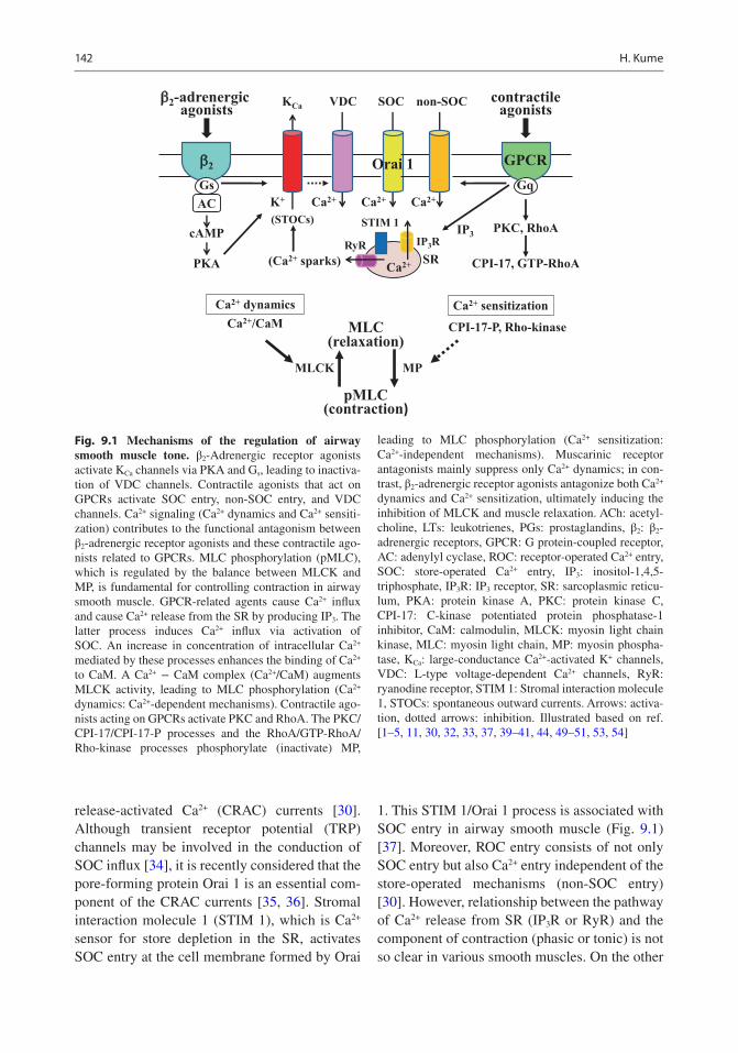
Contractile agonists acting on G protein-coupled receptors (GPCRs), such as methacholine (MCh), histamine, prostaglandins, leukotrienes, and endothelin, initially cause phasic contraction of airway smooth muscle, subsequent to tonic con-traction with increasing concentration of intra-cellular Ca2+ ([Ca2+]i) mediated by Ca2+ influx passing through various Ca2+ channels (Ca2+ dynamics) [30]. When these agents (ligands) are connected to the GPCRs, receptor-operated Ca2+ (ROC) entry is activated [31], and then Ca2+ is released from sarcoplasmic reticulum (SR) via the production of inositol-1,4,5-triphosphate receptor (IP3R) and ryanodine receptors (RyR) in airway smooth muscle (Fig. 9.1) [32, 33]. This Ca2+ release from SR activates store-operated capacitative Ca2+ (SOC) entry, that is, Ca2+
9 Role of Airway Smooth Muscle in Inflammation Related to Asthma and COPD
142
release-activated Ca2+ (CRAC) currents [30]. Although transient receptor potential (TRP) channels may be involved in the conduction of SOC influx [34], it is recently considered that the pore-forming protein Orai 1 is an essential com-ponent of the CRAC currents [35, 36]. Stromal interaction molecule 1 (STIM 1), which is Ca2+ sensor for store depletion in the SR, activates SOC entry at the cell membrane formed by Orai
1. This STIM 1/Orai 1 process is associated with SOC entry in airway smooth muscle (Fig. 9.1) [37]. Moreover, ROC entry consists of not only SOC entry but also Ca2+ entry independent of the store-operated mechanisms (non-SOC entry) [30]. However, relationship between the pathway of Ca2+ release from SR (IP3R or RyR) and the component of contraction (phasic or tonic) is not so clear in various smooth muscles. On the other
Ca2+
contractileagonistsnon-SOC
IP3
MLCK MP
SOC
Ca2+ Ca2+
cAMP
PKA
AC
Ca2+/CaM MLC
pMLC(contraction)
(relaxation)
Ca2+ dynamics Ca2+ sensitization
VDC
Ca2+K+
KCab2-adrenergic
agonists
(Ca2+ sparks)
PKC, RhoA
CPI-17, GTP-RhoA
CPI-17-P, Rho-kinase
IP3RRyRSR
STIM 1
Orai 1
(STOCs)
GPCRGq
GPCRGs
b2
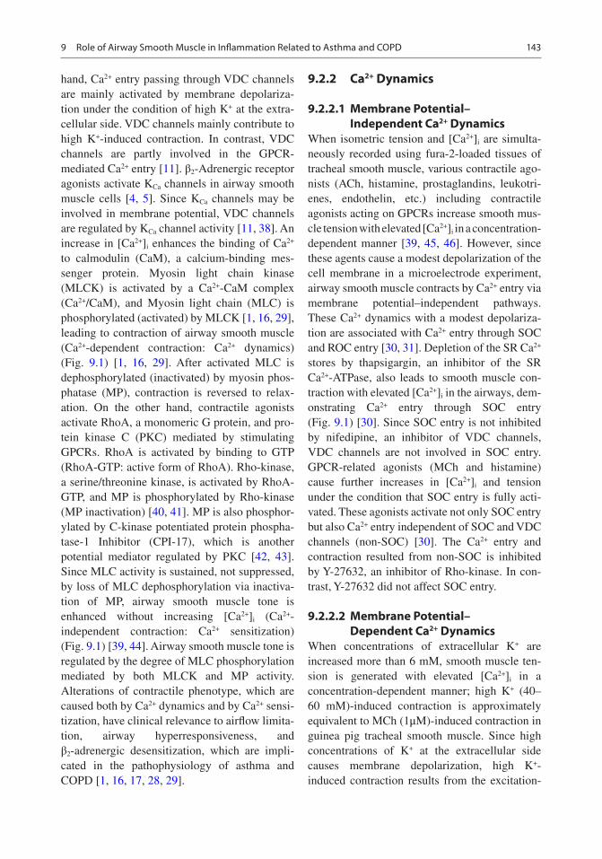
Fig. 9.1 Mechanisms of the regulation of airway smooth muscle tone. β2-Adrenergic receptor agonists activate KCa channels via PKA and Gs, leading to inactiva-tion of VDC channels. Contractile agonists that act on GPCRs activate SOC entry, non-SOC entry, and VDC channels. Ca2+ signaling (Ca2+ dynamics and Ca2+ sensiti-zation) contributes to the functional antagonism between β2-adrenergic receptor agonists and these contractile ago-nists related to GPCRs. MLC phosphorylation (pMLC), which is regulated by the balance between MLCK and MP, is fundamental for controlling contraction in airway smooth muscle. GPCR-related agents cause Ca2+ influx and cause Ca2+ release from the SR by producing IP3. The latter process induces Ca2+ influx via activation of SOC. An increase in concentration of intracellular Ca2+ mediated by these processes enhances the binding of Ca2+ to CaM. A Ca2+ − CaM complex (Ca2+/CaM) augments MLCK activity, leading to MLC phosphorylation (Ca2+ dynamics: Ca2+-dependent mechanisms). Contractile ago-nists acting on GPCRs activate PKC and RhoA. The PKC/CPI-17/CPI-17-P processes and the RhoA/GTP-RhoA/Rho-kinase processes phosphorylate (inactivate) MP,
leading to MLC phosphorylation (Ca2+ sensitization: Ca2+-independent mechanisms). Muscarinic receptor antagonists mainly suppress only Ca2+ dynamics; in con-trast, β2-adrenergic receptor agonists antagonize both Ca2+ dynamics and Ca2+ sensitization, ultimately inducing the inhibition of MLCK and muscle relaxation. ACh: acetyl-choline, LTs: leukotrienes, PGs: prostaglandins, β2: β2- adrenergic receptors, GPCR: G protein-coupled receptor, AC: adenylyl cyclase, ROC: receptor-operated Ca2+ entry, SOC: store-operated Ca2+ entry, IP3: inositol-1,4,5- triphosphate, IP3R: IP3 receptor, SR: sarcoplasmic reticu-lum, PKA: protein kinase A, PKC: protein kinase C, CPI-17: C-kinase potentiated protein phosphatase-1 inhibitor, CaM: calmodulin, MLCK: myosin light chain kinase, MLC: myosin light chain, MP: myosin phospha-tase, KCa: large-conductance Ca2+-activated K+ channels, VDC: L-type voltage-dependent Ca2+ channels, RyR: ryanodine receptor, STIM 1: Stromal interaction molecule 1, STOCs: spontaneous outward currents. Arrows: activa-tion, dotted arrows: inhibition. Illustrated based on ref. [1–5, 11, 30, 32, 33, 37, 39–41, 44, 49–51, 53, 54]
H. Kume
143
hand, Ca2+ entry passing through VDC channels are mainly activated by membrane depolariza-tion under the condition of high K+ at the extra-cellular side. VDC channels mainly contribute to high K+-induced contraction. In contrast, VDC channels are partly involved in the GPCR-mediated Ca2+ entry [11]. β2-Adrenergic receptor agonists activate KCa channels in airway smooth muscle cells [4, 5]. Since KCa channels may be involved in membrane potential, VDC channels are regulated by KCa channel activity [11, 38]. An increase in [Ca2+]i enhances the binding of Ca2+ to calmodulin (CaM), a calcium-binding mes-senger protein. Myosin light chain kinase (MLCK) is activated by a Ca2+-CaM complex (Ca2+/CaM), and Myosin light chain (MLC) is phosphorylated (activated) by MLCK [1, 16, 29], leading to contraction of airway smooth muscle (Ca2+-dependent contraction: Ca2+ dynamics) (Fig. 9.1) [1, 16, 29]. After activated MLC is dephosphorylated (inactivated) by myosin phos-phatase (MP), contraction is reversed to relax-ation. On the other hand, contractile agonists activate RhoA, a monomeric G protein, and pro-tein kinase C (PKC) mediated by stimulating GPCRs. RhoA is activated by binding to GTP (RhoA-GTP: active form of RhoA). Rho-kinase, a serine/threonine kinase, is activated by RhoA-GTP, and MP is phosphorylated by Rho-kinase (MP inactivation) [40, 41]. MP is also phosphor-ylated by C-kinase potentiated protein phospha-tase-1 Inhibitor (CPI- 17), which is another potential mediator regulated by PKC [42, 43]. Since MLC activity is sustained, not suppressed, by loss of MLC dephosphorylation via inactiva-tion of MP, airway smooth muscle tone is enhanced without increasing [Ca2+]i (Ca2+-independent contraction: Ca2+ sensitization) (Fig. 9.1) [39, 44]. Airway smooth muscle tone is regulated by the degree of MLC phosphorylation mediated by both MLCK and MP activity. Alterations of contractile phenotype, which are caused both by Ca2+ dynamics and by Ca2+ sensi-tization, have clinical relevance to airflow limita-tion, airway hyperresponsiveness, and β2-adrenergic desensitization, which are impli-cated in the pathophysiology of asthma and COPD [1, 16, 17, 28, 29].
9.2.2 Ca2+ Dynamics
9.2.2.1 Membrane Potential–Independent Ca2+ Dynamics
When isometric tension and [Ca2+]i are simulta-neously recorded using fura-2-loaded tissues of tracheal smooth muscle, various contractile ago-nists (ACh, histamine, prostaglandins, leukotri-enes, endothelin, etc.) including contractile agonists acting on GPCRs increase smooth mus-cle tension with elevated [Ca2+]i in a concentration- dependent manner [39, 45, 46]. However, since these agents cause a modest depolarization of the cell membrane in a microelectrode experiment, airway smooth muscle contracts by Ca2+ entry via membrane potential–independent pathways. These Ca2+ dynamics with a modest depolariza-tion are associated with Ca2+ entry through SOC and ROC entry [30, 31]. Depletion of the SR Ca2+ stores by thapsigargin, an inhibitor of the SR Ca2+-ATPase, also leads to smooth muscle con-traction with elevated [Ca2+]i in the airways, dem-onstrating Ca2+ entry through SOC entry (Fig. 9.1) [30]. Since SOC entry is not inhibited by nifedipine, an inhibitor of VDC channels, VDC channels are not involved in SOC entry. GPCR-related agonists (MCh and histamine) cause further increases in [Ca2+]i and tension under the condition that SOC entry is fully acti-vated. These agonists activate not only SOC entry but also Ca2+ entry independent of SOC and VDC channels (non-SOC) [30]. The Ca2+ entry and contraction resulted from non-SOC is inhibited by Y-27632, an inhibitor of Rho-kinase. In con-trast, Y-27632 did not affect SOC entry.
9.2.2.2 Membrane Potential–Dependent Ca2+ Dynamics
When concentrations of extracellular K+ are increased more than 6 mM, smooth muscle ten-sion is generated with elevated [Ca2+]i in a concentration- dependent manner; high K+ (40–60 mM)-induced contraction is approximately equivalent to MCh (1μM)-induced contraction in guinea pig tracheal smooth muscle. Since high concentrations of K+ at the extracellular side causes membrane depolarization, high K+-induced contraction results from the excitation-
9 Role of Airway Smooth Muscle in Inflammation Related to Asthma and COPD
144
contraction coupling, different from GPCR-related agonists; VDC channels are involved in this mechanism. Outward K+ currents are suppressed under the condition of higher con-centrations of extracellular K+; K+ channel clos-ing generates smooth muscle tension. In contrast, K+ channel opening leads to smooth muscle relaxation. VDC channel/KCa channel processes may be involved in the membrane potential–mediated Ca2+ dynamics. Activation of KCa chan-nels serves as a brake on vasoconstriction in pulmonary vessels [47, 48]. Membrane hyperpo-larization mediated by activation of KCa channels is proposed as the mechanism of bitter tastant- induced relaxation of airway smooth muscle [49], although an alternative pathway may also be an explanation. Since the membrane potential is elevated by inactivation of KCa channels, air-way smooth muscle contraction may be caused by VDC channel activation via membrane depo-larization [11].
In fura-2-loaded strips of tracheal smooth muscle, verapamil, an inhibitor of VDC chan-nels, inhibits MCh-induced contraction with reduced [Ca2+]i; however, relaxant effects of verapamil are not so dramatic. VDC channels are partly involved in contraction mediated by GPCR- related agonists. Iberiotoxin (IbTX), an inhibitor of KCa channels, enhances muscarinic contraction with elevated [Ca2+]i in airway smooth muscle. Since these effects of IbTX on tension and [Ca2+]i are attenuated by verapamil [11, 38], KCa channel inactivation results in con-traction with elevated [Ca2+]i via opening VDC channels arisen from depolarization of cell membrane, whereas KCa channel activation results in relaxation with reduced [Ca2+]i via VDC channel inactivation arisen from hyperpo-larization of cell membrane.
When [Ca2+]i is increased by Ca2+ entry resulted from various pathways explained before (Ca2+ dynamics), MLCK is activated by Ca2+/CaM, leading to smooth muscle contraction via phosphorylation of MLC (Fig. 9.1). In airway smooth muscle, alteration of contractility regu-lated by Ca2+ dynamics is involved in the patho-physiology of asthma and COPD, such as airflow limitation, airway hyperresponsiveness, and β2-
adrenergic desensitization [1, 16, 17, 28, 29]. It is useful to suppress Ca2+ dynamics for improving these pathological conditions in the airways.
9.2.3 Ca2+ Sensitization
9.2.3.1 Characteristics of RhoA/Rho-Kinase
An increase in [Ca2+]i results in airway smooth muscle contraction (Ca2+ dynamics, Ca2+-dependent contraction) [11, 39]. However, it is generally considered that muscarinic receptor agonists and histamine increase tension without a marked increase in [Ca2+]i. This phenomenon is referred to as Ca2+ sensitization (Ca2+-independent contraction) (Fig. 9.1) [50, 51] and is associated with G protein-coupled mecha-nisms. Rho is a monomeric G protein that belongs to the Ras superfamily. The Rho family makes up a major branch that contains Rho, Rac, and CdC42. Rho has isoforms of A-G; however, most of the function is described based on stud-ies of RhoA. RhoA exhibits both GDP/GTP binding activity and GTPase activity, and it acts as a molecular switch between a GDP-bound inactive state (GDP- RhoA) and a GTP-bound active state (GTP- RhoA). When cells are stimu-lated with agonists related to GPCRs, GDP-RhoA is converted to GTP-RhoA. RhoA and Rho-kinase are widely distributed to many organs, including the respiratory system. Rho-kinase (160 kDa) is an effector molecule of RhoA [52, 53]. Rho-kinase activated by GTP-RhoA interacts with MP, and suppresses MP activity by phosphorylating threonine 696 and 853 of myosin phosphatase targeting subunit 1 (MYPT1), a myosin-binding subunit (Fig. 9.1) [54, 55]. Rho-kinase has effects on contraction resulted from Ca2+ sensitization, stress fiber for-mation due to actin (cytoskeletal) reorganiza-tion, cell migration, and cell proliferation [40, 56]. These phenomena are implicated in the major characteristics in the pathophysiological of asthma and COPD, such as airflow limitation, airway hyperresponsiveness, β2-adrenergic desensitization, eosinophil recruitment, and air-way remodeling [1, 16, 17, 28, 29].
H. Kume
145
9.2.3.2 Role of RhoA/Rho-Kinase on Tension
Y-27632, a pyridine derivative, was developed as a specific Rho-kinase inhibitor. Y-27632 relaxes vascular smooth muscle with reducing sensitivity to intracellular Ca2+ [41]. The effects of Y-27632 on MCh-induced contraction were analyzed by using strips of guinea pig airway smooth muscle treated with fura-2. In strips of guinea pig airway smooth muscle treated with fura-2, Y-27632 inhibits contraction induced by GPCR-related agonists, such as MCh, histamine, prostaglan-dins, and leukotrienes, in a concentration- dependent manner, but there is no significant decrease in [Ca2+]i [39]. Y-27632 inhibits the phosphorylation of MYPT1, which is an effective protein for Rho-kinase action on MP in airway smooth muscle cells, in a concentration- dependent manner [55]. Fasudil hydrochloride (HA-1077), a specific inhibitor of Rho-kinase, is used clinically to suppress cerebral vasospasm following subarachnoid hemorrhage [57]. In allergen sensitized mice, HA-1077 suppresses MCh-induced lung resistance in a dose- dependent manner [58], indicating that Rho-kinase inhibi-tion results in a decrease of bronchoconstriction. Alteration of contractility of airway smooth mus-cle regulated by Ca2+ sensitization is also involved in airflow limitation, airway hyperresponsive-ness, and β2-adrenegic desensitization [1, 16, 17, 28, 29].
9.2.4 Role of Ca2+ Signaling on β2- Adrenergic Action
β2-adrenergic receptor agonists (isoproterenol, procaterol, salbutamol) result in a concentration- dependent inhibition in both tension and F340/F380 induced by MCh-induced contraction in the fura- 2- loaded tissues of guinea pig tracheal smooth muscle [2, 3]. However, under the condition that these β2-adrenergic receptor agonists cause roughly complete inhibition in tension, the values of F340/F380 are still higher than that at the basal level [2, 3]. The concentration-inhibition curves for these β2-adrenergic receptor agonists against MCh in tension are significantly dissociated from
those curves in F340/F380 [2, 3]. These results dem-onstrate that a reduction in tension is significantly greater than that in F340/F380 in β2-aderenergic action on airway smooth muscle. The tension–F340/F380 curve for SKF-96365 (3–100μM), a non- selective inhibitor of Ca2+ influx, against MCh is on the lower side than those curves for these β2- adrenergic receptor agonists. In contrast, the ten-sion–F340/F380 curve for Y-27632 (3–100μM), a specific inhibitor of Rho-kinase, is on the upper side than those curves for these β2-adrebergic receptor agonists. The curves for these β2- adrenergic receptor agonists exist between the curves for SKF-96365 and Y-27632 [2, 3]. These results demonstrate that a decrease not only in Ca2+ dynamics but also in Ca2+ sensitization con-tributes to β2-adrenergic action on airway smooth muscle. On the other hand, glycopyrronium (a muscarinic receptor antagonist) causes a concentration- dependent inhibition of MCh- induced contraction with a marked reduction in [Ca2+] in fura-2-loaded tissues of tracheal smooth muscle [2], different from β2-adrenergic receptor agonists. The concentration-inhibition curve for glycopyrronium against MCh in tension is not dissociated from those curves in F340/F380 [2]. A decrease in Ca2+ sensitization may not be involved in the relaxant effect of a muscarinic receptor antagonist on muscarinic contraction. Involvement of Ca2+ signaling is not consistent between β2-adrenergic receptor agonists and muscarinic receptor antagonists.
9.3 Large-Conductance Ca2+-Activated K+ Channels
9.3.1 General
Large-conductance Ca2+-activated K+ (KCa) chan-nels are densely distributed on smooth muscle cell membrane in various organs including human airway smooth muscle [59–61]. KCa chan-nels have a large conductance of about 250 pS in symmetrical 135–150 mM K+ medium, as com-pared to other K+ channels, and these channels are highly selective for K+ despite their large con-ductance [62]. In freshly isolated human bron-
9 Role of Airway Smooth Muscle in Inflammation Related to Asthma and COPD

146
chial smooth muscle cells, single currents of KCa channels have been recorded in the electrophysi-ological technique such as cell-attached patches, inside-out patches, and outside-out patches [63, 64]. Typical action potentials are not observed in airway smooth muscle cells under physiological conditions (weak excitability). This lack of action potentials may result from a marked increase in outward K+ conductance of the plasma mem-brane passing through KCa channels upon depo-larization [65]. Augmented K+ conductance of the membrane may lead to an inhibition in excit-ability in airway smooth muscle. Application of a K+ channel opener results in a decrease in lung resistance (bronchodilation) [66]. Spontaneous phasic contractions can be generated along with electrical activities by applying KCa channel inhibitors, such as charybdotoxin (ChTX) and iberiotoxin (IbTX) [67]. Outward K+ currents passing through KCa channels may be functioning in an important regulatory role in airway smooth muscle cells [68]. β2-adrenergic receptor agonists increase KCa channel activity, and in contrast, muscarinic receptor agonists decrease this chan-nel activity [4, 5, 8, 9]. Therefore, this channel may be target molecule in the functional antago-nism between β2-adreneric and muscarinic recep-tors [1, 16, 17, 28, 29, 69].
9.3.2 Structure
KCa channels are composed of a tetramer formed by pore-forming α-subunits along with acces-sory β-subunits, and these channels are activated by increased membrane potential and increased [Ca2+]i. The α-subunit is ubiquitously expressed by mammalian tissues and encoded by a single gene (Slo, KCNMA1) [70, 71]. The α-subunit transmembrane domains comprise seven membrane- spanning segments (S0-S6) with extracellular loops and share homology with all voltage-gated K+ channels with six transmem-brane domains (S1-S6) and a pore helix. S1-S4 are arranged in a bundle that forms the voltage- sensing component; S5-S6 and pore helices contribute to form the pore-forming component and the K+ selective filter [72]. The C-terminal
tail contributes to the Ca2+-sensing ability of this channel with a pair of Ca2+-sensing domains that regulate the conductance of K+ (RCK), that is, RCK1 and RCK2 [73]. Although the Ca2+ sensor of KCa channels has high specificity for Ca2+, other factors including divalent cations also influence the opening of these channels. Magnesium (Mg2+) enhances activation of these channels via a distinct binding site in the volt-age sensor and RCK1 domain [74]. On the other hand, intracellular protons (H+) attenuate the opening of KCa channels [10, 75]. KCa channels are associated with modulatory β-subunits, which are expressed in a cell-specific manner and have unique regulatory actions on these channels. The β-subunits bring about diversity of KCa channels. There are four distinct β-subunits, β1–4, which are encoded by KCNMB1, KCNMB2, KCNMB3, and KCNMB4. These β-subunits in these channels consist of two transmembrane domains with intracellular N- and C-termini and a long extra-cellular loop [76].
9.3.3 Electrical Characteristics
The unitary amplitude of KCa channels is approx-imately 5 pA under the condition of approxi-mately 6 mM K+ at the cytosolic side and approximately 130 mM K+ at the extracellular side held at 0 mV in tracheal smooth muscle cells [4]. Ca2+ sensitivity of KCa channels is increased by intracellular Mg2+, as is the case in vascular muscle [77]; in contrast, Ca2+ sensitivity of this channel is decreased by intracellular H+ in tra-cheal smooth muscle [10]. KCa channel activity is markedly inhibited by intracellular acidification by shortening the open state of the channel. On the other hand, intracellular alkalization has an opposite effect (increasing Ca2+ sensitivity and lengthening the open state of the channel). In the single-channel recording using outside-out patches of guinea pig and canine tracheal muscle cells, currents of KCa channels are reversibly blocked by external application of scorpion venom such as charybdotoxin (ChTX) or iberio-toxin (IbTX), selective antagonists of KCa chan-
H. Kume
147
nels. This effect is not a result of reduced current amplitude; rather, it is caused by reducing the open-state probability (nPo), the fraction of the time during which the channel is open [8, 78]. In contrast, tetraethylammonium (TEA, 1 mM) strongly reduces the unitary amplitude of single KCa channel current, different from the effects of ChTX (100 nM) on these channels without affect-ing current amplitude [60]. KCa channels are not affected by 4-aminopyridine (4-AP, 1 mM).
9.3.4 Effects on Ca2+ Signaling
In excitation-contraction coupling of airway smooth muscle cells [79], local increases in Ca2+ concentrations occur due to focal releases of Ca2+ through ryanodine receptors (RyR) from the sarcoplasmic reticulum (SR), termed Ca2+ sparks (Fig. 9.1). KCa channels are markedly opened by the Ca2+ sparks from SR close to the sarcolemma, resulting in spontaneous outward currents (STOCs) (Fig. 9.1). The coupling of ryanodine- mediated Ca2+ sparks to KCa channel-mediated STOCs, which is enhanced by β1 sub-unit, causes hyperpolarization of smooth muscle cells, leading to smooth muscle relaxation via reduction of Ca2+ entry. In KCa channel β1 subunit knockout mice, tracheal contraction induced by a muscarinic receptor agonist is enhanced as compared to wild-type mice, and not only the single channel activity of KCa channels in an inside-out patch but also STOCs in a whole cell configuration are markedly attenuated in tra-cheal smooth muscle cells of knockout mice as compared to wild-type mice [80]. IbTX (30 nM) enhances methacholine- induced contraction with elevating [Ca2+]i in airway smooth muscle, and verapamil, an inhibitor of VDC channels, suppresses the effect of IbTX on both tension and [Ca2+]i, demonstrating that KCa channel inhi-bition augments contraction via a Ca2+ entry passing through VDC channels [11]. Therefore, KCa channel activity regulates the tone of airway smooth muscle; however, the Ca2+ sparks via ryanodine receptors may not be directly involved in this KCa channel-mediated bronchoconstric-tion and bronchodilation [81].
9.3.5 Effects on β2-Adrenergic Action
β2-Adrenergic receptor agonists cause relaxation of human and guinea pig tracheal smooth mus-cles with membrane hyperpolarization in the intracellular microelectrode technique [82, 83]. These agents also inhibit tracheal smooth muscle contraction with reducing [Ca2+]i in a simultane-ous recording isometric tension and F340/F380 using fura-2-loaded tissues [2, 3]. The relaxant effects of cAMP-related agents, such as isopro-terenol and forskolin, on muscarinic contraction are significantly reduced in the presence of ChTX, a selective inhibitor of KCa channels [84–86]. This phenomenon may result from Ca2+ dynamics based on KCa channel activation medi-ated by membrane hyperpolarization (Fig. 9.1).
9.3.5.1 Protein Kinase AApplication of PKA (10 units/mL) to the cyto-solic side of inside-out membrane patches revers-ibly increases nPo of KCa channels with no changes in the amplitude of single-channel cur-rents in tracheal smooth muscle cells [4, 5], and the recovery from this activation is significantly delayed in the presence of okadaic acid, an inhib-itor of protein phosphatases [4]. The open state of KCa channel may be enhanced by phosphoryla-tion of this channel protein. External application of isoproterenol (0.2 μM), a β2-adrenergic recep-tor agonist, and okadaic acid (10μM) also increases KCa channel activity in the cell-attached patch-clamp configuration, and the recovery from this activation was also significantly delayed by okadaic acid [4]. These findings demonstrate that PKA-mediated phosphorylation of KCa chan-nel protein is involved in the β2-adrenerigic action on this channel (Fig. 9.1) [87]. Moreover, exter-nal application of forskolin (10μM), a direct acti-vator of adenylyl cyclase, increases KCa channel activity in tracheal smooth muscle cells [84].
9.3.5.2 Stimulatory G Protein of Adenylyl Cyclase
External application of isoproterenol increases the open state of KCa channels without changes in the unitary amplitude in outside-out patches in
9 Role of Airway Smooth Muscle in Inflammation Related to Asthma and COPD
148
the presence of guanosine triphosphate (GTP, 100μM) at the cytosolic side of the patch [5, 8]. The recombinant α-subunit (αs) of the stimula-tory G protein of adenylyl cyclase (Gs) preincu-bated with GTP-γ-S (αs*GTPγS, 100–1000 pM) similarly activates KCa channel in a concentration- dependent manner when applied to the cytosolic side of inside-out patches [8]. KCa channel activ-ity is directly enhanced by Gs (membrane- delimited action), independent of cAMP-dependent protein phosphorylation (Fig. 9.1) [5, 8]. The effect of PKA on the gating kinetics of KCa channels is distinct from that of αs, that is, PKA acts on the mean duration of the long openings; in contrast, αs acts on the proportion of long open-time events [5]. KCa channels are acti-vated by PKA (cAMP-dependent processes) and αs (cAMP-independent processes); PKA and αs affect these channels independently, that is, dual pathway [5] (Fig. 9.1).
β2-Adrenergic receptor agonists cause mem-brane hyperpolarization in tracheal smooth mus-cle [82, 83]. This phenomenon may result from KCa channel activation by these agents. The relax-ant effects of cAMP-related agents, such as iso-proterenol and forskolin, on muscarinic contraction are reduced in the presence of a selective inhibitor of KCa channels [84–86]. Activation of KCa channels may be associated with β2-adrenergic action on airway smooth mus-cle. After Gs activity is irreversibly enhanced by incubation with cholera toxin (2μg/mL) for 6 h, MCh-induced contraction is significantly attenu-ated, and this effect of cholera toxin is reversed in the presence of ChTX [7, 69]. Hence, the Gs pro-tein/KCa channel stimulatory linkage may con-tribute to β-adrenergic relaxation in airway smooth muscle (Fig. 9.1).
9.3.6 Effects on Muscarinic Action
Methacholine (MCh)-induced contraction is sig-nificantly enhanced with elevating [Ca2+]i in the presence of iberiotoxin, a selective inhibitor of KCa channels, in a simultaneous recording of iso-metric tension and F340/F380 of fura-2-loaded tis-sues of guinea pig tracheal smooth muscle [11,
38]. Airway muscarinic contraction may result from Ca2+ dynamics mediated not only by ROC processes but also by KCa channel inactivation (VDC processes).
9.3.6.1 Inhibitory G Protein of Adenylyl Cyclase
External application of MCh causes a marked inhibition in KCa channel activity without changes in the amplitude of single-channel currents in outside-out patches of porcine or canine tracheal muscle cells [8, 9, 45]. This MCh-induced inhibi-tion of KCa channels is potentiated by application of GTP in the cytosolic side, and in contrast, is abolished by incubation (4–6 h) with pertussis toxin (0.1–1.0μg/mL), which blocks signal trans-duction through ADP ribosylation of Gi, the inhibitory G protein of adenylyl cyclase [9]. The decreased nPo of KCa channels results from a reduction in channel open times, probably reflect-ing a decrease in the Ca2+ sensitivity of the chan-nel. The muscarinic inhibition of KCa channels may be partly responsible for the prolonged sup-pression by acetylcholine of STOCs following a transient increase [88, 89]. MCh-induced con-traction of tracheal smooth muscle is signifi-cantly attenuated after incubation with pertussis toxin (1.0μg/mL for 6 h), and this effect of per-tussis toxin is reversed in the presence of ChTX [69]. The Gi protein/KCa channel inhibitory link-age may be involved in the muscarinic-induced contraction in airway smooth muscle [1, 16, 17, 28, 29, 69].
9.3.6.2 Muscarinic M2 ReceptorsGi protein couples with the M2 subtype of musca-rinic receptors, leading to an inhibition in cAMP. These muscarinic M2 receptors exist on the surface of airway smooth muscle cells. A selective muscarinic M2 receptor antagonist (AF- DX 116, a benzodiazepine derivative) suppresses MCh-induced contraction of tracheal smooth muscle in a concentration-dependent manner [69]. Muscarinic M3 receptors, which are coupled with Gq, are the major muscarinic receptors that coupled to muscarinic receptor agonists. However, muscarinic M2 receptors also contrib-ute to airway smooth muscle contraction; KCa
H. Kume
149
channels regulate this M2 muscarinic action [9, 69, 90].
9.3.7 Dual Regulation by G Proteins
KCa channel antagonists attenuate β2-adrenergic relaxation [69, 85, 86], and in contrast, enhance muscarinic contraction in tracheal smooth mus-cle [11, 69]. KCa channel activity is markedly increased by β2-adrenergic receptor agonists, and in contrast, this channel activity is markedly sup-pressed by muscarinic receptor agonists under the experimental condition that these two agents are sequentially applied to identical outside-out patches with GTP at the cytosolic side [8]. Moreover, internal application of GTP causes an activation of KCa channel in the presence of β2-adrenergic receptor agonists at extracellular side in insideout patches, and in contrast, causes KCa channel suppression in the presence of musca-rinic receptor agonists in the same condition [8]. The activation process is mediated by the stimu-latory G protein, Gs; in contrast, the suppression process is mediated by the inhibitory G protein, Gi, that is, dual regulation by G proteins con-nected to β2-adrenergic and muscarinic M2 recep-tors [8]. The functional antagonism between β2-adrenergic and muscarinic action converges on a single KCa channel current. Therefore, KCa channels may be key molecules in the regulation of airway smooth muscle tone [1, 16, 17, 28, 29, 69].
9.3.8 Regulation by Other Factors
9.3.8.1 NO, cGMPNitric oxide (NO), which is primarily generated by nitric oxide synthase (NOS) in the endothe-lium, results in smooth muscle relaxation on vessels via hyperpolarization of the cell mem-brane [91, 92]. NO also increases KCa channel activity in vascular smooth muscle; NO-induced vasodilation is attenuated by blockade of KCa channel activity [93]. The NO/3′-5′-cyclic gua-nosine monophosphate (cGMP) pathway plays
an important role in smooth muscle relaxation in vessels and airways. KCa channel activity is mark-edly enhanced by cGMP-mediated processes, suggesting that cGMP-induced relaxation of smooth muscle results from activation of these channels [94, 95]. Vascular smooth contraction is enhanced in the KCa channel α-subunit null mice as compared to wild-type mice [96]. This phe-nomenon is caused by an impaired response to cGMP-dependent vasorelaxation, indicating that KCa channels are an important effector for cGMP- mediated action, similar to the cAMP/PKA pro-cesses (see 3.5.1.). Protein kinase G (PKG) increases KCa channel activity via the NO/cGMP pathway [97, 98]. Mechanisms of NO-induced KCa channel activation consists of dual pathway, that is, PKG-dependent phosphorylation [99] and NO direct action (PKG-independent) on channel protein [100]. PKG may also be cross-activated by cAMP to stimulate KCa channels [101]. Since the stimulatory effect of NO on KCa channels is abolished by knockdown of the β-subunit with antisense, the β-subunit acts as a mediator of NO [102].
9.3.8.2 Reactive Oxygen SpeciesReactive oxygen species (ROS), which are syn-thesized during oxidative stress in endothelial and smooth muscle cells, exerts physiological and pathophysiological effects on smooth muscle via altering the intracellular reduction and/or oxi-dation (redox) status [103]. The redox state leads to the gating of KCa channels [104]. However, the effects of redox are complex. Preferential oxida-tion of methionine increases the activity of KCa channels, whereas oxidation of cysteines reduces the channel activity [102, 106]. KCa channel activity is enhanced by hydrogen peroxide (H2O2) in pulmonary arterial smooth muscle, resulting in vasodilation mediated by membrane hyperpolar-ization [107]. H2O2 may directly bind to KCa channels to regulate them, or it may increase this channel activity via the phospholipase A2- arachidonic acid pathway and metabolites of lipoxygenase [108]. On the other hand, H2O2 causes contraction of tracheal smooth muscle with elevating [Ca2+]i in a concentration-
9 Role of Airway Smooth Muscle in Inflammation Related to Asthma and COPD
150
dependent fashion [109]. Moreover, peroxynitrite (ONOO−), an oxidant generated by the reaction of NO and superoxide, causes smooth muscle contraction in cerebral arterial resulted from inhibiting KCa channel activity [110].
9.3.8.3 Arachidonic AcidArachidonic acid and its metabolites such as 20-hydroxyeicosatetraenoic acid (20-HETE) and epoxyeicosatrienoic acids (EETs) contrib-ute to regulation of vascular smooth muscle tone. Arachidonic acid and EETs cause vasodi-lation as a result of an augmentation in KCa channel activity [111, 112]. 20-HETE also causes relaxation of airway smooth muscle with membrane hyperpolarization resulted from acti-vation of KCa channels [113]. Acute hypoxia reduced the generation of 20-HETE, and subse-quently the inhibitory action of 20-HETE on KCa channels results in relaxation of cerebral arterial smooth muscle [114]. On the other hand, 20-HETE acts as a vasoconstrictor via a decrease in KCa channel activity in renal arterial smooth muscle, and PKC is involved in this phenome-non [115].
9.4 Characteristic Action of Bronchodilators on Airway Smooth Muscle
9.4.1 General
GPCR-related agents such as β2-adrenergic receptor agonists and muscarinic receptor antag-onists are generally used as bronchodilators to improve symptoms and lung function for patients with asthma and COPD. The potency of these GPCR-related agents depends on its receptor affinity and intrinsic efficacy, which are influ-enced by pathophysiology of diseases and exces-sive administration. Therefore, alteration of affinity to its receptor and intrinsic efficacy may result in decreases/increases in the effects that these GPCR-related agents originally have. Although these issues are clinically important, little is known about clinical relevance of affinity to its receptor and intrinsic efficacy.
9.4.2 Intrinsic Efficacy
Intrinsic efficacy (intrinsic activity) refers to the ability of an agent to activate its receptors with-out regard for their concentration. Some agonists completely activate their receptors (full agonists), while other agonists activate their receptors only partially (partial agonists). The two subtypes of partial agonists are weak partial agonists, which have lower efficacy, and strong partial agonists, which have higher efficacy [16, 17, 28, 29, 116, 117]. Therefore, partial agonists need to occupy a large fraction of these receptors to produce an equivalent effect that full agonists achieve by occupying many fewer receptors. When the num-ber of these receptors is decreased, and the func-tion of these receptors is disordered, the ability of partial agonists to relax airway smooth muscle becomes less than their initial effect [16, 17, 28, 29, 116, 117]. On the other hand, full agonists resist reducing their responsiveness even under the conditions of reduced receptor number and disordered receptor function [16, 17, 28, 29, 116, 117]. Intrinsic efficacy would provide an impor-tant parameter for the rational clinical use of bronchodilators.
Intrinsic efficacy is commonly measured indi-rectly as a response to activation of the post- receptor signal transduction pathways; this response can be physiological (change in smooth muscle relaxation in vitro and airway resistance in vivo) [118]. Intrinsic efficacy depends mark-edly on variable factors in the target cells, such as the number of receptors and functional antago-nism (activation of an opposing signal transduc-tion process) [116]. In cells with a higher number of receptors (spare receptors), activation of a small fraction of receptors is sufficient to gener-ate a full response [16, 17, 28, 29, 116, 117]. On the other hand, in cells with a lower number of receptors or functional antagonisms (desensitiza-tion or contraction of airway smooth muscle), even though a higher fraction of the receptors is activated, a full response may not be achieved [16, 17, 28, 29, 116, 117]. Many patients with COPD are older patients. Not only excessive exposure to bronchodilators, but also aging con-tributes to reduced receptor numbers and disor-
H. Kume
151
dered receptor functions. Hence, in the clinical use of bronchodilators, intrinsic efficacy may affect the expression of the effects of these agents on airway smooth muscle. The EC50 and the max-imal effects in the concentration-inhibition curves for an agent against MCh express its potency and intrinsic efficacy, respectively [16, 17, 28, 29, 116]. When the functional antagonism was intensified by application of MCh (10μM, roughly 90% of the maximal contraction), iso-proterenol causes complete relaxation against this contraction, indicating that isoproterenol behaves as a full agonist. In contrast, complete inhibition did not occur with other β2-adrenergic receptor agonists [16, 17, 28, 29, 116, 117]. The maximal effects in the curves for these other ago-nists (excluding isoproterenol) are attenuated, indicating that these other agonists behave as par-tial agonists. Based on values of their intrinsic efficacy, they are classified into two types, that is, strong partial agonists (indacaterol, formoterol, procaterol, olodaterol vilanterol) and weak par-tial agonists (salbutamol, salmeterol, tulobuterol) [16, 17, 29]. Isoproterenol, a full agonist, causes β2-aderergic desensitization greater than partial agonists, indicating that excessive application of a full agonist leads to reduced responsiveness to β2-adrenergic receptor agonists in airway smooth muscle [11, 17, 28, 29, 38, 119]. In contrast, tulobuterol, which is the weakest partial agonist, causes a modest reduction in the relaxant effect, even in cases of repeatedly excessive exposure to tulobuterol [17, 28, 29, 120]. However, a loss of β2-adrenergic action in partial agonists after exposure to these agonists is less potent in ago-nists with higher values of intrinsic efficacy (strong partial agonists) than in agonists with lower values of intrinsic efficacy (weak partial agonists) [17]. In a meta-analysis, indacaterol, which has a highest value of intrinsic efficacy in partial agonists, is most effective in improving lung function and clinical symptoms in patients with COPD [121].
On the other hand, in concentration-inhibition curves, muscarinic receptor antagonists (atro-pine, tiotropium, glycopyrronium, umeclidin-ium) cause complete inhibition against MCh (10 μM)-induced contraction of tracheal smooth
muscle; values of EC50 are not significantly dif-ferent in these muscarinic antagonists [38]. These antagonists behave as full antagonists. In a meta- analysis compared to placebo, these muscarinic receptor antagonists have no significant differ-ence in increasing lung function for patients with COPD [122].
9.4.3 Allosteric Effects
Allosteric modulators connect to GPCRs (seven transmembrane receptors) at the allosteric site that is topographically distinct from the ortho-steric site, and they result in an alteration in receptor conformation. Allosteric GPCR modula-tors impact on the orthosteric binding pocket and alter association or dissociation rates of an ortho-steric ligand (affinity modulation). Allosteric effects also affect intracellular responses and alter the signaling capacity (intrinsic efficacy) of an orthosteric ligand (efficacy modulation) [123.124]. Antagonism of agonist response is caused by a reduction in affinity and/or efficacy resulted from allosteric effects. Isoproterenol (a full β2-adrenergic agonist) competitively antago-nizes MCh (10μM)-induced contraction, indicating that this agent acts on orthosteric sites, not on allosteric sites in β2-adrenergic receptors [17, 28, 29, 38]. In contrast, partial β2-adrenergic agonists, such as formoterol, procaterol, inda-caterol, olodaterol, vilanterol, salmeterol, and salbutamol, noncompetitively antagonize MCh (10μM)-induced contraction (efficacy is attenu-ated by stimulating allosteric site), indicating that these agonists behave as allosteric modulators against β2-adrenergic receptors (Fig. 9.2) [17, 28, 29, 38, 125]. Since allosteric modulators merely tune the signal in the receptors and have no effects on the receptors without endogenous ligands, partial agonists that act as allosteric modulators are less potent in causing tachyphy-laxis (desensitization) after excessive exposure [17, 28, 29, 38, 125]. In concentration-inhibition curves for an agent, a reduction in maximal per-cent inhibition from complete inhibition indi-cates efficacy modulation with an agent (inhibition in response to orthosteric site involve-
9 Role of Airway Smooth Muscle in Inflammation Related to Asthma and COPD
152
ment). The ranking of alterations in efficacy modulation of partial β2- adrenergic agonists is in reverse order to values of their intrinsic efficacy. In concentration- inhibition curves for an agent, value of EC50 of an agent is lower than that of isoproterenol, indicating that an agent causes an augmentation in affinity (association rate) to a ligand at an orthosteric site. The rank of augmen-tation in affinity modulation of partial β2-adrenergic agonists is also in reverse order to values of their EC50. On the other hand, all of muscarinic receptor antagonists cause complete inhibition against muscarinic contraction, and values of EC50 are not significantly different between these four antagonists [38]. Affinity and intrinsic efficacy of muscarinic receptor antago-nists may not depend on each agent. Muscarinic receptor antagonists operate upon orthosteric
sites, and do not act on allosteric sites on musca-rinic receptors (Fig. 9.2) [38, 125].
9.4.4 Synergistic Effects of Bronchodilators
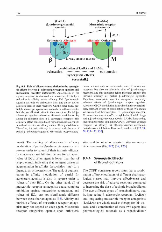
The COPD consensus report states that a combi-nation of bronchodilators of different pharmaco-logical classes may improve effectiveness and decrease the risk of adverse reactions compared to increasing the dose of a single bronchodilator. The two different types of bronchodilators, that is, long-acting β2-adrenergic receptors (LABAs) and long-acting muscarinic receptor antagonists (LAMAs), are widely used as therapy for this dis-ease, and a combination of these two agents has pharmacological rationale as a bronchodilator
OrthostericAllostericOrthosteric Allosteric
b M
b2-Adrenergic partial agonists
Muscarinic receptor antagonists
ACh
(a)(b)
relaxation contractionsynergistic effects
(crosstalk)
combination of LABA and LAMA
(LABA) (LAMA)
airway smooth muscle
Fig. 9.2 Role of allosteric modulation in the synergis-tic effects between β2-adrenergic receptor agonists and muscarinic receptor antagonists. Antagonism of the agonist response is observed via allosteric effects by a reduction in affinity and/or efficacy. Full β2-adrenergic agonists act only on orthosteric sites, and do not act on allosteric sites in their receptors. On the other hand, par-tial β2-adrenergic agonists act not only on orthosteric sites but also on allosteric sites in their receptors. Partial β2- adrenergic agonists behave as allosteric modulators. By acting on allosteric sites in β2-adrenergic receptors, this allosteric effect causes reduced responsiveness to agonists in orthosteric sites via affinity and/or efficacy modulation. Therefore, intrinsic efficacy is reduced with the use of partial β2-adrenergic agonists. Muscarinic receptor antag-
onists act not only on orthosteric sites of muscarinic receptors but also on allosteric sites of β2-adrenergic receptors, and this allosteric action increases affinity and intrinsic efficacy of partial β2-adrenergic agonists. Therefore, muscarinic receptor antagonists markedly enhance effects of β2-adrenergic receptor agonists. Allosteric GPCR modulation is involved in the synergisti-cally relaxant effects of combination of these two agents via crosstalk of their receptors. β: β2-adrenergic receptor, M: muscarinic receptor, ACh: acetylcholine, LABA: long- acting β2-adrenergic receptor agonist, LAMA: long-acting muscarinic receptor antagonist, GPCR: G protein-coupled receptor. (a): affinity, (b): efficacy. Arrows: activation, dotted arrows: inhibition. Illustrated based on ref. [17, 29, 38, 123–125, 132]
H. Kume
153
therapy [16, 17, 28, 29, 38, 126–128]. Clinical trials have demonstrated that LABA/LAMA combination is beneficial to therapy for COPD (improving symptoms and lung function, and reducing exacerbations) [129–132].
Protein allosterism is the change in protein reactivity at one site arising from a molecule binding on the protein at another site. When one agent acts on its specific GPCRs, the effect of another agent on its specific GPCRs is altered. The effects of these two agents are mutually enhanced, leading to synergistic effects. Allosteric GPCR modulators lead to alteration in pharmacological properties such as affinity, effi-cacy, and agonism/inverse agonism [123, 124]. Since allosteric effects may be caused by the interaction mediated by ligands for GPCRs [123, 133], synergistic effects between β2-adrenergic receptor agonists and muscarinic receptor antag-onists against muscarinic contraction may result from allosteric GPCR modulation in airway smooth muscle [17, 28, 29, 38]. Treated with per-tussis toxin and application of AF-DX 116 mark-edly shift the concentration-inhibition curves for isoproterenol against MCh to the left, and values of EC50 at each condition are markedly decreased. Muscarinic M2 antagonism enhances affinity for β2-adenergic receptor agonists via acting on allo-steric sites on β2-adenergic receptors (Fig. 9.2) [17, 28, 29]. In contrast, ChTX markedly shifts these curves for isoproterenol to the right, indi-cating that antagonists of KCa channels reduce affinity for β2-adenergic receptor agonists by muscarinic M2 receptor activation [17, 28, 29].
In concentration-inhibition curves, isoproter-enol completely antagonizes muscarinic contrac-tion [17, 28, 29, 38], and the complete inhibition is not attenuated at higher concentrations that produce the maximal relaxation. Isoproterenol operates orthosteric sites on β2-adrenergic recep-tors, and does not operate on these receptors, demonstrating that isoproterenol acts as a full agonist [16, 17, 28, 29, 38, 117]. In contrast, since β2-adrenergic receptor agonists except for isoproterenol and adrenaline incompletely antag-onize muscarinic contraction, these agonists cause an inhibition in the signal capacity induced by efficacy modulation (reduced responsiveness
to orthosteric sites via allosteric effects) (Fig. 9.2) [16, 17, 28, 29, 38, 117]. These β2-adrenergic agonists cause a concentration-dependent con-traction at higher concentrations that produce the maximal relaxation [38]. These agonists reduce intrinsic efficacy via operating allosteric sites on β2-adrenergic receptors as allosteric modulators (partial agonists) (Fig. 9.2) [38]. In the concentration- inhibition curves for these partial β2-adreneric receptor agonists with lower con-centrations of these muscarinic receptor antago-nists, values of EC50 for these curves are markedly decreased; the maximal effects of these partial β2-adreneric receptor agonists are markedly aug-mented to complete inhibition at each experi-mental condition [38]. Moreover, these partial β2-adreneric receptor agonists do not cause con-traction in a concentration-dependent manner at higher concentrations that produce the maximal relaxation (complete inhibition), different from the curves without lower concentration of musca-rinic receptor antagonists [38]. Muscarinic recep-tor antagonists may act not only upon orthosteric sites on muscarinic receptors but also upon allo-steric sites on β2-adrenergic receptors, and these antagonists enhance both affinity and efficacy to β2-adrenergic receptor agonists; as a result, syn-ergistic effects may be generated via crosstalk between these two GPCRs (Fig. 9.2) [38]. This synergism causes independent of the effects of muscarinic receptor antagonists on orthosteric sites on their receptors.
9.5 Role of Airway Smooth Muscle on Inflammation (Phenotype Plasticity)
9.5.1 General
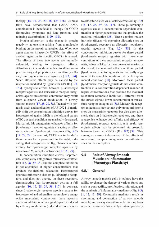
Airway smooth muscle cells in culture have the ability to change the degree of various functions such as contractility, proliferation, migration, and the synthesis of inflammatory mediators (Fig. 9.3) [1, 12, 13, 29]. Contractile mediators result in shortening and contraction of airway smooth muscle, and airway smooth muscle has long been regarded as tissues that mainly contract passively
9 Role of Airway Smooth Muscle in Inflammation Related to Asthma and COPD
154
in response to various mediators for bronchocon-striction released from other cells. Increased con-tractile property of tracheal smooth muscle may be fundamental abnormality of asthma. Contractile response to muscarinic agonists and histamine in human bronchial smooth muscle from patients with asthma is greater than that from healthy subjects. This phenomenon is caused by increased proliferation of airway smooth muscle cells because an increase in thick-ening of airway wall, which is resulted from an increased airway smooth muscle mass, contrib-utes to contractile hyperresponsiveness. Airways smooth muscle cells change to a proliferative phenotype in response to contractile agents, inflammatory mediators, and growth factors. In the presence of proliferating stimuli, airway smooth muscle cells change into a synthetic phe-notype; these cells release several inflammatory mediators under various conditions of stimula-tion. Alteration of airway smooth muscle cells from a contractile to a synthetic or a proliferative phenotype is involved in the pathophysiology of asthma and COPD, such as in airflow limitation, airway hyperresponsiveness, β2-adrenergic desensitization, and airway remodeling (Fig. 9.3).
9.5.2 Contractile Phenotype
In airway smooth muscle cell culture, phenotype plasticity is observed when cells grow to sub- confluence in the presence of serum. A prolifera-tive phenotype develops in airway smooth muscle cells under these conditions that is characterized by decreased expression of contractile proteins including smooth muscle–myosin heavy chain (sm-MHC), calponin, smooth muscle α action (sm-α actin), desmin, MLCK, and caldesmon [12, 29]. In contrast, airway smooth muscle cells with a contractile phenotype are characterized by augmented expression of contractile proteins and retain their ability to contract in response to vari-ous spasmogens. Trangestin (SM22), soothelin, metavinculin, and caveolin-1 are involved in modulation of airway smooth muscle cells toward a contractile phenotype [12, 29].
9.5.3 Synthetic and Proliferative Phenotypes
In addition to the effects of these endogenous factors, airway smooth muscle can change from
Contractile
Hyper-contractile
Synthetic/proliferative
growth factorsadhesion molecules
fibronectin, etc.
serum deprivationTGF-b, insulin
airway smooth muscle cells
airwayhyper-
responsiveness
airwayremodeling
intractable asthma
alterations of contractility, proliferation, migration
Fig. 9.3 Phenotype switching in airway smooth mus-cle cells. Important factors for phenotype switching are shown. Inflammatory processes alter phenotype of airway smooth muscle between the contractile phenotype and the synthetic/proliferative or hyper-contractile phenotype. These phenotype changes enhance contractility, migra-
tion, proliferation, and synthesis of inflammatory sub-stances in airway smooth muscle cells, resulting in hyperresponsiveness and remodeling in the airways that cause an increase in the severity of asthma. Illustrated based on ref. [1, 12, 13, 29]
H. Kume
155
one phenotype to another after exposure to vari-ous exogenous stimuli including extracellular matrix (ECM, in particular collagen type 1 and fibronectin), PDGF, and TGF-β [13, 134]. Airway smooth muscle cells derived from healthy donors are less proliferative than those derived from asthmatic donors, who show alteration toward a more proliferative phenotype [135, 136]. After exposure to IL-13 and PDGF-BB, expression of the SR Ca2+ ATPase (a Ca2+ transporter) is attenu-ated, leading to recapitulation of a more secretory and proliferative phenotype [137]. A synthetic phenotype is characterized by an increase in syn-thetic organelles for protein and lipid synthesis such as the Golgi apparatus and numerous mito-chondria, leading to an augmented proliferative capacity. Modulation toward proliferative and synthetic phenotypes is also associated with an increase in non-muscle MHC, l-caldesmon, vimentin, α/β-PKC, and CD44 homing cellular adhesion molecule [12]. Cells with this pheno-type show increased proliferative capacity with a diminished abundance of contractile proteins, leading to attenuation of responses to contractile agents [12]. In airway smooth cell culture, 20–60% of the cells have a secretory phenotype; on the other hand, approximately 50% of the cells express proliferative capacity, indicating that cytokine production and proliferation may be overlapping and not independent functions [138].
9.5.4 Hyper-Contractile Phenotype
In contractile and proliferative states, intermedi-ate or extreme phenotypes of each state may exist. Previous reports have demonstrated that prolonged starvation of canine airway smooth muscle causes a hyper-contractile phenotype (a third putative phenotype) [139, 140], which may contribute to hyperresponsiveness although this phenotype has not been replicated in human air-way smooth muscle. Markers for this phenotype include a lack of smooth muscle myosin-B (SM- B; an isoform of MHC), and increases in expres-sion of MLCK and muscarinic M3 receptors. In human airway smooth muscle cells, prolonged
serum starvation causes an increase in expression of muscarinic M3 receptors on the surface of cells derived from healthy volunteers, but not on cells derived from patients with asthma. On the other hand, exposure to muscarinic receptor agonists for a longer period reduces expression of contrac-tile proteins and responsiveness of airway smooth muscle cells [141].
9.5.5 Ca2+ Handling
The plasticity of cells that allows them to change from a contractile phenotype to other phenotypes (proliferation, migration, or secretion of chemi-cal mediators) may be associated with Ca2+ dynamics [18, 19] and Ca2+ sensitization [20–24]. Phenotype plasticity in airway smooth muscle cells is associated with an alteration in the expres-sion of ion channels such as voltage-gated sodium, inward rectifying K+, and KCa channels [64]. KCa channels that are regulated by G pro-teins (Gs, Gi) contribute to Ca2+ dynamics, by regulating the passage of Ca2+ through VDC channels via membrane potential. In contrast, the phenotype plasticity in vascular smooth muscle cells is associated with various Ca2+ handling regulators such as SOC, ROC, transient receptor potential channel type C (TRCP), Orai l and Stromal interacting model 1 (STIM1) [142]. On the other hand, since RhoA/Rho-kinase acts on contractility and proliferation in airway smooth muscle, Ca2+ sensitization induced by this path-way may also contribute to phenotype change in this tissue. Exposure of airway smooth muscle to S1P results in airway hyperresponsiveness (hyper-contractile phenotype) that is mediated by Ca2+ sensitization via inactivation of myosin phosphatase, which links Gi and RhoA/Rho- kinase processes [55]. Inhibition of airway smooth muscle cell proliferation (proliferative phenotype) by simvastatin is due to prevention of geranylgeranylation of RhoA, which causes an increase in Ca2+ sensitization not by farnesylation of Ras, independent of reducing cholesterol syn-thesis. The inhibitory effect of simvastatin on cell proliferation is caused by Rho-kinase-induced Ca2+ sensitization [21].
9 Role of Airway Smooth Muscle in Inflammation Related to Asthma and COPD
156
9.5.6 Regulation of Phenotype Switching
Phenotype switching in airway smooth muscle is regulated by dynamic processes that are influ-enced by changes in the microenvironment of the cells. In vitro cell proliferation is increased by various factors such as peptide growth factors, agonists of Gq/i-involved GPCRs, inflammatory mediators and ECM proteins (collagen type I and fibronectin) [143–146]. Many of these factors are increased in the vicinity of the airway smooth muscle by structural cells of the airways, includ-ing by airway smooth muscle cells themselves in asthma [147–150]. In contrast, cell proliferation is inhibited by various factors such as glucocorti-costeroids, agonists of Gs-involved GPCRs, NO, insulin, PGs, and ECM proteins (chondroitin sul-fate, decorin, and laminins) [151–156]. Moreover, prolonged serum deprivation, insulin, and TGFβ induce a hypercontractile phenotype character-ized by decreased proliferative response, increased contractive response, and enhanced expression of contractile proteins, such as sm-α- actin, sm-MHC, sm-MLCK, and calponin [157–159].
9.5.7 Modulation of Cell Phenotype by Cell Culture
Since phenotype change is markedly influenced by the surrounding conditions, this phenomenon may be due to the experimental environment used for analysis of the cell biology of airway smooth muscle in vitro. After single cells are iso-lated from airway smooth muscle bundles, these cells transiently enhance expression of contrac-tile markers, and rapidly change to a synthetic/proliferative phenotype under the condition of exposure to serum-rich medium [13]. It is there-fore possible that such phenotype change is an artifact of cell culture conditions in vitro. Little is known regarding whether this phenotype change occurs in vivo. This problem still remains to be solved. Although airway smooth muscle cell models using classical 2-dimensional cell type culture systems have provided a controlled
environment suitable for assessing long-term control of cellar responses [160], there may be a limit as to what can be clarified using this method. Further research is required to increase the physiological relevance of these models [161].
9.5.8 Interaction Between Airway Smooth Muscle and Inflammatory Cells
Contractility of airway smooth muscle cells is altered by exposure to tryptase and S1P, which are released from mast cells, and Lyso-PC, which is synthesized in the membrane of various inflam-matory cells (Fig. 9.4) [55, 162–164]. Ca2+ sensi-tization by RhoA/Rho-kinase processes contributes to this alteration of contractility. In sensitized mice by allergen challenges, eosino-phil infiltration and responsiveness to MCh are markedly increased in the airways; pre-treatment with Rho-kinase inhibitors such as Y-27632 or fasudil hydrochloride (HA-1077) markedly sup-presses increases in eosinophil recruitment and MCh-induced lung resistance in the respiratory tracts in a dose-dependent manner (Fig. 9.4) [58]. Thalidomide also inhibits both hyperresponsive-ness and eosinophil inflammation in the respira-tory tracts in sensitized mice by allergen challenges [165]. Pre-exposure of Lyso-PC and S1P to tracheal smooth muscle results in reduced responsiveness to β2-aderenergic receptor ago-nists via the Rho-kinase-induced Ca2+ sensitiza-tion [163, 164], and administration of Lyso-PC to guinea pigs enhances eosinophil recruitment and resistance in the respiratory system (Fig. 9.4) [166]. S1P also increases mRNA and protein expression of vascular cell adhesion molecule (VCAM)-1 when S1P is applied to pulmonary endothelial cells, leading to eosinophil infiltra-tion to the airways, and this upregulation of VCAM-1 is attenuated by C3 exoenzyme and Y-27632 [167]. Y-27632 reduces not only the number of eosinophils but also macrophages and neutrophils in an animal model of allergic asthma [22]. Hence, S1P causes eosinophil recruitment, hyperresponsiveness, and remodeling in the air-
H. Kume
157
ways by RhoA/Rho-kinase processes [55, 167, 168].
After exposure to adenosine triphosphate (ATP), MCh-induced contraction is markedly enhanced without elevating [Ca2+]i in fura-2- loaded tissues of guinea pig tracheal smooth muscle (Fig. 9.4) [169]. This phenomenon is inhibited by Y-27632, a selective inhibitor of Rho-kinase, and suramin, a non-selective P2 receptor inhibitor [169]. Pre-incubation with ATPγS, a non-hydrolysable analogue of ATP and α,β-meATP, a P2X agonist, also significantly
increase methacholine-induce contraction [169]. In asthma, eosinophils are infiltrated to around the airway, leading to injury and detachment of airway epithelium. ATP released from these injured epithelial cells act on airway smooth muscle, resulting in airway hyperresponsiveness by RhoA/Rho-kinase-induced Ca2+ sensitization via the P2X receptors. Therefore, Ca2+ sensitiza-tion by RhoA/Rho-kinase processes contributes to the interaction between airway smooth muscle and inflammatory cells related to asthma [1, 16, 23, 24, 29, 170].
airway smooth muscle
inflammatory cells
ATP, isoprostanes, H2O2ONOO−
Phenotype changing
Lyso-PC, S1P, tryptaseTGF-b1, PDGF
epithelial cells
Ca2+ sensitizationCa2+ dynamicsRhoA/Rho-kinaseG protein/KCa/VDC
Y
oxidative stress
airflow limitationairway hyperresponsivenessb2-adrenergic desensitization
airway remodeling
asthma COPD
TNFaIL-5, IL-13, IL-17, IL-1b
leukotriene D4, prostaglandin D2
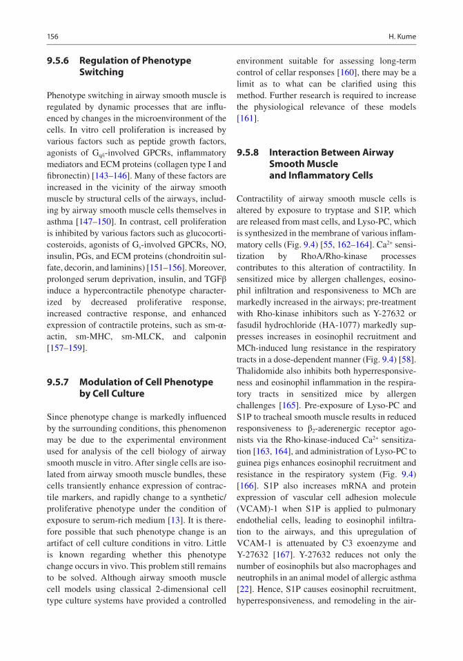
Fig. 9.4 Role of inflammatory cells on airway smooth muscle cells in the pathophysiology of asthma and COPD. In the respiratory tracts, inflammatory cells (eosinophils, mast cells) release interleukins, growth fac-tors (PDGF, TGF-β1), lipid mediators (Lyso-PC, S1P), and serine protease (tryptase). Oxidative stress generates isoprostanes, H2O2, and ONOO−. Injured epithelium releases ATP and these growth factors. These substances contribute to alterations of airway smooth muscle func-tions by affecting Ca2+ dynamics due to the G protein/KCa channel/VDC channel linkage and by affecting Ca2+ sen-sitization due to the RhoA/Rho-kinase processes. These inflammatory processes cause not only alterations in con-tractility but also changes to the proliferative phenotype in airway smooth muscle, referred to as a phenotype change. Contractility change is involved in airflow limitation, air-
way hyperresponsiveness, and β2-adrenergic desensitiza-tion; proliferative change is involved in airway remodeling due to cell proliferation and cell migration. Therefore, the G protein/KCa channel/VDC channel linkage and the RhoA/Rho-kinase processes are involved in almost all of the principal mechanisms of asthma and COPD. These pathways involved in Ca2+ dynamics and Ca2+ sensitiza-tion are molecular targets for therapy of these diseases. Lyso-PC: lysophosphatidylcholine, S1P: sphingosine 1-phosphate, PDGF: Platelet-derived growth factor, TGF- β1: transforming growth factor beta 1, IL: interleukin, H2O2: hydrogen peroxide, ONOO−: peroxynitrite, VDC: L-type voltage-dependent Ca2+ channels, KCa: large- conductance Ca2+-activated K+ channels. Illustrated based on ref. [1, 17, 20, 21, 24, 54, 55, 58, 109, 136, 144–147, 162–169, 171, 176–188, 195–197]
9 Role of Airway Smooth Muscle in Inflammation Related to Asthma and COPD
158
9.6 Role of Airway Smooth Muscle in the Pathophysiology of Asthma and COPD
9.6.1 General
An alteration of phenotype (contractile ~ syn-thetic/proliferative) in airway smooth muscle cells is caused by the inflammatory processes in the airways related to the pathophysiology of obstructive pulmonary diseases, such as asthma and COPD (Fig. 9.3). Ca2+ signaling by both Ca2+ dynamics and Ca2+ sensitization is involved in this phenotype change of airway smooth muscle cells resulted from interaction with airway con-stituent cells (inflammatory cells and epithelial cells), leading to airflow limitation, airway hyper-responsiveness, β2-adrenergic desensitization, and airway remodeling associated with these dis-eases (Fig. 9.4).
9.6.2 Airflow Limitation (Bronchoconstriction)
Airway smooth muscle contraction caused by various spasmogens (ACh, histamine, prosta-glandins, or leukotrienes) is associated with air-flow limitation, which is a characteristic feature of asthma and COPD. These agonists generate force in airway smooth muscle with increasing [Ca2+]i by Ca2+ dynamics via Ca2+ entry passing through SOC, non-SOC, and VDC (Fig. 9.1). Sphingosine 1-phosphate (S1P: a bioactive lyso-phospholipid) [55], tryptase (trypsin-like neutral serine-class protease) and SLIGKV (non- enzymatic activator of protease-activated recep-tor 2, PAR2) [162] released from mast cells cause airway smooth muscle contraction with increas-ing [Ca2+]I (Fig. 9.4). Therefore, S1P and tryptase may be involved in the pathophysiology of asthma as novel mediators. ATP is released from injured airway epithelium during the inflamma-tory processes implicated in the pathophysiology of asthma. Extracellular ATP causes contraction of airway smooth muscle with increasing [Ca2+]i (Fig. 9.4) [169]. Furthermore, oxidative stress
and mechanical stress are related to the patho-physiology of not only COPD but also asthma. 8-iso-prostaglandin F2α, an isoprostane [171], and hydrogen peroxide (H2O2) [109] produced by oxidative stress cause contraction of airway smooth muscle by increasing [Ca2+]I (Fig. 9.4). Therefore, ATP, H2O2, and 8-iso-prostaglandin F2α may be involved in the pathophysiology of asthma as novel mediators.
Y-27632 suppresses smooth muscle contrac-tion induced by spasmogens such as MCh, hista-mine, prostaglandins, and leukotrienes, which are involved in the pathophysiology of asthma and COPD, in a concentration-dependent man-ner, with no significant decrease in [Ca2+]i in strips treated with fura-2 in guinea pig trachealis [30, 39]. Y-27632 also inhibits the following fac-tors in a concentration-dependent manner with a modest effect on [Ca2+]i: 1) contraction due to S1P and tryptase released from mast cells [55, 162]; 2) contraction due to isoprostanes and hydrogen peroxide (H2O2) produced by oxidative stress [109, 171]; and 3) contraction due to ATP synthesized in injured airway epithelium [169]. These factors of contractility, which are impli-cated in the pathophysiology of asthma and COPD, cause force generation in airway smooth muscle via both Ca2+ dynamics and Ca2+ sensiti-zation [172]. Force maintenance is caused by Ca2+ sensitization induced by Rho-kinase [173]. PKC, which is an intracellular signal transduc-tion pathway for GPCR activation, also causes contraction of airway smooth muscle mediated by both Ca2+ dynamics and Ca2+ sensitization [42, 43].
9.6.3 Airway Hyperresponsiveness
Airway hyperresponsiveness is a hallmark of asthma, and any therapy cannot cure this charac-teristic feature of this disease. Airway hyperre-sponsiveness is also observed in some patients with COPD [174, 175]. This airway disorder is clinically defined as increased responsiveness to muscarinic receptor agonists (ACh and MCh) and histamine using provocation test. Airway hyperresponsiveness is due to various inflamma-
H. Kume
159
tory stimulations involved in the pathophysiol-ogy of asthma, such as antigens, chemical mediators, cytokines, and eicosanoids. In a post-mortem study of airway smooth muscle strips of patients with asthma, the response to histamine and ACh is greater than in healthy individuals [176]. In human airway smooth muscle passively sensitized with human asthmatic serum, histamine- induced contraction is significantly elevated [177]. When airway smooth muscle is exposed for an extended period of time to inter-leukin (IL)-5, IL-13, IL-17, or tumor necrosis factor (TNF)α, which are released from inflam-matory cells and epithelial cells in airways, con-traction due to muscarinic receptor agonists and KCl is significantly increased (Fig. 9.4) [178–180]. This enhancement of contractility induced by TNFα may be involved in Ca2+ sensitization via RhoA/Rho-kinase [181]. In the presence of a lower concentration of leukotriene C4, KCl- induced contraction is markedly augmented, and this enhanced contraction due to high concentra-tions of K+ is attenuated by Y-27632, a selective inhibitor of Rho-kinase [182]. After airway smooth muscle is exposed to S1P released from mast cells or ATP released from damaged epithe-lial cells, MCh-induced contraction is markedly increased without an increase in [Ca2+]i, and its augmented response to MCh is markedly sup-pressed by Y-27632 (Fig. 9.4) [55, 169, 183]. Furthermore, pre-treatment of 8-iso- prostaglandin E2, an isoprostane, causes an increase in response to CCh in airway smooth muscle, and its augmented contractility is also suppressed by Y-27632 (Fig. 9.4) [184]. After cultured human bronchial smooth muscle cells were exposed for 15 min to serum-free medium in the absence (control) and presence of S1P (3μM), relative proportion of RhoA-GTP to total RhoA (RhoAGTP/total RhoA) is significantly increased compared with the control [21, 55]. S1P (0.3–3μM) causes a concentration-depen-dent increase in MYPT1 (Thr850) phosphoryla-tion in the cultured human bronchial smooth muscle cells. An increase in phosphorylation of MYPT1 (Thr850) by 3μM S1P is significantly inhibited in the presence of Y-27632 (0.1–1.0μM) in a concentration- dependent manner [55]. These
observations indicate that airway hyperrespon-siveness is caused by direct interactions among inflammatory cells, airway epithelial cells, and airway smooth muscle cells. Ca2+ sensitization due to Rho-kinase-induced MYPT1 phosphory-lation is involved in the airway hyperresponsive-ness [54, 55]. Geranylgeranyltransferase activates RhoA activity [21]; airway hyperresponsiveness is attenuated by suppression of geranylgeranyl-transferase in mice [185]. Alterations of sensitiv-ity to Ca2+ in airway smooth muscle may play a key role in this phenomenon. Therefore, inflam-matory processes involved in asthma directly affect the function of airway smooth muscle cells via the RhoA/Rho-kinase processes. In airway smooth muscle cells, this phenotypic change for contractility induced by not only Ca2+ sensitiza-tion but also cytoskeleton reorganization (cell stiffness), which are caused by Rho-kinase, may cause an augmented response to contractile ago-nists (airway hyperresponsiveness) [1, 186, 187]. Lung resistance in response to MCh is increased in mice sensitized by allergen challenges more than in control mice (airway hyperresponsive-ness). Fasudil hydrochloride (HA-1077), an inhibitor of Rho-kinase, significantly inhibits the augmented response to MCh by allergen chal-lenges [58]. On the other hand, Ca2+ dynamics also results in an alteration in the contractile phe-notype of airway smooth muscle, leading to increased responsiveness to contractile agonists (airway hyperresponsiveness) [188]. Acidification of esophageal lumen increases the contractile response to ACh and KCl in guinea pig trachealis mediated by activation of VDC channels and Rho-kinase [189], indicating that both Ca2+ dynamics and Ca2+ sensitization play key roles in airway hyperresponsiveness (Fig. 9.4).
9.6.4 Desensitization of β2- Adrenergic Receptors
An excessive activation of β2-adrenergic recep-tors results in reduced responsiveness to an ago-nist. This phenomenon is referred to as desensitization of β2-adrenergic receptors. The phosphorylation of β2-adrenergic receptors leads
9 Role of Airway Smooth Muscle in Inflammation Related to Asthma and COPD
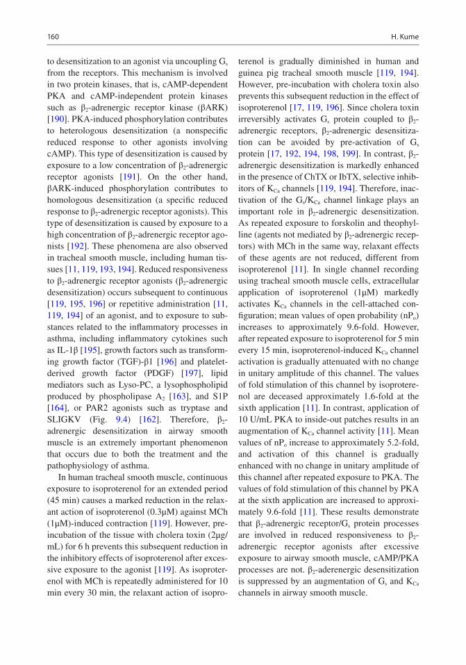
160
to desensitization to an agonist via uncoupling Gs from the receptors. This mechanism is involved in two protein kinases, that is, cAMP-dependent PKA and cAMP-independent protein kinases such as β2-adrenergic receptor kinase (βARK) [190]. PKA-induced phosphorylation contributes to heterologous desensitization (a nonspecific reduced response to other agonists involving cAMP). This type of desensitization is caused by exposure to a low concentration of β2-adrenergic receptor agonists [191]. On the other hand, βARK-induced phosphorylation contributes to homologous desensitization (a specific reduced response to β2-adrenergic receptor agonists). This type of desensitization is caused by exposure to a high concentration of β2-adrenergic receptor ago-nists [192]. These phenomena are also observed in tracheal smooth muscle, including human tis-sues [11, 119, 193, 194]. Reduced responsiveness to β2-adrenergic receptor agonists (β2-adrenergic desensitization) occurs subsequent to continuous [119, 195, 196] or repetitive administration [11, 119, 194] of an agonist, and to exposure to sub-stances related to the inflammatory processes in asthma, including inflammatory cytokines such as IL-1β [195], growth factors such as transform-ing growth factor (TGF)-β1 [196] and platelet- derived growth factor (PDGF) [197], lipid mediators such as Lyso-PC, a lysophospholipid produced by phospholipase A2 [163], and S1P [164], or PAR2 agonists such as tryptase and SLIGKV (Fig. 9.4) [162]. Therefore, β2- adrenergic desensitization in airway smooth muscle is an extremely important phenomenon that occurs due to both the treatment and the pathophysiology of asthma.
In human tracheal smooth muscle, continuous exposure to isoproterenol for an extended period (45 min) causes a marked reduction in the relax-ant action of isoproterenol (0.3μM) against MCh (1μM)-induced contraction [119]. However, pre- incubation of the tissue with cholera toxin (2μg/mL) for 6 h prevents this subsequent reduction in the inhibitory effects of isoproterenol after exces-sive exposure to the agonist [119]. As isoproter-enol with MCh is repeatedly administered for 10 min every 30 min, the relaxant action of isopro-
terenol is gradually diminished in human and guinea pig tracheal smooth muscle [119, 194]. However, pre-incubation with cholera toxin also prevents this subsequent reduction in the effect of isoproterenol [17, 119, 196]. Since cholera toxin irreversibly activates Gs protein coupled to β2-adrenergic receptors, β2-adrenergic desensitiza-tion can be avoided by pre-activation of Gs protein [17, 192, 194, 198, 199]. In contrast, β2-adrenergic desensitization is markedly enhanced in the presence of ChTX or IbTX, selective inhib-itors of KCa channels [119, 194]. Therefore, inac-tivation of the Gs/KCa channel linkage plays an important role in β2-adrenergic desensitization. As repeated exposure to forskolin and theophyl-line (agents not mediated by β2-adrenergic recep-tors) with MCh in the same way, relaxant effects of these agents are not reduced, different from isoproterenol [11]. In single channel recording using tracheal smooth muscle cells, extracellular application of isoproterenol (1μM) markedly activates KCa channels in the cell-attached con-figuration; mean values of open probability (nPo) increases to approximately 9.6-fold. However, after repeated exposure to isoproterenol for 5 min every 15 min, isoproterenol- induced KCa channel activation is gradually attenuated with no change in unitary amplitude of this channel. The values of fold stimulation of this channel by isoprotere-nol are deceased approximately 1.6-fold at the sixth application [11]. In contrast, application of 10 U/mL PKA to inside-out patches results in an augmentation of KCa channel activity [11]. Mean values of nPo increase to approximately 5.2-fold, and activation of this channel is gradually enhanced with no change in unitary amplitude of this channel after repeated exposure to PKA. The values of fold stimulation of this channel by PKA at the sixth application are increased to approxi-mately 9.6- fold [11]. These results demonstrate that β2- adrenergic receptor/Gs protein processes are involved in reduced responsiveness to β2- adrenergic receptor agonists after excessive exposure to airway smooth muscle, cAMP/PKA processes are not. β2-aderenergic desensitization is suppressed by an augmentation of Gs and KCa channels in airway smooth muscle.
H. Kume
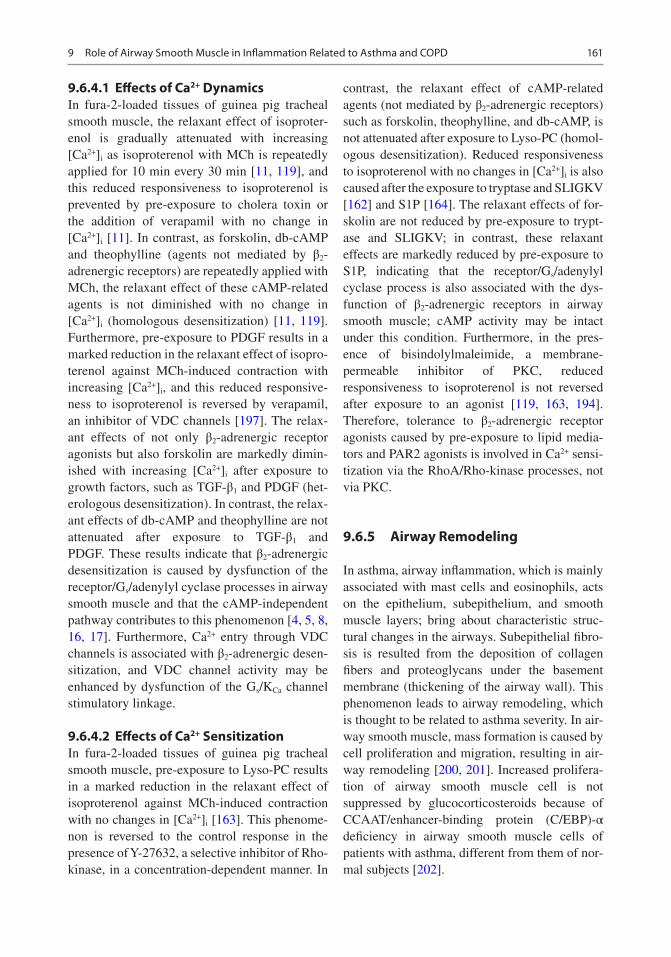
161
9.6.4.1 Effects of Ca2+ DynamicsIn fura-2-loaded tissues of guinea pig tracheal smooth muscle, the relaxant effect of isoproter-enol is gradually attenuated with increasing [Ca2+]i as isoproterenol with MCh is repeatedly applied for 10 min every 30 min [11, 119], and this reduced responsiveness to isoproterenol is prevented by pre-exposure to cholera toxin or the addition of verapamil with no change in [Ca2+]i [11]. In contrast, as forskolin, db-cAMP and theophylline (agents not mediated by β2-adrenergic receptors) are repeatedly applied with MCh, the relaxant effect of these cAMP-related agents is not diminished with no change in [Ca2+]i (homologous desensitization) [11, 119]. Furthermore, pre-exposure to PDGF results in a marked reduction in the relaxant effect of isopro-terenol against MCh-induced contraction with increasing [Ca2+]i, and this reduced responsive-ness to isoproterenol is reversed by verapamil, an inhibitor of VDC channels [197]. The relax-ant effects of not only β2-adrenergic receptor agonists but also forskolin are markedly dimin-ished with increasing [Ca2+]i after exposure to growth factors, such as TGF-β1 and PDGF (het-erologous desensitization). In contrast, the relax-ant effects of db-cAMP and theophylline are not attenuated after exposure to TGF-β1 and PDGF. These results indicate that β2- adrenergic desensitization is caused by dysfunction of the receptor/Gs/adenylyl cyclase processes in airway smooth muscle and that the cAMP- independent pathway contributes to this phenomenon [4, 5, 8, 16, 17]. Furthermore, Ca2+ entry through VDC channels is associated with β2- adrenergic desen-sitization, and VDC channel activity may be enhanced by dysfunction of the Gs/KCa channel stimulatory linkage.
9.6.4.2 Effects of Ca2+ SensitizationIn fura-2-loaded tissues of guinea pig tracheal smooth muscle, pre-exposure to Lyso-PC results in a marked reduction in the relaxant effect of isoproterenol against MCh-induced contraction with no changes in [Ca2+]i [163]. This phenome-non is reversed to the control response in the presence of Y-27632, a selective inhibitor of Rho- kinase, in a concentration-dependent manner. In
contrast, the relaxant effect of cAMP-related agents (not mediated by β2-adrenergic receptors) such as forskolin, theophylline, and db-cAMP, is not attenuated after exposure to Lyso-PC (homol-ogous desensitization). Reduced responsiveness to isoproterenol with no changes in [Ca2+]i is also caused after the exposure to tryptase and SLIGKV [162] and S1P [164]. The relaxant effects of for-skolin are not reduced by pre-exposure to trypt-ase and SLIGKV; in contrast, these relaxant effects are markedly reduced by pre-exposure to S1P, indicating that the receptor/Gs/adenylyl cyclase process is also associated with the dys-function of β2-adrenergic receptors in airway smooth muscle; cAMP activity may be intact under this condition. Furthermore, in the pres-ence of bisindolylmaleimide, a membrane- permeable inhibitor of PKC, reduced responsiveness to isoproterenol is not reversed after exposure to an agonist [119, 163, 194]. Therefore, tolerance to β2-adrenergic receptor agonists caused by pre-exposure to lipid media-tors and PAR2 agonists is involved in Ca2+ sensi-tization via the RhoA/Rho-kinase processes, not via PKC.
9.6.5 Airway Remodeling
In asthma, airway inflammation, which is mainly associated with mast cells and eosinophils, acts on the epithelium, subepithelium, and smooth muscle layers; bring about characteristic struc-tural changes in the airways. Subepithelial fibro-sis is resulted from the deposition of collagen fibers and proteoglycans under the basement membrane (thickening of the airway wall). This phenomenon leads to airway remodeling, which is thought to be related to asthma severity. In air-way smooth muscle, mass formation is caused by cell proliferation and migration, resulting in air-way remodeling [200, 201]. Increased prolifera-tion of airway smooth muscle cell is not suppressed by glucocorticosteroids because of CCAAT/enhancer-binding protein (C/EBP)-α deficiency in airway smooth muscle cells of patients with asthma, different from them of nor-mal subjects [202].
9 Role of Airway Smooth Muscle in Inflammation Related to Asthma and COPD
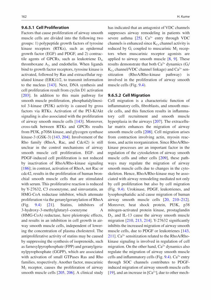
162
9.6.5.1 Cell ProliferationFactors that cause proliferation of airway smooth muscle cells are divided into the following two groups: 1) polypeptide growth factors of tyrosine kinase receptors (RTKs), such as epidermal growth factor (EGF) and PDGF, and 2) contrac-tile agents of GPCRs, such as leukotriene D4, thromboxane A2, and endothelin. When ligands bind to growth factor receptors, tyrosine kinase is activated, followed by Ras and extracellular reg-ulated kinase (ERK)1/2, to transmit information to the nucleus [143]. Next, DNA synthesis and cell proliferation result from cyclin D1 activation [203]. In addition to this main pathway for smooth muscle proliferation, phosphatidylinosi-tol 3-kinase (PI3K) activity is caused by gross factors via RTKs. Activation of the PI3-K/Akt signaling is also associated with the proliferation of airway smooth muscle cells [143]. Moreover, cross-talk between RTKs and GPCRs results from PI3K, p70S6 kinase, and glycogen synthase kinase-3 (GSK-3) [143, 204]. Involvement of the Rho family (RhoA, Rac, and Cdc42) is still unclear in the control mechanisms of airway smooth muscle cell proliferation. EGF- and PDGF-induced cell proliferation is not reduced by inactivation of RhoA/Rho-kinase signaling [186]; in contrast, activation of RhoA, not Rac or cdc42, results in the proliferation of human bron-chial smooth muscle cells that are stimulated with serum. This proliferative reaction is reduced by Y-27632, C3 exoenzyme, and simvastatin, an HMG-CoA reductase inhibitor, which attenuate proliferation via the geranylgeranylation of RhoA (Fig. 9.4) [21]. Statins, inhibitors of 3-hydroxy- 3-methylglutaryl–coenzyme A (HMG-CoA) reductase, have pleiotropic effects, and results in an inhibition in cell growth in air-way smooth muscle cells, independent of lower-ing the concentration of plasma cholesterol. The antiproliferative activity of statins may be caused by suppressing the synthesis of isoprenoids, such as farnesylpyrophosphate (FPP) and geranylgera-nylpyrophosphate (GGPP), which are associated with activation of small GTPases Ras and Rho families, respectively. Another factor, muscarinic M2 receptor, causes the proliferation of airway smooth muscle cells [205, 206]. A clinical study
has indicated that an antagonist of VDC channels suppresses airway remodeling in patients with severe asthma [25]. Ca2+ entry through VDC channels is enhanced since KCa channel activity is reduced by Gi coupled to muscarinic M2 recep-tors when muscarinic receptor agonists are applied to airway smooth muscle [8, 9]. These results demonstrate that both Ca2+ dynamics (Gi/KCa channel/VDC channel linkage) and Ca2+ sen-sitization (RhoA/Rho-kinase pathway) is involved in the proliferation of airway smooth muscle cells (Fig. 9.4).
9.6.5.2 Cell MigrationCell migration is a characteristic function of inflammatory cells, fibroblasts, and smooth mus-cle cells, and this function results in inflamma-tory cell recruitment and smooth muscle hyperplasia in the airways [207]. The extracellu-lar matrix enhances the migration of airway smooth muscle cells [208]. Cell migration arises from contraction involving actin, myosin reac-tions, and actin reorganization. Since RhoA/Rho- kinase processes are an important factor in the regulation of the cytoskeleton of airway smooth muscle cells and other cells [209], these path-ways may regulate the migration of airway smooth muscle cells due to changes in the cyto-skeleton. Hence, RhoA/Rho-kinase may be asso-ciated with airway remodeling mediated not only by cell proliferation but also by cell migration (Fig. 9.4). Urokinase, PDGF, leukotrienes, and lysophosphatidic acid cause migration of human airway smooth muscle cells [20, 210–212]. Moreover, heat shock protein, PI3K, p38 mitogen- activated protein kinase, prostaglandin D2, and IL-13 cause the airway smooth muscle migration [210, 213, 214]. Y-27632 significantly inhibits the increased migration of airway smooth muscle cells, due to PDGF or leukotrienes [143, 211]. Ca2+ sensitization related to the RhoA/Rho- kinase signaling is involved in regulation of cell migration. On the other hand, Ca2+ dynamics also regulate the migration of airway smooth muscle cells and inflammatory cells (Fig. 9.4). Ca2+ entry through SOC channels contributes to PDGF- induced migration of airway smooth muscle cells [19], and an increase in [Ca2+]i due to other mech-
H. Kume
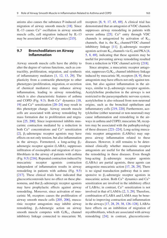
163
anisms also causes the substance P-induced cell migration of airway smooth muscle [18]. Since IL-13 causes Ca2+ oscillation in airway smooth muscle cells, cell migration induced by IL-13 may be associated with Ca2+ dynamics [215].
9.7 Bronchodilators on Airway Inflammation
Airway smooth muscle cells have the ability to alter the degree of various functions, such as con-tractility, proliferation, migration, and synthesis of inflammatory mediators [1, 12, 13, 29]. The plasticity from a contractile phenotype to other phenotypes (proliferation, migration, or secretion of chemical mediators) may enhance airway inflammation, leading to airway remodeling, which is also characteristic features of asthma and COPD (Fig. 9.3). Both Ca2+ dynamics [18, 19] and Ca2+ sensitization [20–24] may result in this phenotype change. Airway smooth muscle cells are associated with airway remodeling by mass formation due to proliferation and migra-tion [25, 200]. Since isoproterenol inhibits mus-carinic contraction mediated by a reduction in both Ca2+ concentrations and Ca2+ sensitization [2], β2-adrenerguc receptor agonists may have effects on not only tension, but also inflammation in the airways. Formoterol, a long-acting β2- adrenergic receptor agonist (LABA), suppresses infiltration of eosinophils and migration of myo-fibroblasts in the airway of patients with asthma (Fig. 9.5) [216]. Repeated contraction induced by muscarinic receptor agonists contraction independent of inflammation results in airway remodeling in patients with asthma (Fig. 9.5) [217]. These clinical trials have indicated that glucocorticosteroids have no effect on these phe-nomena, and that β2-adrenergic receptor agonists may have prophylactic effects against airway remodeling. Moreover, since activation of mus-carinic M2 receptors causes the proliferation of airway smooth muscle cells [205, 206], musca-rinic receptor antagonists may inhibit airway remodeling. β2-Adrenergic action on airway smooth muscle competes with Gi/KCa channel inhibitory linkage connected to muscarinic M2
receptors [8, 9, 17, 45, 69]. A clinical trial has demonstrated that an antagonist of VDC channels suppresses airway remodeling in patients with severe asthma [25]. Ca2+ entry through VDC channels is antagonized by activation of KCa channels, that is, the KCa channel/VDC channel inhibitory linkage [11]. β2-adrenergic receptor agonists activate KCa channels via Gs and PKA [4, 5, 8, 69], indicating that these agonists may be useful for preventing airway remodeling resulted from a reduction in VDC channel activity [218].
Since atropine, a muscarinic receptor antago-nist, suppresses Gi/KCa channel inhibitory linkage induced by muscarinic M2 receptors [8, 9], these antagonists may have effects not only against ten-sion, but also against inflammation in the air-ways, similar to β2-adrenergic receptor agonists. Acetylcholine production in the airways is not restricted to the parasympathetic nervous system; acetylcholine is also released from non-neuronal origins, such as the bronchial epithelium and inflammatory cells [219, 220]. Furthermore, ace-tylcholine (either neuronal or non-neuronal) may cause inflammation and remodeling in the air-ways in asthma and COPD; muscarinic M3 recep-tors may be associated with the pathophysiology of these diseases [221–224]. Long-acting musca-rinic receptor antagonists (LAMAs) may sup-press airway inflammation related to these diseases. However, it still remains to be deter-mined clinically whether muscarinic receptor antagonists are useful for the inflammation and the remodeling in these diseases. Even though long-acting β2-adrenergic receptor agonists (LABAs) are partial agonists, these agents can antagonize muscarinic action [17, 28, 29]. There is no signal transduction pathway that is unre-sponsive to β2-adrenergic receptor agonists in muscarinic action. Both Ca2+ dynamics and Ca2+ sensitization are involved in the inhibitory effect of LABAs; in contrast, Ca2+ sensitization is not involved in that of LAMAs [2, 3, 29]. Therefore, combination of LABA and LAMA may be bene-ficial to improving contraction and inflammation in the airways [17, 28, 29, 38, 126–128]. LABAs have effects on an imbibition of migration of myofibroblasts, which are associated with airway remodeling [24]; in contrast, glucocorticoste-
9 Role of Airway Smooth Muscle in Inflammation Related to Asthma and COPD
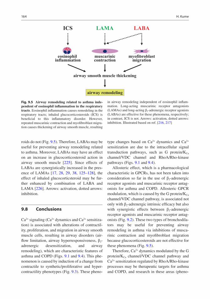
164
roids do not (Fig. 9.5). Therefore, LABAs may be useful for preventing airway remodeling related to asthma. Moreover, LABAs may have an effect on an increase in glucocorticosteroid action in airway smooth muscle [225]. Since effects of LABAs are synergistically increased in the pres-ence of LAMAs [17, 28, 29, 38, 125–128], the effect of inhaled glucocorticsteroid may be fur-ther enhanced by combination of LABA and LAMA [226]. Arrows: activation, dotted arrows: inhibition.
9.8 Conclusions
Ca2+ signaling (Ca2+ dynamics and Ca2+ sensitiza-tion) is associated with alterations of contractil-ity, proliferation, and migration in airway smooth muscle cells, resulting in airway disorders (air-flow limitation, airway hyperresponsiveness, β2- adrenergic desensitization, and airway remodeling), which are characteristic features of asthma and COPD (Figs. 9.1 and 9.4). This phe-nomenon is caused by induction of a change from contractile to synthetic/proliferative and hyper- contractility phenotypes (Fig. 9.3). These pheno-
type changes based on Ca2+ dynamics and Ca2+ sensitization are due to the intracellular signal transduction pathways, such as G protein/KCa channel/VDC channel and RhoA/Rho-kinase pathways (Figs. 9.1 and 9.4).
Allosteric effect, which is a pharmacological characteristic in GPCRs, has not been taken into consideration so far in the use of β2-adrenergic receptor agonists and muscarinic receptor antag-onists for asthma and COPD. Allosteric GPCR modulation, which is caused by the G protein/KCa channel/VDC channel pathway, is associated not only with β2-adrenergic intrinsic efficacy but also with synergistic effects between β2-adrenergic receptor agonists and muscarinic receptor antag-onists (Fig. 9.2). These two types of bronchodila-tors may be useful for preventing airway remodeling in asthma via inhibitions of musca-rinic contraction and myofibroblast migration because glucocorticosteroids are not effective for these phenomena (Fig. 9.5).
Therefore, Ca2+ dynamics modulated by the G protein/KCa channel/VDC channel pathway and Ca2+ sensitization regulated by RhoA/Rho-kinase processes may be therapeutic targets for asthma and COPD, and research in these areas (pheno-
ICS LAMA
airway remodeling
LABA
eosinophil inflammation
muscariniccontraction
myofibroblastmigration
airway smooth muscle thickening
Fig. 9.5 Airway remodeling related to asthma inde-pendent of eosinophil inflammation in the respiratory tracts. Eosinophil inflammation causes remodeling in the respiratory tracts; inhaled glucocorticosteroids (ICS) is beneficial to this inflammatory disorder. However, repeated muscarinic contraction and myofibroblast migra-tion causes thickening of airway smooth muscle, resulting
in airway remodeling independent of eosinophil inflam-mation. Long-acting muscarinic receptor antagonists (LAMAs) and long-acting β2-adrenergic receptor agonists (LABAs) are effective for these phenomena, respectively; in contrast, ICS is not. Arrows: activation, dotted arrows: inhibition. Illustrated based on ref. [216, 217]
H. Kume
165
type changes and allosteric effects) may provide novel strategies in the development of agents for these diseases that will be effective for both bron-choconstriction and airway inflammation.
References
1. Kume H. RhoA/Rho-kinase as a therapeutic target in asthma. Curr Med Chem. 2008;15:2876–85.
2. Oguma T, Kume H, Ito S, et al. Involvement of reduced sensitivity to Ca2+ in β-adrenergic action on airway smooth muscle. Clin Exp Allergy. 2006;36:183–91.
3. Fukunaga K, Kume H, Oguma T, et al. Involvement of Ca2+ signaling in the synergistic effects between muscarinic receptor antagonists and β2-adrenoceptor agonists in airway smooth muscle. Int J Mol Sci. 2016;17(9):1590.
4. Kume H, Takai A, Tokuno H, et al. Regulation of Ca2+-dependent K+-channel activity in tracheal myo-cytes by phosphorylation. Nature. 1989;341:152–4.
5. Kume H, Hall IP, Washabau RJ, et al. β-adrenergic agonists regulate KCa channels in airway smooth muscle by cAMP-dependent and -independent mechanisms. J Clin Invest. 1994;93:371–9.
6. Tomita T, Kume H. Electrophysiology of potassium channels in airways smooth muscle. In: Raeburn D, Giembycz MA, editors. Airways smooth muscle: development and regulation of contractility. Basel: Birkhauser Verlag; 1994. p. 163–84.
7. Kume H. Large-conductance calcium-activated potassium channels. In: Wang YX, editor. Calcium signaling in airway smooth muscle cells. New York: Springer; 2013. p. 49–83.
8. Kume H, Graziano MP, Kotlikoff MI. Stimulatory and inhibitory regulation of calcium-activated potas-sium channels by guanine nucleotide-binding pro-teins. Proc Natl Acad Sci U S A. 1992;89:11051–5.
9. Kume H, Kotlikoff MI. Muscarinic inhibition of single KCa channels in smooth muscle cells by a pertussis-sensitive G protein. Am J Physiol. 1991;261:C1204–9.
10. Kume H, Takagi K, Satake T, et al. Effects of intra-cellular pH on calcium-activated potassium chan-nels in rabbit tracheal smooth muscle. J Physiol. 1990;424:445–57.
11. Kume H, Ishikawa T, Oguma T, et al. Involvement of Ca2+ mobilization in tachyphylaxis to β-adrenergic receptors in trachealis. Am J Respir Cell Mol Biol. 2003;29:359–66.
12. Halayko AJ, Tran T, Gosens R. Phenotype and functional plasticity of airway smooth muscle: role of caveolae and caveolins. Proc Am Thorac Soc. 2008;5:80–8.
13. Wright DB, Trian T, Siddiqui S, et al. Phenotype modulation of airway smooth muscle in asthma. Pulm Pharmacol Ther. 2013;26:42–9.
14. Mahn K, Ojo OO, Chadwick G, et al. Ca2+ homeo-stasis and structural and functional remodel-ling of airway smooth muscle in asthma. Thorax. 2010;65:547–52.
15. Koopmans T, Anaparti V, Castro-Piedras I, et al. Ca2+ handling and sensitivity in airway smooth muscle: emerging concepts for mechanistic understanding and therapeutic targeting. Pulm Pharmacol Ther. 2014;29:108–20.
16. Kume H. Ca2+ dynamics and Ca2+ sensitization in the regulation of airway smooth muscle tone. In: Sakuma K, editor. Muscle cell and tissue. Rijeka: InTech; 2015. p. 289–330.
17. Kume H, Fukunaga K, Oguma T. Research and development of bronchodilators for asthma and COPD with a focus on G protein/KCa channel link-age and β2-adrenergic intrinsic efficacy. Pharmacol Ther. 2015;156:75–89.
18. Li M, Shang YX, Wei B, et al. The effect of sub-stance P on asthmatic rat airway smooth muscle cell proliferation, migration, and cytoplasmic calcium concentration in vitro. J Inflamm (Lond). 2011;8:18.
19. Suganuma N, Ito S, Aso H, et al. STIM1 regulates platelet-derived growth factor-induced migration and Ca2+ influx in human airway smooth muscle cells. PLoS One. 2012;7:e45056.
20. Parameswaran K, Cox G, Radford K, et al. Cysteinyl leukotrienes promote human airway smooth muscle migration. Am J Respir Crit Care Med. 2002;166:738–42.
21. Takeda N, Kondo M, Ito S, et al. Role of RhoA inactivation in reduced cell proliferation of human airway smooth muscle by simvastatin. Am J Respir Cell Mol Biol. 2006;35:722–9.
22. Schaafsma D, Bos IS, Zuidhof AB, et al. The inhaled Rho kinase inhibitor Y-27632 protects against allergen-induced acute bronchoconstriction, air-way hyperresponsiveness, and inflammation. Am J Physiol Lung Cell Mol Physiol. 2008;295:L214–9.
23. Possa SS, Charafeddine HT, Righetti RF, et al. Rho-kinase inhibition attenuates airway respon-siveness, inflammation, matrix remodeling, and oxidative stress activation induced by chronic inflammation. Am J Physiol Lung Cell Mol Physiol. 2012;303:L939–52.
24. Gerthoffer WT, Solway J, Camoretti-Mercado B. Emerging targets for novel therapy of asthma. Curr Opin Pharmacol. 2013;13:324–30.
25. Girodet PO, Dournes G, Thumerel M, et al. Calcium channel blocker reduces airway remodeling in severe asthma: a proof-of-concept study. Am J Respir Crit Care Med. 2015;191:876–83.
26. Seibold MA, Wang B, Eng C, et al. An african- specific functional polymorphism in KCNMB1 shows sex-specific association with asthma severity. Hum Mol Genet. 2008;17:2681–90.
27. Goldklang MP, Perez-Zoghbi JF, Trischler J, et al. Treatment of experimental asthma using a single small molecule with anti-inflammatory
9 Role of Airway Smooth Muscle in Inflammation Related to Asthma and COPD
166
and BK channel-activating properties. FASEB J. 2013;27:4975–86.
28. Kume H, Ito S. Role of large-conductance calcium- activated potassium channels on airway smooth muscle in physiological and pathological conditions. In: Kume H, editor. Potassium channels in health and disease. New York: Nova Science Publishers; 2017. p. 41–120.
29. Kume H. Research and development for anti- asthmatic agents with a focus on phenotype chang-ing by Ca2+ signaling in airway smooth muscle cells. In: Rahman AU, editor. Frontiers in clinical drug research - anti allergy agents, vol. 3. Sharjah: Bentham; 2018. p. 116–81.
30. Ito S, Kume H, Yamaki K, et al. Regulation of capac-itative and noncapacitative receptor-operated Ca2+ entry by Rho-kinase in tracheal smooth muscle. Am J Respir Cell Mol Biol. 2002;26:491–8.
31. Murray RK, Kotlikoff MI. Receptor-activated cal-cium influx in human airway smooth muscle cells. J Physiol. 1991;435:123–44.
32. Ay B, Prakash YS, Pabelick CM, et al. Store-operated Ca2+ entry in porcine airway smooth muscle. Am J Physiol Lung Cell Mol Physiol. 2004;286:L909–17.
33. Du W, Stiber JA, Rosenberg PB, et al. Ryanodine receptors in muscarinic receptor-mediated broncho-constriction. J Biol Chem. 2005;280:26287–94.
34. Salido GM, Sage SO, Rosado JA. TRPC channels and store-operated Ca2+ entry. Biochim Biophys Acta. 2009;1793:223–30.
35. Feske S, Gwack Y, Prakriya M, et al. A mutation in Orai1 causes immune deficiency by abrogating CRAC channel function. Nature. 2006;441:179–85.
36. Zhang SL, Yeromin AV, Zhang XH, et al. Genome- wide RNAi screen of Ca2+ influx identifies genes that regulate Ca2+ release-activated Ca2+ channel activity. Proc Natl Acad Sci U S A. 2006;103:9357–62.
37. Spinelli AM, González-Cobos JC, Zhang X, et al. Airway smooth muscle STIM1 and Orai1 are upregulated in asthmatic mice and mediate PDGF- activated SOCE, CRAC currents, proliferation, and migration. Pflügers Arch. 2012;464:481–92.
38. Kume H, Nishiyama O, Isoya T, et al. Involvement of allosteric effect and KCa channels in crosstalk between β2-adrenergic and muscarinic M2 recep-tors in airway smooth muscle. Int J Mol Sci. 2018;19(7):1999.
39. Ito S, Kume H, Honjo H, et al. Possible involvement of Rho kinase in Ca2+ sensitization and mobilization by MCh in tracheal smooth muscle. Am J Physiol Lung Cell Mol Physiol. 2001;280:L1218–24.
40. Kimura K, Ito M, Amano M, et al. Regulation of myosin phosphatase by Rho and Rho-associated kinase (Rho-kinase). Science. 1996;273:245–8.
41. Uehata M, Ishizaki T, Satoh H, et al. Calcium sen-sitization of smooth muscle mediated by a Rho- associated protein kinase in hypertension. Nature. 1997;389:990–4.
42. Mukherjee S, Trice J, Shinde P, et al. Ca2+ oscilla-tions, Ca2+ sensitization, and contraction activated
by protein kinase C in small airway smooth muscle. J Gen Physiol. 2013;141:165–78.
43. Dixon RE, Santana LF. A Ca2+- and PKC-driven regulatory network in airway smooth muscle. J Gen Physiol. 2013;141:161–4.
44. Yoshii A, Iizuka K, Dobashi K, et al. Relaxation of contracted rabbit tracheal and human bronchial smooth muscle by Y-27632 through inhibition of Ca2+ sensitization. Am J Respir Cell Mol Biol. 1999;20:1190–200.
45. Wang YX, Fleischmann BK, Kotlikoff MI. Modulation of maxi-K+ channels by voltage- dependent Ca2+ channels and methacholine in single airway myocytes. Am J Physiol. 1997;272:C1151–9.
46. Iwata S, Ito S, Iwaki M, et al. Regulation of endothelin- 1-induced interleukin-6 production by Ca2+ influx in human airway smooth muscle cells. Eur J Pharmacol. 2009;605(1–3):15–22.
47. Gutman GA, Chandy KG, Grissmer S, et al. International Union of Pharmacology. LIII. Nomenclature and molecular relationships of voltage-gated potassium channels. Pharmacol Rev. 2005;57:473–508.
48. Bonnet S, Archer SL. Potassium channel diversity in the pulmonary arteries and pulmonary veins: implications for regulation of the pulmonary vascu-lature in health and during pulmonary hypertension. Pharmacol Ther. 2007;115:56–69.
49. Zhang CH, Lifshitz LM, Uy KF, et al. The cellular and molecular basis of bitter tastant-induced bron-chodilation. PLoS Biol. 2013;11:e1001501.
50. Somlyo AP, Somlyo AV. Signal transduction and reg-ulation in smooth muscle. Nature. 1994;372:231–6.
51. Somlyo AP, Somlyo AV. Ca2+ sensitivity of smooth muscle and nonmuscle myosin II: modulated by G proteins, kinases, and myosin phosphatase. Physiol Rev. 2003;83:1325–58.
52. Ishizaki T, Maekawa M, Fujisawa K, et al. The small GTP-binding protein Rho binds to and activates a 160 kDa Ser/Thr protein kinase homologous to myotonic dystrophy kinase. EMBO J. 1996;15:1885–−93.
53. Matsui T, Amano M, Yamamoto T, et al. Rho- associated kinase, a novel serine/threonine kinase, as a putative target for the small GTP binding protein Rho. EMBO J. 1996;15:2208–16.
54. Wilson DP, Susnjar M, Kiss E, et al. Thromboxane A2-induced contraction of rat caudal arterial smooth muscle involves activation of Ca2+ entry and Ca2+ sensitization: rho-associated kinase-mediated phos-phorylation of MYPT1 at Thr-855, but not Thr-697. Biochem J. 2005;389:763–74.
55. Kume H, Takeda N, Oguma T, et al. Sphingosine 1-phosphate causes airway hyper-reactivity by Rho-mediated myosin phosphatase inactivation. J Pharmacol Exp Ther. 2007;320:766–−73.
56. Amano M, Chihara K, Kimura K, et al. Formation of actin stress fibers and focal adhesions enhanced by Rho-kinase. Science. 1997;275:1308–11.
57. Seto M, Sasaki Y, Hidaka H, et al. Effects of HA1077, a protein kinase inhibitor, on myosin phosphoryla-
H. Kume

167
tion and tension in smooth muscle. Eur J Pharmacol. 1991;195:267–72.
58. Taki F, Kume H, Kobayashi T, et al. Effects of Rho-kinase inactivation on eosinophilia and hyper- reactivity in murine airways by allergen challenges. Clin Exp Allergy. 2007;37:599–607.
59. McCann JD, Welsh MJ. Calcium-activated potas-sium channels in canine airway smooth muscle. J Physiol. 1986;372:113–27.
60. Green KA, Foster RW, Small RC. A patch-clamp study of K+ channel activity in bovine isolated tracheal smooth muscle cells. Br J Pharmacol. 1991;102:871–8.
61. Saunders HH, Farley JM. Pharmacological proper-ties of potassium currents in swine tracheal smooth muscle. J Pharmacol Exp Ther. 1992;260:1038–44.
62. Lattorre R, Oberhauser A, Labarca P, et al. Varieties of calcium-activated potassium channels. Annu Rev Physiol. 1989;51:385–99.
63. Snetkov VA, Hirst SJ, Twort CH, et al. Potassium currents in human freshly isolated bronchial smooth muscle cells. Br J Pharmacol. 1995;115:1117–25.
64. Snetkov VA, Hirst SJ, Ward JP. Ion channels in freshly isolated and cultured human bronchial smooth muscle cells. Exp Physiol. 1996;81:791–804.
65. Kirkpatrick CT. Tracheobronchial smooth muscle. In: Bulbrung E, Brading AE, Jones AW, Tomita T, editors. Smooth muscle; An assessment of cur-rent knowledge. London: Edward Arnold; 1981. p. 385–95.
66. Ando T, Kume H, Urata Y, et al. Effects of JTV-506, a new K+ channel activator, on airway smooth mus-cle contraction and systemic blood pressure. Clin Exp Allergy. 1997;27:705–13.
67. Isaac L, McArdle S, Miller NM, et al. Effects of some K+-channel inhibitors on the electrical behav-ior of guinea-pig isolated trachealis and on its responses to spasmogenic drugs. Br J Pharmacol. 1996;117:1653–62.
68. Berkefeld H, Fakler B, Schulte U. Ca2+-activated K+ channels: from protein complexes to function. Physiol Rev. 2010;90:1437–59.
69. Kume H, Mikawa K, Takagi K, et al. Role of G proteins and KCa channels in the muscarinic and β-adrenergic regulation of airway smooth muscle. Am J Physiol. 1995;268:L221–−9.
70. Atkinson NS, Robertson GA, Ganetzky B. A com-ponent of calcium-activated potassium channels encoded by the Drosophila slo locus. Science. 1991;253:551–5.
71. Butler A, Tsunoda S, McCobb DP, et al. mSlo, a com-plex mouse gene encoding “maxi” calcium- activated potassium channels. Science. 1993;261:221–4.
72. Wallner M, Meera P, Toro L. Determinant for beta- subunit regulation in high-conductance voltage- activated and Ca2+-sensitive K+ channels: an additional transmembrane region at the N terminus. Proc Natl Acad Sci U S A. 1996;93:14922–7.
73. Jiang Y, Pico A, Cadene M, et al. Structure of the RCK domain from the E. coli K+ channel and dem-
onstration of its presence in the human BK channel. Neuron. 2001;29:593–601.
74. Shi J, Krishnamoorthy G, Yang Y, et al. Mechanism of magnesium activation of calcium-activated potas-sium channels. Nature. 2002;418:876–80.
75. Park JK, Kim YC, Sim JH, et al. Regulation of mem-brane excitability by intracellular pH (pHi) changers through Ca2+-activated K+ current (BK channel) in single smooth muscle cells from rabbit basilar artery. Pflügers Arch. 2007;454:307–1.
76. Knaus HG, Folander K, Garcia-Calvo M, et al. Primary sequence and immunological characteriza-tion of β-subunit of high conductance Ca2+-activated K+ channel from smooth muscle. J Biol Chem. 1994;269:17274–8.
77. Trieschmann U, Isenberg G. Ca2+-activated K+ chan-nels contribute to the resting potential of vascular myocytes. Ca2+-sensitivity is increased by intracel-lular Mg2+-ions. Pflügers Arch. 1989;414:S183.
78. Murray MA, Berry JL, Cook SJ, et al. Guinea-pig isolated trachealis; the effects of charybdotoxin on mechanical activity, membrane potential changes and the activity of plasmalemmal K+-channels. Br J Pharmacol. 1991;103:1814–8.
79. ZhuGe R, Sims SM, Tuft RA, et al. Ca2+ sparks acti-vate K+ and Cl- channels, resulting in spontaneous transient currents in guinea-pig tracheal myocytes. J Physiol. 1998;513:711–8.
80. Semenov I, Wang B, Herlihy JT, et al. BK channel β1-subunit regulation of calcium handling and con-striction in tracheal smooth muscle. Am J Physiol Lung Cell Mol Physiol. 2006;291:L802–10.
81. Tazzeo T, Zhang Y, Keshavjee S, et al. Ryanodine receptors decant internal Ca2+ store in human and bovine airway smooth muscle. Eur Respir J. 2008;32:275–84.
82. Honda K, Satake T, Takagi K, et al. Effects of relaxants on electrical and mechanical activities in the guinea-pig tracheal muscle. Br J Pharmacol. 1986;87:665–71.
83. Honda K, Tomita T. Electrical activity in iso-lated human tracheal muscle. Jpn J Physiol. 1987;37:333–6.
84. Hiramatsu T, Kume H, Kotlikoff MI, et al. Role of calcium-activated potassium channels in the relax-ation of tracheal smooth muscles by forskolin. Clin Exp Pharmacol Physiol. 1994;21:367–75.
85. Jones TR, Charette L, Garcia ML, et al. Selective inhibition of relaxation of guinea-pig trachea by charybdotoxin, a potent Ca2+-activated K+ channel inhibitor. J Pharmacol Exp Ther. 1990;255:697–706.
86. Miura M, Belvisi MG, Stretton CD, et al. Role of potassium channels in bronchodilator responses in human airways. Am Rev Respir Dis. 1992;146:132–−6.
87. Nara M, Dhulipala PD, Wang YX, et al. Reconstitution of β-adrenergic modulation of large conductance, calcium-activated potassium (maxi-K) channels in Xenopus oocytes. Identification of the
9 Role of Airway Smooth Muscle in Inflammation Related to Asthma and COPD
168
cAMP-dependent protein kinase phosphorylation site. J Biol Chem. 1998;273:14920–4.
88. Hisada T, Kurachi Y, Sugimoto T. Properties of membrane currents in isolated smooth muscle from guinea-pig trachea. Pflügers Arch. 1990;416:151–61.
89. Saunders HH, Farley JM. Pharmacological proper-ties of potassium currents in swine tracheal smooth muscle. J Pharmacol Exp Ther. 1992;260:1038–44.
90. Semenov I, Wang B, Herlihy JT, et al. BK channel β1 subunits regulate airway contraction secondary to M2 muscarinic acetylcholine receptor mediated depolarization. J Physiol. 2011;589:1803–17.
91. Tare M, Parkington HC, Coleman HA, et al. Hyperpolarization and relaxation of arterial smooth muscle caused by nitric oxide derived from the endo-thelium. Nature. 1990;346:69–71.
92. Mitchell JA, Ali F, Bailey L, et al. Role of nitric oxide and prostacyclin as vasoactive hormones released by the endothelium. Exp Physiol. 2008;9:141–7.
93. Archer SL, Huang JM, Hampl V, et al. Nitric oxide and cGMP cause vasorelaxation by activation of a charybdotoxin-sensitive K channel by cGMP- dependent protein kinase. Proc Natl Acad Sci U S A. 1994;91:7583–7.
94. Mikawa K, Kume H, Takagi K. Effects of BKCa chan-nels on the reduction of cytosolic Ca2+ in cGMP- induced relaxation of guinea-pig trachea. Clin Exp Pharmacol Physiol. 1997;24:175–81.
95. Nara M, Dhulipala PD, Ji GJ, et al. Guanylyl cyclase stimulatory coupling to KCa channels. Am J Physiol Cell Physiol. 2000;279:C1938–−45.
96. Sausbier M, Arntz C, Bucurenciu I, et al. Elevated blood pressure linked to primary hyperaldosteronism and impaired vasodilation in BK channel- deficient mice. Circulation. 2005;112:60–8.
97. White RE, Lee AB, Shcherbatko AD, et al. Potassium channel stimulation by natriuretic pep-tides through cGMP-dependent dephosphorylation. Nature. 1993;361:263–−6.
98. Stockand JD, Sansom SC. Mechanism of activa-tion by cGMP-dependent protein kinase of large Ca2+-activated K+ channels in mesangial cells. Am J Physiol. 1996;271:C1669–77.
99. Peng W, Hoidal JR, Farrukh IS. Regulation of Ca2+-activated K+ channels in pulmonary vascular smooth muscle cells: role of nitric oxide. J Appl Physiol. 1996;81:1264–72.
100. Bolotina VM, Najibi S, Palacino JJ, et al. Nitric oxide directly activates calcium-dependent potas-sium channels in vascular smooth muscle. Nature. 1994;368:850–3.
101. White RE, Kryman JP, El-Mowafy AM, et al. cAMP-dependent vasodilators cross-activate the cGMP-dependent protein kinase to stimulate BKCa channel activity in coronary artery smooth muscle cells. Circ Res. 2000;86:897–905.
102. Wu L, Cao K, Lu Y, et al. Different mechanisms underlying the stimulation of KCa channels by nitric oxide and carbon monoxide. J Clin Invest. 2002;110:691–700.
103. Ray PD, Huang BW, Tsuji Y. Reactive oxygen spe-cies (ROS) homeostasis and redox regulation in cel-lular signaling. Cell Signal. 2012;24:981–90.
104. Wang ZW, Nara M, Wang YX, et al. Redox regu-lation of large conductance Ca2+-activated K+ channels in smooth muscle cells. J Gen Physiol. 1997;110:35–44.
105. Zeng XH, Xia XM, et al. Redox-sensitive extracellu-lar gates formed by auxiliary β subunits of calcium- activated potassium channels. Nat Struct Biol. 2003;10:448–54.
106. Santarelli LC, Wassef R, Heinemann SH, et al. Three methionine residues located within the regulator of conductance for K+ (RCK) domains confer oxidative sensitivity to large-conductance Ca2+-activated K+ channels. J Physiol. 2006;571:329–48.
107. Miura H, Bosnjak JJ, Ning G, et al. Role for hydro-gen peroxide in flow-induced dilation of human coronary arterioles. Circ Res. 2003;92:e31–40.
108. Barlow RS, El-Mowafy AM, White RE. H2O2 opens BKCa channels via the PLA2-arachidonic acid signal-ing cascade in coronary artery smooth muscle. Am J Physiol Heart Circ Physiol. 2000;279:H475–83.
109. Kojima K, Kume H, Ito S, et al. Direct effects of hydrogen peroxide on airway smooth muscle tone: roles of Ca2+ influx and Rho-kinase. Eur J Pharmacol. 2007;556:151–6.
110. Liu Y, Terata K, Chai Q, et al. Peroxynitrite inhib-its Ca2+-activated K+ channel activity in smooth muscle of human coronary arterioles. Circ Res. 2002;91:1070–6.
111. Roman RJ. P-450 metabolites of arachidonic acid in the control of cardiovascular function. Physiol Rev. 2002;82:131–85.
112. Clarke AL, Petrou S, Walsh JV Jr, et al. Modulation of BKCa channel activity by fatty acids: struc-tural requirements and mechanism of action. Am J Physiol Cell Physiol. 2002;283:C1441–53.
113. Morin C, Sirois M, Echave V, et al. Functional effects of 20-HETE on human bronchi: hyper-polarization and relaxation due to BKCa channel activation. Am J Physiol Lung Cell Mol Physiol. 2007;293:L1037–44.
114. Gebremedhin D, Yamaura K, Harder DR. Role of 20-HETE in the hypoxia-induced activation of Ca2+-activated K+ channel currents in rat cerebral arte-rial muscle cells. Am J Physiol Heart Circ Physiol. 2008;294:H107–20.
115. Zou AP, Fleming JT, Falck JR, et al. 20-HETE is an endogenous inhibitor of the large-conductance Ca2+-activated K+ channel in renal arterioles. Am J Phys. 1996;270:R228–37.
116. Hanania NA, Sharafkhaneh A, Roger B, et al. β-Agonist intrinsic efficacy: management and clinical significance. Am J Respir Crit Care Med. 2002;165:1353–8.
117. Kume H. Clinical use of β2-adrenergic receptor ago-nists based on their intrinsic efficacy. Allergol Int. 2005;54:89–97.
H. Kume
169
118. Lemoine H, Overlack C. Highly potent β2 sympa-thomimetics convert to less potent partial agonists as relaxants of guinea pig tracheae maximally con-tracted by carbachol. Comparison of relaxation with receptor binding and adenylate cyclase stimulation. J Pharmacol Exp Ther. 1992;261:258–70.
119. Kume H, Takagi K. Inhibition of β-adrenergic desen-sitization by KCa channels in human trachealis. Am J Respir Crit Care Med. 1999;159:452–60.
120. Kume H, Kondo M, Ito Y, et al. Effects of sustained- release tulobuterol on asthma control and β-adrenoceptor function. Clin Exp Pharmacol Physiol. 2002;29:1076–83.
121. Donohue JF, Betts KA, Du EX, et al. Comparative efficacy of long-acting β2-agonists as monotherapy for chronic obstructive pulmonary disease: a net-work meta-analysis. Int J Chron Obstruct Pulmon Dis. 2017;12:367–81.
122. Ismaila AS, Huisman EL, Punekar YS, et al. Comparative efficacy of long-acting muscarinic antagonist monotherapies in COPD: a systematic review and network meta-analysis. Int J Chron Obstruct Pulmon Dis. 2015;10:2495–517.
123. Conn PJ, Christopoulos A, Lindsley CW. Allosteric modulators of GPCRs: a novel approach for the treatment of CNS disorders. Nat Rev Drug Discov. 2009;8:41–54.
124. Kenakin TP. 7TM receptor allostery: putting num-bers to shapeshifting proteins. Trends Pharmacol Sci. 2009;30:460–9.
125. Kume H. Role of bronchodilators in therapy for COPD – mechanisms of LABA and LAMA on air-way smooth muscle. Nihon Rinsho. 2016;74:813–9. [Article in Japanese]
126. Cazzola M, Molimard M. The scientific rationale for combining long-acting β2-agonists and mus-carinic antagonists in COPD. Pulm Pharma Ther. 2010;23:257–67.
127. Dale PR, Cernecka H, Schmidt M, et al. The phar-macological rationale for combining muscarinic receptor antagonists and β-adrenoceptor agonists in the treatment of airway and bladder disease. Curr Opin Pharmacol. 2014;16:31–42.
128. Calzetta L, Matera MG, Cazzola M. Pharmacological interaction between LABAs and LAMAs in the airways: optimizing synergy. Eur J Pharmacol. 2015;761:168–73.
129. Bateman ED, Mahler DA, Vogelmeier CF, et al. Recent advances in COPD disease management with fixed-dose long-acting combination therapies. Expert Rev Respir Med. 2014;8:357–79.
130. Wedzicha JA, Decramer M, Ficker JH, et al. Analysis of chronic obstructive pulmonary disease exacerbations with the dual bronchodilator QVA149 compared with glycopyrronium and tiotropium (SPARK): a randomized, double-blind, parallel- group study. Lancet Respir Med. 2013;1:199–209.
131. Decramer M, Anzueto A, Kerwin E, et al. Efficacy and safety of umeclidinium plus vilanterol versus tiotropium, vilanterol, or umeclidinium monothera-
pies over 24 weeks in patients with chronic obstruc-tive pulmonary disease: results from two multicentre, blinded, randomized controlled trials. Lancet Respir Med. 2014;2:472–86.
132. Buhl R, Maltais F, Abrahams R, et al. Tiotropium and olodaterol fixed-dose combination versus mono- components in COPD (GOLD 2-4). Eur Respir J. 2015;45:969–79.
133. Kenakin T, Christopoulos A. Signaling bias in new drug discovery: detection, quantification and therapeutic impact. Nat Rev Drug Discov. 2013;12:205–16.
134. Dekkers BG, Schaafsma D, Nelemans SA, et al. Extracellular matrix proteins differentially regulate airway smooth muscle phenotype and function. Am J Physiol Lung Cell Mol Physiol. 2007;292:L1405–13.
135. Johnson PR, Roth M, Tamm M, et al. Airway smooth muscle cell proliferation is increased in asthma. Am J Respir Crit Care Med. 2001;164:474–7.
136. Johnson PR, Burgess JK, Underwood PA, et al. Extracellular matrix proteins modulate asthmatic airway smooth muscle cell proliferation via an autocrine mechanism. J Allergy Clin Immunol. 2004;113:690–6.
137. Mahn K, Hirst SJ, Ying S, et al. Diminished sarco/endoplasmic reticulum Ca2+ ATPase (SERCA) expression contributes to airway remodeling in bronchial asthma. Proc Natl Acad Sci U S A. 2009;106:10775–80.
138. Sukkar MB, Stanley AJ, Blake AE, et al. ‘Proliferative’ and ‘synthetic’ airway smooth mus-cle cells are overlapping populations. Immunol Cell Biol. 2004;82:471–8.
139. Hirst SJ, Walker TR, Chilvers ER. Phenotypic diversity and molecular mechanisms of airway smooth muscle proliferation in asthma. Eur Respir J. 2000;16:159–77.
140. Mitchell RW, Halayko AJ, Kahraman S, et al. Selective restoration of calcium coupling to musca-rinic M(3) receptors in contractile cultured airway myocytes. Am J Physiol Lung Cell Mol Physiol. 2000;278:L1091–100.
141. Stamatiou R, Paraskeva E, Vasilaki A, et al. Long- term exposure to muscarinic agonists decreases expression of contractile proteins and responsive-ness of rabbit tracheal smooth muscle cells. BMC Pulm Med. 2014;14:39.
142. Berra-Romani R, Mazzocco-Spezzia A, Pulina MV, et al. Ca2+ handling is altered when arterial myo-cytes progress from a contractile to a proliferative phenotype in culture. Am J Physiol Cell Physiol. 2008;295:C779–90.
143. Hirst SJ, Martin JG, Bonacci JV, et al. Proliferative aspects of airway smooth muscle. J Allergy Clin Immunol. 2004;114:S2–17.
144. Hirst SJ, Barnes PJ, Twort CH. PDGF isoform- induced proliferation and receptor expression in human cultured airway smooth muscle cells. Am J Physiol. 1996;270:L415–28.
9 Role of Airway Smooth Muscle in Inflammation Related to Asthma and COPD

170
145. Gosens R, Meurs H, Bromhaar MM, et al. Functional characterization of serum- and growth factor-induced phenotypic changes in intact bovine tracheal smooth muscle. Br J Pharmacol. 2002;137:459–66.
146. Dekkers BG, Bos IS, Zaagsma J, et al. Functional consequences of human airway smooth muscle phe-notype plasticity. Br J Pharmacol. 2012;166:359–67.
147. Bai TR, Cooper J, Koelmeyer T, et al. The effect of age and duration of disease on airway struc-ture in fatal asthma. Am J Respir Crit Care Med. 2000;162:663–9.
148. Howarth PH, Knox AJ, Amrani Y, et al. Synthetic responses in airway smooth muscle. J Allergy Clin Immunol. 2004;114:S32–50.
149. Chan V, Burgess JK, Ratoff JC, et al. Extracellular matrix regulates enhanced eotaxin expression in asthmatic airway smooth muscle cells. Am J Respir Crit Care Med. 2006;174:379–85.
150. Araujo BB, Dolhnikoff M, Silva LF, et al. Extracellular matrix components and regulators in the airway smooth muscle in asthma. Eur Respir J. 2008;32:61–9.
151. Burgess JK, Ge Q, Boustany S, et al. Increased sen-sitivity of asthmatic airway smooth muscle cells to prostaglandin E2 might be mediated by increased numbers of E-prostanoid receptor. J Allergy Clin Immunol. 2004;113:876–81.
152. D’Antoni ML, Torregiani C, Ferraro P, et al. Effects of decorin and biglycan on human airway smooth muscle cell proliferation and apoptosis. Am J Physiol Lung Cell Mol Physiol. 2008;294:L764–71.
153. Dekkers BG, Schaafsma D, Tran T, et al. Insulin- induced laminin expression promotes a hypercon-tractile airway smooth muscle phenotype. Am J Respir Cell Mol Biol. 2009;41:494–504.
154. Dekkers BG, Bos IS, Halayko AJ, et al. The laminin β1-competing peptide YIGSR induces a hypercon-tractile, hypoproliferative airway smooth muscle phenotype in an animal model of allergic asthma. Respir Res. 2010;11:170.
155. Roscioni SS, Dekkers BG, Prins AG, et al. cAMP inhibits modulation of airway smooth muscle phe-notype via the exchange protein activated by cAMP (Epac) and protein kinase A. Br J Pharmacol. 2011;162:193–209.
156. Yan H, Deshpande DA, Misior AM, et al. Anti- mitogenic effects of β-agonists and PGE2 on air-way smooth muscle are PKA dependent. FASEB J. 2011;25:389–97.
157. Ma X, Wang Y, Stephens NL. Serum depriva-tion induces a unique hypercontractile phenotype of cultured smooth muscle cells. Am J Physiol. 1998;274:C1206–14.
158. Gosens R, Nelemans SA, Hiemstra M, et al. Insulin induces a hypercontractile airway smooth muscle phenotype. Eur J Pharmacol. 2003;481:125–31.
159. Schaafsma D, McNeill KD, Stelmack GL, et al. Insulin increases the expression of contractile phe-notypic markers in airway smooth muscle. Am J Physiol Cell Physiol. 2007;293:C429–39.
160. Hall IP, Kotlikoff MI. Use of cultured airway myo-cytes for study of airway smooth muscle. Am J Physiol. 1995;268:L1–11.
161. Ceresa CC, Knox AJ, Johnson SR. Use of a three- dimensional cell culture model to study airway smooth muscle-mast cell interactions in airway remodeling. Am J Physiol Lung Cell Mol Physiol. 2009;296:L1059–66.
162. Kobayashi M, Kume H, Oguma T, et al. Mast cell tryptase causes homologous desensitization of β-adrenoceptors by Ca2+ sensitization in tracheal smooth muscle. Clin Exp Allergy. 2008;38:135–44.
163. Kume H, Ito S, Ito Y, et al. Role of lysophosphatidyl-choline in the desensitization of β-adrenergic recep-tors by Ca2+ sensitization in tracheal smooth muscle. Am J Respir Cell Mol Biol. 2001;25:291–8.
164. Makino Y, Kume H, Oguma T, et al. Role of sphingosine- 1-phosphate in β-adrenoceptor desen-sitization via Ca2+ sensitization in airway smooth muscle. Allergol Int. 2012;61:311–22.
165. Asano T, Kume H, Taki F, et al. Thalidomide attenu-ates airway hyperresponsiveness and eosinophilic inflammation in a murine model of allergic asthma. Biol Pharm Bull. 2010;33:1028–32.
166. Nishiyama O, Kume H, Kondo M, et al. Role of lysophosphatidylcholine in eosinophil infiltration and resistance in airways. Clin Exp Pharmacol Physiol. 2004;31:179–84.
167. Sashio T, Kume H, Takeda N, et al. Possible involve-ment of sphingosine-1-phosphate/Gi/RhoA path-ways in adherence of eosinophils to pulmonary endothelium. Allergol Int. 2012;61:283–93.
168. Fuerst E, Foster HR, Ward JP, et al. Sphingosine-1- phosphate induces pro-remodelling response in air-way smooth muscle cells. Allergy. 2014;69:1531–9.
169. Oguma T, Ito S, Kondo M, et al. Roles of P2X recep-tors and Ca2+ sensitization in extracellular adenosine triphosphate-induced hyperresponsiveness in airway smooth muscle. Clin Exp Allergy. 2007;37:893–900.
170. Schaafsma D, Gosens R, Zaagsma J, et al. Rho- kinase inhibitors: a novel therapeutical intervention in asthma? Eur J Pharmacol. 2008;585:398–406.
171. Shiraki A, Kume H, Oguma T, et al. Role of Ca2+ mobilization and Ca2+ sensitization in 8-iso-PGF2α- induced contraction in airway smooth muscle. Clin Exp Allergy. 2009;39:236–45.
172. Bai Y, Sanderson MJ. The contribution of Ca2+ sig-naling and Ca2+ sensitivity to the regulation of air-way smooth muscle contraction is different in rats and mice. Am J Physiol Lung Cell Mol Physiol. 2009;296:L947–58.
173. Lan B, Deng L, Donovan GM, et al. Force mainte-nance and myosin filament assembly regulated by Rho-kinase in airway smooth muscle. Am J Physiol Lung Cell Mol Physiol. 2015;308:L1–10.
174. Zanini A, Cherubino F, Zampogna E, et al. Bronchial hyperresponsiveness, airway inflammation, and reversibility in patients with chronic obstructive pul-monary disease. Int J Chron Obstruct Pulmon Dis. 2015;10:1155–61.
H. Kume
171
175. Kume H, Hojo M, Hashimoto N. Eosinophil inflam-mation and hyperresponsiveness in the airways as phenotypes of COPD, and usefulness of inhaled glu-cocorticosteroids. Front Pharmacol. 2019;10:765.
176. Bai TR. Abnormalities in airway smooth muscle in fatal asthma. Am Rev Respir Dis. 1990;141:552–7.
177. Schmidt D, Rabe KF. Immune mechanisms of smooth muscle hyperreactivity in asthma. J Allergy Clin Immunol. 2000;105:673–82.
178. Rizzo CA, Yang R, Greenfeder S, et al. The IL-5 receptor on human bronchus selectively primes for hyperresponsiveness. J Allergy Clin Immunol. 2002;109:404–9.
179. Tliba O, Deshpande D, Chen H, et al. IL-13 enhances agonist-evoked calcium signals and contractile responses in airway smooth muscle. Br J Pharmacol. 2003;140:1159–62.
180. Kudo M, Melton AC, Chen C, et al. IL-17A produced by αβ T cells drives airway hyper-responsiveness in mice and enhances mouse and human airway smooth muscle contraction. Nat Med. 2012;18:547–54.
181. Hunter I, Cobban HJ, Vandenabeele P, et al. Tumor necrosis factor-alpha-induced activation of RhoA in airway smooth muscle cells: role in the Ca2+ sensiti-zation of myosin light chain20 phosphorylation. Mol Pharmacol. 2003;63:714–21.
182. Setoguchi H, Nishimura J, Hirano K, et al. Leukotriene C4 enhances the contraction of porcine tracheal smooth muscle through the activation of Y-27632, a rho kinase inhibitor, sensitive pathway. Br J Pharmacol. 2001;132:111–8.
183. Rosenfeldt HM, Amrani Y, Watterson KR, et al. Sphingosine-1-phosphate stimulates contraction of human airway smooth muscle cells. FASEB J. 2003;17:1789–99.
184. Liu C, Tazzeo T, Janssen LJ. Isoprostane-induced airway hyperresponsiveness is dependent on inter-nal Ca2+ handling and Rho/ROCK signaling. Am J Physiol Lung Cell Mol Physiol. 2006;291:L1177–84.
185. Chiba Y, Sato S, Hanazaki M, et al. Inhibition of geranylgeranyltransferase inhibits bronchial smooth muscle hyperresponsiveness in mice. Am J Physiol Lung Cell Mol Physiol. 2009;297:L984–91.
186. Gosens R, Schaafsma D, Meurs H, et al. Role of Rho- kinase in maintaining airway smooth muscle con-tractile phenotype. Eur J Pharmacol. 2004;483:71–8.
187. An SS, Fabry B, Trepat X, et al. Do biophysical properties of the airway smooth muscle in culture predict airway hyperresponsiveness? Am J Respir Cell Mol Biol. 2006;35:55–64.
188. Tao FC, Tolloczko B, Eidelman DH, et al. Enhanced Ca2+ mobilization in airway smooth muscle con-tributes to airway hyperresponsiveness in an inbred strain of rat. Am J Respir Crit Care Med. 1999;160:446–53.
189. Cheng YM, Cao AL, Zheng JP, et al. Airway hyper-responsiveness induced by repeated esophageal infusion of HCl in guinea pigs. Am J Respir Cell Mol Biol. 2014;51:701–8.
190. Benovic JL, Strasser RH, Caron MG, et al. β-adrenergic receptor kinase: identification of a novel protein kinase that phosphorylates the agonist- occupied form of the receptor. Proc Natl Acad Sci U S A. 1986;83:2797–801.
191. Clark RB, Kunkel MW, Friedman J, et al. Activation of cAMP-dependent protein kinase is required for heterologous desensitization of adenylyl cyclase in S49 wild-type lymphoma cells. Proc Natl Acad Sci U S A. 1988;85:1442–6.
192. Hausdorff WP, Bouvier M, O’Dowd BF, et al. Phosphorylation sites on two domains of the β2- adrenergic receptor are involved in distinct path-ways of receptor desensitization. J Biol Chem. 1989;264:12657–65.
193. Mizutani H, Kume H, Ito Y, et al. Different effects of β-adrenoceptor desensitization on inhibitory actions in guinea-pig trachealis. Clin Exp Pharmacol Physiol. 2002;29:646–54.
194. Kume H, Takagi K. Inhibitory effects of Gs on desensitization of β-adrenergic receptors in tracheal smooth muscle. Am J Physiol. 1997;273:L556–64.
195. Koto H, Mak JC, Haddad EB, et al. Mechanisms of impaired β-adrenoceptor-induced airway relaxation by interleukin-1β in vivo in the rat. J Clin Invest. 1996;98:1780–7.
196. Ishikawa T, Kume H, Kondo M, et al. Inhibitory effects of interferon-γ on the heterologous desensi-tization of β-adrenoceptors by transforming growth factor-β1 in tracheal smooth muscle. Clin Exp Allergy. 2003;33:808–15.
197. Ikenouchi T, Kume H, Oguma T, et al. Role of Ca2+ mobilization in desensitization of β-adrenoceptors by platelet-derived growth factor in airway smooth muscle. Eur J Pharmacol. 2008;591:259–65.
198. Sudo Y, Kume H, Ito S, et al. Effects of direct and indirect activation of G protein of adenylyl cyclase on the subsequent response to β-adrenergic receptor agonists in human trachealis. Arzneimittelforschung. 2002;52:803–12.
199. Finney PA, Belvisi MG, Donnelly LE, et al. Albuterol-induced downregulation of Gsα accounts for pulmonary β2-adrenoceptor desensitization in vivo. J Clin Invest. 2000;106:125–35.
200. Bara I, Ozier A, Tunon de Lara JM, et al. Pathophysiology of bronchial smooth muscle remod-eling in asthma. Eur Respir J. 2010;36:1174–84.
201. Girodet PO, Ozier A, Bara I, et al. Airway remod-eling in asthma: new mechanisms and potential for pharmacological intervention. Pharmacol Ther. 2011;130:325–37.
202. Roth M, Johnson PR, Borger P, et al. Dysfunctional interaction of C/EBPα and the glucocorticoid recep-tor in asthmatic bronchial smooth-muscle cells. N Engl J Med. 2004;351:560–74.
203. Billington CK, Kong KC, Bhattacharyya R, et al. Cooperative regulation of p70S6 kinase by recep-tor tyrosine kinases and G protein-coupled recep-tors augments airway smooth muscle growth. Biochemistry. 2005;44:14595–605.
9 Role of Airway Smooth Muscle in Inflammation Related to Asthma and COPD
172
204. Gosens R, Dueck G, Rector E, et al. Cooperative regulation of GSK-3 by muscarinic and PDGF receptors is associated with airway myocyte pro-liferation. Am J Physiol Lung Cell Mol Physiol. 2007;293:L1348–58.
205. Oenema TA, Mensink G, Smedinga L, et al. Cross- talk between transforming growth factor-β1 and muscarinic M2 receptors augments airway smooth muscle proliferation. Am J Respir Cell Mol Biol. 2013;49:18–27.
206. Placeres-Uray FA, Febres-Aldana CA, Fernandez- Ruiz R, et al. M2 muscarinic acetylcholine receptor modulates rat airway smooth muscle cell prolifera-tion. World Allergy Organ J. 2013;6:22.
207. Madison JM. Migration of airway smooth muscle cells. Am J Respir Cell Mol Biol. 2003;29:8–11.
208. Parameswaran K, Radford K, Zuo J, et al. Extracellular matrix regulates human airway smooth muscle cell migration. Eur Respir J. 2004;24:545–51.
209. Hirshman CA, Emala CW. Actin reorganization in airway smooth muscle cells involves Gq and Gi-2 acti-vation of Rho. Am J Physiol. 1999;277:L653–61.
210. Irani C, Goncharova EA, Hunter DS, et al. Phosphatidylinositol 3-kinase but not tuberin is required for PDGF-induced cell migration. Am J Physiol Lung Cell Mol Physiol. 2002;282:L854–62.
211. Carlin SM, Resink TJ, Tamm M, et al. Urokinase signal transduction and its role in cell migration. FASEB J. 2005;19:195–202.
212. Hirakawa M, Karashima Y, Watanabe M, et al. Protein kinase A inhibits lysophosphatidic acid- induced migration of airway smooth muscle cells. J Pharmacol Exp Ther. 2007;321:1102–8.
213. Hedges JC, Dechert MA, Yamboliev IA, et al. A role for p38(MAPK)/HSP27 pathway in smooth muscle cell migration. J Biol Chem. 1999;274:24211–9.
214. Parameswaran K, Radford K, Fanat A, et al. Modulation of human airway smooth muscle migra-tion by lipid mediators and Th-2 cytokines. Am J Respir Cell Mol Biol. 2007;37:240–7.
215. Matsumoto H, Hirata Y, Otsuka K, et al. Interleukin-13 enhanced Ca2+ oscillations in airway smooth muscle cells. Cytokine. 2012;57:19–24.
216. Kelly MM, O’Connor TM, Leigh R, et al. Effects of budesonide and formoterol on allergen-induced airway responses, inflammation, and airway remodeling in asthma. J Allergy Clin Immunol. 2010;125:349–56.
217. Grainge CL, Lau LC, Ward JA, et al. Effect of bron-choconstriction on airway remodeling in asthma. N Engl J Med. 2011;364:2006–15.
218. Dekkers BG, Pehlic A, Mariani R, et al. Glucocorticosteroids and β2-adrenoceptor agonists synergize to inhibit airway smooth muscle remodel-ing. J Pharmacol Exp Ther. 2012;342:780–7.
219. Wessler I, Kirkpatrick CJ. Acetylcholine beyond neurons: the non-neuronal cholinergic system in humans. Br J Pharmacol. 2008;154:1558–71.
220. Kistemaker LE, Gosens R. Acetylcholine beyond bronchoconstriction: roles in inflammation and remodeling. Trends Pharmacol Sci. 2015;36:164–71.
221. Pera T, Zuidhof A, Valadas J, et al. Tiotropium inhibits pulmonary inflammation and remodel-ling in a guinea pig model of COPD. Eur Respir J. 2011;38:789–96.
222. Meurs H, Oenema TA, Kistemaker LE, et al. A new perspective on muscarinic receptor antagonism in obstructive airways diseases. Curr Opin Pharmacol. 2013;13:316–23.
223. Kistemaker LE, Bos ST, Mudde WM, et al. Muscarinic M3 receptors contribute to allergen- induced airway remodeling in mice. Am J Respir Cell Mol Biol. 2014;50:690–8.
224. Gosens R, Gros N. The mode of action of anticholin-ergics in asthma. Eur Respir J. 2018;52(4):1701247. Published online 2018 Oct 4
225. Barnes PJ. Scientific rationale for inhaled combina-tion therapy with long-acting β2-agonists and corti-costeroids. Eur Respir J. 2002;19:182–91.
226. Kerstjens HAM, Maspero J, Chapman KR, et al. IRIDIUM trial investigators. Once-daily, single- inhaler mometasone-indacaterol-glycopyrronium versus mometasone-indacaterol or twice-daily fluticasone-salmeterol in patients with inadequately controlled asthma (IRIDIUM): a randomized, double- blind, controlled phase 3 study. Lancet Respir Med. 2020;S2213–S2600(20):30190–9.
H. Kume