rodolfo-micrornas activate natural killer cells through toll-like
TRANSCRIPT

doi:10.1182/blood-2012-07-441360Prepublished online April 11, 2013;
and Jianhua YuBenson Jr., Craig Hofmeister, Xiaoming He, Kalpana Ghoshal, Steven M. Devine, Michael A. CaligiuriTiffany Hughes, Min Wei, Jianying Zhang, Shunzong Yuan, Sumeet Sandhu, Sumithira Vasu, Don M. Shun He, Jianhong Chu, Lai-Chu Wu, Hsiaoyin Mao, Yong Peng, Christopher A. Alvarez-Breckenridge, MicroRNAs activate natural killer cells through toll-like receptor signaling
http://bloodjournal.hematologylibrary.org/site/misc/rights.xhtml#repub_requestsInformation about reproducing this article in parts or in its entirety may be found online at:
http://bloodjournal.hematologylibrary.org/site/misc/rights.xhtml#reprintsInformation about ordering reprints may be found online at:
http://bloodjournal.hematologylibrary.org/site/subscriptions/index.xhtmlInformation about subscriptions and ASH membership may be found online at:
digital object identifier (DOIs) and date of initial publication. theindexed by PubMed from initial publication. Citations to Advance online articles must include
final publication). Advance online articles are citable and establish publication priority; they areappeared in the paper journal (edited, typeset versions may be posted when available prior to Advance online articles have been peer reviewed and accepted for publication but have not yet
Copyright 2011 by The American Society of Hematology; all rights reserved.20036.the American Society of Hematology, 2021 L St, NW, Suite 900, Washington DC Blood (print ISSN 0006-4971, online ISSN 1528-0020), is published weekly by
For personal use only. at CAPES CONSORTIUM on May 7, 2013. bloodjournal.hematologylibrary.orgFrom

1
MicroRNAs Activate Natural Killer Cells through
Toll-like Receptor Signaling
Shun He1,2, Jianhong Chu1,2, Lai-Chu Wu3, Hsiaoyin Mao4, Yong Peng4, Christopher A.
Alvarez-Breckenridge1, Tiffany Hughes4, Min Wei4, Jianying Zhang5,
Shunzong Yuan1, Sumeet Sandhu1, Sumithira Vasu1,2,6, Don M. Benson Jr. 1,2,
Craig Hofmeister1,2, Xiaoming He1,7, Kalpana Ghoshal1,8, Steven M. Devine1,2, 6,
Michael A Caligiuri1,2,6,*, and Jianhua Yu1,2,6,*
1The Ohio State University Comprehensive Cancer Center, Columbus, Ohio 43210, USA; 2Division of Hematology, Department of Internal Medicine, College of Medicine, The Ohio State University, Columbus, Ohio 43210, USA; 3Department of Molecular and Cellular Biochemistry, School of Biomedical Science, The Ohio State University, Columbus, OH 43210, USA; 4Department of Molecular Virology, Immunology, and Medical Genetics, School of Biomedical Science, The Ohio State University, Columbus, Ohio 43210, USA; 4Center for Biostatistics, The Ohio State University, Columbus, Ohio 43210, USA; 6Blood and Marrow Transplantation Program, The James Cancer Hospital, The Ohio State University, Columbus, Ohio 43210, USA; 7Department of Biomedical Engineering, The Ohio State University, Columbus, Ohio 43210, USA; 8Department of Pathology, College of Medicine, The Ohio State University, Columbus, Ohio 43210, USA Running title: NK Cell Activation by MicroRNAs
*Correspondence: Jianhua Yu, Ph.D. or Michael A. Caligiuri, M.D., The Ohio State
University, Biomedical Research Tower 816, 460 West 12th Avenue, Columbus, OH
43210. Phone: (614)-292-4158; Fax: (614)-688-4028; Email: [email protected] or
Blood First Edition Paper, prepublished online April 11, 2013; DOI 10.1182/blood-2012-07-441360
Copyright © 2013 American Society of Hematology
For personal use only. at CAPES CONSORTIUM on May 7, 2013. bloodjournal.hematologylibrary.orgFrom

2
Key Points
• MicroRNAs activate natural killer cells through a TLR-NF-κB signaling
pathway and may have therapeutic applications in cancer.
ABSTRACT
MicroRNAs (miRNAs) bind to complementary sequences of target mRNAs, resulting in
translational repression or target degradation and thus gene silencing. MiRNAs are
abundantly found in circulating blood, yet whether, as a class of regulatory molecules,
they may interact with human natural killer (NK) cells has not been explored. Here we
found that the treatment of human NK cells with several mature miRNAs, in the presence
of a low concentration of IL-12, induced CD69 expression, IFN-γ production, and
degranulation marker CD107a expression. In vivo, infusion of several miRNAs alone in
murine peripheral blood also resulted in comparable NK but not T cell activation.
Furthermore, miRNA administration significantly protected mice from tumor
development in an NK cell-dependent manner. Mechanistically, we found that miRNA
stimulation led to downstream activation of NF-κB, an effect that was blunted by a block
in Toll-like receptor (TLR) 1 signaling and was attenuated in lymphoma patients.
Knockdown of TLR1 resulted in less activation by miRNAs. Collectively, we show that
miRNAs have a capacity to selectively activate innate immune effector cells that is at
least in part via the TLR1-NF-κB signaling pathway. This may be important in the
normal host defense against infection and/or malignant transformation.
For personal use only. at CAPES CONSORTIUM on May 7, 2013. bloodjournal.hematologylibrary.orgFrom

3
Introduction
MicroRNAs (miRNAs) are small, noncoding RNAs first described in C. elegans in 1993
1. Later, it was found that their deletion or deregulation was associated with cancer
development 2. Starting with about 70-100 nucleotides (nt), the pre-miRNA are processed
into an 18-25 nt mature, single-stranded RNA by the ribonuclease Dicer and the RNA-
induced silencing complex (RISC) 3. The mature miRNAs bind to 3’ untranslated regions
(UTRs) of target mRNAs, either inducing degradation of mRNAs in the presence of the
RISC complex, or blocking protein translation processes 4. Computational analysis has
predicted that over one-third of human protein-coding genes (including some tumor
suppressor genes and oncogenes) are regulated by miRNAs 5. MiRNAs play essential and
pleiotropic roles in both physiologic and disease processes, including normal cell
development, as well as malignant transformation and metastasis 6, 7.
More recent evidence has shown that miRNAs reside and circulate in the blood and other
body fluids of both healthy donors and patients with diseases including cancer 8. These
miRNAs, which are detectable in serum or plasma, can serve as potential markers for
cancer diagnosis, prognosis or even targets for treatment 9. For example, levels of miR-
155 increase in patients with breast cancer or lung cancer in comparison to levels found
in healthy donors, and changes in miR-155 expression levels also correlate with the
metastasis of breast cancer 10, 11. Similarly, significantly higher levels of circulating miR-
21 are found in patients with hepatocellular carcinoma (HCC) and breast cancer in
For personal use only. at CAPES CONSORTIUM on May 7, 2013. bloodjournal.hematologylibrary.orgFrom

4
comparison to healthy donors 12, 13. Interestingly, expression of miR-15b in the
cerebrospinal fluid is strikingly increased in patients with glioma compared to healthy
donors 14. MiR-122 is the most dominant miRNA found in the liver, and constitutes 70%
of the cloned hepatic miRNA in adult mice 15. In patients with chronic hepatitis or HCC,
levels of circulating miR-122 were demonstrated to be higher in comparison to levels in
healthy donors 16, 17. Some miRNAs are also found to be down-regulated in cancer
patients 18. However, the biological functions of the circulating miRNAs remain largely
unknown in both normal and disease states.
Natural killer (NK) cells are a critical component of innate immunity in that they often
provide the first line of defense against malignant transformation and viral infection. We
and others found that miRNAs are expressed by NK cells and intrinsically regulate their
function and development 19-22. However, to the best of our knowledge, whether extrinsic
or circulating miRNAs might be able to activate NK cells has not been explored. In this
study, using both in vitro and in vivo approaches, we found that synthetic circulating
miRNAs and miRNA-containing exosomes freshly isolated from healthy donors have a
capacity to activate NK cells. This appears to occur via a Toll-like receptor (TLR)
signaling pathway. Our study suggests that we have identified a novel role for miRNAs
in the innate immune response.
For personal use only. at CAPES CONSORTIUM on May 7, 2013. bloodjournal.hematologylibrary.orgFrom

5
Methods
Cell culture
Primary human NK cells, human PBMCs, mouse spleen cells were cultured in complete
RPMI 1640 media (Invitrogen) containing 10% FBS, penicillin (100 U/ml), and
streptomycin (100 μg/ml). Cells were cultured at 37°C supplemented with 5% CO2. The
human IL-2-dependent NK cell line NK-92, a generous gift of Dr. Hans G. Klingemann
(Rush University Medical Center, Chicago, IL) 23 was cultured similarly except that 20%
FBS was used.
Mice
Eight-week old C57BL/6 and athymic nude mice were obtained from The Jackson
Laboratory. All animal work was approved by The Ohio State University Animal Care
and Use Committee.
Human NK cell isolation
Human NK cells were first enriched from the peripheral blood of healthy donors
(American Red Cross) with the RosetteSep NK cell enrichment mixture (StemCell
Technologies) and Ficoll-Paque Plus (Amersham) centrifugation. The enriched NK cells
were further purified by positive selection using anti-CD56 MACS beads (Miltenyi
Biotec). NK cells with purity over 99%, which was confirmed by flow cytometry, were
used (Fig. S1). PBMCs from lymphoma patients were first stained with CD3-PE and
CD19-PE, and subjected to negative selection for NK cells using MACS LS Columns
For personal use only. at CAPES CONSORTIUM on May 7, 2013. bloodjournal.hematologylibrary.orgFrom

6
(Miltenyi Biotec). The enriched NK cells were then further purified by FACS cell sorting
after being stained with CD3-FITC and CD56-APC antibodies. For healthy donors,
PBMCs were directly stained with CD3-FITC and CD56-APC antibodies and subjected
to sorting for NK cells. The purity of sorted NK cells immediately lysed for Real-time
RT-PCR analysis is over 97%. All human work was approved by The Ohio State
University Institutional Review Board.
Cell stimulation by miRNAs
Purified human primary NK cells, PBMCs, or freshly isolated mouse splenocytes were
resuspended at 1 × 10⁶ cells per 100 μl complete RPMI 1640 media, then plated on a 96-
well plate in the presence of recombinant human (rh) IL-12 (Genetics Institute Inc).
MiRNAs were placed in complex with DOTAP, a cationic liposomal formulation
(Roche), according to manufacturer’s instruction. Briefly, 2 μg miRNAs were dissolved
in 25 μl hepes-buffered saline (HBS), combined with 10 μg DOTAP solution in 25 μl
HBS, and incubated for 15 minutes. Next, 50 μl of complete RPMI 1640 media were
added to the miRNAs-DOTAP mixture and mixed well before adding to each well pre-
seeded with cells. The final concentration of DOTAP is 50 μg/ml, and the final
concentration of miRNA is 10 μg/ml. Vehicle control for all experiments consisted of 50
μg/ml of DOTAP, and RNA-control for all experiments consisted of 50 μg/ml of DOTAP
complexed with 10 μg/ml of a non-specific, single-stranded control RNA called RNA41,
which is similar in size to miRNA24. The cells were stimulated for 36 h (unless specified)
with miRNAs and 2.5 ng/mL rhIL-12. The dose of 2.5 ng/ml rhIL-12 is less than that
typically used for stimulation of NK cells (10 ng/ml). For the TLR blocking assay, the
For personal use only. at CAPES CONSORTIUM on May 7, 2013. bloodjournal.hematologylibrary.orgFrom

7
aforementioned cells were pre-incubated with TLR1 (InvivoGen), TLR3 (Hycult Biotech)
or TLR6 (InvivoGen) blocking antibodies or control IgG (Equitech Bio) at a
concentration of 10 μg/ml for 1.5 h prior to stimulation with miRNAs. The blocking Abs
were also kept in the culture during the stimulation with miRNAs.
Flow cytometric analysis
The stimulated cells were stained with mAbs at 4°C for 20 minutes, washed with PBS,
and fixed with 1% formalin, followed by FACS analysis using an LSRII (BD Bioscience)
to detect surface expression of each antigen. Human NK cells were gated as CD56+CD3- ,
and mouse NK cells were gated as NK1.1+CD3− cells within the lymphocyte gate. Anti-
human mAbs used were: CD3-FITC, CD56-APC, CD107a-PE, and CD69-PE. Anti-
mouse mAbs used were: CD3-FITC, NK1.1-APC, CD69-PE, and CD107a-PE. All mAbs
were purchased from BD Bioscience.
Real-time reverse-transcriptase(RT)-PCR and enzyme-linked immunosorbent assay
(ELISA)
Total RNA was extracted and reverse-transcribed into cDNA. IFN-γ mRNA expression
level was determined by Real-time RT-PCR using Taqman PCR Master Mix (Applied
Biosystems), while TLRs, p65 and IRAK1 mRNA expression levels were assessed using
SYBR Green Master Mix (Applied Biosystems). Expression levels were normalized to an
18S or β-actin internal control and then analyzed by the ΔΔCt method. The sequences of
primers and/or probes were available upon request. To detect secreted IFN-γ protein,
For personal use only. at CAPES CONSORTIUM on May 7, 2013. bloodjournal.hematologylibrary.orgFrom
8
cells were stimulated with miRNAs and IL-12 as described above, and cell-free
supernatants were analyzed by ELISA as previously described 25.
CD107a degranulation assay
To detect the capacity of NK cells for cytotoxic activity, human NK cells and murine
splenocytes were stimulated in vitro and treated in vivo with miRNAs, respectively. A
total of 0.5 million miRNA-stimulated human and murine NK cells were then incubated
at the ratio of 1:1 with K562 and YAC-1 target cells, respectively, however only human
NK cells were co-cultured with a low (2.5 ng/ml) concentration of IL-12. Subsequently,
2.5 μl anti-human or 1 μl anti-mouse CD107a antibody was added to this co-culture for 1
h. Then 1μl/ml of the secretion inhibitor, monensin (eBioScience), was added for an
additional incubation of 3 h. The cells were washed with PBS and stained with CD3
(human and mouse) and CD56 (human) or NK1.1 mAbs (mouse), and analyzed via flow
cytometry using an LSRII.
In vivo miRNA stimulation
The complexes of miRNAs and vehicle, Lipofectamine 2000 (Invitrogen), were prepared
according to manufacturer’s instruction. Briefly, 30 μl of Lipofectamine 2000 was mixed
with 20 μg RNA-Ctl, miR-122 or miR-15b dissolved in 170 μl PBS. The liposome
complexes were administered intravenously (200 μl/mouse) into mice through tail veins.
Four days later, the injected mice were sacrificed, and total splenocytes were isolated for
degranulation assay or stained with aforementioned mAbs for flow cytometric analysis.
For personal use only. at CAPES CONSORTIUM on May 7, 2013. bloodjournal.hematologylibrary.orgFrom

9
Immunoblotting
Total protein lysates were prepared with T-Per (Tissue protein extraction reagent,
ThermoScientific) supplemented with proteinase and phosphatase inhibitors. Proteins
were resolved on a 4-20% SDS-PAGE gel and transferred onto PVDF membranes
(Amersham). The antibodies used for blotting are: TLR1 (Cell Signaling), phospho (p)-
p65 (Cell Signaling), p65 (Cell Signaling) and β-actin (Santa Cruz).
TLR1 shRNA knockdown
A TLR1 shRNA plasmid was constructed by inserting RNA interference (RNAi)
sequences into pSUPER-retrovirus vector expressing GFP. Viruses were prepared by
transfecting the shRNA plasmid and packaging plasmids into phoenix cells. Infection
was performed as previously described 26. Briefly, NK-92 cells were co-cultured with
virus-containing media and centrifuged at 1800 rpm at 32°C for 45 minutes, then
incubated for 2-4 h at 32°C. This infection cycle was repeated twice. Upon completion
of this infection, GFP positive cells were sorted on a FACSAria II cell sorter (BD
Bioscience). Knockdown of TLR1 in the sorted NK-92 cells was confirmed by
immunoblotting.
Bioluminescent imaging
Balb/C mice-derived A20 B-cell lymphoma cells were retrovirally transduced with a
Pinco-pGL3-luc/GFP plasmid and the GFP positive cells were sorted by a FACSAria II
cell sorter (BD Biosciences). Then 1 × 10⁵ luciferase(luc)-expressing A20 cells were
injected into each athymic nude mice via tail veins. MiRNAs were administered via a
For personal use only. at CAPES CONSORTIUM on May 7, 2013. bloodjournal.hematologylibrary.orgFrom

10
tail-vein injection at the following three different time points: 3 days prior to, 4 days and
18 days after A20 implantation. For NK depletion, each mouse was administered 200 μg
TM-β1 (IL-2/15Rβ) mAb intraperitoneally twice on day 6 prior to and day 3 after A20
cells implantation. Three weeks after A20 implantation, mice were injected with luciferin
(150 mg/kg body weight, Gold Biotechnology), anesthetized with isoflurane and imaged
using an IVIS-100 Imaging system (Xenogen).
Statistics
Data were compared by Student’s two tailed t test. p < 0.05 was considered statistically
significant.
For personal use only. at CAPES CONSORTIUM on May 7, 2013. bloodjournal.hematologylibrary.orgFrom

11
Results
MiRNAs enhance surface expression of the activation marker, CD69, on human NK
cells
We synthesized several miRNAs previously demonstrated to be present in the circulation,
including miR-122, miR-15b, miR-21, and miR-155 and placed them in culture with
highly purified human NK cells (Fig. S1). Alone, each of these miRNAs was unable to
activate NK cells (data not shown). However, when placed in culture for 36 h in the
presence of a low concentration of IL-12 (2.5 ng/ml), we observed a two-to-three-fold
increase in the surface expression of CD69 for each miRNA (Fig. 1A, B). In contrast, a
non-specific, single-stranded control RNA termed RNA41 24 that is of similar size to
miRNAs and was prepared in an identical fashion and incubated for an equal amount of
time at an identical concentration with human NK cells did not significantly induce CD69
surface expression (Fig. 1A, B). Similar results were also obtained with either a longer
(72 h) or shorter (24 h) exposure (not shown). To further verify whether this effect was
sequence-specific, we mutated miR-122 and miR-15b sequences by substituting uridine
(U) with guanosine (G) in the miR sequence. We found that these two mutated miRNAs
completely lost their capability to activate NK cells (Fig. S2).
Consistent with these data obtained with synthetic miRNAs, we also found that CD-9
expressing-exosomes isolated from healthy donor serum and containing relatively high
For personal use only. at CAPES CONSORTIUM on May 7, 2013. bloodjournal.hematologylibrary.orgFrom

12
concentrations of miRNAs such as miR-122 and miR-21, were able to significantly
activate NK cells purified from the corresponding (autologous) donors (Fig. S3).
Treatment with miRNAs augments human NK cell IFN-γ production and
degranulation
High expression of activation marker CD69 on NK cells is usually coupled with
functional activation 27. We therefore assessed primary human NK cells for secretion of
IFN-γ following miRNA stimulation, and we observed variable but consistently
significant increases of IFN-γ protein secretion in each instance (Fig. 2A, upper panel).
The data were confirmed by Real-time RT-PCR (Fig. 2A, lower panel), and were also
found to be dependent of the concentration of miRNAs (Fig. 2B). Similar results were
also observed in the NK cell line NK-92 (Fig. S4). Although the extent of NK-92
activation was less, most likely due to IL-2 pre-stimulation of this IL-2 dependent cell
line, these data exclude the possibility that the activation of NK cells by miRNAs was
caused by contamination of other immune cell subsets. Consistent with the results of
CD69 surface expression, the control RNA41 did not induce IFN-γ expression in NK
cells (Fig. 2A). Hereafter, we included only miR-122 and miR-15b for our experimental
conditions in assessing NK cell activation because of their stronger stimulation of IFN-γ
expression, compared to the other two miRNAs (miR-21 and miR-155) (Fig. 2A).
For personal use only. at CAPES CONSORTIUM on May 7, 2013. bloodjournal.hematologylibrary.orgFrom

13
Upon activation with cytokines, human CD56bright NK cells secrete abundant IFN-γ;
while, in contrast, CD56dim NK cells produce negligible amounts of IFN-γ in response to
cytokine stimulation 28, 29. Interestingly, upon miRNA stimulation, both CD56bright and
CD56dim NK cells become activated, resulting in higher expression of CD69 and IFN-γ
secretion when compared to parallel cultures treated with vehicle alone (Figs. S5A and
S5B).
To determine whether miRNAs also stimulate NK cell cytotoxic effector functions, we
measured CD107a expression by a flow cytometric assay to quantify degranulation of
primary human NK cells upon contact with K562 tumor cell targets in the presence of IL-
12. We observed that, compared to the controls, CD107a expression underwent a
moderate, but statistically significant, increase following stimulation with miR-122 and
miR-15b (Figs. 2C and 2D). A 51Cr-release cytotoxicity assay with K562 targets was
also performed, but did not reach statistical significance because of inter-donor variation
in cytotoxic activity (not shown).
MiRNAs activate NK cells in vivo
To support the physiological relevance for our findings, we treated normal wild-type
mice with miR-122 and miR-15b by injection of one dose of miRNAs, without
administration of exogenous IL-12, and sacrificed the mice four days later. Flow
cytometric analysis indicated that NK cells from miRNA-treated mice have 2- to 3-fold
higher surface expression of the activation marker CD69 in comparison to that from
For personal use only. at CAPES CONSORTIUM on May 7, 2013. bloodjournal.hematologylibrary.orgFrom

14
either vehicle- or RNA-Ctl sequence-treated mice (Fig. 3A). In vivo NK cell activation
by miRNAs was also evidenced by ex vivo co-culture with YAC tumor cells in the
absence of any exogenous IL-12, which resulted in a statistically significant increase in
CD107a expression (Fig. 3B).
MiRNAs do not activate T cells in vivo or in vitro
In contrast to NK cells, human T cells were not activated by miRNAs when assessed in
culture of whole peripheral blood mononuclear cells (PBMCs) (Fig. 4A). Moreover,
mouse T cells were not found to be activated following the in vivo infusion of miRNA
(Fig. 4B). These data suggest that miRNAs may selectively activate the innate immune
response without activating adaptive (T-cell) immune response.
Activation of NK cells by miRNAs occurs via the Toll-like receptor signaling
pathway
TLR9 has been shown to recognize and respond to bacterial and viral or synthetic
deoxyoligonucleotides containing unmethylated CpG dinuceotide motifs 30. Murine
TLR7 and human TLR8 have also been shown to recognize viral RNA 24, 31. We therefore
speculated that miRNAs might be activating NK cells through the TLR8 or TLR9
pathway. We first determined mRNA expression levels of TLR8 and TLR9, as well as
other TLRs, in resting human NK cells as well as those co-stimulated with IL-12 and
miRNAs. Surprisingly, we found that TLR8 and TLR9 are negligibly expressed in both
For personal use only. at CAPES CONSORTIUM on May 7, 2013. bloodjournal.hematologylibrary.orgFrom

15
resting and activated human NK cells, suggesting that activation of NK cells by miRNAs
is unlikely to occur through these receptors. In contrast, we found that resting human NK
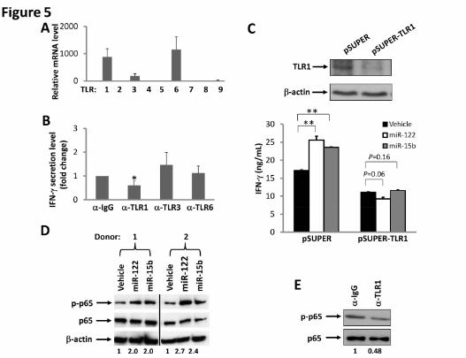
cells express relatively higher transcript levels of TLR1, TLR3 and TLR6 (Fig. 5A).
Accordingly, we disrupted TLR signaling by blocking each of these three TLRs with
their respective blocking antibodies. For TLR1, we used an antibody with a capability to
block signaling activated by its ligand, Pam3CSK4 (Fig. S6). We found that blockade of
TLR1 in primary NK cells significantly reduced miRNA-mediated induction of IFN-γ
production by ~50%, while blockade of TLR3 and TLR6 had no significant effect on NK
cell activation (Fig. 5B). To further demonstrate that TLR1 may participate in miRNA-
induced NK cell activation, a short hairpin RNA (shRNA) approach was undertaken. We
first confirmed that TLR1, but not TLR3, was successfully knocked down in NK-92 cells
(Figs. 5C and S7). We then found that TLR1 knockdown caused NK-92 cells to lose their
capability for miRNA-mediated activation as no increase of IFN-γ production was
observed, while the empty vector (pSUPER)-transduced control cells remained
responsive to miRNAs (Fig. 5C). A confocal microscopy study of HEK293T cells co-
transfected with miRNAs and TLR1-YFP fusion plasmid indicated that miRNAs and
TLR1 protein colocalize with each other within these cells (Fig. S8). Collectively, these
data suggest that NK cell activation by miRNAs occurs at least in part via the TLR1
signaling pathway, while this might be different in other types of immune cells, as most
recently reported 32.
NF-κB represents a signaling pathway down-stream of several TLRs, including TLR1 33.
To further test our hypothesis that miRNAs activate NK cells via TLR signaling, we
For personal use only. at CAPES CONSORTIUM on May 7, 2013. bloodjournal.hematologylibrary.orgFrom

16
assessed NF-κB activation in NK cells after treatment with miRNAs. We found that,
while the total level of p65 protein, a transactivation component of NF-κB signaling, was
unchanged, treatment with miRNAs induced an increase in the phosphorylation of p65 in
primary NK cells primed with IL-12 (Fig. 5D). To further confirm that miRNA-induced
activation of NK cells requires TLR1-mediated NF-κB signaling, we blocked TLR1 with
its blocking antibody, and found that the signaling blockade inhibited phosphorylation of
p65 (Fig. 5E). Importantly, when we treated purified primary human NK cells with miR-
122 and miR-15b in the same way as we did to determine p65 activation, we did not
observe any phosphorylation of IRF3, an activation event specifically mediated via TLR3
(data not shown).
MiRNAs enhance anti-tumor activity of NK cells in vivo to control tumor
development
A hallmark of NK cell function is to kill tumor cells or virally-infected cells. To further
validate the physiological relevance of NK activation by miRNAs, we treated mice with
miR-122 prior to and post implantation of A20 lymphoma cells, in the presence or
absence of NK cell depletion. To exclude the possible T-cell effect, athymic nude mice
were utilized. As shown in Fig. 6, after 3 doses of miR-122 treatment, the growth of A20
tumor cells in mice was significantly suppressed when compared with vehicle-
administered mice. However, this effect was largely abrogated when the NK cells were
depleted by TM-β1 mAb treatment, suggesting that miRNA suppressed tumor growth at
least in part through NK cell activation. More robust inhibition of tumor growth was
For personal use only. at CAPES CONSORTIUM on May 7, 2013. bloodjournal.hematologylibrary.orgFrom

17
observed when we repeated our experiment with more frequent miRNA injections and a
longer duration of the treatment (Fig. S9).
Components of the TLR signaling pathway are downregulated in NK cells from
cancer patients
The data presented above provided strong evidence for miRNAs having a role in
combating tumors by directly activating NK cells. Yet still, some cancer patients with
disease progression have high levels of circulating miRNAs, which suggests that there
may be a mechanism(s) by which tumors escape surveillance by miRNA-activated NK
cells. To investigate this, we purified NK cells from PBMCs of lymphoma patients, who
are reported to have elevated expression of miRNAs such as miR-155 18. We found that
although there was no significant difference of TLR1 expression between healthy donors
and cancer patients (Fig. S10), p65 and IRAK1, the two main components in the TLR1-
NF-κB signaling pathway, were consistently and significantly downregulated in NK cells
from lymphoma patients (Figs. 7A and 7B). We speculate that higher miRNAs in cancer
patients may therefore be unable to activate NK cells in order to control tumor
progression, and we will look to validate this in future studies.
For personal use only. at CAPES CONSORTIUM on May 7, 2013. bloodjournal.hematologylibrary.orgFrom

18
Discussion
MiRNAs exist in the peripheral blood of healthy donors, as well as in patients with
cancer 8. As supported by our in vivo anti-tumor data, it is conceivable that miRNAs may
boost NK cells’ ability to participate in surveillance against malignant transformation or
infectious insult in otherwise healthy individuals. On the other hand, the prevalence of
circulating miRNAs in some cancer patients also imply that other factors secreted by
malignant tumors, such as TGF-β, might somehow serve to disarm this form of innate
immune activation. In addition, in lymphoma patients we found that both p65 and IRAK1
have consistently lower mRNA expression in NK cells when compared to those in
healthy donors, suggesting that downregulaton of NF-κB signaling in NK cells might be
a potential underlying mechanism by which tumors evade immune surveillance by
miRNAs. However, this should not imply that modulation of miRNAs in cancer patients
is irrelevant. In fact, high levels of miR-21 expression are associated with a more
favorable clinical outcome for diffuse large B-cell lymphoma 34, and during this
manuscript preparation, a published report showed that systemic mi-RNA-34a delivery
abrogates in vivo tumor growth of this disease 35.
We found the activation induced by miRNAs to be relatively specific for innate immune
effector cells (i.e., NK cells), and absent in T cells, both in vitro and in vivo. This finding
may have application in circumstances when selective activation of these relatively
distinct arms of the immune response is desired. For example, in the setting of blood and
marrow transplantation, the NK cell innate immune response can function to kill
For personal use only. at CAPES CONSORTIUM on May 7, 2013. bloodjournal.hematologylibrary.orgFrom

19
activated T cells thereby contributing to the suppression of donor T cell-mediated graft
versus host disease 36, 37
We worked to ensure that the NK cell activation noted in the presence of miRNAs was
not due to non-specific binding and activation by contaminants. Our HPLC-purified
RNA negative control was prepared and handled in an identical fashion to our miRNAs,
making it unlikely that NK cell activation in response to the miRNAs was the result of
endotoxin or other contaminants. With regard to the RNA control, we used a non-
specific, single-stranded RNA called RNA41, which is similar in size to miRNAs 24, in
the majority of in vitro experiments, and did not see induction of IFN-γ or CD107a
degranulation. Further, to rule out the potential bias due to the use of a single RNA
(RNA41) as a control, we also mutated several nucleotides of miR-122 and miR-15b and
found that all NK cell activation was lost.
In addition to the potential of miRNAs for cancer diagnosis and prognosis, miRNAs are
also promising for therapeutic applications and they are on their way to clinical trials.
Our findings suggest that those miRNAs which function as tumor suppressors, e.g. miR-
122 38 included in the current study may be promising agents for treatment of cancer
because, on one hand, they may directly and specifically target oncogene expression, yet
on the other hand, they are also able to activate innate immune effector cells against
tumor cells. Our discovery may also shed new light on the manner in which the host
mounts an immune response against infectious pathogens and/or malignant
For personal use only. at CAPES CONSORTIUM on May 7, 2013. bloodjournal.hematologylibrary.orgFrom

20
transformation, as well the manner by which tumors or pathogens edit the immune
response to escape immune activation by circulating miRNAs.
In summary, here, we provide what we believe to be the first evidence that miRNAs, as a
class of regulatory molecules, directly activate both human and mouse NK cells, and this
NK cell activation occurs at least in part via the TLR1 signaling pathway. This indentifies
a new function of miRNAs with physiological relevance, and their potential for
applications in preventing or treating cancer and infections either alone or as an adjuvant.
For personal use only. at CAPES CONSORTIUM on May 7, 2013. bloodjournal.hematologylibrary.orgFrom

21
Acknowledgements
This project was supported by grants from NCI (CA155521 to J.Y. and CA095426 &
CA068458 to M.A.C.), a 2012 scientific research grant from the National Blood
Foundation (to J.Y.), an institutional research grant (IRG-67-003-47) from the American
Cancer Society (to J.Y.), and The Ohio State University Comprehensive Cancer Center
Pelotonia grant (to J.Y.).
Authorship
S.H. designed research work, performed experiments and wrote the paper. J.C., L-C.W.,
H.M., Y.P., T.H., M.W., S.Y., and S.S. performed some experiments. C.A.A-B, S.V.,
D.M.B., C.H., X.H., K.G., S.D. designed some of the research work. J.Z. analyzed data.
M.A.C. designed some of the research work and proofread the manuscript. J.Y. had a
conceptual idea, designed research, performed some experiments, and wrote the paper.
Disclosure Conflict of Interest
The authors declare no competing financial interests.
For personal use only. at CAPES CONSORTIUM on May 7, 2013. bloodjournal.hematologylibrary.orgFrom

22
References
1. Wightman B, Ha I, Ruvkun G. Posttranscriptional regulation of the heterochronic
gene lin-14 by lin-4 mediates temporal pattern formation in C. elegans. Cell. 1993;75(5):855-862.
2. Calin GA, Dumitru CD, Shimizu M, et al. Frequent deletions and down-regulation of micro- RNA genes miR15 and miR16 at 13q14 in chronic lymphocytic leukemia. Proc Natl Acad Sci U S A. 2002;99(24):15524-15529.
3. Bartel DP. MicroRNAs: genomics, biogenesis, mechanism, and function. Cell. 2004;116(2):281-297.
4. Bartel DP. MicroRNAs: target recognition and regulatory functions. Cell. 2009;136(2):215-233.
5. Lewis BP, Burge CB, Bartel DP. Conserved seed pairing, often flanked by adenosines, indicates that thousands of human genes are microRNA targets. Cell. 2005;120(1):15-20.
6. Chen CZ, Li L, Lodish HF, Bartel DP. MicroRNAs modulate hematopoietic lineage differentiation. Science. 2004;303(5654):83-86.
7. Rovira C, Guida MC, Cayota A. MicroRNAs and other small silencing RNAs in cancer. IUBMB Life. 2010;62(12):859-868.
8. Schwarzenbach H, Hoon DS, Pantel K. Cell-free nucleic acids as biomarkers in cancer patients. Nat Rev Cancer. 2011;11(6):426-437.
9. Hu Z, Chen X, Zhao Y, et al. Serum microRNA signatures identified in a genome-wide serum microRNA expression profiling predict survival of non-small-cell lung cancer. J Clin Oncol. 2010;28(10):1721-1726.
10. Roth C, Kasimir-Bauer S, Pantel K, Schwarzenbach H. Screening for circulating nucleic acids and caspase activity in the peripheral blood as potential diagnostic tools in lung cancer. Mol Oncol. 2011;5(3):281-291.
11. Roth C, Rack B, Muller V, Janni W, Pantel K, Schwarzenbach H. Circulating microRNAs as blood-based markers for patients with primary and metastatic breast cancer. Breast Cancer Res. 2010;12(6):R90.
12. Asaga S, Kuo C, Nguyen T, Terpenning M, Giuliano AE, Hoon DS. Direct serum assay for microRNA-21 concentrations in early and advanced breast cancer. Clin Chem. 2011;57(1):84-91.
13. Tomimaru Y, Eguchi H, Nagano H, et al. Circulating microRNA-21 as a novel biomarker for hepatocellular carcinoma. J Hepatol. 2012;56(1):167-175.
14. Baraniskin A, Kuhnhenn J, Schlegel U, et al. Identification of microRNAs in the cerebrospinal fluid as biomarker for the diagnosis of glioma. Neuro Oncol. 2012;14(1):29-33.
15. Lagos-Quintana M, Rauhut R, Yalcin A, Meyer J, Lendeckel W, Tuschl T. Identification of tissue-specific microRNAs from mouse. Curr Biol. 2002;12(9):735-739.
16. Chen CJ, Lee MH. Early diagnosis of hepatocellular carcinoma by multiple microRNAs: validity, efficacy, and cost-effectiveness. J Clin Oncol. 2011;29(36):4745-4747.
For personal use only. at CAPES CONSORTIUM on May 7, 2013. bloodjournal.hematologylibrary.orgFrom

23
17. Waidmann O, Bihrer V, Pleli T, et al. Serum microRNA-122 levels in different groups of patients with chronic hepatitis B virus infection. J Viral Hepat. 2012;19(2):e58-65.
18. Fang C, Zhu DX, Dong HJ, et al. Serum microRNAs are promising novel biomarkers for diffuse large B cell lymphoma. Ann Hematol. 2012;91(4):553-559.
19. Cichocki F, Felices M, McCullar V, et al. Cutting edge: microRNA-181 promotes human NK cell development by regulating Notch signaling. J Immunol. 2011;187(12):6171-6175.
20. Fehniger TA, Wylie T, Germino E, et al. Next-generation sequencing identifies the natural killer cell microRNA transcriptome. Genome Res. 2010;20(11):1590-1604.
21. Kim TD, Lee SU, Yun S, et al. Human microRNA-27a* targets Prf1 and GzmB expression to regulate NK-cell cytotoxicity. Blood. 2011;118(20):5476-5486.
22. Trotta R, Chen L, Ciarlariello D, et al. MiR-155 regulates IFN-gamma production in natural killer cells. Blood. 2012.
23. Gong JH, Maki G, Klingemann HG. Characterization of a human cell line (NK-92) with phenotypical and functional characteristics of activated natural killer cells. Leukemia. 1994;8(4):652-658.
24. Heil F, Hemmi H, Hochrein H, et al. Species-specific recognition of single-stranded RNA via toll-like receptor 7 and 8. Science. 2004;303(5663):1526-1529.
25. Yu J, Wei M, Becknell B, et al. Pro- and antiinflammatory cytokine signaling: reciprocal antagonism regulates interferon-gamma production by human natural killer cells. Immunity. 2006;24(5):575-590.
26. Becknell B, Trotta R, Yu J, et al. Efficient infection of human natural killer cells with an EBV/retroviral hybrid vector. J Immunol Methods. 2005;296(1-2):115-123.
27. Borrego F, Pena J, Solana R. Regulation of CD69 expression on human natural killer cells: differential involvement of protein kinase C and protein tyrosine kinases. Eur J Immunol. 1993;23(5):1039-1043.
28. Caligiuri MA. Human natural killer cells. Blood. 2008;112(3):461-469. 29. Cooper MA, Fehniger TA, Turner SC, et al. Human natural killer cells: a unique
innate immunoregulatory role for the CD56(bright) subset. Blood. 2001;97(10):3146-3151.
30. Takeshita F, Gursel I, Ishii KJ, Suzuki K, Gursel M, Klinman DM. Signal transduction pathways mediated by the interaction of CpG DNA with Toll-like receptor 9. Semin Immunol. 2004;16(1):17-22.
31. Diebold SS, Kaisho T, Hemmi H, Akira S, Reis e Sousa C. Innate antiviral responses by means of TLR7-mediated recognition of single-stranded RNA. Science. 2004;303(5663):1529-1531.
32. Fabbri M, Paone A, Calore F, et al. MicroRNAs bind to Toll-like receptors to induce prometastatic inflammatory response. Proc Natl Acad Sci U S A. 2012;109(31):E2110-2116.
33. West AP, Koblansky AA, Ghosh S. Recognition and signaling by toll-like receptors. Annu Rev Cell Dev Biol. 2006;22:409-437.
For personal use only. at CAPES CONSORTIUM on May 7, 2013. bloodjournal.hematologylibrary.orgFrom
24
34. Lawrie CH, Gal S, Dunlop HM, et al. Detection of elevated levels of tumour-associated microRNAs in serum of patients with diffuse large B-cell lymphoma. Br J Haematol. 2008;141(5):672-675.
35. Craig VJ, Tzankov A, Flori M, Schmid CA, Bader AG, Muller A. Systemic microRNA-34a delivery induces apoptosis and abrogates growth of diffuse large B-cell lymphoma in vivo. Leukemia. 2012.
36. Olson JA, Leveson-Gower DB, Gill S, Baker J, Beilhack A, Negrin RS. NK cells mediate reduction of GVHD by inhibiting activated, alloreactive T cells while retaining GVT effects. Blood. 2010;115(21):4293-4301.
37. Shlomchik WD. Graft-versus-host disease. Nat Rev Immunol. 2007;7(5):340-352. 38. Bai S, Nasser MW, Wang B, et al. MicroRNA-122 inhibits tumorigenic properties
of hepatocellular carcinoma cells and sensitizes these cells to sorafenib. J Biol Chem. 2009;284(46):32015-32027.
For personal use only. at CAPES CONSORTIUM on May 7, 2013. bloodjournal.hematologylibrary.orgFrom

25
Figure Legends
Figure 1: MiRNAs increase CD69 surface expression on NK cells. (A) Highly purified
(≥ 99%) human NK cells were treated with miRNAs, DOTAP vehicle control or non-
specific, single-stranded RNA, RNA41 (RNA-Ctl) 24, for 36 h in the presence of a low
concentration of IL-12 (2.5 ng/ml). The treated cells were then subjected to flow
cytometric analysis to determine the percentage of CD69+ cells. Representative data from
1 out of 6 donors with similar results are shown. (B) Summary of data from 4 donors
obtained in 1 experiment. * indicates p < 0.05 and error bars represent S.D.
Figure 2. MiRNAs increase IFN-γ production by NK cells. (A) Highly purified (≥
99%) human NK cells were treated with miRNAs, DOTAP vehicle control or non-
specific, single-stranded RNA, RNA41 (RNA-Ctl) 24, for 36 h in the presence of a low
concentration of IL-12. Supernatants were harvested for ELISA and cells were harvested
for Real-time RT-PCR analysis to determine the levels of IFN-γ secretion (upper panel)
and gene expression (lower panel), respectively. Gene expression of the vehicle was
normalized to 1 and experimental values are each presented as fold change compared to
that of the vehicle. Data shown represent 1 of 3 donors with similar results. (B) Cells
were treated, and data were collected as described in (A), with the exception that
concentrations of miR-122 were varied, as indicated on the X axis. Data suggest that
miR-122 activates NK cells in a dose-dependent fashion. (C, D) Highly purified human
NK cells were treated with miRNAs or DOTAP vehicle control for 36 h in the presence
of a low concentration of IL-12. The cells were then incubated with K562 tumor cells at a
For personal use only. at CAPES CONSORTIUM on May 7, 2013. bloodjournal.hematologylibrary.orgFrom

26
ratio of 1:1 (effector: target). After 4 h, CD107a expression was assessed by flow
cytometric analysis. Shown in C are representative data from 1 out of 5 donors with
similar results, and D demonstrates summary data of 5 donors. In all panels, error bars
represent S.D. * indicates p < 0.05 , and ** indicates p < 0.01.
Figure 3. MiRNAs activate NK cells in vivo. (A) Mice were treated in vivo with
vehicles or 20 μg RNA-Ctl, miR-122 or miR-15b for 4 days. The treated mice were then
sacrificed, and total splenocytes were isolated for flow cytometric analysis to measure
CD69 surface expression after gating on CD3-NK1.1+ NK cells. Representative data from
1 out of 6 mice with similar results (left) as well as summary data from 3 mice (right) in
one experiment are shown. (B) The prepared splenocytes from A were co-cultured with
YAC-1 tumor cells for 3 h without any exogenous IL-12, and were subjected to flow
cytometric analysis of CD107a expression after gating on CD3-NK1.1+ NK cells. For (A)
and (B), * and ** indicate p < 0.05 and p < 0.01, respectively, and error bars represent
S.D.
Figure 4. MiRNAs do not activate T cells in vivo. (A) Human PBMCs were stimulated
with miRNAs as described in Fig. 1A and were subjected to flow cytometric analysis of
CD69 surface expression within CD3+ T cells. Depicted are representative data from 1 of
6 donors (top), as well as summary data from 3 donors in one experiment (bottom). Data
suggest that miRNAs do not significantly change human T cell surface expression of
CD69. (B) Mice were treated in vivo and cells were prepared for flow cytometric analysis
as described in Fig. 5A. Depicted are representative data from 1 of 6 mice with similar
For personal use only. at CAPES CONSORTIUM on May 7, 2013. bloodjournal.hematologylibrary.orgFrom

27
data (left), as well as summary data from 3 mice in one experiment (right), suggesting
that miRNAs do not significantly change murine T cell surface expression of CD69 in
vivo. For (A) and (B), error bars represent S.D.
Figure 5. Interaction of TLRs and miRNAs. (A) Real-time RT-PCR was utilized to
determine the level of TLR mRNA expressed in highly purified human NK cells. The
mRNA level of TLR5 was found lowest and was normalized to 1. The mRNA level of
other TLRs was presented as relative to that of TLR5. Data are shown as the average of 3
donors. (B) Purified human NK cells were pre-incubated for 1.5 h with a non-specific
IgG or anti-TLR1, anti-TLR3, or anti-TLR6-blocking antibody (α). Cells were then
stimulated with miR-122 as described in Fig. 1A in the presence of the blocking antibody,
and supernatants were harvested to measure IFN-γ protein via ELISA. The concentration
of IFN-γ in the purified NK cells incubated with IgG and miR-122 was arbitrarily set at 1.
Data were averaged from 3 donors. * indicates p < 0.05 and error bars represent S.D. (C)
NK-92 cells were infected with pSUPER-TLR1-GFP retroviruses, and stably transfected
cells were sorted based on GFP expression. Both the vector-transduced cells (pSUPER)
and the TLR1 knockdown cells (pSUPER-TLR1, confirmed by immunoblotting, upper
panel) were stimulated with miR-122 or miR-15b as described in Fig. 1A, and cell-free
supernatants were collected to assess IFN-γ secretion via ELISA (lower panel). **
indicates p < 0.01 and error bars represent S.D. (D) Purified human NK cells were
stimulated with miR-122 or miR-15b as described in Fig. 1A, and subsequently subjected
to immunoblotting using p65 and phospho-p65 (p-p65) antibodies. β-actin
immunoblotting was included to demonstrate equal loading of total protein. Data shown
For personal use only. at CAPES CONSORTIUM on May 7, 2013. bloodjournal.hematologylibrary.orgFrom

28
represent 2 of 4 donors with similar results. Numbers beneath each lane represent
quantification of p-p65 by densitometry, normalized by p65. (E) The experiment was
performed as in C except that anti-TLR1 blocking mAb or its control IgG was included in
the culture in the presence of miR-122. Data shown represent 1 of 3 donors with similar
results. Numbers beneath each lane represent quantification of p-p65 by densitometry,
normalized by p65.
Figure 6. MiRNA stimulation enhances anti-tumor activity of NK cells in vivo. (A)
Ventral bioluminescence imaging of mice bearing A20 lymphoma. Athymic nude mice
were injected with 1 × 10⁵ luciferase-expressing A20 cells via tail veins and subjected to
miRNA stimulation alone or combined with TM-β1 treatment according to the schedule
described in the Materials and Methods. The pseudo color indicates the relative signal
strength for tumor growth, with strongest in red and weakest in purple. (B) Shown are
quantification summary of units of photons per second per mouse from (A). Data are
shown as mean ± SD from each group of mice. *, p < 0.05; **, p < 0.01.
Figure 7. Downregulation of the NF-κB signaling pathway components in NK cells
from lymphoma patients. (A, B) NK cells were isolated from PBMCs of both healthy
donors and lymphoma patients as described in the Materials and Methods. The purified
NK cells were then immediately subjected to RNA extraction and cDNA synthesis. The
expression levels of p65 (A) and IRAK1 (B) were determined by SYBR Green Real-time
RT-PCR assay.
For personal use only. at CAPES CONSORTIUM on May 7, 2013. bloodjournal.hematologylibrary.orgFrom

For personal use only. at CAPES CONSORTIUM on May 7, 2013. bloodjournal.hematologylibrary.orgFrom

For personal use only. at CAPES CONSORTIUM on May 7, 2013. bloodjournal.hematologylibrary.orgFrom

For personal use only. at CAPES CONSORTIUM on May 7, 2013. bloodjournal.hematologylibrary.orgFrom

For personal use only. at CAPES CONSORTIUM on May 7, 2013. bloodjournal.hematologylibrary.orgFrom
For personal use only. at CAPES CONSORTIUM on May 7, 2013. bloodjournal.hematologylibrary.orgFrom

For personal use only. at CAPES CONSORTIUM on May 7, 2013. bloodjournal.hematologylibrary.orgFrom

For personal use only. at CAPES CONSORTIUM on May 7, 2013. bloodjournal.hematologylibrary.orgFrom